
EPIDEMIOLOGICAL FEATURES OF NATURAL Mycoplasma hyopneumoniae INFECTION
IN PIGS IN SPAIN
Marina Sibila Vidal Bellaterra, Juny 2004

UNIVERSITAT AUTÒNOMA DE BARCELONA FACULTAT DE VETERINÀRIA
DEPARTAMENT DE SANITAT I ANATOMIA ANIMALS
EPIDEMIOLOGICAL FEATURES OF NATURAL Mycoplasma hyopneumoniae INFECTION
IN PIGS IN SPAIN
Marina Sibila Vidal
Memòria presentada per optar al grau de doctora en veterinària Bellaterra, Juny del 2004

Maria Calsamiglia i Costa, investigadora del Centre de Recerca en Sanitat Animal (CReSA), i Joaquim Segalés i Coma, professor titular del Departament de Sanitat i Anatomia Animals de la Universitat Autònoma de Barcelona Certifiquen: Que la tesis doctoral titulada “Epidemiological features of natural Mycoplasma hyopneumoniae infection in pigs in Spain”, de la que és autora la llicenciada en Biologia, Marina Sibila Vidal, s’ha realitzat als laboratoris de la Unitat d’histologia i Anatomia patològica de la facultat de Veterinària de la Universitat Autònoma de Barcelona sota la nostra direcció. I perquè així consti, a tots els efectes, firmem el present certificat a Bellaterra, 21 de Juny del 2004 Maria Calsamiglia Costa Joaquim Segalés i Coma Directora de Tesis Tutor de la Tesis

El treball presentat en aquesta Tesis, forma part de les investigacions realitzades en el Centre de Recerca en Sanitat Animal (CReSA)

Literature Review
LITERATURE REVIEW

Literature Review
1
1 Etiologic agent:
1.1 Mycoplasmas and Porcine Mycoplasmas
Mycoplasma hyopneumoniae (M. hyopneumoniae) is a member of the
Class Mollicutes which lacks of cell wall (mollis, soft; cutis, skin),
Family Mycoplasmataceae (sterol required for growth), Genus
Mycoplasma (Tully et al., 1996). Mollicutes are phylogenetically related
to the Gram-positive bacteria.
Nowadays, Mycoplasmas are considered the simplest self-replicating
organisms known. Despite their genetic simplicity, the members of the
Class Mollicutes are considered as major animal and plant pathogens
world-wide. The minimal characteristics to differentiate Mycoplasmas
from other eubacteria are: permanent lack of cell wall, tendency to form
fried egg colonies on solid media, pass through membrane filters (450-
220 nm pore size), low genome G+C content (23-40 mol%) and small
genome size (Mycoplasma hyopneumoniae =1070 bp) (Minion et al.,
2002a). With the smallest genomes known for self-living organism their
number of genes approaches the minimal set of genes required for life.
Because of their low number of genes they have limited metabolic
capabilities and they are well adapted to or dependant on their hosts
(Garnier et al., 2001). The extremely low guanine + cytosine (G+C)
imposes further restrictions on the amount of genetic information
available to Mycoplasma cells, leading to the complex nutritional
requirements and the parasitic or saprophytic mode of life. The
Mycoplasma cell is bound by a plasma membrane and the enclosed
cytoplasm contains ribosomes and a circular double-stranded DNA
molecule (Razin et al., 1998). The major antigenic determinants
described in these wall-less organisms are membrane proteins,

Literature Review
2
glycolipids and lipoglicans found in the cell membrane (Razin et al.,
1998).
Although some Mycoplasmas are commensals and belong to the
normal flora of their hosts, most species are pathogens. Many Mollicutes
are inhabitants of the mucous membranes of the respiratory, urogenital or
gastrointestinal tract of vertebrates (Tully et al., 1996). Moreover,
arthropods and plants are also possible hosts of mycoplasma infections
(Razin et al., 1998).
Besides M. hyopneumoniae, there are two other mycoplasmal species
that can cause disease in swine: M. hyorhinis inhabits the upper
respiratory tract (nasal cavity) and can occasionally cause poliserositis
and arthritis in young pigs; M. hyosynoviae inhabits the nasal cavity and
pharynx of pigs and can cause non-suppurative arthritis without
polyserosits in grow-finishing pigs (Whittlestone et al., 1979). On the
other hand, M. flocculare, appears to be a common inhabitant of the
porcine lung and nasal cavities of pigs but is essentially non-pathogenic
(Amstrong et al., 1981). Friis (1973, cited in Ross et al., 1999) presented
evidence that M. flocculare produced lymphocytic infiltration in the nasal
mucosa and peribronchial areas of gnobiotics pigs inoculated with this
organism. However, there is still no evidence that these findings are of
significance in naturally occurring respiratory disease. Although, a recent
study suggested that M.hyopneumoniae may not be the only Mycoplasma
important in respiratory disease in swine (Strait et al., 2003).
Several other species of Mollicutes are infrequently found in the
porcine respiratory tract and are of questionable pathogenecity: these
include M. sualvi and M. hyopharyngis and several species of
acholeoplasmas (Ross, 1999).
Among all porcine mycoplasmas, M. hyorhinis and M. flocculare are
the most commonly studied in relation with M.hyopneumoniae. Cross-

Literature Review
3
reactions in serologic techniques, as well as M.hyopneumoniae culture
overgrown with these two mycoplasmas have been extensively described
(Nicolet et al., 1980, Feld et al., 1992).
M. flocculare has many morphological, growth and antigenic
similarities to M. hyopneumoniae (Ross, 1999). Results of DNA-DNA
hybridization studies have indicated that there is some relation between
M. hyopneumoniae and M. flocculare (10-11%); in contrast, M.
hyopneumoniae and M flocculare only cross-hybridize with nonporcine
Mycoplasmas at levels about 3% (Stemke et al., 1985).
Because the rRNA genes are among the most conserved genetic
elements, comparisons of these sequences have become an important
phylogenetic tool. Comparison of these genes in different Mycoplasma
species have shown that M.hyopneumoniae, M. flocculare and M.
hyorhinis are very closely related, particularly the first two (Stemke et al.,
1994a). The homology between M. hyopneumoniae and M. flocculare
16S ribosomal sequence is 95,6% (Stemke et al., 1992). The genome of
M.hyopneumoniae was found to be larger (1070 compared to 890 kb)
than M. flocculare (Robertson et al., 1990). Presumably, pathogenicity of
M. hyopneumoniae is facilitated by the absent genes in M. flocculare. An
example could be the gene for ciliary adhesin (adh), wich is believed to
be a virulence factor in M.hyopneumoniae (Hsu and Minion, 1998) and
was not detected by hybridization in M flocculare nor in M. hyorhinis
(Wilton et al., 1998).
1.2 Mycoplasma hyopneumoniae
1.2.1 Enzootic Pneumonia history
Initially, it was thought that the chronic pneumonia observed in
growing and fattening pigs was caused by a virus and consequently
termed as virus pneumonia (Betts, 1952, Whittlestone, 1979). The basis
Literature Review
4
for this idea was the transmission of the disease to pigs with bacteria-free
filtrates and because penicillin and sulfamides did not affect the
infectivity of affected lung material (Whittlestone, 1979). Some years
later, evidences that lead to the identification of a Mycoplasma-related
organism as the etiologic agent were: the efficacy of tetracycline
antibiotics against the disease, the size of the causal agent (0.2-0.45 µm);
the Giemsa staining; and the growth of the agent in media free of living
cells clearly demonstrating that it was a nonviral microorganism
(Whittlestone, 1979). Mare and Switzer (1965) obtained the first isolate
of the microorganism and identified it as M. hyopneumoniae (strain 11 or
VP119). The same year, Goodwin et al. (1965) induced EP to pigs
inoculated with another isolated, which was named of Mycoplasma
suipneumoniae (strain J). To reach a consensus, the Academy of
Taxonomy of Mycoplasmatales, decided to designate a neotype strain and
proposed the strain J (M. suipneumoniae) as the neotype strain of M.
hyopneumoniae.
1.2.2 Main Immunogens
A better characterization of putative M hyopneumoniae antigens
would help in detecting virulence factors, in pathogenecity studies, in
serological differentiation of porcine Mycoplasmas, and finally in
developing efficaceous vaccines. Up to date, several immunogens have
been described:
A 54kDa protein located within the cell membrane with cytopathic
effect was described (Geary and Walczak, 1985, Ross and Young , 1993).
Debey and Ross, showed that the attachment of M. hyopneumoniae to
ciliated epithelium was necessary to induce cytopathic effect in tracheal
organ cultures from newborn piglets (Ross and Young, 1993).

Literature Review
5
Another well studied antigen is a cytosolic 36kDa protein (p36). This
protein was cloned and expressed (Strasser et al., 1991), and afterwards
characterized (Haldiman et al., 1993) as lactate dehydrogenase (LDH).
This cytosolic protein induces an early immune response in naturally and
experimentally infected pigs, is apparently highly conserved among
different M.hyopneumoniae strains and does not cross-react with other
porcine Mycoplasmas (Strasser et al., 1991). Frey et al., (1994) used an
LDH expressed in E. coli for the development of a specific LDH-ELISA.
Anti-LDH titres persist until 21 weeks post-infection, in contrast to the
antibody titres against membrane proteins which decreased at 12 weeks
post-infection. On the contrary, Caron et al., (2000b) found no correlation
of clinical and pathological findings with an IIF specific test using p36 as
antigen.
Another antigen used to develop new serodiagnostic tests was the
ABC transporter ATP-binding protein (Blanchard et al., 1996,
Subramaniam et al. 2000).
It has been proposed that cytoadherence of M.hyopneumoniae to porcine
cilia might be a mutifactorial process. At the moment two adhesin
proteins have been postulated: a 97kDa protein, named p97 (Zhang et al.,
1995) and a 110kDa protein, named p110 (Chen et al., 1998). (See
pathogenesis). Induction of humoral immune response after p97
recombinant vaccines is controversial. King et al., (1997) and Shimoji et
al., (2003) were not able to induce serum antibody production, while
Chen et al., (2001) observed a remarkable IgG responses in immunized
pigs with a p97 RR1 vaccine. However, Shimoji et al., (2003), suggest
that intranasal vaccination of YS-19 strain could prime a cell-mediated
immune response against p97.

Literature Review
6
Other M.hyopneumoniae putative immunogens are: a 46kDa (p46)
surface antigen (Futo et al., 1995) and a 65Kda (p65) lipoprotein, and a
74.5 kDa heat shock protein (hsp) (Brooks and Faulds 1989).
Heat shock proteins have been proposed as good immunogen to
vaccine development. Scherm et al., (2002), identified and sequenced the
Hsp60 gene of several mycoplasma species (including
M.hyopneumoniae) and confirmed that the Mycoplasmal Hsp60 is
immunogenic in natural infections.
Ross and Young (1993) identified strain specific immunogens of
M.hyopneumoniae pathogenic strain 232 and non-pathogenic strain J.
Strain 232 antigens were immunoblotted with convalescent serum
previously adsorbed with either strain 232 or strain J whole cell. Results
of this study revealed that there were 6 putative specific antigens
abundant in the strain 232, which were not detected in strain J. These 6
antigens were: 145kDa, 97kDa, 86 kDa, 75kDa, 60kDa, and 46kDa.
1.2.3 M.hyopneumoniae strains
Since the designation of strain J, a non-virulent strain, as the neotype
strain of M.hyopneumoniae, several studies have looked for differences
between field strains. Ro and Ross (1983) demonstrated by growth-
inhibition and metabolic-inhibition tests some heterogeneicity among the
strains they tested. Moreover, Frey et al. (1992) found by enzyme
restriction digestion run in a field inversion gel electrophoresis (FIGE)
that chromosomes of M.hyopneumoniae strains were quite different. In
1996, (Artiushin and Minion, 1996) found differences in RAPD
(randomly amplified polymorphic DNA) band DNA pattern even within
isolates from the same geographical location.
In 1998, Wilton et al., showed that the number of tandem amino acid
repeats in R1 of ciliar-adhesin p97 varied consistently between strains.
Literature Review
7
Moreover, intraespecies variation among field USA isolates by total
protein profile, glycoprotein profile, and size difference in the amplified
PCR product of p97 adhesin repeat region R1 has been also described
(Lin, 2001). PCR amplifications of R1 region of seven commercial
vaccines produced also different size of amplicons (Lin, 2001).
Although genetic heterogeneity between field strains had been
extensively reported, it had not been related with virulence differences. In
2003, Vicca et al. analyzed also by RAPD six M.hyopneumoniae field
isolates and linked it to genetic and virulence differences. Based on
clinical and pathological parameters, the isolates tested were classified as
highly, moderately or non-virulent isolates. Moreover, a 5000pb RAPD
band was found in the highly and moderately virulent isolates but not in
the avirulent ones. Recently, differences in virulence factors between
field isolates were studied in a challenge trial. In that study, differences in
percentage of lung affection were observed among the groups inoculated
with different field isolates (Strait et al, 2004).
Some species of pathogenic mycoplasmas (M. hyorhinis, M. bovis, M.
gallisepticum) have genetic machinery for altering their surface
characteristics (antigenic variation). It remains to be determined whether
these mechanisms are also characteristic of M.hyopneumoniae and to
what extent they may explain apparent genetic and antigenic differences
between strains (Desrosiers 2001).

Literature Review
8
2 The disease: Enzootic Pneumonia and PRDC
Mycoplasma hyopneumoniae is the principal etiologic agent of
Mycoplasmal pneumonia (MPS) or Enzootic Pneumonia (EP) in pigs. EP
is a heterogeneous disease that results from combined infctions with
M.hyopneumoniae and one or more opportunistic, secondary bacterial
pathogens such as Pasteurella multocida, Mycoplasma hyorhinis,
Streptococcus suis, Haemophilus parasuis, Bordetella bronchiseptica,
Actinomyces pyogenes and Actinobacillus pleuroneumonia (Done et al.,
1997). The two most frequent association are M.hyopneumonia with A.
pleuropneumoniae (Yagishaji et al., 1984), and with P. Multocida
(Ciprián et al., 1988)
Nowadays, EP is found in all pig producing countries. In fact, abattoir
surveys in many countries have demonstrated high percentages of EP
seropositive pigs (Falk and Lium, 1991, Wallgren et al., 1993, Wallgren
et al., 1994) and EP lung lesions (Falk and Lium, 1991, Lium and Falk,
1991). In Spain, an abattoir studyshowed a 96% of slaughter pigs had
lung lesions, from which a 68.8% were compatible with EP
characteristics lung lesions (Cubero, 2000). These results, lead to
consider M. hyopneumoniae the most economically significant bacterial
respiratory pathogen.
In the last decade, a new respiratory syndrome named as Porcine
respiratory disease complex (PRDC) emerged as a serious health problem
in most of pig producing countries. This respiratory complex includes
both viral and bacterial agents: Porcine reproductive and respiratory
syndrome virus (PRRSV), Mycoplasma hyopneumoniae, swine Influenza
virus (SIV), Pasteurella multocida, Actinobacillus pleuropneumoniae,
Haemophilus parasuis, Streptococcus suis, Porcine circovirus type 2
(PCV2) and Aujeszky’s disease virus (ADV) (Thacker, 2001a).

Literature Review
9
Pneumonia in pigs with PRDC is due to a combination of both viral and
bacterial agents, being PRRSV and Mycoplasma hyopneumoniae the
main causative agents (Thacker et al., 1999).
PRDC is characterized by slow growth, decreased feed efficiency,
anorexia, fever, cough, and dyspnea in finishing pigs (Thacker, 2001a).
PRDC typically occurs at 14 to 20 weeks of age, being also referred as
the “18 week wall”. It has been described in all kinds of management and
facilities schemes, including intensive production systems.
2.1 Clinical presentation
Enzootic pneumonia is characterized by a high morbidity and low
mortality. Although pigs of all ages are susceptible to be infected with
M. hyopneumoniae (Piffer and Ross 1984, Kobisch et al., 1993). EP is
usually not observed in animals younger than 6 weeks of age (Ross,
1999). The rarity of disease in pigs under 6 weeks of age suggests that
transmission from sow to suckling pigs is inefficient. This inefficiency
may be due to little shedding from sows or the relative protection of pigs
afforded by colostral immunity (Stevenson et al., 1998). From 6 weeks to
3 months of age, EP is infrequently observed. Finally, the peak
prevalence of EP occurs later, at market weight (Done, 1997).
Severity of symptoms depends on the intensity of infection, presence
of secondary infections and enviromental conditions (Ross et al., 1999,
Leon et al., 2001). When Mycoplasma infection is not complicated with
secondary pathogens, the disease can take a subclinic course (no
coughing) or mild clinical signs consisting on chronic nonproductive
cough and reduced rate of gain and feed efficiency (Desrosiers 2001,
Maes, 1996). When infection with secondary pathogens occurs, other
symptoms such as laboured breathing and fever, and even death may
occur (Kobisch, 2000).

Literature Review
10
Under field conditions, EP incubation period is very variable.
Coughing is not constant and is generally noticeable 2 to 6 weeks after
infection and ceases after 2 months post infection (Kobisch et al., 1993,
Maes 1996, Done 1997, Sorensen et al., 1997). However, considerable
variation may occur. In incubation period cases, development of clinical
signs may take up to 16 months (Piffer and Ross; 1984, Done 1997).
Inoculation studies have shown that the incubation period and the
development of clinical signs are dose-dependant (Ross, 1999, Stevenson
et al., 1998). At high doses, clinical pneumonia may develop following an
incubation period of 11 days. At moderate doses, clinical pneumonia
develops after a prolonged incubation period of up to 4-6 weeks. And at
low doses, only subclinical (no coughing) infection develops (Stevenson
et al., 1998).
Although in field cases is unknown, a similar dose-dependency is
expected. This dependency has also been linked to a certain percentages
of pigs being infected (Calsamiglia et al., 1999b). High percentages of
infected pigs lead to a faster spread in the nurseries and earlier disease
outbreak (Pijoan, 1995).
Quantification of pathogen’s DNA present in clinically and
subclinically infected pigs would help to elucidate the necessary dose to
elicit lung lesions and the consequent cough.
2.2 Gross lesions
Mycoplasmal pneumonia is a catarrhal bronchopneumonia with a
cranioventral distribution involving cranial, middle and intermediate
lobes as well as cranioventral portion of caudal lobes (Taylor et la.,1996).
The intermediate, the left cardiac and the left apical lobes are affected less
frequently. The diaphragmatic lobes are rarely affected. This distribution

Literature Review
11
may reflect the airflow patterns, as the right apical lobe receives a stem
bronchus directly from the trachea (Livigston et la., 1972, Maes, 1996).
In early stages of infection gross lesions are characterized by purple to
gray areas of consolidation. The lung is more collapsed than in control
lungs and when the lesion is incised, the consistency is meaty but not
excessively firm (Christensen et al.,1999). In the acute phase of the
disease, the lobules are swollen and the airways contain copious amounts
of stick white exudates (Armstrong et al., 1983). If samples are taken at
this stage, large numbers of Mycoplasma cells will be found. In the
chronic phase, lesions are contracted rather than swollen and contain the
causative agent in low numbers (Armstrong et al.,1983, Amanfu et la.,
1984, Livingston et la., 1972). Livingston (1972) described that in
uncomplicated Mycoplasma infection, gross lesions will run a course of 5
to 6 weeks, after which lesions start to regress.
When secondary bacterial infections occur, gross lesions have a
more grayish color and are firmer due to formation of fibrous tissue,
leading to a purulent bronchopneumonia (Christensen et al., 1999).
Correlation between onset of clinical symptoms (coughing) and
pneumonic lesions has been described (Kobisch et al., 1993). In this
study, macroscopic lesions and coughing were seen after 1 and 2 weeks
post infection, respectively. After 7-9 weeks post infection, lesions
recovered and coughing disappeared.
In experimental inoculations with pure M. hyopneumoniae, there is
rarely more than 20% of lung affection. However, in field cases,
complications by secondary invaders lead to more extended lesions (up to
80-90% of lung involvement) (Done, 1997).

Literature Review
12
2.3 Microscopic lesions
In early stages of the disease, loss of cilia, exfoliation and small
accumulations of neutrophils in lumina around airways can be seen. As
the disease progresses, there are increased numbers of lymphoctes in
peribronchial, peribronchiolar and perivascular spaces (Ross, 1999).
Acute cases may show minimal lymphoid hyperplasia and a
predominantly neutrophilic alveolar exudate (Taylor et al., 1996). In
chronic stage of the disease, there is lymphoid hyperplasia or cuffing and
the consequent thickening of the interalveolar septa. In recovering
lesions, collapsed alveoli, alveolar emphysema and lymphoid nodules can
be seen (Ross, 1999). Lymphoid hyperplasia itself may be related to
clinical signs, because pressure from aggregates of lymphoid tissue may
obliterate the lumen of the softwalled bronchiole and cause collapse of
surrounding alveoli (Bakersville 1981).
The overall chronology in experimental inoculations would be the
following: two to four weeks post inoculation (PI), macroscopic lesions
are maximal and cuffing appears, at 8 weeks pI macroscopic lesions start
healing and if no secondary infections occur only few macroscopic
lesions remain at 10 weeks pI. Approximately at 4 months pI, cuffing
begins to disappear (cited in Maes, 1996).
2.4 Seroconversion
Time of seroconversion depends on the infection pressure present, the
management factors established in the farm and also the serological assay
used (Maes, 1996).
In intratracheal inoculation experiments, serum antibodies were first
detected in some pigs at 2 –4 weeks PI, reaching the peak of antibody
titres after 11-12 weeks PI (Piffer et al., 1984, Suter et al., 1985, Messier
and Ross, 1990, Sheldrake et al., 1990, Kobisch et al., 1993, Le Potier, et

Literature Review
13
al.,1994, Djordjevic et al., 1994). However, not all animals seroconverted
at the same time. In most of these studies, it took some weeks to detect
serum antibodies in all tested pigs. In aerosol infected pigs, Sorensen et
al., (1997) reported seroconversion as early as 8 days PI, but it took a
further 5 weeks for all animals to become positive. In conditions more
similar to natural infections, seroconversion is delayed. Armstrong et al.,
(1983), reported that intranasal exposed pigs seroconverted earlier and
developed higher titres than contact-exposure pigs.
In natural conditions, time to seroconversion appears to be more
delayed. Thus seroconversion occurs mainly in the growing-to-finishing
units (8-24 weeks of age), after 6-9 weeks post infection (Lam et al.,
1972, Sheldrake et al., 1990, Yagihashi et al. 1993, Djordjevic et al.,
1994, Morris et al. 1995, Sitjar et al., 1996, Andreasen et al. 2000, Leon
et al., 2001, Vicca et al., 2002). In a field study where clinically infected
herds were compared with subclinically infected herds, differences in the
seroconversion patterns were observed (Vicca et al., 2002). At 15 and 18
weeks of age, percentage of seropositive pigs was significantly higher in
the clinically than in the subclinically infected herds. While
seroconversion in clinically infected herds was observed between 12 and
15 weeks of age, seroconversion in subclinical herds occurred between 15
and 18 weeks of age.
Different degrees of correlation between time of seroconversion and
gross lung lesions have been reported (Falk and Lium 1991, Yagihashi et
la., 1993, Morris et al, 1995, Sitjar et al., 1996, Leon et al,. 2001). Time
elapsed between onset of clinical signs or pneumonic lesions to
seroconversion seems to be increased in naturally infected pigs compared
to experimentally infected pigs. A delay of approximately 8 weeks
between peak pneumonia and seroconversion has been described (Suter
1985, Young and Ross, 1987, Sitjar et la., 1996). This delay was
Literature Review
14
attributed to the poorly invasive character of Mycoplasma and the
consequent detection of IgG antibodies by the serological test (Sitjar et
la., 1996).
Seroconversion has been correlated not only with onset of pneumonic
lesions but also with the extent of these lesions (Kobisch et al., 1993,
Sitjar et al., 1996). Andreasen et al., (2001b) was in agreement with Sitjar
et. al. (1996), reporting that pigs seroconverting to M.hyopneumoniae
close to slaughter expressed the largest extent of Mycoplasma-like
catarrhal pneumonia.
Relation of seroconversion with average daily gain is controversial.
Wallgren et al., 1998 reported that pigs seroconverting early in life tended
to have a decreased weight gain. On the contrary, others found that
pneumonia occurring later in life has the largest impact on daily weight
gain. (Christensen et al., 1999, Sitjar et al., 1996). And finally, no
significant differences between M.hyopneumoniae seronegative or
sepositive pigs with weight gain was reported by Morris et al., (1995) and
Andreasen et al, (2001a).
2.5 Experimental Inoculations
Reproduction of EP in laboratory animals has been difficult.
Administration of cultured bacteria or lung homogenates via intranasal
(Armstrong et al., 1983, Kobisch et al., 1987) or intratracheal (Suter et al.,
1985, Messier et al., 1990, Sherldrake et al., 1990, Djordjevic et al., 1997,
Kurth et al., 2002) routes has been used to reproduce te disease (Suter et
al., 1985, Messier et al., 1990, Sherldrake et al., 1990, Strasser et al.,
1992, Kobisch et al., 1993, Djordjevic et al., 1997, Calsamiglia et al.,
1999a, Thacker et al., 2000a, Thacker et al., 2000b, Ruiz et al., 2002,
Thacker et al., 2002). However, these methods have the disadvantage that
animals need to be sedated, and that the dose administered is difficult to

Literature Review
15
measure (Czaja et al., 2002). Moreover, intratracheally administration
may induce an inflammatory response in the inoculated tissue (Czaja et
al., 2002). Another alternative is intranasal or intratracheal administration
of 105-109 color changing units (CCU) of in-vitro cultured organism. The
main drawback of these type of inoculations is that they are dose-
dependant, and very high doses are needed to achieve clinical disease
(Stevenson et al., 1998). Finally, aerosol administration of an in-vitro
cultured M.hyopneumoniae (Sorensen et al., 1997, Czaja et al., 2002) has
also been described. Cazja et al., (2002) reported that small aerosol
inoculation doses are very effective in producing EP lung lesions,
probably because the aerosol deposits the bacteria directly on their site of
colonization.
3 Diagnosis
Combination of the different diagnostic tools along with clinical
evaluation of the pigs will ensure an accurate diagnostic of the health
status of the herd.
3.1 Lesion detection
3.1.1 Gross Lesions
Scoring systems to evaluate respiratory disease based on gross
pathological lung lesions have been described by several authors
(Morrison et al., 1985, Lium and Falk 1991, Hannan et al., 1982, Hurnik
et al., 1993,). Since most of these scoring methods are based on a visual
subjective estimation of the proportion of lung affected, it may evolve to
possible errors (Davies et al., 1995). Thirty is the number of lungs that
has been suggested to be inspected in order to reliably estimate the
prevalence and severity of pneumonia on a herd level (Davies et al.,
1995).

Literature Review
16
Although gross lesions are suggestive of enzootic pneumonia, they are
not pathognomonic, as other organisms, such as Swine influenza virus
(SIV) might produce similar lesions (Ross, 1999, Done et al., 1991,
Thacker et al., 2001c). Also, Fuentes and Pijoan (1987) were able to
induce lesions indistinguishable from that caused by M. hyopneumoniae
by co-infecting pigs with pseudorabies virus and P. multocida.
Moreover, lesion resolution may lead to a false negative diagnosis of
early mycoplasmosis in slaughter checks (Livingston et al., 1972,
Sorensen et al., 1997).
Hence, the subjectivity of the scoring method, regression of lung
lesions and the non-specificity of these lesions make the diagnosis based
solely on gross lung lesions a poor indicator of lifetime pneumonia.
Armstrong et al. (1984), recommended that the final diagnosis regarding
infection of an individual animal should be based on a laboratory follow-
up in combination with clinical and pathological findings.
3.1.2 Microscopic lesions
Microscopic lung lesions present in animals infected with M.
hyopneumoniae have been classified based on their characteristics and
severity (Livingston et al., 1972, Morris et al., 1995, Calsamiglia et al.,
2000a). Calsamiglia et al., 2000a suggested a modification of other two
criteria previously described, grading the lesions from 0-4. No lesions
were scored 0. Lesions characterized by inflammation different from
categories 3 and 4 (such as interstitial pneumonia or purulent
bronchopneumonia) were given a score 1. Light to moderate cellular
infiltrates (macrophages ad lymphocytes) with mild cellular exudate into
the airways were graded 2. Scores 1 and 2 were considered non-
diagnostic of M.hyopneumoniae infection. Lesions with perivascular and
peribronchiolar lymphoplasmaytic hyperplasia, pneumocytic type II

Literature Review
17
hyperplasia and vacuolation, edema fluid in the alveolar spaces with
neutophils, macrophages and plasma cells were graded 3. When these
latter lesions were present together with peribronchial and perivascular
lymphoid nodules, they were graded 4. Scores 3 and 4 were considered to
be typical of M. hyopneumoniae infection. However, they are not
pathognomic of M. hyopneumoniae. Swine Influenza Virus microscopic
lesions consist of epithelial disruption in the bronchioles and interstitial
pneumonia. Mild to peribronchiolar and perivascular lymphocytic
infiltration occurs at all levels of the airways (Thacker et al., 2001c)
Although, presence of M.hyopneumoniae specific microscopic lesions
is considered diagnostic of M. hyopneumoniae infection, detection of the
pathogen in animals without lesions has also been described (Sorensen et
al., 1997, Calsamiglia et al., 2000a). Therefore, diagnostic based solely
on microscopic lesions may mislead animals in early or in recovering
phases of the infection.
3.2 Agent detection: Microbiological Culture
Cultivation is considered as the “gold standard” technique to a
definitive diagnosis of M.hyopneumoniae infection (Sorensen et al.,
1997). Sorensen et al. (1997), compared culture with IF, ELISA and PCR
and found that culture was the most sensitive technique at late stages of
the disease (when fewer Mycoplasmas were present). However, it is used
in few laboratories due to the difficulty, time required for isolation of this
pathogen in field samples (usually 7-10 days) and because cultures are
frequently overgrown by M. hyorhinis and M. flocculare (Maes, 1996).

Literature Review
18
3.3 Antigen Detection: Immunofluorescents (IFA),
Immunohistochemestry (IHC)
These techniques are considered specific to detect M.hyopneumoniae
on lung tissue smears. The main drawback of these techniques is that
diagnosis is done once the animals have been slaughtered and that the
sensitivity of the assay is easily compromised if tissues are not handled
properly. In IFA, the tissue is embedded with polyethylene glycol and
afterwards frozen. In frozen tissue sections, microestructures are most
frequently broken and difficult to recognize, and the use of polyclonal
antisera may result in non-specific detection of other closely related
Mycoplasmas (Cheikh Saad Bouh et al., 2003). A positive IFA test is
associated with the acute-stage of pneumonia, where high numbers of
Mycoplasmal cells are present (Ross, 1999). Amanfu et al. (1984)
showed that after an experimental inoculation, 10e4 Mycoplasmas per
gram of lung tissue were necessary to obtain an IFA positive result. In
consequence, the disadvantage of this technique is that only acutely
infected pigs, harboring a high amount of bacteria, would give a positive
signal. Therefore, low sensitivity, difficulty in obtaining specific-
antibodies, and artifacts due to freeze procedures has limited the use of
IFA in diagnostic laboratories.
Immunohistochemestry (IHC) uses lung tissue in formalin-fixed
parafin-embbeded tissue blocks. IHC is thought to be sensitive, specific
and allows the pathologist to observe the presence of the targeted antigen
and the typical lesions attributed to a particular diseas (Halbur, 1997).
Use of monoclonal and policlonal antibodies in IHC has been also used to
detect M.hyopneumoniae antigens and to determine cellular populations
present in lungs lesions of M.hyopneumoniae infected pigs (Sarradell et
al., 2003, Rodríguez et al., 2004). Moreover, enzimatic staining with
Literature Review
19
peroxidase reactions (Immunoperoxidase, (IP)) in sections of formalin-
fixed paraffin-embedded porcine lungs has been also used to diagnose M.
hyopneumoniae in naturally and experimentally infected pigs (Doster et
al., 1988). In 2003, Cheikh Saad Bouh (2003) developed and compared
an IP (paraffin-embedded lung tissue) versus IFA (frozen tissue), both
techniques against the p46 and p65 membrane proteins. The advantage of
IP labeling techniques versus IFA is that the pathological lesions and the
cell types involved in the inflammatory process can be both visualized,
and therefore antigen detection and microscopic lesions related (Cheihk
Saad Bouh et al., 2003).
3.4 DNA detection:
3.4.1 in situ hibridization (ISH)
Detection of nucleic acid in formalin-fixed paraffin-embedded
tissues by ISH has also been reported (Kwon et al., 1999, Kwon et al.,
2002). ISH is used to describe the specific location of M. hyopneumoniae
in the respiratory cells of affected tissues (Kwon et al., 1999).
Hybridization signals have been seen in the bronchial and bronchiolar
epithelium, alveolar and interstitial macrophages, and type I pneumocytes
of M. hyopneumoniae naturally infected pigs. The main drawback of ISH
is that lung samples from dead pigs are needed and that is a time-
consuming technique. Therefore, ISH is a very interesting technique to
perform pathogenesis studies but when a rapid diagnostic in live animals
is required, it is not adequate.
3.4.2 Polymerase Chain Reaction (PCR)
Polymerase Chain Reaction (PCR) is a technique that allows the
amplification of the DNA fragment flanked by two known sequences.
Detection of the pathogen’s DNA has some advantages over other

Literature Review
20
diagnostic techniques: the infectious agent is detected rather than the
immune response, this test is quicker than culture, there is less
interference with the treatment using antibiotics, it is very sensitive and
specific, it can be performed in live or dead animals and from diverse
kind of samples.
As all diagnostic techniques, PCR has some disadvantages: a higher
risk of contamination due to its high sensitivity, it cannot be associated
with lesion detection as in IHC or ISH, and since only a small fragment
of the DNA is amplified, no further bacterial characterization can be
performed, such as antibiograms or serotypeing, Also, the technique
amplifies DNA from live and dead organisms, which can make
interpretation of positive results difficult (Calsamiglia et al., 1999, Ruiz et
al., 2002a, 2002b ). Conventional PCR are qualitative techniques, since it
only informs about the presence of the microorganism but not about the
load.
In 1991, the first PCR test to specifically amplify M.hyopneumoniae
was described (Harasawa et al., 1991). Since then, different PCR tests
have been reported: single PCR (Harasawa et al., 1991, Artiushin et al.,
1993, Mattson et a., 1995, Blanchard et al., 1996, Sorensen et al., 1997,
Baumaister et al., 1998, Caron et al., 2000a), multiplex PCR (Caron et al.,
2000a), nPCR (Stärk et al., 1998, Calsamiglia et al., 1999a, Verdin et al.,
2000a, Kurth et al., 2002) and PCR with an internal control (Verdin et al.,
2000b, Kurth et al., 2002).
The sensitivity of PCR tests has improved since the first described
assay. Currently, some of the above mentioned nPCR are able to detect
one M. hyopneumoniae cell per assay (Stärk et al., 1998, Verdin et al.
2000a, Kurth et al., 2002).
A great variety of samples have been used to detect M.hyopneumoniae
DNA from infected pigs: nasal swabs (Mattson et al. 1995, Sorensen et

Literature Review
21
al., 1997, Calsamiglia et al., 1999a, 1999b, 2000b, Caron et al., 2000a,
Ohlinger et al., 2000, Verdin et al., 2000a, 2000b, Batista et al., 2002,
Bosch et al., 2000, Kurth et al., 2002, Ruiz et al., 2002a, 2002b, 2002c,
2002d, 2003, Vicca et al., 2002), bronchial swabs (Sorensen et al., 1997,
Calsamiglia et al. 2000a, Caron et al., 2000a, Kurth et al., 2002), tracheal
swabs (Kurth et al., 2000, Ruiz et al., 2002a) tonsillar swabs (Kurth et al.,
2002), tracheobronchial washings (Mattson et al. 1995, Blanchard et al.,
1996 Verdin et al., 2000a, 2000b) broncho-alveolar fluids (Baumesiter et
al., 1998, Kurth et al., 2002) lung tissue homogenates (Mattson et al.
1995, Caron et al., 2000a, Rautiainen and Wallgren, 2001), lung tissue
(Thongkumkoon et al., 2000, Hipple et al., 2002), and filtered air from
infected barns (Stärk et al., 1998, Fano et al., 2003).
Most of the respiratory samples listed above have the disadvantage
that they are taken once the animal has been sacrificed (bronchial swab,
bronchoalveolar fluids and lung tissue) or they are difficult or laborious
to take in live animals (tonsillar and tracheal swabs). On the contrary,
nasal swabs can be obtained from live or dead pigs and is easy to sample.
Although detection of M.hyopneumoniae from nasal swabs confers
several advantages, several points need to be considered when its results
are interpreted. The fact that the DNA detected in nasal swabs by nPCR
may belong to viable and nonviable cells, arises the question whether
these animals are infected or only eliminating dead bacteria (Calsamiglia
et al., 1999a). On the other hand, colonization of ciliated epithelium has
only been described in the lower respiratory tract cells (Mebus and
Underdahl, 1977), and not in the epithelium of the nasal cavities. In fact,
cultivation of M.hyopneumoniae from nasal swabs is extremely difficult,
supporting the idea that levels of the pathogen in nose are very low
(Mattson et al., 1995, Sorensen et al., 1997). Therefore, remains unclear if
a positive nPCR result obtained from nasal samples reflects the

Literature Review
22
colonization of lower respiratory tract and consequent macroscopic and
microscopic lung lesion development. In fact, Calsamiglia et al., (2000a)
found that nPCR detection in bronchi (bronchial swab) corralated very
well with histopathology lesions characteristic of M. hyopneumoniae.
Another point of interest regarding nasal swab detection is for how
long the animal would be nPCR positive. Ruiz et al. (2002a), described a
ihigh an intermittent shedding of M.hyopneumoniae along the disease
course in experimentally infected pigs by SEM and nPCR. According to
the author, shortly after infection, nasal swabs are positive due to few
Mycoplasma cells attached to the cilia. These cells are presumably dead
cells being cleared by the mucus escalator. Following this initial period,
Mycoplasma cells are firmly attached, there is little mucus clearing,
leading to a negative nPCR nasal swab. And finally a later proliferation
of Mycoplasmas cells forming microcolonies, will probably result in the
late positive nasal swab. In another study, Ruiz et al., 2002c, reported the
nPCR detection in nasal swabs from 72 naturally infected pigs from two
different groups, for a period of 7 weeks. A 55 and 65% of total
populations were positive by nPCR during nursery period, being most of
the pigs positive at one sampling time.
In order to test the most adequate sampling sites for M.
hyopneumoniae detection, Kurth et al., (2002), tested different samples
(nasal swabs, tracheobronchial brush, tracheobronchial swabs, tonsillar
swab, and bronchoalveolar lavage fluid) from experimentally infected
pigs. According to the author, the most reliable sites to detect
M.hyopneumoniae, determined for experimentally infected pigs, were
BAL fluid and tracheobronchial brushes. With these samples a nPCR is
not required since there is sufficient load of organisms to be detected in a
single PCR. On the contrary, if nasal swabs are sampled, a nPCR is
needed. Moreover, failure in detection of M.hyopneumoniae in tonsillar
Literature Review
23
swabs was also described. It should be taken into account that the
different samples tested in this work, came from different challenge
experiments, and not from natural infection cases. This can be important
when trying to elucidate the natural colonization sites and their
significance in disease.
Although PCR techniques to diagnose M.hyopneumoniae infection
described so far are very sensitive and very specific, a very important
information is missing: the bacterial load present in a given sample. A
novel quantitative PCR test for M. hyopneumoniae that uses a
temperature cycler and an ELISA reader has been described (Minion,
2002b), and our group is currently developing a TaqMan PCR assay for
M.hyopneumoniae quantification.
3.5 Antibody detection techniques: complement fixation test (CF),
enzyme-linked immunosorbent assay (ELISA)
Serological test are among the most commonly used strategies for
practitioners to evaluate health status of pig herds. Detection of antibodies
against M.hyopneumoniae may be done by complement fixation test (CF)
assay and the most frequently used, enzyme-linked immunosorbent assay
(ELISA).
CF is based on complement consumption by antigen-antibody
complexes present in serum samples. Although IgM and IgG are capable
of fixing the complement, the former do it more efficiently. Therefore,
higher CF titers suggest high IgM levels, corresponding to a recent
exposure (Halbur, 1997). This fact may lead to false negative results in
later stages of infection, while the microorganism can be still isolated in
the lungs. (Roberts et al., 1970, Armstrong et al., 1983, Bereiter et al.,
1990). Moreover, cross-reactions with M. flocculare and M. hyorhinis
have been also described (Roberts et al., 1970). Therefore, due to

Literature Review
24
sensitivity and specificity problems, CF is not frequently used to test
M.hyopneumoniae serologic response.
Nowadays, ELISA tests are the most widely used serologic techniques
used to detect antibodies against M.hyopneumoniae. The main
advantages of ELISA test versus other serological test are the following: a
large number of samples can be tested automatically and concomitantly,
all classes of immunoglobulins can be detected, gives quantitatively
measurable results, is very sensitive and is not expensive (Calsamiglia et
al., 1999).
The first developed ELISA test was an indirect ELISA with crude
antigen coating the plastic wells (Ross, 1999). Use of the crude antigen
solubilized with SDS lead to cross-reactions with M. flocculare and M.
hyorhinis. Nicolet et al., (1980), showed that solubilizing the antigen with
Tween 20 detergent enhanced the test specificity. However, cross-
reaction with M. flocculare was still not eliminated. Finally, Bereiter et
al.,(1990) established an OD cut-point to distinguish M. flocculare cross-
reactivity from a M.hyopneumoniae specific antibody response.
Some years later, test specificity was determined by the detector
antibody rather than the antigen. Feld et al. (1992), developed a blocking
or competitive ELISA (c-ELISA) using a monoclonal antibody (MAb)
against the 74kDa protein. Using MAb against a particular protein, the
specificity was enhanced, even when incompletely purified antigen was
used.
Blocking ELISA using MAb against the 40kDa membrane protein (Le
Potier et al.,1994), 43kDa protein (Djorjevic et al., 1994) and LDH-
ELISA. (Frey et al., 1994) have also been described.
Sensitivity and specificity of the different available ELISA tests have
been compared in several studies (Le Potier et al., 1994, Levonen et al.,
1999, Erlandson et al., 2002, Chitik et al., 2000, Strait et al., 2004).

Literature Review
25
Results from these studies are controversial and lead to confusing
conclusions. According to Pijoan et al. (1994), while Tween-ELISA is
very suitable for serum profiling because has a greater sensitivity and
lower price, c-ELISA has a higher specificity and therefore is suitable for
negative high-health herds monitoring.
A non-invasive method for antibody detection is using colostrum
samples which may contain locally produced IgA and IgG and IgM
transferred from serum. Several authors have described and approved the
colostrum sampling, highlighting its utility for regular herd monitoring
and in reducing cross-reactions with common inhabitants of the pigs’
respiratory tract (Levonen et al., 1994 Rautiainen et al. 1998, 2000). The
main drawback is that collection of colostrum should be done after
farrowing and therefore constant monitoring of the sows is required
(Stärk et al., 2000). If sampling is performed immediately after farrowing,
positive ELISA test results are observed earlier and at higher frequency
than in serum among naturally infected pigs (Yagihashi et al., 1993,
Morris et al., 1994, Stärk et al., 2000, Leon et al., 2001).
4 Pathogenesis
4.1 Adherence and Ciliostasis
M.hyopneumoniae binds specifically to the sulfated glycolipid
receptor on the cilia of swine respiratory epithelia (Zhang et al., 1994a).
Three glycolipids were identified as adherence receptors, named La, Lb,
LC, being the latter one the strongest binding site (Zhang et al., 1994a).
This specific adherence has been confirmed by in vivo and in vitro studies
(Livingston et al., 1972, Mebus and Underdahl, 1977, Zielinski et al.,
1990a, Zielinski et al., 1990b, Debey et al., 1992, Blanchard et al., 1992, ,
Zielinski and Ross, 1993, Zhang et al., 1994b, Debey and Ross 1994,

Literature Review
26
Young et al., 2000). This pathogen does not colonize any other cell type
of the upper respiratory tract (Mebus and Underdahl, 1977, Kobisch et al.,
2000).
M. flocculare is able to adhere to epithelial cells, suggesting that
receptors for this non-pathogenic Mycoplasma are also present, but are
different from the receptors of M.hyopneumoanie (Young et al., 2000).
Moreover, the different ability of the pathogenic versus non-pathogenic
M.hyopneumoniae strains to adhere to cilia (i,e: Strain J), suggests that
adherence to the cilia is associated with the induction of Mycoplasma
pneumonia (Young et al., 2000). In fact, failure to induce disease with
Strain J was first attributed to the decrease in pathogenecity after high-
passages in vitro (Zielisnki et al., 1990a), but it was finally correlated
with the low adherence activity of this strain (Zhang et al., 1995).
• The organism does not penetrate the airway epithelium. Early in
the infection process, it forms micro colonies at the tip of the cilia, later
on in the interciliary space and sometimes in contact with the microvilli
(Jacques et al., 1992, Blanchard et al., 1992,). As the infection progresses,
there is a reduction of ciliary activity, a gradual loss of cilia, microcolony
formation and accumulation of M.hyopneumoniae in adjacent cells.
Finally, a complete loss of cilia and destruction and exfoliation of
epithelial cells is observed (Jacques et al., 1992, Debey and Ross, 1994).
Limited information is available on the mechanism which triggers the
damage of cilia or active inflammation in the host lung after the
attachment of the pathogen to the epithelial cells. It has been
hypothesized that M. hyopneumoniae may increase the intracellular free
Ca2+ of ciliated epithelial cells, which would serve as an intracellular
signal to induce loss of cilia (Debey et al., 1993, Zhang et al., 1994, Park
et al., 2002). Park et al., 2002, showed that neither M.hyopneumoniae
strain J or M. flocculare could induce the increase intracellular calcium

Literature Review
27
Besides the reduced ciliary activity there is a hypersecretion and
altered glycoprotein production in goblet cells, leading to the presence of
viscous exudate in the airways (DeBey et al., 1992).
In 1995, a 97-kDa surface protein was described as a
M.hyopneumoniae adhesin by immunoblot assay (Zhang et al.1995).
Nowadays, it is known that p97 has two repetitive sequences named
RR1and RR2 (Hsu et al., 1997) and that RR1 participates directly in the
adhesion process (Hsu and Minion 1998). In fact eight units of R1 repeat
sequence are needed for adherence to the cilia (Minion et al, 2000).
The incomplete inhibition of adhesion to cilia by the p97 monoclonal
antibody (Hsu and Minion, 1998, Chen te al., 1998), the variability in the
number of repeats in different M.hyopneumoniae field strains (Wilton et
al. 1998) and that strain’s J p97 binding epitope is fully functional (Hsu
and Minion 1998), support the hypothesis that other adhesins may exist.
In fact, a 110kDa protein (p110) was described by Chen (1998) as another
putative M.hyopneumoniae adhesin. The existence of several adhesin
proteins would increase the specificity of attachement.
4.2 Evasion and modulation of host immune response
The slow and ineffective immune response, measured by low levels of
antibodies in the respiratory tract (Minion et al., 2002a), can be
considered as another pathogenic mechanism. M. hyopneumoniae
modulates the host immune response inducing immunpathogenic
changes:
• Antigenic variation is a pathogenic mechanism, described also in
other Mycoplasma species such as M. bovis and M, gallisepticum
(Minion et al., 2002a). These antigenic variation is mainly based on the
spontaneously variation in size of the repetitive sequence of lipoproteins,
present in high quantities in Mycoplasma membranes (Minion et al.,

Literature Review
28
2002a). In M. hyopneumoniae, antigenic variation of adhesins has also
been suggested as pathogenic mechanism used to evade the porcine
immune system (Zhang et al., 1995).
• Phagocytosis by alveolar macrophages is the principal means by
which pathogens are cleared from the lung. Regulation of the phagocytic
activity of pulmonary alveolar macrophages in M.hyopneumoniae
infection has been observed. While infection with M. hyopneumoniae
stimulated the alveolar macrophages functions, when infection was
complicated with secondary pathogens (i.e A. pleuropneumoniae)
suppression of the phagocytic activity response occur (Carusso and Ross,
1990). This potential suppression of the phagocytic response may
represent a predisposition of the host to secondary bacterial infections.
(Carusso and Ross, 1990)
• M. hyopneumoniae induces macrophages and monocytes to
produce proinflamatory cytokines including TNF-α, IL-1-α and β, and,
IL6 in BALT of experimentally infected pigs (Asai et al., 1993, Asai et
al., 1994,). The fact that TNF-α and IL-1 are activation factors of T
lymphocytes, would explain their association with the development of
pneumonic lesions. Recently, an increase of IL-2, IL-4 and TNF-α and, in
lesser extent, of IL-1 (-α and β) and IL-6 were detected by
immunohistochemical labeling in BALT of naturally infected pigs, which
showed intense lymphoid hyperplasia (Rodríguez et al., 2004). The
mechanism by which M. hyopneumoniae activates this process is still
unknown, but it has been suggested that lipoproteins may be involved.
Generally, inflammation is a proper response of the host’s immune
system to fight against infection. But in M.hyopneumoniae infection, lung
lesions and specially lymphoreticular hyperplasia of the BALT may be a
consequence of the increased expression of these proinflamatory

Literature Review
29
citokines (Rodríguez et al., 2004). Moreover, this inflammation appears
to be an important factor in the potentiation of PRRSV-induced
pneumonia (Thacker et al., 1999, Thanawongnuwech et al., 2000).
• Implication of the immune system in mycoplasma penumonia
development is evidenced by the aggregation of lymphocytes and
monocytes into the lung tissue. Accumulation of lymphocytes in lung
lesions is probably caused by a specific (Roberts et al., 1973) and non-
specific (Messier and Ross, 1991) stimulation of mitogenesis. Messier
and Ross, (1991) suggested that the mitogenic factor responsible for the
non-specific stimulation of lymphocytes may be found in
M.hyopneumoniae membranes. On the other hand, Kishima et al. (1985),
reported the supressive effect of M.hyopneumoniae on phytohemagluinin-
induced transformation of swine lymphocytes. Cytopathic factors might
be involved in the supression of lymphocyte transformation (Kishima and
Ross, 1985).
• Debey et al., (1993) reported higher concentrations of calcium in
neutrophils incubated with pathogenic strains of M.hyopneumoniae than
in those incubated with a non-pathogenic strains. Recently, it has been
hypothesized that this increase of free calcium would be an intracellular
signal to induce loss of cilia (Debey et al., 1993, Zhang et al., 1994, Park
et al., 2002).
5 Epidemiology
5.1 Transmission
M.hyopneumoniae, like other respiratory pathogens, is primarily
transmitted through respiratory exudates and secretions that are spread in
large droplets through direct contact or coughing/sneezing in close
proximity (Stevenson et al., 1998). Morris et al. (1995), reported that
direct contact was the only significant variable associated with

Literature Review
30
seroconversion of infected pigs. This “nose-to-nose contact” transmission
and the consequent recirculation of the microorganism in a herd can occur
by three different ways:
1) Transmission of M.hyopneumoniae from sow-to-piglet (vertical
transmission) (Ross, 1999). Presumably, low parity sows or gilts have
lower immunity and excrete more microorganisms than other sows
(Maes, 1996), although Calsamiglia et al. (2000b), obtained opposite
results. According to Morris et al. (1995), pigs in direct contact with
seropositive gilts, were seven times more likely to seroconvert than those
in only indirect contact;
2) Horizontal transmission among pen mates, especially after piglets
are pooled together;
3) Transmission from older pigs to younger pigs in finishing units
with continuous flow production systems (Sheldrake et al., 1990, Ross et
al., 1999). The critical period for M.hyopneumoniae transmission of
seems to be around the beginning of the finishing period, when pigs have
low concentrations of antibodies against this agent (Sheldrake et al.,
1990, Clark et al., 1991, Leon et al., 2001).
Recently, different colonization patterns among pigs sired by different
boars have been suggested (Ruiz et al., 2002a). These findings would
introduce a possible genetic effect on M.hyopneumoniae susceptibility
and transmission. However, the small sample size tested in this study, and
the fact that the role of the sow was not studied makes further research on
this topic necessary to reach a conclusion.
The main source of infection is believed to be the sub-clinically
infected pigs (carrier pigs), although, airborne transmission has been also
proposed (Desrosiers, 2004). Nowadays, airborne transmission is gaining
consistency as a very important route of transmission (Goodwin, 1985;
Stärk et a., 1992, Thomsen and Jorsal 1992, Morris et la., 1995, Stärk et

Literature Review
31
al., 1998, Leon et al., 2001, Desrosiers 2004). Airborne particles
containing the microorganism are generated by animals sneezing and
coughing and in lower concentrations exhaled in the normal breath (Stärk,
1999). Goodwin (1985) suggested that aerosol transmission between
farms could occur. In fact, the author found that the risk of a herd
becoming infected with M.hyopneumoniae was inversely related to the
proximity of other pigs. As a consequence, the minimum distance
between pig farms to avoid air-bone transmission was determined to be
less than 3 Km (Goodwin et la., 1985). Moreover, Zuang et al., (2002)
found that the risk of infection was strongly associated with pig density
and distance to the neighboring farms. In Denmark, Thomsen proposed
airborne transmission and spread through trade as the two major sources
of M.hyopneumoniae infections in Mycoplasma-free SPF herds (Jorsal
and Thomsen, 1988, Thomsen et al, 1992). The authors also stated that
the risk of being infected was greater when there was proximity to large
infected herds rather than to a small herd. Another finding that supports
airborne transmission is the seasonal pattern of M.hyopneumoniae
infections and reinfections. Jorsal and Thomsen (1988), found also that
the wind had an impact on infections, referred to as windborne
transmission. The risk of infections increased in larger herds, and average
daily temperature, as well as rainfall, were negatively associated with M.
hyopneumoniae infections.
Recently, Fano et al. (2002), reported the infection of negative pigs
placed in a trailer 6 meters away from M.hyopneumoniae intratracheally
inoculated pigs. Finally, Cardona et al. (2003), detected M.
hyopneumoniae’s DNA, in air at 1, 75 and 150 meters away from where
the microorganism were aerolized.
Apart from sow-to-pig, pig-to-pig and airborne transmission, indirect
transmission through fomites has also been suggested (Goodwin et al.,

Literature Review
32
1985). Batista et al., (2002) investigated the role of personnel as
mechanical vectors of transmission. In this study where standard hygiene
protocols were used, M.hyopneumoniae was not transmitted to naive pigs
by personnel who were in a weekly contact with infected pigs.
Although all these possibilities of M.hyopneumoniae transmission
exist, it is proposed that its spread is slow and quite inefficient since
infection among pen mates does not always take place (Morris et al.,
1995, Stevenson et al., 1998, Torremorell et al., 2000).
5.2 Environmental and management factors:
Clinical signs, severity of lung lesions and economic impact of EP are
influenced by other factors a part from M. hyopneumoniae infection.
These factors may affect the presence of the pathogen or may affect the
susceptibility of an individual animal and consequently the establishment
of the infection. (Stärk et al., 2000).
Following are the main factors with an important effect on EP
development:
• Purchase policy: purchase of pigs or gilts, number of pigs
purchased and multisource purchase policy seem to be important
risk factors. Fattening herds which buy large number of pig have
the greatest risk of infection (Done et al., 1991, Thomsen et al.,
1992, Maes, 1996, Stärk et al,. 2000).
• Production system: all-in/all-out strategies imply a routinely
cleaning and disinfecting before entering new pigs in the barn.
These systems ensure a more clean and stable environment and
therefore a lower infectious pressure (Done et al., 1991, Klark et
al., 1991a, Hurnik et al., 1994, Stärk et al,. 2000, Maes et al.,
2000). Moreover, separation of production sites (2 or 3- site

Literature Review
33
systems) or medicated eraly weaning strategies decrease the chance
of vertical transmission (Harris et al., 1988).
• Herd Size: increasing herd size, increases the risk of
infection. (Thomsen et al., 1992)
• Management factors influencing airborne particles: stocking
density, ventilation, air volume per pig, use of bedding, manure
disposal and feeding system (Hurnik et al., 1994, Stärk et al.,
2000).
• Climatic factors. In autumn and winter infections and
reinfections of M.hyopneumoniae were are more likely to occur
(Goodwin et la., 1985, Jorsal and Thomsen, 1988, Särk et al.,
1992). This seasonal pattern is likely due to the more favorable
conditions for survival of the microorganism. M.hyopneumoniae
survives for at least 31 days in water at temperatures of 2 to 7º C
(Goodwin et la., 1985). Although low temperature and high indoors
humidity are associated with peak of pneumonia prevalence’s, high
humidity outdoors may help in reducing pneumonia indoors, by
increasing the sedimentation of particles (Done et al., 1991).
• Other risk factors described are: dust, amonia, carbon
dioxide and presence of other microbes (Donham, 1991). Amonia,
reduces the ciliary activity and therefore enhances the attachment
of respiratory pathogens.
Overall, paying attention to the environmental and management
conditions cited above, EP prevalence at a herd level could be reduced
(Yagihashi et al., 1993, Christensen et al., 1999). However, application of
good management conditions does not imply absence of
M.hyopneumoniae infection and vice versa. Vicca et al., (2002) compared
the M.hyopneumoniae infection pattern in five subclinically infected
farms with poor housing and management conditions with five clinically

Literature Review
34
infected farms with good housing and management conditions. In this
study it was concluded that additional factors different from housing and
management conditions, such as strain differences, may determine the
infection pattern and clinical course of EP.
5.3 Herd Monitoring:
For many years, seroprofiles have been one of the most used
strategies by practitioners to test health herd status. Seroprofiles provide
information of levels and duration of maternal antibodies, time of
seroconversion and the moment of seroconversion, but not on the time of
infection. Frequently, time of infection in live animals has been deduced
from time of seroconversion (Sheldrake 1990, Kobisch et al., 1993, Le
Potier et al., 1994, Morris et al., 1995, Sitjar et al., 1996, Sorensen 1997,
Leon et al, 2001). In viral or other bacterial systemic agents,
seroconversion occurs in a more predictible time period after infection,
and therfore the extrapolation seroconversion-moment of infection is
accurate. Since seroconversion of M. hyopneumoniae in the field occurs 2
to 9 weeks after infection (Morris et al., 1995, Sitjar et al., 1996,
Sorensen 1997, Leon et al, 2001), seroprofiles in combination with
bacterioprofiles (by nPCR from nasal swabs) have been proposed and is
widely used (Calsamiglia et la, 1999b, Calsamiglia et al, 2000b, Bosch et
al., 2000, Ohlinger et al., 2000, Batista et al., 2002, Vicca et al, 2002)
Using this combination of techniques provides useful information
on M.hyopneumoniae infections dynamics and prevalence in vaccinated
and non vaccinated infected herds (Calsamiglia et al., 1999b), in
clinically and subclinically infected herds (Vicca et al., 2002), and in
herds dually co-infected with PRRSV (Bosch et al., 2000). Moreover
transmission from sow to piglet (Calsamiglia et al., 2000b, Bosch et al.,
2000, Ruiz et al., 2002a, Ruiz et al., 2003) and by personnel (Batista et

Literature Review
35
al., 2002) has been also studied. An important limitation of these studies
are that they are transversal studies rather than longitudinal. In
crossectional studies, the profile obtained does not reflect the infection
dynamics in a group of animals, but represents a compositions of
infection status of different age-groups at one point in time. To better
understand infection, transmission and appearance of disease,
longitudinal studies should be performed.
6 Immunity
Protective immunity is transferred from sow to piglets via colostrum
(Wallgren et al., 1998). M.hyopneumoniae serum antibody levels in sows
decrease from 4 weeks ante partum to partus, leading the sows more
susceptible to be infected at the time of farrowing. M. hyopneumoniae
antibody levels in colostrum are comparable to that in sow’s serum 4
weeks ante partum (Wallgren et al., 1998, Rautianen and Wallgren,
2001).
Older parity sows are supposed to transmit less pathogen to their
offspring than young parity sows (Goodwin et al., 1965), probably
because older sows have been exposed longer time to the pathogen and
have had time to develop immunity towards M. hyopneumoniae.
However, controversial results have been published describing the
immunity of older parity sows. Morris et al, 1994. found higher antibody
concentrations in older sows, explaining the positive association between
parity with duration of maternal antibodies; and Rautiainen et al., 2000,
reported that older sows (parity >5) in endemically infected herds, were
three times more likely to have antibodies to M.hyopneumoniae in their
colostrums than younger sows. On the contrary, Calsamiglia et al.
(2000b), found that older parity sows (third to seventh) had the potential
to spread the microorganisms to their piglets (were nPCR positive in

Literature Review
36
nasal swabs), and also found an inverse relationship between parity and
sow antibody titres. Finally, Rautiainen and Wallgren (2001) no
association between the antibody level in colostrum and the age of the
sows.
Humoral response to M.hyopneumoniae varies between sows and the
antibody level of serum of the offspring is related to the antibody level of
sow colostrum (Wallgren et la., 1998, Rautianen and Wallgren, 2001). If
piglets receive sufficient quantity of antibodies to M.hyopneumoniae in
colostrum, they appear to be protected from M.hyopneumoniae for up to
14 days of age (Rautiainen and Wallgren , 2001). In fact, the median half
life of serum antibodies was established by Morris et al (1994) as 15.8
days. The persistence of these antibodies was related to the initial
antibody concentration, persisting much longer if there was a high
maternal antibody transfer to the piglet (Yagishahi et al., 1993, Leon et
al., 2001).
The role of humoral and cell-mediated immune response against
M.hyopneumoniae infection and the mechanism that confers protection
against this infection are still under debate. While some authors have
reported evidence that serum antibodies are involved in the partial
protection against M.hyopneumoniae infection (Lam and Switzer 1971,
Suter et al., 1985), others have reported the contrary: a lack of correlation
of serum antibody titers with protection against experimental challenge
(Djordejevic et al.,1997, Thacker et al., 2000b). An autoimmune response
elicited by M. hyopneumoniae infection has been proposed to explain the
correlation of antibodies and lung lesions (Baumgartner and Nicolet,
1984). Thacker et al., (1998) found that neither blood lymphocyte
proliferative response nor serum antibody titers correlated with protection
on an individual pig basis, but that group antibody titers were related to
group percentage pneumonic tissue. Moreover, Suter et al., (1985)
Literature Review
37
observed a maximum antibody response during the establishment of the
lesions and Messier et al., (1990) highlighted the coincidence on time of
local antibody response, resolution of pneumonic lesions and decrease in
number of microorganisms. Sarradell et al., 2003, determined the cellular
populations present in lung and especially in BALT. Hyperplasia
observed in EP field cases consisted of macrophages, dendritic cells, T
and B lymphocytes, and IgG+ and IgA+ plasma cells, suggesting that
local humoral immunity appears to play an important role in
M.hyopneumoniae infection.
Because M. hyopneumoniae colonizes the respiratory epithelium in a
non invasive way, induction of secretory IgA may play a role in the
defense against the infection. In fact, Sheldrake et al., (1993) and
Sarradell et al., (2003) supported the idea that protection is mainly based
on cell- mediated immunity and local secretion of IgA. In another
experimental challenge,protection was attributed to interferon-gamma-
secreting T lymphocytes and antibodies present at the mucosal sites
(Thacker et al., 2000b). On the contrary, an experimental challenge where
T-cell response was supressed by thymectomy, a decreased severity of
microscopic pneumonic lesions and an increase of M. hyopneumoniae
dissemination in tissues was observed (Tajima et al., 1984).
The role of cytokines in the immune response against
M.hyopneumoniae, seems also to be double. Firstly, expression of several
cytokines (IL-1, IL-2, IL-4, IL-6, and TNFα) was higher in the BALT of
infected tissues than in controls, indicating that these cytokines play an
important role in lymphoreticular hyperplasia (Rodríguez et al., 2004).
On the contrary, these cytokines, may contribute to the development of an
adequate humoral and cellular immune response by activation of
macrophages and lymphocyte (Rodríguez et al., 2004).

Literature Review
38
Since local humoral immune response seems to play an important role
in protective immunity against M.hyopneumoniae, further research on
specific M.hyopneumoniae IgA expression and production in naturally
and infected pigs should be done.
7 Vaccination
Vaccination with commercial bacterins has become an important tool
to control M.hyopneumoniae infections. These vaccines are usually
inactivated preparations consisting on membrane or whole-cells adjuvant
preparations (Thacker et al., 1997). It has been demonstrated that these
vaccines do not prevent from colonization of the lungs but induce partial
protection against development of gross lesions (Maes et al., 1999). The
beneficial effect of vaccination include: improved daily gain, feed
conversion ratio, shorter time to reach slaughter weight, reduced clinical
signs, reduced of prevalence and severity of lung lesions and occasionally
reduced mortality (Scheidt et al., 1994, Dohoo et al., 1996, Maes et al.,
1999, Okada et al., 1999, Wallgren et al., 2000, Kyriakis et al., 2001,
Pallarés et al., 2001, Dawson et al., 2002, Llopart et al., 2002, Moreau et
al., 2004)
Although the exact mechanism of protection is not understood, it is
suggested that both local mucosal humoral response and cell-mediated
immune response play an important role (Thacker et al., 2000a).
According to Thacker et al., (2000a) reduced pneumonia, elevated serum
antibody level after challenge and elevated antibody in BAL fluid,
provide evidence that a vaccine has induced an effective immune
response. Okada et al., (2000a) suggested that vaccination contributes to
a decreased TNF-α production, and therefore, inflammatory response in
lung is diminished resulting in fewer lung lesions.

Literature Review
39
Different vaccination schedules have been applied depending on the
type of herd, production systems and infection patterns. In farms were
M.hyopneumoniae infections occur in the early production stages (e.g.
continuous production systems), double vaccination of pigs in farrowing
and in nursery units or one-shot vaccination at 3 weeks or older are
usually administered. (Dohoo et al., 1996, Kyriakis et al., 2001, Dawson
et al., 2002, Kuhn et al., 2004, Haesebrouck et al, 2004). Effect of two-
shot versus one-shot schedules have been reported to be similar in several
field trials (Reynaud et a., 1998) or better (Kyriakis et al., 2001, Ruiz et
al, 2002c). In case of infection occurring during the finishing period (e.g.
all-in/all-out or early weaning strategies), vaccination is advised later, at
10 weeks of age. (Wallgren et al., 2000, Haesebrouck et al, 2004).
According to Maes et al., (2000b), single strategic vaccination is only
useful when time of infection (seroconversion) is known and when pigs
become infected some weeks after the start of the fattening period.
Cases of vaccine failure appear to be increasing in frequency.
Different reasons have been proposed: improper injection techniques,
antigenic differences between field strains and vaccine strains, presence
of the disease at the moment of vaccination time, immunosupression due
to stress, or co-infections at vaccination, short duration of immunity and
interference of maternal antibodies in the immune response induced by
the vaccine (Thacker et al., 2001b, Vicca et al., 2002).
It is known that there is passive transmission of antibodies from
M.hyopneumoniae vaccinated sows to their offspring (Kobisch et al.,
1987, Kristensen et al., 2004). According to Ruiz et al. (2003),
vaccination of sows could be use to control M.hyopneumoniae infection,
since the author showed that vaccinating sows reduced the prevalence of
nPCR positive pigs at weaning.

Literature Review
40
Maternal antibody (MDA) reduced the severity of lesions induced by
challenge, but they had no impact on the level of infection (Lam and
Switzer 1971). Moreover, Thacker et al., (2000) reported that piglets with
maternal immunity had less severe lung lesions after challenge than
piglets from naïve sows. In an experimental challenge, a group of sows,
from a M.hyopneumoniae free herd, were vaccinated prior to farrowing to
induce high levels of MDA (Thacker et al., 2001). In that study, there was
no difference in the level of protection provided by vaccination between
pigs from vaccinated and unvaccinated sows. It has been suggested that
titre of maternal antibodies at the time of piglet vaccination may play a
more important role than the age of pigs (Jayappa et al., 2001, Hodgings
et al. 2004). In fact, Hodgings et al (2004) described active immune
response induced by vaccination in pigs as young as 2 weeks of age, in
spite of moderate titres of maternal antibodies.
Protective effect of other vaccination strategies have been studied in
experimental challenges. Lung lesion reduction was reported by
vaccinating with an inactivated vaccine prepared from culture supernatant
of M.hyopneumoniae (Okada et al., 2000a, 200b). In order to elicit a local
humoral immune response, oral immunizations have also been tested,
obtaining different degrees of protection (Fagan et al,. 2001, Lin et al.,
2003). Also several recombinant vaccines have been constructed. Fagan
et al, (1996) reported a reduction of lung lesions and higher average daily
gain, by vaccinating pigs with a recombinant vaccines that included nrdF
gene. Only minimal protection against lung lesions was provided by a
Mhp1 protein fused to glutathione S-transferase (King et al., 1997). Chen
et al., (2001) achieved a marked increase of IgG against a recombinant
chimera containing the RR1 repeat region of the p97 adhesin and
pseudomonas exotoxin. Afterwards, p97 was expressed in an attenuated
Erysipelothrix rhusiopathiae YS-19 recombinant protein (Shimoji et al.,

Literature Review
41
2003). In latter study, although serum antibody production was not
observed, a higher stimulation index of peripheral blood mononuclear
cells in vaccinated pigs was achieved (Shimoji et al., 2003). Chen et al.,
2003, reported the use of the heat shock protein antigen p42 as a DNA
vaccine which was able to induce both TH1 and TH2 immune response.
Finally, preliminary studies of a life vaccine using avirulent
M.hyopneumoniae strains have also been described. Utrera et al., (2002),
produced a temperature mutant of M.hyopneumoniae and evaluated its
application as a live vaccine in susceptible pigs. Reduction in severity of
pneumonic lesions and improved on ADG levels were observed in
intranasal and intramuscular vaccinated pigs.
Differences in virulence within field strains may be involved in the
efficiency of M.hyopneumoniae vaccines. From an economic point of
view, further studies searching for antigenic and virulence differences
between M.hyopneumoniae field strains should be carried out to design
better vaccines against the disease.

AIMS

Aims
43
The main objective of this study was to better understand the
dynamics of M.. hyopneumoniae infection by serumprofiles and
bacterioprofiles at different respiratory sites and correlate them to
macroscopic and microscopic lung lesions in naturally infected pigs. This
main objective has been divided in three sub objectives:
1. To determine the involvement of M.hyopneumoniae in respiratory
outbreaks in herds of pigs with serumprofiles and bacterium
profiles; and to determine if the dynamics of M.hyopneumoniae
infection differed between 3-sites versus 1-or-2 site production
systems.
2. To determine whether there is a correlation between
M.hyopneumoniae detection in two upper respiratory sites (nasal
cavity and tonsil) and its correlation in bronchi, as well as the
presence of microscopic lesions.
3. To study longitudinally M.hyopneumoniae infection in three
respiratory sampling sites (nasal, tonsil and bronchi) in naturally
infected pigs at different ages and correlate this detection with
seroconversion, macroscopic and microscopic EP compatible lung
lesions.

Chapter 1
Chapter 1
Dynamics of Mycoplasma hyopneumoniae infection in
12 farms with different production systems
The Canadian Journal of Veterinary Research, 2004; 68: 12-18

Article
2004;68:000–000 The Canadian Journal of Veterinary Research 1
Dynamics of Mycoplasma hyopneumoniae infection in 12 farmswith different production systems
Marina Sibila, Maria Calsamiglia, Dolors Vidal, Llorenç Badiella, Álvaro Aldaz, Jens C. Jensen
A b s t r a c tThis study had 2 objectives: 1) to determine the involvement of Mycoplasma hyopneumoniae in respiratory outbreaks in herds of pigs, with the use of a nested polymerase chain reaction (nPCR) and an enzyme-linked immunosorbent assay (ELISA); and 2) to determine if the dynamics of M. hyopneumoniae infection differ between 3-site versus 1- or 2-site production systems (in which at least farrowing/gestation and nursery pigs are on the same site). Animals of different ages from 12 Spanish farms with respi-ratory problems were randomly sampled. Blood samples and nasal swabs were collected in a single farm visit, and ELISA and nPCR tests, respectively, were performed. All the farms demonstrated M. hyopneumoniae. According to the proportions of infected animals and the appearance of clinical signs in the different age groups, the farms were divided into 2 groups: farms in which M. hyopneumoniae probably played an important role in the observed respiratory outbreak and farms in which M. hyopneumoniae was not the main agent involved in the outbreak. Although seroconversion occurred in most herds in the finishing units, the number of seropositive pigs in the first group of farms was greater than the number in the second group. Statistically significant differences (P 0.0001) between farms with a 1- or 2-site production system versus those with a 3-site production system were detected in nPCR results but not in rates of seroconversion. The farm effect also had a great influence on both controlled param-eters: the pathogen’s DNA and antibody detection. Thus, although M. hyopneumoniae was present in all the studied farms, there were significant differences in the infection dynamics and clinical implications according to the type of production system, and M. hyopneumoniae colonization and seroconversion were greatly influenced by the effect of the individual farm.
R é s u m é
(Traduit par Docteur Serge Messier)
Centre de Recerca en Sanitat Animal (Sibila, Calsamiglia) y Departamento de Sanitat i d’Anatomia Animals, Facultat de Veterinària (Vidal); Edifici V, Campus de Bellaterra, UAB 08193, Bellaterra, Barcelona, Spain; Servei d’Estadística, Edifici C, Campus de Bellaterra, UAB 08193, Bellaterra, Barcelona, Spain (Badiella); Pfizer, S.A., Av. Europa, 20B, Parque Empresarial de La Moraleja 28108, Alcobendas, Madrid, Spain (Aldaz); Pfizer ApS, Lautrupvang 8 DK-2750, Ballerup, Denmark (Jensen).
Address all correspondence and reprint requests to Dr. Maria Calsamiglia; telephone: 93-5811599; fax: 93-5813142; e-mail: [email protected]
Received March 10, 2003. Accepted August 28, 2003.

2 The Canadian Journal of Veterinary Research 2000;64:0–00 2000;64:0–00 The Canadian Journal of Veterinary Research 3
I n t r o d u c t i o nMycoplasma hyopneumoniae is the primary agent of enzootic
pneumonia (EP) in pigs. This chronic worldwide disease causes important economic losses. The use of just 1 diagnostic technique [culture, serology, or polymerase chain reaction (PCR)] to implement prevention programs and to perform epidemiologic studies could be inappropriate owing to limitations inherent in each technique. A definitive diagnosis should involve the combination of some of these diagnostic tools (1). The combination of serology [enzyme-linked immunosorbent assay (ELISA)] and nested PCR (nPCR) in nasal swabs has provided new and useful information about M. hyopneumoniae infection and dynamics (1). The detection of microorganisms in nasal swabs provides accurate information about the time of infection (2,3), whereas serology indicates the time of seroconversion, which is variable in mycoplasma infection in swine (1).
Different studies have shown that the clinical outcome of M. hyopneumoniae infection is dependent on environmental and management factors. The latter include high replacement rates (4), multiple-origin nursery or fattening stock (5), continuous-flow practices (6), and high animal density, all these factors having a great impact on disease outcome (4). In contrast, all-in/all-out procedures in all production stages can reduce the prevalence and severity of EP (7). Modern swine production systems include 3 sites, for the 3 stages of production (farrowing/gestation, nursery, and fattening) (8), which implies a further step in breaking transmission between different age groups (9).
The objectives of this study were: 1) to determine the involvement of Mycoplasma hyopneumoniae in respiratory outbreaks in herds of pigs, with the use of nPCR and ELISA; and 2) to determine if the dynamics of M. hyopneumoniae infection differ between 3-site versus 1- or 2-site production systems (in which at least farrowing/gestation and nursery pigs are on the same site).
M a t e r i a l s a n d m e t h o d s
HerdsTwelve Spanish herds with clinical respiratory symptoms (coughing)
in the nursery or fattening stage, or both stages, were included in the study. Of the 12 farms, 7 had more than 2000 sows, 3 had 1000 to 2000 sows, and 2 had about 700 sows (Table I). Of the 12 farms, 5 were using M. hyopneumoniae vaccination (Stellamune, Pfizer), 8 were using a specific medication against the microorganism at the time of sampling, and 1 farm was not vaccinating or medicating.
SamplesSamples were taken from different age groups during a single visit,
starting with suckling piglets and then sampling at different weekly intervals depending on the available groups in each farm. Sampled animals were selected randomly. The age range was 1 to 28 wk. Nasal swabs (Culturette; Becton Dickinson, Le Pont de Claix, France) were obtained by swabbing the mucosa of both nostrils, reaching deeply into the turbinates. Blood samples were collected from the jugular vein into tubes without anticoagulant (5-mL Venoject; Terumo Europe,
Madrid, Spain). Samples were individually identified, refrigerated, and delivered by express mail to the laboratory, where they were processed.
DNA extractionThe nasal swabs were resuspended in 1000 L of sterile
phosphate-buffered saline (PBS) and vigorously vortexed; 400 L of the suspension was used for DNA extraction. The samples were processed with a DNA extraction kit (Nucleospin Blood; Macherey-Nagel GmbH & Co KG, Düren, Germany), according to the manufacturer’s instructions. To test for contamination during the extraction procedure, we included a negative control, using PBS as the extraction substrate, in each group of processed samples.
Polymerase chain reactionThe 4 stages of the PCR process (DNA extraction, mix preparation,
DNA amplification, and electrophoresis) were conducted in 4 different rooms to minimize contamination. We performed nPCR using primers and conditions previously described (3). The specificity of this technique had previously been assessed with the use of DNA of different mycoplasmas and acholeplasmas, as well as other bacterial species found in the respiratory tract of swine, of which only M. hyopneumoniae was amplified (3). The nPCR has a sensitivity of 80 mycoplasmal cells (3).
Two sets of primers from the 16S ribosomal gene of M. hyopneumoniae were used. Briefly, 2.5 L of the DNA preparation was used as PCR templates in the first reaction and 0.5 L in the second reaction. Amplification was performed in a final volume of 25 L. The reaction mixture consisted of 200 nM of each primer, 0.2 mM of each nucleotide (Amersham–Pharmacia Biotech, Spain), 1 PCR buffer (Ecogen, Barcelona, Spain), 4 mM MgCl2 (Ecogen), and 1 U of Taq DNA polymerase (Ecogen).
The 2 reactions were performed in a thermocycler (GeneAmp PCR system 9700; Applied Biosystems, Spain) under the same conditions: 30 cycles, denaturation at 94°C for 30 s, annealing at 60°C for 45 s, and extension at 72°C for 30 s. In each run, we included with every 4 test samples a negative control consisting of millipore water, as well as a positive control in the last position (DNA extracted from a pure culture of M. hyopneumoniae). If 1 negative control was PCR-positive, all positive samples were tested again.
The amplified products (300 base pairs) were run in a 2% agarose gel with 0.05 mg/mL of ethidium bromide and then visualized and pictured (Bio-1d V.96; Vilber Lourmat, Marine la Vallee, France).
Enzyme linked immunosorbent assayBlood samples were centrifuged at 760 g for 10 min at 4°C; the
serum was used for the ELISA technique. Antibodies against M. hyopneumoniae were assayed with a monoclonal blocking ELISA (DAKO M. hyopneumoniae, ELISA kit; DAKO A/S, Glostrup, Denmark). The sensitivity of the monoclonal antibody used (mAb17, 74 kDa) has been shown to be 103 CCU in the 20 M. hyopneumoniae strains tested (10). No reactions of the mAb 17 were observed with 105 CCU of M. flocculare, M. hyorhinis, M. hyosynoviae, or M. dispar.
The inhibition percentage (IP) was calculated considering the optical density (OD) of each sample as well as the negative control. Classification of individual animals on the basis of IP values was as

2 The Canadian Journal of Veterinary Research 2000;64:0–00 2000;64:0–00 The Canadian Journal of Veterinary Research 3
follows: IP 30%, negative; IP 50%, positive; IP 30% and 50%, suspicious. The percentage was obtained from the following formula: % IP = (mean negative value OD sample OD) (mean negative value OD).
On farms 4, 5, 7, and 9, where other respiratory pathogens were suspected to be present, we also performed serologic tests for antibodies against porcine respiratory and reproductive syndrome virus (PRRSV) (Porcine Respiratory and Reproductive Syndrome virus Antibody test kit; IDEXX Europe, BV, The Netherlands), Aujeszky’s disease virus (Pseudorabies Virus gpI Antibody Test Kit; IDEXX Europe), swine influenza virus (SIV) (Civtest suis Influenza; Hipra, Spain), and Actinobacillus pleuropneumoniae (Civtest suis App.; Hipra).
Other techniquesImmunohistochemistry was also used to detect SIV antigen (on
farm 2) and in situ hybridization to detect porcine circovirus type 2 (11) (on farms 3 and 5, where high mortality rates and poor performance in the finishing pigs were observed).
Statistical analysisTo study the relationships between variables, we performed a
descriptive univariate test using analysis of variance and chi-squared tests. To analyze the influence of different variables on M. hyopneumoniae infection in 1-site or 2-site versus 3-site production systems, we used 2 multivariate linear mixed models, measuring infection by M. hyopneu-moniae DNA detection in the first model and by antibody detection in the second model. The factors included in both models were age, production system (3-site versus 1- or 2-site), vaccination, medication, and farm (random effect). The farm effect included factors that were not controlled in the study, such as stocking densities, farm localization, genetics, weather, nutrition, and concomitant infections, among others. Vaccination and medication were nested within farm. All analyses were based on type III sums of squares.
To study the dynamics of M. hyopneumoniae infection in 1- or 2-site (n = 5) versus 3-site (n = 7) production systems, we tested the differences
in least square means (LSMeans). To compare the prevalence of M. hyopneumoniae antibodies in the 12 tested farms, we performed a chi-squared test with the ELISA results. The level of significance was set to P values lower than 0.05.
The statistical analysis was performed with the use of the com-puter software (SAS System for Windows, version 8.0; SAS Institute Inc., Cary, North Carolina, USA).
R e s u l t s
Respiratory outbreak by nPCR and serology resultsA total of 704 nasal swabs and 706 serum samples were processed.
Both techniques detected M. hyopneumoniae infection on all 12 farms, but at different percentages. Table II presents the percentages of positive pigs with each test in the 5 age groups. The 12 farms were divided into 2 groups according to the M. hyopneumoniae nPCR results and the appearance of clinical signs: A) farms in which the proportion of nPCR-positive pigs was zero or very low when coughing was observed and remained low ( 30%) throughout the older groups (farms 2, 3, 4, 5, 7, and 12); and B) farms in which the proportion of nPCR-positive pigs was high when coughing was observed and increased in the older groups (farms 1, 6, 8, 9, 10, and 11).
In group A, the microorganism’s genome was detected in a low proportion of the tested nursery pigs (0 to 15.8%) and in growers and finishers (0 to 30%). An exception was farm 7, where 62.5 % of weaning pigs (3 wk of age) were already colonized, although the proportion decreased and remained low (10% to 30%) in the nursery and fattening pigs. Further serologic analyses indicated concurrent infections with other major respiratory pathogens when respiratory signs were observed. An outbreak of postweaning multisystemic wasting syndrome was diagnosed in farm 3, starting in late-nursery pigs. In farm 5, antibodies to a low-virulence strain of A. pleuropneumoniae appeared at 15 wk of age. In farm 12, antibodies against PRRSV appeared at 10 wk of age.
Table I. Main characteristics of the 12 farms tested for the presence of Mycoplasma hyopneumoniae
Farm number
Production system(number of sows)1
Vaccination against M. hyopneumoniae2
Medication against
M. hyopneumoniaeOther respiratory pathogens detected1 or 2 sites 3 sites Yes/No Age (wk) Yes/No Stage
1 4000 No Yes F PRRSV2 2400 1000 Yes 1–3 Yes N, F SIV
3 6000 No Yes N, F PCV24 750 No Yes N, F PRRSV, App, PCV25 2600 No No PRRSV, SIV, App6 7000 Yes 1–3 No PRRSV, PCV2, App, P. multocida7 5250 Yes 1–3 No PRRSV8 1500 Yes 3–6 Yes PRRSV, App9 600 Yes 1–3 No PRRSV, ADV, SIV10 1500 No Yes F App, H. parasuis11 2400 No Yes PRRSV, PCV2, ADV12 1064 No Yes N, F PRRSV, ADV, App, H. parasuis, P. multocida
N — nursery; F — finishing; PRRSV — porcine reproductive and respiratory syndrome virus; SIV — swine influenza virus; PCV2 — porcine circovirus type 2; App — Actinobacillus pleuropneumoniae; P. — Pasteurella; ADV — Aujeszky’s disease virus; H. — Haemophilus

4 The Canadian Journal of Veterinary Research 2000;64:0–00 2000;64:0–00 The Canadian Journal of Veterinary Research 5
In all the farms of group B, colonization occurred several weeks earlier than the appearance of clinical signs, and the proportion of nPCR-positive animals markedly increased when the clinical signs appeared.
Seroconversion occurred in all the finishing units. However, the proportions of seropositive pigs in group A (17.4%) and group B (23.4%) were significantly different [P 0.05, odds ratio = 0.69 (95% confidence interval, 0.47 to 1.01)].
Results for farms with 1- or 2-site versus 3-site production systems
The univariate analysis showed that the age of the tested animals had a statistically significant effect on the nPCR and ELISA results (Table III). Medication had no significant effect on nPCR or ELISA results, but vaccination had a significant effect on nPCR detection of M. hyopneumoniae (Table IV).
The multivariate model showed that all the variables analyzed had a significant effect on nPCR detection of M. hyopneumoniae (Table V). The farm was the variable with the greatest effect, followed by the type of production system. Vaccinated and medicated pigs reared in
3-site production systems had the lowest probability of being nPCR-positive (5%) and vaccinated and medicated pigs reared in 1- or 2-site productions systems the greatest probability (59.52%).
On the other hand, detection of antibodies against M. hyopneu-moniae was significantly influenced by only farm, medication, and age (Table V). As in the nPCR analyses, the probability of having antibodies against M. hyopneumoniae increased with age.
Infection with M. hyopneumoniae was first detected by nPCR at different ages and in different proportions among the tested farms. Although not statistically significant, piglets were already colonized in the nurseries in higher proportions in the 1- or 2-site farms (4 out of 5) than in the 3-site farms (4 out of 7). In addition, in farms with 1- or 2-site systems, the percentage of infected pigs increased progressively with age, whereas in farms with 3-site production systems, the percentage decreased with age in the nurseries and abruptly increased in the fattening pigs (Figure 1). However, the probability of detecting the pathogen’s DNA in nasal swabs increased with age in both types of farm (P 0.0351).
Seroconversion was observed rather late in the finishing period in both types of farm (Figure 2). In fact, no significant differences in
Table II. Percentages of pigs positive for M. hyopneumoniae by nested polymerase chain reaction (nPCR) or enzyme-linked immunosorbent assay (ELISA) by farm and age
Farm no.
% positive by nPCR; age (wk) % positive by ELISA; age (wk)
1–5 6–10 11–15 16–20 21–25 1–5 6–10 11–15 16–20 21–252 10
(0.0)10
(0.0)20
(5.0)10
(0.0)10
(30.0)10
(10.0)10
(60.0)20
(35.0)10
(10.0)10
(15.0)
3 10 (0.0)
30 (0.0)
10 (0.0)
10 (20.0)
10 (20.0)
30 (0.0)
10 (0.0)
10 (35.0)
4 10 (10.0)
30 (6.7)
10 (30.0)
10 (0.0)
30 (6.7)
10 (30.0)
9 (33.3)
5 19 (15.8)
10 (0.0)
10 (20.0)
10 (30.0)
19 (0.0)
10 (0.0)
10 (0.0)
10 (35.0)
7 16 (62.5)
20 (10.0)
20 (10.0)
10 (30.0)
16 (12.5)
20 (0.0)
10 (0.0)
10 (0.0)
10 (65.0)
12 10 (0.0)
20 (0.0)
10 (30.0)
20 (10.0)
10 (50.0)
18 (11.1)
8 (62.5)
18 (27.8)
1 27 (0.0)
19 (0.0)
8 (12.5)
5 (40.0)
9 (55.6)
27 (40.7)
19 (10.5)
8(12.5)
5 (80.0)
9 (66.7)
6 10 (10.0)
20 (5.0)
20 (20.0)
10 (70.0)
10 (20.0)
20 (10.0)
20 (25.0)
10 (100.0)
8 10 (20.0)
20 (45.0)
10 (50.0)
10 (20.0)
10 (60.0)
10 (0.0)
30 (0.0)
10 (10.0)
10 (20.0)
10 (30.0)
9 20 (15.0)
10 (10.0)
10 (40.0)
8 (37.5)
20 (15.0)
10 (20.0)
10 (30.0)
8 (87.5)
10 19 (15.8)
10 (50.0)
9 (66.7)
9 (77.8)
19 (42.1)
10 (20.0)
9 (33.3)
9 (33.3)
11 20 (20.0)
20 (10.0)
27 (29.6)
9 (55.6)
20 (0.0)
20 (0.0)
29 (0.0)
10 (30.0)
5 (60.0)
Note: Shading indicates occurrence of clinical signs (coughing)

4 The Canadian Journal of Veterinary Research 2000;64:0–00 2000;64:0–00 The Canadian Journal of Veterinary Research 5
time of seroconversion between 1- or 2-site systems and 3-site systems were found (Table V). However, in farms 1 and 3 (3-site systems) and farm 12 (1-site system), seroconversion occurred at the same time when the microorganism was first detected by nPCR.
D i s c u s s i o nIn this study, the presence of M. hyopneumoniae in 12 Spanish farms
with respiratory problems was assessed. The results indicated the presence of the organism on all the farms, although with different infection dynamics, proportions of infected animals, and clinical implications.
In the farms in which the proportion of animals nPCR-positive for M. hyopneumoniae remained low or was absent in the age groups in which coughing was recorded, the pathogen was probably not playing a central role in the clinical respiratory problems observed. The other respiratory agents detected in the affected groups may have triggered the respiratory outbreak.
In the farms in which the proportions of infected pigs were high and increasing when clinical signs occurred, M. hyopneumoniae probably played an important role in the respiratory outbreak, although other agents were also concomitantly found.
As previously described (1,12), in most farms, colonization occurred several weeks before clinical signs appeared. It seems that a certain, unknown percentage of animals must be infected to elicit the onset of clinical signs (1). In farm 7, however, a high percentage of weaning piglets were infected without showing clinical signs. A possible explanation is the protective effect of maternal immu-
nity (13), which is still present in 3-wk-old piglets, as indicated by the presence of antibodies. Another possible explanation is that the tested animals were still in the incubation period. Moreover, the proportion of colonized piglets decreased with age on this farm. The low stocking densities could explain the decrease in colonization observed in fattening pigs (4,14), since the fatteners sampled were selected replacement gilts.
Seroconversion occurred on all farms in the finishing stages, independent of mycoplasma involvement in the respiratory process, time of colonization, or production system. Therefore, in contrast to nPCR, the ELISA test performed was useful in determining the presence of M. hyopneumoniae but did not indicate if M. hyopneu-moniae was involved in the clinical signs. These results support the idea that the time of seroconversion is highly variable; it may depend on infectious dose (15) and on the onset and severity of pneumonic lesions (16).
Although the microorganism circulated in all the farms, lower proportions of positive animals were detected than in a study conducted in the midwestern United States (1). The percentage of infected animals was lower even when M. hyopneumoniae seemed to play a central role in the respiratory process. Possible explanations for this finding include the wider use of antibiotics in feed in the Spanish herds tested compared with the US herds used in the previous study.
Significant differences were found between nPCR results from the 2 categories of production system studied. The fact that nursery colonization was greater in the farms with a 1- or 2-site system could support the idea that the use of multiple isolated sites decreases the
Table III. Total numbers and percentages of positive pigs in the different age groups
Test
No. (and %) positive; age (wk) P value in univariate model1–5 6–10 11–15 16–20 21–25
nPCR 103 (15.53) 217 (11.06) 165 (16.36) 143 (23.78) 76 (48.68) 0.0001
ELISA 103 (20.39) 215 (10.70) 165 (13.33) 142 (23.24) 81 (56.79) 0.0001
Table IV. Total numbers and percentages of positive pigs among those medicated or vaccinated
Test
Medicated Vaccinated
No Yes P valuea No Yes P valuea
nPCR 223 (21.97) 481 (18.50) 0.2812 410 (16.83) 294 (23.47) 0.0292
ELISA 223 (21.52) 483 (20.08) 0.6594 412 (18.93) 294 (22.79) 0.2116a In univariate model
Table V. Type 3 GEE analysis of nPCR and ELISA results
Variable
nPCR ELISA
DF Chi-squared Pr chi-squared DF Chi-squared Pr chi-squared
Age 1 4.44 0.0351 1 8.49 0.0036
Production system 1 13838.6 0.0001 1 0.22 0.6403
Vaccination 1 9.34 0.0022 1 1.67 0.1963
Medication 1 26.72 0.0001 1 4.85 0.0276
Farm effect 8 6.06e14 0.0001 8 7.18e12 0.0001
DF — degrees of freedom

6 The Canadian Journal of Veterinary Research 2000;64:0–00 2000;64:0–00 The Canadian Journal of Veterinary Research 7
chance of vertical transmission of disease (8). The evolution of infection observed in the 1- or 2-site systems suggests a progressive transmission of the microorganism, whereas in the 3-site systems the appearance of the microorganism was more abrupt. This difference is probably a consequence of the interruption of horizontal transmission by the physical separation of the pigs in a 3-site system. The particular infection dynamics observed in the 3-site systems may, in part, explain the new presentation of respiratory outbreaks in modern swine production systems: acute and affecting late-finishing pigs (17).
Besides the observed difference in infection dynamics in the 2 systems, overall an animal reared in a 1- or 2-site production system had a greater probability of being colonized by M. hyopneu-moniae than an animal reared in a 3-site production system.
Vaccination and medication strategies significantly lowered the percentage of infected animals as determined by nPCR. However, the number of farms with 1 or 2 sites versus 3 sites with and without treatment was insufficient to properly quantify these effects.
In conclusion, this study showed that 1) M. hyopneumoniae was present in all the studied farms, although with different infection dynamics and clinical implications; 2) the dynamics of M. hyopneumoniae
infection differed significantly between the farms with a 1- or 2-site system and those with a 3-site system; and 3) M. hyopneumoniae colonization and seroconversion were greatly influenced by the effect of each individual farm.
A c k n o w l e d g m e n t sThis work was financially supported by Pfizer Animal Health.
The authors thank Merche Mora, Eva Huerta, and Maria de la Sierra Espinar for nPCR technical assistance. We also thank Cristina Ramírez, Unitat de Malalties Infeccioses, Facultat de Veterinària de la Universitat Autònoma de Barcelona, for conducting the ELISA tests. We are grateful to the veterinarians who helped with the collections and carefully completed the epidemiologic surveys. Special thanks to Dr. Joaquim Segalés for helping in the preparation of this manuscript.
R e f e r e n c e s 1. Calsamiglia M, Pijoan C, Bosch GJ. Profiling Mycoplasma
hyopneumoniae in farms using serology and a nested PCR technique. Swine Health Prod 1999;7:263–268.
2. Mattson JG, Bergstrom K, Wallgren P, Johansson KE. Detection of Mycoplasma hyopneumoniae in nose swabs from pigs by in vitro amplification of the 16S rRNA gene. J Clin Microbiol 1995;33:893–897.
3. Calsamiglia M, Pijoan C, Trigo A. Application of a nested polymerase chain reaction assay to detect Mycoplasma hyopneumoniae from nasal swabs. J Vet Diagn Invest 1999;11:246–251.
4. Done SH. Enzootic pneumonia (mycoplasmosis) revisited. Pig J, Referred Section 1996;38:40–61.
5. Castryk F, Devriese LA, Hormmez J, Cassimon P, Miry CL. Bacterial agents associated with respiratory disease in young feeder pigs. Proc 12th Int. Pig Vet Soc Congr, Lausanne, Switzerland 1990:112.
6. Lindqvist JO. The relationship between environmental factors and pathological lesions recorded at slaughter. Acta Vet Scand 1974;51:17–54.
7. Clark LK, Scheidt AB, Armstrong CH. The effect of all-in/all-out management on pigs from a herd with enzootic pneumonia. Vet Med 1991;86:946–951.
8. Harris LD, Alexander TJ. Methods of disease control. In: Straw BE, D’Allaire S, Mengeling WL, Taylor DJ, eds. Disease of Swine, 8th ed. Ames, Iowa: Iowa State University Press, 1999:1077–1078.
9. Harris LD. Alternative approaches to eliminating endemic diseases and improving performance of pigs. Vet Rec 1988;123:422–423.
10. Feld NC, Qvist P, Ahrens P, Friis NF, Meyling A. A monoclonal blocking ELISA detecting serum antibodies to Mycoplasma hyopneumoniae. Vet Microbiol 1992;30:35–46.
11. Rosell C, Segalés J, Plana-Durán J, et al. Pathological, immuno-histochemical, and in-situ hybridazation studies of natural cases of postweaning multisystemic wasting syndrome (PMWS) in pigs. J Comp Pathol 1999;120:59–78.
Figure 1: Mean percentage of animals positive for Mycoplasma hyopneu-moniae infection by nested polymerase chain reaction in farms with a 1- or 2-site production system ( ) versus those with a 3-site ( ) system.
Figure 2: Mean percentage of positive animals by enzyme-linked immuno-sorbent assay in the same farms.
Per
cent
age
(%)
Per
cent
age
(%)
Weeks of age
Weeks of age

6 The Canadian Journal of Veterinary Research 2000;64:0–00 2000;64:0–00 The Canadian Journal of Veterinary Research 7
12. Sørensen V, Ahrens P, Barford K, et al. Mycoplasma hyopneumoniae infection in pigs: duration of the disease and evaluation of four diagnostic assays. Vet Microbiol 1997;54:23–34.
13. Lam KM, Switzer WP. Mycoplasmal pneumonia of swine: active and passive immunizations. Am J Vet Res 1971;32:1737–1741.
14. Maes D, Verdonck M, Deluyker H. Enzootic pneumonia in pigs. Vet Q 1996;12:104–109.
15. Piffer IA, Young TF, Petenate A, Ross RF. Comparison of comple-ment fixation test and enzyme-linked immunosorbent assay for
detection of early infection with Mycoplasma hyopneumoniae. Am J Vet Res 1984;45:1122–1126.
16. Sitjar M, Noyes EP, Simon X, Pijoan C. Relationship among seroconversion to Mycoplasma hyopneumoniae, lung lesions, and production parameters in pigs. Swine Health Prod 1996;4:273–277.
17. Dee S. The porcine respiratory disease complex: Are subpopula-tions important? Swine Health Prod 1996;4:147–149.

Chapter 2
Chapter 2
Association between Mycoplasma hyopneumoniae at
different respiratory sites and presence of
histopathological lung lesions detection
Veterinary Record, 2004, 155(2): 57-59

Chapter 2
53
Association between Mycoplasma hyopneumoniae at different
respiratory sites and presence of histopathological lung lesionsdetection
M. Sibila, M. Calsamiglia, J. Segalés, C. Rosell
Mycoplasma hyopneumoniae (M.hyopneumoniae) is the primary etiologic
agent of swine enzootic pneumonia (EP), a chronic respiratory disease
worldwide distributed. Culture of the microorganism is considered the gold
standard technique to diagnose M.hyopneumoniae infection in pigs (Ross
1999, Desrosiers 2001). However, the fastidious and slow growth of the
pathogen makes the implementation of this technique difficult in diagnostic
laboratories. Therefore, EP diagnostics is usually based on macroscopic
lesion observation in slaughterchecks and serology (Sitjar and others 1996,
Desrosiers 2001). In the past few years, due to their specificity, sensitivity,
and easiness, several polymerase chain reaction (PCR) techniques have
been developed to detect M. hyopneumoniae DNA in live and dead pigs.
Since the first descriptions of M. hyopneumoniae nested PCR (nPCR)
(Stärk and others 1998; Calsamiglia and others 1999a), the technique has
been used to detect this M. hyopneumoniae from air samples (Stärk and
others 1998), nasal, tonsillar and bronchial sites (Mattson and others 1995,
Blanchard and others 1996, Sørensen and others 1997, Calsamiglia and
others 1999a, b, 2000a, b, Caron and others 2000, Verdin and others 2000a,
b, Kurth and others 2002, Ruiz and others 2002, Vicca and others 2002).
Monitoring M. hyopneumoniae infection by nPCR from nasal swabs alone
or together with serology has been described under field conditions
(Calsamiglia and others 1999a, b, 2000a,b, Vicca and others 2002) or in
experimentally challenged pigs (Calsamiglia and others 1999a, Kurth and
others 2002).

Chapter 2
54
However, it is still unknown whether detection of M.hyopneumoniae
DNA in nasal cavities is indicative of the presence of the microorganism in
bronchi (pathogen’s main site of infection) or of the presence of EP
characteristic lung lesions. Also, it is not known if nasal cavities are the
most suitable detection site in the upper respiratory tract. Thus, the
objective of the present study was to correlate M. hyopneumoniae detection
by nPCR in two upper-respiratory sites (nasal cavity and tonsil), with its
detection in bronchi as well as the presence of characteristic EP
microscopic lesions. This information will enable better making
recommendations for proper monitoring M. hyopneumoniae infection in
live pigs.
In total, 195 pigs were included in this study. From these, 155 pigs
ranging from 1 weeks to 3 months of age, showing different clinical signs
(mainly, respiratory signs, growth retardation, diarrhoea and central
nervous symptoms), were submitted for postmortem examination to the
Pathology Diagnostic Service of the Veterinary School of Barcelona. The
remaining 40 pigs were selected from a Spanish slaughterhouse, based on
the presence of compatible EP macroscopic lung lesions. Nasal, tonsillar
and bronchial swabs (Culturette TM, Becton Dickinson, Sparks, USA) and
lung samples were collected from each animal. Nasal and tonsillar swabs
were obtained during the necropsy or while the animals were hanging by
the hind legs in the production chain at slaughterhouse. Nasal swabs were
obtained by swabbing the nasal mucosa of both nostrils, reaching deeply
into the turbinates, while tonsil swabs were obtained by swabbing the
surface of the tonsil thoroughly but gently. Bronchial swabs and lung
samples were obtained from the lungs already separated from the carcase,
from pneumonic areas, when present, and from the cranial and middle
lobes when not present.

Chapter 2
55
Histopathological slides from lung samples (apical, middle and
cranial portion of the diaphragmatic lobes) were examined using a light
microscope and scored in a blind fashion. Histopathological lesions were
graded from 0 to 4, following the previously described criteria (Livingston
and others 1972, Calsamiglia and others 2000a), where 0 to 2 scores were
classified as non-diagnostic EP microscopic lesions and 3 to 4 scores were
considered compatible EP microscopic lesions.
DNA extraction and nPCR for M. hyopneumoniae amplification
were performed as previously described (Calsamiglia and others 1999a).
Statistical analysis was performed using Chi-square tests (EpiInfo 6,
Version 6.04b, World Health Organization. Genova, Switzerland). To
analyse the relationship between nPCR results and lung lesions, animals
were divided with regard to the presence of non-diagnostic (0-2
histopathological score) or diagnostic (3-4 histopathological scoring) EP
microscopic lung lesions. Odds ratio (OR) and its confidence interval from
the significant p-values were calculated. The kappa test (Analyse-It
Software) was performed to measure the correlation between nPCR results
at different respiratory sites and microscopic lung lesions
The findings of the nPCR and EP microscopic lesions are
summarized in table 1. Overall, 91 out of 195 (46,6 %) pigs had at least one
swab positive by nPCR, whereas only 49 pigs (25,1%) presented EP
compatible microscopic lung lesions. From these 49 pigs (9 submitted to
the veterinary school), 34 had a positive nPCR in the three tested sites,
whereas 12 were nPCR positive in 1 or 2 locations (Table 2). The
remaining 3 pigs were nPCR negative to all tested swabs. Of the 104
(53,3%) animals negative by nPCR in the three examined sites, 101 did not
present EP compatible lung lesion. A statistically significant relationship
(p<0.05) between the number of M. hyopneumoniae nPCR positive swabs
and presence of EP compatible microscopic lung lesions was found.

Chapter 2
56
When detection of M. hyopneumoniae genome in the three different
respiratory sites was analysed, a significant relationship (p<0.05) and a
substantial agreement between the nasal (K=0.76) and tonsil (K=0.70) with
bronchial swabs were found. The chance of finding a positive bronchial
sample was almost 58 times greater (OR=58.02, [22.04-158.69]) in an
animal with a positive nasal swab than in an animal with a negative nPCR
nasal swab, and 38 times (OR=37. 83, [15.19 -97.13]) greater when the
tonsillar swab was positive. Both upper-respiratory sites (nasal and tonsillar
swabs) were also significantly related (p<0.05, K=0.74, OR =56.42 [20.96-
158.36]) between them. Separately, each detection site (tonsillar, nasal, or
bronchial swabs) was significantly correlated (p<0.05) and showed a
moderate association (k=0.44, k=0.55 and k=0.57, respectively) with
presence of EP compatible microscopic lung lesions. The chance of finding
these lesions in a pig was greater when the bronchial swab was nPCR
positive (OR=31.35 [10.72-98.6]), followed by nPCR positive nasal swab
(OR=24,54 [8.98-70.73]) and finally a nPCR positive tonsillar swab
(OR=9.48 [4.26-21.44]).
The results obtained in this study indicated that detection of M.
hyopneumoniae in nasal swabs is a good indicator of its presence in
bronchi, the main site of action of the pathogen. Although both upper
respiratory locations (nasal and tonsil) were substantially corralated with
M. hyopneumoniae detection in bronchi, the microorganism was more
readily detected in nasal (76 pigs) than in tonsillar samples (69 pigs). These
results and the fact that nasal swabbing is easier than tonsil sampling,
indicate that nasal swabs are the most suitable of the two upper respiratory
sites tested for M. hyopneumoniae detection in live animals.
The results of our study are in disagreement with a recently
published work (Kurth and others 2002) where nasal and tonsillar swabs
were not reliable indicators of M. hyopneumoniae in an experimentally-

Chapter 2
57
induced infection. However, in that study, where animals were
intratracheally challenged, comparison of the three different detection sites
was not carried out on the same animal. The sampling method, different
route of infection, dose, conditions of exposure and management conditions
of challenged pigs versus farm animals could explain the divergent results.
More in agreement with our findings, Ruiz and others (2002) reported
detection of the pathogen’s DNA in high percentage of nasal and tracheal
swabs up to 4 weeks post-inoculation in intratracheally-challenged pigs.
The correlation between M. hyopneumoniae nPCR detection in
bronchi and presence of EP microscopic lung lesion has been already
described (Calsamiglia and others 2000a). Surprisingly, detection of M.
hyopneumoniae in both upper respiratory sites and the presence of EP
characteristic lesions gave the same moderate correlation as that between
M.hyopneumoniae in bronchial swabs and the presence of EP lesions.
These results further support that detection of M. hyopneumoniae in upper
respiratory sites could be used as an indicator of the presence of EP
characteristic microscopic lung lesions in alive pigs.
In summary, this study showed that detection of M. hyopneumoniae
in upper respiratory sites is in agreement with its detection in bronchi; the
three respiratory sites, are good indicators of presence of EP microscopic
lung lesions, although bronchial and nasal swabs are the best choices;
andthe more nPCR positive respiratory sites, the higher association with
presence of EP compatible microscopic lung lesions.
ACKNOWLEDGMENTS
The authors thank Miquel Nofrarías for helping in sampling, Merche
Mora, Eva Huerta, and Maria de la Sierra Espinar for nPCR technical
support, and Blanca Pérez, Pere Losada, Silvia Usero and Raquel Viñas for
the assistance with the histopathology.

Chapter 2
58
REFERENCES
BLANCHARD, B., KOBISCH, M., BOVE, J.M., SAILLARD, C (1996)
Polymerase chain reaction for Mycoplasma hyopneumoniae in
tracheobronchiolar washings from pigs. Molecular Cellular Probes 10,
15-22
CALSAMIGLIA, M., PIJOAN, C., TRIGO, A. (1999a) Application of a
nested polymerase chain reaction assay to detect Mycoplasma
hyopneumoniae from nasal swabs. Journal Veterinary Diagnostic
Investigation 11, 246-251
CALSAMIGLIA, M., PIJOAN, C., BOSCH, G.J. (1999b) Profiling
Mycoplasma hyopneumoniae in farms using serology and a nested PCR
technique. Swine Health and Production 7, 263-268
CALSAMIGLIA, M., COLLINS, J. E., PIJOAN, C. (2000a) Correlation
between presence of enzootic pneumonia lesions and detection of
Mycoplasma hyopneumoniae in bronchial swabs by PCR. Veterinary
Microbiology 76, 299-303
CALSAMIGLIA, M., PIJOAN, C. (2000b). Colonisation state and
colostral immunity to Mycoplasma hyopneumoniae of different sows
parity. Veterinary Record 146, 530-532.
CARON, J., OUARDANI, M., DEA, S. (2000). Diagnosis of Mycoplasma
hyopneumoniae and Mycoplasma hyorhinis Infections in pigs by PCR
amplification of the p36 and p46 genes. Journal of Clinical
Microbiology 38, 1390-1396
DESROSIERS, R. (2001). A review of some aspects of the epidemiology,
diagnosis, and control of Mycoplasma hyopneumoniae infections.
Swine Health and Production 9, 233-237
KURTH, K.T., HSU, T., SNOOK, E.R, THACKER, E.L., THACKER,
B.J., MINION, F.C. (2002) Use of a Mycoplasma hyopneumoniae

Chapter 2
59
nested polymerase chain reaction test to determine the optimal sites in
swine. Journal of Veterinary Diagnostic Investigation 14, 463-469.
LIVINGSTON, C. W., STAIR, E. L., UNDERHAL, N. R. MEBUS, C. A.
(1972) Pathogenesis of Mycoplasma Pneumonia in Swine. American
Journal of Veterinary Research 33, 2249- 2258.
MATTSON, J. G., BERGSTROM, K., WALLGREN, P., JOHANSSON,
K.E. (1995) Detection of Mycoplasma hyopneumoniae in nose swabs
from pigs by in vitro amplification of the 16S rRNA gene. Journal of
Clinical Microbiology 33, 893-897.
ROSS, R.F. (1999) Mycoplasma Diseases. In Disease of Swine. 8th edn..
Eds Straw, B.E., D’Allaire, S., Mengeling, W.L. & Taylor, D.J. Iowa
State University Press, Ames, pp. 495-505.
RUIZ, A., GALINA, L, PIJOAN, C. (2002) Mycoplasma hyopneumoniae
colonization of pigs sired by different boars. Canadian Journal of
Veterinary Research 66, 79-85.
SITJAR, M., NOYES, E., SIMON, X. PIJOAN, C. (1996) Relationship
among seroconversion to Mycoplasma hyopneumoniae, lung lesions,
and production parameters in pigs. Swine Health Production 4, 273-
277
STÄRK, K., NICOLET, FREY, J. (1998) Detection of Mycoplasma
hyopnuemoniae by Air Sampling with a nested PCR assay. Applied
Enviromental Microbiology 64, 543-548
SØRENSEN, V., AHRENS, P., BARFORD, K., FEENSTRA, A. A.,
FELD, N. C., FRIIS, N. F., BILLE-HANSEN, V., JENSEN, N. E.,
PEDERSEN, M. W. (1997) Mycoplasma hyopneumoniae infection in
pigs: Duration of the disease and evaluation of four diagnostic assays.
Veterinary Microbiology 54, 23-14
VERDIN, E., KOBISCH, E., BOVÉ, J.M., GARNIER, M., SAILLARD. C
(2000a) Use of an internal control in a nested-PCR assay for

Chapter 2
60
Mycoplasma hyopneumoniae detection and quantification in
tracheobronchiolar washings from pigs. Molecular Cellular Probes 14,
365-372.
VERDIN, E., SAILLARD, C., LABBÉ, A., BOVÉ, J.M., KOBISCH E.
(2000b) A nested-PCR assay for the detection of Mycoplasma
hyopneumoniae in tracheobronchiolar washings from pigs. Veterinar
Microbiology 76, 31-40
VICCA, J., MAES, D., THERMOTE, L., PEETERS, J., HAESEBROUCK,
F., DE KRUIF, A. (2002) Patterns of Mycoplasma hyopneumoniae
infections in Belgian Farrow-to-Finish pig herds with diverging
disease-course Journal Veterinary Medicine B 49, 349-353.

Chapter 2
61
Table 1: nPCR results in pigs for the detection of M hyopneumoniae
in nasal, bronchial and tonsil swabs and lung lesion scores graded from 0 to
4, where 0 to 2 is calssified as non-diagnostic of enzootic pneumonia (EP)
and 3 to 4 is considered compatible with EP microscopic lung lesions
nPCR Results Microscopic EP lung lesion
Nasal cavity
Bronchi Tonsil Non-diagnostic
(0-2 score) Diagnostic (3-4 score)
Total (%)
+ - - 5 0 5 (2.6)
- + - 4 3 7 (3.6)
- - + 4 0 4 (2.0)
+ + - 3 7 10 (5.1)
- + + 4 0 4 (2.0)
+ - + 4 2 6 (3.1)
+ + + 21 34 55 (28.2)
- - - 101 3 104
(53.3)
Total 146 49 195
(100.0)

Chapter 2
62
Table 2: Number of positive swabs by nPCR correlated with non-
diagnostic (0-2 histopathological score) or diagnostic (3-4 histopathological
score) of microscopic EP compatible microscopic lung lesions in tested
pigs.
Microscopic EP compatible microscopic lung
lesions
Nº of positive
swabs by nPCR
Non-diagnostic
(0-2 score) %
Diagnostic (3-4 score) %
Total (%)
0 101 97.1 3 2.9 104 53.3
1 13 81.3 3 18.7 16 8.2
2 11 55 9 45 20 10.2
3 21 38.2 34 61.8 55 28.2
Total 146 74.9 49 25.1 195 100

Chapter 3
Chapter 3
Correlation between localization of Mycoplasma
hyopneumoniae in respiratory airways with
macroscopic and microscopic lung lesions in a
longitudinal study
Chapter 3
64
CORRELATION BETWEEN LOCALIZATION OF
Mycoplasma hyopneumoniae IN RESPIRATORY AIRWAYS WITH
MACROSCOPIC AND MICROSCOPIC LUNG LESIONS IN A
LONGITUDINAL STUDY
M. Sibila, M. Calsamiglia, M. Nofrarías, S. López-Soria ,J.
Segalés, P. Riera, D. Llopart, C. Artigas
INTRODUCTION
Mycoplasma hyopneumoniae (M. hyopneumoniae) is the etiologic
agent of Enzootic Pneumonia (EP) of pigs and is also thought to play a
central role in the porcine respiratory disease complex (PRDC) (Thacker
et al., 2001a). Nowadays, M.hyopneumoniae infection dynamics in field
trials is tested by serumprofiles with combination of bacterioprofiles (by
nPCR from nasal swabs) (Calsamiglia et al., 1999b, Bosch et al., 2002,
Ohlinger et al., 2002, Vicca et al., 2002, Sibila et al., 2004a). The main
drawback of these studies is that they are transversal rather than
longitudinal; in other words, the profiles obtained represent the infectious
status of pigs of different ages and different batches, but they do not
reflect the evolution of infection through the life time of a group of pigs.
M.hyopneumoniae bacterioprofiles have been performed with the
amplification of the agent’s DNA directly from nasal swabs, providing
information on the timing of nasal colonization by the pathogen.
Although this information is very useful, care must be taken in the proper
interpretation of the results (Calsamiglia et al., 1999a). Since the PCR
technique allows detection of viable and nonviable Mycoplasma cells, it
is not known if positive results in nasal cavities indicate a true infection
and/or presence of EP macroscopic and microscopic compatible lesions

Chapter 3
65
(Calsamiglia et al., 2000). This question has been addressed in a few
studies. Firstly, a good correlation between detection of the pathogen’s
DNA in bronchial swabs and presence of characteristic EP microscopic
lung lesions was observed (Calsamiglia et al., 2000); moreover, in a
recent study, three respiratory sites (nasal cavity, tonsil and bronchi),
were shown to be correlated with the presence of EP-compatible
microscopic lung lesions, being bronchi and nasal cavity the best choices
(Sibila et al., 2004b). However, both studies (Calsamiglia et al., 2000,
Sibila et al., 2004b) were performed using pigs received in veterinary
diagnostic laboratories, from different farms, ages and health status. No
information on the chronology of infection in the different respiratory
sites and its correlation with the onset of lung macroscopic and
microscopic lesions at the herd level have been generated.
The objectives of the present work were to study longitudinally the
detection of M. hyopneumoniae at different respiratory tract levels and
their correlation with seroconversion, macroscopic and microscopic EP
characteristic lesions.
MATERIALS AND METHODS
Farm and assessment of M.hyopneumoniae-related disease
A one-site 240-sow, farrow-to-finish herd with continuous flow
production system was selected to perform this study. The farm had
respiratory disease in the fattening stage. To confirm M. hyopneumoniae–
related disease, 34 animals were analysed at slaughter, by taking nasal
swabs, blood samples and gross lung evaluation. A 100% and 64% of the
tested pigs were positive by nPCR and serology, respectively. Mean
macroscopic lung score was 9,16 based on the classification by Hannan et

Chapter 3
66
al., 1982. The results of these analyses confirmed the presence of
pneumonia problems associated with by M.hyopneumoniae in the farm.
Animals and Samples
One hundred and ninety five pigs from three farrowing batches
were included in this study. Two groups were born in September 2002
(A, n=58 pigs; B, n=77 pigs) and the third one in April 2003 (C, n=60
pigs). Piglets included in the study were ear-tagged. From 3 weeks
onward, necropsies of randomly selected pigs were performed. A mean of
six pigs were necropsied at 3, 6, 9, and 12 weeks of age, and a mean of
nine pigs were necropsied at 15, 18 and 22 weeks of age. The remaining
pigs were slaughtered at 25 –28 weeks of age.
Pigs between 3 and 22 weeks were euthanized with an intravenous
overdose of sodium pentobarbital. Blood samples (5ml Venoject, Terumo
Europe, Madrid, Spain) from the jugular vein were taken immediately
after. Afterwards pigs were necropsied and lung samples were taken, as
well as nasal, tonsillar and bronchial swabs (Culturette TM, Becton
Dickinson, Sparks, USA).
EP compatible macroscopic lung lesions were registered on a
lungs’ diagram with a maximum score of 35 points (Hannan et al., 1982).
Once the lung lesions were recorded, bronchial swabs were taken, and
lung sample from pneumonic areas, if present, and from the cranial and
middle lobes when not present were fixed by immersion in buffered
formalin.
Swab samples and blood were individually identified, refrigerated
and transported to the laboratory, where they were processed immediately
thereafter.
ELISA Test

Chapter 3
67
Blood samples were centrifuged at 760 g during 10 minutes at 4ºC and
the obtained sera were tested by a monoclonal blocking ELISA
(CIVTEST SUIS MYCOPLASMA HYOPNEUMONIAE, Laboratorios
HIPRA. Girona, Spain).
The inhibition percentage (IP) was calculated taking into account the
optical densities (OD) of each sample as well as the negative control.
Classification of individual animals, based on IP values, was as follows:
IP< 30%, negative; IP> 50%, positive; IP >30% and < 50%, doubtful.
This percentage was obtained from the following formula:
Mean Negative value OD- sample OD
% IP =
Mean Negative value OD
DNA Extraction
Swabs were resuspended in 1ml of sterile PBS and vigorously
vortexed. Four hundred microliters of the suspension were used for DNA
extraction. DNA was extracted using a commercial kit (Nucleospin®
Blood, Macherey-Nagel GmbH & Co KG Düren, Germany) and the
samples were processed according to the manufacturer’s instructions. In
order to test for contaminations during the extraction procedure, a
negative control was included using PBS as extraction substrate in each
group of processed samples.
nPCR
Each stage of the PCR process (DNA extractions, mix preparations,
DNA amplifications and electrophoresis) was carried out in four different
rooms to minimize contaminations. A nested polymerase chain reaction
(nPCR) was performed using primers and conditions previously described
(Calsamiglia et al., 1999a). Each PCR reaction contained several negative

Chapter 3
68
controls, consisting of millipore water, included every four test samples,
as well as a positive control in the last position (DNA extracted from a
pure culture of M. hyopneumoniae). If one negative control was PCR
positive, all positive samples were repeated. The amplified products (300
bp) were run in a 2% agarose gel with 0.05 mg/ml of ethidium bromide,
and then visualized and pictured (Bio-1d V.96, Vilber Lourmat, France).
Macroscopic lung lesion score
The extent of lung lesions observed at post mortem examination
was drawn onto a lung’s diagram (Hannan et al., 1982). Each lobe was
arbitrary allotted a maximum possible score of 5. The pneumonic area of
each lobe was then assessed and expressed as a fraction of 5 to give the
pneumonic score per lobe. The maximum total score possible per lung
was 35.
Microscopic lung lesion score
Histopathologic slides from lung samples (apical, middle and
cranial portion of the diaphragmatic lobes) were examined using a light
microscope and scored in a blind fashion. Histopathologic lesions were
graded from 0 to 4, following previously described criteria (Calsamiglia
et al., 2000). Absence of lesions was scored 0. Lesions characterized by
inflammation different from categories 3 and 4 (such as interstitial
pneumonia or purulent bronchopneumonia) were scored as 1 . Light to
moderate cellular infiltrates (macrophages and lymphocytes) with mild
cellular exudates into the airways were graded 2. Scores 1 and 2 were
considered non-diagnostic of M. hyopneumoniae. Lesions with
perivascular and peribronchiolar lymphoplasmacytic hyperplasia,
pneumocytic type II hyperplasia and vacuolation, edema fluid in the
alveolar spaces with neutrophils, macrophages and plasma cells were

Chapter 3
69
scored 3. Lesions with the characteristics of grade three, together with
peribronchial and perivascular lymphoid follicles were scored 4. Scores 3
and 4 were considered highly compatible with lesions of M.
hyopneumoniae infection.
Statistical analysis.
Chi-square analysis was used to correlate detection of
M.hyopneumoniae in the three respiratory sites among them as well as
with macroscopic and microscopic score lung lesions. To analyse the
relationship between nPCR results and lung lesions, animals were divided
regarding presence of non-diagnostic (0-2 Histopathologic score) or
diagnostic (3-4 Histopathologic scoring) EP microscopic lung lesions.
Odds ratio (OR) and its confidence interval from the significant p-values
were given. The kappa test was performed to measure the agreement
between nPCR results and microscopic lung lesions from different
respiratory sites. Statistical analyses were performed with SAS system for
windows version 8.0 (SAS Institute Inc, Cary, North Carolina, USA).
RESULTS
Overall, M.hyopneumoniae was detected in greater percentages in
bronchial swabs (52%), than tonsillar (31%) and nasal (22%) swabs. A
significant relationship (p<0.001) but a fair concordance between
tonsillar and nasal swab nPCR results (K=0.335, OR= 5.48 [2.50-12.13])
and bronchial and nasal swab results (K=0.246, OR=12.23 [5,08-30,47])
were observed. On the other hand, tonsillar and bronchial swab nPCR
results were significantly related and had a moderate agreement (p<0.001
,K= 0.439) between them.
Moreover, bronchial swab PCR results were significantly related
(p<0.001) and had a moderate agreement (K= 0.416, OR= 6.88 (3.39-

Chapter 3
70
14.10)) with microscopic scores 3-4 (microscopic lesions compatible with
EP). On the contrary, tonsillar and nasal swabs results were in slight
agreement (K=0.197, OR= 3.33 (1.55-7.28) p=0.006, and K=0.212 (0.92-
5.00) p=0.55, respectively) with presence of EP compatible microscopic
lung lesions.
When the number of nPCR positive respiratory sites in the same
animal was analysed, some significant relationships were observed. The
number of positive nPCR swabs (one, two or three of them) was greater
in older age groups (p<0.001). Also, as the number of nPCR positive
swabs increased, more pigs showed microscopic lesions compatible with
EP (p<0.001) (table 1). Hence, percentage of pigs with microscopic lung
lesions compatible with EP also increased with the age (p<0.001). Mean
gross lung lesions were significantly lower when any of the tested swabs
were positive (p<0.001).
Statistically significant differences (p<0.001, OR=7,56 (2,84-
20,85)) were also observed when the number of pigs showing
macroscopic and microscopic lesions were compared. There were 50
(25,6%) pigs that did not show any macroscopic lesions, although
microscopic lesion with score 1-4 were observed. From these 50 pigs, 31
(62%) had a microscopic 3-4 score.
When the number of positive swabs were analysed by group, it was
observed that group C had statistically significant (p<0.005) greater
number of pigs with two or three respiratory sites nPCR positive than
group A and group B (Table 1).
Different dynamics of infection at different respiratory levels were
observed in the three groups of animals tested. In group A, M.
hyopneumoniae infection was first detected at 15 weeks of age, being the
detection rate in bronchial swabs always in higher percentages than in
other respiratory sites tested (Fig 1). While the rate of M. hyopneumoniae

Chapter 3
71
tonsillar detection increased over time, percentages of pigs with nPCR
positive nasal swabs were intermittent. Seroconversion occurred at 15
weeks of age coinciding with the first detection of the pathogen by nPCR.
On the contrary, gross and EP-compatible microscopic lesions (scores 3-
4) appeared at 12 and 9 weeks earlier, respectively, than pathogen’s
detection (Figs. 4 and 5).
In group B, M.hyopneumoniae DNA was detected in a tonsillar
swab from a 3-week-old pig (out of 9) (Fig 2). At 15 weeks
seroconversion was detected, which coincided with the detection of
M.hyopneumoniae DNA in the three respiratory sites and with the
increase of the mean of macroscopic lung lesions. The number of positive
nPCR results increased over time, being almost always greater in higher
percentages in bronchial swabs. As in group A, percentages of detection
in nasal swabs were very variable from one week to the following
sampling. Macroscopic lesions appeared at 15 weeks of age reaching a
maximum at 18 and 22 weeks of age (Fig 4). EP-compatible microscopic
lesions appeared at 6 weeks of age and increased until 12 weeks of age
(Fig 5). At 15 weeks of age, although a 66,6% and 16,6% of bronchial
and tonsillar swabs were PCR positive, none of the tested pigs showed
EP-compatible microscopic lesion (scored 3-4) . At 18 weeks of age,
microscopic lesion scores 3-4 reappeared with increasing percentages of
affected pigs until slaughter age.
In group C, overall percentages of M. hyopneumoaniae detection
by nPCR throughout time were greater than in groups A and B (Fig 3).
M.hyopneumoniae was detected initially observed in a bronchial swab at
3 weeks of age. In the following samplings (6 weeks of age), the
pathogen was also detected in tonsillar swabs. From 12 weeks onwards,
nasal swabs became positive, being always in lower percentages than
tonsillar and bronchial swabs. In this group of pigs, seroconversion
Chapter 3
72
occurred at 9 weeks of age. Macroscopic lung lesions appeared at 3
weeks of age and were discontinuously found in the successive sampling
times weeks, peaking at 15 weeks of age. EP-compatible microscopic
lung lesions appeared at 6 weeks of age and in increasing percentages
thereafter.
DISCUSSION
Overall, M.hyopneumoniae was detected in greater percentages in
bronchi, followed by tonsil and finally in nasal cavity in all three groups.
As in previous studies, bronchial swabs were better predictors of EP-
compatible microscopic lung lesions than swabs from upper respiratory
sites (Calsamiglia et al., 2000, Sibila et al., 2004b). Moreover, a better
agreement between M.hyopneumoniae detection in bronchi and tonsils
than in bronchi and nasal cavities was found. Therefore, in the animals
tested in this study, tonsils seemed the best tested upper respiratory site to
predict M. hyopneumoniae infection at the bronchial level. These results
are in partial disagreement with the ones obtained in an experimental
challenge, where detection of M.hyopneumoniae in different sampling
sites was compared (Kurth et al. 2002). In this latter study, nPCR
detection in nasal and tonsillar swabs was very low (15.3% and 0%,
respectively) compared to the detection in bronchial swabs (83.3%). It
should be taken into account, that samples tested in that study came from
different animals and that these pigs were intratracheally inoculated. The
route of inoculation, differences in nPCR conditions and sensitivity,
number of tested animals and origin of the samples, may contribute to
explain the variable rate of low detection observed in upper respiratory
sites among different studies.
The results obtained in the present work, are in disagreement with a
recent published study (Sibila et al., 2004b), where 195 pigs received at a
Chapter 3
73
Pathological diagnostic laboratory were used. In that study, nasal swabs
were better correlated with nPCR M.hyopneumoniae detection in
bronchial swabs than tonsillar swabs. Hence, the relative proportion of
nasal detection was lower in this field study than in the study using pigs
received at the diagnostic laboratory. A possible explanation for these
findings could be the stress due to transportation of the animals to the
laboratory prior to necropsy. An increase of M.hyopneumoniae shedding
in nasal cavities due to stressfull situations has been proposed (Ruiz et al.,
2003). This hypothesis is also supported by challenge studies, where
clinical disease and pathogen loads were potentiated in dexamethasone
coinoculated pigs versus single inoculated pigs with the pathogen (Vecht
et al., 1989; Narita et al., 1985). On the other hand, a significant
proportion of pigs received at the diagnostic service were already dead.
The change in microbial distribution some hours after the animal’s death
could also partially explain the observed differences. A wider distribution
of bacteria after the animal’s death has been described for Streptococcus
suis (Rovira, personal communication). On the contrary, most of the
samples tested in the present study, were taken at the farm. Only the
samples at slaughter were from pigs that suffered stress from
transportation. It remains unexplained why only the relative proportion of
nasal swabs was different in both studies, whereas the correlation
between tonsillar and bronchial swabs remained similar.
Another interesting finding related with nasal swab detection was
that percentages varied from one week to the following sampling. This
intermittent detection has been already described in a field trial (Ruiz et
al., 2002a) and vaccination challenge trial (Ruiz et al., 2002b). According
to the authors, shedding of M. hyopneumoniae in nasal swab was
intermittent, depending on the stage of infection at the time of sampling.
Chapter 3
74
On the other hand, bronchi had, as expected, the best correlation
with microscopic lung lesions compatible with EP, coinciding with
previously reported results (Calsamiglia et al., 2000, Sibila et al., 2004b).
On the contrary, a slight correlation between both nasal and tonsillar
swabs and EP characteristic microscopic lesions was observed. All these
results reflect that the main site of multiplication and action of
M.hyopneumoniae is the bronchi. However, the higher the number of
nPCR positive swabs, the higher the probability of having EP-compatible
lesions..
Relationship between macroscopic and microscopic lung lesions
was difficult to assess. While the macroscopic lesion score reflect the
extent of lesion, the microscopic score measures the severity and
specificity of the sampled lung fragment, which included a portion of
lesion, if grossly observed. There were some lung samples that showed
EP-compatible histopathologic lesions without any macroscopic lesions.
In those cases, microscopic lesions were probably too localized and of
minimal extension that they were not seen macroscopically.
Results obtained from bacteriumprofiles, serumprofiles and lung
lesion analyses, suggest that different dynamics of infection among
groups of the same farm can be seen. M. hyopneumoniae detection at the
different respiratory levels suggested that groups A and B suffered from
M. hyopneumoniae infection at mid-late finishing periods. Moreover
presence of EP-compatible microscopic lesions, prior to the detection of
the microorganism in any of the tested sites (groups A and B), suggest
that these lesions might be caused by other pathogens different from M.
hyopneumoniae. In fact, gross histopathologic lesions caused by Swine
influenza virus (SIV) infections are very difficult to distinguish from M.
hyopneumoniae induced lesions (Thacker et al., 2001b). Moreover,
seroconversion against SIV was detected in slaughtered pigs of these

Chapter 3
75
groups (data not shown), indicating that this virus had circulated among
the pigs during their production period.
In these two groups of pigs (A and B), seroconversion coincided with
a sudden increase of M. hyopneumoniae detection in bronchus, which was
associated with an increased mean macroscopic lung lesion score and
with highest percentage of lungs with EP- compatible microscopic
lesions. These results would support two previously suggested
hypotheses: 1) a critical mass of infected pigs should be necessary to
elicit clinical signs and seroconversion (Calsamiglia et al., 1999b) and 2)
severity of infection may affect magnitude and time of seroconversion
(Sitjar et al., 1996).
In group C, early colonization (3-6 weeks of age) in tonsil and bronchi
was observed. This early detection coincided with appearance of
microscopic lesions (6 weeks of age) but not with an increase of mean
macroscopic lung score nor seroconversion. As in groups A and B, a clear
seroconversion occurred when a high proportion of pigs was infected.
Only in that stage (where high percentages of bronchi were positive, and
microscopic and macroscopic lesions were observed), the microorganism
was detected in nasal swabs.
In conclusion, the chronology of M. hyopneumoniae infection at
different levels of the respiratory tract in the three groups of naturally
infected pigs has been described. M. hyopneumoniae was detected in
greater percentages in bronchi followed by tonsil and finally in nasal
cavities. In this study, M.hyopneumoniae detection in tonsil was better
correlated with its presence in bronchi than in nasal cavity. Nasal swab
detection of M.hyopneumoaniae might be useful to detect severe
infections, when macroscopic lesions are already present. Moreover, this
study has shown that differences in dynamics of infection within the same
farm may exist.
Chapter 3
76
Therefore, the assumption that dynamics of infection is relatively
static in each farm, is probably not too acurate. Variability of infection
dynamics within groups of the same farm, difficults the prediction of
dynamics of infection in the following groups and therefore the
implemention of medication/vaccination strategies.
ACKNOWLEDGMENTS
The authors are very grateful to Ivan Díaz, Humberto Ramírez, Mercè
Roca, Nilsa De Deus, Maria Fort, Álex Olvera, Gemma Castella, Jean
Paul Cano, José Gregorio, Llorenç Grau for helping in farm sampling.
We also thank the farmer (Josep Palol) and the animal care taker (Simba)
for taking care of the pigs. And finally, we thank Anna Espinal for
helping in the statistical analysis, Merche Mora and Eva Huerta for nPCR
technical support, and Mònica Pérez for the assistance in histopathology.
REFERENCES
Bosch G, Zhou C: 2000, Using nested PCR and serology to determine
disease patterns in herds dually infected with PRRS virus and
Mycoplasma hyopneumoniae. In Proccedings of the 16th IPVS
Congress, Melbourne, Australia, P451
Calsamiglia M, Pijoan C, Trigo A: 1999a, Application of a nested
polymerase chain reaction assay to detect Mycoplasma
hyopneumoniae from nasal swabs. J. Vet. Diagn. Invest. 11, 246-
251.
Calsamiglia M, Pijoan C , Bosch GJ: 1999b, Profiling Mycoplasma
hyopneumoniae in farms using serology and a nested PCR
technique. Swine Health Prod. 7, 263-268.
Calsamiglia M, Collins JE, Pijoan C: 2000, Correlation between the
presence of enzootic pneumonia lesions and detection of

Chapter 3
77
Mycoplasma hyopneumoniae in bronchial swabs by PCR. Vet.
Microbiol. 76, 299-303.
Hannan PCT, Bhogal BS, Fish JP: 1982, Tylosin tartrate and tiamulin
effects on experimental piglet pneumonia induced with pneumonic
pig lung homogenate containing Mycoplasma, bacteria and viruses.
Res. Vet. Sci. 33: 76-88.
Kurth KT, Hsu T, Snook ER, Thacker EL, Thacker BJ, Minion FC: 2002,
Use of a Mycoplasma hyopneumoniae nested polymerase chain
reaction test to determine the optimal sampling sites in swine. J.
Vet. Diagn. Invest. 14. 463-469.
Narita M, Haritani M, Moriwaki M: 1985, Pseudorabies virus in
dexamethasone-treated pigs. Vet. Pathol. 22: 417-419 .
Ohlinger VF, Pesch S, Schagemann, Behrens G, Grummert t H, Grätz T,
Heggeman R, Wilms-Schulze Kump A: 2000, The use of
polymerase Chain reaction (PCR) and Serology (ELISA) to
determine Mycoplasma hyopneumoniae infection in German pig
herds. In Proccedings of the 16th IPVS Congress, Melbounre,
Australia. P453
Ruiz A, Pijoan C: 2002a, Shedding of Mycoplasma hyopneumoniae from
commercial naturally infected pigs. In Proccedings of the 17th IPVS
Congress. Ames (Iowa) USA. P284
Ruiz A, Galina L, Pijoan C: 2002b, Mycoplasma hyopneumoniae of pigs
sired by differents boars. Can. J.Vet. Res. 66: 79-85.
Ruiz A, Utrera V, Pijoan C: 2003 Effect of Mycoplasma sow vaccination
on piglet colonization at weaning. J. Swine. Health. Prod. 11(3):
131-135.
Sibila M, Calsamiglia M, Vidal D, Badiella Ll, Áldaz A, Jensen JC:
2004a, Dynamics of Mycoplasma hyopneumoniae infections in 12

Chapter 3
78
farms with different production systems. Can. J. Vet. Res. 68: 12-
18
Sibila M, Calsamiglia M, Segalés J, Rosell C: 2004b, Correlation of
Mycoplasma hyopneumoniae detection in different respiratory sites
with histopathologic lung lesions. Vet. Rec. In press
Sitjar M, Noyes EP, Simon X, Pijoan C: 1996, Relationships among
seroconversion to Mycoplasma hyopneumoniae, lung lesions and
production parameters in pigs. Swine. Health. Prod. 4(6): 273-277.
Thacker EL: 2001a, Immunology of the porcine respiratory disease
complex. Immunolgy. 17(3): 551-565
Thacker EL, Thacker BJ, Janke BH: 2001b, Interaction between
Mycoplasma hyopneumoniae and Swine influenza virus. J. Clin.
Microbiol. 39(7): 2525-2530.
Vecht V, Arends JO, van der Molen EJ, van Leengoed LAMG: 1989,
Differences in virulence between two strains of Streptococcus suis
type II after experimentally induced infection of newborn germ free
pigs. Am. J. Vet. Res. 50(7): 1037-1043
Vicca J, Maes D, Thermote L, Peeters J, Hasesbrouck F, Kruif A: 2002,
Patterns of Mycoplasma hyopneumoniae infections in Belgian
farrow-to-finish pigs herds with diverging-course. J. Vet. Med. B.
49. 349-353.

Chapter 3
79
Table 1: Number of nPCR positive swabs in the three tested groups
GROUP TOTAL Nº of
nPCR
Positive
swabs
A B C
0 31 31 18 80
1 14 26 10 50
2 9 14 18 41
3 4 6 14 24
Total 58 77 60 195

Chapter 3
80
Figure 1: Percentage of seropositive pigs (line) and nPCR positive
nasal (white), tonsillar (black), and bronchial (grey) swabs by ages in
group A.
0
10
20
30
40
50
60
70
80
90
100
3 6 9 12 15 18 22 25
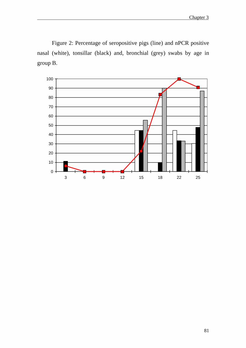
Chapter 3
81
Figure 2: Percentage of seropositive pigs (line) and nPCR positive
nasal (white), tonsillar (black) and, bronchial (grey) swabs by age in
group B.
0
10
20
30
40
50
60
70
80
90
100
3 6 9 12 15 18 22 25
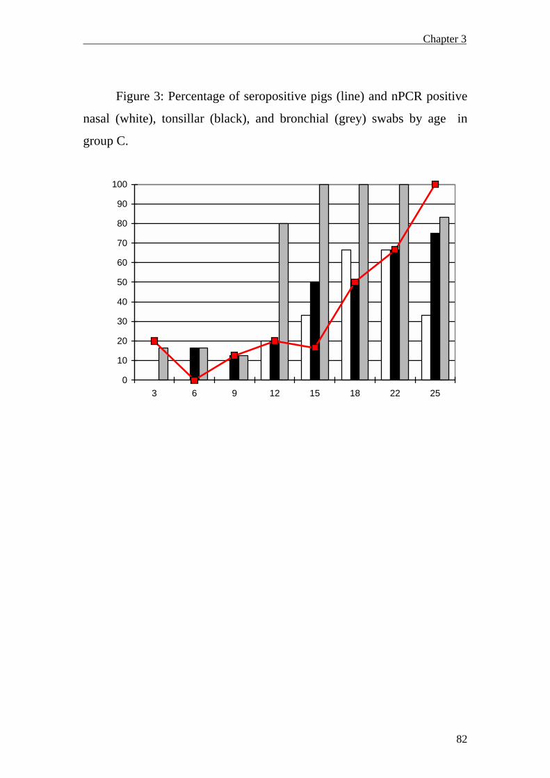
Chapter 3
82
Figure 3: Percentage of seropositive pigs (line) and nPCR positive
nasal (white), tonsillar (black), and bronchial (grey) swabs by age in
group C.
0
10
20
30
40
50
60
70
80
90
100
3 6 9 12 15 18 22 25

Chapter 3
83
Figure 4: Mean macrosopic lung lesions score in group A (black),
group B (white) and group C (grey) by age
0,0
5,0
10,0
15,0
20,0
25,0
30,0
35,0
3 6 9 12 15 18 22 25

Chapter 3
84
Figure 5: Percentage of microscopic lung lesions compatibles with
Enzootic pneumonia in group A (black), group B (white) and group C
(grey) by age
0
10
20
30
40
5060
70
80
90
100
3 6 9 12 15 18 22 25

General Discussion
GENERAL DISCUSSION

General Discussion
86
In this section, a general discussion on the conclusions obtained in
each study, their contributions and their limitations on the knowledge on
M.hyopneumoniae infection will be addressed.
In order to control a disease, it is critical to understand its
transmission and circulation of the causative agent within a herd and
factors that are involved in the manifestation of the clinical outcome. The
first step in studying a disease is to diagnose it; in case of infectious
disease, this point is initially based on the detection of the etiological
agent. In the case of M. hyopneumoniae, bacteriological culture is
difficult and time-consuming, and for these reasons molecular tools have
been developed and used in upper respiratory tract samples to monitor the
presence of agent in field studies. The research presented in this Thesis
assess the dynamics of infection of M. hyopneumoniae at a herd level and
its relationship with production systems used in a transversal field trial
(Chapter 1), and with EP-compatible microscopic and macroscopic
lesions and/or disease in a longitudinal study (Chapter 3). Moreover,
correlation of M.hyopneumoniae detection in upper respiratory site with
detection in bronchi, and with EP microscopic and macroscopic lung
lesions at individual pig level has been also addressed (Chapter 2).
Relationships between detection of M.hyopneumoniae in upper and lower
respiratory sites needs to be clarified in order to interpret herd monitoring
results using nPCR from upper respiratory site samples.
The studies on infection dynamics show that the factor with
greatest effect on M. hyopneumoniae circulation pattern was the farm
(Chapter 1). Even within a farm (Chapter 3), significant differences in the
dynamics of infection between different batches were observed, which

General Discussion
87
were also related with different degrees and timings of M.
hyopneumoniae –related lesions appearance.
Moreover, the type of production system (1/2-site versus 3-site
herds) is another factor that influences M.hyopneumoniae infection
dynamics. In modern swine production, at approximately 18-20 weeks of
age, pigs appear to experience severe outbreaks, resulting in elevated
morbidity and even increased mortality (Dee, 1996). This clinical
condition has been defined and characterized as porcine respiratory
disease complex (PRDC), where M. hyopneumoniae plays a major role
(Thacker et al., 1999). PRDC appears to be closely associated with
multisite production systems (Dee, 1996). Results of our field trial in
regards M.hyopneumoniae, involving 12 Spanish herds clearly support
this idea. M. hyopneumoanie colonization in nurseries was lower and
occurred later in time in 3-site versus in 1/2-site production systems.
Moreover, an abrupt increase in the proportion of infected animals,
associated with late respiratory outbreaks was seen in the multisite
production systems.
Besides the influence of production systems, involvement of M.
hyopneumoniae in respiratory disease observed in these farms was
analysed. Although M. hyopneumoniae infection was detected in all
farms, it was not always involved in the respiratory disease. An important
contribution of this work is that, although seroconversion and M.
hyopneumoniae infection were detected in all herds, clinical and
subclinical infections were described. This is important for decision
making on the control strategies to implement at the farm. Serumprofiles
were not discriminative to determine the involvement of M.
hyopneumoniae in a clinical outbreak. Bacteriumprofiles were more
useful in determining M. hyopneumoniae involvement in respiratory
disease, since an increase of the proportion of infected pigs (based on

General Discussion
88
detection of M. hyopneumoniae in nasal swabs) was clearly related with
appearance of the clinical outbreak. The idea that a certain percentage of
infected pigs is necessary to induce disease is supported in both field
studies (Chapers 1 and 3), together with other recently published studies
(Vicca et al., 2002). Although not discriminative, the proportion of
seropositive pigs was also greater in clinically than in subclinically
infected herds.
So far, there are three crossectional studies from different
countries, USA (Calsamiglia et al., 1999b), Belgium (Vicca et al., 2002)
and Spain (Chapter 1) in which infection dynamics was studied using
serumprofiles in combination with bacteriumprofiles. Within the M.
hyopneumoniae clinically affected farms, proportions of infected pigs
when the outbreak occurred varied between the three studies (USA: 70-
100%, Spain: 10-70%, Belgium: 8-24%). These differences in
proportions may be explained by: differences in sensitivity, use of
antibiotics, different genetic lines, presence of co-infections, number of
farms tested, and inherent characteristics of each farm.
An important limitation of the abovementioned studies is that they
are transversal rather than longitudinal. Therefore, the information
obtained reflects the M. hyopneumoniae infection status of a different
group of animals in a point in time rather than the evolution of infection
throughout life time of a particular group of pigs.
Dynamics of serocoversion against M. hyopneumoniae was another
issue addressed in both, transversal and longitudinal field trials. Several
authors have described the time of seroconversion under field conditions
as slow, delayed and very variable (Morris et al. 1995, Sitjar et al., 1996,
Calsamiglia et a., 1999b). In the two field trials described in this Thesis,
time of seroconversion was also variable. Results obtained in Chapter 3
indicate that seroconversion was more related to detection of M.

General Discussion
89
hyopneumoniae in bronchial swabs rather than in nasal or tonsillar swabs,
and to the detection of a higher percentage of pigs with a positive nPCR
bronchial swab. Although the relation with proportions of pigs infected
pigs at bronchi level, seroconversion was still variable with respect to the
age in which these proportion increase.
In chapter 1, information regarding vaccination and medication
strategies conducted in the tested farms was provided by the farmer.
These factors were, therefore, considered as non-controlled variables.
However, they were tested in a univariate and multivariate statistical
model. The first model showed that vaccination against
M.hyopneumoniae had a significant effect on pathogen’s nPCR detection.
On the contrary, when the effect of all variables was analysed in the
multivariate analysis, both vaccination and medication had a significant
effect on lowering the nPCR results. These results are in agreement with a
M. hyopneumoniae vaccination field trial, where prevalence of nPCR-
positive piglets at weaning was lower in groups of vaccinated sows (Ruiz
et al., 2003). This reduction could be due to lower levels of shedding in
vaccinated sows and/or an increased maternal immunity, and therefore
lower levels of nasal M. hyopneumoniae colonization in the offspring.
However, the effect of vaccination and medication strategies on
colonization (nPCR) at the sow and piglets level should be studied in
more detail.
Results from Chapter 1 and Chapter 3 highlighted that early
colonization of M.hyopneumoniae occurs at different levels of the
respiratory tract. This is the first time that M. hyopneumoniae infection is
detected at the lung level (bronchial swabs) in such an early stage (3
weeks of age).
Another objective aimed in this Thesis was to assess the
significance of detection M.hyopneumoniae in nasal swabs. Our initial

General Discussion
90
hypothesis was that nasal swabs are adequate sampling sites to monitor
M. hyopneumoniae infection in live pigs. Chapters 2 and 3 addressed this
point; the first study, using animals received at the Servei de Diagnòstic
Veterinari at the Veterinary school of the Universitat Autònoma de
Barcelona, and the latter one in a longitudinal study carried in a farm
suffering from mycoplasma-related disease. As expected, in both studies
bronchial swabs were the best predictors of presence of microscopic and
gross EP lesions coinciding with previously published studies
(Calsamiglia et al., 2000). However, to sample bronchial swabs from live
animals is not possible. Surprisingly, the detection in nasal swabs and the
level of prediction was different in Chapters 2 and 3. The relative
proportion of nasal detection was lower in the field study than in the
study using pigs received at the diagnostic laboratory. An increase of
M.hyopneumoniae shedding in nasal cavities due to stressful situations
(such as transport) or due to increase multiplication of bacteria after the
animal’s death have been proposed as potential explanations for a larger
proportion nPCR positive nasal samples obtained in Chapter 2.
Another interesting finding in Chapter 3 is the discontinuous
detection of M. hyopneumoniae in nasal swabs. Ruiz et al,. (2002a)
reported a high but discontinuous pattern of nasal and tracheal detection
in an challenge study. Moreover, in a longitudinal field study, where
nursery pigs were sampled 7 times during the nursery period, most of the
pigs were positive only in one sampling (Ruiz et al., 2002c). Data from
other analyses performed in our group (data not shown) demonstrated that
detection in nasal swabs throughout the live of pigs is discontinuous: a
maximum of three positive nasal samplings out of nine were detected in
the same animal. Moreover, these three positive swabs were not always
detected in consecutive samplings. From the results of these previously
published studies (Ruiz et al., 2002a, Ruiz et al., 2002c) and the work

General Discussion
91
performed by our group, it can be proposed that M.hyopneumoniae nasal
swab detection by nPCR is not as frequent and constant as previously
thought, and that detection of M. hyopneumoniae in nasal swabs becomes
more constant when microscopic and macroscopic lesions exist.
Although tonsillar sampling in live animals is more laborious than
nasal samplings, it is possibly more adequate to monitor M.
hyopneumoniae detection at a farm level. Tonsillar swabs are currently
being used to detect other swine pathogens such as Actinabacillus
pleuropneumoniae (Chiers, 2004). This respiratory pathogen, usually
attaches to the cells of the terminal bronchioli and alveolar epithelial
cells, but it is also detected in upper respiratory sites (Chiers, 2004).
Studies using electronic microscopy have shown that, in carrier pigs, the
pathogen can reside in tonsillar crypts for long periods without
developing the disease. Therefore, tonsillar sampling is considered a
useful site for detection of A. pleuropneumoniae sublinically infected
pigs. Shedding of this pathogen in nasal secretions only takes place at the
time of acute lung infection (Chiers, 2004). Taking into account these
observations, studies addressing the interactions of M.hyopneumoniae
with cells of both upper respiratory sites will help in interpreting its
detection in tonsil and nasal cavities.
In Chapters 2 and 3, analysis of microscopic lesions was
performed. Results of these studies and from previous works (Calsamiglia
et al., 2000), showed that when bronchial swabs are positive the
probability to show EP-compatible microscopic lesions in the animal is
high. However, presence of EP-compatible lesions does not always imply
M. hyopneumoniae detection in bronchi. In Chapter 3, EP-compatible
microscopic lesions appeared in pigs from groups A and B before
M.hyopneumoniae was detected in bronchial swabs. Swine influenza
virus (SIV) is known to cause microscopic lesions compatible with the

General Discussion
92
defined scores 3 and 4 (EP-compatible microscopic lesions). SIV
antibodies were detected in slaughter pigs, indicating that infection with
this agent occurred in those groups of pigs.
One limitation of the longitudinal study presented in Chapter 3
was that clinical signs (such as coughing) were not properly recorded. In
that study, clinical signs were only recorded when observed on the
sampling day. Therefore, the time of appearance and number of animals
affected are unknown.
A complementary and informative approach to M.hyopneumoniae
dynamics of infection should be the knowledge on bacterial loads.
Quantification of Mycoplasma cells will help to relate the proportion of
infected animals and individual bacterial loads, and better define the
concept “pressure of infection” used in these studies. Also, chronology of
infection at the different respiratory levels will be better interpreted and
presumably more easily related with microscopic and macroscopic
lesions and disease. Therefore, an interesting future research line on
M.hyopneumoniae would be the use of a quantitative PCR method in
order to assess these latter points.

Conclusions
CONCLUSIONS

Conclusions
94
1. A certain percentage of M. hyopneumoniae infected pigs seems
to be necessary to induce clinical signs. Thus,
M.hyopneumoniae clinically infected herds have greater
percentages of nPCR positive pigs than subclinically infected
herds without a clinical outbreak.
2. The Production system type, may affect M.hyopneumoniae
colonization and infection dynamics: 3- site production systems
had later colonization than 1/2-site systems, and a more abrupt
increase of infected pigs was observed in the finishers, which
was associated with disease.
3. M. hyopneumoniae infection dynamics is greatly influenced by
characteristics inherent to each farm.
4. Seroconversion in field situations is very variable and is related
with certain percentage of infected pigs, mainly at the bronchi
levels, to be elicited.
5. Although combination of serumprofiles with bacteriumprofiles
provide very useful information on M. hyopneumoniae infection
dynamics, differences between different batches of the same
farm occur, and therefore its application for decision making on
vaccination/medication programs in subsequent groups is not so
reliable.
6. M. hyopneumoniae nasal swab detection was not so frequent
and constant as previously thought. In the longitudinal study, it
was shown that nasal swab detection becomes more reliable
when microscopic and macroscopic already exist.
7. Tonsillar swabs are an alternative sampling site for monitoring
M. hyopneumoniae infection in field studies. Detection of
M.hyopneumoniae in tonsillar swabs was observed even in

Conclusions
95
farrowing and nursery pigs, previously to the appearance of
microscopic and macroscopic lung lesions.
8. Detection of M.hyopneumoniae in bronchial swabs was greater
than in nasal and tonsillar swabs, and is the best predictor of
EP-compatible microscopic lesions. Early colonization was also
detected using bronchial swabs.

References
96
REFERENCES
Andreasen M, Peter Nielsen J, Baekbo P, Willberg P, Botner A: 2000, A
longitudinal study for serogical patterns of respiratory infections in
nine infected Danish swine herds. Prev. Vet. Med. 45:221-235.
Andreasen M, Mousing J, Krogsgaard Thomsen L: 2001a, No overall
relationship between average daily gain and the serological
response to Mycoplasma hyopneumoniae and Actinobacillus
pleuropneumoniae in eight chronically infected Danish swine
herds. Prev. Vet. Med. 49: 19-28.
Andreasen M, Mousing J, Krogsgaard Thomsen L: 2001b, No simple
association between time elapsed from seroconversion until
slaughter and the extent of lung lesions in Danish swine. Prev. Vet.
Med. 52:147-161
Amanfu W, Weng CN, Ross RF, Barnes HJ: 1984, Diagnosis of
Mycoplasmal pneumonia of swine: Sequential study by direct
immunofluorescence. A.J.V.R 45(7): 1349-1352.
Asai T, Munenori O, Ono M, Irisawa T, Mori Y, Yokomizo Y, Sato S:
1993, Increased levels of tumor necrosis factor and interleukin 1 in
bronchoalveolar lavage fluids from pigs infected with Mycoplasma
hyopneumoniae. Vet. Immnuol Immunopathol 38:253-260.
Asai T, Okada T, Ono M, Mori Y, Yokomizo Y, Sato S: 1994, Detection
of IL-6 and PGE 2 in bronchoalveolar lavage fluid of pigs
experimentally infected with Mycoplasma hyopneumoniae. Vet.
Immunol. Immunopathol. 44: 97-102
Armstrong CH: 1981, Porcine Mycoplasmas. Chapter 6. p68-83.
Armstrong CH: 1983, Comparison of the enzyme-linked immunosorbent
assay and the indirect hemagglutination and complement fixation
tests for detecting antibodies to Mycoplasma hyopneumoniae. Can.
J. Comp. Med. 47: 464-470

References
97
Artiushin S, Stipkovits L, Minion FC: 1993, Development of polymerase
chain reaction primers to detect Mycoplasma hyopneumoniae. Mol.
Cell. Probes. 7:381-385.
Artiushin S, Minion C: 1996. Arbitrarily primed PCR analysis of
Mycoplasma hyopneumoniae field isolates demonstrates genetic
heterogeneity. Int. J. Syst. Bacteriol. 46.324-328.
Bakersville, A: 1972, Development of the Early lesions in Experimental
enzootic pneumonia of Pigs: an ultrastructural and histological
study. Res. Vet. Sci. 13: 570-578
Baumeister AK, Runge M, Ganter M, Feesntra AA, Delbeck, Kirchhoff
H. Detection of Mycoplasma hyopneumoniae in Bronchioalveolar
Lavage Fluids of pigs by PCR. J. Clin. Microbiol. 36(7): 1984-
1988.
Baumgartner A, Nicolet J: 1984, Detection of antibodies against
membrane glycoprotein of swine erythrocytes after experimental
infection with M.hyopneumoniae. Vet. Immuno. Immunopathol.
73-79
Batista L, Pijoan C, Ruiz A, Utrera V, Dee S: 2002, Assessment of
transmission of Mycoplasma hyopneumoniae by personnel. 12(2):
75-77. In Proceedings of the 17th IPVS Congress, Ames, Iowa.
P285
Blanchard B, Vena MM, Cacalier A, Le Lannic J, Gouranton J, Kobisch
M: 1992, Electron microscopic observation of the respiratory tract
of PSF piglets inoculated with M. hyopneumoniae. Vet. Microbiol.
30: 329-341.
Blanchard B, Kobisch M, Bové JM, Saillard C: 1996, Polymerae chain
reaction for Mycoplasma hyopneumoniae detection in
tracheobronchial washings from pigs. Mol.Cell.Probes. 10:15-22.

References
98
Bereiter M, Young TF, Joo HS, Ross RF: 1990, Evaluation of the ELISA
and comparison to the complement fixation test and radial
immunodiffusion enzyme assay for detection of antibodies against
Mycoplasma hyopneumoniae in swine serum. Vet. Microbiol. 25:
177-192.
Betts AO. 1952, Respiratory Diseases of pigs V- some clinical and
epidemiological aspects of virus pneumonia. Vet. Rec. 63: 283
Bosch G, Zhou C: 2000, Using nested PCR and serology to determine
disease patterns in herds dually infected with PRRS virus and
Mycoplasma hyopneumoniae. In Proceedings of the 16th IPVS
Congress, Melbourne, Australia, P451
Brooks E, Fauld D: 1989, The Mycoplasma hyopneumoniae 74,5kD
antigen elicits neutralizing antibodies and shares sequences
similarity with heat-shock proteins. In Vaccines. Ed. Lerner RA,
Ginsberg H, Chanock RM, brown F. New York: Cold Spring
Harbor Laboratory Press. 89.pp 265-269
Cardona AC, Pijoan C, Dee SA: 2003, Assessment of Mycoplasma
hyopneumoniae aerosol movement. Proceedings of Leman
Conference. P16.
Calsamiglia M, Pijoan C, Trigo A: 1999a, Application of a nested
polymerase chain reaction assay to detect Mycoplasma
hyopneumoniae from nasal swabs. J. Vet. Diagn. Invest. 11, 246-
251.
Calsamiglia M, Pijoan C , Bosch GJ: 1999b, Profiling Mycoplsama
hyopneumoniae in farms using serology and a nested PCR
technique. Swine Health Prod. 7, 263-268.
Calsamiglia M, Collins JE, Pijoan C: 2000a,Correlation between the
presence of enzootic pneumonia lesions and detection of

References
99
Mycoplasma hyopneumoniae in bronchial swabs by PCR. Vet.
Microbiol. 76, 299-303.
Calsamiglia M, Pijoan C: 2000b, Colonisation state and colostral
immunity to Mycoplasma hyopneumoniae of different party sows.
Vet. Rec. 146: 530-532.
Caron J, Ourdani M, Dea S: 2000a, Diagnosis and differentiation of
Mycoplasma hyopneumoniae and Mycoplasma hyorhinis infections
in Pigs by PCR amplification of the p36 p46 genes. 38(4): 1390-
1396.
Caron J, Sawyer N, ben Abdel Moumen B, Cheikh Saad Bouh K, Dea S:
2000b, Species-Specific Monoclonal Antibodies to Escherichia
coli-Expressed p36 cytosoloic protein of Mycoplasma
hyopneumoniae. Clin. Diag. Lab. Immunol. 7(4): 528-535.
Caruso JP, Ross rf: 1990, Effects of Mycoplasma hyopneumoniae and
Actinobacillus (Haemophilus) pleuropneumoiae infections on
alveolar macrophage functions in swine. Am. J. Vet. Res. 51(2):
227-231.
Cheikh Saad Bouh K, Shareck F, Dea S: 2003, Monoclonal Antibodies to
Escherichia Coli-Expressed p46 and p65 membranous proteins for
Specific immunodetection of Mycoplasma hyopneumoniae in
Lungs of infected pigs. Clin. Diagn. Lab. Immunol. 459-468.
Chen JR, Lin JH, Weng CN, Lai SS: 1998, Identification of a novel
adhesin-like glycoprotein from Mycoplasma hyopneumoniae. Vet.
Microbiol, 62: 97-110.
Chen JR, Liao CW, Mao SJT, Weng CN: 2001, A recombinant chimera
composed of repeat region RR1 of Mycoplasma hyopneumoniae
adhesin with Pseudomonas exotoxin: in vivo evaluation of specific
IgG response in mice and pigs. Vet. Microbiol. 80: 347-357.
References
100
Chen YL, Wang SN, Yang WJ, Chen YJ, Lin HH, Shiuan D: 2003,
Expression and immunogenecity of Mycoplasma hyopneumoniae
Heat shock protein antigen p42 by DNA vaccination. Infect.
Immuni. 71(3): 1155-1160
Chiers K: 2004, Detection of pigs carrying Actinobacillus
pleuropneumoniae in their upper respiratory tract using polymerase
chain reaction. PhD Thesis. University of Gent, Gent. Belgium
Chittick, W: 2000, A novel approach for reporting of serologic antibody
results using a competitive Mycoplasma hyopneumoniae ELISA. In
Proceedings of the 16th IPVS Congress, Melbourne, Australia.
P456.
Christensen G, Sorensen V, Mousing J: 1999, Disease of the Respiratory
System, In Diseases of Swine, 8th Ed. Eds. Straw BE, D’Allaire S,
Mengeling WL, Taylor DJ. Iowa State University Press, Ames. P
913-941.
Ciprián A, Pijoan C, Cruz T, Camacho J, Tortora J, Colmenares G,
Lopez-Revilla R, de la Garza M: 1988, Mycoplasma
hyopneumoniae increases susceptibility of pigs to experimental
Pasteurella multocida. Can. J. Vet. Res. 52: 434-438
Clark LK, Armstrong Ch, Freeman MJ, Scheidt AB, Snads-Freeman L,
Knox K: 1991, Investing the transmission of Mycoplasma
hyopneumoniae in a swine herd with enzootic pneumonia. Vet.
Med. May, 543-550.
Cubero Martín, G: 2000, El matadero como herramienta para la mejora
de la producción. III Foro Porcino, 24-25 de Enero, Madrid.
Czaja T, Kanci A, Lloyd LC, Markham PF, Whithear KG, Browning GF:
2002, Induction of enzootic pneumonia in pigs by the
administration of an aerosol of in vitro-cultured Mycoplasma
hyopneumoniae. Vet. Rec. 150: 9-11
References
101
Davies PR, Banhson PB, Grass Jj, Marsh WE, Dial GD: 1995,
Comparison of methods for measurement of enzootic pneumonia
lesions in pigs. Am. J. Vet. Res. 56(6): 709-714.
Dawson A, Harvey RE, Thevasagayam Sj, Sherington J, Peters AR:
2002, Studies of the field efficacy and safety of a single-dose
Mycoplasma hyopneumoniae vaccine for pigs. Vet. Rec. 151: 535-
538.
DeBey MC, Jacbson CD, Ross RF: 1992, Histochemical and
morphologic changes of porcine airway epithelial cells in response
to infection with Mycoplasma hyopneumoniae. Am.J.Vet.Res.
53(9): 1705-1710.
DeBey MC, Roth JA, Ross RF: 1993, Enhancement of increase in
intracellular calcium concentration in stimulated neutrophils by
Mycoplasma hyopneumoniae. Vet. Res. Communications, 17: 249-
257.
DeBey MC and Ross RF: 1994, Ciliostasis and loss of cilia induced by
Mycoplasma hyopneumoniae in porcine tracheal organ culture.
Infec. Immun. 62(12): 5312-5318.
Dee S: 1996, The porcine respiratory disease cmplex: are subpopulations
important?. Swine. Health. Prod. 4: 147-149.
Desrosiers R: 2001, A review of some aspects of the epidemiology,
diagnosis, and control of Mycoplasma hyopneumoniae infections.
J. Swine Health. Prod 9(5): 233-237.
Desrosiers R: 2004, Epidemiology, diagnosis and control of swine
diseases. Proceeding of AASV congress.Des Moines. Iowa. 9-37
Djordjevic SP, Eamens GJ, Romalis LF, Saunders MM: 1994, An
improved enzyme linked immunosorbent assay (ELISA) for the
detection of porcine serum antibodies against Mycoplasma
hyopneumoniae. Vet. Microbiol. 39: 261--274

References
102
Djordjevic SP, Eamens GJ, Romalis LF, Nicholis PJ, Taylor V, Chin J:
1997, Serum and mucosal antibody response and protection in pigs
vaccinated against Mycoplasma hyopneumoniae with vaccines
containing a denatured membrane antigen pool and adjuvant. Aus.
Vet. J. 75: 504-511
Dohoo IR, Montgomery ME: 1996, A field trial to evaluate a
Mycoplasma hyopneumoniae vaccine: effects on lung lesions and
growth rates in swine. Can. Vet. J.37: 299-302.
Done SH: 1991, Environmental factors affecting the severity of
pneumonia in pigs. Vet. Rec. June 22: 582-586.
Done SH: 1997, Enzootic Pneumonia (mycoplasmosis) revisited. The pig
Journal. p 40-61.
Donham KJ: 1991, Association of enviromental air contaminants with
disease and productivity in swine. Am. J. Vet. Res. 52: 1723-1730
Doster AR, Boh Chang Lin: 1988, Identification of Mycoplasma
hyopneumoniae in formalin-fixed porcine lung, using and indirect
immunoperoxidase method. Am.J.Vet.Res. 49(10): 1719-1721
Erlandson K, Thacker B, Wegner m, Evans R, Thacker E: 2002,
Evaluation of three antibody ELISA test for Mycoplasma
hyopneumoniae. In Proceedings of the 17th IPVS Congress. Ames,
Iowa. P74.
Falk K, Lium BM: 1991, An abattoir survey of pneumonia and pleuritis
in Slaughter weight swine from 9 selected herds. Acta. Vet. Scand.
32, 79-88.
Fagan PK, Djordjevic SP, Eamens GJ, Chin J, Walker MJ: 1996,
Molecular characterization of ribonucleotide reductase (nrdF) gene
fragment of Mycoplasma hyopneumoanie and assessment of the
recombinant product as an experimental vaccine for enzootic
pneumonia. Infect. Immun. Mar. 1060-1064

References
103
Fagan PK, Walker MJ, Chin J, Eamens GJ, , Djordjevic SP: 2001, Oral
immunization of swine with attenuated Salmonella typhimurium
aroA SL3261 expressing a recombinant antigen of Mycoplasma
hyopneumoanie (NrdF) primes the immune system for NrdF
specific secretory IgA response in the lungs. Microbial. Pathogen.
30:101-110
Fano E, Pijoan C, Dee S: 2003, Evaluation of aerosol transmission of
Mycoplasma hyopneumoniae and porcine reproductive and
respiratory syndrome virus (mixed infection) under field
conditions. Proceedings of Leman Conference. p15. St Paul.
Minnesota
Feld NC, Qvist P, Ahrens P, Friis NF, Meyling A: 1992, A monoclonal
blocking ELISA detecting serum antibodies to Mycoplasma
hyopneumoniae. Vet. Microbiol. 30: 35-46.
Frey J, Haldimann A, Nicolet J: 1992, Chromosomal heterogeneity of
Various Mycoplasma hyopneumoniae field strains. Int. J. Syst.
Bacteriol. 42(2): 275-280.
Frey J, Haldimann A, Kobisch m, Nicolet J: 1994, Immune response
against the l-lactate dehydrogenase of Mycoplasma hyopneumoniae
in enzootic pneumonia of swine. Microbiol Pathogenesis 17: 313-
322.
Fuentes M, Pijoan C: 1987, Pneumonia in pigs induced by intranasal
challenge exposure with Pseudorabies virus and Pastereulla
multocida. Am. J. Vet. Res. 48. 1446-1448
Futo S, Seto Y, Okada M, Sato A, Suzuki T, kawai K, Imada Y, Mori Y:
1995, Recombinant 46-Kilodalton surface antigen (p46) of
Mycoplasma hyopneumoniae expressed in Escerichia Coli can be
used for early specific diagnosis of Mycoplasmal pneumonia of

References
104
swine by enzyme-linked immunosorbent assay. J. Clin. Micribiol.
33(3): 680-683.
Garnier M, Foissac X, Gaurivaud P, Laigret F, Renaudin J, Saillard C,
Bove JM: 2001, Mycoplasmas, plants, insects vectors: a
matrimonial triangle. Acad. Sci. Paris. In Life science. 324: 923-
928
Geary SJ, Walczak EM: 1985, Isolation of cytopathic factor from
Mycoplasma hyopneumoniae. Infect. Immun. 48: 576-578
Goodwin RFW, Pomeroy AP, Whittlestone P: 1965, Production of
Enzootic Pneumonia with a Mycoplasma. Vet. Rec. 77.1247-1249.
Goodwin RFW: 1985, Apparent reinfection of enzootic pneumonia-free
pig herds: Search for possible causes. Vet. Rec. 690-694.
Haesebrouck F, Pasmans F, Chiers K, Maes D, Ducatelle R, Decostere A:
2004, Efficacy of vaccines against bacterial diseases in swine: what
can we expect?. Vet. Microbiol. 100: 255-268.
Halbur PG: 1997, Making the diagnosis with Serology, antigen Detection
and PCR Mycoplasmal Pneumonia in modern swine production
units. In Proceeding of the ISU Veterinary Medicine Seminar. p3-
7
Haldimann A, Nicolet J, Frey J: 1993, DNA sequence determination and
biochemical analysis of the immunogenic protein p36, the lactate
dehydrogenase (LDH) of Mycoplasma hyopneumoniae. J. General.
Microbiol. 139: 317-323.
Hannan PCT, Bhogal BS, Fish JP: 1982, Tylosin tartrate and tiamulin
effects on experimental piglet pneumonia induced with pneumonic
pig lung homogenate containing Mycoplasma, bacteria and viruses.
Res. Vet. Sci. 33: 76-88

References
105
Harasawa R, Koshimizu K, Takeda O, Uemori T, Asada K, kato I: 1991,
Detection of Mycoplasma DNA by the polymerase Chain Reaction.
Moll. Cell. Probes. 5:103-109.
Harris DL: 1988, Alternative approaches to eliminating endemic disease
and improving performance of pigs. Vet. Rec. 15: 422-423.
Hipple K, Ruebling K, Minion FC, Thacker B, Thacker E: 2002, A PCR
assay to detect Mycoplasma hyopneumoniae in formalin fixed
tissues. In Proceedings of the 17th IPVS Congress, Ames, Iowa.
P453.
Hodgings D, Schewen PE, Dewey CE: 2004, Influence of age and
maternal antibodies on antibody response of neonatal piglets
vaccinated against Mycoplasma hyopneumoniae. J. Swine. Health.
Prod. 12(1):10-16.
Hsu T, Artiushin S, Minion FC. 1997, Cloning ad functional analysis of
the p97 swine cilium adhesin gene of Mycoplasma hyopneumoniae.
J. Bacteriol. 179: 1317-1323.
Hsu T, Minion FC: 1998, Molecular analysis of the p97 cilium adhesin
operon of Mycoplasma hyopneumoniae. Gene. 214: 13-23
Hurnik D, Hanna PE, Dohoo IR: 1993 Evaluation of Rapid Gross visual
appresial of Swine lungs at slaughter as a Diagnostic screen for
enzootic pneumonia. Can. J. Vet. Res. 57: 37-41
Hurnik D, Dohoo IR, Bate LA: 1994, Types of farm management as risk
factors for swine respiratory disease. Prev. Vet. Med. 20, 147-57
Jacques M, Blanchard B, Foiry B, Girard C, Kobisch M: 1992, In vitro
colonization of porcine trachea by Mycoplasma hyopneumoniae.
Ann. Rec. Vét. 23: 239-347.
Jayappa H, Davis R, Rapp-Gabrielsen V, Wasmen T, Thacker E: 2001,
Evaluation of the efficacy of Mycoplasma hyopneumoniae bacterin
following immunization of young pigs in the presence of varying

References
106
levels of maternal antibodies. In Proceedings of AASV, Nashville.
Tennesse. P237-241.
Jorsal SE, Thomsen BL: 1988, A cox regression analysis of risk factors
related to Mycoplasma suipneumoniae reinfection in Danish SPF-
herds. Acta. Vet. Scand (suppl) 84: 436-437.
King KW, Faulds DH, Rosy EL, Yancey RJ: 1997, Characterization of
the gene encoding Mhp1 from Mycoplasma hyopneumoniae and
examination of Mhp1’s vaccine potential. Vaccine 15(1): 25-35
Kishima M, Ross RF: 1985, Supressive effect of nonviable Mycoplasma
hyopneumoniae on phytohemagglutinin-indced transformation of
swine lymphocytes. Am. J. Vet. Res. 46(11): 2366-2368.
Klark LK, Armstrong CH, Freeman MJ, Scheidt AB, Sands-Freeman l,
Knox K: 1991a, Investigating the transmission of Mycoplasma
hyopneumoniae in swine herd with enzootic pneumonia. Vet. Med.
May. 543-550.
Klark LK, Armstrong CH, Knox K, Mayrose V: 1991b, The effect of all-
in/all-out management pigs from a herd with enzootic pneumonia.
Vet. Med. 946:951.
Kobisch M, Quillien L, Tillo JP, Wroblewski: 1987 The Mycoplasma
hyopneumoniae plasma membrane as a vaccine against porcine
enzootic pneumonia. Ann. Inst. Pasteur. 138: 693-705
Kobisch M, Blanchard b, Le Potier MF: 1993, Mycoplasma
hyopneumoniae infection in pigs: duration of the disease and
resistance to reinfection. Vet. Res. 24: 67-77
Kobisch M: 2000, Mycoplasma disease in pigs-old diseases still causing
trouble. In Proceedings of the 16th IPVS Congress, Melbourne,
Australia. P434-438.
Kristensen CS, Andreasen m, Ersboll AK, Nielsen JP: 2004 Antibody
response in sows and piglets following vaccination against

References
107
Mycoplasma hyopneumoniae, Pasteurella toxigenic multocida,
and Actinobacillus pleuropneumoniae. Can. J. Vet. Res. 68: 66-70.
Kuhn MA: 2004, Comparative safety evaluation of a one-dose
M.hyopneumoniae bacterin (Respisure-One) and a competitor one-
dose M.hyopneumoniae bacterin. In Proceedings of Proc AASV
Congress, Des Moines , Iowa. p 267-270.
Kurth KT, Hsu T, Snook ER, Thacker EL, Thacker BJ, Minion FC: 2002,
Use of a Mycoplasma hyopneumoniae nested polymerase chain
reaction test to determine the optimal sampling sites in swine. J.
Vet. Diagn. Invest. 14. 463-469.
Kyriakis SC, Alexopoulos C, Vlemmas J, Sarris k, Lekkas S, Koutsoviti-
Papadopulou M, Saoulidis K: 2001, Field study on the efficacy of
two different vaccination schedules with Hyoresp in a
Mycoplasmas hyopneumoniae infected commercial herd.
J.Vet.Med.b. 48: 675-684.
Kwon D, Chae C: 1999, Detection and localization of M. hyopneumoniae
DNA in lungs from naturally infected pigs by in situ hibridization
using a digoxigenin-labeled probe. Vet. Pathol. 36. 308-313.
Kwon D, Choi C, Chae C: 2002, Chronologic localization of Mycoplasma
hyopneumoniae in experimentally infected pigs. Vet. Pathol. 39:
584-587.
Lam KM, Switzer WP: 1972, Mycoplasmal pneumonia of swine: active
and passive immunizations. Am. J. Vet. Res. 32: 1737-1741.
Leon EA; MAdec F, Taylor NM, Kobisch M: 2001, Seroepidemiology of
Mycoplasma hyopneumoniae in pigs from farrow-to-finish farms.
Vet. Microbiol. 78: 331-341
Le Potier MF, Abiven P, Kobisch M: 1994, A blocking ELISA using a
monoclonal antibody for the serological detection of Mycoplasma
hyopneumoniae. Res. Vet. Science. 56: 338-345.

References
108
Levonen K: 1994, Detection of enzootic pneumonia in pig herds using an
enzyme-linked immunosorbent assay in sow colostrum. Res. Vet.
Science. 56: 111-113.
Levonen K, Sihvo E, Veijalainen P: 1999, Comparison of two
commercial enzyme-linked immunosorbent assays for the detection
of antibodies against Mycoplasma hyopneumoniae and correlation
with herd status. J. Vet. Diagn. Invest. 11: 547-549.
Lin BC: 2001, Intraspecies differentiation of Mycoplasma
hyopneumoniae field strains in the United States. In Proceeding of
AASP. Nashville, Tennesse. p225-235.
Lin JH, Weng CN, Liao CW, Yeh KS, Pan MJ: 2003, Protective effects
of oral microencapsulated Mycoplasma hyopneumoniae vaccine
prepared by Co-spray drying method. J. Vet. Med. Sci. 65(1): 69-
74.
Lium BM, Falk K: 1991, An abattoir survey of pneumonia and pleuritis
in Slaughter weight swine from 9 selected herds. Acta. Vet. Scand.
32, 55-65
Livingston CW, Stair EL, Underdahl MS, Mebus CA: 1972, Pathogenesis
of Mycoplasmal Pneumoniae in Swine. Am. J. Vet. Res. 33(11):
2249-2258
Llopart D, Casal J, Clota J, Navarra I, March R, Riera P, Artigas C: 2002,
Evaluation of the field efficacy and safety of a Mycoplasma
hyopneumoniae vaccine in finishing pigs. The Pig Journal. 49. 70-
83.
Maes D, Verdonck M, Deluyker H, de Kruif A: 1996, Enzootic
Pneumonia in Pigs. The Vet. Quarterly. 18(3): 104-109.
Maes D, Deluyker H, Verdonck M, Castryck F, Miry C, Vrijens B,
Verbeke W, Viane J, de Kruif A: 1999, Effect of vaccination

References
109
against Mycoplasma hyopneumoniae in pig herds with an all-in/all-
out production system. Vaccine. 17. 1024-1034.
Maes D, Deluyker H, Verdonck M, Castryck F, Miry C, Vrijens B, Kruif
A: 2000, Herd factors associated with the seroprevalence of four
major respiratory pathogens in slaughter pigs from farrow-to-finish
pig herds. Vet. Res. 313: 327.
Maes D, Verdonck M, Verbeke W, Viane J, de Kruif A: 2000b,
Mycoplasma hyopneumoniae: benefit to cost of vaccination. In
Proceeding of AASP.2000. p327-333.
Mare CJ, Switzer WP: 1965, New species: Mycoplasma hyopneumoniae
a causative agent of virus pig pneumonia. Vet. Med. 60: 841-846
Mattson JG, Bergdtröm K, Wallgren P, Johansson KE: 1995, Detection of
Mycoplasma hyopneumoniae in nose swabs from pigs by in vitro
amplification of the 16SrRNA gene. J. Clin. Microbiol. 33(4). 893-
897.
Mebus CA, Underdahl NR: 1977, Scanning electron microscopy of
trachea and bronchi from gnobiotic pigs inoculated with
Mycoplasma hyopneumoniae. Am. J. Vet. Res. 43: 1249-1254.
Messier S, Ross RF. 1990, Interactions of Mycoplasma hyopneumoniae
membranes with porcine lymphocytes. Am. J. Vet. Res. 52: 1497-
1502
Messier S, Ross RF: 1991, Interactions of Mycoplasma hyopneumoniae
membranes with porcine lymphocytes. Am. J. Vet. Res. 52(9):
1497-1502
Minion FC, Adams C, Hsu T: 2000, R1 region of p97 mediates adherence
of Mycoplasma hyopneumoniae to swine cilia. Infect. Immun 68:
3056-3060.
Minion FC: 2002a, Molecular pathogenesis of Mycoplasma animal
respiratory pathogens. Frontiers in Bioscience. 7:1410-1422
References
110
Minion FC, Kurth K, Lovekamp M, Ruebeling K, Thacker B, Thacker E:
2002b, A novel quantitative polymerase Chain reaction test for
Mycoplasma hyopneumoniae. In Proceedings of the 17th IPVS
Congress, Ames, Iowa. P77.
Moreau IA, Miller GY, Bahnson PB: 2004, Effects of Mycoplasma
hyopneumoniae vaccine on pigs naturally infected with
M.hyopneumoniae and porcine reproductive and respiratory
syndrome virus. 22: 2328-2333.
Morris CR, Gardner IA, Hietala SK, Carpenter TE: 1994, enzootic
pneumonia: Comparison of cough and Lung lesion as predictor of
weight gain in Swine. Can. J. Vet. Res. 59: 197-204.
Morris CR, Gardner IA, Hietala SK, Carpenter TE, Anderson RJ, Parker
KM: 1994, Persistence of passively acquired antibodies to
Mycoplasma hyopneumoniae in a swine herd. Prev.Vet. Med. 21:
29-41.
Morris CR, Gardner IA, Hietala Sk, Carpenter Te, Anderson RJ, Parker
KM: 1995, Seroepidemiologic study of natural transmission of
Mycoplasma hyopneumoniae in a swine herd.
Morrison RB, Pijoan C, Hilley HD: 1985, Microorganism associated with
pneumonia in slaughter weight swine. Can. J. Comp. Med. 49: 129-
137
Nicolet J, Paroz P, Bruggmann S: 1980, Tween 20 soluble proteins of
Mycoplasma hyopneumoniae as antigen for an enzyme linked
immunosorbent assay. Res. Vet. Sci. 29: 305-309.
Ohlinger VF, Pesch S, Schagemann, beherns g, grumert H, Grätz T,
Heggeman R, Wilms-Schulze Kump A: 2000, The use of
polymerase Chain reaction (PCR) and Serology (ELISA) to
determine Mycoplasma hyopneumoniae infection in German pig

References
111
herds. In Proceedings of the 16th IPVS Congress, Melbourne,
Australia. P453
Okada M, Sakano T, Senna K, Maruyama T, Murofushi J, Okonogi H,
Sato S: 1999, evaluation of Mycoplasma hyopneumoniae
inactivated vaccine under field conditions. J. Vet. Med. Sci. 6(10):
1131-1135.
Okada M, Asai T, Ono M, Sakano T, Sato S. 2000a, Cytological and
immunological changes in bronchoalveolar lavage fluid and
histological observations of lung lesions in pigs immunized with
Mycoplasma hyopneumoniae inactivated vaccine prepared from
broth culture supernate. Vaccine 18: 2825-2831.
Okada M, Asai T, Ono M, Sakano T, Sato S: 2000b, Protective effect of
vaccination with culture supernatant of M. hyopneumoniae against
experimental infection in pigs. J. Vet. Med. B. 47: 527-533
Pallarés FJ, Gómez S, Muñoz A: 2001, Evaluation of zootechincal
parameters of vaccinating against swine enzootic pneumonia under
field conditions. Vet. Rec. 148:104-107.
Park S, Yibchok-Anun S, Cheng H, Yong TF, Thacker El, Minion FC,
Ross RF, Hsu WH: 2002, Mycoplasma hyopneumoniae increases
intracellular calcium release in porcine ciliated tracheal cells. Infec.
Immun. 70(5): 2502-2506.
Piffer IA, Ross RF: 1984, Effect of age on susceptibility of pigs to
Mycoplasma hyopneumoniae pneumonia. Am. J. Vet. Res. 45(3):
478-481.
Pijoan C. Serology of Mycoplasma hyopneumoniae. 1994, In Proceedings
of Leman Swine Conference. St. Paul, Minnesota, p8.
Pijoan C: 1995, Disease of high-health pigs: some ideas on pathogenesis.
In Proceedings of Leman Swine Conference. St. Paul, Minnesota,
p 16-17

References
112
Rautiainen E: 1998, The prevalence of Mycoplasma hyopneumoniae in
Pig herds in Western Finland based on the demonstration of
antibodies in Colostrum by ELISA. Acta. Vet. Scan. 39(3): 325-
330.
Rautiainen E, Tuovinen V, Levonen K: 2000, Monitoring antibodies to
Mycoplasma hyopneumoniae in sow Colostrum- a tool to document
freedom of infection. Acta. Vet. Scand. 41(3): 213-225.
Rautianen E, Wallgren P: 2001, Aspects of the transmission of protection
against Mycoplasma hyopneumoniae from sow to offsprings. J.
Vet. Med. B. 48. 55-65.
Razin S, Yogev D, Naot Y: 1998, Molecular Biology and pathogenicity
of Mycoplasmas. Microbiology and Molecular Biology reviews.
62(4): 1094-1156.
Reynaud G, Martinon O, Chouvet C, Brun A, Milward BF: 1998, Clinical
field trial with Mycoplasma hyopneumoniae bacterin (Hyoresp). In
Proceedings of the 15th IPVS Congress, Birmingham, England.
p150
Ro LH, Ross RF: 1983. Comparison of Mycoplasma hyopneumoniae
strains by serologic methods. Am. J. Vet. Res. 44:2087-2094
Roberts Dh, Little TWA: 1970, Serological studies in pigs with
Mycoplasma hyopneumoniae. J.Comp. Pathol. 80:211-220.
Roberts DH: 1973, preliminary studies on the cell-mediated immune
response in pigs to Mycoplasma hyopneumoniae. Br. Vet. J. 129:
427.
Robertson JA; Pyle LE, Stemke GW, Finch LR: 1990, Human
ureaplasmas show diverse genome sizes by pulse-field
electrophoresis. Nucleic. Acid. Res. 18: 1451-1455
Rodríguez F, Ramírez GA, Sarradell J, Andrada M, Lorenzo H: 2004,
Immunhitochemical labelling of cytokines in lug lesions of pigs

References
113
naturally infected with Mycoplasma hyopneumoniae. J. Comp.
Path. 130: 306-312.
Ross RF and Young TF: 1993, The nature and detection of Mycoplasmal
immunogens. Vet Microbiol. 37. 369-380.
Ross RF: 1999, Mycoplasmal diseases. 8th Ed. Eds. Straw BE, D’Allaire
S, Mengeling WL, Taylor DJ. Iowa State University Press, Ames
In Diseases of Swine, p495-505
Ruiz A, Galina L, Pijoan C: 2002a, Mycoplasma hyopneumoniae of pigs
sired by differents boars. C. J. Vet. Res. 66: 79-85
Ruiz A, Pijoan C: 2002b, Use and interpretation of n-PCR from nasal
swabs for Mycoplasma hyopneumoniae diagnosis. In Proceedings
of the 17th IPVS Congress, Ames, Iowa. P79
Ruiz A, Pijoan C: 2002c Shedding of Mycoplasma hyopneumoniae from
commercial naturally infected pigs. In Proceedings of the 17th IPVS
Congress, Ames, Iowa. P284
Ruiz A, Batista L, Pijoan C: 2002d Effect of different vaccination
protocols in Mycoplasma hyopneumoniae infection. In Proceedings
of the 17th IPVS Congress, Ames, Iowa. P334.
Ruiz AR, Utrera V, Pijoan C:2003 Effect of Mycoplasma sow
vaccination on piglet colonization at weaning. Swine. Health. Prod.
11(3): 131-135.
Sarradell J, Andrada M, Ramírez AS, Fernández A, Gómez-Villamandos
JC, Jover A, Lorenzo H, Herráez P, Rodríguez F: 2003, A
morphologic and immunohistochemical study of the bronchus-
associated lymphoid tissue of pigs naturally infected with
Mycoplasma hyopneumoniae. Vet. Pathol. 40: 395-404.
Scheidt AB, Mayrose VB, Van Alstine WG, Clark LK, Cline TR,
Einstein ME: 1994, The effect of vaccinating pigs for Mycoplasmal

References
114
pneumonia in a swine herd affected by enzootic pneumonia. Swine.
Health. Prod. 2(1): 7-11
Scherm B, Gerlach GF, Runge M: 2002, Analysis of heat shock protein
60 encoding genes of Mycoplasmas and investigations concerning
their role in immunity and infection. Vet. Microbiol. 89:141-150.
Sheldrake RF, Gardner Ia, Saunders MM, Romalis LF: 1990, Serum
antibody response to Mycoplasma hyopneumoniae measured by
enzyme-linked immunosorbent assay after experimental and
natural infection in pigs. Aust. Vet J. 67: 39-42
Sheldrake RF, Romalis LF, Saunders MM: 1993, Serum and mucosal
antibody response against Mycoplasma hyopneumoniae following
intraperitoneal vaccination and challenge pigs with M.
hyopneumoniae. Res. Vet. Sci. 55: 371-376
Shimoji Y, Osihi E, Muneta Y, Nosaka H, Mori Y: 2003, Vaccine
efficacy of the attenuated Erysipelothrix rhusopathiae YS-19
expressing a recombinant protein of Mycoplasma hyopneumoniae
p97 adhesin against mycoplasmal pneumonia of swine. Vaccine.
21: 532-537
Sitjar M, Noyes EP, Simon X, Pijoan C: 1996, Relationships among
seroconversion to Mycoplasma hyopneumoniae, lung lesions and
production parameters in pigs. Swine. Health. Prod. 4(6): 273-277.
Sørensen V, Barford K, Feld NC: 1992, Evaluation of a monoclonal
blocking ELISA and IHA for antibodies to Mycoplasma
hyopneumoniae in SPF-pig Herds. Vet. Rec. 130: 488-490
Sørensen V, Ahrens p, Barfod k, feenstra AA, Feld NC, Friis NF, Bille-
Hansen V, Jensen NE, Pedersen MW: 1997, Mycoplasma
hyopneumoniae infection in pigs: duration of the disease and
evaluation of four diagnostic assays. Vet. Micribiol. 54:23-34

References
115
Stärk KDC, Keller H, Eggenberg E: 1992, Risk factors for the reinfection
of specific pathogen free pig breeding herds with enzootic
pneumonia. Vet Rec. 131: 512-535.
Stärk KD, Nicolet J, Frey J: 1998, Detection of Mycoplasma
hyopneumoniae by air sampling with a nested PCR assay. App.
Envirom. Microb. 64:543-548.
Stärk KD: 1999, The Role of Infectious Aerosols in Disease
Transmission in Pigs. The Vet. Journal. 158: 164-181.
Stärk KD: 2000, Epidemiological Investigation of the influence of
environmental risk factor on respiratory disease in swine- A
literature Review. Vet. Journal. 159: 37-56.
Stemke GW, McIntyre DJ, Roy KL, Stemler ME, Robertson JA: 1985,
Guanine-plus-cytosine compositions of and deoxyribonucleic acid
hibridization comparisons between Mycoplasma hyopneumoniae
and Mycoplasma flocculare. Int. J. Syst. Bacteriol. 35. 527-529
Stemke GW, Laigret F, Grau O, Bové JM: 1992, Phylogenetic
relationship of three porcine mycoplasmas, Myoplasma
hyopneumoniae, Mycoplasma flocculare and Mycoplasmas
hyorhinis, and complete 16sRNA sequence of M. flocculare. Int.J.
Syst. Bacteriol. 42: 220-225.
Stemke GW, Phan R, Young TF, Ross RF: 1994a, Differentiation of
Mycoplasma hyopneumoniae, M. flocculare, and M. hyorhinis on
the basis of amplification of a 16SrRNA gene sequence. 55(1): 81-
84.
Stemke GW, Huang Y, Laigret F, Bové JM: 1994b, Cloning the
ribosomal TNA operons of Mycoplasma flocculare and comparison
with those of Mycoplasma hyopneumoniae. Microbiology. 140:
857-860.
References
116
Stevenson GW: 1998, Bacterial pneumonia in swine. In Proceedings of
15th IPVS Congress. Birmingham, England. p11-20
Strait EL, Erickson BZ, Ruebling K, Lovekamp M, Minion C, Thacker
EL: 2003, Who dunit? The characterization of a Mycoplasma field
isolate obtained from a high health status swine herd. In
Proceedings of AASV Congress. Orlando, Florida. p33-34
Strait EL, Erickson BZ, Thacker EL: 2004, Analysis of Mycoplasma
hyopneumoniae field isolated. In Proceedings of AASV Congress,
Des Moines , Iowa. p 95-96.
Strasser m, Frey J, Bestetti G, Kobisch M, Nicolet J: 1991, Cloning and
expression of species-specific early immunogenic 36-kilodalton
protein of Mycoplasma hyopneumoniae in Escherichia Coli. Infect.
Immun.59(4): 1217-1222.
Sorensen V, Ahrens P, Barfod k, Feenstra A, Feld nc, Friis NF, Bille-
hansen V, Jensen NE, Pedersen MW: 1997, Mycoplasma
hyopneumoniae infection in pigs: Duration of the disease and
evaluation of four diagnostic assays. Vet. Microbiol. 54: 23-34.
Subramaniam S, Frey J, Huang B, Djordjevic S, Kwan J: 2000,
Immunoblot assays using recombinant antigens for the detection of
Mycoplasma hyopneumoniae antibodies. Vet. Microbiol. 75: 99-
106
Suter M, Kobisch M, Nicolet J: 1985, Stimulation of Immunoglobulin-
containing cells and isotype-specific antibody response in
experimental Mycoplasma hyopneumoniae infection in specific-
pathogen-free pigs. Infect. Immun. 49: 615-620.
Tajima M, Yagihashi T, Nunoya T, Takeuchi A, Ohashi F: 1984,
Mycoplasma hyopneumoniae infection in pigs immunosupressed
by thymectomy and treatment with antithymocyte serum. Am. J.
Vet. Res. 45: 1928-1932

References
117
Taylor DJ: 1996, The lung: Pathology of the pig, a diagnostic guide. Ed
Sims Ld, Glastonbury JRW, Agriculture Victoria. P219-238.
Thacker BJ, Thacker EL Halbur PG, Minion F, Young T, Erickson B,
Thanawngnuwech R: 2000, The influence of maternally-derived
antibodies on Mycoplasma hyopneumoniae infection. In
Proceedings of 16th IPVS Congress, Melbourne, Australia. P454
Thacker BJ, Thacker EL: 2001. Influence of maternally-derived
antibodies on the efficacy of Mycoplasma hyopneumoniae bacterin.
In Proceedings of AASV. Nashville, Tennessee. P:513-515.
Thacker EL: 1997, Mycoplasma Vaccines: what we know and what we
don’t know. Mycoplasmal Pneumonia in modern swine production
units. In Proceeding of the ISU Veterinary Medicine Seminar.
p11-13
Thacker EL, Thacker BJ, boettcher TB, Jayappa H: 1998, Comparison of
antibody production, lymphocyte stimulation and protection
induced by four commercial Mycoplasma hyopneumoniae
bacterins. Swine. Health. Produc. 6(3): 107-112.
Thacker EL, Halbur PG, Ross RF, Thanawngnuwech R, Thacker BJ:
1999, Mycoplasma hyopneumoniae potentiation of porcine
reproductive and respiratory syndrome virus induced Pneumonia. J.
Clinical. Microbiol. 37(3): 620-627
Thacker EL, Thacker BJ, Young TF, Halbur PG: 2000a. Effect of
vaccination on the potentiation of porcine reporductive and
respiratory syndrome virus (PRRSV)-induced pneumonia by
Mycoplasma hyopneumoniae. Vaccine. 18:1244-1252.
Thacker EL, Thacker BJ, Kuhn M, Hawkins PA; Waters R: 2000b,
Evaluation of local and systemic immune responses induced by
intramuscular injection of Mycoplasma hyopneumoniae bacterin to
pigs. Am. J. Vet. Res. 61(11): 1384-1389.

References
118
Thacker EL: 2001a, Immunology of the porcine Respiratory Disease
complex. Immunol. 17(3): 551-565.
Thacker EL: 2001b, Mycoplasma diagnosis and immunity. In
Proceedings of AASV, Nashville, Tennessee. P467-469
Thacker EL, Thacker BJ, Janke BH: 2001c, Interaction between
Mycoplasma hyopneumoniae and Swine influenza virus. J. C.
Microbiol. 39(7): 2525-2530.
Thongkumkoon P, Damrongwatanapokin t, Wongsawang S, Sarikaputi
M: 2000, Detection of Mycoplasma hyopneumoniae from field
sample using a PCR technique. In Proceedings of 16th IPVS
Congress, Melbourne, Australia. P450.
Thomsen bl, Jorsal SE, Andersen S, Willeberg P: 1992, The Cox
regression model applied to risk factors analyses of infection in the
breeding and multiplying herds in Danish SPF systems. Prev. Vet.
Med. 12: 287-297
Torremorell M, Pijoan C, Mendoza S, Ruiz A. 2000, Mycoplasma
hyopneumoniae transmission in swine nursery pens with either
open or solid partition design. In Proceedings of the 16th IPVS
Congress, Melbourne, Australia. P447.
Tully JG: 1996, Mollicute-host Interralationships: Current concepts and
diagnostic implications. In Molecular and Diagnostic Procedures in
Mycoplasmology, Vol II. Diagnostic procedures. Academic press,
inc., San Diego, California. p1-9
Utrera V, Mendoza S, Pijoan C: development of a live avirulent vaccine
against Mycoplasma hyopneumoniae. In Proceedings of 17th IPVS
Congress, Ames, Iowa. P289
Verdin E, Saillard C, Labbé A, Bové JM, Kobisch M: 2000a, A nested
PCR assay for the detection of Mycoplasma hyopneumoniae in
tracheobronchial washings from pigs. 76: 31-40.

References
119
Verdin E, Kobisch M, Bové JM, Garnier M, Saillard C: 2000b, Use of
internal control in a nested-PCR assay for Mycoplasma
hyopneumoniae detection and quantification in tracheobronchial
washings from pigs. Mol. Cell. Probes. 14: 365-372.
Vicca J, Maes D, Thermote L, Peeters J, Hasesbrouck F, Kruif A: 2002,
Patterns of Mycoplasma hyopneumoniae infections in Belgian
farrow-to-finish pigs herds with diverging-course. J. Vet. Med. B.
49. 349-353.
Vicca J, Stakenborg T, Maes D, Butaye P, Peeters J, de Kruif A,
Haesebrouck F: 2003. Evaluation of virulence Mycoplasma
hyopneumoniae field isolates. Vet. Microbiol. 97:177-190
Wallgren P, Artursson K, Fossum C, V.Alm G: 1993, Incidence of
infections in pigs bred for slaughter revealed by elevated serum of
interferon and development of antibodies to M. hyopneumoniae and
A.pleuropneumoniae. J. Vet. Med. B, 40: 1-12
Wallgren P, Beskow P, Fellstróm C, Resntröm HM: 1994, porcine lung
lesions at slaughter and their correlation to the incidence of
infections by Mycoplasma hyopneumoniae and Actinobacillus
pleuorpneumoniae during the rearing period. J. Vet. Med. B. 41:
441-452.
Wallgren P, Schwan O, Mattson S, Fossum C: 1998, Humoral immune
response to Mycoplasma hyopneumoniae in sows and offspring
following an outbreak of mycoplsmosis. Vet. Microbiol. 60: 193-
205.
Wallgren P, Vallgarda J, Lindberg M, Eliason-Selling L, Segall T: 2000,
The efficacy of different vaccination strategies against Mycoplasma
hyopneumoanie. In Proceedings of the 16th IPVS Congress,
Melbourne, Australia. P461

References
120
Wilton JL, Scarman AlL, Walker MJ, Djordevic SP: 1998, Reiterated
repeat region variability in the ciliary adhesin gene of Mycoplasma
hyopneumoniae. Microbiol. 144: 1931-1943.
Whittlestone P: 1979, Porcine Mycoplasmas, In the Mycoplasmas, Vol 2.
Academic Press.p. 133-176.
Yagihashi T, Kazama S, Tajima M: 1993, Seroepidemiology of
mycoplasmal pneumonia of swine in Japan as surveyed by an
enzyme-linked immunosorbent assay. Vet. Microbiol. 34: 155-166.
Yagihashi T, Nunoya t, Mitui T, Tajima M: 1984, Effect of Mycoplasma
hyopneumoniae infection on the development of Haemophilus
pleuropneumoniae pneumonia in pigs. Nippon Juigaku Zasshi. 46:
705-713
Young TF, Ross RF: 1987, Assessment of antibody response of swine
infected with Mycoplasma hyopneumoniae by immunoblotting.
Am. J. Vet. Res. 48:651-656
Young T, Thacker EL, Erickson BZ, Ross RF: 2000, A tissue culture
system to study respiratory ciliary epithelial adherence of selescted
swne Mycoplasmas. Vet. Microbiol. 71:269-279
Zielinski GC, Young T, Ross RF, Rosenbuch RF: 1990a, Adherence of
Mycoplasma hyopneumoniae to cell monoloayers. Am. J. Vet Res.
51: 339-343.
Zielinski GC, Young T, Ross RF: 1990b, Effect of growth in cell
cultures and strain on virulence of Mycoplasma hyopneumoniae for
swine. Am. J. Vet Res. 51: 344-348.
Zielinski GC, Ross RF: 1993, Adherence to Mycoplasma
hyopneumoniae to porcine ciliated respiratory tract cells. Am. J.
Vet. Res 54: 1262-1269.

References
121
Zhang Q, Young TF, Ross RF: 1994, Glycolipid receptor for attachment
of Mycoplasma hyopneumoniae to porcine respiratory ciliated
cells. 62(10): 4367-4373.
Zhang Q, Young TF, Ross RF: 1994, Microtier plate adherence assay and
receptor analogs for Mycoplasma hyopneumoniae. Infect. Immun.
63: 1013-1019.
Zhang Q, Young TF, Ross RF: 1995, Identification and Characterization
of a Mycoplasma hyopneumoniae Adhesin. Infect Immun. 63(3):
1013-1019.
Zuang Q, Wachmann H, Mortensen S, Barford K: 2002, Incidence of
Actinobacillus pleuropneumoniae serotype 2 and Mycoplasma
hyopneumoniae infections in the Danish SPF pigs herds and risk
factor for infections. In Proceedings of the 17th IPVS Congress,
Ames, Iowa. P228


