
Engineering a Selectable Marker for Hyperthermophiles1
2
3
4
5
6
7
8
9
10
11
12
13
14
15
16
17
18
19
20
21
22
23
24
25
26
27
28
29
30
Stan J.J. Brouns1§, Hao Wu1, Jasper Akerboom1, Andrew P. Turnbull2, Willem M. de Vos1, John van
der Oost1
1. Laboratory of Microbiology, Department of Agrotechnology and Food Sciences, Wageningen
University, Hesselink van Suchtelenweg 4, 6703 CT Wageningen, The Netherlands.
2. Proteinstrukturfabrik c/o BESSY GmbH, Albert Einstein Strasse 15, D-12489 Berlin, Germany.
§ To whom correspondence should be addressed; [email protected], tel.: +31-317-483110,
fax: +31-317-483829.
RUNNING TITLE
Crystal Structure of a Thermostable Bleomycin Binding Protein
KEYWORDS
Directed evolution, thermostability, selection marker, bleomycin, hyperthermophiles, X-ray
crystallography
Abbreviations
BBP: Bleomycin Binding Protein, Shble: BBP from Streptoalloteichus hindustanus, BlmA: BBP from
Streptomyces verticillus, BlmT: BBP from transposon Tn5, BlmS: BBP from plasmid pUB110, HTS:
High Temperature Shble, RMSD: root-mean-square deviation.
Data deposition
The synthetic gene sequence of HTS has been deposited in Genbank (accession number:
AY780486). The atomic coordinates and experimental structure factors of the HTS protein have been
deposited in the Protein Data Bank (PDB-ID: 1XRK), Research Collaboratory for Structural
Bioinformatics, Rutgers University, New Brunswick, NY (http://www.rcsb.org/pdb).
1
JBC Papers in Press. Published on January 7, 2005 as Manuscript M413623200
Copyright 2005 by The American Society for Biochemistry and Molecular Biology, Inc.
by guest on March 17, 2018
http://ww
w.jbc.org/
Dow
nloaded from

SUMMARY1
2
3
4
5
6
7
8
9
10
11
12
13
14
15
16
17
18
19
20
21
Limited thermostability of antibiotic resistance markers has restricted genetic research in the field of
extremely thermophilic archaea and bacteria. In this study, we have employed directed evolution and
selection in the thermophilic bacterium Thermus thermophilus HB27 to find thermostable variants of a
bleomycin binding protein from the mesophilic bacterium, Streptoalloteichus hindustanus. In a single
selection round, we identified 8 clones bearing 5 types of double mutated genes that provided
T. thermophilus transformants with bleomycin resistance at 77°C, while the wild-type gene could only
do so up to 65°C. Only 6 different amino acid positions were altered, three of which were glycine
residues. All variant proteins were produced in Escherichia coli and analyzed biochemically for
thermal stability and functionality at high temperature. A synthetic mutant resistance gene with low GC
content was designed which combined 4 substitutions. The encoded protein showed up to 17°C
increased thermostability and unfolded at 85°C in the absence of bleomycin, whereas in its presence
the protein unfolded at 100°C. Despite these highly thermophilic properties, this mutant was still able
to function normally at mesophilic temperatures in vivo. The mutant protein was co-crystallized with
bleomycin and the structure of the binary complex was determined to a resolution of 1.5 Å. Detailed
structural analysis revealed possible molecular mechanisms of thermostabilization and enhanced
antibiotic binding, which included the introduction of an intersubunit hydrogen bond network, improved
hydrophobic packing of surface indentations, reduction of loop flexibility and -helix stabilization. The
potential applicability of the thermostable selection marker is discussed.
2
by guest on March 17, 2018
http://ww
w.jbc.org/
Dow
nloaded from

INTRODUCTION1
2
3
4
5
6
7
8
9
10
11
12
13
14
15
16
17
18
19
20
21
22
23
24
25
26
27
28
29
30
Despite the vast amount of protein sequences and structures from micro-organisms that grow
optimally at temperatures above 80 °C, improving a protein’s thermal stability is still a challenging
task. This is mainly because the laws governing protein stability are not easily extracted, since they
are highly variable and complex (1,2). It seems generally accepted that the extreme stability of certain
natural proteins results from the cumulative effect of small adaptations in protein architecture and
amino acid composition. Although some of these stabilizing features, such as optimized surface ion
pair networks (3), are unlikely to be engineered into a protein of interest, other strategies like -helix
capping (4), and the introduction of disulfide bonds and prolines in -turns (5) can be applied very
successfully when carefully designed on the basis of a high resolution crystal structure. However, in
many cases atomic resolution three dimensional information of a protein is unavailable. Directed
evolution approaches, by contrast, do not require any structural information, and commonly rely on
random mutagenesis and recombination followed by screening or selection schemes (1,6).
Thermostability screens of mutant libraries are usually carried out by applying a thermal challenge at
nonpermissive temperatures, after which the remaining functionality of the individual clones is tested
(7,8). To explore sufficient sequence space, requires the testing of large numbers of mutant clones,
which necessitates high throughput approaches such as the use of robotics. Conversely, efficient
selection procedures allow the testing of a large set of variants while reducing the effort of finding
improved ones to a minimum.
A convenient selection system for finding protein variants in a library with improved
thermostability is based on in vivo screening in a thermophilic expression host. Cloning and selection
in thermophilic micro-organisms such as Geobacillus stearothermophilus (30 to 60 °C) or Thermus
thermophilus (50 to 80 °C), mimics natural evolution, but is only applicable when the gene of interest
encodes a protein that is of biological relevance to growth or survival of the host organism (9,10). The
selective pressure can be fine-tuned by raising the temperature of growth, enabling only hosts that
bear thermo-adapted variants to grow on solid media. For instance, a combination of in vitro
mutagenesis methods and in vivo selection schemes have led to a highly thermostable kanamycin
nucleotidyltransferase gene that is able to function at temperatures up to 79 °C (11). Such mutant
selection markers have permitted the development of genetic tools which are very useful in the study
of gene-function relationships in thermophilic bacteria (12).
3
by guest on March 17, 2018
http://ww
w.jbc.org/
Dow
nloaded from

In contrast to thermophiles (optimum temperature for growth 60 - 80 °C), antibiotic-based
genetic systems for hyperthermophilic bacteria and archaea (optimum temperature for growth > 80
°C) are still in their infancy. This is primarily due to the absence of thermostable antibiotics and their
corresponding resistance factors, since most known antibiotic producing micro-organisms are
mesophilic bacteria and fungi. Often, the common antibiotics cannot be used, since many of them are
unstable at high temperatures, or hyperthermophiles are simply insensitive to them (13). The
glycopeptide bleomycin is an exception, since it is a highly thermostable molecule and effective
against many aerobic micro-organisms and eukaryotic cell-lines (14,15). The bleomycin family of
antibiotics, including phleomycin and tallysomycin, are DNA- and RNA-cleaving glycopeptides that are
produced by the actinomycetes Streptoalloteichus hindustanus and Streptomyces verticillus. As little
as a few hundred bleomycin molecules can effectively kill aerobic cells (16). For this reason,
bleomycin is currently clinically employed as an antitumor agent against squamous cell carcinomas
and malignant lymphomas (17). Resistance against bleomycin-like antibiotics is conferred by
N-acetylation, deamidation and sequestration of the molecule (15). The latter mechanism involves
bleomycin binding proteins (BBPs) which have been found only in mesophilic Bacteria. Two proteins,
Shble and BlmA, provide self-immunity for bleomycin-producers St. hindustanus and S. verticillus,
respectively (14,18), and may be involved the transport and excretion of the molecule (19). Two
genes, blmT and blmS, are located on the Klepsiella pneumoniae transposon Tn5 (20) and on the
Staphylococcus aureus plasmid pUB110 (21), respectively. All four proteins are highly negatively-
charged cytoplasmic proteins of around 14 kDa, which form homodimers that bind two positively
charged antibiotic molecules at a hydrophobic subunit interface cleft (19,22,23). The small protein size
and the wide applicability of the drug have made both shble and blmT popular dominant selection
markers in vector systems for lower and higher eukaryotes, bacteria and halophilic archaea
(15,24,25). This prompted us to investigate whether we could thermostabilize Shble and BlmS to
allow for its application in aerobic thermophiles and hyperthermophiles.
31
32
33
34
35
36
37
38
39
40
41
42
43
44
45
46
47
48
49
50
51
52
53
54
55
56
57
58
59
60
In this study, we have performed directed evolution using selection in the thermophilic
bacterium T. thermophilus, and obtained various mutant proteins which could operate at highly
thermophilic growth conditions. Their enhanced performance at high temperature was analyzed
biochemically and possible stabilizing effects were identified.
4
by guest on March 17, 2018
http://ww
w.jbc.org/
Dow
nloaded from

EXPERIMENTAL PROCEDURES 1
2
3
4
5
6
7
8
9
10
11
12
13
All chemicals were of analytical grade and purchased from Sigma. Primers were obtained
from MWG Biotech AG (Ebersberg, Germany). Polymerase chain reactions were performed with Pfu
TURBO (Stratagene) unless stated otherwise. Bleomycin A2 (Bleocin, Calbiochem) was used for all
selections. Escherichia coli HB101 (F- hsdS20 (rB-, mB-) ara-14 galK2 lacY1 leuB6 mcrB mtl-1 proA2
recA13 rpsL20 supE44 thi-1 xyl-5 (StrR)) (26) was used for cloning purposes and routinely
transformed by electroporation.
Generation of a bleomycin based shuttle vector
Bacillus subtilis 168 8G5 carrying pUB110 was kindly provided by Dr. S. Bron (University of
Groningen, The Netherlands) and the plasmid was isolated by Qiagen Miniprep according to the
manufacturer's instructions. The blmS gene was PCR amplified with primers BG1407
(sense) 5'-GGAGGTGCATATGAGAATGTTACAGTCTATCCC-3' and BG1240 (antisense)
5'-CGCGTCTAGA
14
TTAGCTTTTTATTTGTTGAAAAAAG-3' (NdeI and XbaI sites underlined).
Chromosomal DNA of Streptoalloteichus hindustanus (ATCC 31158) was prepared according to
standard procedures (27) and used for PCR amplification of the shble gene with primers
BG1410 (sense) 5'-TGAGGCATATG
15
16
17
GCCAAGTTGACCAGTGCCG-3' and BG1411 (antisense)
5'-GATCCTCTAGA
18
TTAGTCCTGCTCCTCGGCCACG-3' (NdeI and XbaI sites underlined). PCR
products were digested and ligated into E. coli - T. thermophilus shuttle-vector pMK18 (28) (Biotools,
Madrid, Spain) thereby replacing the kanamycin nucleotidyltransferase gene. Ligation mixtures were
transformed into E. coli HB101 and transformants were plated on 1.5% LB agar plates supplemented
with 3 g/ml bleomycin. Both blmS and shble provided resistance against the antibiotic, giving rise to
the 4434 bp plasmid pWUR111 and the 4404 bp plasmid pWUR112, respectively.
19
20
21
22
23
24
25
26
27
28
29
30
Mutant library construction
Error-prone PCR was carried out using two different polymerases, namely Taq (Amersham)
and Mutazyme (Genemorph kit, Stratagene). This approach was chosen to complement the
transition and transversion bias of each enzyme to provide a more complete mutational
spectrum of the PCR product. For the error-prone amplification, flanking primers
5
by guest on March 17, 2018
http://ww
w.jbc.org/
Dow
nloaded from

BG1412 (sense) 5'-CGACCCTTAAGGAGGTGTGAGGCATATG-3' and BG1408 (antisense)
5'-CGAGCTCGGTACCCGGGGATCCTCTAGA
31
TTA-3' (NdeI and XbaI site underlined) were designed
to allow variation throughout the entire coding sequence, between the start- and stop-codon (indicated
in boldface). Taq polymerase based PCR reactions were performed as previously described (29). A
50 l PCR reaction contained 5 ng of pWUR112, 5 pmol of each primer, 0.2 mM of dATP and dGTP,
1 mM of dCTP and dTTP, 5 U of polymerase, 3 mM MgCl2 and three concentrations of MnCl2 (0.1, 0.3
and 0.5 mM). The mixture was thermocycled as follows: 95 °C (4 min), 30 cycles of 94 °C (30 sec), 55
°C (45 sec) and 72 °C (25 sec) post-dwelled for 4 min at 72 °C. Mutazyme PCR reactions were
prepared according to the manufacturer's instructions and thermocycled as above using an elongation
time of 50 seconds.
32
33
34
35
36
37
38
39
40
41
42
43
44
45
46
47
48
49
50
51
52
53
54
55
56
57
58
59
Randomly mutated PCR products were cloned into vector pMK18 and transformed into E. coli
HB101. A total of approximately 10.000 Taq and 10.000 Mutazyme derived clones were resuspended
in 50 ml LB media supplemented with antibiotic and grown in 1 L media to early stationary phase.
Plasmids were subsequently harvested using a Miniprep plasmid isolation kit (Qiagen).
Selection in Thermus thermophilus
Thermus thermophilus HB27 was kindly provided by Dr. J. Berenguer (Autonomous
University of Madrid, Spain). Cells were routinely cultivated at 70 °C in a Ca2+ (3.9 mM) and Mg2+ (1.9
mM) rich media (28) containing 8 g/l tryptone, 4 g/l yeast extract and 3 g/l NaCl dissolved in Evian
mineral water (pH 7.7, after autoclaving) (Evian-les-Bains, France). Transformation of T. thermophilus
was essentially performed by the method of Koyama (30). Frozen cell aliquots were resuspended in
25 ml of media and grown at 150 rpm to an OD600 of 0.8. The culture was then diluted 1:1 in
preheated media and incubated for another hour. Next, plasmids were added to 0.5 ml of culture and
the mixture was incubated for 2 - 3 hours at 70 °C with occasional shaking before being plated on 3%
agar plates (Beckton Dickinson) supplemented with 30 g/ml kanamycin or 15 g/ml bleomycin
(Calbiochem) for selection. Colonies appeared within 36 hours at 60 - 70 °C. At temperatures above
70 °C, 1% Gelrite plates (Roth, Karlsruhe, Germany) were used, supplemented with 100 g/ml
kanamycin or 20 g/ml bleomycin for selection. Colonies were grown overnight in liquid media
containing 30 g/ml kanamycin or 5 g/ml bleomycin. T. thermophilus plasmid DNA was prepared
6
by guest on March 17, 2018
http://ww
w.jbc.org/
Dow
nloaded from
using a plasmid Miniprep-kit (Qiagen) after a 2 mg/ml pre-incubation with lysozyme for 30 minutes at
37 C.
60
61
62
63
64
Gene cloning, overexpression and protein purification
Wild-type and double mutant shble genes were PCR amplified from their respective
pWUR112 plasmids using primers BG1503 (sense) 5'-GATGGCCATGGCCAAGTTGACCAGTGC-3'
and BG1504 (antisense) 5'-GCCGCAAGCTT
65
AGTCCTGCTCCTCGGCC-3' (NcoI and HindIII site
underlined). PCR products were cloned into vector pET26b (Novagen) and fused to an Erwinia
carotovora pectate lyase (pelB) signal sequence allowing periplasmic protein overexpression in E. coli
BL21(DE3) (Novagen). Periplasmic fractions of 1 L cultures were prepared by osmotic shock
according to the manufacturers instructions and dialyzed overnight against 20 mM Tris-HCl (pH 7.5).
Samples were loaded onto a MonoQ HR 5/50 connected to a FPLC system (Amersham) and eluted
using a 1 M NaCl gradient. Shble containing fractions were pooled and dialyzed against a 10 mM
NaPi buffer (pH 7.0) supplemented with 50 mM NaCl and subsequently purified by size exclusion
chromatography using a Superdex 200 HR 10/30 column (Amersham).
66
67
68
69
70
71
72
73
74
75
76
77
78
79
80
81
82
83
84
85
86
87
88
89
Synthetic gene construction
A synthetic mutant shble gene based on archaeal codon usage was constructed by
oligonucleotide assembly PCR (31). This gene contains the point mutations Gly18Glu, Asp32Val,
Leu63Gln and Gly98Val, and has a GC content of 40.8% compared to 70.2 % of wild-type shble. The
synthetic gene was denominated HTS (High Temperature Shble). The sequence has been deposited
to Genbank (accession number AY780486).
Assembly PCR mixtures contained 10 oligonucleotides (BG1542 - BG1451, Supplementary Table 1)
with an overlap of 20 bases. Both flanking primers were 40 bases in length whereas the 8 central
primers consisted of 80 to 90 bases. The assembly PCR mixture contained 2.5 M of each primer, 0.2
mM of dNTP's and 0.05 U/ l of Pfu polymerase. The mixture was thermocycled at 94 °C (30 sec), 55
°C (30 sec) and 72 °C (60 sec) for 40 cycles. The PCR products were purified over a PCR purification
column (Qiagen) and diluted 1:1 in fresh PCR mix containing only both flanking primers BG1542 and
BG1551 at 0.1 M concentration and were thermocycled according to standard procedures. PCR
products of the expected size were isolated from agarose gel using Qiaex II gel extraction kit
7
by guest on March 17, 2018
http://ww
w.jbc.org/
Dow
nloaded from

(Qiagen), digested with NdeI and BglII and cloned into vector pET26b. This allowed for efficient
cytoplasmic protein overproduction in E. coli BL21(DE3)-RIL (Novagen). Positive clones were picked
from LB agar plates containing 3 g/ml bleomycin and 50 g/ml chloramphenicol. A 4 L culture was
grown at 37 °C until OD600 0.5, induced with 0.5 mM isopropyl- -D-thiogalactopyranoside (IPTG) and
was incubated for another 5 hours. Cells were harvested, resuspended in 20 mM Tris-HCl (pH 7.5)
and sonicated. Cell extracts were clarified by centrifugation (30 min, 26,500xg, 4 °C) and applied to a
70 ml Q-Sepharose Fast Flow (Amersham) anion exchange column. Proteins were eluted by a 1 M
NaCl gradient, High Temperature Shble (HTS) containing fractions were pooled and concentrated
over a YM10 filter (Amicon) and further purified by size exclusion chromatography as described
above.
90
91
92
93
94
95
96
97
98
99
100
101
102
103
104
105
106
107
108
109
110
111
112
113
114
115
116
117
118
119
DNA sequencing
Inserts of plasmids used in this study were sequenced by Westburg Genomics (Wageningen,
The Netherlands).
Protein quantitation
Protein concentrations were determined by using a Bradford assay (32) (Bio-Rad). Purified
proteins were quantified from OD280 measurements using a protein extinction coefficient of 29000 M-
1cm-1 (33).
DNA protection assay
Protein functionality assays were essentially performed as described elsewhere (14),
employing a 10-fold excess molar concentration of protein over bleomycin A2 (Calbiochem). In each
assay, 0.2 µg of PstI linearized plasmid, pUC19, was used. Assays were performed by first incubating
DNA and protein shortly at 85 °C, after which bleomycin A2, dithiothreitol (DTT) and Fe2+ were
sequentially added to the reaction mixture.
Circular dichroism spectroscopy
CD experiments were performed on a Jasco J-715 spectropolarimeter (Jasco, Tokyo, Japan)
equipped with a PTC-348WI Peltier temperature control system. Far-UV CD measurements were
8
by guest on March 17, 2018
http://ww
w.jbc.org/
Dow
nloaded from

conducted with Suprasil quartz cuvettes (Hellma Benelux, Rijswijk, The Netherlands) with a 1 mm cell
length. During all experiments, the sample cell chamber was purged by dry N2 gas at a flow rate of 10
L/min. In temperature-induced unfolding experiments, the cuvette containing 1.7 µM of protein sample
in degassed 10 mM NaPi (pH 7.0) and 50 mM NaCl, was heated from 25 to 95 °C at 0.4 °C/min and
subsequently cooled to 25 °C at the same rate. The ellipticity at 205 nm was measured every 0.5 °C
with a 2 second response time to monitor the loss of -sheet and -turn secondary structure
elements. The bandwidth of the measurement was set to 1.0 nm and the sensitivity to 100 mdeg.
Data were corrected for the temperature dependent ellipticity of a blanc without protein. Averaged
data of two independent scans were fit according to a two state model of unfolding and the apparent
temperature unfolding midpoint (Tm) was derived from van ’t Hoff plots.
120
121
122
123
124
125
126
127
128
129
130
131
132
133
134
135
136
137
138
139
140
141
142
143
144
145
146
147
148
Fluorescence spectroscopy
Fluorescence experiments were performed on a Varian Cary Eclipse fluorimeter (Varian,
Middelburg, The Netherlands) equipped with a four-cuvette Peltier multicell holder and PCB-150
waterbath. All measurements were performed in 3 ml Suprasil quartz cuvettes (Hellma Benelux,
Rijswijk, The Netherlands) with a 1 cm pathlength. A magnetic stirring bar ensured a homogeneous
sample temperature. The temperature of the sample was recorded by a temperature probe inside one
of the four samples. Spectra and thermal unfolding curves were recorded of 1.7 M protein solutions
in degassed 10 mM NaPi (pH 7.0) buffer supplemented with 50 mM NaCl. Tryptophans were excited
at 295 nm and fluorescence was recorded from 300 to 550 nm with both the excitation and the
emission slits set to 5 nm. During temperature-induced unfolding and refolding studies, fluorescence
emission intensities were monitored at 315 nm from 27 to 92 °C at a heating and cooling rate of 0.4
°C/min. Data were corrected for the fluorescence emission of corresponding blanc solutions. Data of
two independent scans were treated and fit as described above.
Differential scanning calorimetry (DSC)
DSC measurements were performed on a Microcal III system (Setaram, Caluire, France).
Degassed protein samples of 0.28 mg/ml (20 M) in 10 mM NaPi (pH 7.0) and 50 mM NaCl in the
presence and absence of an 8-fold molar excess bleomycin A2, were heated from 20 to 120 °C at 0.5
9
by guest on March 17, 2018
http://ww
w.jbc.org/
Dow
nloaded from

°C/min. Midpoint temperatures of unfolding were determined by curved baseline analysis from two
independent scans.
149
150
151
152
153
154
155
156
157
158
159
160
161
162
163
164
165
166
167
168
169
170
171
172
173
174
175
176
177
178
Protein crystallization, data collection and processing
The HTS protein was extensively dialyzed against 10 mM NaPi, pH 7.0 and was subsequently
crystallized by the sitting drop method of vapor diffusion at 20 °C and a protein concentration of 3.3
mg/ml in the presence of a 10 fold molar excess of bleomycin A2 - HCl (Calbiochem). Crystals grew
optimally using 2.0 M Ammonium-Sulfate as the precipitant in 0.1 M Sodium-Acetate buffer, pH 4.6.
Data were collected from a single flash-frozen native crystal (100K) to 1.5 Å resolution using a
MAR345 imaging plate at the Protein Structure Factory beamline BL14.2 of the Free University of
Berlin at the BESSY synchrotron source (Berlin, Germany). All data were reduced with DENZO and
SCALEPACK (34). The crystal used for data collection had unit cell parameters a= 44.0 Å, b=66.6 Å
and c=47.2 Å and =117.4° and belonged to space group P21 with a dimer in the asymmetric unit.
The structure of HTS was determined by molecular replacement using the program MOLREP
(35) and the Streptomyces verticillus BlmA dimer (PDB-ID: 1JIE) (23) as the search model. The initial
phases were improved using the free-atom refinement method together with automatic model tracing
in ARP/wARP (36). TLS parameters were determined and TLS-restrained refinement was performed
using REFMAC (37). Several rounds of iterative model building and refinement followed and water
molecules were added using ARP/wARP (36). The final model (comprising 241 amino acids, 288
water molecules, 2 bleomycin molecules and 3 sulphate ions), refined using data between 30 and 1.5
Å resolution, has an R- and free R-factor of 17.4 and 19.4 %, respectively, with good geometry.
Residues Met1, Glu122, Gln123 and Asp124 in chain A and Met1, Gln123 and Asp124 in chain B are
not visible in the electron density map and therefore have been excluded from the model. Additionally,
the side chains of two residues in chain A (Asp36 and Arg87), four side chains in chain B (Glu21,
Asp36, Arg87 and Glu122), and the -aminopropyldimethylsulfonium moiety of bleomycin have been
truncated in the final model. The stereochemical quality of the model and the model fit to the
diffraction data were analyzed with the programs PROCHECK (38) and SFCHECK (39).
The coordinates and experimental structure factors have been deposited in the Protein Data
Bank with accession number 1XRK. Figures were prepared with Swiss-PDBviewer v3.7 SP5 (40) and
rendered with POV-Ray v3.6.
10
by guest on March 17, 2018
http://ww
w.jbc.org/
Dow
nloaded from

RESULTS & DISCUSSION1
2
3
4
5
6
7
8
9
10
11
12
13
14
15
16
17
18
19
20
21
22
23
24
25
26
27
28
29
30
Selection of stabilized variants of Shble
Randomly mutated shble genes were introduced in the E. coli - T. thermophilus shuttle vector
pMK18 under the control of the promoter of the surface layer protein A (slpA) from Thermus
thermophilus HB8 (41). This promoter is known to drive efficient transcription of the single selection
marker in both bacteria. An error-prone library of approximately 20.000 functional clones was
generated in E. coli HB101. Colonies appeared of similar size and there was no difference between
the mutant and wild-type shble phenotype. The plasmid library was harvested and transformed into
Thermus thermophilus HB27, making use of its high natural competence (30). Thermus clones
appeared on bleomycin-containing plates up to 65 °C after transformation with the wild-type shble
shuttle vector, whereas wild-type blmS was unable to generate a resistant phenotype at either 50 or
65 °C. The transformation efficiency of the shble shuttle vector was approximately five times lower at
65 °C than the kanamycin-based vector pMK18 (28). This difference might be due to the lethal effect
of bleomycin and the non-catalytic nature of its elimination, which requires at least one protein
molecule per bleomycin molecule.
Upon increasing the temperature of selection, a dramatic decrease in the number of colonies
was observed after transformation of 8 g of mutant library DNA. While 1200 colonies appeared at 67
°C, this number decreased to 800 at 69 °C, 600 at 70 °C, 106 at 75 °C and 8 at 77 °C. No colonies
appeared at 78 and 80 °C. Plating efficiencies at these temperatures have been reported to be
severely reduced, which complicates selection up to 85 °C, the maximum temperature of growth (11).
The eight Thermus clones found at 77 °C (termed 77-1 to 77-8) were grown overnight in selective
media at 70 °C and their plasmids were isolated, transformed into E. coli HB101, and subsequently
re-isolated and their inserts were sequenced. This revealed that all variants were double mutants
bearing, in total, 6 different amino acid substitutions and 3 silent mutations (Table 1). Five types of
double mutants could be distinguished at the protein level and two sets of double mutants were
identical. Remarkably, 3 out of 6 mutations found were glycine substitutions, of which glycine 98 was
replaced by either a valine or a serine. The fact that only double mutants were found, seems to be a
clear indication of the high stringency that was employed during selection. Interestingly, some
substitutions, such as Leu63Gln, had occurred in combination with either Gly18Glu, Asp32Val or
11
by guest on March 17, 2018
http://ww
w.jbc.org/
Dow
nloaded from

Gly98Val, which may point to the independent effects of the different mutations. A multiple sequence
alignment of BBPs and the position of the mutations are shown in Figure 1. To assess the reason why
these mutants performed better at elevated temperatures in vivo, we produced and purified wild-type
Shble and all double mutants and studied their biochemical behavior in vitro. Furthermore, a synthetic
quadruple mutant gene with low GC content was designed by combining mutation Gly18Glu,
Asp32Val, Leu63Gln and Gly98Val. The protein, designated HTS (High Temperature Shble), was
produced, purified and biochemically analyzed. The HTS protein was crystallized in complex with
bleomycin A2 and its structure determined.
31
32
33
34
35
36
37
38
39
40
41
42
43
44
45
46
47
48
49
50
51
52
53
54
55
56
57
58
59
60
Thermal unfolding
Shble variants were subjected to temperature-induced equilibrium unfolding experiments in
the presence and absence of bleomycin. The protein was found to unfold largely irreversible, since
only 40% of the native folded signal was regained after slow cooling of the thermally unfolded protein.
Therefore only apparent midpoint temperatures of unfolding (Tm) could be calculated. The results are
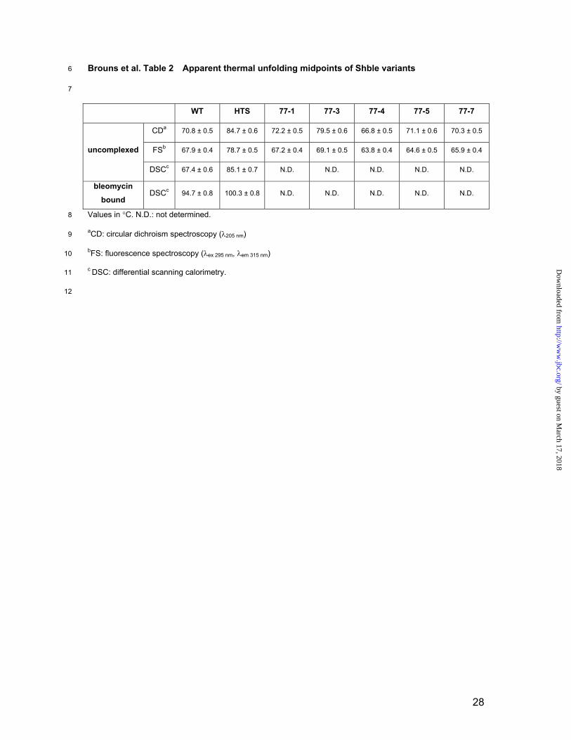
summarized in Table 2.
In the absence of the antibiotic, wild-type Shble appears to be a very stable protein. This is
remarkable, since St. hindustanus grows optimally at 28 °C (42). It is often found, however, that
proteins for which low biological turnover is beneficial for a host, are prone to little local unfolding and
hence are less susceptible to proteolytic attack (43). Structurally, Shble which serves a function of
self-immunity, might well be adapted to meet these criteria by its compactness, relatively high
secondary structure content, high surface charge and by its embedded N and C-termini (19). The
unfolding data also clearly show the strong stabilizing effect of ligand binding on the thermostability of
the BBP, since the apparent unfolding midpoint temperature increases 27.3 °C upon bleomycin
binding. This effect has also been recognized in other ligand binding proteins, such as streptavidin
and avidin, which become extremely thermostable in the presence of biotin (44). In the absence of
bleomycin, the stability of the various mutants is rather different. Of the double mutants, only 77-3
(Asp32Val, Leu63Gln) seems to have a marked increase in Tm as observed with circular dichroism
spectroscopy (CD) and fluorescence spectroscopy (FS), while numbers 77-1 (Leu63Gln, Gly98Val),
77-5 (Arg31Leu, Gly98Ser) and 77-7 (Gly18Glu, Leu63Gln) remain virtually unchanged. Surprisingly,
mutant 77-4 (Arg31Leu, Gly40Ala) displays significantly lower Tm values compared to the wild-type.
12
by guest on March 17, 2018
http://ww
w.jbc.org/
Dow
nloaded from
Quadruple mutant HTS, which combines non-redundant mutations found in 77-1, 77-3 and 77-7,
displays a profound increase of 13.9, 10.8 and 17.7 °C in stability in the absence of the antibiotic as
found by CD, FS and differential scanning calorimetry (DSC), respectively. In its presence, the
complex becomes hyperthermostable, unfolding at a temperature of just over 100 °C, 5.6 °C higher
compared to the wild-type protein.
61
62
63
64
65
66
67
68
69
70
71
72
73
74
75
76
77
78
79
80
81
82
83
84
85
86
87
88
89
90
To our surprise, the double mutants 77-1, 77-5 and 77-7 had almost unchanged apparent
melting temperatures compared to the wild-type. This can be understood by realizing that in vivo,
some amino acid changes may prevent instances of local protein unfolding, and therefore may avoid
further unfolding and subsequent proteolytic attack. However, this is not necessarily reflected in its in
vitro melting temperature, which is a measure of its global stability. Only when the weakest point of a
structure was compensated (Asp32Val and Leu63Gln in 77-3), an increase of its melting temperature
from 70.8 to 79.5 °C with CD, and 67.9 to 69.1 °C with FS was observed. Adding mutation Gly98Val
from 77-1 and Gly18Glu from 77-7 to 77-3, giving rise to HTS, further increased its melting
temperature as one would expect. This observation is analogous to the findings of extensive work
which has been conducted with the neutral protease from Geobacillus stearothermophilus, where
interactions close to the N-terminus were found to be limiting the global stability (5).
Mutants improve DNA protection against bleomycin at high temperature
In vitro DNA protection assays were performed with the various Shble mutants in order to test
whether the resistant phenotype of T. thermophilus at 77 °C was due to improved protection against
the DNA degrading capability of bleomycin. The result of this is shown in Figure 2. At 25 °C, no
significant differences in band intensities are observed. A 30-minute thermal pre-incubation of the
protein at 85 °C, however, revealed a drastic loss of function in mutant 77-4. Differences between the
wild-type and mutants became pronounced when bleomycin binding capabilities were tested at 85 °C.
At this temperature, the DNA was protected best by 77-1 and HTS, followed by 77-4, 77-5, 77-3, 77-7
and the wild-type. Surprisingly, mutant 77-4, which displayed a low temperature unfolding midpoint
and high thermal inactivation at 85 °C, apparently bound bleomycin effectively at high temperature
conditions. So although the global stability of this mutant was decreased, it had improved bleomycin
binding characteristics, which in itself, stabilizes the protein dramatically as observed by DSC
measurements for the wild-type. These results indicate that some of the double mutants have
13
by guest on March 17, 2018
http://ww
w.jbc.org/
Dow
nloaded from

improved the bleomycin binding properties compared to the wild-type, which confirms the findings of
the in vivo selection procedure in Thermus thermophilus. Possible structural explanations for the
improved functionality at higher temperature are discussed below.
91
92
93
94
95
96
97
98
99
100
101
102
103
104
105
106
107
108
109
110
111
112
113
114
115
116
117
118
119
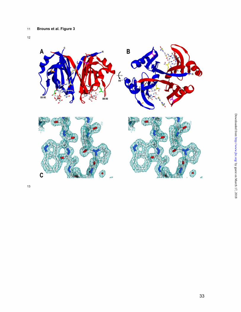
Overall structure description
The quadruple mutant HTS was crystallized in the presence of bleomycin A2 and its structure
was determined to 1.50 Å resolution (Table 3). The crystals grown belong to space group P21 with
unit cell parameters a= 44.0 Å, b= 66.6 Å, c= 47.2 Å and = 117.4° and a dimer in the asymmetric unit
(Figure 3A and 3B). Representative electron density is shown in Figure 3C. The structure forms a
compact, homodimeric / protein of 121 amino acids (Met1, Gln123 and Asp124 are disordered) in
which two bleomycin A2 molecules are accommodated in binding pockets at the dimer interface.
These pockets consist of a hydrophilic concavity which runs into a hydrophobic intersubunit crevice.
The dimer is maintained by alternate N-terminal 1-strand hydrogen bonding between both monomers
and by Van der Waals interactions at the largely hydrophobic subunit contact (19,45). Three sulphate
ions are present at the surface of the dimer of which two form ion pairs with Arg104 of both chains.
The presence of a dimer in the asymmetric unit allowed the identification of certain symmetry
deviations between both monomers. A backbone superimposition of both chains (RMSD: 0.38 Å,
Table 4) only revealed large differences in a random coil region comprising of residues Asp88, Ala89
and Ser90 (Figure 3A and 3B), which is spatially close to the carbamoyl group of the D-mannose
moiety of bleomycin (Figure 4A). Their respective C atoms deviate 2.0, 5.1 and 1.6 Å in position,
while giving rise to almost oppositely pointing amino acid side chains. In contrast to the bleomycin
bound and unbound BlmA structure, backbone B-factors in this region are only marginally higher
compared to the average value, suggesting a rigid conformation (23,45). The difference in orientation
of this loop might therefore be the result of sequential binding of two bleomycin molecules. Unlike
BlmA, no symmetry related differences were observed in the region between amino acids 100 to 103.
The topology of the HTS protein complex and the mode of bleomycin binding are similar to other
BBPs. An overview of available structures is given in Table 4.
The structure of the HTS mutant in complex with bleomycin completes the list of structural
information of three BBPs with and without their ligands, hereby contributing to our understanding of
14
by guest on March 17, 2018
http://ww
w.jbc.org/
Dow
nloaded from

these proteins in general. Moreover, it has revealed several molecular features, which can account for
increased protein stability and improved functionality at higher temperature in vivo and in vitro.
120
121
122
123
124
125
126
127
128
129
130
131
132
133
134
135
136
137
138
139
140
141
142
143
144
145
146
147
148
149
Structural effects of mutations
Introduction of an intersubunit hydrogen bond network
The structure of the dimer shows that each of the two bleomycin A2 molecules is bound by
the concerted action of 21 amino acids. Due to its intersubunit location, both binding sites are
composed of residues from either subunit. These include Val32, Phe33, Glu35, Phe38, Ser51, Ala52
and Val53 of one subunit and Pro59, Asp60, Asn61, Thr62, Gln63, Trp65, Phe86, Ala89, Trp102,
Ala107, Arg109, Gly113, Cys115 and His117 of the other. The crystal structure clearly reveals the
central role of mutation Leu63Gln which was found in 3 out of 5 different double mutants. Gln63 is
involved in an extensive hydrogen bond network at the bottom of the bleomycin binding concavity
(Figure 5A). It is noteworthy that the carbonyl side chains (O 1) of both Gln63 residues in the dimer
act as terminal hydrogen bond acceptors of a five-molecule water channel present at the dimer
interface. A second hydrogen bond is accepted from the side chain hydroxyl group (O ) of Ser51 of
the adjacent subunit. The amide side chain (N 2) of Gln63 forms a hydrogen bond with one of two
water molecules trapped between the bleomycin and the surface of the protein. The presence of a
leucine at position 63 would most likely not have allowed for a hydrogen-bond network of this size.
The advantage of an amino acid compatible with hydrogen bonding at position 63 is also evident from
the alignment, which indicates that without exception the other four BBPs have a serine at this specific
site (Figure 1). Although the mutant structure without bleomycin is not available, we speculate that an
intersubunit hydrogen bond between Gln63 and Ser51 can persist even without the antibiotic bound,
giving rise to a beneficial interaction that might stabilize the dimer at high temperatures.
Hydrophobic packing of surface indentations
In wild-type Shble and BlmA, Asp32 is located on the edge of the intersubunit binding groove
for the bithiazole moiety and tail-region of bleomycin (Figure 4A) (19,23,45). In bleomycin A2, B2 and
in phleomycin D1 the tail is positively charged which suggests involvement of Asp32 in electrostatic
stabilization or ligand recognition. From NMR studies it has become clear that in the bound state no
strong interactions occur between the protein and the positively charged tail of bleomycin (46). These
15
by guest on March 17, 2018
http://ww
w.jbc.org/
Dow
nloaded from

data are supported by the absence of electron density for the -aminopropyldimethylsulfonium moiety
of bleomycin A2 in the binary complex structure of BlmA and HTS, suggesting a disordered
conformation of the tail end (Figure 4B) (23). This might have allowed for an amino acid substitution to
valine, which extends the hydrophobic bithiazole binding cleft at the dimer interface fitting nicely within
a highly hydrophobic environment consisting of Phe33, Phe38, Val42, Thr47 and Phe49 (Figure 5B).
In addition, both BlmT and BlmS sequences also contain a valine at the corresponding position
(Figure 1). From a thermodynamic point of view, a mutation introducing surface hydrophobicity is
generally believed to be unfavorable and has therefore rarely been investigated in directed
mutagenesis studies. Nevertheless, some studies have reported significant improvements in protein
stability by placing bulky hydrophobic amino acids at the surface of a neutral protease from
Geobacillus stearothermophilus (47). Recent findings using Bacillus licheniformis -amylase have
clearly indicated that hydrophobic surface residues can indeed be extremely stabilizing by improving
hydrophobic packing of surface indentations, hereby reinforcing subsurface secondary structure
elements (48). This might explain the enhanced secondary structure preservation at high
temperatures as inferred from CD of mutant 77-3. Strikingly, modeling of Gly40Ala into the wild-type
structure (not shown) revealed close spatial proximity to Asp32 (5 Å between C and 3.5 Å between
C atoms), which may also underline a similar need for hydrophobicity in this part of the protein.
Mutation Arg31 to Leu, which occurred in two types of double mutants came as a surprise, since it is
involved in a surface ion pair with Asp25 in wild-type Shble. Apparently this electrostatic interaction
does not counterweight beneficial effects of improved hydrophobic packing among residues Val20,
Thr24, Val34, and Val41.
150
151
152
153
154
155
156
157
158
159
160
161
162
163
164
165
166
167
168
169
170
171
172
173
174
175
176
177
178
179
Reduction of surface loop flexibility
From previous crystallographic and NMR studies of BBPs, it has become clear that the loop
following Gly98 in Shble will change its conformation upon binding of the antibiotic (22,23,45,46). This
conformational change enables the tryptophan at position 102 to stack optimally with the hydrophobic
bithiazole moiety of bleomycin (Figure 4A), packing both thiazole rings tightly against Phe33 and
Phe38 of the adjacent subunit. In both BlmA and Shble, Gly98, located on the edge of a small -
strand leading towards the binding loop, seems to have a hinge function (Figure 5C). The bending
motion of the backbone is also clearly reflected in large Phi and Psi torsion angle changes of more
16
by guest on March 17, 2018
http://ww
w.jbc.org/
Dow
nloaded from
than 20° upon the binding of bleomycin. At high temperatures, however, this flexibility might have
caused problems leading to local unfolding or a decreased bleomycin binding ability. A substitution for
either a valine or serine as observed, would increase the rigidity of the loop and could therefore
restore the binding capacity at high temperature.
180
181
182
183
184
185
186
187
188
189
190
191
192
193
194
195
196
197
198
199
200
201
202
203
204
205
206
207
208
209
-Helix stabilization
Mutation Gly18Glu introduces a glutamate at position N3 in the first turn of the largest -helix
of the protein. Statistical analysis as well as experimental studies have shown that glutamates are
energetically highly favored over glycines at the third position in an -helix (49,50). This effect is most
likely caused by the stabilizing effect of the negatively charged side chain on the helix macro dipole.
To our surprise, chain A of the crystal structure revealed the formation of a genuine i, i + 5 -helix
surface ion-pair between Glu21 and Arg26, which was absent in the wild-type structure (Figure 5D).
This new ion pair may have been the result of repulsion of anionic glutamate side chains of position
18 and 21, directing the latter towards the C-terminal arginine. Although i, i + 5 -helical surface ion
pairs do not give rise to strong ionic interactions at ambient temperatures (51), they might be more
favorable at higher temperatures. Theoretical models have indicated that the energetic cost of
desolvating charged groups is much less at 100 °C, due to a drop in the dielectric constant of water
(52). This is currently the best explanation for the fact that proteins from extreme thermophiles have
large ion pair networks at their surfaces, which are thought to be involved in maintaining structural
integrity (3). Additionally, a minor beneficial effect of this mutation could be the introduction of
additional negative surface charge, which enhances electrostatic attraction of the cationic antibiotic
under physiological conditions.
Laboratory versus natural evolution of thermostability
In this study, several possible mechanisms of adaptation to high temperature were identified,
such as the introduction of a hydrogen bond network, improved hydrophobic packing of surface
indentations, reduction of loop flexibility and -helix stabilization. Remarkably, half of all mutations
found were glycine replacements, which could point to protein stabilization by decreasing the entropy
of the unfolded state (53). Although this could be a general strategy of stabilization, proteins from
hyperthermophiles do not have a lower glycine content than their mesophilic counterparts, but rather a
17
by guest on March 17, 2018
http://ww
w.jbc.org/
Dow
nloaded from

slightly increased one (54). Their predicted proteomes do have an increased propensity for charged
(Arg, Lys and Glu) and bulky aliphatic (Ile and Val) amino acids, which has mostly come at the cost of
polar residues (Asn, Gln, Ser and Thr) (54). This is fully in agreement with the requirements for the
elevated numbers of surface salt bridges and improved hydrophobic core packing that has generally
been recognized in these types of proteins. These are just two of a multitude of mechanisms that
proteins from hyperthermophilic micro-organisms have employed to deal with extreme temperatures
(2,55,56).
210
211
212
213
214
215
216
217
218
219
220
221
222
223
224
225
226
227
228
229
230
231
232
233
234
235
236
237
238
239
Recently, several other random mutagenesis studies have also reported large improvements
in thermostability by applying directed evolution approaches. A mesophilic xylanase of family 11 was
stabilized by over 35 °C by combining nine mutations found separately after extensive screening. The
activity of this mutant was optimized by saturation mutagenesis of all mutated positions, yielding an
enzyme variant with highly enhanced properties for high-temperature applications (8). In another
study, a highly thermostable esterase containing seven mutations was evolved in six rounds of
random mutagenesis, recombination and screening (7). The resulting enzyme was crystallized and its
structure determined (57). The structure revealed that improved stability was due to altered core
packing, -helix stabilization, the introduction of surface salt bridges and reduction of flexibility in
surface loops. From these and many other directed evolution and site-directed mutagenesis studies, it
has become apparent that (i) proteins can be stabilized substantially by small numbers of mutations,
(ii) these mutations are often located at the protein surface and (iii) their effects are usually additive.
As few as 2 out of 12 amino acid differences between a mesophilic and thermophilic cold shock
protein turned out to be responsible for the difference in thermostability (58). The remaining variation
in sequence might just have occurred as a result of neutral sequence drift or specific properties
required by the host, such as solubility, turnover and molecular interactions (59).
Despite the fact that only a small number of mutations are required to render a protein
thermostable, finding those mutations remains a difficult task. Apart from screening vast numbers of
random mutants in microtiter plates, in vitro and in vivo selection schemes offer great advantages in
order to reduce the effort to encounter improved variants. Currently, two main strategies are available.
One of these thermostabilizing selection methods is called Proside (Protein stability increased
by directed evolution) which is based on the empirically derived inverse correlation between protein
thermostability and proteolytic susceptibility (60). In a phage-display-like procedure, a library of
18
by guest on March 17, 2018
http://ww
w.jbc.org/
Dow
nloaded from

mutants is fused between two domains of E. coli filamentous phage Fd gene-3-protein (G3P) which is
then subjected to proteases while inducing local unfolding of the target protein by means of
temperature or chemical denaturants. The resulting instable fusion protein variants are cleaved,
causing only the surviving, more stable phages to be found after infection. It was found that in an ionic
denaturant, non-polar surface interactions were optimized, whereas at elevated temperature variants
with improved surface electrostatics were selected (59).
240
241
242
243
244
245
246
247
248
249
250
251
252
253
254
255
256
257
258
259
260
261
262
263
264
265
266
267
268
269
Cloning and selection in a thermophile, as conducted in this study, is a second directed
evolution strategy which can lead to rapid improvements in thermostability. Despite its simplicity, very
few studies using this technique have been reported. This is most likely due to its limited applicability,
since the gene of interest rarely confers any biologically relevant function for growth or survival of the
thermophile. Nonetheless, functionally stabilized mutants have been reported up to 79 °C for a
kanamycin nucleotidyltransferase (9,11) and 58 °C for a chloramphenicol acetyltransferase (61).
Auxotrophic knockout strains for leucine biosynthesis were complemented with thermolabile
counterpart genes from Bacillus subtilis and Saccharomyces cerevisiae and adapted to higher
temperature by serial accumulation of beneficial mutations in the in-trans introduced 3-
isopropylmalate dehydrogenase genes (62,63). A hybrid -galactosidase consisting of B.
stearothermophilus and T. thermophilus peptide regions was adapted to function at 67 °C by selection
for growth on melibiose as the sole carbon and energy source (64).
Concluding remarks
In this study, several double mutants of a bleomycin binding protein were isolated with
enhanced performance at high temperature both in vivo and in vitro. Structural analysis showed that
the mutations gave rise to different means of stabilization in four parts of the protein. A combined
mutant gene with a low GC content was created, which can serve as an antibiotic resistance marker
for aerobic and micro-aerophilic mesophiles, thermophiles and hyperthermophiles. This may allow the
development of efficient shuttle-vectors and knockout strategies for hyperthermophilic archaea and
bacteria based on positive selection schemes. Moreover, the high GC content double mutant genes
will now permit multigene knockout strategies in thermophiles such as Thermus thermophilus,
allowing further exploration and exploitation of thermophilic microbial sources.
19
by guest on March 17, 2018
http://ww
w.jbc.org/
Dow
nloaded from

ACKNOWLEDGEMENTS1
2
3
4
The authors thank Dr. J. Berenguer for helpful suggestions and Anton Korteweg for technical
assistance with DSC. This work was supported by a grant from the European Union in the framework
of the SCREEN project (contract QLK3-CT-2000-00649).
20
by guest on March 17, 2018
http://ww
w.jbc.org/
Dow
nloaded from

REFERENCES
1. Arnold, F. H., Wintrode, P. L., Miyazaki, K., and Gershenson, A. (2001) Trends Biochem Sci
26, 100-106
2. Vieille, C., and Zeikus, G. J. (2001) Microbiol Mol Biol Rev 65, 1-43
3. Karshikoff, A., and Ladenstein, R. (2001) Trends Biochem Sci 26, 550-556
4. Serrano, L., and Fersht, A. R. (1989) Nature 342, 296-299
5. Van den Burg, B., Vriend, G., Veltman, O. R., Venema, G., and Eijsink, V. G. (1998) Proc Natl
Acad Sci U S A 95, 2056-2060
6. Stemmer, W. P. (1994) Nature 370, 389-391
7. Giver, L., Gershenson, A., Freskgard, P. O., and Arnold, F. H. (1998) Proc Natl Acad Sci U S
A 95, 12809-12813
8. Palackal, N., Brennan, Y., Callen, W. N., Dupree, P., Frey, G., Goubet, F., Hazlewood, G. P.,
Healey, S., Kang, Y. E., Kretz, K. A., Lee, E., Tan, X., Tomlinson, G. L., Verruto, J., Wong, V.
W., Mathur, E. J., Short, J. M., Robertson, D. E., and Steer, B. A. (2004) Protein Sci 13, 494-
503
9. Liao, H., McKenzie, T., and Hageman, R. (1986) Proc Natl Acad Sci U S A 83, 576-580
10. Tamakoshi, M., Yamagishi, A., and Oshima, T. (1995) Mol Microbiol 16, 1031-1036
11. Hoseki, J., Yano, T., Koyama, Y., Kuramitsu, S., and Kagamiyama, H. (1999) J Biochem
(Tokyo) 126, 951-956
12. Friedrich, A., Prust, C., Hartsch, T., Henne, A., and Averhoff, B. (2002) Appl Environ Microbiol
68, 745-755
13. Noll, K. M., and Vargas, M. (1997) Arch Microbiol 168, 73-80
14. Gatignol, A., Durand, H., and Tiraby, G. (1988) FEBS Lett 230, 171-175
15. Sugiyama, M., and Kumagai, T. (2002) J Biosci Bioeng 93, 105-116
16. Poddevin, B., Orlowski, S., Belehradek, J., Jr., and Mir, L. M. (1991) Biochem Pharmacol 42
Suppl, S67-75
17. Sikic, B. I. (1986) Cancer Surv 5, 81-91
18. Sugiyama, M., Thompson, C. J., Kumagai, T., Suzuki, K., Deblaere, R., Villarroel, R., and
Davies, J. (1994) Gene 151, 11-16
19. Dumas, P., Bergdoll, M., Cagnon, C., and Masson, J. M. (1994) Embo J 13, 2483-2492
21
by guest on March 17, 2018
http://ww
w.jbc.org/
Dow
nloaded from
20. Genilloud, O., Garrido, M. C., and Moreno, F. (1984) Gene 32, 225-233
21. Semon, D., Movva, N. R., Smith, T. F., el Alama, M., and Davies, J. (1987) Plasmid 17, 46-53
22. Maruyama, M., Kumagai, T., Matoba, Y., Hayashida, M., Fujii, T., Hata, Y., and Sugiyama, M.
(2001) J Biol Chem 276, 9992-9999
23. Sugiyama, M., Kumagai, T., Hayashida, M., Maruyama, M., and Matoba, Y. (2002) J Biol
Chem 277, 2311-2320
24. Drocourt, D., Calmels, T., Reynes, J. P., Baron, M., and Tiraby, G. (1990) Nucleic Acids Res
18, 4009
25. Nuttall, S. D., Deutschel, S. E., Irving, R. A., Serrano-Gomicia, J. A., and Dyall-Smith, M. L.
(2000) Biochem J 346 Pt 2, 251-254
26. Boyer, H. W., and Rouland-Dussoix, R. (1969) J Mol Biol 41, 459-472
27. Sambrook, J., Fritsch, E. F., and Maniatis, T. (1989) Molecular Cloning: A Laboratory Manual,
2nd Ed., Cold Spring Harbor Laboratory, Cold Spring Harbor, NY
28. de Grado, M., Castan, P., and Berenguer, J. (1999) Plasmid 42, 241-245
29. Shafikhani, S., Siegel, R. A., Ferrari, E., and Schellenberger, V. (1997) Biotechniques 23,
304-310
30. Koyama, Y., Hoshino, T., Tomizuka, N., and Furukawa, K. (1986) J Bacteriol 166, 338-340
31. Stemmer, W. P., Crameri, A., Ha, K. D., Brennan, T. M., and Heyneker, H. L. (1995) Gene
164, 49-53
32. Bradford, M. M. (1976) Anal Biochem 72, 248-254
33. Rondeau, J. M., Cagnon, C., Moras, D., and Masson, J. M. (1989) J Mol Biol 207, 645-646
34. Otwinowski, A., and Minor, W. (1997) Meth Enzymol 276, 307-326
35. Vagin, A. A., and Teplyakov, A. (1997) J Appl Cryst 30, 1022-1025
36. Perrakis, A., Morris, R., and Lamzin, V. S. (1999) Nat Struct Biol 6, 458-463
37. Murshudov, G. N., Vagin, A. A., and Dodson, E. J. (1997) Acta Crystallogr D Biol Crystallogr
53, 240-255
38. Laskowski, R. A., MacArthur, M. W., Moss, D. S., and Thornton, J. M. (1993) J Appl Cryst 26,
283-291
39. Vaguine, A. A., Richelle, J., and Wodak, S. J. (1999) Acta Crystallogr D Biol Crystallogr 55 (
Pt 1), 191-205
22
by guest on March 17, 2018
http://ww
w.jbc.org/
Dow
nloaded from

40. Guex, N., and Peitsch, M. C. (1997) Electrophoresis 18
41. Fernandez-Herrero, L. A., Olabarria, G., and Berenguer, J. (1997) Mol Microbiol 24, 61-72
42. Tomita, K., Nakakita, Y., Hoshino, Y., Numata, K., and Kawaguchi, H. (1987) Int J Syst
Bacteriol 37, 211-213
43. Jaswal, S. S., Sohl, J. L., Davis, J. H., and Agard, D. A. (2002) Nature 415, 343-346
44. Gonzalez, M., Argarana, C. E., and Fidelio, G. D. (1999) Biomol Eng 16, 67-72
45. Kawano, Y., Kumagai, T., Muta, K., Matoba, Y., Davies, J., and Sugiyama, M. (2000) J Mol
Biol 295, 915-925
46. Vanbelle, C., Brutscher, B., Blackledge, M., Muhle-Goll, C., Remy, M. H., Masson, J. M., and
Marion, D. (2003) Biochemistry 42, 651-663
47. Van den Burg, B., Dijkstra, B. W., Vriend, G., Van der Vinne, B., Venema, G., and Eijsink, V.
G. (1994) Eur J Biochem 220, 981-985
48. Machius, M., Declerck, N., Huber, R., and Wiegand, G. (2003) J Biol Chem 278, 11546-11553
49. Iqbalsyah, T. M., and Doig, A. J. (2004) Protein Sci 13, 32-39
50. Penel, S., Hughes, E., and Doig, A. J. (1999) J Mol Biol 287, 127-143
51. Smith, J. S., and Scholtz, J. M. (1998) Biochemistry 37, 33-40
52. Elcock, A. H. (1998) J Mol Biol 284, 489-502
53. Van den Burg, B., and Eijsink, V. G. (2002) Curr Opin Biotechnol 13, 333-337
54. Cambillau, C., and Claverie, J. M. (2000) J Biol Chem 275, 32383-32386
55. Kumar, S., and Nussinov, R. (2001) Cell Mol Life Sci 58, 1216-1233
56. Vogt, G., Woell, S., and Argos, P. (1997) J Mol Biol 269, 631-643
57. Spiller, B., Gershenson, A., Arnold, F. H., and Stevens, R. C. (1999) Proc Natl Acad Sci U S
A 96, 12305-12310
58. Perl, D., Mueller, U., Heinemann, U., and Schmid, F. X. (2000) Nat Struct Biol 7, 380-383
59. Martin, A., Sieber, V., and Schmid, F. X. (2001) J Mol Biol 309, 717-726
60. Sieber, V., Pluckthun, A., and Schmid, F. X. (1998) Nat Biotechnol 16, 955-960
61. Turner, S. L., Ford, G. C., Mountain, A., and Moir, A. (1992) Protein Eng 5, 535-541
62. Akanuma, S., Yamagishi, A., Tanaka, N., and Oshima, T. (1998) Protein Sci 7, 698-705
63. Tamakoshi, M., Nakano, Y., Kakizawa, S., Yamagishi, A., and Oshima, T. (2001)
Extremophiles 5, 17-22
23
by guest on March 17, 2018
http://ww
w.jbc.org/
Dow
nloaded from

64. Fridjonsson, O., Watzlawick, H., and Mattes, R. (2002) J Bacteriol 184, 3385-3391
65. Thompson, J. D., Gibson, T. J., Plewniak, F., Jeanmougin, F., and Higgins, D. G. (1997)
Nucleic Acids Res 25, 4876-4882
24
by guest on March 17, 2018
http://ww
w.jbc.org/
Dow
nloaded from

FIGURE LEGENDS1
2
3
4
5
6
7
8
9
10
11
12
13
14
15
16
17
18
19
20
21
22
23
24
25
26
27
28
29
30
Figure 1 Structural alignment of bleomycin binding proteins from different microbial
sources
Shble (PDB-ID: 1BYL) from St. hindustanus (19), BlmA (PDB-ID: 1QTO) from S. verticillus
ATCC15003 (45), SvP from S. verticillus ATCC21890, BlmT (PDB-ID: 1ECS) from K. pneumoniae
transposon Tn5 (22) and BlmS from S. aureus plasmid pUB110. The alignment was created by
backbone superimposition of the three structures and expanded with the SvP and BlmS sequences by
realignment using ClustalX v1.81 (65) while maintaining the original gaps. The HTS structure was
used for residue numbering and topology assignment (black arrows: -strand, checkered boxes: -
helix). Mutations are indicated by arrows.
Figure 2 DNA protection assay
Digital photographs of 1% agarose gels showing the degree of DNA protection by Shble variants
against the strand scission action of bleomycin A2. A) assay at 25 °C for 10 minutes. B) assay at 25
°C for 10 minutes after protein preincubation at 85 °C for 30 minutes. C) assay at 85 °C for 10
minutes.
Figure 3 Structure and electron density of HTS in complex with bleomycin A2
Ribbon diagram showing the dimeric structure of the fourfold mutant Shble in complex with bleomycin
A2. Mutations are indicated by stick representations. Chain A in blue, chain B in red, Gly18Glu in
green, Asp32Val in pink, Leu63Gln in yellow and Gly98Val in orange. A) side view. B) viewed from
the N- and C-terminal side (top view) C) stereo view of the electron density around residue Pro9 and
Trp65 contoured at 2 . Residues are colored according to the CPK color scheme, water molecules
are represented by red spheres.
Figure 4 Chemical diagram and electron density of bleomycin A2
A) Schematic representation of bleomycin A2. B) Electron density around bleomycin A2 contoured at
1.5 . The diagram indicates the missing electron density around the -aminopropyldimethylsulfonium
moiety suggesting a disordered conformation.
25
by guest on March 17, 2018
http://ww
w.jbc.org/
Dow
nloaded from

Figure 5 Structural effects of the individual mutations31
32
33
34
35
36
37
38
39
40
41
42
43
44
45
A) Leu63Gln. Ribbon diagram showing the hydrogen bond network at the dimer interface. Thr62,
Gln63, Ser51 and bleomycin A2 are shown together with the intersubunit water channel. B) Asp32Val.
Ribbon diagram showing the hydrophobic intersubunit bleomycin tail binding crevice. Val32 may be
involved in improved hydrophobic packing of this surface indentation among amino acids Phe33,
Phe38, Val42, Thr47 and Phe49. C) Gly98Val. Ribbon representation showing a loop between Pro92
and Pro111 which is involved in bleomycin binding. Val98 is located at a former hinge region which
enables Trp102 to stack the bithiazole tail against Phe33 and Phe38. The electron density revealed
two alternative sidechain rotamers for Val98 in chain A (not shown) and a single side chain
conformation in chain B. D) Gly18Glu. Side-by-side comparison of -helix one formed between Asp15
and Leu27 in the wild-type and mutant crystal structures of Shble. The existence of a surface ion-pair
between Glu21 and Arg26 is visible in the electron density (contoured at 1.5 ). Interatomic distances
are indicated in Å. CPK color coding was used for amino acids and bleomycin A2. Chain A is
indicated in blue, chain B in red. Water molecules are represented by red spheres. Hydrogen bonds
are depicted by green dotted lines.
26
by guest on March 17, 2018
http://ww
w.jbc.org/
Dow
nloaded from

TABLES1
2
3
4
Brouns et al. Table 1 Nucleotide and amino acid substitutions of Shble variants
Residue number 18 31 32 40 63 98
wild-type Gly Arg Asp Gly Leu Gly
77-1 / 77-2 / 77-8 GlnT188A
ValG293T
77-3a ValA95T
GlnT188A
77-4 / 77-6b LeuG92T
AlaG119C
77-5c LeuG92T
SerG292A
77-7 GluG53A
GlnT188A
Clones containing silent nucleotide substitutions: aC360A, bG63A, cG30A.5
27
by guest on March 17, 2018
http://ww
w.jbc.org/
Dow
nloaded from
Brouns et al. Table 2 Apparent thermal unfolding midpoints of Shble variants6
7
WT HTS 77-1 77-3 77-4 77-5 77-7
CDa 70.8 ± 0.5 84.7 ± 0.6 72.2 ± 0.5 79.5 ± 0.6 66.8 ± 0.5 71.1 ± 0.6 70.3 ± 0.5
FSb 67.9 ± 0.4 78.7 ± 0.5 67.2 ± 0.4 69.1 ± 0.5 63.8 ± 0.4 64.6 ± 0.5 65.9 ± 0.4 uncomplexed
DSCc 67.4 ± 0.6 85.1 ± 0.7 N.D. N.D. N.D. N.D. N.D.
bleomycinbound
DSCc 94.7 ± 0.8 100.3 ± 0.8 N.D. N.D. N.D. N.D. N.D.
Values in C. N.D.: not determined.8
9
10
11
12
aCD: circular dichroism spectroscopy ( 205 nm)
bFS: fluorescence spectroscopy ( ex 295 nm, em 315 nm)
c DSC: differential scanning calorimetry.
28
by guest on March 17, 2018
http://ww
w.jbc.org/
Dow
nloaded from

Brouns et al. Table 3 Data collection and refinement statistics13
14
X-ray data collection statistics
Wavelength (Å) 0.90830
Resolution (Å) 30 – 1.5 (1.53 – 1.50)
Total observations 87740 (3602)
Unique observations 36444 (1714)
Completeness (%) 94.4 (88.6)
I / (I) 20.3 (3.9)
Rsyma 0.043 (0.189)
Refinement statistics
Resolution (Å) 30.0 - 1.5
Rworkb 0.174
Rfreec 0.194
RMSD bond distances (Å) 0.01
RMSD bond angles (˚) 1.664
Total number of non-H atoms
Av. protein B value (Å2)
1883
13.3
Number of solvent molecules
Av. solvent B value (Å2)
288
27.0
Number of bleomycin atoms
Av. ligand B value (Å2)
182
22.8
Number of Sulphate ion atoms
Av. Sulphate ion B value (Å2)
15
31.4aRsym = hkl i |I-<Ii>| / hkl i Ii, where Ii is the intensity of a given measurement and the sums are over all
measurements and reflections. Values in parentheses refer to the highest resolution shell.
15
16
17
18
19
bRwork = ||F(obs)| - |F(calc)|| / |F(obs)| for the 95% of the reflection data used in refinement.
cRfree = ||F(obs)| - |F(calc)|| / |F(obs)| for the remaining 5%.
29
by guest on March 17, 2018
http://ww
w.jbc.org/
Dow
nloaded from

Brouns et al. Table 4 Crystal structures of bleomycin binding proteins20
21
Protein / Source PDB-ID Form Resolution(Å)
BackboneRMSD (Å) Reference
1BYL uncomplexed 2.30 0.63a (19)ShbleStreptoalloteichus
hindustanus 1XRK bleomycin A2bound 1.50 0.38b this study
1QTO uncomplexed 1.50 0.77a (45)
1JIE bleomycin A2 bound 1.80 0.73a
BlmAStreptomyces
verticillus1JIF Cu2+-bleomycin A2
bound 1.60 0.75a(23)
1ECS uncomplexed 1.70 1.18aBlmTKlepsiella pneumoniae
transposon Tn5 1EWJ bleomycin A2 bound 2.50 1.15a
(22)
aAveraged backbone superimposition RMSD values compared to HTS chain A and B 22
23 bBackbone superimposition RMSD values of HTS chain A to B
30
by guest on March 17, 2018
http://ww
w.jbc.org/
Dow
nloaded from

FIGURES1
2
3
4
Brouns et al. Figure 1
5
6
31
by guest on March 17, 2018
http://ww
w.jbc.org/
Dow
nloaded from

Brouns et al. Figure 2 7
8
9
10
32
by guest on March 17, 2018
http://ww
w.jbc.org/
Dow
nloaded from
Brouns et al. Figure 3 11
12
13
33
by guest on March 17, 2018
http://ww
w.jbc.org/
Dow
nloaded from

Brouns et al. Figure 4 14
15
16
34
by guest on March 17, 2018
http://ww
w.jbc.org/
Dow
nloaded from

Brouns et al. Figure 517
18
19
35
by guest on March 17, 2018
http://ww
w.jbc.org/
Dow
nloaded from

John van der OostStan J. J. Brouns, Hao Wu, Jasper Akerboom, Andrew P. Turnbull, Willem M. de Vos and
thermostable Bleomycin binding proteinEngineering a selectable marker for hyperthermophiles: Crystal structure of a
published online January 7, 2005J. Biol. Chem.
10.1074/jbc.M413623200Access the most updated version of this article at doi:
Alerts:
When a correction for this article is posted•
When this article is cited•
to choose from all of JBC's e-mail alertsClick here
Supplemental material:
http://www.jbc.org/content/suppl/2005/01/12/M413623200.DC1
by guest on March 17, 2018
http://ww
w.jbc.org/
Dow
nloaded from


