Download - Endocrinal Emergencies In ICU - BU

Endocrinal Emergencies In ICU
An Essay
for Complete fulfillment of Master Degree in Anesthesiology
Presented By
Asmaa Mohammed Abdel Fattah
(M.B.; B.Ch.)
Under Supervision Of
PROF. DR. Ehab Ahmed Abd Elrahman
Professor of Anesthesiology and Intensive Care
Faculty of Medicine
Benha University
DR. Ahmed Abd Elhameed Hassan
Lecturer of Anesthesiology and Intensive Care
Faculty of Medicine
Benha University
Faculty of Medicine
Benha University
2016

الرعايت المركزة الحالاث الحرجت المتعلقت بالغدد الصماء فى وحدة
رسالة مقدمة
للحصولذعلىذدرجةذماجودتيرذالتخدير
من ةمقدم
اسماءذمحمدذعبدالفتاحطبوب/
تحت اشرافإيهاب احمد عبدالرحمنالأستاذ الدكتىر/
د الحميد حسن احمد عبالدكتىر/
كلوةذالطب
بنهاذجامعةذ
2016

First and foremost thanks to "ALLAH" for his help to fulfill this work.
I would like to express my deepest gratitude to DR. Ehab Ahmed Abd
Elrahman, professor of anesthesiology, and Intensive Care faculty of medicine,
Benha University for her kind guidance and supervision.
My thanks to Dr. Ahmed Abd Elhameed Hassan, Lecturer of
Anesthesiology and Intensive Care , faculty of medicine Benha University for his
continuous encouragement & supervision.
Last but not least, I wish also to express my sincere thanks and respect to my
professors, seniors, colleagues ,my family and all persons who helped me in
completing this work.
Asmaa Mohammed Abdel Fattah

Contents
i
CONTENTS
Page Chapter
List of Abbreviations
List of Tables
List of Figures
Review of literature
1 Chapter 1: Anatomy and Physiology
12 Chapter 2: DIABETIC KETOACIDOSIS (DKA)
28 Chapter 3: THYROID STORM
44 Chapter 4: MYXEDEMA COMA
66 Chapter 5: ADDISONIAN CRISIS
08 Chapter 6: PHEOCHROMOCYTOMA
484 Summary
107 References
Arabic summary

List of Abbreviations
i
List of Abbreviations
ABG arterial blood gases
ACE angiotensin converting enzyme
ACLS advanced cardiac life support
ADH antidiuretic hormone
APACHE acute physiology and chronic health evaluation
AVP arginine vasopressin
BUN blood urea nitrogen
CBC complete blood count
CHF congestive heart failure
CI confidence interval
CPK creatine phosphokinase
CSF cerebrospinal fluid
CT computed tomography
CVP central venous pressure
DHEA dehydroepiandrosterone
DKA diabetic ketoacidosis
DM diabetes mellitus
DOC drug of choice
ED emergency department
EDTA ethylenediaminetetraacetic acid

List of Abbreviations
ii
ECG electrocardiogram
FSH follicle stimulating hormone
GH growth hormone
GI gastrointestinal
HEENT head ,eyes ,ears ,nose and throat
HSV herpes simplex virus
HU hounsfield units
IL-2 interleukin-2
LH leutinizing hormone
LP lumbar puncture
MEN multiple endocrine neoplasia
MI myocardial infarction
MIBG metaiodobenzyl guanidine
MRI magnetic resonance imaging
NMS neuroleptic malignant syndrome
NPH neutral protamine hagedron
beta- OHBbetahydroxybutyrate
PCR polymerase chain reaction
PET positron emission tomography
PMN polymorphonuclear neutrophil
PP pancreatic polypeptide

List of Abbreviations
iii
PRL prolactin releasing hormone
PTH parathyroid hormone
PTHrP parathyroid harmone related peptide
PTU propylthiouracil
SCII subcutaneous continuous insulin infusion
TPO thyroid peroxidase
TRH thyrotropin releasing hormone
TSH thyroid hormone releasing hormone
UTI urinary tract infection
VBG venous blood gases
WBC white blood cell

List of Tables
i
List of Tables
Table No. Contents Page
Table (1) Manifestations of thyrotoxicosis versus thyroid
storm 31
Table (2) symptoms and signs in patients with
pheochromocytoma 86
Table (3) sensitivity and specificity of lab tests in
pheochromocytoma 88

List of Figures
i
List of Figures
Fig. No. Title Page
Fig. (1) Anatomy and blood supply of pituitary and
hypothalamus 2
Fig. (2) Functional anatomy of the endometrium 3
Fig.(3) The Thyroid gland and its relations 5
Fig. (4) Regulation of the thyroid hormones T3 and T4 7
Fig.(5)
Schematic diagram of the adrenal gland showing
its 3 cortical layers and blood supply. G =
glomerulosa, F = fasciculata, R = reticularis, M =
medulla, C = cortex
10

Anatomy and Physiology
1
Anatomy and Function of the Pituitary Gland
The pituitary gland, or hypophysis, is an endocrine gland about the
size of a pea and weighing 0.5 gram. It is a protrusion off the bottom of
the hypothalamus at the base of the brain, and rests in a small, bony
cavity (sella turcica) covered by a dural fold (diaphragma sellae). The
pituitary fossa, in which the pituitary gland sits, is situated in the
sphenoid bone in the middle cranial fossa at the base of the brain. It is
considered a master gland. The pituitary gland secretes hormones
regulating homeostasis, including tropic hormones that stimulate other
endocrine glands. It is functionally connected to the hypothalamus by the
median eminence. Located at the base of the brain, the pituitary is
composed of two lobes: the anterior pituitary (adenohypophysis) and the
posterior pituitary (neurohypophysis). The pituitary is functionally linked
to the hypothalamus by the pituitary stalk, whereby hypothalamic
releasing factors are released and, in turn, stimulate the release of
pituitary hormones. Although the pituitary gland is known as the master
endocrine gland, both of its lobes are under the control of the
hypothalamus. If there are problems with the pituitary gland, it can cause
an irregular condition known as gigantism (Molitch, 2000).

Anatomy and Physiology
2
Anterior pituitary
The anterior pituitary synthesizes and secretes important endocrine
hormones, such as ACTH, TSH, PRL, GH, endorphins, FSH, and LH.
These hormones are released from the anterior pituitary under the
influence of the hypothalamus. Hypothalamic hormones are secreted to
the anterior lobe by way of a special capillary system, called the
hypothalamic-hypophyseal portal system (James et al., 2006).As seen in
fig.1
Figure 1. Anatomy and blood supply of pituitary and
hypothalamus(Barkovich, 2000).
Posterior pituitary
The posterior pituitary stores and releases Oxytocin, most of which
is released from the paraventricular nucleus in the hypothalamus
Antidiuretic hormone (ADH, also known as vasopressin and AVP,
(arginine vasopressin), the majority of which is released from the
supraoptic nucleus in the hypothalamus. Oxytocin is one of the few
hormones to create a positive feedback loop. For example, uterine

Anatomy and Physiology
3
contractions stimulate the release of oxytocin from the posterior pituitary,
which, in turn, increases uterine contractions. This positive feedback loop
continues throughout labor (James et al., 2006).
Anatomy and function of the Pancreas
Insulin is produced from islets of the pancreas, so the anatomy and
function of the pancreas are very important. The pancreas is a gland in
the digestive and endocrine system of vertebrates. It is both an endocrine
gland producing several important hormones, including insulin, glucagon,
and somatostatin, as well as an exocrine gland, secreting pancreatic juice
containing digestive enzymes that pass to the small intestine. These
enzymes help in the further breakdown of the carbohydrates, protein, and
fat in the chime (Medvei, 1993).
Figure 2 . Anatomy of the pancreas (Cabrera et al. 2006)
Endocrine function :

Anatomy and Physiology
4
The part of the pancreas with endocrine function is made up of
approximately a million cell clusters called islets of Langerhans. Four
main cell types exist in the islets. They can be classified by their
secretion: α cells secrete glucagon(increase Glucose in blood), β cells
secrete insulin (decrease Glucose in blood), δ cells secrete somatostatin
(regulates/stops α and β cells), and PP cells secrete pancreatic polypeptide
(Rother, 2007).
Exocrine function:
In contrast to the endocrine pancreas, which secretes hormones into
the blood, the exocrine pancreas produces digestive enzymes and an
alkaline fluid (referred to as pancreatic juice), and secretes them into the
small intestine through a system of exocrine ducts in response to the
small intestine hormones secretin and cholecystokinin. Digestive
enzymes include trypsin, chymotrypsin, pancreatic lipase, and pancreatic
amylase, and are produced and secreted by acinar cells of the exocrine
pancreas. Specific cells that line the pancreatic ducts, called centroacinar
cells, secrete a bicarbonate- and salt-rich solution into the small intestine
(Lawrence et al., 2008).
Anatomy and Function of Thyroid gland
The thyroid gland is one of the largest endocrine glands in the body.
This gland is found in the neck inferior to the thyroid cartilage and at
approximately the same level as the cricoid cartilage. The thyroid gland is
a butterfly shaped organ and is composed of two cone-like lobes or
wings: right lobe and left lobe, and is also connected with the isthmus.

Anatomy and Physiology
5
Figure 3. The Thyroid gland and its relations (Williams et al., 1995)
The organ is situated on the anterior side of the neck, lying against
and around the larynx and trachea, reaching posteriorly the
oesophagus and carotid sheath. It starts cranially at the oblique line on
the thyroid cartilage and extends inferiorly to the fifth or sixth tracheal
ring. It is difficult to demarcate the gland's upper and lower border with
vertebral levels because it moves position in relation to these during
swallowing (Yalcin and Ozan 2006) .
The thyroid controls how quickly the body uses energy,
makes proteins, and controls how sensitive the body should be to
other hormones. The thyroid participates in these processes by producing
thyroid hormones, principally thyroxine (T4) and triiodothyronine (T3).
These hormones regulate the rate of metabolism and affect the growth
and rate of function of many other systems in the
body. Iodine and tyrosine are used to form both T3 and T4. The thyroid

Anatomy and Physiology
6
also produces the hormone calcitonin, which plays a role in calcium
homeostasis. The thyroid is controlled by the hypothalamus and pituitary.
The gland gets its name from the Greek word for "shield", after the shape
of the related thyroid cartilage. Hyperthyroidism and hypothyroidism are
the most common problems of the thyroid gland (Baskin et al., 2002).
Physiology :
The primary function of the thyroid is production of the hormones:
thyroxine (T4), triiodothyronine (T3), and calcitonin. Up to 80% of the T4
is converted to T3 by peripheral organs such as
the liver, kidney and spleen. T3 is about ten times more active than T4.
Thyroxine (T4) is synthesised by the follicular cells from
free tyrosine and on the tyrosine residues of the protein called
thyroglobulin (Tg). Iodine is captured with the "iodine trap" by
the hydrogen peroxide generated by the enzyme thyroid peroxidase
(TPO) and linked to the 3' and 5' sites of the benzene ring of the tyrosine
residues on Tg, and on free tyrosine. Upon stimulation by the thyroid-
stimulating hormone (TSH), the follicular cells reabsorb Tg
and proteolytically cleave the iodinated tyrosines from Tg, forming T4
and T3 (in T3, one iodine atom is absent compared to T4), and releasing
them into the blood. Deiodinase enzymes convert T4 to T3. Thyroid
hormone that is secreted from the gland is about 90% T4 and about 10%
T3 (Bianco et al., 2002).

Anatomy and Physiology
7
Figure 4. Regulation of the thyroid hormones T3 and T4 .(Hill et al., 1998)
Cells of the brain are a major target for the thyroid hormones T3 and
T4. Thyroid hormones play a particularly crucial role in brain maturation
during fetal development. A transport protein, organic anion co-
transporting polypeptide 1c1 (OATP1C1), has been identified that seems
to be important for T4 transport across the blood brain barrier. A second
transport protein, monocarboxylate transporter (MCT8), is important for
T3 transport across brain cell membranes. Non genomic actions of T4 are
those that are not initiated by liganding of the hormone to intranuclear
thyroid receptor. These may begin at the plasma membrane or within
cytoplasm. Plasma membrane-initiated actions begin at a receptor on the
integrin alphaV beta3 that activates ERK1/2. This binding culminates in
local membrane actions on ion transport systems such as the Na(+)/H(+)
exchanger or complex cellular events including cell proliferation. These
integrins are concentrated on cells of the vasculature and on some types
of tumor cells which in part explains the proangiogenic effects of
iodothyronines and proliferative actions of thyroid hormone on some

Anatomy and Physiology
8
cancers including gliomas. T4 also acts on the mitochondrial genome via
imported isoforms of nuclear thyroid receptors to affect several
mitochondrial transcription factors. Regulation of actin polymerization by
T4 is critical to cell migration in neurons and glial cells and is important
to brain development (Jansen et al., 2005).
T3 and T4 regulation : (fig 4)
The production of thyroxine and triiodothyronine is regulated by
thyroid-stimulating hormone (TSH), released by the anterior pituitary.
The thyroid and thyrotropes form a negative feedback loop: TSH
production is suppressed when the T4 levels are high, and vice versa. The
TSH production itself is modulated by thyrotropin-releasing
hormone (TRH), which is produced by the hypothalamus and secreted at
an increased rate in situations such as cold (in which an accelerated
metabolism would generate more heat). TSH production is blunted
by somatostatin (SRIH), rising levels of glucocorticoids and sex
hormones (estrogen and testosterone), and excessively high blood iodide
concentration. An additional hormone produced by the thyroid
contributes to the regulation of blood calcium levels. Parafollicular
cells produce calcitonin in response to hypercalcemia. Calcitonin
stimulates movement of calcium into bone, in opposition to the effects
of parathyroid hormone (PTH). However, calcitonin seems far less
essential than PTH, as calcium metabolism remains clinically normal
after removal of the thyroid, but not the parathyroids (Kester et al., 2004).
The use of iodised salt is an efficient way to add iodine to the diet. It
has eliminated endemic cretinism in most developed countries, and some
governments have made the iodination of flour, cooking oil or salt
mandatory. Potassium iodide and sodium iodide are typically used forms

Anatomy and Physiology
9
of supplemental iodine. As with most substances, either too much or too
little can cause problems. Recent studies on some populations are
showing that excess iodine intake could cause an inceased prevelence
of autoimmune thyroid disease resulting in permanent hypothyroidism
(Patrick, 2008).
Anatomy and physiology of adrenal glands
The adrenal glands each weigh approximately 4 grams in term
neonates at birth, which is equivalent to that of adult glands; however,
adrenal size decreases by approximately 50% to 60% within the 1st week
of life. The adrenal glands are made up of an inner medulla and an outer
cortex that are linked by vascular supply and hormonal influence. Within
the mature adrenal cortex are 3 functionally distinct zones:
(1) the glomerulosa, comprising approximately 15% of the gland;
(2) fasciculata, the largest zone, comprising approximately 75% of the
gland; and
(3) the reticularis, comprising approximately 10% of the gland. The
adrenal medulla is regulated by the sympathetic nervous system and
secretes catecholamines, whereas the 3 zones of the cortex secrete
steroid hormones categorized, respectively, as mineralocorticoids,
glucocorticoids, and sex steroids. Mineralocorticoid production,
exemplified by aldosterone, is principally regulated by the renin-
angiotensin axis and by ambient potassium levels.
Mineralocorticoids affect sodium and potassium homeostasis, and
deficiencies in their production or action cause hyponatremia,
hyperkalemia, and dehydration. Glucocorticoid and adrenal sex steroid
production are primarily regulated by pituitary corticotrophin

Anatomy and Physiology
10
(adrenocorticotropic hormone [ACTH]) and hypothalamic
corticotrophin-releasing hormone, secreted mainly in the early morning
hours. Cortisol is the main glucocorticoid, and dehydroepiandrosterone
(DHEA) is the main adrenal sex hormone (Rozansky, 2006).
Figure 5. Schematic diagram of the adrenal gland showing its 3 cortical
layers and blood supply. G = glomerulosa, F = fasciculata, R =
reticularis, M = medulla, C = cortex (Barbara and John 2000).
Glucocorticoids are generally catabolic, promoting protein and lipid
breakdown and inhibiting protein synthesis. The effects of cortisol
counterregulate those of insulin, increasing the concentration of glucose
by stimulating gluconeogenesis and by decreasing glucose utilization in
muscle. Amino acids and glycerol produced by catabolic actions of
cortisol on protein and fat are used as gluconeogenic substrates. The net
effect is increased production and conservation of glucose for use by
essential tissues, such as the brain and red blood cells, at the expense of
less essential tissues during times of stress or starvation.

Anatomy and Physiology
11
Supraphysiological doses of exogenous glucocorticoids suppress growth
by antagonizing the production and action of growth hormones (GHs).
Cortisol contributes to the maintenance of normal blood pressure through
several mechanisms. Under normal baseline conditions, cortisol increases
urine flow by stimulating glomerular filtration rate and decreasing water
resorption. At high concentrations, cortisol acts as a mineralocorticoid
agonist, causing sodium and water retention(Bradley et al., 2001).
Other vascular actions of cortisol include stimulating
angiotensinogen synthesis by the liver and increasing vascular sensitivity
to vasopressors. In the adrenal medulla, cortisol is required for the
enzymatic activity of phenylethanolamine N-methyltransferase, which
converts norepinephrine to epinephrine. Epinephrine stimulates cardiac
output, as well as hepatic glucose production. Cortisol decreases capillary
permeability, as well as the production and activity of nitrous oxide and
the vasodilatory kinin and prostaglandin systems during stress, preventing
life-threatening hypotension. Cortisol or aldosterone deficiencies, or both
kinds, often result in shock if unrecognized and untreated (Bradley et al.,
2001).

Diabetic Ketoacidosis (DKA)
12
DIABETIC KETOACIDOSIS (DKA)
Introduction:
Diabetic ketoacidosis
Diabetic ketoacidosis (DKA) remains a significant complication of
diabetes in both the United States and around the world. Diabetic
ketoacidosis remains a significant complication of diabetes in both the
United States and worldwide with its associated high rates of hospital
admissions. Therefore, it becomes vital that the healthcare professional be
able to manage the hyperglycemic crises associated with diabetes.
Moreover, with increasing healthcare costs and a changing healthcare
system, prevention of diabetic ketoacidosis remains essential. Though
management of diabetic ketoacidosis has followed a set algorithm for
many years, there are exciting management alternatives on the horizon
such as subcutaneous insulin administration for uncomplicated DKA
patients. By understanding DKA, including its pathogenesis, presentation,
treatment, and prevention, admissions may be decreased and length of
stay shortened (Michael et al., 2013).
There are two major hyperglycemic crises associated with diabetes:
diabetic ketoacidosis and the hyperosmotic hyperglycemic state. There
are ~120,000 admissions for diabetic ketoacidosis and hyperglycemic
hyperosmolar state per year in the United States alone Diabetic
ketoacidosis primarily results from insulin deficiency and hyperglycemic
hyperosmolar state (HHS) from severe insulin resistance. Both of the
crises result in subsequent glucagon and counter-regulatory hormone
excess from lack of suppression from insulin Normally, with elevated
blood glucose, as occurs after a digested meal, there is production and

Diabetic Ketoacidosis (DKA)
13
release of insulin by the beta cells in the islets of Langerhans. With this
surge of insulin, the production of new glucose is suppressed
appropriately. Conversely, in a state of starvation, there is an increase in
counter regulatory hormones such as glucagon in which stores are
appropriately mobilized and glucose production increased. This is a
catabolic state, which allows for sustenance in times when nutrition is not
available (Christine, Saraceni, et al., 2013).
Mortality/Morbidity
When diabetic ketoacidosis (DKA) is treated properly, it rarely
causes any residual effects. The overall mortality rate from DKA ranges
from 1-10% of all DKA admissions, according to hospital facilities and
the experiences of people who have dealt with this acute metabolic
condition. Better understanding of the pathophysiology of DKA and
proper monitoring and correction of electrolytes has resulted in
significant reduction in the overall mortality rate from this life-
threatening condition in most developed countries. Mortality rates from
DKA have markedly decreased from 7.96% 20 years ago to 0.67% (Lin,
Huang 2005).
Best results are always observed in patients treated in ICUs during
the first 1-2 days of hospitalization.
In contrast, the mortality rate still is high in developing countries and
among nonhospitalized patients. This high mortality rate illustrates the
necessity of early diagnosis and the implementation of effective
prevention programs.
Cerebral edema remains the most common cause of mortality,
particularly in young children and adolescents. Cerebral edema frequently
results from rapid intracellular fluid shifts. Other causes of mortality

Diabetic Ketoacidosis (DKA)
14
include severe hypokalemia, adult respiratory distress syndrome, and
comorbid states (eg, pneumonia, acute myocardial infarction) (Zargar et
al., 2009).
Race:
The incidence of diabetic ketoacidosis is higher in whites because of the
higher incidence of type 1 diabetes in this racial group
(Zargar et al., 2009).
Sex:
The incidence of diabetic ketoacidosis (DKA) is slightly greater in
females than in males for reasons that are unclear. Recurrent DKA is
frequently seen in young women with type 1 diabetes mellitus (DM) and
is mostly caused by the omission of insulin treatment
(Zargar et al., 2009).
Age:
Among persons with type 1 diabetes, diabetic ketoacidosis is much
more common in young children and adolescents than it is in
adults(Zargar et al., 2009)..
Causes:
Patients with type 1 diabetes
Diabetic ketoacidosis (DKA) present at diagnosis of type 1 diabetes
due to acute insulin deficiency (occurs in 25% of patients).
Poor compliance with insulin through the omission of insulin
injections either due to lack of patient or guardian education or as a
result of psychological stress, particularly in adolescents.
Bacterial infection and intercurrent illness (eg, UTI, vomiting).

Diabetic Ketoacidosis (DKA)
15
Klebsiella pneumoniae (the leading cause of bacterial infections
precipitating DKA).
Medical, surgical, or emotional stress.
Brittle diabetes.
Idiopathic (no identifiable cause).
Insulin infusion catheter blockage.
Mechanical failure of insulin infusion pump(Bowden et al., 2008).
Patients with type 2 diabetes
Intercurrent illness (eg, myocardial infarction, pneumonia, prostatitis,
UTI).
Medications(eg, corticosteroids,pentamidine,clozapine).
(Potenza et al., 2009).
Pathophysiology:
In diabetic ketoacidosis (DKA), the balance between catabolism and
anabolism is, in a sense, broken. With the lack of insulin, there is
decreased storage of glucose, increased breakdown of glycogen stores,
and increased synthesis of glucose in both the liver and kidney. To add to
the overall hyperglycemic state, there is also a concomitant decreased
utilization of glucose in peripheral tissues The situation is complicated by
the fact that in this more catabolic state there is breakdown of proteins to
form new amino acids that in turn are used to build glucose Moreover,
the risk of DKA increases with any increased stress state. In a so-called
“stressed state,” there is a relative abundance of epinephrine and cortisol.
Epinephrine acts to block the action of insulin and stimulates the release
of glucagon. Growth hormone also has a similar role as epinephrine and
cortisol. In a stressed state, such as infection, myocardial infarction,

Diabetic Ketoacidosis (DKA)
16
intoxication, pregnancy, or stroke there is an increased demand for
insulin, but a diminished supply by the stress put on the pancreas
(Potenza et al., 2009).
While elevated blood glucose from the increased glycogenolysis and
gluconeogenesis is certainly a major problem, the cornerstone of DKA
lies in ketogenesis. Insulin is normally the most important regulator in
production and utilization of ketones. Insulin will inhibit lipolysis and
oxidation of free fatty acids. Insulin also increases oxidation of ketones in
the peripheral tissues Thus there is both overproduction and
underutilization of ketones in an insulin-deficient state Also, glucagon
itself will stimulate hormone-sensitive lipase, which in turn mobilizes
adipose stores and converts triglycerides to free fatty acids These free
fatty acids are then transported across the mitochondrial membrane, and
they are eventually used for synthesis of ketones, namely in the form of
acetoacetic acid, which is oxidized to form betahydroxybutyrate or
decarboxylated to form acetone. Unfortunately, with ketone
overproduction, peripheral tissues cannot utilize these molecules and
ketosis predominates Conversely, in HHS there is usually enough insulin
to suppress ketogenesis, but not control blood sugars In HHNK, blood
sugars are usually higher as ketoacidosis produces more severe symptoms
and presentation is usually earlier (Potenza et al., 2009).
Many of the remaining problems with DKA are from the resultant
osmotic diuresis. Elevated blood glucose shifts water into the
extracellular compartment. However, the expansion of the extra-cellular
compartment is short lived as the ability to reabsorb glucose at the level
of the renal tubule is limited and osmotic diuresis occurs. Thus,
glycosuria and polyuria result. Water losses are typically greater than
electrolyte losses, and thus there is an increased serum osmolality

Diabetic Ketoacidosis (DKA)
17
Polydipsia results from the hyperosmolarity after osmoreceptors are
triggered in the brain. Many of the other symptoms may result from the
pro-inflammatory state of DKA, and elevated cytokines have been
documented during diabetic ketoacidosis Sodium tends to be low
secondary to the fact that glucose is osmotically active and will draw
fluids into the extracellular space. Potassium is variable based on the
degree of acidosis and the time of presentation of the DKA
(Potenza et al., 2009).
Management of DKA
Diagnosis:
Symptoms:
Insidious increased thirst (ie, polydipsia) and urination (ie, polyuria)
are the most common early symptoms of diabetic ketoacidosis (DKA).
Nausea and vomiting usually occur and may be associated with
diffuse abdominal pain.
Generalized weakness and fatigability may occur.
Altered consciousness in the form of mild disorientation or confusion
is a possible symptom. Although frank coma is uncommon, it may
occasionally occur when the condition is neglected or if dehydration
or acidosis is severe.
Symptoms of possible associated intercurrent infection may include
fever, dysuria, coughing, malaise, and arthralgia, among others.
Acute chest pain or palpitation may occur in association with
myocardial infarction. Painless infarction is not uncommon in patients
with diabetes and should always be suspected in elderly patients.

Diabetic Ketoacidosis (DKA)
18
Patients may present with a history of failure to comply with insulin
therapy or missed insulin injections due to vomiting or psychological
reasons.
History of rapid weight loss is a symptom in patients who are newly
diagnosed with type 1 diabetes (Lin et al., 2005).
Physical signs:
Signs of dehydration - Weak and rapid pulse, dry tongue and skin,
hypotension, and increased capillary refill time.
Patient odor - Characteristic acetone odor
Signs of acidosis - Shallow rapid breathing or air hunger (Kussmaul or
sighing respiration), abdominal tenderness, and disturbance of
consciousness
Although these signs are not usual in all cases of diabetic ketoacidosis
(DKA), their presence signifies a severe form of DKA.
Emphasizing that no direct correlation exists between the degree of
acidosis, hyperglycemia, and the disturbances in the level of
consciousness is important. (Lin et al., 2005)
Signs of intercurrent illness - Myocardial infarction, urinary tract
infection (UTI), pneumonia, and perinephric abscess, among others:
Noticing that the body temperature may be within the reference range
or low, even in the presence of intercurrent infection, is particularly
important.
Search for signs of infection is mandatory in all case (Lin et al., 2005)
Diabetic Ketoacidosis (DKA)
19
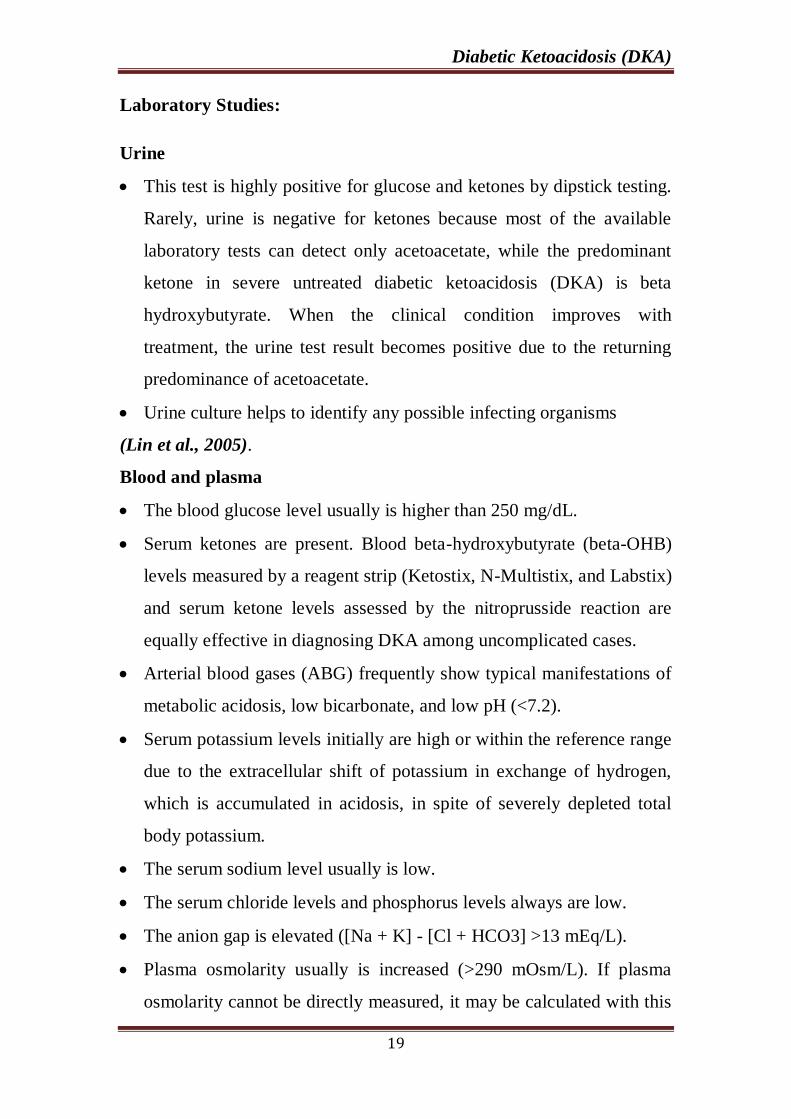
Laboratory Studies:
Urine
This test is highly positive for glucose and ketones by dipstick testing.
Rarely, urine is negative for ketones because most of the available
laboratory tests can detect only acetoacetate, while the predominant
ketone in severe untreated diabetic ketoacidosis (DKA) is beta
hydroxybutyrate. When the clinical condition improves with
treatment, the urine test result becomes positive due to the returning
predominance of acetoacetate.
Urine culture helps to identify any possible infecting organisms
(Lin et al., 2005).
Blood and plasma
The blood glucose level usually is higher than 250 mg/dL.
Serum ketones are present. Blood beta-hydroxybutyrate (beta-OHB)
levels measured by a reagent strip (Ketostix, N-Multistix, and Labstix)
and serum ketone levels assessed by the nitroprusside reaction are
equally effective in diagnosing DKA among uncomplicated cases.
Arterial blood gases (ABG) frequently show typical manifestations of
metabolic acidosis, low bicarbonate, and low pH (<7.2).
Serum potassium levels initially are high or within the reference range
due to the extracellular shift of potassium in exchange of hydrogen,
which is accumulated in acidosis, in spite of severely depleted total
body potassium.
The serum sodium level usually is low.
The serum chloride levels and phosphorus levels always are low.
The anion gap is elevated ([Na + K] - [Cl + HCO3] >13 mEq/L).
Plasma osmolarity usually is increased (>290 mOsm/L). If plasma
osmolarity cannot be directly measured, it may be calculated with this

Diabetic Ketoacidosis (DKA)
20
formula: plasma osmolarity = 2 (Na + K) + BUN/3 + glucose/18.
Urine osmolarity also is increased.
Even in the absence of infection, CBC shows increased WBC count.
BUN frequently is increased.
Blood culture findings may help to identify any possible infecting
organisms(Lin et al., 2005).
Frequency of laboratory studies
Blood tests for glucose should be performed hourly (until patient is
stable, then every 6 h).
Serum electrolyte determinations should be obtained hourly (until
patient is stable, then every 6 h).
BUN should be performed initially.
ABG should be performed initially, followed with bicarbonate as
necessary(Glaser et al., 2008).
Imaging Studies:
Plain chest radiograph may reveal signs of pneumonia.
If it occurs during therapy, magnetic resonance imaging (MRI) is
helpful in detecting early cerebral edema and should only be ordered if
altered consciousness is present (Glaser et al., 2008).
Other Tests:
Electrocardiogram (ECG) :
This test may reveal signs of acute myocardial infarction that could be
painless in patients with diabetes, particularly in those with autonomic
neuropathy.
T-wave changes may produce the first warning sign of disturbed
serum potassium levels.
Low T wave and apparent U wave always signify hypokalemia, while
peaked T wave is observed in hyperkalemia.

Diabetic Ketoacidosis (DKA)
21
ECG should be performed every 6 hours during the first day, unless
the patient is monitored (Glaser et al., 2008).
Treatment:
Immediate medical Care:
Managing diabetic ketoacidosis (DKA) in an ICU during the first 24-
48 hours is always advisable. When treating DKA, the points that must be
considered and closely monitored include correction of fluid loss with IV
fluids; correction of hyperglycemia with insulin; correction of electrolyte
disturbances, particularly potassium loss; correction of acid-base balance;
and treatment of concurrent infection if present Paying great attention to
the correction of fluid and electrolyte loss during the first hour of
treatment, followed by gradual correction of hyperglycemia and acidosis,
always is advisable. Correction of fluid loss makes the clinical picture
clearer and may be sufficient to correct acidosis. The presence of even
mild signs of dehydration means that at least 3 liters of fluid already have
been lost (Glaser et al., 2008).
Fluids: Initial correction of fluid loss is either by isotonic sodium
chloride solution or by lactated Ringer solution.
Administer 1 liter over the first 30 minutes.
Administer 1 liter over the second hour.
Administer 1 liter over the following 2 hours.
Administer 1 liter every 4 hours, depending on the degree of
dehydration and central venous pressure (CVP) readings.
When the patient becomes euvolemic, the physician may switch to
half the isotonic sodium chloride solution, particularly if
hypernatremia exists.

Diabetic Ketoacidosis (DKA)
22
When blood sugar decreases to less than 180 mg/dL, isotonic sodium
chloride solution is replaced with 5-10% dextrose with half isotonic
sodium chloride solution (William et al ,2014)
When insulin treatment is started in patients with DKA, several
points must be considered. A low-dose insulin regimen has the advantage
of not inducing the severe hypoglycemia or hypokalemia that may be
observed with a high-dose insulin regimen.Only short-acting insulin is
used for correction of hyperglycemia. Subcutaneous absorption of insulin
is reduced in DKA because of dehydration; therefore, using intravenous
routes is preferable. (William et al ,2014)
SC use of the fast-acting insulin analog (lispro) has been tried in
pediatric DKA (0.15 U/kg q2h). The results were shown to be
comparable to IV insulin, but ketosis took 6 additional hours to resolve.
Such technically simplified methods may be cost-effective and may
preclude admissions to intensive care units in patients with mild cases.
Use of subcutaneous insulin analog (aspart) has been shown to be
effective as well in adults.The initial insulin dose is a continuous IV
insulin infusion using an infusion pump, if available, at a rate of 0.1
U/kg/h. A mix of 24 units of regular insulin in 60 mL of isotonic sodium
chloride solution usually is infused at a rate of 15 mL/h (6 U/h) until the
blood glucose level drops to less than 180 mg/dL; the rate of infusion
then decreases to 5-7.5 mL/h (2-3 U/h) until the ketoacidotic state
corrected. (Timothy et al, 2014)
Larger volumes of an insulin and isotonic sodium chloride solution
mixture can be used, providing that the infusion dose of insulin is similar.
Larger volumes may be easier in the absence of an IV infusion pump (eg,
60 U of insulin in 500 mL of isotonic sodium chloride solution at a rate of
50 mL/h.The optimal rate of glucose decline is 100 mg/dL/h. Do not

Diabetic Ketoacidosis (DKA)
23
allow the blood glucose level to fall below 200 mg/dL during the first 4-5
hours of treatment. Hypoglycemia may develop rapidly with correction of
ketoacidosis due to improved insulin sensitivity(Timothy et al, 2014).
Allowing blood glucose to drop to hypoglycemic levels is a common
mistake that usually results in a rebound ketosis derived by counter-
regulatory hormones. Rebound ketosis necessitates a longer duration of
treatment. The other hazard is that rapid correction of hyperglycemia and
hyperosmolarity may shift water rapidly to the hyperosmolar intracellular
space and may induce cerebral edema. Although DKA was a common
problem in patients with diabetes who were treated with continuous
subcutaneous insulin infusion through insulin infusion pumps, the
incidence of DKA was reduced with the introduction of pumps equipped
with sensitive electronic alarm systems that alert users when the infusion
catheter is blocked(William et al , 2014).
Electrolyte correction:
Potassium
If the potassium level is greater than 6 mEq/L, do not administer
potassium supplement.
If the potassium level is 4.5-6 mEq/L, administer 10 mEq/h of
potassium chloride.
If the potassium level is 3-4.5 mEq/L, administer 20 mEq/h of
potassium chloride.
Monitor serum potassium levels hourly, and the infusion must stop if
the potassium level is greater than 5 mEq/L.
Monitoring of serum potassium must continue even after potassium
infusion is stopped in the case of (expected) recurrence of
hypokalemia (William et al , 2014).

Diabetic Ketoacidosis (DKA)
24
In severe hypokalemia, not to starting insulin therapy is advisable
unless potassium replacement is underway in order to avoid
potentially serious cardiac dysrhythmia that may result from
hypokalemia.
Correction of acid-base balance:
Sodium bicarbonate only is infused if decompensated acidosis starts to
threaten the patient's life, especially when associated with either sepsis
or lactic acidosis.
If sodium bicarbonate is indicated, 100-150 mL of 1.4% concentration
is infused initially. This may be repeated every half hour if necessary.
Rapid and early correction of acidosis with sodium bicarbonate may
worsen hypokalemia and cause paradoxical cellular acidosis
(Pugliese et al., 2009).
Treatment of concurrent infection:
In the presence of infection, administer proper antibiotics guided by
the results of culture and sensitivity studies.
Starting empiric antibiotics on suspicion of infection until culture
results are available may be advisable (Weber et al., 2009).
Follow-up:
Further Inpatient Care:
Patients are not usually discharged from the hospital unless they
have switched back to their daily insulin regimen without recurrence of
ketosis. When the condition is stable, pH is greater than 7.3, and
bicarbonate is greater than 18 mEq/L, the patient is allowed to eat a meal
preceded by an SC dose of regular insulin. Insulin infusion can be
discontinued 30 minutes later. If the patient is still nauseated and cannot
eat, continue dextrose infusion and administer regular or ultra–short-
acting insulin SC every 4 hours according to blood glucose level, while

Diabetic Ketoacidosis (DKA)
25
trying to maintain blood glucose values at 100-180 mg/dL. In established
patients with diabetes, SC long-acting insulin (eg, insulin glargine,
Detemir, Ultralente) should be initiated back at the same dose that was
used prior to diabetic ketoacidosis. However, if neutral protamine
Hagedorn (NPH) insulin was previously used, start back at the usual dose
only when the patient eats well and is able to retain meals without
vomiting; otherwise, the dose should be reduced to avoid hypoglycemia
during its peak efficacy period. In newly diagnosed patients with type 1
DM, a careful estimate of the long-acting insulin dose should be
considered. Starting with smaller doses generally is recommended to
avoid unneeded hypoglycemia (Weber et al., 2009).
Complications:
The most widely recognized complications of DKA treatment
include exogenous insulin-induced hypoglycemia and hypokalemia. The
effect of bicarbonate therapy may also worsen hypokalemia. These
complications may be avoided with the use of dextrose-containing
solutions when blood glucose falls below 250 mg/dL with a concomitant
reduction in the rate of insulin delivery as well as the addition of
potassium to replacement fluids Additionally, abrupt discontinuation of
intravenous insulin therapy after resolution of DKA without overlapping
subcutaneous insulin coverage may precipitate hyperglycemia Although
rare in adult patients(Michael et al., 2013).
cerebral edema is a complication of DKA treatment with significant
morbidity and mortality Its hallmarks include rapid deterioration in the
level of consciousness and headache. Other manifestations include
seizure, bradycardia, incontinence, respiratory arrest, and eventual brain-
stem herniation. Theoretically, cerebral edema develops when water
is osmotically driven into the central nervous system;

Diabetic Ketoacidosis (DKA)
26
plasma osmolality declines too rapidly during replacement of sodium and
water deficits in DKA treatment Gradual correction of
the hyperosmolar state in addition to adding dextrose to intravenous
fluids when blood glucose falls below 250 mg/dL may avert this
risk(Michael et al., 2013). .
Hypoxemia and noncardiogenic pulmonary edema may result
secondary to falling colloid osmotic pressure and subsequent increase in
lung water content and diminished lung compliance A rare but highly
morbid complication of DKA is ARDS Ominous signs include a widened
A-a gradient, dyspnea, hypoxemia, rales or infiltrates during routine
resuscitation as well as severe acidemia Lastly, vascular thrombosis may
occur in the setting of critical illness and low dose heparin or low
molecular weight heparin should be considered for prophylaxis
(Christine, Saraceni, et al., 2013). .
Nonspecific myocardial injury may occur in severe DKA, which is
associated with minute elevations of myocardial biomarkers (troponin T
and CK-MB) and initial ECG changes compatible with myocardial
infarction (MI). Acidosis and very high levels of free fatty acids could
cause membrane instability and biomarker leakage. Coronary
arteriography usually is normal, and patients usually recover fully without
further evidence of ischemic heart disease. Regardless of the
pathogenesis, the presence of minute biomarker elevations and ECG
changes do not necessarily signify MI in DKA (Moller et al., 2005).
Microvascular changes consistent with diabetic retinopathy have
been reported prior to and after treatment of DKA; however, the blood-
retinal barrier does not experience the same degree of perturbation as the
blood-brain barrier does (Martin et al., 2005).

Diabetic Ketoacidosis (DKA)
27
Prognosis:
The prognosis of properly treated patients with diabetic
ketoacidosis is excellent, especially in younger patients if intercurrent
infections are absent. The worst prognosis is usually observed in patients
who are older with severe intercurrent illnesses, eg, myocardial
infarction, sepsis, or pneumonia, especially when they are treated outside
an ICU. The presence of deep coma at the time of diagnosis,
hypothermia, and oliguria are signs of poor prognosis (Martin et al.,
2005).

Thyroid Storm
28
THYROID STORM
Introduction:
Thyroid storm is a clinical manifestation of an extreme hyperthyroid
state that results in significant morbidity or disability or even death.
Previously, thyroid storm was a common complication of toxic goiter
surgery during intraoperative and postoperative stages. Preoperative
control of the thyrotoxic state and use of radioiodine ablation has greatly
reduced this phenomenon. Today, thyroid storm more commonly is seen
in a thyrotoxic patient with intercurrent illness or surgical emergency.
Early recognition and prompt intervention are necessary
(Migneco et al., 2005).
Epidemiology:
Presently, incidence is less than 10% among patients hospitalized for
thyrotoxicosis(Migneco et al., 2005).
Mortality/Morbidity:
Thyroid storm, considered a fulminating state, is fatal when
untreated. Although methods of diagnosis and management have
improved considerably, reported mortality still is 20-30%. Although it
can develop in toxic adenoma or multinodular toxic goiter, thyroid storm
is more commonly seen in toxicity secondary to Grave's disease
(Nakamura et al., 2005).

Thyroid Storm
29
Sex:
Age and sex predilection depends on the etiology of thyrotoxicity.
Graves disease more frequently develops in females (ie, male-to-female
ratio ranges from 1:7 to 1:10); multinodular goiter more often manifests
in the elderly population (Nakamura et al., 2005).
Causes:
A precipitating factor usually is found with thyroid storm. Presently,
the most common cause of thyroid storm is intercurrent illness or
infection (ie, medical storm)
Some causes that rapidly increase the thyroid hormone levels include
the following:
Surgery, thyroidal or nonthyroidal.
Radioiodine therapy.
Withdrawal of antithyroid drug therapy.
Vigorous thyroid palpation.
Iodinated contrast dye.
Thyroid hormone ingestion.
Other common precipitants include the following:
Infection.
Emotional stress.
Tooth extraction.
Diabetic ketoacidosis.
Hypoglycemia.

Thyroid Storm
30
Trauma.
Bowel infarction.
Toxemia of pregnancy.
Pulmonary embolism.
Cerebrovascular accident (Migneco et al., 2005).
Management of thyroid storm
Diagnosis:
History:
Clinical features form the hallmark in diagnosing thyroid storm.
Most patients have goiter, and many of those with Graves disease have
concurrent ophthalmopathy. Frequently, a past history of thyroid disease
that has been partially treated exists (Nakamura et al., 2005).
Physical examination:
An accentuation of signs and symptoms is seen in uncomplicated
thyrotoxicosis. The point of transition from uncomplicated thyrotoxicosis
to thyroid storm is difficult to ascertain. Very few criteria define the
change. However, certain clinical features (eg, high-grade fever,
decompensation of one or more organ systems secondary to the severe
state of hypermetabolism) reveal its onset (Tietgens and Leinung 2006).
The Following table presents some changes in the symptoms and
signs of thyroid storm when compared with uncomplicated
thyrotoxicosis. Importantly, some findings of thyroid storm (eg, atrial
dysrhythmia) may also occur in uncomplicated thyrotoxicosis.

Thyroid Storm
31
Therefore, the table represents only guidelines, not specific criteria
to define thyroid storm (Tietgens and Leinung 2006).
Uncomplicated Thyrotoxicosis
Thyroid Storm
1.Heat intolerance, diaphoresis 1.Hyperpyrexia, temperature in
excess of 106o F, dehydration
2.Sinus tachycardia, heart rate 100-
140
2.Heart rate faster than 140
beats/min, hypotension, atrial
dysrhythmias, congestive heart
failure
3.Diarrhea, increased appetite with
loss of weight
3. Nausea, vomiting, severe diarrhea,
abdominal pain, hepatocellular
dysfunction-jaundice
4.Anxiety, restlessness 4.Confusion, agitation, delirium,
frank psychosis, seizures, stupor
or coma
Table (1): Manifestations of thyrotoxicosis versus thyroid storm
(Tietgens and Leinung 2006).
Certain unusual presentations include chest pain, acute abdomen,
status epilepticus, stroke, acute renal failure due to rhabdomyolysis, and
apathetic thyroidism. apathetic thyroidism (ie, masked hyperthyroidism)
60 years ago. Apathetic thyroidism more frequently was seen in elderly
patients but since has been described in all ages. Patients in this variant
group present without goiter, ophthalmopathy, or prominent symptoms of
hyperthyroidism. These patients have a low pulse rate and a propensity to
develop thyroid storm due to delay in diagnosis (Tietgens and Leinung
2006).

Thyroid Storm
32
Differential diagnosis:
Postoperative complications (eg, sepsis, hemorrhage, septicemia,
transfusion drug reactions) mimic the thyrotoxic state. Previous history of
hyperthyroidism, precipitating factors, increased T3 and T4 levels, and
decreased thyroid stimulating hormone (TSH) levels help to establish the
diagnosis of thyroid storm. (Morgan et al., 2006).
Thyroid storm can be differenciated from malignant hyperthermia by
that it is not associated with muscle rigidity, elevated creatine kinase, or a
marked degree of metabolic (lactic) and respiratory acidosis.The
neuroleptic malignant syndrome (NMS) is a rare, but life-threatening,
idiosyncratic reaction to a neuroleptic medication. The syndrome is
characterized by fever, muscular rigidity, altered mental status, and
autonomic dysfunction. Although potent neuroleptics (eg, haloperidol,
fluphenazine) are more frequently associated with NMS, all antipsychotic
agents, typical or atypical, may precipitate the syndrome (Strawn et al.,
2007).
Laboratory Studies:
Presently, no specific diagnostic criteria to establish the diagnosis of
thyroid storm exist.
Burch and Wartofsky have constructed an excellent clinical diagnostic
point scale to facilitate a semiquantitative distinction between
uncomplicated thyrotoxicosis, impending storm, and established
thyroid storm. Laboratory findings in thyroid storm are consistent with
those of thyrotoxicosis and include the following:
Elevated T3 and T4 levels.

Thyroid Storm
33
Elevated T3 uptake.
Suppressed TSH levels.
Elevated 24-hour radioiodine uptake.
Elevated T4 and decreased TSH are the only abnormal findings needed
for conformation of thyrotoxicosis. Treatment should not be withheld
for any laboratory confirmation of hyperthyroidism when thyroid
storm is suspected clinically. A 2-hour radioiodine uptake is advisable
if thyroid storm is suspected and no past history of hyperthyroidism
exists(Strawn et al., 2007).
Other abnormal laboratory values that point toward decompensation
of homeostasis include the following:
Increased BUN and creatinine kinase.
Electrolyte imbalance from dehydration, anemia, thrombocytopenia,
and leukocytosis.
Hepatocellular dysfunction as shown by elevated levels of
transaminases, lactate dehydrogenase, alkaline phosphatase, and
bilirubin.
Elevated calcium levels.
Hyperglycemia (Strawn et al., 2007).

Thyroid Storm
34
Treatment:
Medical Care:
Management of thyroid storm is a multi-step process. Blocking the
synthesis, secretion, and peripheral action of the thyroid hormone is the
ideal therapy. Aggressive supportive therapy then is used to stabilize
homeostasis and reverse multiorgan decompensation. Additional measures
are taken to identify and treat the precipitating factor, followed by
definitive treatment to avoid recurrence. Thyroid storm is a fulminating
crisis that demands an intensive level of care, continuous monitoring, and
vigilance (Rosenberg, 2006).
Blocking thyroid hormone synthesis:
Antithyroid compounds propylthiouracil (PTU) and methimazole
(MMI) are used to block the synthesis of the thyroid hormone.
propylthiouracil also blocks peripheral conversion of T4 to T3 and hence
is preferred in thyroid storm over methimazole. methimazole is the
common agent used in hyperthyroidism. propylthiouracil and
methimazole block the incorporation of iodine into thyroglobulin within 1
hour of ingestion. A history of hepatotoxicity or agranulocytosis from
previous thioamide therapy precludes use of propylthiouracil and
methimazole. PTU is considered as a second-line drug therapy, except in
patients who are allergic or intolerant to methimazole, or for women who
are in the first trimester of pregnancy(Rosenberg, 2006).
Blocking thyroid hormone secretion:
After initiation of antithyroid therapy, hormone release can be
inhibited by large doses of iodine, which reduce thyroidal iodine uptake.
Lugol solution or saturated solution of potassium iodide can be used.

Thyroid Storm
35
Iodine therapy should be administered after approximately 1 hour
following administration of PTU or MMI; iodine used alone helps to
increase thyroid hormone stores and may increase the thyrotoxic state.
The iodinated x-ray contrast agent, sodium ipodate, can be administered
instead of iodine and also inhibits peripheral conversion of T4 to T3.
Potassium iodide (KI) decreases thyroidal blood flow and hence is used
preoperatively in thyrotoxicosis. Patients intolerant to iodine can be
treated with lithium, which also impairs thyroid hormone release. Patients
unable to take PTU or MMI also can be treated with lithium, as use of
iodine alone is debatable. Unlike iodine, lithium is not subject to the
escape phenomenon; lithium blocks the release of thyroid hormone
throughout its administration(Rosenberg 2006).
Plasmapheresis, plasma exchange, peritoneal dialysis exchange
transfusion, and charcoal plasma perfusion are other techniques used to
remove excess circulating hormone. Presently, these techniques are
reserved for patients who do not respond to the initial line of management
(Ingbar, 2000).
Blocking peripheral action of thyroid hormone:
Propranolol is the drug of choice to counter peripheral action of
thyroid hormone. Propranolol blocks beta-adrenergic receptors and
prevents conversion of T4 to T3. It produces dramatic improvement in
clinical status and greatly ameliorates symptoms Propranolol produces
the desired clinical response in thyroid storm only after large doses.
Intravenous administration of propranolol requires continuous monitoring
of cardiac rhythm (Ingbar, 2000).
Presently, esmolol is the ultra-short-acting beta-blocking agent used
successfully in thyrotoxicosis and thyroid storm. Noncardioselective beta-

Thyroid Storm
36
blockers (eg, propranolol, esmolol) cannot be used in patients with
congestive cardiac failure, bronchospasm, or history of asthma.
Guanethidine or reserpine can be used instead in these cases Successful
treatment with reserpine in cases of thyroid storm resistant to large doses
of propranolol has been documented. However, guanethidine and
reserpine cannot be used in the presence of cardiovascular collapse or
shock (Ingbar, 2000).
Supportive measures:
- Aggressive fluid and electrolyte therapy is needed for
dehydration and hypotension. This excessive hypermetabolic state, with
increased intestinal transit and tachypnea, leads to immense fluid loss.
Fluid requirements may increase to 3-5 L/day. Therefore, invasive
monitoring is advisable in elderly patients and in those with congestive
cardiac failure (Ingbar, 2000).
Pressor agents can be used when hypotension persists following
adequate fluid replacement. Add glucose to IV fluids for nutritional
support. Multivitamins, especially vitamin B-1, are added to prevent
Wernicke encephalopathy (Ingbar, 2000).
Hyperthermia:
is treated through central cooling and peripheral heat dissipation.
Acetaminophen is the drug of choice, as aspirin may displace thyroid
hormone from binding sites and increase severity of thyroid storm.
Cooling blankets, ice packs, and alcohol sponges encourage dissipation of
heat. Use of a cooled humidified oxygen tent is advised (Rosenberg
2006).

Thyroid Storm
37
Use of glucocorticoids in thyroid storm is associated with improved
survival rates. Initially, glucocorticoids were used to treat potential
relative insufficiency due to accelerated production and degradation
owing to the hypermetabolic state. However, the patient may have type 2
autoimmune deficiency, in which Graves disease coexists with absolute
adrenal insufficiency . Glucocorticoids reduce iodine uptake and antibody
titers of thyroid-stimulating antibodies with stabilization of the vascular
bed. In addition, dexamethasone and hydrocortisone have an inhibitory
effect on conversion of T4 to T3. Therefore, a stress dose of
glucocorticoid (eg, hydrocortisone, dexamethasone) now is
routine(Scholz et al., 2003).
Cardiac decompensation, although seen more frequently in elderly
patients, may appear in younger patients and in patients without
underlying cardiac disease Digitalization is required to control the
ventricular rate in patients with atrial fibrillation (Scholz et al., 2003).
Anticoagulation drugs may be needed for atrial fibrillation and can
be administered in the absence of contraindications (Martin, 2009).
Medications:
Antiadrenergic drugs:
Promptly administer antiadrenergic drugs (eg, propranolol) to
minimize sympathomimetic symptoms. Propranolol is administered
orally or via nasogastric tube at a dose of 60-80 mg every 4-6 hours and
the dose adjusted based on heart rate and blood pressure. It may also be
given intravenously when necessary for rapid onset of action (0.5-1 mg
over 10 min followed by 1-2 mg over 10 min every few hours, adjusted
based on vital signs). It is important to avoid propranolol in conditions

Thyroid Storm
38
such as asthma, chronic obstructive pulmonary disease, peripheral
vascular disease, or decompensated heart failure. Cardioselective beta
blockers such as atenolol or metoprolol may be administered in patients
with reactive airway disease, and calcium channel blockers may be used
when beta blockers are contraindicated. The use of intravenous short
acting beta-1 blockers, such as esmolol (loading dose of 250-500 mcg/kg,
followed by an infusion of 50-100 mcg/kg per minute), allows quick dose
titration with minimization of side effects (Madhusmita et al, 2016).
Dosing of beta blockers for thyroid storm in a pediatric population:
Propranolol: Neonates: 2 mg/kg per day PO/NGT divided every 6-
12 hours; Children: 0.5-4 mg/kg per day PO/NGT divided every 6 hours
(not to exceed 60 mg per day) or 0.01-0.02 mg/kg IV over 10 minutes
(may repeat over 10’ every few hours to a maximum cumulative dose of
5 mg) (Rosenberg 2006).
Esmolol:
Loading dose: 250-500 mcg/kg over 1 minute, repeat as needed,
maintenance dose: 50-100 mcg/kg per minute IV infusion(Rosenberg
2006).
Thionamides:
Correct the hyperthyroid state. Administer antithyroid medications
to block further synthesis of thyroid hormones (THs) (Rosenberg 2006).
High-dose propylthiouracil (PTU) is preferred over methimazole for
treatment of severe thyroid storm because of its early onset of action and
capacity to inhibit peripheral conversion of T4 to T3. Methimazole may
be used in less severe cases. Dosing for thyroid storm in adults is as

Thyroid Storm
39
follows: PTU 200 mg every four hours or methimazole 20 mg orally
every four to six hours; these drugs may need to be administered through
a nasogastric tube(Stephen et al, 2016).
Dosing of PTU for thyroid storm in children: Neonates: 5-10 mg/kg
per day PO/NGT divided every 6-8 hours; Children: 15-20 mg/kg per day
PO/NGT divided every 6-8 hours (up to 40 mg/kg per day has been used;
not to exceed 1200 mg per day (Stephen et al, 2016).
The US Food and Drug Administration (FDA) has added a boxed
warning, the strongest warning issued by the FDA, to the prescribing
information for PTU.
The boxed warning emphasizes the risk for severe liver injury and
acute liver failure, some of which have been fatal. The boxed warning
also states that PTU should be reserved for use in those who cannot
tolerate other treatments such as methimazole, radioactive iodine, or
surgery(Madhusmita et al, 2016).
The decision to include a boxed warning was based on the FDA's
review of postmarketing safety reports and meetings held with the
American Thyroid Association, the National Institute of Child Health and
Human Development, and the pediatric endocrine clinical community
(Madhusmita et al, 2016).
The FDA has identified 32 cases (22 adult and 10 pediatric) of
serious liver injury associated with PTU. Among adults, 12 deaths and 5
liver transplants occurred; among the pediatric patients, 1 death and 6
liver transplants occurred. (Stephen et al, 2016). PTU is indicated for
hyperthyroidism due to Graves' disease. These reports suggest an
increased risk for liver toxicity with PTU compared with methimazole.

Thyroid Storm
40
Serious liver injury has been identified with methimazole in 5 cases (3
resulting in death(Stephen et al, 2016).
PTU is now considered as a second-line drug therapy for treatment
of hyperthyroidism in general (though not thyroid storm), except in
patients who are allergic or intolerant to methimazole, or women who are
in the first trimester of pregnancy. Rare cases of embryopathy, including
aplasia cutis, have been reported with methimazole during pregnancy. For
more information, The FDA recommends the following criteria be
considered for prescribing PTU. Reserve PTU use for during first
trimester of pregnancy or for patients who are allergic to or intolerant of
methimazole. Closely monitor patients undergoing PTU therapy for signs
and symptoms of liver injury, especially during the first 6 months after
initiation of therapy. For suspected liver injury, promptly discontinue
PTU therapy and evaluate for evidence of liver injury and provide
supportive care (Stephen et al, 2016).
PTU should not be used in pediatric patients unless the patient is
allergic to or intolerant of methimazole and no other treatment options are
available. Counsel patients to promptly contact their health care provider
for the following signs or symptoms: fatigue, weakness, vague abdominal
pain, loss of appetite, itching, easy bruising, or yellowing of the eyes or
skin. If the patient is given PTU during treatment of thyroid storm, this
should be switched to methimazole at the time of discharge unless
methimazole is contraindicated. If methimazole is contraindicated,
alternative methods to treat hyperthyroidism should be considered after
discharge, such as radioactive iodine or surgery(Stephen et al, 2016).

Thyroid Storm
41
Iodine compounds:
Administer iodine compounds (Lugol iodine or potassium iodide)
orally or via a nasogastric tube to block the release of THs (at least 1 h
after starting antithyroid drug therapy). In adults, KI is a given at a dose
of 5 drops every 6 hours, or Lugol's iodine at a dose of 10 drops every 8
hours. If available, intravenous radiocontrast dyes such as ipodate and
iopanoate can be effective in this regard. These agents are particularly
effective at preventing peripheral conversion of T4 to T3(Stephen et al,
2016).
Dosing of iodine compounds for thyroid storm in children:
SSKI (50 mg iodide per drop): Neonates: 2 drops PO/NGT every 6-8
hours; Children: 2-5 drops PO/NGT every 6 hours
Lugol's iodine (8 mg iodine/drop): 10 drops PO/NGT every 8
hours(Stephen et al, 2016).
Glucocorticoids
Administer glucocorticoids to decrease peripheral conversion of T4
to T3. This may also be useful in preventing relative adrenal insufficiency
due to hyperthyroidism and improving vasomotor symptoms.
Hydrocortisone is administered intravenously at a dose of 100 mg every 8
hours or dexamethasone at a dose of 1-2 mg every 6 hours
(Madhusmita et al, 2016).
Dosing of glucocorticoids for thyroid storm in children:
Hydrocortisone: 5 mg/kg (up to 100 mg) intravenously every 6-8
hours
Dexamethasone: 0.1-0.2 mg/kg per day divided every 6-8 hours

Thyroid Storm
42
Bile acid sequestrants prevent reabsorption of free THs in the gut
(released from conjugated TH metabolites secreted into bile through the
enterohepatic circulation). A recommended dose is 4 g of cholestyramine
every 6 hours via a nasogastric tube (Madhusmita et al, 2016).
Treatment of the underlying condition, if any, that precipitated
thyroid storm and exclude comorbidities such as diabetic ketoacidosis
and adrenal insufficiency. Infection should be treated with antibiotics.
Rarely, as a life-saving measure, plasmapheresis has been used to treat
thyroid storm in adults. Iodine preparations should be discontinued once
the acute phase resolves and the patient becomes afebrile with
normalization of cardiac and neurological status. Glucocorticoids should
be weaned and stopped and the dose of thioamides adjusted to maintain
thyroid function in the normal range. Beta-blockers may be discontinued
once thyroid function normalizes. If the patient is given PTU during
treatment of thyroid storm, this should be switched to methimazole at the
time of discharge unless methimazole is contraindicated. If there is a
contraindication for the use of methimazole, alternative methods to treat
hyperthyroidism should be considered after discharge, such as radioactive
iodine or surgery. (Madhusmita et al, 2016)
Patients with contraindications to thionamides need to be managed
with supportive measures, aggressive beta blockade, iodine preparations,
glucocorticoids and bile acid sequestrants for about a week in preparation
for a thyroidectomy. Plasmapheresis may be attempted if other measures
are not effective (Madhusmita et al, 2016).

Thyroid Storm
43
Prevention:
Identification of precipitating factors:
Surgery and anesthesia induction, labor, thioamide withdrawal, and
use of radioiodine are known precipitants of thyroid storm. However,
these precipitants may not be discovered frequently.
Precipitating factors are not found in all patients, but a meticulous
search improves chances for a successful outcome.
Chest radiographs and blood, urine, and sputum cultures may be
needed to identify intercurrent illness (eg, infection).
Judicious use of empiric antibiotics is needed if no obvious source is
found (Stephen et al, 2016).
Prevention of recurrence:
Prevention of a recurrent crisis should be the main objective until
completion of definitive therapy.
Vigilant monitoring of signs and symptoms of hyperthyroidism during
preoperative or pre-anesthetic evaluation is paramount. =
Consider precipitating factors when deciding on treatment modalities.
Adequate control of the thyrotoxic state prior to initiation of definitive
therapy is important. Carry out procedures only after the patient is
euthyroid (Stephen et al, 2016).

Myxedema Coma
44
MYXEDEMA COMA
Introduction:
myxedema has been applied to several clinical entities and is often
used interchangeably with severe hypothyroidism, the common clinical
condition in which the thyroid gland produces abnormally low levels of
hormones. Myxedema also refers to 2 different dermatologic conditions.
Pretibial myxedema, an uncommon skin disorder, occurs not in cases of
hypothyroidism but in hyperthyroid states, including, most commonly,
Graves disease. The term pretibial is somewhat misleading, because the
condition can affect other areas of the body and could more accurately be
called localized dermopathy. The other skin condition, called myxedema,
occurs in severe, long-standing hypothyroid states and is caused by the
deposition of mucopolysaccharides within the dermis. Myxedema coma,
an uncommon but life-threatening form of untreated hypothyroidism with
physiological decompensation (Wall, 2000). The condition occurs in
patients with long-standing, untreated hypothyroidism and is usually
precipitated by a secondary insult, such as climate-induced hypothermia,
infection, or another systemic condition, or drug therapy. Patients with
myxedema coma have changes in their mental status, including lethargy,
stupor, delirium, or coma. A more appropriate term for myxedema coma
is myxedema crisis; we often use the term myxedema coma/crisis
(Fliers and Wiersinga 2003).
Epidemiology:
hypothyroidism is a common disorder in the older population; in
the United States, the condition is present in 8% of women and 2% of
men older than 50 years. Myxedema coma is a rare consequence of

Myxedema Coma
45
untreated hypothyroidism. In areas in which the population ingests
sufficient iodine, the most common cause of hypothyroidism is
autoimmune thyroid disease and thyroid ablation therapy, with a
prevalence of approximately 8% of women aged 50 years or older
(Kwaku and Burman 2007).
In regions where not enough iodine is ingested, the most common
cause of hypothyroidism is iodine deficiency, with the prevalence of
hypothyroidism correlating with the iodine content of the diet. Severe
hypothyroidism (neonatal thyrotropin [TSH] >5 mU/L in >40% of births)
and cretinism are observed with severe iodine deficiency (< 20 mcg/dL).
Iodine deficiency of this magnitude is generally observed only in isolated,
mountainous regions of South America, Africa, and Asia. The prevalence
of myxedema coma/crisis in the populations of these areas is unknown
(Rodriguez et al., 2004).
Mortality/Morbidity:
Myxedema coma/crisis is a metabolic and cardiovascular
emergency. If the condition is not promptly diagnosed and treated, the
mortality rate is approximately 50% or more. Even with immediate
recognition and appropriate medical intervention, mortality rates of up to
25% are observed. Factors suggesting a poor prognosis are a body
temperature of less than 93° F, persistent hypothermia that is
unresponsive to 72 hours of therapy, advanced age, bradycardia (< 44
beats per min), sepsis, myocardial infarction, and hypotension. In
addition, a study found that the patient's admission level of
consciousness, as well as his/her score on the Glasgow Coma Scale and
on the Acute Physiology and Chronic Health Evaluation (APACHE) II,
were most predictive of survival (Rodriguez et al., 2004).

Myxedema Coma
46
Race
No studies suggest a race or ethnic predilection to myxedema
coma/crisis(Rehman et al., 2005).
Sex
Myxedema coma/crisis is approximately 4-8 times more common in
women than in men, corresponding to the increased incidence of
hypothyroidism in women(Rehman et al., 2005).
Age
The incidence of hypothyroidism increases with age; the
physiological decompensation of severe hypothyroidism, myxedema
coma/crisis, occurs primarily in the elderly (Rehman et al., 2005).
However, this condition should not be automatically ruled out in young
adults(Rehman et al., 2005).
Causes:
Myxedema coma/crisis is a physiologic decompensation of severe
primary or secondary hypothyroidism that is usually caused by additional
physiologic stress. Specific types of such stress are as follows:
Infection/systemic illness.
Cold environmental temperatures.
Trauma.
Burns.
Decreased cerebral blood flow/cerebrovascular accident.

Myxedema Coma
47
Decreased cardiac output/congestive heart failure.
Respiratory acidosis (increased P CO2 , decreased P O2 ).
Drugs: Tranquilizers, Sedatives, Anesthetics, Analgesics/ narcotics,
Amiodarone, Rifampin, Beta blockers, Lithium, Phenytoin, Diuretics.
GI hemorrhage.
Hypoglycemia (Rehman et al., 2005).
Pathophysiology:
Myxedema coma/crisis is a form of decompensated hypothyroidism
in which adaptations are no longer sufficient Essentially, all organ
systems are affected. Myxedema coma/crisis occurs most commonly in
older women with long-standing, undiagnosed or undertreated
hypothyroidism who experience an additional significant stress, such as
infection, a systemic disease, certain medications, and exposure to a cold
environment.When hypothyroidism is long-standing, physiologic
adaptations occur. Reduced metabolic rate and decreased oxygen
consumption result in peripheral vasoconstriction, which maintains core
temperature. The number of beta-adrenergic receptors is reduced, usually
with preservation of alpha-adrenergic receptors and circulating
catecholamines, causing beta/alpha-adrenergic imbalance, diastolic
hypertension, and reduced total blood volume(Wall, 2000).
Metabolic
Thyroid hormones are critical for cell metabolism and organ
function. With an inadequate supply, organ tissues do not grow or mature,
energy production declines, and the action of other hormones is affected.
Although weight gain is common, severe obesity is rarely secondary to

Myxedema Coma
48
hypothyroidism alone. However, long-standing, untreated
hypothyroidism may result in years of inactivity, eventually with a large
increase in weight. Because of decreased drug metabolism, overdoses of
medications (eg, morphine, hypnotics, anesthetic agents, sedatives) can
occur and can even precipitate myxedema crisis (Michiels et al., 2006)
Neurologic
Although the condition is called myxedema coma, the absence of
coma does not exclude the diagnosis of this disorder. The presenting
mental status may be lethargy or stupor. The exact mechanisms causing
changes in mental status are not known. Brain function is influenced by
reductions in cerebral blood flow and oxygen delivery, a lack of
thyroxine (T4) and triiodothyronine (T3), and reductions in oxygen and
glucose consumption; all of these factors are probably involved.
Hyponatremia brought on by renal dysfunction may be an additional
cause of altered mental function (Michiels et al., 2006)
Cardiovascular
The heart is profoundly depressed, with bradycardia and decreased
contractility causing low stroke volume and cardiac output. These
changes are caused by decreased production of myocyte contractile
proteins and enzymes, including NA+/K
+ adenosine triphosphatase
(NA+/K
+ ATPase), as a result of low levels of gene transcription in the
absence of T3. Increased systemic vascular resistance occurs; although
the causes appear to be multifactorial, a study suggests that in many
cases, the increase is secondary to decreased T3 levels. Nonspecific ST-
and T-wave inversion changes, low voltage, and ventricular arrhythmias
may be noted. Plasma volume is decreased, and capillary permeability is

Myxedema Coma
49
increased, leading to fluid accumulation in tissue and spaces and possibly
causing pericardial effusions. (Michiels et al., 2006)
Pulmonary
Typically, the lungs are not severely affected. Respiratory muscle
dysfunction may be compromised, and depressed ventilatory drive and
increased alveolar-arterial oxygen gradient are common. Fluid
accumulation may cause pleural effusions and decreased diffusing
capacity. Ventilation-perfusion mismatch is common, contributing to
hypercapnia. Dysfunction of other organ systems may have profound
effects. Severe obesity, if present, causes decreased lung volumes,
diffusion capacity, and flow rates and may be the primary cause of the
hypoventilation, hypoxia, hypercarbia, and depressed respiratory drive
that is often noted in these patients. However, hypothyroidism may also
have a direct impact, because the condition can cause obstructive sleep
apnea that resolves with thyroid replacement (even without weight loss).
(Michiels et al., 2006)
Renal
Kidney function may be severely compromised, partly because of
low cardiac output and vasoconstriction that causes a low glomerular
filtration rate. Reduced levels of NA+/K
+ ATPase decrease sodium
reabsorption and impair free water excretion, resulting in hyponatremia,
which is usually present in myxedema coma (Michiels et al., 2006).
Gastrointestinal
Severe or even mild hypothyroidism decreases intestinal motility.
Patients with myxedema coma can present with gastric atony, megacolon,
or paralytic ileus. Malabsorption has also been reported. Ascites, while

Myxedema Coma
50
uncommon, may occur due to increased capillary permeability,
congestive heart failure, or other mechanisms.
Diagnosis:
History:
Although most patients with myxedema coma/crisis have a long
history of hypothyroidism, undiagnosed or undertreated myxedema
coma/crisis may be the initial manifestation of a hypothyroid state. A
history of generalized fatigue, cold intolerance, constipation, and dry skin
(common features of long-standing hypothyroidism) are usually present.
These features slowly progress to lethargy, delirium, or coma.Faster
progression may occur, precipitated by overmedication, stroke,
congestive heart failure, trauma, exposure to cold environmental
temperatures, or an infection, such as pneumonia. Relatives or friends
who know the patient may be able to report a history of long-standing
fatigue, weight gain, hair and skin changes, edema, constipation, and cold
intolerance. Most cases of myxedema coma/crisis occur during the winter
in women aged 60 years or older, although a case of a woman presenting
in labor has been reported (Sheu et al., 2007).
Physical signs:
Hypothermia is usually present in myxedema coma/crisis. If mercury
thermometers are used, the degree of hypothermia may not be
recognized unless the temperature of the thermometer is lowered
before checking the patient's temperature. Moreover, use of special
thermometers that record well below 90°F may be necessary in order
to determine the patient's actual temperature and to monitor
rewarming.
Absence of fever in the presence of infection can be expected.

Myxedema Coma
51
The following features are extremely common, although some are not
invariably present:
Hypotension/shock.
Hypothermia.
Decreased pulse pressure, normal systolic pressure, elevated diastolic
pressure, slow pulse and respiration rates.
Periorbital, nonpitting edema; facial swelling or coarseness;
macroglossia; enlargement of the tonsils, nasopharynx, and larynx;
coarse or thinning hair (Sheu et al., 2007).
Thyroid - Enlarged, not palpable, scar suggesting previous
thyroidectomy.
Lungs - Slow respiration rate, hypoventilation, congestion, pleural
effusions, consolidation.
Heart - Soft or distant heart sounds, diminished apical impulse,
bradycardia, enlarged heart, pericardial effusion.
Abdomen - Distension secondary to ileus and/or ascites, diminished or
absent bowel sounds. (Sheu et al., 2007).
Bladder distension.
Extremities - Cold, nonpitting edema of the hands and feet.
Skin/nails - Cool, pale, dry, scaly, thickened skin; dry, brittle nails;
ecchymoses, purpura, sallowness due to carotenemia.
Neuromuscular - Confusion, stupor, obtundation, coma, slow speech,
seizures, reflexes with a slow relaxation phase (Sheu et al., 2007).
Laboratory Studies:
Laboratory values are essential for the workup of myxedema
coma/crisis; however, if the condition is suspected, treatment must be
initiated immediately without waiting for the results (Sheu et al., 2007).

Myxedema Coma
52
Free T4 and T3 levels are low or undetectable.
The TSH level may be elevated, indicating a primary thyroid disorder.
A low or normal TSH level with low levels of T4 and T3 may indicate
central (pituitary) hypothyroidism or the suppression of TSH
production by severe illness or drugs, such as dopamine or high-dose
glucocorticoids. A serum cortisol level should be determined before
beginning intravenous steroids (Sheu et al., 2007).
Serum electrolytes and serum osmolality - Hyponatremia with low
serum osmolality is common.
Serum creatinine - Because of decreased renal perfusion, the levels are
usually elevated.
Serum glucose - Hypoglycemia is common but may also suggest
adrenal insufficiency.
Complete blood count (CBC) with differential - Bands and/or a left
shift may be the only sign of infection.
Creatine kinase (CK) - CK levels are often elevated, and fractionation
indicates skeletal (not cardiac) muscle injury unless a myocardial
infarction was the precipitating event.
Arterial blood gases - Increased P CO2 and decreased P O2 are found.
Pan-culture for sepsis (Sheu et al., 2007).

Myxedema Coma
53
Imaging Studies:
Chest radiographs: Obtain chest radiographs for all patients.
Cardiomegaly, pericardial effusion, congestive heart failure, and/or
pleural effusion are observed (Sheu et al., 2007).
Cardiological Studies:
Electrocardiogram: Sinus bradycardia, low-amplitude QRS
complexes, a prolonged QT interval, and flattened or inverted T waves
are noted(Sheu et al., 2007).
Treatment:
Management of airway and airway protection from aspiration in case
of patients with poor consciousness level should be the utmost priority.
Endotracheal intubation or tracheostomy with mechanical ventilation may
be performed. Arterial blood gas should be monitored frequently to
ensure adequate oxygenation and correction of hypercarbia. Sedatives
and other drugs may exacerbate the respiratory depression and may delay
the weaning of ventilator support (Vivek Mathew et al., 2011)
Medical Care:
Myxedema crisis/coma is a life-threatening condition; therefore,
patients with this disorder must be stabilized in an intensive care unit.
The first 24-48 hours are critical. If the diagnosis is considered likely,
immediate and aggressive administration of multiple interventions is
necessary to lower an otherwise high rate of mortality. Initial priorities
include the following: Mechanical ventilation if respiratory
acidosis/hypercapnia/hypoxia is significant.Immediate intravenous
thyroid hormone replacement while awaiting confirmatory test results (T4

Myxedema Coma
54
and TSH), even if the diagnosis of myxedema coma is only probable.
Because GI absorption is compromised, intravenous therapy is
mandatory. Whether to use T4 alone, combined T4 and T3, or T3 alone
remains a subject of controversy. Deiodinase conversion of T4 to the
active hormone T3 is reduced in these patients, and T3 administration
may be advisable. However, T3, because of its more immediate action
and short half-life, may be more likely to cause arrhythmias, particularly
if myocardial function is compromised. The usual conversion to an
intravenous dose of T4 is approximately one half to two thirds of the oral
dose. An intravenous loading dose of 500-800 mcg of levothyroxine is
followed by a daily intravenous dose of 50-100 mcg; the daily dose is
administered until the patient is able to take medication by mouth. Use
caution in elderly persons and in patients with coronary artery disease or
myocardial infarction, because full-dose T4 therapy may worsen
myocardial ischemia by increasing myocardial oxygen consumption.
Some authorities advocate the use of additional intravenous T3, at 10-20
mcg every 8-12 hours, especially in young patients with low
cardiovascular risk. Because of the rarity of the condition, randomized
trials comparing different treatment modalities are not available.
Observational studies are not in agreement regarding whether low or high
dose T4 or T3 replacement reduces mortality (Rodriguez et al., 2004).
In light of the possibility of adrenal insufficiency, stress steroid
replacement after a cortisol level is obtained .After a baseline cortisol
level is ascertained, initiate hydrocortisone at 5-10 mg/hr. Continue
therapy unless the random cortisol level on admission indicates adrenal
function without abnormalities, in which case, hydrocortisone may be
stopped without tapering. (Jordan, 1995).

Myxedema Coma
55
Passive rewarming using ordinary blankets and a warm room (rapid
and external rewarming are contraindicated).
Treatment of associated infection.
Correction of severe hyponatremia (sodium level < 120 mEq/L) with
saline, free water restriction.
Broad-spectrum antibiotics with modification of the antibiotic regimen
based on culture results.
Correction of hypoglycemia with intravenous dextrose.
Treatment of severe hypotension with cautious administration of 5-
10% glucose in half-normal or normal saline (or hypertonic saline if
severely hyponatremic, ie, < 120 mEq/L) .
Dose adjustment of any medication to compensate for decreased renal
perfusion, drug metabolism, etc(Taguchi et al., 2007).
Infection:
The precipitating event in myxedema coma/crisis is often overt or
occult bacterial infection.
Fever and elevated white blood cell (WBC) count are usually absent,
although a left shift and/or bands may be observed.
Pan-culture and initiate empiric broad-spectrum antibiotic treatment,
which can be narrowed if the source of infection is identified.
If culture results remain negative, antibiotics may be
discontinued(Taguchi et al., 2007).

Myxedema Coma
56
Myocardial ischemia
Myocardial infarction may be the precipitating event in older patients,
or it may subsequently occur (Taguchi et al., 2007).
Troponin tests are also sometimes used to evaluate people for heart
injury due to causes other than a heart attack or to distinguish signs and
symptoms such as chest pain that may be due to other causes. Testing
may also be done to evaluate people with angina if their signs and
symptoms worsen.
Troponin tests are sometimes ordered along with other cardiac
biomarkers, such as CK–MB or myoglobin. However, troponin is the
preferred test for a suspected heart attack because it is more specific for
heart injury than other tests (which may be elevated in the blood with
skeletal muscle injury) and remain elevated for a longer period of time.
A high troponin and even slight elevations may indicate some degree of
damage to the heart. When a person has significantly elevated troponin
levels and, in particular, a rise in the results from a series of tests done
over several hours, then it is likely that the person has had a heart attack
or some other form of damage to the heart. Levels of troponin can
become elevated in the blood within 3 or 4 hours after heart injury and
may remain elevated for 10 to 14 days. In people with angina, an elevated
troponin may indicate that their condition is worsening and they are at
increased risk of a heart attack. (American Association for Clinical
Chemistry, 2016).
A test called high-sensitivity troponin detects the same protein that
the standard test does, just at much lower levels. Because this version of
the test is more sensitive, it becomes positive sooner and may help detect
heart injury and acute coronary syndrome earlier than the standard test.

Myxedema Coma
57
The troponin test may also be positive in people with stable angina and
even in people with no symptoms. When it is elevated in these
individuals, it indicates an increased risk of future heart events such as
heart attacks. Currently, this test is not approved in the U.S., but research
is ongoing and it may become available in the near future. It is already
routinely used as a cardiac biomarker Serial CK determinations with
fractionation assist in the diagnosis and treatment of an acute coronary
event. CK levels are often elevated in myxedema coma/crisis but are
usually of muscle origin(American Association for Clinical Chemistry,
2016)
If ischemia or infarction is diagnosed, or if the patient has significant
risk factors for coronary artery disease, institute thyroid replacement at
low doses(American Association for Clinical Chemistry, 2016).
Fluid management
the choice is between fluid supplementation for hypotension and
fluid restriction for hyponatremia. A pragmatic approach in mild
hyponatremia will be to advice fluid restriction with replacement to cover
the daily losses taking care to supplement glucose, sodium, and potassium
In a situation of severe hyponatremia, it may be prudent to administer 3%
sodium chloride along with furosemide, so that serum sodium may be
elevated by 3-4 meq/L to tide over the immediate crisis A rapid correction
of chronic hyponatremia might put patients at risk for central pontine
myelinolysis Treatment with furosemide will prevent fluid overloading
associated with hypertonic saline. In experimental hypothyroidism, the
impaired response to an acute water load was shown to be reversed by a
Vasopressin receptor antagonist (V2R antagonist) Future research with

Myxedema Coma
58
V2R antagonists in treating hyponatremia associated with myxedema
crisis may prove to be interesting( Raiz Misgar, et al., 2011)
Hypothermia
It may be managed by external warming, but the accompanying
vasodilatation may precipitate hypotension. Hypotension requires careful
infusion of dextrose saline solutions and vasopressors if required. A
search for other causes of hypotension like sepsis, myocardial infarction,
pericardial effusion, and occult bleeding should be initiated. In a setting
of concomitant adrenal insufficiency, hydrocortisone supplementation is
also required for correction of hypotension.( Sujoy Ghosh et al., 2011)
Hypocortisolemia
It may be due to primary or secondary adrenal insufficiency. The
clinical features of myxedema crisis and cortisol deficiency may overlap.
Hyperpigmentation, hyperkalemia, hypercalcemia, and previous history
of on and off steroid use must be sought. Thyroid hormone replacement
may increase cortisol clearance and may aggravate cortisol deficiency. If
the facilities for HPA axis evaluation are not available on an emergency
basis, steroid therapy may start and a formal evaluation of axis is done at
a later date when patient is stable. Intravenous hydrocortisone is preferred
at a rate of 50 mg every 6 hours ( Kaushik Pandit et al., 2011).
It cannot be overemphasized that the precipitating factors require
urgent attention with antibiotics in case of infection, hemodialysis for
associated renal failure, and comprehensive care of multiorgan
dysfunction. (Sheu et al., 2007).

Myxedema Coma
59
Volume status:
- Intravenous glucose and normal saline should be carefully
administered, because patients are usually volume overloaded and prone
to congestive heart failure from the reduced cardiac function of
hypothyroidism. If severely hyponatremic (sodium level < 120 mEq/L),
consider administration of small amounts of hypertonic saline followed
by intravenous furosemide to improve volume status. Generally,
hypotension is resistant to the usual drugs until thyroid hormone and
glucocorticoids (if insufficient) are administered. If hypotension does not
improve with prudent fluid replacement, whole blood can be transfused.
Finally, cautious administration of dopamine can be used (Rimar et al.,
2007).
Surgical Care:
Stabilize patients in myxedema coma on T4 and glucocorticoids
prior to surgical procedures. In life-threatening situations, administer a
loading dose of T4 and glucocorticoids before induction of anesthesia.
Careful cardiovascular monitoring with a Swan-Ganz catheter is required.
Endotracheal intubation - Decreased ventilatory drive, CO 2
retention, and hypoxemia all necessitate mechanical respiratory
assistance to prevent cardiovascular collapse and worsening of
hypoxia and hypercapnia.
Cardiac monitoring in an intensive care unit (Taguchi et al., 2007).
Myxedema coma/crisis is a medical emergency and requires close
monitoring and stabilization.
Patients are at risk of myocardial ischemia.

Myxedema Coma
60
Central venous pressure or Swan-Ganz catheter monitoring -
Hypotension signifies loss of blood volume from bleeding or vascular
redistribution and must immediately be corrected. Temperature
monitoring - This requires the use of a rectal probe to determine true core
temperature and to monitor rewarming(Rimar et al., 2007).
Diet:
Motility of the GI tract is usually decreased; therefore, withhold food
until the patient is alert and extubated and normal bowel sounds are
present; at that time, gradually introduce soft foods.
Activity:
Once stable, patients may progress to usual activity as their strength
allows. Physical therapy may be useful for patients who are debilitated.
Medications:
The goals of pharmacotherapy are to increase thyroid hormone
levels, reduce morbidity, and prevent complications.
Thyroid hormones:
Immediate administration of intravenous levothyroxine is necessity
if myxedema coma/crisis is considered likely. Controversy exists
regarding whether additional treatment with T3 is necessary. When the
patient is eating and ambulating, oral T4 may be substituted.
Levothyroxine (Synthroid, Levoxyl):
In active form, influences growth and maturation of tissues. Involved
in normal growth, metabolism, and development. IV dosage form has a
long half-life (may be administered qd and is the preferred route of

Myxedema Coma
61
administration in patients with myxedema coma/crisis because GI tract
absorption may be compromised). Preferred by many authorities, because
the onset of action is slow and sustained, making adverse effects less
likely to occur and serum levels easier to monitor. Administering only T4
assumes normal conversion to T3 by deiodinase activity, which is usually
compromised in severe illness. IV dose of T4 is approximately one half to
two thirds of the PO dose. Lower doses recommended if patient has
uncontrolled atrial arrhythmia or recent MI (Brent et al., 2008).
Liothyronine (Cytomel, Triostat)
Synthetic form of the natural thyroid hormone, T3, converted from
T4. T3 is the active form, but because peripheral conversion of T4 to T3
is compromised in patients who are hypothyroid, some authorities suggest
combined IV T4 and T3 in these patients. However, patients with
cardiovascular disease are at greater risk of arrhythmia and infarction.
T3 has a short half-life and must be administered q8h. Because of
concerns about abrupt onset and fluctuating concentrations in tissues,
experts advise coadministration of T3 with T4(Brent et al., 2008).
Corticosteroids:
Corticosteroids have anti-inflammatory properties and cause
profound and varied metabolic effects. They modify the body's immune
response to diverse stimuli (Brent et al., 2008).
Hydrocortisone (Solu-Cortef, Hydrocortone):
Patients presenting with myxedema coma/crisis may have adrenal
insufficiency, and stress doses of IV steroids must be administered along

Myxedema Coma
62
with initial thyroid replacement until adrenal function has been
determined to be normal (Brent et al., 2008).
Follow-up:
Further Inpatient Care:
Closely monitor vital signs, electrolytes, and glucose until the levels
are within reference ranges and the patient is alert.
Substitute oral medications for intravenous ones in patients who are
extubated and eating.
Watch for signs of infection, myocardial ischemia, and congestive
heart failure.
Patients who, before hospitalization, did not take their thyroid
medication regularly must be evaluated to determine whether they
require assistance in taking their thyroid hormone replacement every
day.
Institute physical therapy to assist in strength training and
reconditioning. (Wartofsky, 2006).
Further Outpatient Care:
Follow-up care is necessary to ensure compliance with thyroid
hormone replacement.
If primary hypothyroidism was diagnosed, assess the TSH level every
6 weeks and adjust the T4 dose. Once a normal TSH level is obtained,
it may be monitored yearly. If compliance is an issue, check the
patient every 3-6 months.

Myxedema Coma
63
In hypothyroidism secondary to pituitary dysfunction, monitor free T4
levels. The TSH level is not an accurate measure of thyroid function.
Obtain assurance that the precipitants of the initial presentation will
not recur. (Wartofsky, 2006).
Prevention:
Patients with a history of thyroid resection or ablation for
hyperthyroidism and persons with a history of Hashimoto thyroiditis are
at risk for developing hypothyroidism, and the TSH level should be
monitored yearly. Such patients should be informed that hypothyroidism
could occur in the future. They should understand the symptoms that
signal the condition and the need to seek medical attention for appropriate
testing.
Patients who are likely to be noncompliant with medication regimens
must have their thyroid function closely monitored.
In cold climates, inadequately heated residences are a significant
cause of myxedema coma/crises in patients with undiagnosed or
inadequately treated hypothyroidism. (Wartofsky, 2006).
Complications: -
Adrenal crisis is a major complication if patients presenting with
myxedema coma/crisis also have adrenal insufficiency and are not treated
concomitantly with stress doses of intravenous corticosteroids.
Myocardial infarction can cause myxedema coma/crisis but may also be a
complication of intravenous treatment with thyroid replacement
hormones in patients whose myocardial function is already precarious.
(Rodriguez et al., 2004).

Myxedema Coma
64
Prognosis:
Observational studies have analyzed the predictors of survival for
patients presenting with myxedema coma.
In a study of 11 patients with myxedema coma in which 7 survived,
statistically significant factors correlated with mortality included the
following:
Coma on entry
Lower Glasgow Coma Scale
High APACHE II score
The following factors were not significantly correlated with survival:
Age.
Body temperature.
Heart rate.
Free T4, TSH.
In a study of 23 consecutive patients presenting with myxedema
coma, 11 of whom survived, statistically significant predictors of
mortality included the following :
Hypotension and bradycardia at presentation.
Need for mechanical ventilation.
Hypothermia that is not responsive to treatment.
Sepsis.

Myxedema Coma
65
Lower Glasgow Coma Scale.
High APACHE II score.
High Sequential Organ Failure Assessment (SOFA) score. The SOFA
score at baseline was most predictive, and a day 3 score of more than
6 was predictive of a poor outcome (Dutta et al., 2008).

Addisonian Crisis
66
ADDISONIAN CRISIS
Introduction:
Adrenal crisis and severe acute adrenocortical insufficiency are often
difficult to be diagnosed that may result in severe morbidity and mortality
when undiagnosed or ineffectively treated. Although it is thought by
experts that more than 50 steroids are produced within the adrenal cortex
cortisol and aldosterone are by far the most abundant and physiologically
active (Hahner et al., 2009).
In primary adrenocortical insufficiency, glucocorticoid and
mineralocorticoid properties are lost; however, in secondary
adrenocortical insufficiency (ie, secondary to disease or suppression of
the hypothalamic-pituitary axis), mineralocorticoid function is preserved.
Although suppression of the hypothalamic-pituitary axis from chronic
exogenous steroid use is the most common cause of secondary adrenal
insufficiency, the possibility of hypopituitarism due to hypothalamic-
pituitary disease must be considered. With acute hypopituitarism, other
hormone deficiencies must be identified and treated in addition to treating
adrenal insufficiency with corticosteroids (Hahner et al., 2009) .
For instance, if a patient with panhypopituitarism due to Sheehan
syndrome (postpartum pituitary infarction) is only treated for adrenal
crisis, severe cardiovascular compromise from the untreated associated
hypothyroidism likely occurs. Death can result if the hypothyroid state is
not diagnosed . Every emergency physician should be familiar with
adrenocortical insufficiency, which is a potentially life-threatening entity.
The initial diagnosis and decision to treat are presumptive and are based
on history, physical examination, and, occasionally, laboratory findings.

Addisonian Crisis
67
Delay in treatment while attempting to confirm this diagnosis can result
in poor patient outcomes
Epidemiology:
In USA, primary adrenocortical insufficiency is an uncommon
disorder with an incidence in Western populations near 50 cases per
1,000,000 persons. With the advent of widespread corticosteroid use,
however, secondary adrenocortical insufficiency due to steroid
withdrawal is much more common. Approximately 6,000,000 persons in
the United States are considered to have undiagnosed adrenal
insufficiency, which is clinically significant only during times of
physiologic stress. Primary adrenocortical insufficiency has multiple
etiologies; however, 80% of cases in the United States are caused by
autoimmune adrenal destruction. Glandular infiltration by tuberculosis is
the second most frequent etiology(Hahner et al., 2009).
In patients with primary adrenocortical insufficiency due to
idiopathic autoimmune lymphocytic infiltration, the presence of other
associated endocrine disorders must be entertained. Consider
polyglandular autoimmune disorders (PGAs) such as Schmidt syndrome.
Schmidt syndrome (PGA type II) includes adrenal insufficiency,
autoimmune thyroid disease, and, occasionally, insulin-dependent
diabetes mellitus. Adrenal insufficiency usually occurs in these patients
when they are older than 20 years. In approximately 40-50% of patients
with PGA II, the first manifestation of the syndrome is adrenal
insufficiency (Hahner et al., 2009).
PGA type I includes hypoparathyroidism and mucocutaneous
candidiasis in conjunction with adrenal insufficiency. The full triad may
manifest in approximately 30% of patients with PGA type I.

Addisonian Crisis
68
Mortality/Morbidity
Acute adrenocortical insufficiency is a difficult diagnosis to make.
The disorder rarely occurs without concomitant injury or illness. Many of
the presenting signs and symptoms are nonspecific. For instance,
postoperative fever may presumptively be treated as infection or systemic
inflammatory response syndrome when it may be a subtle indicator of
adrenal insufficiency Left untreated, a patient with acute adrenal
insufficiency has poor prognosis for survival. Therefore, treatment upon
clinical suspicion is mandatory. Any delay in management while waiting
for diagnostic confirmation cannot be justified (Hahner et al., 2009).
Sex
Although primary adrenocortical insufficiency affects men and
women equally, women are affected 2-3 times more often by the
idiopathic autoimmune form of adrenal insufficiency (Hahner et al.,
2009).
Age
In idiopathic autoimmune adrenal insufficiency, the diagnosis is
most often discovered in the third to fifth decades of life; however, it is
particularly important to recognize that adrenocortical insufficiency is not
limited to any specific age group (Hahner et al., 2009).
Causes :
Exogenous steroid withdrawal, surgery, volume loss, trauma,
asthma, hypothermia, alcohol, myocardial infarction, fever,
hypoglycemia, pain, psychoses or depression, anesthesia (eg, etomidate).
(Hahner et al., 2009).

Addisonian Crisis
69
Etomidate, a parenteral hypnotic agent, is a steroid synthesis
inhibitor. Although the use of continuous etomidate infusion in ICU fell
from favor due to reports of adrenal crisis, single-dose etomidate for
induction of anesthesia is common for the hemodynamically unstable
patient or in patients who may not tolerate wide variance in heart rate or
blood pressure. A case is presented of acute adrenocortical insufficiency
and crisis after a standard induction dose of etomidate. Acute adrenal
insufficiency should be suspected in ICU patients who have undergone
general anesthesia with etomidate induction and present with hypotension
refractory to standard vasopressor (J Intensive Care Med, 2007).
Pathophysiology:
Adrenal medullae normally secrete 80% epinephrine and 20%
norepinephrine. Sympathetic stimulation results in secretion. The adrenal
cortex produces cortisol, aldosterone, and androgens. Cortisol is produced
from 2 hydroxylations of 17alpha-hydroxyprogesterone. Cortisol, also
known as hydrocortisone, is 90-93% protein bound (primarily by
corticosteroid-binding globulin) (Guyton et al., 2000).
Physiologic effects of glucocorticoids:
Glucocorticoids are nonspecific cardiac stimulants that activate
release of vasoactive substances. In the absence of corticosteroids, stress
results in hypotension, shock, and death. Glucocorticoids act as follows
to:
Stimulate gluconeogenesis and decrease cellular glucose use.
Mobilize amino acids and fatty acids.
Inhibit the effects of insulin.

Addisonian Crisis
70
Give rise to ketone bodies in metabolism (ketogenesis).
Elevate RBC and platelet levels.
Exhibit anti-inflammatory effects, including the following:
Maintenance of normal vascular response to vasoconstrictors.
Opposition to increases in capillary permeability.
Inhibition of interleukin-2 (IL-2) production by macrophages
Stimulation of polymorphonuclear neutrophil (PMN) leukocytosis.
Reduction of adherence of macrophages to endothelium.
Depletion of circulating eosinophils and lymphocytes.
Reduction of circulating lymphocytes (primarily T cells) (Guyton et
al., 2000).
Physiologic effects of aldosterone:
Aldosterone is produced by multiple hydroxylations of
deoxycorticosterone and is normally 60% protein bound. The renin-
angiotensin system stimulates aldosterone release. Increased potassium
stimulates aldosterone production, and decreased potassium inhibits
production. Chronic adrenocorticotropic hormone (ACTH) deficiency
may inhibit production The primary actions of aldosterone cause the
kidneys, gut, and salivary/sweat glands to affect electrolyte balance. The
primary targets are the kidneys; these organs stimulate reabsorption of
sodium and secretion of potassium and hydrogen ions. The kidneys' effect
on sodium and potassium depend on the intake of these cations (ie,

Addisonian Crisis
71
increased sodium intake = increased potassium secretion). The effects on
hydrogen probably can occur independently(Guyton et al., 2000).
Persistent aldosterone excess results in atrial natriuretic factor
release and renal hemodynamic changes for compensation. Congestive
heart failure (CHF) and cirrhosis with ascites are exceptions that cause
progressive sodium retention. Excess aldosterone results in sodium
retention, hypokalemia, and alkalosis. Aldosterone deficiency results in
sodium loss, hyperkalemia, and acidosis. Hyperkalemia stimulates
aldosterone release to improve potassium excretion. Aldosterone is the
first-line defense against hyperkalemia (Guyton et al., 2000).
Primary adrenal insufficiency:
Primary adrenal insufficiency, which can be acute or chronic, may
be caused by the anatomic destruction of the gland. This destruction can
have various causes, including tuberculosis or fungal infection, other
diseases infiltrating the adrenal glands, and hemorrhage. However, the
most frequent cause is idiopathic atrophy, which is probably autoimmune
in origin(Guyton et al., 2000).
Primary adrenal insufficiency also may be caused by metabolic
failure (eg, insufficient hormone production). This failure may be a result
of congenital adrenal hyperplasia, enzyme inhibitors (eg, metyrapone), or
cytotoxic agents (eg, mitotane) (Guyton et al., 2000).
Primary adrenocortical insufficiency is rare and occurs at any age.
The male-to-female ratio is 1:1 (Guyton et al., 2000).

Addisonian Crisis
72
Secondary adrenal insufficiency:
Secondary adrenal insufficiency may be caused by hypopituitarism
due to hypothalamic-pituitary disease or may result from suppression of
the hypothalamic-pituitary axis by exogenous steroids or endogenous
steroids (ie, tumor). Secondary adrenocortical insufficiency is relatively
common. Extensive therapeutic use of steroids has greatly contributed to
increased incidence (Guyton et al., 2000).
Acute adrenocortical insufficiency:
Adrenal crisis may result from an acute exacerbation of chronic
insufficiency, usually caused by sepsis or surgical stress. Acute adrenal
insufficiency also can be caused by adrenal hemorrhage (eg, usually
septicemia-induced Waterhouse-Friderichsen syndrome (fulminant
meningococcemia) and anticoagulation complications. Steroid
withdrawal is the most common cause of acute adrenocortical
insufficiency and almost exclusively causes glucocorticoid deficiency.
(Guyton et al., 2000)
Management of addisonian crisis
Diagnosis:
History:
The following are important elements in the history of patients with
adrenal crisis or adrenal insufficiency:
Weakness (99%).
Pigmentation of skin (98%).
Weight loss (97%).

Addisonian Crisis
73
Abdominal pain (34%)
Salt craving (22%).
Diarrhea (20%).
Constipation (19%).
Syncope (16%).
Vitiligo (9%) (Marik et al., 2008).
Physical signs:
Physical findings in patients with adrenal insufficiency are subtle
and nonspecific Patients with mineralocorticoid insufficiency may show
signs of sodium and volume depletion (eg, orthostatic hypotension,
tachycardia).Evidence of hyperpigmentation is observed, particularly in
areas exposed to the sun or areas subject to friction or pressure.
(Lelubre and Lheureux 2008).
Laboratory Studies:
The following should be assessed in patients with suspected adrenal
crisis or adrenal insufficiency:
CBC count.
Electrolyte levels.
BUN level.
Creatinine level.
Cortisol level
Serum calcium level.
Thyroid function (possibly performed in ED but unlikely to influence
immediate management) (Hahner et al., 2009).

Addisonian Crisis
74
Imaging Studies:
Chest radiograph.
CT scan:
A CT scan of the abdomen may show hemorrhage in the adrenals,
calcification of the adrenals (seen with tuberculosis), or metastasis.
In cases of secondary adrenal insufficiency, a head CT scan may
show destruction of the pituitary (ie, empty sella syndrome) or a pituitary
mass lesion (Simm et al., 2004).
Other Tests:
Adrenocorticotropic hormone (ACTH) stimulation test
Note: In emergent situations, do not delay treatment of presumed
adrenal insufficiency during diagnostic testing. Treatment with
dexamethasone allows ACTH stimulation testing without affecting or
interfering with the measurement of serum cortisol levels.
Obtain baseline serum cortisol and ACTH levels.
Administer 0.25 mg (250 mcg) of cosyntropin (synthetic ACTH)
intravenously (IV) or intramuscularly (IM).
Repeat cortisol levels every 30 minutes (some authors recommend 60
min) and 6 hours after ACTH administration.
Normal response is indicated when the cortisol level doubles in
response to ACTH stimulation.
In adrenal insufficiency, serum cortisol levels fail to rise after ACTH
administration.
Electrocardiograph (ECG): Elevated peaked T waves may indicate
hyperkalemia (Simm et al., 2004).

Addisonian Crisis
75
Treatment:
Emergency Department Care:
Maintain airway, breathing, and circulation in patients with adrenal
crisis.
Use coma protocol (ie, glucose, thiamine, naloxone).
Use aggressive volume replacement therapy (dextrose 5% in normal
saline solution [D5NS]).
Correct electrolyte abnormalities as follows:
Hypoglycemia (67%).
Hyponatremia (88%).
Hyperkalemia (64%).
Hypercalcemia (6-33%).
Use dextrose 50% as needed for hypoglycemia.
Administer hydrocortisone 100 mg intravenously (IV) every 6 hours.
During adrenocorticotropic hormone (ACTH) stimulation testing,
dexamethasone (4 mg IV) can be used instead of hydrocortisone to
avoid interference with testing of cortisol levels.
Administer fludrocortisone acetate (mineralocorticoid) 0.1 mg every
day.
Always treat the underlying problem that precipitated the crisis.
(Anand Swaminathan ,2015)
Consultations:
Endocrine consultation following admission is beneficial. If no
endocrinologist is available, a general internist can manage the
process. Emergency management should be implemented in the ED

Addisonian Crisis
76
prior to consultation when sufficient clinical suspicion for this
diagnosis is present.
ICU admission is necessary for most patients with acute adrenal
insufficiency and adrenal crisis (Anand Swaminathan ,2015).
Medications:
One of the goals in treating adrenal insufficiency is glucocorticoid
replacement Electrolyte and metabolic abnormalities, as well as
hypovolemia, must also be corrected. In addition, address the event
precipitating abrupt decompensation (Hahner and Allolio 2009).
Steroid Replacement:
Initial Dose
Hydrocortisone 100mg
IV preferred but can be given IM if necessary Stress dose
hydrocortisone has mineralocorticoid activity (20mg = 0.1mg Florinef)
Continued dosing: Hydrocortisone 25mg Q6h
(Anand Swaminathan ,2015).
Consider dexamethasone 4mg IV if no known diagnosis of adrenal
insufficiency (does not interfere with ACTH stimulation test likely to be
performed by inpatient team Even if residual adrenal fx, hydrocortisone
dose the same, IVF requirements may just be lower. Less acute illness
may consider 50-100 mg hydrocortisone IM q 6h

Addisonian Crisis
77
Hypoglycemia
Treat severe hypoglycemia with 1-2 gm/kg of D50 Consider infusion
of D5NS for continued hypoglycemia Check FS glucose Q1-2 hours to
ensure improving hypoglycemia(Anand Swaminathan ,2015).
Hypotension
IVF bolus of 30 cc/kg Consider using D5NS for resuscitation if the
patient has concomitant hypoglycemia
Blood/urine cultures, antibiotics if infection suspected
Improvement in BP and clinical picture should occur within 1 hour
of 1st dose hydrocortisone
If Na >130 consider change to D5 ½ NS to avoid rapid rise Na If
Na <130 and rate of rise slow, continue D5 NS (Trudi Cloyd, 2015)
Hyperkalemia
Usually normalized with fluids and steroid replacement Monitor K
Q2-3 hours as may fall during initial rehydration (Trudi Cloyd, 2015).
Dexamethasone (decadrone)
Alternative to hydrocortisone to avoid interference with testing of
cortisol levels.
Dosing:
Adult
4 mg IV; repeat q2-6h if necessary.

Addisonian Crisis
78
Pediatric:
0.03-0.15 mg/kg/d IV divided q6-12h. (Trudi Cloyd, 2015)
Further Inpatient Care:
Inpatient care of adrenal insufficiency should consist of the
following:
Employ supportive measures as necessary.
Correct electrolyte abnormalities.
Perform judicious volume resuscitation.
Continuously monitor and administer glucose.
Once the patient stabilizes, usually by the second day, the
corticosteroid dose may be reduced and then tapered. Oral maintenance
can usually be achieved by the fourth or fifth day. (Trudi Cloyd, 2015)
.Mineralocorticoid administration is not needed unless a corticosteroid
with low mineralocorticoid activity (eg, dexamethasone) is used, or
cortisol/corticosteroid administration has been reduced to near
maintenance levels. Mineralocorticoid administration is usually not
necessary for treatment of secondary adrenocortical insufficiency . Pursue
and manage precipitating factors of adrenal crisis or insufficiency.
Infectious etiologies commonly precipitate adrenal crisis. Recognition
and treatment of causative factors are crucial aspects of managing adrenal
hypofunction (Simm et al., 2004).

Addisonian Crisis
79
Further Outpatient Care:
Maintenance of cortisol levels may be achieved by administering
hydrocortisone 15-20 mg orally (PO) every morning and 5-10 mg PO
between 4:00-6:00 PM every afternoon. Maintenance mineralocorticoid
levels may be achieved by administering 9alpha-fluorocortisol 0.05-0.1
mg every morning. (This treatment is necessary only for primary
adrenocortical insufficiency) . Periodically assess blood pressure, body
weight, and electrolytes.Advise patients to increase their cortisol dosage
during times of physical stress (Gilliland, 2003).

Pheochromocytoma
80
PHEOCHROMOCYTOMA
Introduction:
Pheochromocytoma is a rare catecholamine-secreting tumor derived
from chromaffin cells. When such tumors arise outside of the adrenal
gland, they are termed extra-adrenal pheochromocytomas, or
paragangliomas. Because of excessive catecholamine secretion,
pheochromocytomas may precipitate life-threatening hypertension or
cardiac arrhythmias. If the diagnosis of a pheochromocytoma is
overlooked, the consequences could be disastrous, even fatal; however, if
a pheochromocytoma is found, it is potentially curable. The term
pheochromocytoma (in Greek, phios means dusky, chroma means color,
and cytoma means tumor) refers to the color the tumor cells acquire when
stained with chromium salts. Roux performed the first surgical resection
of a pheochromocytoma in Lausanne, Switzerland in 1926. Later the
same year, Charles Mayo performed the first surgical resection in the
United States (Waguespack et al., 2010).
Recent studies
Parnaby et al., 2010 investigated whether significant differences
exist between perioperative hemodynamic changes arising from
laparoscopic adrenalectomy (LA) for pheochromocytomas and those
stemming from LA for other types of adrenal tumors. The study included
34 patients who underwent LA for pheochromocytomas (total resections
= 35) and 104 patients who underwent LA for other tumors (total
resections = 106). Severe hypertension was associated with 5 of the 35
pheochromocytoma resections (14.3%), compared with 2 of the 106
nonpheochromocytoma resections (1.9%), and there was a significant

Pheochromocytoma
81
increase in the need for intraoperative hypertensive treatment in the
pheochromocytoma patients. However, no instances of transient or
persistent systolic blood pressure of greater than 220 mm Hg occurred in
either group, and no significant differences in recovery room
hemodynamic parameters, the frequency of persistent hypotension, or the
occurrence of heart rates greater than 120/min were noted between the 2
groups (Parnaby et al., 2010).
Epidemiology:
Frequency:
In USA Pheochromocytomas are rare, reportedly occurring in 0.05-
0.2% of hypertensive individuals. Patients may be completely
asymptomatic. A retrospective study from the Mayo Clinic revealed that
in 50% of cases, the diagnosis was made at autopsy Approximately 10%
of pheochromocytomas are discovered incidentally. Pheochromocytomas
may occur in certain familial syndromes, including multiple endocrine
neoplasia (MEN) 2A and 2B, neurofibromatosis, and VON HIPPEL-
LINDAU (VHL) disease (Lenders et al., 2005).
Mortality/Morbidity:
Although pheochromocytomas are rare, making the diagnosis is
critical because the malignancy rate is 10%, they may be associated with
a familial syndrome (10%), they are 10% recurrent, 10% extraadrenal,
10% bilateral, they may precipitate life-threatening hypertension, and the
patient may be cured completely with their removal. Cardiovascular
morbidity: Many cardiac manifestations are associated with
pheochromocytomas. Hypertension is the most common complication.
Cardiac arrhythmias, such as atrial and ventricular fibrillation, may occur
because of excessive plasma catecholamine levels. Other complications

Pheochromocytoma
82
include myocarditis, signs and symptoms of myocardial infarction,
dilated cardiomyopathy, and pulmonary edema, either of cardiac or
noncardiac origin (Lenders et al., 2005).
Neurologic complications:
Apheochromocytoma-induced hypertensive crisis may precipitate
hypertensive encephalopathy, which is characterized by altered mental
status, focal neurologic signs and symptoms, or seizures. Other
neurologic complications include stroke due to cerebral infarction or an
embolic event secondary to a mural thrombus from a dilated
cardiomyopathy. Intracerebral hemorrhage also may occur because of
uncontrolled hypertension (Lenders et al., 2005).
Race
Pheochromocytomas occur in people of all races, although they are
diagnosed less frequently in blacks(Lenders et al., 2005).
Sex
Pheochromocytomas occur with equal frequency in males and
females(Lenders et al., 2005).
Age
Pheochromocytomas may occur in persons of any age. The peak
incidence, however, is between the third and the fifth decades of life.
Approximately 10% occur in children. In children, 50% of
pheochromocytomas are solitary intra-adrenal, 25% are present
bilaterally, and 25% are extra-adrenal (Lenders et al., 2005).

Pheochromocytoma
83
Pathophysiology:
The clinical manifestations of a pheochromocytoma result from
excessive catecholamine secretion by the tumor. Catecholamines
typically secreted, either intermittently or continuously, include
norepinephrine and epinephrine; rarely, dopamine is secreted. The
biological effects of catecholamines are well known. Stimulation of
alpha-adrenergic receptors results in elevated blood pressure, increased
cardiac contractility, glycogenolysis, gluconeogenesis, and intestinal
relaxation. Stimulation of beta-adrenergic receptors results in an increase
in heart rate and contractility Catecholamine secretion in
pheochromocytomas is not regulated in the same manner as in healthy
adrenal tissue. Unlike the healthy adrenal medulla, pheochromocytomas
are not innervated, and catecholamine release is not precipitated by neural
stimulation. The trigger for catecholamine release is unclear, but multiple
mechanisms have been postulated, including direct pressure, medications,
and changes in tumor blood flow(Boulkina et al., 2007).
Relative catecholamine levels also differ in pheochromocytomas.
Most pheochromocytomas secrete norepinephrine predominantly,
whereas secretions from the normal adrenal medulla are composed of
roughly 85% epinephrine. Familial pheochromocytomas are an exception
because they secrete large amounts of epinephrine. Thus, the clinical
manifestations of a familial pheochromocytoma differ from those of a
sporadic pheochromocytoma (Karger and Basel 2014).

Pheochromocytoma
84
Management of pheochromocytoma
Diagnosis:
History:
The classic history of a patient with a pheochromocytoma includes
spells characterized by headaches, palpitations, and diaphoresis in
association with severe hypertension. These 4 characteristics together are
strongly suggestive of a pheochromocytoma. In the absence of these 3
symptoms and hypertension, the diagnosis may be excluded. The spells
may vary in occurrence from monthly to several times per day, and the
duration may vary from seconds to hours. Typically, they worsen with
time, occurring more frequently and becoming more severe as the tumor
grows. Symptoms include headache, diaphoresis, palpitations, tremors,
nausea, weakness, anxiety, epigastric pain, flank pain, constipation,
weight loss (Elenkova et al., 2010).
Pheochromocytomas are known to occur in certain familial syndromes.
These include MEN 2A and 2B, neurofibromatosis (von Recklinghausen disease),
and VHL disease. The MEN 2A and 2B syndromes, which are autosomally
inherited, have been traced to germline mutations in the ret proto-oncogene. The
ret proto-oncogene, located on chromosome 10, encodes a tyrosine kinase
receptor involved in the regulation of cell growth and differentiation.
Pheochromocytomas occur bilaterally in the MEN syndromes in as many as 70%
of cases Pheochromocytomas may produce calcitonin, opioid peptides,
somatostatin, corticotropin, and vasoactive intestinal peptide. Corticotropin
hypersecretion has caused Cushing syndrome, and vasoactive intestinal peptide
overproduction causes watery diarrhea (Elenkova et al., 2010).

Pheochromocytoma
85
Physical signs:
The clinical signs associated with pheochromocytomas include
hypertension, postural hypotension, retinopathy, fever, pallor, tremor,
café au lait spots, and neurofibromas.
Clinical signs:
Hypertension (paroxysmal in 50% of cases).
Postural hypotension (from volume contraction).
Hypertensive retinopathy.
Weight loss.
Pallor.
Fever.
Tremor.
Neurofibromas.
Café au lait spots: These are patches of cutaneous pigmentation that
vary from 1-10 mm and occur any place on the body. Characteristic
locations include the axillae and intertriginous areas (groin). The name
refers to the color of the lesions, which varies from light to dark
brown.
Tachyarrhythmias.
Pulmonary edema.
Cardiomyopathy.
Ileus (Elenkova et al., 2010).

Pheochromocytoma
86
Causes:
signs Symptoms
Hypertension ++++ Headaches ++++
Sustained hypertension ++ Palpitations ++++
Paroxysmal hypertension ++ Anxiety nervousness +++
Postural hypertension + Tremulousness ++
Tachycardia or reflex
bradycardia
+++ Weakness fatigue ++
Excessive sweating ++++ Nausea vomiting +
Pallor ++ Pain in chest abdomen +
Flushing + Dizziness or faintness +
Weight loss + Paresthesias +
Fasting hyperglycemia ++ Constipation (rarely
diarrhea)
+
Decreased gastrointestinal
motility
+ Visual disturbances +
Increased respiratory rate +
Table (2) : symptoms and signs in patients with pheochromocytoma
Precipitants of a hypertensive crisis are anesthesia induction, opiates,
dopamine antagonists, cold medications, Radiographic contrast media, drugs that
inhibit catecholamine reuptake, such as tricyclic antidepressants and cocaine,
childbirth (Gergics et al., 2009).
Laboratory Studies:
The choice of diagnostic test should be based on the clinical
suspicion of a pheochromocytoma. Plasma metanephrine testing has the
highest sensitivity (99%) for detecting a pheochromocytoma, but it has a
lower specificity (89%). In comparison, a 24-hour urinary collection for
catecholamines and metanephrines has a sensitivity of 90% and a
specificity of 98% (de Jong et al., 2009).

Pheochromocytoma
87
High-risk patients, including those who have a genetic syndrome that
predisposes them to pheochromocytoma (eg, MEN 2A or 2B, VHL
disease or neurofibromatosis, a prior history of a pheochromocytoma,
a family history of a pheochromocytoma), should be screened with
plasma metanephrine testing. In these scenarios, a higher-sensitivity
test that lacks specificity is justified.
A fractionated plasma free metanephrine level may be measured in a
standard venipuncture sample taken from a seated, ambulatory patient.
Patients at lower risk for a pheochromocytoma, including those with
flushing spells, poorly controlled hypertension, should be screened
with a 24-hour urine collection for catecholamines and metanephrines.
This test has a high specificity and acceptable sensitivity.
Perform a 24-hour urine collection for creatinine, total
catecholamines, vanillylmandelic acid, and metanephrines.
Provocative testing, although used in the past, rarely is needed. Agents
used in the past to provoke a catecholamine surge include histamine,
tyramine, glucagon, and metoclopramide. Suppression tests using
phentolamine and clonidine can also be used for diagnostic purposes
(de Jong et al., 2009).
Chromogranin A is an acidic monomeric protein that is stored with
and secreted with catecholamines. Plasma levels of chromogranin A
reportedly are 83% sensitive and 96% specific for identifying a
pheochromocytoma. Chromogranin A levels are sometimes used to detect
recurrent pheochromocytomas (Kudva et al., 2003).

Pheochromocytoma
88
test SEN SPEC
Ucatechols
83% 88%
Utotal metanephrines
76% 94%
Ucatechols+metaneph
90% 98%
UVMA
63% 94%
Plasma catecholamines 85% 80%
Plasma metanephrines 99% 89%
Table (3): sensitivity and specificity of lab tests in pheochromocytoma
Imaging Studies:
Over 90% of pheochromocytomas are located within the adrenal glands,
and 98% are within the abdomen. Extra-adrenal pheochromocytomas develop
in the paraganglionchromaffin tissue of the sympathetic nervous system. They
may occur anywhere from the base of the brain to the urinary bladder.
Common locations for extra-adrenal pheochromocytomas include the organ of
Zuckerkandl (close to origin of the inferior mesenteric artery), bladder wall,
heart, mediastinum, and carotid and glomusjugulare bodies
(de Jong et al., 2009).
Imaging studies should be performed only after biochemical studies
have confirmed the diagnosis of pheochromocytoma (Baid et al., 2009).
Magnetic resonance imaging (MRI) This is preferred over computed
tomography (CT) scanning. MRI has a reported sensitivity of up to 100% in
detecting adrenal pheochromocytomas, does not require contrast, and does not

Pheochromocytoma
89
expose the patient to ionizing radiation. MRI is also superior to CT scanning
for detecting extra-adrenal pheochromocytomas. In approximately 70% of
cases, pheochromocytomas appear hyperintense on T2-weighted images
because of their high water content (Baid et al., 2009).
Abdominal CT scanning has an accuracy of 85-95% for detecting
adrenal masses with a spatial resolution of 1 cm or greater CT scanning is
less accurate for lesions smaller than 1 cm. Differentiating an adenoma
from a pheochromocytoma is more difficult using CT scanning. Although
it has been thought that the use of intravenous contrast poses a risk of
inducing hypertensive crisis in patients with pheochromocytomas, a
controlled, prospective study in patients receiving low-osmolar CT
contrast and a retrospective review in patients who received nonionic
contrast concluded that this use of contrast is safe, even in patients not
receiving alpha or beta blockers(Bessell-Browne and O'Malley 2007).
Scanning with iodine-131–labeled metaiodobenzylguanidine:
A scan with iodine-131 (131
I)–labeled metaiodobenzylguanidine (MIBG) is
reserved for cases in which a pheochromocytoma is confirmed biochemically
but CT scanning or MRI does not show a tumor. The molecular structure of
iodine-123 (123
I) MIBG resembles norepinephrine and concentrates within
adrenal or extra-adrenal pheochromocytomas. This isotope has a short half-life
and is expensive. It frequently is used in cases of familial pheochromocytoma
syndromes, recurrent pheochromocytoma, or malignant pheochromocytoma
(de Jong et al., 2009).
Positron emission tomography (PET) scanning:
This has been used as an imaging modality and has shown
promising results. PET with 18F-fluorodeoxyglucose, which is

Pheochromocytoma
90
selectively concentrated as part of the abnormal metabolism of many
neoplasms, has been demonstrated to detect occult pheochromocytomas.
Pheochromocytomas usually show increased uptake on PET scanning, as
do adrenal metastases. The most impressive results to date have been with
6-[18F] fluorodopamine PET scanning and carbon-11 hydroxyephedrine
PET scanning. Results of these studies suggest that PET scanning
performed with both of these radioisotopes is extremely useful in the
detection and localization of pheochromocytomas. Further study results
with these agents are eagerly awaited (Trampal et al., 2004).
MR spectroscopy:
Initial studies have suggested that MR spectroscopy can be used to
distinguish pheochromocytomas from other adrenal masses (Faria et al.,
2007 and Kim et al., 2009). Specifically, a resonance signature of 6.8
ppm appears to be unique to pheochromocytomas; the signature
apparently is attributable to the catecholamines and catecholamine
metabolites present in pheochromocytomas (Kim et al., 2009).
Other Tests:
Once the diagnosis of pheochromocytoma is made, perform
additional studies to rule out a familial syndrome, such as MEN 2A or
2B:
Obtain a serum intact parathyroid hormone level and a simultaneous
serum calcium level to rule out primary hyperparathyroidism (part of
MEN 2A).
Perform screening for mutations in the ret proto-oncogene in any
patient with a familial syndrome or to distinguish a sporadic

Pheochromocytoma
91
pheochromocytoma from a familial pheochromocytoma (Kim et al.,
2009).
Patients with seizures, unexplained shock, weight loss,
cardiomyopathy, neurofibromatosis, and/or orthostatic hypotension
should be screened for pheochromocytomas.
Treatment:
Preoperative preparation:
The chronic elevation of catecholamines and rapid fluctuations in
circulating catecholamine levels produce a number of significant
cardiovascular changes which require attention preoperatively. Most
obviously there are abrupt marked changes in arterial pressure often
accompanied by a tachycardia. These changes can be extreme with
systolic pressures in excess of 200 mm Hg and diastolic pressures over
100 mm Hg. In patients with co-existing heart disease these pressures,
particularly in association with a heart rate in excess of 100 beats per
minute, impose a considerable workload on the heart and can lead to
myocardial ischaemia (as a result of the high intraventricular pressures
and shortening of diastole, during which the myocardium is perfused). In
contrast, postural hypotension can also occur during periods when there is
no catecholamine release. This is in part due to the relative hypovolaemia
which results from prolonged alpha-adrenergic stimulation. That is,
chronic peripheral vascular vasoconstriction adjusts the ‘normal’
intravascular volume to a lower value. In addition, chronic stimulation of
the alpha-adrenergic receptors will lead to a change in receptor density
and responsiveness which blunts the normal homeostatic response to
postural change. Preoperative preparation is therefore aimed at
controlling the surges and drops in arterial pressure and restoring blood

Pheochromocytoma
92
volume to normal. It is useful for the anaesthetist to be involved at an
early stage in the preoperative preparation (George et al., 2010).
Preoperative drug therapy:
The initial therapy is the use of alpha-adrenergic blockade, usually
phenoxybenzamine but other alpha blockers such as doxazosin or
prazosin have also been used. This controls surges in arterial pressure and
restores the blood volume by blocking the chronic vasoconstriction.
Following alpha-adrenergic blockade it may be necessary to introduce
beta-adrenergic blockade (e.g., atenolol) to control reflex tachycardia.
Labetolol has a theoretical advantage in producing some alpha blockade
as well but this is insufficient on its own. Virtually, every class of
antihypertensive drugs has been used in the control of hypertension in
patients with phaeochromocytoma but, other than alpha blockade, there
are no particular indications based on mechanism of action. Due to the
relative rarity of this disease, there is an overall lack of randomized
studies comparing the different pharmacological approaches (George et
al., 2010).

Pheochromocytoma
93
Alpha-adrenergic block:
Phenoxybenzamine is the ‘traditional’ agent used preoperatively in
phaeochromocytoma patients and it is still the most widely used. It is a
long-acting agent which produces blockade of both α1- and α2-
adrenergicreceptors and thus acts both pre- and post-synaptically.It is
usually started at a dose of 10 mg twice daily,but it may require a total
daily dose of up to 100 mg.It has a relatively slow onset and the dose
should be adjusted only every 2–3 days to allow for this. (Kocak et al.,
2002).
Various guidelines have been proposed as the target for preoperative
alpha blockade. These include: maintenance of normotension at rest;
pressures less than 160/90 mmHg for 24 hours; the presence of
orthostatic hypotension;and a systolic arterial pressure > 90 mm Hg on
standing. These effects usually take at least seven days to be established
and thus treatment for two weeks before the operation is generally
recommended. Phenoxybenzamine is associated with several side effects,
including tachycardia, miosis, gastrointestinal upsets and postural
hypotension (Kocak et al., 2002).
Doxazosin and prazosin are quinazoline derivatives which have
selective post-junctional α1 inhibitory activity. They therefore produce a
fall in arterial pressure with less of an increase in heart rate than is seen
with phenoxybenzamine. Doxazosin is given in a dose of 2–16 mg per
day in a once daily dose, and prazosin over a similar range up to a
maximum of 20 mg per day in divided doses. Both drugs will produce
postural hypotension at higher doses. Both have been used successfully in
the preoperative preparation of patients with phaeochromocytoma (Kocak
et al., 2002).

Pheochromocytoma
94
Several studies have compared the use of phenoxybenzamine,
doxazosin and prazosin in the preparation of patients with
phaeochromocytoma The rarity of the disease means that these studies are
of relatively small numbers. However, the preoperative haemodynamic
stability achieved is comparable withall the agents. The argument is put
forward that thelonger duration of action of phenoxybenzamine may lead
to greater postoperative haemodynamic instability,but this is not a
consistent finding (Prys-Roberts and Farndon 2002).
Urapidil is a short-acting, post-junctional, competitive α1-antagonist
which also has a central 5-HT(serotonin) agonist action. It can be given
orally or by intravenous infusion. This drug has been available in some
European and Asian countries for several years but not in the UK or the
USA However, it is apotentially interesting drug for wider use in the
managementof phaeochromocytoma. Several studies from France have
described its use in the acute management of phaeochromocytoma, with
an infusion of 10–15mg / h for 3 days preoperatively providing reasonable
haemodynamic stability A recent study has evaluatedthe use of an even
higher dosage regimen (Tauzin-Fin et al., 2004).
Beta-adrenergic blockade may be required following establishment
of alpha blockade. Alpha blockade, particularly with phenoxybenzamine
(α1 and α2), can produce significant tachycardia and even
tachyarrhythmias. However, it is important that beta blockade is not
started before alpha blockade as this will result in severe hypertension –
alpha-mediated peripheral vasoconstriction without the modulation of the
beta-mediated vasodilation in other vessel beds(Gosse et al., 2009).
.

Pheochromocytoma
95
Atenolol 25–50 mg daily is appropriate and this has largely replaced
the use of propranolol. There is potentially some attraction in the use of
labetolol in these patients as it has both alpha and beta adrenergic
blocking effects. However, the beta effect outweighs that of the alpha
effect by a consider able margin and, while it may be a useful alternative
for controlling tachycardia following alpha blockade, it should not be
used as the first line treatment for the reasons stated above (Gosse et al.,
2009).
Calcium channel blockers have been used in the preoperative
preparation. They exert their effect by inhibiting calcium transport in
vascular smooth muscle. The most widely used agent is nicardipine,
which has the advantage that it can be used orally preoperatively and by
infusion peroperatively. Orthostatic hypotension is much less of a
problem than with the use of alpha-adrenergic blockade. Preoperative
control similar to that achieved with adrenergic blockade has been
claimed Calcium channel blockers have also been used as an additional
agent when adrenergic blockade (alpha and beta) has not produced
adequate control of arterial pressure (Pacak et al., 2007).
Metyrosine an alternative, or additional, method of reducing the
effect of circulating catecholamines is to interfere with their synthesis.
Metyrosine is a competitive inhibitor of tyrosine hydroxylase, a key
enzyme in catecholamine synthesis. It acts to deplete the catecholamine
stores in the body and thus to minimise surges in arterial pressure. The
dose is 1–2 g per day in divided doses. It is not widely used as a sole
agent but is given in some centres as additional therapy in patients who
still have poor control despite adrenergic blocking drugs. Unfortunately,
it has quite marked systemic side effects as it can cross the blood brain

Pheochromocytoma
96
barrier, particularly at higher doses, and these include sedation,
depression, anxiety and diarrhea (Pacak et al., 2007).
In summary, alpha-adrenergic blockade is the mainstay of
preoperative control in patients with phaeochromocytoma.
Phenoxybenzamine is still the mostwidely used agent for this in the UK.
Beta-adrenergic blockade is often added to control the heart rate after
alpha blockade is established. Alternatively, calcium channel blockade
can be used successfully. As noted earlier, there is a need for randomised
controlled studies comparing these treatment options (Tiberio et al.,
2008).
Perioperative management:
It is important that the team involved in the management of a patient
(physician, surgeon and anaesthetist) communicate well and are all
involved in preparing the patient for surgery. In most countries,
phaeochromocytoma are referred to specialist centres for management
and this is to be commended. It is obviously more appropriate that these
relatively rare cases are dealt with by a team that manage 10–20 per year
rather than a team that see only 1 case every 5–10 years. For an
anaesthetist managing a phaeochromocytoma patient for the first time it is
sensible to use techniques and drugs that they are familiar with as this
reduces the variables involved.
Surgical management:
The advent of laparoscopic surgery has changed the surgical
approach to resection of phaeochromocytoma (Tiberio et al., 2008). The
majority of tumours are now resected laparoscopically but large, bilateral
or malignant tumours may require laparotomy. The majority of

Pheochromocytoma
97
phaeochromocytoma can be removed laparoscopically using a
retroperitoneal approach with the patient in a lateral position with some
break on the operating table. Larger or bilateral tumours may require a
supine transperitoneal approach. The same positioning options are
required for open surgery. When laparoscopic resection was first
introduced 15 years ago the upper-size limit of resectable tumour was set
at 6 cm. However, with increasing experience tumours up to 15 cm in
diameter have been removed laparoscopically (Tiberio et al., 2008).
Anaesthetic management:
Premedication:
The patient’s medication, including antihypertensive therapy,
should be continued through to the operation. However, if
phenoxybenzamine is being used, the last dose should be given the day
before surgery. As stressful situations can be a trigger to catecholamine
release, it is appropriate to prescribe an anxiolytic (benzodiazepine)
preoperatively(Tiberio et al., 2008).
Monitoring:
The physiological changes and stress involved in induction of
anaesthesia can trigger catecholamine release. It is therefore essential that
good cardiovascular monitoring (ECG, oximetry and invasive arterial
pressure) is in place before induction. Central venous access for
monitoring and for administration of vasoactive drugs is required. In
patients with compromised cardiovascular function, either secondary to
the phaeochromocytoma or from any other cause, it may be of value to
include a method of measuring cardiac output as the operation can entail
marked changes in intravascular volume, sympathetic activity and venous

Pheochromocytoma
98
return. It is essential to check arterial gases and blood glucose at regular
intervals throughout surgery(Tiberio et al., 2008).
Induction of anaesthesia and tracheal intubation can trigger severe
hypertension in patients with phaeochromocytoma. It is essential that
induction is carried out in a smooth unhurried manner to minimise the
risk of stimuli such as hypotension or the pressor response to intubation.
A technique using propofol and a shortacting opioid such as remifentanil
is appropriate. Drugs which have the potential to produce histamine
release should be avoided (George et al., 2010).
Following muscle relaxation with a non-depolarising agent,
adequate time should be allowed for the block to develop before
attempting tracheal intubation to prevent a pressor response. The use of
suxamethonium is best avoided as both the potential for histamine release
and the effect of muscle fasciculation can trigger massive catecholamine
release from the phaeochromocytoma. If a rapid sequence induction is
essential, e.g., severe reflux, high dose rocuronium is the best option. Due
to its sympathomimetic effects, ketamine is not appropriate in these
patients (George et al., 2010).
Maintenance:
It is usual to use a balanced technique to maintain anaesthesia with
an inhalational agent and shortacting opioids. The vasodilatory effects of
isoflurane and sevoflurane may be of some benefit and both have been
used successfully in these patients. Similarly, all of the short-acting
opioids have been used, including remifentanil (George et al., 2010).
The role of epidural anaesthesia in patients with
phaeochromocytoma is less clear. With the majority of surgical

Pheochromocytoma
99
procedures now being laparoscopic there is less need for extensive intra
and postoperative analgesia. The arguments for the use of an epidural
perioperatively are: provision of good intra and postoperative analgesia
(especially in open procedures); the vasodilatory effect requires
correction with a fluid load which can minimize the likelihood of
hypotension after the tumour is resected;and that the lower arterial
pressure from vasodilatation helps to minimise surges in pressure during
tumour handling. The arguments against are: placement of the epidural
catheter (positioning of the patient, direct pressure and accompanying
stress) may trigger a catecholamine surge; hypotension from
vasodilatation may also do this; it introduces another (?unnecessary)
variable to the patient’s haemodynamic status; and that after laparoscopic
surgery extensive analgesia is not required. A reasonable synthesis of
these arguments may be that the use of an epidural should be considered
for open surgical procedures with limited use before resection of the
tumour but then used to provide good postoperative analgesia (George et
al., 2010).
Intraoperative problems:
There are two major problems that will be encountered
intraoperatively: hypertension during resection, and hypotension
following devascularisation of the tumour. While preoperative
preparation may minimize the incidence of the latter, the former is a
consistent problem in phaeochromocytoma surgery (Weismann et al.,
2006).
Hypertension:
Severe hypertension can abruptly occur at several stages of the
procedure including induction, retroperitoneal insufflation and tumour

Pheochromocytoma
100
handling. The rise in arterial is abrupt and severe with systolic pressures
in excess of 200 mm Hg and diastolic pressures over 100 mm Hg being
not uncommon (Atallah et al., 2001). This may be accompanied by a
tachycardia, particularly if the patient has not been given beta-adrenergic
blockers preoperatively. Tumour handling or manipulation is the most
consistent stimulus and can produce life-threatening rises in arterial
pressure, which can precipitate myocardial infarction, cardiac failure,
pulmonary oedema and cerebrovascular effects. However, this can occur
in some patients before surgery during placement of lines, tracheal
intubation, movement and positioning of the patient (e.g., into the lateral
position for surgery), or insufflation with carbon dioxide. It is essential,
therefore, that the anaesthetist has a strategy to deal with these
complications well before inducing anaesthesia and has an
antihypertensive agent available immediately for infusion (preferably
using a central line) (Atallah et al., 2001).
Many drugs have been used successfully including: sodium nitroprusside,
phentolamine, esmolol, nicardipine, magnesium sulphate and urapidil .Again,
using an agent that the anaesthetist is familiar with is probably more important
than themechanism of action. It is important to remember that the action of these
agents may be altered by the antihypertensivedrugs given preoperatively
(Atallah et al., 2001)..
Sodium nitroprusside (SNP) is an inorganic ferrous salt which is a
potent vaso- and venodilator, producing its action through release of
nitric oxide (NO). In contact with blood it rapidly decomposes to produce
NO. As NO is a potent but short-acting dilator, SNP must be given by
continuous infusion. Its potential advantages are a rapid onset and offset,
and its potency. Disadvantages include severe hypotension with a
compensatory e.g., adrenergic blockers attenuating the compensatory

Pheochromocytoma
101
mechanisms), toxicity from the iron metabolites which produce
thiocyanate and tachyphylaxis. It is given in a dose of 0.5–1.5 μg / kg /
min, max. 8 μg kg / min (Atallah et al., 2001).
Phentolamine is a competitive α1 and α2 antagonist which has been
widely used to control hypertensive episodes in phaeochromocytoma
patients. It has a relatively rapid onset and a duration of action of around
one hour. It is given by infusion. It is potentially of use in the alarming
situation of dealing with a previously undiagnosed phaeochromocytoma
in an anaesthetized patient undergoing surgery. The infusion should be
started cautiously as severe hypotension can occur. (Baid et al., 2009).
Esmolol is a short-acting (nine minute half-life) cardioselective beta-
adrenergic blocker which has been used, often in combination with one of
the other drugs described here, to control intraoperative hypertension and
tachycardia. Its short action is of benefit following removal of the tumour
as prolonged beta-adrenergic blockade is a disadvantage at that stage
(Baid et al., 2009).
Nicardipine: Magnesium sulphate has a number of antiadrenergic
effects which are mediated mainly through its actions on calcium
channels. Calcium is required for the release of catecholamines from the
adrenal medulla and magnesium appears to block its action. Magnesium
has effects on L-type calcium channels in membranes and the
sarcoplasmic reticulum. It also has vasodilatory and antiarrhythmic
effects and a small, dose-dependent myocardial depressant effect(Baid et
al., 2009).
Magnesium sulphate has been used successfully to control arterial
pressure intraoperatively in phaeochromocytoma patients In patients
pretreated with phenoxybenzamine or prazosin, (James, 1989).

Pheochromocytoma
102
The onset of hypertensive episodes is abrupt and is triggered by
events such as induction of anaesthesia, movement of the patient,
insufflation and, in particular, handling of the tumour. These episodes are
accompanied by surges of catecholamines which may achieve very high
concentrations. For example, during one study peak norepinephrine
(noradrenaline) concentrations of 200–300 ng ml-1 (normal < 510 pg ml-
1) and epinephrine (adrenaline) concentrations of 150–200 ng ml-1
(normal < 170 pg ml-1) were measured during insufflation and tumour
handling As catecholamines have a short duration of action the episodes
are self-limiting, if the stimulus is removed. Thus all procedures must be
done in a controlled manner; for example, insufflation should be done
gradually and unnecessary manipulation of the tumour avoided. The
surgeon must be prepared to stop operating for several minutes to allow
severe arterial pressure/heart rate increases to be brought back under
control (Tauzin et al., 2004).
Hypotension:
Following ligation of the tumour’s venous drainage, there is an
abrupt removal of sympathetic activity. This can result in severe and
refractory hypotension, particularly if the patient has had incomplete
alpha blockade preoperatively. Some patients may require a
vasoconstrictor/inotropic infusion (e.g., norepinephrine) for some hours
postoperatively. One of the arguments for the use of alpha-adrenergic
blockade for a sufficiently long period preoperatively is to allow
restoration of intravascular volume, which can help to minimise the
immediate hypotensive effect of withdrawal of catecholamines.
Treatment of the hypotension should include intraoperative volume
loading and minimising the infusion of antihypertensive drugs As
mentioned briefly above, there are good reasons not to use long-acting

Pheochromocytoma
103
hypotensive drugs to control arterial pressure during the earlier part of
surgery. Similarly, it is advisable to avoid the use of long acting alpha-
and beta-adrenergic blockade immediately pre and perioperatively; for
example, the last dose of phenoxybenzamine is given the day before
surgery. It is said that with good preoperative alpha blockade using
phenoxybenzamine (with beta blockade if required) and intraoperative
fluid replacement, the need for postoperative inotropes is rare (George et
al., 2010).
Postoperative care:
In addition to routine postoperative care, it is appropriate to have the
patient in a high dependency unit (HDU). Intra-arterial pressure
monitoring should be continued to detect any episodes of hyper-
orhypotension. Likewise, central venous pressure monitoring should be
used to direct fluid and (if required) inotrope therapy. Blood glucose
should be monitored as insulin levels may rise following removal of the
suppressioncaused by the catecholamines. Postoperative analgesia can be
provided with opioids or, if the procedure was an open one, with epidural
analgesia (George et al., 2010).

Summary
104
SUMMARY
Only recently have endocrine disorders in critically ill patients been
given detailed consideration.
Diabetic ketoacidosis (DKA) and hyperglycaemic hyperosmolar
state (HHS) are the major hyperglycaemic crises associated with diabetes
mellitus. They are frequently considered as separate conditions but as will
be shown in the following discussions they really represent two ends of a
spectrum of hyperglycaemic and metabolic derangement, and not
infrequently exist in combination. As many as 30–33% of all
hyperglycaemic crisis admissions can be expected to have a mixed
DKA/HHS metabolic picture.
Diabetic ketoacidosis (DKA) is an acute, major, life-threatening
complication of diabetes. DKA mainly occurs in patients with type 1
diabetes, but it is not uncommon in some patients with type 2 diabetes.
DKA is defined clinically as an acute state of severe uncontrolled
diabetes that requires emergency treatment with insulin and intravenous
fluids. Biochemically, DKA is defined as an increase in the serum
concentration of ketones greater than 5 mEq/L, a blood glucose level of
greater than 300 mg/dL (although it is usually much higher) and
metabolic acidosis.
Thyroid storm is a clinical manifestation of an extreme hyperthyroid
state that results in significant morbidity or disability or even death.
Previously, thyroid storm was a common complication of toxic goiter
surgery during intraoperative and postoperative stages. Preoperative
control of the thyrotoxic state and use of radioiodine ablation has greatly
reduced this phenomenon. Today, thyroid storm more commonly is seen

Summary
105
in a thyrotoxic patient with intercurrent illness or surgical emergency.
Early recognition and prompt intervention are necessary to prevail in
management of this phenomenon.
myxedema coma, an uncommon but life-threatening form of
untreated hypothyroidism with physiological decompensation.The
condition occurs in patients with long-standing, untreated hypothyroidism
and is usually precipitated by a secondary insult, such as climate-induced
hypothermia, infection, or another systemic condition, or drug therapy.
Patients with myxedema coma have changes in their mental status,
including lethargy, stupor, delirium, or coma. A more appropriate term
for myxedema coma is myxedema crisis; we often use the term
myxedema coma/crisis.
Adrenal crisis and severe acute adrnocortical insufficiency are often
elusive diagnoses that may result in severe morbidity and mortality when
undiagnosed or ineffectively treated.
In primary adrenocortical insufficiency, glucocorticoid and
mineralocorticoid properties are lost; however, in secondary
adrenocortical insufficiency (ie, secondary to disease or suppression of
the hypothalamic-pituitary axis), mineralocorticoid function is preserved.
Every emergency physician should be familiar with adrenocortical
insufficiency, which is a potentially life-threatening entity. The initial
diagnosis and decision to treat are presumptive and are based on history,
physical examination, and, occasionally, laboratory findings. Delay in
treatment while attempting to confirm this diagnosis can result in poor
patient outcomes.

Summary
106
Pheochromocytoma is a rare catecholamine-secreting tumorderived
from chromaffin cells. When such tumors arise outside of the adrenal
gland, they are termed extra-adrenal pheochromocytomas, or
paragangliomas. Because of excessive catecholamine secretion,
pheochromocytomas may precipitate life-threatening hypertension or
cardiac arrhythmias. If the diagnosis of a pheochromocytoma is
overlooked, the consequences could be disastrous, even fatal; however, if
a pheochromocytoma is found, it is potentially curable.

References
107
References
A
American Association for Clinical Chemistry, 2016: Myxedema coma: a
well-known but unfamiliar medical emergency. Fluid management
in Myxedema Updated: December 17, 2015
Anand Swaminathan , (2015): Addisonian Crisis, Adrenal Gland,
Adrenal Insufficiency , December 9, 2015
Atallah F, Bastide-Heulin T, Soulie M, et al., (2001): Haemodynamic
changes during retroperitoneoscopic adrenalectomy for
phaeochromocytoma. Br J Anaesth; 86: 731–3.
B
Baid SK, Lai EW, Wesley RA, et al. (2009): Brief communication:
radiographic contrast infusion and catecholamine release in patients
with pheochromocytoma. Ann Intern Med. Jan 6;150(1):27-32.
Barbara Young and John Heath W (2000): Wheater’s Functional
Histology, a text and colour atlas, 4th edition ,CHURCHILL
LIVINGSTONE; p. 320, Figure 17.12.
Barkovich AJ (2000): Pediatric Neuroimaging. 3rd ed. Philadelphia:
Lippincott Williams & Wilkins: 281 –354.
Baskin HJ, Cobin RH, Duick DS, et al., (2002): American Association
of Clinical Endocrinologists. "Medical Guidelines For Clinical
Practice For The Evaluation And Treatment Of Hyperthyroidism
And Hypothyroidism". Endocrine Practice 2002; 8 (6): 457–69.

References
108
Bessell-Browne R and O'Malley ME (2007): CT of pheochromocytoma
and paraganglioma: risk of adverse events with i.v. administration of
nonionic contrast material. AJR Am J Roentgenol. Apr;188(4):970-4.
Bianco AC, Salvatore D, Gereben B, et al., (2002): "Biochemistry,
cellular and molecular biology, and physiological roles of the
iodothyronine selenodeiodinases". Endocr Rev; 23 (1): 38–89.
Boulkina LS, Newton CA, Drake AJ 3rd
and Tanenberg RJ. (2007):
Acute myocardial infarction attributable to adrenergic crises in a
patient with pheochromocytoma and neurofibromatosis 1. Endocr
Pract. May-Jun;13(3):269-73.
Bowden SA, Duck MM and Hoffman RP (2008): Young children (<5
yr) and adolescents (>12 yr) with type 1 diabetes mellitus have low
rate of partial remission: diabetic ketoacidosis is an important risk
factor. Pediatr Diabetes. Jun 2008;9(3 Pt 1):197-201.
Bradley P, Tobias JD 19. Ten S, New M and Maclaren N (2001):
Clinical review 130: Addison's disease . J Clin Endocrinol Metab,
2001;86:2909-22.
Brent GA, Larsen PR, Davies TF (2008): Hypothyroidism thyroiditis.
In: Kronenberg HM, Melmed S, Polonsky KS, et al, eds. Williams
Textbook of Endocrinology.11th
ed. Philadelphia, Pa: Saunders
/Elsevier;15:345-52.
C
Cabrera O, Berman DM, Kenyon NS, et al., (2006): "The unique
cytoarchitecture of human pancreatic islets has implications for islet
cell function". Proceedings of the National Academy of Sciences of
the USA; 103 (7): 2334–9.

References
109
Christine, Saraceni, et al., 2013: Diabetic Ketoacidosis: A Review and
Update: March 2013, Volume 1, Issue 1, pp 10–17
D
de Jong WH, Eisenhofer G, Post WJ, et al., (2009): Dietary influences
on plasma and urinary metanephrines: implications for diagnosis of
catecholamine-producing tumors. J Clin Endocrinol Metab. Jun
30.12:100-6.
Dutta P, Bhansali A, Masoodi SR, et al., (2008): Predictors of outcome
in myxoedema coma: a study from a tertiary care centre. Crit
Care.;12(1):R1.
E
Elenkova A, Matrozova J, Zacharieva S, et al., (2010): Adiponectin - A
possible factor in the pathogenesis of carbohydrate metabolism
disturbances in patients with pheochromocytoma. Cytokine. Apr 10.
F
Faria JF, Goldman SM, Szejnfeld J, et al., (2007): Adrenal masses:
characterization with in vivo proton MR spectroscopy--initial
experience. Radiology. Dec;245(3):788-97.
Fliers E and Wiersinga WM (2003): Myxedema coma. Rev Endocr
Metab Disord. May;4(2):137-41.

References
110
G
George M, Hall, Jennifer M, Hunter and Mark S, Cooper (2010):
pheochromocytroma, preoperative Preparation and perioperative
Management. campridge university; 5:48-53.
Gergics P, Patocs A, Toth M, et al., (2009: Germline VHL gene
mutations in Hungarian families with von Hippel-Lindau disease and
patients with apparently sporadic unilateral phaeochromocytomas.
Eur J Endocrinol. Jul 2.10:170-8.
Gilliland PF (2003): Endocrine emergencies. Adrenal crisis, myxedema
coma, and thyroid storm. Postgrad Med. Nov 2003;74(5):215-20,
225-7.
Glaser NS, Marcin JP, Wootton-Gorges SL, et al., (2008): Correlation
of clinical and biochemical findings with diabetic ketoacidosis-
related cerebral edema in children using magnetic resonance
diffusion-weighted imaging. J Pediatr. Jun 25; 15:150-7.
Gosse P, Tauzin-Fin P, Sesay MB, Sautereau A, Ballanger P (2009):
Preparation for surgery of phaeochromocytoma by blockade of
alpha-adrenergic receptors with urapidil: what dose? J Hum
Hypertens; 23(9): 605–9.
Guyton AC, et al., (2000): The adrenocortical hormones. In: Textbook of
Medical Physiology. 7th ed. WB Saunders:909-19.

References
111
H
Hahner S and Allolio B (2009): Therapeutic management of adrenal
insufficiency. Best Pract Res Clin Endocrinol Metab. Apr;23(2):167-
79.
Hahner S, Loeffler M, Bleicken B, et al., (2009): Epidemiology of
adrenal crisis in chronic adrenal insufficiency - the need for new
prevention strategies. Eur J Endocrinol. Dec 2.13:245-7.
Hill RN, Crisp TM, Hurley PM, et al., (1998): "Risk assessment of
thyroid follicular cell tumors". Environ. Health Perspect.1998; 106
(8): 447–57.
I
Ingbar SH (2000): Management of emergencies. IX. Thyrotoxic storm.
N Engl J Med. Jun 10; 274(22):1252-4.
J
James MFM (1989): Use of magnesium sulphate in the anaesthetic
management of phaeochromocytoma: a review of 17 anaesthetics. Br
J Anaesth; 62: 616–23.
James W, Findling and Hershel R (2006): Cushing's syndrome:
Important issues in diagnosis and management. J Clin Endocrinol
Metab. 91; 3746-53
Jansen J, Friesema ECH, Milici C, et al., (2005): Thyroid hormone
transporters in health and disease. Thyroid:15;757-68.

References
112
K
Kaushik Pandit et al., 2011: Myxedema Coma: A New Look into an Old
Crisis. Institute of Post-Graduate Medical Education & Research,
Calcutta 700020, India Received 1 March 2011; Accepted 12 May
2011
Kester MH, Martinez de Mena R, Obregon MJ, et al., (2004):
"Iodothyronine levels in the human developing brain: major
regulatory roles of iodothyronine deiodinases in different areas". J
Clin Endocrinol Metab; 89 (7): 3117–28.
Kim S, Salibi N, Hardie AD, et al., (2009): Characterization of adrenal
pheochromocytoma using respiratory-triggered proton MR
spectroscopy: initial experience. AJR Am J Roentgenol.
Feb;192(2):450-4.
Kocak S, Aydintug S and Canakci N (2002): Alpha blockade in
preoperative preparation of patients with pheochromocytomas. Int
Surg 2002; 87: 191–4.
Kudva YC, Sawka AM and Young WF Jr (2003): Clinical review 164:
The laboratory diagnosis of adrenal pheochromocytoma: the Mayo
Clinic experience. J Clin Endocrinol Metab. Oct;88(10):4533-9.
Kwaku MP and Burman KD (2007): Myxedema coma. J Intensive Care
Med. Jul-Aug 2007;22(4):224-31.
L
Lawrence JM, Contreras R, Chen W, et al., (2008): "Trends in the
prevalence of preexisting diabetes and gestational diabetes mellitus

References
113
among a racially/ethnically diverse population of pregnant women,
1999–2005". Diabetes Care; 31 (5): 899–904.
Lelubre C and Lheureux PE (2008): Epigastric pain as presentation of
an addisonian crisis in a patient with Schmidt syndrome. Am J
Emerg Med. Feb;26(2):251.e3-4.
Lenders JWM, Eisenhofer G, Mannelli M et al., (2005):
Phaeochromocytoma. Lancet 2005; 366: 665–75.
Lin SF, Lin JD and Huang YY (2005): Diabetic ketoacidosis:
comparisons of patient characteristics, clinical presentations and
outcomes today and 20 years ago. Chang Gung Med
J. Jan ;28(1):24-30.
M
Madhusmita et al, (2016): MD, MPH; Chief Editor: Stephen Kemp, MD,
PhDO verview Presentation DDx Workup Treatment Medication
Updated: Jun 20, 2016
Marik PE, Pastores SM, Annane D, et al., (2008): Recommendations for
the diagnosis and management of corticosteroid insufficiency in
critically ill adult patients: consensus statements from an
international task force by the American College of Critical Care
Medicine. Crit Care Med. Jun; 36(6):1937-49.
Martin D (2009): Disseminated intravascular coagulation precipitated by
thyroid storm. South Med J. Feb; 102(2):193-5.
Martin SL, Hoffman WH, Marcus DM, et al., (2005): Retinal vascular
integrity following correction of diabetic ketoacidosis in children
and adolescents. J Diabetes Complications. Jul-Aug 2005;19(4):233-
7.

References
114
Medvei and Victor Cornelius (1993): The history of clinical
endocrinology. Carnforth, Lancs, U.K: Parthenon Pub. Group; pp.
23–34.
Michael et al., (2013): Diabetic Ketoacidosis: A Review and Update:
March 2013, Volume 1, Issue 1, pp 10–17
Migneco A, Ojetti V and Testa A (2005): Management of thyrotoxic
crisis. Eur Rev Med Pharmacol Sci. Jan-Feb; 9(1):69-74.
Molitch M (2000): Neuroendocrinology and pituitary disease. In: Atlas of Clinical
Endocrinology, vol 4. Philadelphia: Current Medicine,13(4):191-2.
Moller N, Foss AC, Gravholt CH, et al., (2005): Myocardial injury with
biomarker elevation in diabetic ketoacidosis. J Diabetes
Complications. Nov-Dec; 19(6):361-3.
Morgan GE, Maged SM and Michail JM (2006): 4th
edition 2006. hyper
thyroidism36: 806-8.
N
Nakamura S, Nishmyama T and Hanaoka K (2005): [Perioperative
thyroid storm in a patient with undiscovered hyperthyroidism ]
.Masui. Apr; 54(4):418-9.
P
Pacak K (2007): Preoperative management of the pheochromocytoma
patient. J Clin Endocrinol Metab; 92: 4069–79.
Parnaby CN, Serpell MG, Connell JM, et al., (2010): Perioperative
haemodynamic changes in patients undergoing laparoscopic

References
115
adrenalectomy for phaeochromocytomas and other adrenal tumours.
Surgeon. Feb;8(1):9-14.
Patrick L (2008): "Iodine: deficiency and therapeutic considerations"
Altern Med Rev.: 13 (2): 116–27.
Potenza M, Via MA and Yanagisawa RT (2009): Excess thyroid
hormone and carbohydrate metabolism. Endocr Pract. May Jun;
15(3):254-62.
Prys-Roberts C, Farndon JR (2002): Efficacy and safety of doxazosin
for perioperative management of patients with pheochromocytoma.
World J Surg; 26:1037–42.
Pugliese G, Zanuso S, Alessi E, et al., (2009): Self glucose monitoring
and physical exercise in diabetes. Diabetes Metab Res
Rev. Sep 2009;25 Suppl 1:S11-7.
R
Raiz Misgar, et al., 2011: Myxedema Coma: A New Look into an Old
Crisis. Institute of Post-Graduate Medical Education & Research,
Calcutta 700020, India Received 1 March 2011; Accepted 12 May
2011
Rehman SU, Cope DW, Senseney AD, et al., (2005): Thyroid disorders
in elderly patients. South Med J. May; 98(5):543-9.
Rimar D, Kruzel-Davila E, Dori G, et al., (2007): Hyperammonemic
coma--barking up the wrong tree. J Gen Intern Med. Apr;22(4):549-
52.
Rodríguez I, Fluiters E, Pérez-Méndez LF, et al., (2004): Factors
associated with mortality of patients with myxoedema coma . J
Endocrinol. Feb; 180(2):347-50.

References
116
Rosenberg IN (2006): Thyroid storm. N Engl J Med. Nov 5; 283
(19):1052-3.
Rother KI. (2007): "Diabetes treatment—bridging the divide". The New
England Journal of Medicine; 356 (15): 1499–501.
Rozansky DJ (2006): The role of aldosterone in renal sodium transport.
Semin Nephrol; 26:173-181.
S
Scholz GH, Hagemann E, Arkenau C, et al., (2003): Is there a place for
thyroidectomy in older patients with thyrotoxic storm and
cardiorespiratory failure?. Thyroid. Oct; 13(10):933-40.
Sheu CC, Cheng MH, Tsai JR, et al., (2007): Myxedema coma: a well-
known but unfamiliar medical emergency. Thyroid. Apr;17(4):371-
2.
Simm PJ, McDonnell CM and Zacharin MR (2004): Primary adrenal
insufficiency in childhood and adolescence: advances in diagnosis
and management. J Paediatr Child Health. Nov;40(11):596-9.
Strawn JR, Keck PE Jr and Caroff SN (2007): Neuroleptic malignant syndrome.
Am J Psychiatry. Jun; 164(6):870-6.
Sujoy Ghosh et al., 2011:Myxedema Coma: A New Look into an Old
Crisis. Institute of Post-Graduate Medical Education & Research,
Calcutta 700020, India Received 1 March 2011; Accepted 12 May
2011
T

References
117
Taguchi T, Iwasaki Y, Asaba K, et al., (2007): Myxedema coma and
cardiac ischemia in relation to thyroid hormone replacement therapy
in a 38-year-old Japanese woman. Clin Ther. Dec;29(12):2710-4.
Tauzin-Fin P, Sesay M, Gosse P, Ballanger P (2004): Effects of
perioperative а1 block on haemodynamic control during
laparoscopic surgery for phaeochromocytoma. Br J Anaesth; 92:
512–17.
Tiberio GAM, Baiocchi GL, Arru Luca, et al., (2008): Prospective
randomized comparison of laparoscopic versus open adrenalectomy
for sporadic pheochromocytoma. Surg Endosc; 22: 1435–9.
Tietgens ST and Leinung MC (2006): Thyroid storm. Med Clin North
Am. Jan; 79(1):169-84.
Timothy E Corden et al ,2014:ABOUT DIABETIC KETOACIDOSIS
ON MEDSCAPE MD, MBBS, FRCP(Edin), FRCP, FRCPCH; Apr
25, 2014
Trampal C, Engler H, Juhlin C, et al., (2004): Pheochromocytomas:
detection with 11C hydroxyephedrine PET. Radiology.;230:423-42.
Trudi Cloyd, 2015: Addisonian Crisis, Adrenal Gland, Adrenal
Insufficiency,December 9, 2015
V
Vivek Mathew et al., 2011 : Myxedema Coma: A New Look into an Old
Crisis. Institute of Post-Graduate Medical Education & Research,
Calcutta 700020, India Received 1 March 2011; Accepted 12 May
2011

References
118
W
Waguespack SG, Rich T, Grubbs E, et al., (2010): A Current Review of
the Etiology, Diagnosis, and Treatment of Pediatric
Pheochromocytoma and Paraganglioma. J Clin Endocrinol Metab.
Mar 9.;50:99-102.
Wall CR (2000): Myxedema coma: diagnosis and treatment. Am Fam
Physician. Dec 1; 62(11):2485-90.
Wartofsky L. (2006): Myxedema coma. Endocrinol Metab Clin North
Am. Dec;35(4):687-98, vii-viii.
Weber C, Kocher S, Neeser K, et al., (2009): Prevention of diabetic
ketoacidosis and self-monitoring of ketone bodies: an
overview. Curr Med Res Opin. May 2009;25(5):1197-207.
Weismann D, Fassnacht M, Weinberger F, et al., (2006): Intraoperative
haemodynamic stability in patients with phaeochromocytoma –
minimally invasive versus conventional open surgery. Clin
Endocrinol (Oxf); 65: 1365–2265.
William et al ,2014:ABOUT DIABETIC KETOACIDOSIS ON
MEDSCAPE MD, MBBS, FRCP(Edin), FRCP, FRCPCH; Apr 25,
2014
Williams PL et al., (1995): Thyroid Gland. In: Gray's anatomy. New
York, NY: Churchill Livingstone:1891-6.
Y

References
119
Yalçin B and Ozan H (2006): "Detailed investigation of the relationship
between the inferior laryngeal nerve including laryngeal branches
and ligament of Berry". Journal of the American College of
Surgeons; 202 (2):2916.
Z
Zargar AH, Wani AI, Masoodi SR, et al., (2009): Causes of mortality in
diabetes mellitus: data from a tertiary teaching hospital in
India. Postgrad Med J. May; 85 (1003): 227-32.


