ORIGINAL PAPER
Effect of sample moisture content on XRD-estimatedcellulose crystallinity index and crystallite size
Umesh P. Agarwal . Sally A. Ralph . Carlos Baez . Richard S. Reiner . Steve P. Verrill
Received: 5 November 2016 / Accepted: 14 March 2017 / Published online: 18 March 2017
� Springer Science+Business Media Dordrecht (outside the USA) 2017
Abstract Although X-ray diffraction (XRD) has
been the most widely used technique to investigate
crystallinity index (CrI) and crystallite size (L200) of
cellulose materials, there are not many studies that
have taken into account the role of sample moisture on
these measurements. The present investigation
focuses on a variety of celluloses and cellulose
containing materials—from loblolly pine wood to
tunicin, and evaluated moisture-induced changes in
CrI and L200. It was observed that upon introduction of
a small amount of water (5%) into P2O5 dried samples,
for most samples, both absolute intensity of (200)
reflection and its full width at half maximum declined.
Moreover, (200) peak position (2h max) increased
when the samples became moist. Although the extent
of such changes were material dependent, in general, a
greater degree of change was associated with lower
sample CrI. For CrI, maximum and minimum
increases occurred for oven dried NaOH treated red
pine holopulp and tunicin, respectively. For L200,
maximum and minimum increases were for wood and
tunicin, respectively. Moreover, 2h max position for
(200) reflection increased most for the wood and oven
dried NaOH treated red pine holopulp (acid chlorite
delignified milled-wood) and least for tunicin. The
nonparametric statistical test ‘‘sign test’’ further sup-
ported these results. Observations from longer dura-
tion drying experiments, post moistening, indicated
that the changes to the XRD parameters were
reversible to some degree. Based on the findings it is
concluded that for most cellulose materials with Segal
CrI\ 90% the moisture content has a significant
bearing on the XRD-estimated CrI and L200 data.
Consequently, it is essential that when such materials
are compared, their diffractograms should be obtained
under similar levels of sample moisture content.
Keywords Cellulose � X-ray diffraction �Crystallinity � Crystallite size � Moisture
Introduction
Because cellulose materials are naturally produced,
are renewable and sustainable, they are important
commercial feedstocks for a number of industries.
Cellulose is a nature made material which is found in
abundance, mainly in biomass although it’s also
produced by some bacteria (Klemm et al. 2011; Tokoh
et al. 1998) and sea animal (tunicate, Favier et al.
1995). Several reviews that focused on the field of
cellulose have been published (Klemm et al. 2011;
Electronic supplementary material The online version ofthis article (doi:10.1007/s10570-017-1259-0) contains supple-mentary material, which is available to authorized users.
U. P. Agarwal (&) � S. A. Ralph � C. Baez �R. S. Reiner � S. P. Verrill
Forest Products Laboratory, USDA FS, 1 Gifford Pinchot
Drive, Madison, WI 53726-2398, USA
e-mail: [email protected]
123
Cellulose (2017) 24:1971–1984
DOI 10.1007/s10570-017-1259-0
Moon et al. 2011; Zhu et al. 2016). Currently, cellulose
materials are objects of vigorous research and devel-
opment activity for producing renewable energy
(Armstrong et al. 2016) and nanomaterials (Eichhorn
2011; Isogai 2013; Zhu et al. 2016). Fuels and
chemicals that are derived from cellulose have been
getting increased attention (Zhu et al. 2016) due to
cellulose being a renewable and environmentally
friendly material. Similarly, significant research
efforts are focused in areas of materials that incorpo-
rate nanocelluloses (Eichhorn 2011; Klemm et al.
2011; Moon et al. 2011; Zhu et al. 2016). In both these
fields, understanding of aggregated cellulose at
molecular and supramolecular levels is very important
because properties, both physicochemical and
mechanical, as well as reactivity of cellulose materials
are influenced by cellulose structures.
In XRD, depending upon the nature of the sample,
not only CrI depends upon the fraction of crystalline
material but also is a reflection of crystallite size when
latter is varying (French and Santiago Cintron 2013).
XRD has been used to investigate structures of
celluloses and cellulose based materials for a long
time (Ahvenainen et al. 2016; Nishiyama et al. 2002;
Oh et al. 2005; Segal et al. 1959; Sisson 1933). In
particular, the technique has been used extensively to
calculate both CrI and L200. This is largely due to the
X-ray method’s availability and simplicity (Park et al.
2010). Certainly, there are other methods available to
measure these characteristics. For example, to estimate
CrI, NMR (Larsson et al. 1999; Newman 1999;
Peciulyte et al. 2015), Raman spectroscopy (Agarwal
et al. 2010, 2013, 2014; Schenzel et al. 2005), DSC
(Bertran and Dale 1986), sum frequency vibrational
spectroscopy (Barnette et al. 2012), terahertz spec-
troscopy (Vieira and Pasquini 2014), and FT-IR
spectroscopy (Hulleman et al. 1994; Nelson and
O’Connor 1964; Richter et al. 1991) have all been
used. Depending upon the nature of the material under
analysis, compared to XRD, the other techniques may
have their advantages. For instance, compared to XRD,
NMR and Raman are more appropriate for measure-
ment of CrIs of water-containing samples. Similarly, if
a material’s composition is such that it contains lignin
or hemicelluloses (e.g., wood), Raman spectroscopy
provides a more accurate estimation than either XRD
or NMR (Agarwal et al. 2013).
In general, limited information is available on how
the existence of water in a cellulose sample impacts its
XRD profile and therefore, a sample’s estimated CrI
and L200. A few of the publications that have
investigated the role of water focused on the effects
of drying and rewetting of wood (Abe and Yamamoto
2005, 2006; Hill et al. 2010; Nishimura et al. 1981;
Sugino et al. 2007; Zabler et al. 2010) and dehydration
of cotton and bacterial cellulose (Astley et al. 2001;
Fang and Catchmark 2014; Fink et al. 1997; Hu and
Hsieh 2001). While using XRD (WAXS) in transmis-
sion mode, Nishimura et al. (1981) reported that for
wood sections the (200) peak shifted to higher 2h with
increasing moisture content and this observation was
independent of the crystallite orientation. A similar
moisture dependent shift was also observed for ball
milled wood powder. Later investigations by other
researchers Abe and Yamamoto (2005), Sugino et al.
(2007), and Hill et al. (2010) supported such obser-
vations of shifts to higher 2h and d-spacing reduction
upon moistening of wood. Wormald et al. (1996),
using ss NMR, reported role of moisture in converting
disordered cellulose to ordered cellulose. Sugino et al.
(2007) also reported hysteresis in moisture adsorption/
desorption isotherms in wood and that wood crys-
tallinity changed upon drying and rewetting. On the
other hand, other investigators focused on the role of
temperature (Atalla and Whitmore 1978) and fibril
disorder (Lindner et al. 2014) on XRD estimated CrI.
It seems that removal of sample moisture as a disorder
causing factor has received only limited attention.
Using XRD, several investigators studied dehydra-
tion of bacterial cellulose (Astley et al. 2001; Fang and
Catchmark 2014; Fink et al. 1997) and two Groups
have focused on dehydration of cotton (Fang and
Catchmark 2014; Hu and Hsieh 2001). The conclusion
from such investigations was that material dehydration
induced structural changes which were either due to a
reduction in crystal size or an increase in crystal
disorder.
It is well known that cellulose has high affinity for
water. Given that cellulose is a polymer of b-D-glucose
this is not surprising because there are three hydroxy
groups per glucose repeat unit. However, cellulose
does not exist in isolated chain form and its aggregated
supramolecular state dictates how extensively or
nominally it would interact with water. Such attraction
for water has been investigated, for instance, by
measuring hygroexpansive (morphology and dimen-
sional changes) behavior of pulp fibers (Lee et al.
2012). Likewise, on a macro scale, the effect of
1972 Cellulose (2017) 24:1971–1984
123

humidity on cellulose materials and their properties
are well known. Undoubtedly, in a multi-component
material like wood or pulp fibers, some of the observed
effect is likely to be caused by non-cellulosic matrix
components (e.g., hemicellulose) but the majority of
the effect has been attributed to cellulose due to its
being the dominant component (at times C40%),
although the nature of its aggregated state plays an
important role. In one instance, how cellulose crys-
tallinity influences the absorbability of water has been
investigated (Awa et al. 2014). Another study
(Driemeier and Bragatto 2013) reported that cellulose
crystalline width plays a role in monolayer hydration
of various plant celluloses.
This investigation is a first systematic study of the
role of moisture content of cellulose materials on CrI
and L200. In addition to the variation of the CrI among
the selected materials, the ultrastructure of the aggre-
gated cellulose varied between the materials because
the natures of their aggregated states were very
different. The CrIs of the materials varied from high
(tunicin) to low or non-crystalline (loblolly pine
wood). The remaining selected samples, which had
intermediate CrIs, consisted of two microcrystalline
celluloses (cotton and pulp based), four isolated NaOH
treated wood holopulps that were dried differently, a
bleached softwood kraft pulp, and a pulp cellulose
nanocrystal.
Experimental
Materials
Table 1 lists the materials and their compositions that
were used in this study. Tunicate (T, Table 1) was a
gift from Dr. Akira Isogai (Tokyo University, Japan).
Whatman CC31 powder (MCC1, Table 1) was from
Whatman International Ltd., (Maidstone, UK) and
Avicel PH-101 (MCC2, Table 1) was from FMC
Corporation (Newark, Delaware). BNSWKP
(bleached northern softwood kraft pulp, KP, Table 1)
was obtained from NIST (Reference materials 8495,
National Institute of Standards and Technology,
Gaithersburg, MD. Loblolly pine (Pinus taeda L., W,
Table 1) 40 mesh was available at the Forest Products
Laboratory (FPL). NaOH treated wood holopulp
samples (HP1 to HP2od, Table 1) were the same as
used previously (Agarwal et al. 2016) and were
obtained from hardwood aspen (Populous tremu-
loides) and the softwood red pine (pinus resinosa).
It’s important to note that the wood samples were
delignified at room temperature using a procedure
similar to that of Atalla et al. (2014) and that
subsequently the holopulps were treated by alkali at
room temperature (Agarwal et al. 2016). A portion of
the samples HP1 and HP2 were vacuum oven dried
(110 �C for 24 h) and these were called HP1od (oven
dried NaOH treated aspen holopulp) and HP2od (oven
dried NaOH treated red pine holopulp). For production
of CNCs (cellulose nanocrystals; CNC, Table 1),
bleached Kraft pulp was obtained commercially. The
method of Reiner and Rudie (2013) was used to
produce the CNCs from softwood kraft pulp (SWKP).
The sulfur content of the CNCs was 1.1% which was a
measure of their surface charge. This method uses
64% sulfuric acid for hydrolyzing the pulp. A similar
method was used to produce CNCs from wood and has
been reported in detail earlier (Agarwal et al. 2016).
Chemical compositions
Chemical compositions of the samples listed in
Table 1 were obtained. The amounts of Klason lignin
and carbohydrates were determined using the TAPPI
test method (1983) and Davis (1998), respectively.
Reproducibility for Klason lignin was 0.4%, and for
the carbohydrate analysis, the standard deviation was
1% (Davis 1998).
Drying of sample pellets at room temperature
From each of the materials one 100 mg pellet was
prepared using a pellet press. More details can be
found elsewhere (Agarwal et al. 2010).
For further drying the pellets under P2O5 and
sampling purposes, small sampling chambers were
prepared by sealing nylon washers (16 mm O.D.,
11 mm I.D.) onto the centers of cleaned microscope
slides with a silicone-based sealant. Care was taken to
keep the sealant clear of the center hole and without
gaps as seen from the back side. The sealant was left to
cure.
A 9 mm diameter sample pellet was placed inside
the washer and the height of the pellet was less than the
top rim of the washer. A thin layer of high vacuum
grease was then applied to the top rim of the washer.
Each of the slide assemblies was placed in a desiccator
Cellulose (2017) 24:1971–1984 1973
123

over Drierite along with a watch glass containing
anhydrous P2O5 and a rack of thin, square cover slips.
The samples were kept drying for 2 weeks and
initially, the P2O5 was changed daily and then
periodically on as needed basis.
The desiccator was placed in a glove bag along with
a dish of Drierite desiccant. The bag was evacuated
and then purged with dry nitrogen gas 5 times and then
filled to a workable level with dry nitrogen and left
over the weekend.
The desiccator was then opened in the glove bag
and using a forceps a cover slip was dropped onto the
greased washer and pressed to seal. Once all of the
samples were sealed the slides were removed from the
glove-bag and the cover slips gently taped, each side to
the glass slide. The samples were kept sealed in an air-
tight box until analyzed.
X-ray measurements
Wide-angle X-ray scattering (WAXS) profiles were
recorded using a Bruker D8 Discover diffractometer at
the Materials Research Science and Engineering
Center, University of Wisconsin, Madison. The
diffractometer was equipped with a VANTEC-500
detector and the samples were analyzed in the
reflection mode. The incident X-ray radiation was
monochromatic CuKa (k = 1.5418 A) point source.
An X-ray diffractometer beam aperture of 0.5 mm was
used. Except where stated otherwise, pellets were
sampled on both sides and average values of XRD
parameters along with corresponding SD data are
reported. The reported SD represents spread between
repeated measurements.
For WAXS, to minimize exposure to ambient air,
each P2O5 dried sample pellet was kept sealed until
just before sampling when a side tape was cut and the
cover slip pulled back. Diffractograms were recorded
on both sides of all the dry pellets. Subsequently, 5 lL
water was added to the center of the pellet surface
using a syringe. Once the water disappeared from the
surface and was absorbed by the pellet a diffractogram
was obtained. Then, the pellet was overturned and the
other side was analyzed. Additional water was not
applied to the other side. Upon adding the small
amount of water, only loblolly pine pellet (W,
Table 1) did not retain its shape and could not be
turned over. Therefore, in this case, only a single XRD
was obtained after water was added.
To evaluate the effect of drying, post moistening,
another set of XRD experiments was carried out.
Pellets from a subset of samples (T, MCC2, CNC,
HP1, HP2, W; Table 1) were analyzed by XRD.
Initially, these samples were dried under ambient
Table 1 Materials used and their compositions
Sample Description Compositiona, %
KL Ara Gal Glu Xyl Man Hemi
T Tunicate 1.5 ND ND 94.6 ND ND ND
MCC1 Whatman CC31 3.1 ND ND 92.2 0.1 ND ND
MCC2 Avicel PH 101 0.4 ND ND 93.4 0.8 1.7 2.9
CNC BSWKP-CNCsb 0.3 ND ND 85.4 3.1 ND 3.1
KP BNSWKPc 0.5 0.3 ND 81.3 7.5 5.2 14.0
HP1 NaOH treated aspen holopulp 1.6 0.1 0.3 73.0 6.6 3.5 10.5
HP1od Oven dried NaOH treated aspen holopulp 1.6 0.1 0.3 73.0 6.6 3.5 10.5
HP2 NaOH treated red pine holopulp 0.8 0.6 0.9 69.1 3.2 14.4 19.1
HP2od Oven dried NaOH treated red pine holopulp 0.8 0.6 0.9 69.1 3.2 14.4 19.1
W Loblolly pine, 40 mesh 30.7 1.5 2.4 41.3 6.2 9.7 19.8
a KL Klason lignin, Ara arabinan, Gal galactan, Glu glucan, Xyl xylan, Man mannan, Hemi hemicellulose (content of hemicellulose
was obtained by adding all the non-glucose sugars)b Bleached softwood kraft pulp cellulose nanocrystalsc Bleached northern softwood kraft pulp
1974 Cellulose (2017) 24:1971–1984
123

conditions (temperature 25 �C; RH 15%) and pressed
into pellets. Subsequently, XRDs of the sample pellets
were obtained in the following order—(a) in dried
state, (b) in moist state after 5 lL added water was
absorbed by the pellet, (c) in partly dried state—at
15 min interval from step (b), and lastly, (c) after
further drying, at 70 min interval from step (b). In this
XRD investigation, the pellets were analyzed only on
one side.
FWHM and peak position for the (200) reflection
were determined from the diffractograms of the
samples after a Gaussian profile function was fitted
between 2h of 18� and 29�. This was done to smooth
out the effect of noise in the diffractogram. To
minimize the effect of noise, the Gaussian function
fitted between 2h 18� and 29� was only applied for
determining FWHM and the (200) position, but not for
signal intensity. This was done not to artificially
deconvolute the (200) diffraction peak profile because
there are issues with not only deconvolution
approaches but also the assumptions that have to do
with what are the contributors to a sample’s diffrac-
togram. Segal-WAXS CrI was calculated using the
Segal method, Eq. 1 (Segal et al. 1959) with the
amorphous content measured at *18� and crystalline
amount at *22.5� after a background was subtracted
from each of the diffractograms. The diffractograms
were background corrected using ‘‘rubber band cor-
rection’’ (a polynomial correction, in OPUS 7.2,
Bruker Inc.). This method uses—figuratively speak-
ing—a rubber band which is stretched between the
spectrum endpoints. The rubber band follows the
diffractogram minima.
CrI ¼ I22:5 � I18ð Þ � 100=I22:5 ð1Þ
In most cases, the crystallites of cellulose making
up the materials are small and therefore, broadened X-
ray diffractograms were obtained. This broadening is
inversely proportional to the size of the crystallite. The
Scherrer equation, Eq. 2 (Scherrer 1918; Langford and
Wilson 1978) was used to measure the crystallite size,
L200. Here the shape factor (usually called K, depends
upon the shape of the crystallites) is taken as 0.9.
L ¼ 0:9 � k=b � Coshð Þ ð2Þ
To calculate L200 in nanometers, k the X-ray
wavelength is taken as 0.15418 nm, b is the peak
width of the (200) profile at half maximum (FWHM)
in radians. h is the half of the (200) Bragg diffraction
peak position (2h max position) in radians. However,
French and Santiago Cintron (2013) showed that
measured FWHM values increased because of increas-
ing FWHM values. Therefore, experimentally mea-
sured values did not truly reflect actual crystallite sizes
due to increased peak overlap.
Statistical test
The nonparametric ‘‘sign test’’ (Hollander et al. 2014)
was performed for all four response variables (CrI, 2hmax position, FWHM, and L200). However, it must to
be noted that L200 is a function of 2h and FWHM and
therefore, it is not an independent variable. The sign
test is perhaps the simplest of the nonparametric
statistical tests. We describe it below. The R software
environment (R Core Team 2013; The R Project for
Statistical Computing) was used to do the sign test.
There were 10 materials. A pellet was prepared for
each material, and the pellet was measured in both the
dry and moist states. Because of random variation in
response measurement, if there were no systematic
difference between the dry and moist states, for a
given material the probability that the dry measure-
ment (in the present context, any of the four variables
CrI, 2h max, FWHM, or L200) would be less than the
moist measurement would be 1/2. The probability that
the dry measurement would be less than the moist
measurement for all 10 materials would be (1/
2)10 = 1/1024 = 0.00098 (the probability that a flip
of a fair coin comes up heads in 10 consecutive flips).
Similarly, the probability that the dry measurement
would be greater than the moist measurement for all 10
materials would be 0.00098. Thus, if there were no
systematic difference between the dry and moist
measurements, the probability that the ordering of
the dry and moist observations would be the same for
all 10 materials would be 2/1024 = 0.002. Thus, if for
one of the four response variables, we see the same
ordering for all 10 materials (either all the dry
measurements are greater than the moist measure-
ments or all the dry measurements are lower than the
moist measurements), we can reject the hypothesis of
no difference between the dry and moist measure-
ments at a 0.002 significance level. That is, either there
is a systematic difference between the dry and moist
measurements or we have seen a result that occurs
very rarely.
Cellulose (2017) 24:1971–1984 1975
123
Results and discussion
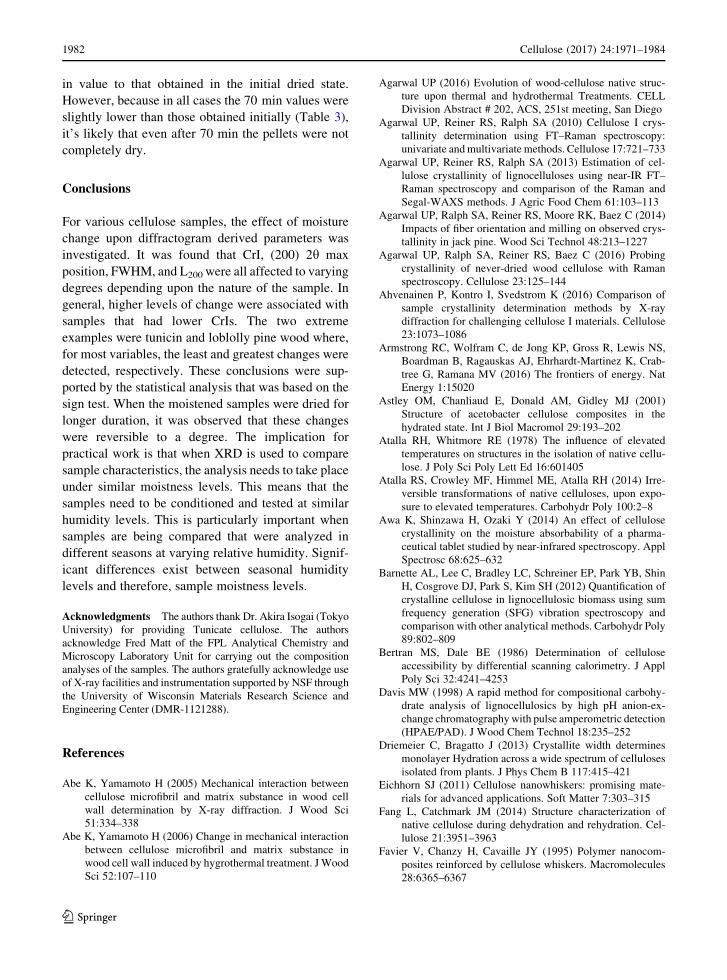
Diffractograms of pellets of the samples listed in
Table 1 were obtained in dry (P2O5 dried) and moist
states. Diffractograms of a few selected samples (T,
MCC2, HP2od, W) are shown in Fig. 1a–d and for
others in Fig SI1 (in the SI section). Except for Fig. 1d
(representing sample W), all other sub-Figs in Fig. 1
and Fig. SI1 consist of four diffractograms—two each
obtained in dry and moist states. For sample W
(Fig. 1d), although two X-ray diffractograms could be
obtained in the dry state, only one could be obtained in
the moist state after 5 lL water was added to the
surface of the pellet. The pellet integrity was lost and it
could not be turned over.
From the diffractograms, obtained in dry and moist
states, various sample parameters (CrI, 2h max,
FWHM, and L200) were determined and are listed in
Table 2. The impact of moisture on the sample
diffractograms can be noted from Figs. 1 and SI1
and on the sample parameters from Figs. 2, 3, 4, 5 and
6 where the differences between dry and moist states
are reported. In Table 2, average data with standard
deviation (SD) for CrI, 2h max position, FWHM, and
L200 are compared. For sample W in the moist state, no
SD is given because only one side of the pellet could
be sampled. Water addition to the samples causes
significantly different effects on the samples. In the
following, each of the four XRD measurements is
discussed individually.
CrI change
Segal CrIs are reported in Table 2. For most samples,
the SD values are reasonable and in the range
previously reported by Agarwal et al. (2014). In that
investigation, compared to the milled wood samples
the authors reported higher SDs for rectangular wood
blocks. It is likely that the higher SDs were caused by
the orientation differences of the cellulose fibers in the
blocks. Among the samples in Table 2 that showed
high SD, most were associated with the moistened
pellets where the diffractograms obtained from the
two sides were quite different (e.g., Fig. 1c).
Based on the data in Table 2, differences between
the moist and dry sample Segal CrIs are plotted in
Fig. 2. Of the ten samples analyzed, the most to least
CrI increase order (% value in parentheses) was as
follows—HP2od (36.7)[W (32.9)[HP1od (29.0)
[KP (25)[HP2(18.1)[CNC (10.6)[HP1 (9.1)
[MCC2 (8.9)[MCC1 (4.0)[T (0.2). Upon sam-
ple moistening (5 lL water addition), the CrI
increased by 37% for HP2od and 33% for W whereas
almost no change occurred for T and very little (4%)
for MCC1, the latter two being highly crystalline
materials (dry CrIs 97 and 94%, respectively,
Fig. 1 X-ray
diffractograms of P2O5 dried
and moistened sample
pellets a T, b MCC2,
c HP2od, d W; os means
other side of the pellet
1976 Cellulose (2017) 24:1971–1984
123

Table 2). In contrast, in the case of W which was P2O5
dried at 25 �C, cellulose molecules are expected to be
present in a state that is only slightly modified from
their existence in the native state (Agarwal et al.
2016). The authors reported that in the native state
*50% of wood cellulose is accessible by water.
Table 2 XRD parameters for P2O5 dried and moist pellets
Sample % CrI, Segal (200) Peak position, 2h (200) FWHM Crystallite size, L200
Dry Moist Dry Moist Dry Moist Dry Moist
T 97.2 ± 0.09 97.4 ± 2.4 22.9 ± 0.01 23.0 ± 0.03 0.89 ± 0.03 0.85 ± 0.09 9.1 ± 0.26 9.55 ± 0.99
MCC1 94.3 ± 0.57 98.0 ± 0.50 22.7 ± 0.03 22.8 ± 0.07 1.28 ± 0.00 1.09 ± 0.01 6.3 ± 0.01 7.46 ± 0.04
MCC2 88.4 ± 1.81 96.3 ± 1.0 22.5 ± 0.00 22.7 ± 0.08 1.87 ± 0.05 1.56 ± 0.00 4.3 ± 0.11 5.20 ± 0.01
CNC 84.4 ± 0.11 93.4 ± 3.2 22.6 ± 0.01 22.7 ± 0.17 2.25 ± 0.01 1.82 ± 0.18 3.6 ± 0.01 4.47 ± 0.32
KP 74.7 ± 0.51 93.0 ± 0.30 22.3 ± 0.01 22.6 ± 0.06 2.21 ± 0.00 1.79 ± 0.08 3.7 ± 0.01 4.54 ± 0.20
HP1 83.4 ± 4.77 91.0 ± 4.3 22.4 ± 0.09 22.6 ± 0.05 2.59 ± 0.14 2.31 ± 0.13 3.1 ± 0.17 3.52 ± 0.20
HP1od 71.5 ± 0.16 92.3 ± 0.10 22.0 ± 0.02 22.5 ± 0.03 3.18 ± 0.06 2.27 ± 0.11 2.5 ± 0.05 3.57 ± 0.18
HP2 76.2 ± 1.27 89.9 ± 1.8 22.2 ± 0.01 22.4 ± 0.19 3.05 ± 0.07 2.60 ± 0.26 2.7 ± 0.06 3.13 ± 0.32
HP2od 65.7 ± 0.17 89.8 ± 7.4 21.9 ± 0.01 22.5 ± 0.25 3.77 ± 0.01 2.74 ± 0.13 2.1 ± 0.01 2.97 ± 0.20
W 55.5 ± 1.72 73.7 ± NA 21.8 ± 0.06 22.3 ± NA 4.49 ± 0.20 3.03 ± NA 1.8 ± 0.08 2.70 ± NA
After adding 5 lL water
Fig. 2 Percentage of increase in CrI upon 5 lL water addition
Fig. 3 2h max position for (200) reflection, W in dried state
(W) and after 5 lL water addition (W_5 lL_H2O); os means
other side of the pellet. Subtracted spectra (W–W_5 lL_H2O),
and (W_os–W_5 lL_H2O) are shown as well Fig. 5 Percentage of decline in FWHM after 5 lL water
addition
Fig. 4 Percentage of increase in 2h max position for (200)
reflection
Cellulose (2017) 24:1971–1984 1977
123

Compared to the dried state, upon addition of water,
significant ordering of the cellulose chains seems to
take place. Additional evidence for this came from the
FWHM data (Table 2, more below), which showed a
significant narrowing of the (200) peak. Previously,
XRD FWHM for (200) and water accessibility of
wood cellulose have been found to be positively
correlated (Agarwal et al. 2016). In that paper, the
authors suggested that the (200) FWHM can be taken
as a measure of existing disorder in cellulose.
On the other hand, due to T’s high crystalline nature,
upon water addition, the sample underwent no such
reordering of the interior cellulose chains (CrI changes
of 0.2%, Fig. 2). Only surface chains are available for
interaction with water. But it appears that such an
interaction was not enough to bring about a meaningful
change in the CrI. This explains why the CrI for the
sample remained unchanged. It is important to note
that the contribution of water’s diffractogram to the
sample diffractograms can be ruled out based on the
fact that (a) the moistened T sample did not show any
significant change in its diffractogram (Fig. 1a, T vs.
T–5 lL–H2O) and (b) water’s diffractogram (Fig. SI2)
indicated that most contribution occurs at 2h[ 27�.The latter is supported by the literature reported water
peak at 28.5� (2h) in XRD (Fang and Catchmark 2014).
Furthermore, because sample XRDs were obtained
after water was completely absorbed by the samples,
it’s likely to be only present in a molecularly dispersed
state and not exist as a separate phase. In any case, in
the XRDs of the samples, no peak due to water at 28.5�(2h) was detected.
Moistening of the samples of kraft pulp (KP), three
of the NaOH treated holocellulose samples (HP2,
HP1od, and HP2od), and CNC showed significant CrI
increases (37–9%; Fig. 2). In contrast, microcrys-
talline celluloses from cotton (MCC1) showed only a
small increase (4%). Additionally, compared to 25� C
dried samples HP1 and HP2, the corresponding oven
dried equivalents HP1od and HP2od showed higher
CrI change (Fig. 2). The latter suggested that oven
drying induced more disorder between cellulose
chains (the CrIs of the oven dried samples were lower
compared to their 25 �C dried equivalents, Table 2)
and this thermally induced disorder seem to have been
quenched to a considerable degree upon water addi-
tion as evidenced by the resultant CrI increases for the
moist samples (Table 2).
In addition to wood (W), three pulp samples (KP,
HP1od, HP2od) showed high % CrI increases (Fig. 2).
To explain the differences in the crystallinity behav-
iors between KP, HP1od, and HP2od their composi-
tional differences were considered. The compositions
of various samples are reported in Table 1. The glucan
content for the samples KP, HP1od, and HP2od are
81.3, 73, and 69.1%, respectively. Moreover, com-
pared to HP1od, both KP and HP2od have slightly
higher amounts of hemicelluloses—10.5% in HP1od
versus 14 and 19.1% in KP and HP2od, respectively
(Table 1). However, in these pulps, the order of CrI
increases does not correlate with the amount of
hemicellulose present. Also, the CrI increase in HP2
is lower compared to that of KP whereas the former
has higher amount of hemicellulose (Table 1). Con-
sequently, in the case of HPod1 and HPod2, there must
be a contribution due to oven drying. Therefore,
hemicellulose difference is not likely to be the reason
why compared to KP, HP1od and HP2od showed
higher CrI increases. Moreover, simply having a
higher cellulose or glucan content does not seem to be
the reason either because compared to HP2, HP1 had
more cellulose but showed lower increase upon water
addition (Fig. 2). The explanation for the latter
observation may have to do with the difference in
the hemicellulose content of the two samples—HP2
had 9% more hemicellulose (Table 1). Therefore, the
only explanation seems to lie in the differences
between the ultrastructures of the celluloses between
the samples (KP, HP1, HP2, HP1od, HP2od). The
cellulose structures and hence their susceptibility to
water is likely to be different due to the fact that these
samples were produced with two different processes.
Whereas KP is a kraft pulp sample which was treated
at high temperature (170 �C) with alkali, both HP1 and
Fig. 6 Percentage of increase in L200 after 5 lL water addition
1978 Cellulose (2017) 24:1971–1984
123

HP2 underwent mild treatments (acid chlorite delig-
nification and mild NaOH treatments both at 25 �C—
Agarwal et al. 2016).
2h max position change
Upon sample moistening (5 lL water addition), in all
cases, the (200) 2h max position shifted slightly to
higher values (Table 2). For the wood sample (W), the
shift can be clearly seen in Fig. 3 where the diffrac-
tograms for dry and moist states are plotted after
background subtraction and (200) peak normalization.
Figure 3 also shows how water addition causes the
X-ray scattering to decline in 12�–22� (2h) and to
increase in 22�–37� (2h). Such changes are likely to be
associated with the alterations in the organization of
the scattering components because sample composi-
tion remains same. As shown in Fig. 4, between the
samples, the % increase in 2h max position varied. The
shift was hardly detectable for T (0.2% increase,
Fig. 4; Table 2), whereas was 12.5 times higher at
2.5% for W (Fig. 4; Table 2). In case of woods, such a
shift has been previously reported (Abe and Yama-
moto 2005, 2006; Hill et al. 2010; Nishimura et al.
1981; Sugino et al. 2007; Zabler et al. 2010). The 2hmax position was also shifted to higher values in most
of the non-wood samples (Fig. 4; Table 2). The
compositional data in Table 1 and plots in Fig. 4
suggest that % increases in 2h max position are not due
to lignin. Comparing the oven (110 �C) versus room
temperature (25 �C) dried samples (HP1od and HP2od
vs. HP1 and HP2), the (200) peak of the former
samples shifted by larger amounts (Fig. 4). This is
likely to be due to the presence of more disordered
cellulose and hemicelluloses in the oven dried mate-
rials compared to their room temperature equivalents.
Because KP’s 2h max position shift was lower
compared to that of HP1od and HP2od samples, it
suggested that compared to KP more disordered
cellulose (and hemicellulose) exists in the holopulp
samples that were heated to 110 �C (HP1od and
HP2od). In particular, in both these oven dried
holopulp samples, cellulose (and hemicellulose) has
the characteristic of becoming more ordered upon
water addition.
It is interesting that, upon sample moistening,
compared to the highly crystalline T, CNC showed
greater shift in (200) 2h max position. This is likely to
be a consequence of the fact that compared to T, CNC
has a smaller L200 (Table 2) and therefore, the latter
sample has higher surface area. This means that CNC
has more cellulose chains on the crystallite surfaces
which in turn, upon moistening, are capable of
reorienting by interacting with the molecules of added
water. The other change could be straightening (by
reorientation) of the chain-sections at the ends of the
CNCs. The net effect of the two processes could be a
reduction in inter-planer distance.
FWHM and L200 change
Except for T which showed hardly any decline in (200)
FWHM (4%, Table 2), all other moist cellulose
materials showed a decline in FWHM (Fig. 5;
Table 2). The highest reduction in FWHM was
observed for the wood sample (W, Fig. 5). The
FWHM decline in T was very small, 4% is within
experimental error (Table 2). In all cases, the narrow-
ing of the (200) peak in the diffractograms seems to be
associated with the reorganization of the cellulose
chains although in samples that contained hemicellu-
lose its role cannot be ruled out. However, the extent of
the cellulose reorganization varies between the sam-
ples and in some cases (e.g., W) it is expected to take
place in the interior of the sample due to the fact that
the interior cellulose chains are water accessible
(Agarwal et al. 2016). In the case of samples other
than W, ‘‘hypothesis of drying stresses’’ can also be
invoked because these samples are known to be
partially crystalline, although for cellulose materials,
the hypothesis of drying stresses has never been
proven. Nevertheless, even assuming no access of
water to polymers located in the interior of the
crystalline fibril, net observed changes would be a
result of two distinct contributions—(a) the drying
stresses part and (b) the water-polymer interaction
induced changes part (occurring in the non-crystalline
regions of the fibrils). Noteworthy in this regard is the
special status of sample W where the contribution
(a) does not exist. In other samples contribution
(a) (and therefore b) will vary depending upon the
level of crystallinity of the sample.
The decline in FWHM of the other samples was
directly responsible for the increased L200 (Eq. 2;
Table 2). The % increase in the L200 is plotted in
Fig. 6. The relationship between FWHM and L200 is
clear from the Scherrer equation (Eq. 2). The order in
which crystallite size increased in the moistened
Cellulose (2017) 24:1971–1984 1979
123

samples was as follows (% values in parentheses)—W
(49.5)[HP1od (40.2)[HP2od (38.2)[CNC
(24.1) = KP (24.1)[MCC2 (20)[HP2 (18) C
MCC1 (17.8)[HP1 (12.5)[T (4.9). Clearly, W
showed the most increase in the crystallite size. It is
important to note that the L200 values are based on the
observed (200) FWHMs and contributions of lignin
and hemicelluloses to the W’s diffractogram, if any,
were still present. Because, in XRD, both lignin and
hemicelluloses contribute in some of the same 2hregions as the cellulose peak (200) (Agarwal et al.
2013), theoretically, one can make the argument that
at least in the case of W the diffractogram needs to be
corrected prior to calculating the L200. However, given
that such a correction is not straightforward and
complexities arise that are not addressable in a
satisfactory way, such a need cannot be justly fulfilled.
Specifically, issues that have to do with the contribu-
tions to the broadening of the wood diffractogram
peak and the extent to which disorganized cellulose
itself is responsible for it. Moreover, unanswered
remains the question about the XRD contributions of
lignin and hemicelluloses when they exist as a separate
phase versus in molecularly distributed form (as they
do in wood). Furthermore, it has become increasingly
clear that various treatments (physical, chemical, and
thermal) of wood modify the native state structure of
cellulose to various degrees (Agarwal 2016; Agarwal
et al. 2016; Atalla et al. 2014; Langan et al. 2014;
Nishiyama et al. 2014; Toba et al. 2013). Upon
chemical treatment, the (200) peak of the treated wood
narrows, but whether this is due to removal of non-
cellulose component(s) or alterations of cellulose
structure is unknown. And, in case both these factors
play a role then to what degree is not easily discern-
able. Therefore, in the case of W and the other
samples, the authors have decided not to make a
correction prior to estimating L200. Besides, in this
investigation, the objective is to determine how
moistening impacts these XRD parameters for the
whole sample.
Sign test
Based on data in Table 2, for all four response
variables (CrI, 2h max position, FWHM, and L200),
either the dry measurement was less than the corre-
sponding moist measurement for all 10 materials, or
the dry measurement was greater than the
corresponding moist measurement for all 10 materials.
Thus, the p values for all four comparisons were 0.002.
If we want to take into account the fact that we are
making four comparisons, then a Bonferroni multiple
comparison approach (Miller 1981) would still yield
0.008 p values. Therefore, the null hypothesis that
there are no differences between the response vari-
ables of moist and dried samples is rejected at this
level.
Correlations of the changes with sample CrIs
The % changes in each of the four parameters (CrI, 2hmax position, FWHM, and L200) were correlated with
dry sample CrIs to find out if these variables depended
upon Segal crystallinities of the samples (Fig. 7). All
the correlations were positive (Fig. 7, R2 between 0.87
and 0.76) which meant that, upon moistening, the %
increases in CrI (Fig. 7a), 2h max position (Fig. 7b),
and L200 (Fig. 7d) declined with increase in the sample
CrIs. In contrast, the % reduction in FWHM declined
with increase in sample CrI (Fig. 7c). The factor that
fluctuated in tandem most with the sample CrIs was %
increase in CrI (Fig. 7a, R2 = 0.87).
Longer duration drying post moistening
To assess the effect of longer duration drying post
moistening, pellets of selected samples (Table 3) that
were initially dried under ambient conditions were
used. As before, diffractograms were obtained from
dried and moist (5 lL H2O) pellets, but only from one
side. The pellets were not turned over. Additionally,
pellet diffractograms were obtained after 15 and
70 min of the pellet moistening step. From the
diffractograms, CrI and FWHM data were obtained
(Table 3). In Fig. 8, the effect of longer duration
drying on the CrIs is shown and it can be noted that in
cases where the CrI had increased upon moistening, it
declined upon drying of the samples. Based on the
comparison of the CrIs that were obtained initially and
after 70 min of pellet drying (Table 3; Fig. 8), for all
samples, the two values were similar.
FWHM data (Table 3) are plotted in Fig. 9 wherein
the effect of longer duration drying can be observed.
As expected (see above), upon water addition, initially
the FWHM declined in most cases but the peak width
increased upon drying (Fig. 9; Table 3). And at
70 min point, for each sample, the FWHM was closer
1980 Cellulose (2017) 24:1971–1984
123

Table 3 CrI and FWHM changes upon 15 and 70 min drying post moistening of 25 �C dried (ambient) pellets
Sample % CrI, Segal (200) FWHM
Dry Moista 15 min later 70 min later Dry Moist 15 min later 70 min later
T 96.4 100.1 98.0 97.2 0.95 0.80 0.94 0.88
MCC2 87.1 98.0 89.8 88.8 1.99 1.56 1.86 1.93
CNC 82.9 94.8 86.7 84.8 2.43 1.88 2.30 2.33
HP1 76.5 94.0 88.2 81.1 2.95 2.25 2.53 2.68
HP2 74.4 90.2 82.9 79.3 3.13 2.55 2.80 2.85
W 62.9 77.8 73.6 67.0 4.00 3.20 3.20 3.60
a After adding 5 lL water
Fig. 8 Segal CrIs in dried (ambient) state, upon 5 lL water
addition, and after 15 and 70 min dryingFig. 9 (200) FWHM in dried state, upon 5 lL water addition,
and after 15 and 70 min drying
Fig. 7 Correlations of
various XRD parameters
with sample dry state Segal
CrI; a % increase in CrI, b %
increase in 2h max position,
c % decline in (200)
FWHM, d % increase in
L200
Cellulose (2017) 24:1971–1984 1981
123
in value to that obtained in the initial dried state.
However, because in all cases the 70 min values were
slightly lower than those obtained initially (Table 3),
it’s likely that even after 70 min the pellets were not
completely dry.
Conclusions
For various cellulose samples, the effect of moisture
change upon diffractogram derived parameters was
investigated. It was found that CrI, (200) 2h max
position, FWHM, and L200 were all affected to varying
degrees depending upon the nature of the sample. In
general, higher levels of change were associated with
samples that had lower CrIs. The two extreme
examples were tunicin and loblolly pine wood where,
for most variables, the least and greatest changes were
detected, respectively. These conclusions were sup-
ported by the statistical analysis that was based on the
sign test. When the moistened samples were dried for
longer duration, it was observed that these changes
were reversible to a degree. The implication for
practical work is that when XRD is used to compare
sample characteristics, the analysis needs to take place
under similar moistness levels. This means that the
samples need to be conditioned and tested at similar
humidity levels. This is particularly important when
samples are being compared that were analyzed in
different seasons at varying relative humidity. Signif-
icant differences exist between seasonal humidity
levels and therefore, sample moistness levels.
Acknowledgments The authors thank Dr. Akira Isogai (Tokyo
University) for providing Tunicate cellulose. The authors
acknowledge Fred Matt of the FPL Analytical Chemistry and
Microscopy Laboratory Unit for carrying out the composition
analyses of the samples. The authors gratefully acknowledge use
of X-ray facilities and instrumentation supported by NSF through
the University of Wisconsin Materials Research Science and
Engineering Center (DMR-1121288).
References
Abe K, Yamamoto H (2005) Mechanical interaction between
cellulose microfibril and matrix substance in wood cell
wall determination by X-ray diffraction. J Wood Sci
51:334–338
Abe K, Yamamoto H (2006) Change in mechanical interaction
between cellulose microfibril and matrix substance in
wood cell wall induced by hygrothermal treatment. J Wood
Sci 52:107–110
Agarwal UP (2016) Evolution of wood-cellulose native struc-
ture upon thermal and hydrothermal Treatments. CELL
Division Abstract # 202, ACS, 251st meeting, San Diego
Agarwal UP, Reiner RS, Ralph SA (2010) Cellulose I crys-
tallinity determination using FT–Raman spectroscopy:
univariate and multivariate methods. Cellulose 17:721–733
Agarwal UP, Reiner RS, Ralph SA (2013) Estimation of cel-
lulose crystallinity of lignocelluloses using near-IR FT–
Raman spectroscopy and comparison of the Raman and
Segal-WAXS methods. J Agric Food Chem 61:103–113
Agarwal UP, Ralph SA, Reiner RS, Moore RK, Baez C (2014)
Impacts of fiber orientation and milling on observed crys-
tallinity in jack pine. Wood Sci Technol 48:213–1227
Agarwal UP, Ralph SA, Reiner RS, Baez C (2016) Probing
crystallinity of never-dried wood cellulose with Raman
spectroscopy. Cellulose 23:125–144
Ahvenainen P, Kontro I, Svedstrom K (2016) Comparison of
sample crystallinity determination methods by X-ray
diffraction for challenging cellulose I materials. Cellulose
23:1073–1086
Armstrong RC, Wolfram C, de Jong KP, Gross R, Lewis NS,
Boardman B, Ragauskas AJ, Ehrhardt-Martinez K, Crab-
tree G, Ramana MV (2016) The frontiers of energy. Nat
Energy 1:15020
Astley OM, Chanliaud E, Donald AM, Gidley MJ (2001)
Structure of acetobacter cellulose composites in the
hydrated state. Int J Biol Macromol 29:193–202
Atalla RH, Whitmore RE (1978) The influence of elevated
temperatures on structures in the isolation of native cellu-
lose. J Poly Sci Poly Lett Ed 16:601405
Atalla RS, Crowley MF, Himmel ME, Atalla RH (2014) Irre-
versible transformations of native celluloses, upon expo-
sure to elevated temperatures. Carbohydr Poly 100:2–8
Awa K, Shinzawa H, Ozaki Y (2014) An effect of cellulose
crystallinity on the moisture absorbability of a pharma-
ceutical tablet studied by near-infrared spectroscopy. Appl
Spectrosc 68:625–632
Barnette AL, Lee C, Bradley LC, Schreiner EP, Park YB, Shin
H, Cosgrove DJ, Park S, Kim SH (2012) Quantification of
crystalline cellulose in lignocellulosic biomass using sum
frequency generation (SFG) vibration spectroscopy and
comparison with other analytical methods. Carbohydr Poly
89:802–809
Bertran MS, Dale BE (1986) Determination of cellulose
accessibility by differential scanning calorimetry. J Appl
Poly Sci 32:4241–4253
Davis MW (1998) A rapid method for compositional carbohy-
drate analysis of lignocellulosics by high pH anion-ex-
change chromatography with pulse amperometric detection
(HPAE/PAD). J Wood Chem Technol 18:235–252
Driemeier C, Bragatto J (2013) Crystallite width determines
monolayer Hydration across a wide spectrum of celluloses
isolated from plants. J Phys Chem B 117:415–421
Eichhorn SJ (2011) Cellulose nanowhiskers: promising mate-
rials for advanced applications. Soft Matter 7:303–315
Fang L, Catchmark JM (2014) Structure characterization of
native cellulose during dehydration and rehydration. Cel-
lulose 21:3951–3963
Favier V, Chanzy H, Cavaille JY (1995) Polymer nanocom-
posites reinforced by cellulose whiskers. Macromolecules
28:6365–6367
1982 Cellulose (2017) 24:1971–1984
123
Fink HP, Purz HJ, Bohn A, Kunze J (1997) Investigation of the
supramolecular structure of never dried bacterial cellulose.
J Macromol Symp 120:207–217
French AD, Santiago Cintron M (2013) Cellulose polymorphy,
crystallite size, and the Segal crystallinity index. Cellulose
20:583–588
Hill SJ, Kirby NM, Mudie ST, Hawley AM, Ingham B, Franich
RA, Newman RH (2010) Effect of drying and rewetting of
wood on cellulose molecular packing. Holzforschung
64:421–427
Hollander M, Wolfe DA, Chicken E (2014) Nonparametric
statistical methods, 3rd edn. Wiley, Hoboken
Hu X-P, Hsieh Y-L (2001) Effects of dehydration on the crys-
talline structure and strength of developing cotton fibers.
Text Res J 71:231–239
Hulleman SHD, Van Hazendonk JM, Van Dam JEG (1994)
Determination of crystallinity in native cellulose from
higher plants with diffuse reflectance Fourier transform
infrared spectroscopy. Carbohydr Res 261:163–172
Isogai A (2013) Wood nanocelluloses: fundamentals and applica-
tions as new bio-based nanomaterials. J Wood Sci 59:449–459
Klemm D, Kramer F, Moritz S, Lindstrom T, Ankerfors M, Gray
D, Dorris A (2011) Nanocelluloses: a new family of nature-
based materials. Angew Chem Int Ed 50:5438–5466
Langan P, Petridis L, O’Neill HM, Pingali SV, Foston M,
Nishiyama Y, Schulz R, Lindner B, Hanson BL, Harton S,
Heller WT, Urban W, Evans B, Gnanakaran S, Ragauskas
AJ, Smith JC, Davison BH (2014) Common processes
drive the thermochemical pretreatment of lignocellulosic
biomass. Green Chem 16:63–67
Langford J, Wilson A (1978) Scherrer after sixty years: a survey
and some new results in the determination of crystallite
size. J Appl Crystallogr 11:102–113
Larsson PT, Hult E-L, Wickholm K, Pettersson E, Iversen T
(1999) 13C-NMR spectroscopy applied to structure and
interaction studies on cellulose I. Solid State Nucl Magn
Reson 15:31–40
Lee JM, Pawlak JJ, Heitmann JA (2012) Dimensional and
hygroexpansive behaviors of cellulose microfibrils (MFs)
from kraft pulp-based fibers as a function of relative
humidity. Holzforschung 66:1001–1008
Lindner B, Petridis L, Langan P, Smith JC (2014) Determination
of cellulose crystallinity from powder diffraction diagrams.
Biopoly 103:67–73
Miller RG Jr (1981) Simultaneous statistical inference.
Springer, New York
Moon RJ, Martini A, Nairn J, Simonsen J, Youngblood J (2011)
Cellulose nanomaterials review: structure, properties and
nanocomposites. Chem Soc Rev 40:3941–3994
Nelson ML, O’Connor RT (1964) Relation of certain infrared
bands to cellulose crystallinity and crystal lattice type. Part
I. Spectra of lattice types I, II, III and of amorphous cel-
lulose. J Appl Poly Sci 8:1311–1324
Newman RH (1999) Estimation of the lateral dimensions of
cellulose crystallites using 13C NMR signal strengths. Solid
State Nucl Magn Reson 15:21–29
Nishimura H, Okano T, Asano I (1981) Fine structure of wood
cell walls. I. Structural features of noncrystalline sub-
stances in wood cell walls. Mokuzai Gakkaishi 27:611–617
Nishiyama Y, Langan P, Chanzy H (2002) Crystal structure and
hydrogen-bonding system in cellulose Ia from synchrotron
X-ray and neutron fiber diffraction. J Am Chem Soc
124:9074–9082
Nishiyama Y, Langan P, O’Neill H, Pingali SV, Harton S (2014)
Structural coarsening of aspen wood by hydrothermal
pretreatment monitored by small- and wide-angle scatter-
ing of X-ray and neutrons on oriented specimens. Cellulose
21:1015–1024
Oh SY, Yoo DI, Shin Y, Kim HC, Kim HY, Chung YS, Park
WH, Youk JH (2005) Crystalline structure analysis of
cellulose treated with sodium hydroxide and carbon diox-
ide by means of X-ray diffraction and FT-IR spectroscopy.
Carbohydr Res 340:2376–2391
Park S, Baker JO, Himmel ME, Parilla PA, Johnson DK (2010)
Cellulose crystallinity index: measurement techniques and
their impact on interpreting cellulase performance.
Biotechnol Biofuels 3:10
Peciulyte A, Karlstrom K, Larsson PT, Olsson E (2015) Impact
of the supramolecular structure of cellulose on the effi-
ciency of enzymatic hydrolysis. Biotechnol Biofuels 8:56
Reiner RS, Rudie AW (2013) Process scale-up of cellulose
nanocrystal production to 25 kg per batch at the Forest
Products Laboratory. In: Postek MT, Moon RJ, Rudie AJ,
Bilodeau MA (eds) Production and applications of cellu-
lose materials. TAPPI Press, Atlanta, pp 21–24
Richter U, Krause T, Schempp W (1991) Untersuchungen zur
Alkalibehandlung von Cellulosefasern. Teil 1. Infrarot-
spektroskopische und Rontgenographische Beurteilung der
A nderung des Ordnungszustandes. Angew Makromol
Chem 185:155–167
R Core Team (2013) R: A language and environment for sta-
tistical computing. R Foundation for Statistical Comput-
ing, Vienna, Austria. http://www.R-project.org
Schenzel K, Fischer S, Brendler E (2005) New method for
determining the degree of cellulose I crystallinity by means
of FT Raman spectroscopy. Cellulose 12:223–231
Scherrer P (1918) Bestimmung der Grosse und der inneren
Struktur von Kolloidteilchen mittels Rontgenstrahlen.
Nachrichten von der Gesellschaft der Wissenschaften,
Gottingen, pp 98–100
Segal L, Creely JJ, Martin AE, Conrad CM (1959) An empirical
method for estimating the degree of crystallinity of native
cellulose using the X-ray diffractometer. Text Res J
29:786–794
Sisson WA (1933) X-ray analysis of fibers, part I, literature
survey. Text Res J 3:295–307
Sugino H, Sugimoto H, Miki T, Kanayama K (2007) Fine
structure changes of wood during moisture adsorption and
desorption process analyzed by X-ray diffraction mea-
surement. Mokuzai Gakkaishi 53:82–89
TAPPI test method (1983) Acid insoluble lignin in wood and
pulp; official test method T-222 (Om). TAPPI, Atlanta
Toba K, Yamamoto H, Yoshida M (2013) Crystallization of
cellulose microfibrils in wood cell wall by repeated dry-
and-wet treatments, using X-ray diffraction technique.
Cellulose 20:633–643
Tokoh C, Takabe K, Fujita M, Saiki H (1998) Cellulose syn-
thesized by Acetobacter xylinum in the presence of acetyl
glucomannan. Cellulose 5:249–261
Vieira FS, Pasquini C (2014) Determination of cellulose crys-
tallinity by terahertz-time domain spectroscopy. Anal
Chem 86:3780–3786
Cellulose (2017) 24:1971–1984 1983
123
Wormald P, Wickholm K, Larsson PT, Iversen T (1996) Con-
versions between ordered and disordered cellulose. Effects
of mechanical treatment followed by cyclic wetting and
drying. Cellulose 3:141–152
Zabler S, Paris O, Burgert I, Fratzl P (2010) Moisture changes in
the plant cell wall force cellulose crystallites to deform.
J Struct Biol 171:133–141
Zhu H, Luo W, Ciesielski PN, Fang Z, Zhu JY, Henriksson G,
Himmel ME, Hu L (2016) Wood-derived materials for
green electronics, biological devices, and energy applica-
tions. Chem Rev 116:9305–9374
1984 Cellulose (2017) 24:1971–1984
123


