
Pierre Karam
EFFECT OF PLATFORM SWITCHING ON MARGINAL BONE RESORPTION
AROUND THE IMPLANT
UNIVERSIDADE FERNANDO PESSOA
Faculdade de Ciências da Saúde
Porto, 2017

Pierre Karam
EFFECT OF PLATFORM SWITCHING ON MARGINAL BONE RESORPTION
AROUND THE IMPLANT
UNIVERSIDADE FERNANDO PESSOA
Faculdade de Ciências da Saúde
Porto, 2017


Pierre Karam
EFFECT OF PLATFORM SWITCHING ON MARGINAL BONE RESORPTION
AROUND THE IMPLANT
Trabalho apresentado à
Universidade Fernando
Pessoa como parte dos
requisitos para a obtenção
do grau de Licenciatura em
Medicina Dentária.
_______________________
Pierre Karam
vi
Resumo
0 conceito de plaform switching, em português, plataforma alterada, referenciado na
literatura, no âmbito da implantologia, que parece permitir uma melhoria na preservação
do osso peri-implantar. Dos estudos sobre esta temática surgem varias hipóteses e
explicações para justificar as melhorias clinicas obtidas com a sua utilização.
Este trabalho procura rever e detalhar, tendo por base uma revisão da literatura
cientifica, os fundamentos biológicos que suportam as vantagens clinicas obtidas com a
utilização da técnica de plataforma alterada.
Uma pesquisa na base de dados "PubMed", foi efetuada considerando os artigos dos
últimos 10 anos. A bibliografia obtida inicialmente foi selecionada pela leitura dos
resumos e posteriormente pela leitura integral das publicações.
O mecanismo pelo qual a plataforma alterada apresenta melhorias clinicas quando
comparado com as técnicas convencionais poderá estar relacionado com distintas
ocorrências, nomeadamente devido a uma modelação biomecânica das componentes
implantares, alteração da localização do microgap, modulação do infiltrado inflamatório
peri-implantar e formação de um espaço biológico horizontal
Palavras chave: plataforma alterada; perda óssea peri-implantar; perda óssea alveolar
coronal; conexão implante-pilar; revisão bibliográfica.

vii
Abstrat
The concept of plaform switching, in Portuguese, changed platform, referenced in the
literature, in the scope of implantology, which seems to allow an improvement in the
preservation of the peri-implant bone. From the studies on this theme several
hypotheses and explanations appear to justify the clinical improvements obtained with
its use.
This work seeks to review and detail, based on a review of the scientific literature, the
biological foundations that support the clinical advantages obtained with the use of the
altered platform technique.
A search in the "PubMed" database was carried out considering articles from the last 10
years. The bibliography obtained initially was selected by the reading of the abstracts
and later by the full reading of the publications.
The mechanism by which the altered platform presents clinical improvements when
compared to conventional techniques may be related to different occurrences, namely
due to a biomechanical modeling of the implant components, alteration of microgap
location, modulation of the peri-implant inflammatory infiltrate and formation of a
Horizontal biological space.
Keywords: altered platform; Peri-implant bone loss; Coronal alveolar bone loss;
Implant-abutment connection; literature review.

viii
TABLE OF CONTENTS
1. INTRODUCTION ....................................................................................................... 1
2. MARGINAL BONE RESORPTION AROUND IMPLANTS ................................... 2
2.1. Generalities .......................................................................................................... 2
2.2. Marginal Bone Loss around Implants ............................................................... 2
2.3. Possible Causes of Early Bone Loss ..................................................................... 4
2.3.1. Biologic Width .............................................................................................. 4
2.3.2. Periodontal Biotype ....................................................................................... 8
2.3.3. Microgap......................................................................................................... 9
2.3.4. Plaque-induced Peri-implantitis .................................................................. 11
2.3.5. Excess Cement ............................................................................................. 12
2.3.6. Occlusal Overload ....................................................................................... 13
2.3.7. Other Factors ............................................................................................... 14
2.3.7.1. Implant Surface Roughness ................................................................. 15
2.3.7.2. Proximity between Implants ................................................................ 16
2.3.7.3. Surgical Trauma ................................................................................... 16
2.3.7.4. Smoking Habit ...................................................................................... 17
2.3.7.5. Diabetic Patients .................................................................................. 17
2.4. Consequences of Marginal Bone Loss around Implants ................................. 17
2.5. Proposed Solutions ............................................................................................. 18
3. PLATFORM SWITCHING AS A SOLUTION TO EARLY CRESTAL BONE
LOSS AROUND IMPLANTS ...................................................................................... 19
3.1. Discovery ............................................................................................................ 19
3.2. Concept and Rationales ...................................................................................... 20
3.2.1. Biologic Rationales ...................................................................................... 21
3.2.2. Biomechanical Rationales ........................................................................... 22
3.2.3. Proof of Concept .......................................................................................... 24
3.3. Design Specifications and Variations ................................................................ 26
3.3.1. Extent of Platform Mismatch ..................................................................... 27
3.3.2. Apical-Coronal Location of the Microgap .................................................. 28
3.3.3. Platform Design .......................................................................................... 30
3.4. Platform Switching in Relation to Other Concepts ............................................ 30
3.4.1. Marginal Bone Loss with Respect to Time ................................................ 31

ix
3.4.2. Proximity to Natural Teeth .......................................................................... 31
3.4.3. Proximity to Other Platform-Switched Implants ........................................ 32
3.4.4. Disconnection and Reconnection of Platform-switched Abutments …….. 33
3.5. Advantages and Disadvantages of Platform Switching ...................................... 35
3.5.1. Advantages .................................................................................................. 35
3.5.2. Disadvantages .............................................................................................. 37
3.5.3. Indications .................................................................................................. 37
4. CONCLUSION ......................................................................................................... 38
5. REFERENCES .......................................................................................................... 40
Effect of Platform Switching on Marginal Bone Resorption Around the Implant
1
1. INTRODUCTION
It is not unreasonable to view technological progress as a self-perpetuating process. The
more useful a technology is, the more rapidly and actively are its limits challenged by
its users. In turn, user demand then drives the necessity for refinements and
improvements in the technology. This is just as true in implant dentistry as in any other
field or discipline.
The practicality and success rate of endosseous oral implants have seen it rapidly
become a treatment of choice in many clinical situations. This surge in popularity has
galvanized a constant evolution, demanding the use of implants in more challenging
ways than were previously ever thought possible. This radical change has been enabled
by a more profound understanding of individual case treatment planning and
improvements in surgical procedures, but also through the advancement of the design of
the implants themselves.
The platform-switched implant design is one of the latest fruits borne from this constant
thirst for progress, and certainly one of its most promising. The concept was introduced
by Gardner (2005), Lazzara and Porter (2006) and Vela-Nebot et al. (2006) when
minimal vertical bone loss was noticed radiographically around implants with
mismatched abutments. Great attention has been given to the concept since then by
practitioners and manufacturers alike, and today, with a wealth of scientifically-backed
theories and clinical experiments at our disposal, we finally find ourselves close to fully
understanding its effects and implications.
It is with that in mind that this paper tackles the following questions: What is platform
switching, and what is the scientific rationale behind it? What is its relationship with
marginal bone loss, and does it truly succeed in reducing it? If so, what are the full
implications of its use in clinical situations, and what new opportunities does it offer to
the discipline of implant treatment?

Effect of Platform Switching on Marginal Bone Resorption Around the Implant
2
2. MARGINAL BONE RESORPTION AROUND IMPLANTS
2.1. Generalities
The skeleton is a metabolically active organ that undergoes continuous remodeling
throughout life. Bone remodeling involves the removal of mineralized bone
by osteoclasts followed by the formation of bone matrix through the osteoblasts that
subsequently become mineralized. The remodeling cycle consists of three consecutive
phases: resorption, during which osteoclasts digest old bone; reversal, when
mononuclear cells appear on the bone surface; and formation, when osteoblasts lay
down new bone until the resorbed bone is completely replaced. Bone remodeling serves
to adjust bone architecture to meet changing mechanical needs and helps to repair
micro-damages in bone matrix preventing the accumulation of old bone (Hadjidakis &
Androulakis 2006).
It enables the substitution of the primary bone (woven bone), which has low load-
bearing capacity, with lamellar bone that is more resistant to load (Lindhe & editors
2008).
Bone remodeling is especially significant in oral implantology, where the success and
longevity of dental implants is highly dependent on the relationship between the implant
components and the surrounding oral tissues (Oh etal 2002) Indeed, the level of peri
implant marginal bone, being a sensitive parameter, is widely considered one of the
most important reference criteria to monitor peri-implant health and evaluate the long-
term success of dental implants.
2.2. Marginal Bone Loss around Implants
Crestal bone loss around oral implants has been reported in most clinical follow-up
studies. In the majority of cases, marginal bone loss during the first year in function is
larger than the annual bone loss during the following years (Laurel/ & Lundgren 2011).
Effect of Platform Switching on Marginal Bone Resorption Around the Implant
3
This bone loss has been regarded as part of a bone remodeling phase (Albrektsson
eta,2014).
It has been suggested that a successful implant might lose an average of 1.5 mm of bone
on both axes during the first year in function and less than 0.2 mm annually in
subsequent years (Albrektsson etal 1986, Astrand etal 2004, Cardaropoli etal 2006).
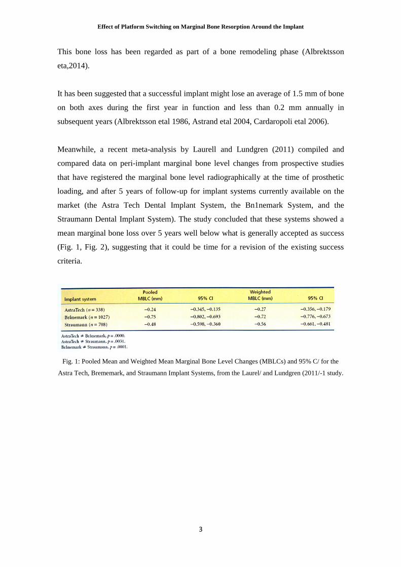
Meanwhile, a recent meta-analysis by Laurell and Lundgren (2011) compiled and
compared data on peri-implant marginal bone level changes from prospective studies
that have registered the marginal bone level radiographically at the time of prosthetic
loading, and after 5 years of follow-up for implant systems currently available on the
market (the Astra Tech Dental Implant System, the Bn1nemark System, and the
Straumann Dental Implant System). The study concluded that these systems showed a
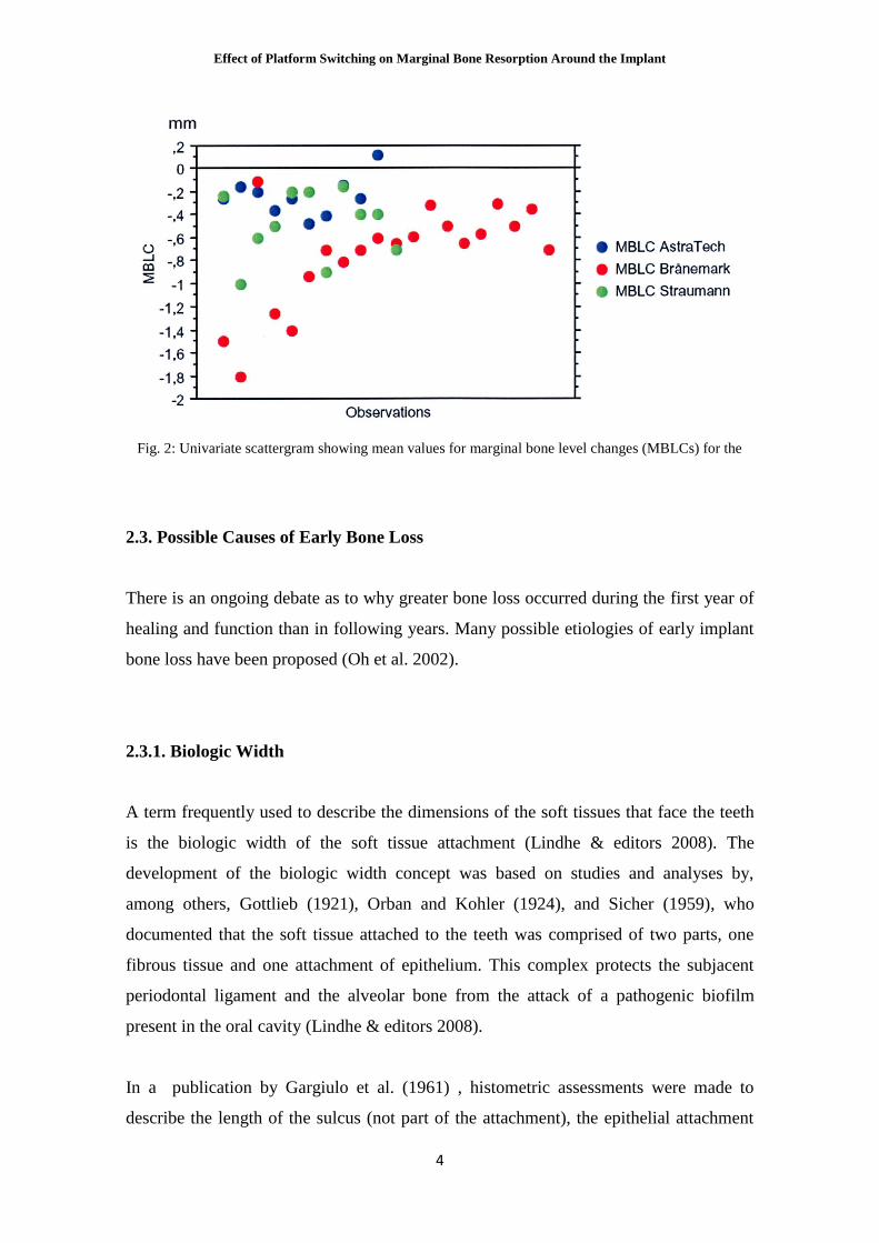
mean marginal bone loss over 5 years well below what is generally accepted as success
(Fig. 1, Fig. 2), suggesting that it could be time for a revision of the existing success
criteria.
Fig. 1: Pooled Mean and Weighted Mean Marginal Bone Level Changes (MBLCs) and 95% C/ for the
Astra Tech, Brememark, and Straumann Implant Systems, from the Laurel/ and Lundgren (2011/-1 study.
Effect of Platform Switching on Marginal Bone Resorption Around the Implant
4
Fig. 2: Univariate scattergram showing mean values for marginal bone level changes (MBLCs) for the
2.3. Possible Causes of Early Bone Loss
There is an ongoing debate as to why greater bone loss occurred during the first year of
healing and function than in following years. Many possible etiologies of early implant
bone loss have been proposed (Oh et al. 2002).
2.3.1. Biologic Width
A term frequently used to describe the dimensions of the soft tissues that face the teeth
is the biologic width of the soft tissue attachment (Lindhe & editors 2008). The
development of the biologic width concept was based on studies and analyses by,
among others, Gottlieb (1921), Orban and Kohler (1924), and Sicher (1959), who
documented that the soft tissue attached to the teeth was comprised of two parts, one
fibrous tissue and one attachment of epithelium. This complex protects the subjacent
periodontal ligament and the alveolar bone from the attack of a pathogenic biofilm
present in the oral cavity (Lindhe & editors 2008).
In a publication by Gargiulo et al. (1961) , histometric assessments were made to
describe the length of the sulcus (not part of the attachment), the epithelial attachment

Effect of Platform Switching on Marginal Bone Resorption Around the Implant
5
(today called junctional epithelium) and the connective tissue attachment (Fig. 3). It was
reported that the average value of sulcus depth was 0.69 mm, and the average values for
the epithelial attachment and connective tissue attachment were 0.97 mm and 1.07 mm,
respectively. The biologic width included the latter two, the epithelial attachment and
connective tissue attachment, which was 2.04 mm. Mean values of the biologic
widthobtained from two recent meta-analyses (Kosyfaki et al. 2010, Schmidt et al.
2013)
Fig. 3: Drawing describing the "biologic width" of the soft tissue attachment at the buccal surface of a
tooth with healthy periodontium. The combined length of the junctional epithelium (epithelial attachment)
and the connective tissue attachment is considered to represent the "biologic width" of the soft tissue
attachment (Lindhe & editors 2008)
Likewise, around dental implants, the epithelial attachment and connective tissue
attachment exist, comprising the biologic seal around dental implants that acts as a
barrier against bacterial invasion and food debris ingress into the implant-tissue
interface. (McKinney et al 1984, Cochran et al 2013)38 85 Cochran et al. (1997)37
documented the soft tissue dimensions and described the biologic width around non-
submerged, one-piece dental implants. This study supported previous reports on soft
tissues around implants (Berglundh et al 1991, Abrahamsson et al1996), and showed
that an area of epithelial attachment with the implant surface occurs similar in

Effect of Platform Switching on Marginal Bone Resorption Around the Implant
6
morphology to that which is found around natural teeth. In addition, an area of
connective tissue contact was found between the apical extension of the junctional
epithelium and the alveolar bone comprising the first bone-to-implant contact. The
dimensions of this biologic width for non-submerged, one-piece implants were
demonstrated to be similar to the dimensions for the same tissues described for natural
teeth. In addition, Hermann et al. (2000) histometrically evaluated the dimensional
change of the biologic width around non-submerged implants and observed that each
dimension of the sulcus depth, epithelial attachment, and connective tissue attachment
changed over time, but within the overall biologic width dimension. A histologic study
by Cochran et al. (2013) presents a more recent confirmation of these notions.
A notable difference between the biologic seal observed around natural teeth and that
which exists around implants is the orientation of the collagen fibers surrounding the
tooth or implant. Around a natural tooth, the collagen fibers of the periodontal ligament
are radially oriented to the dental surface in the cervical area, a direction that maximizes
resistance to tensile forces (Lindhe & editors 2008). In contrast, longitudinal and
circumferential fibers, the axes of which are parallel or oblique to the implant surface,
have been observed around the titanium neck in dental implants (Lindhe & editors
2008).

Effect of Platform Switching on Marginal Bone Resorption Around the Implant
7
Fig. 4: Microphotograph of a tooth with marginal periodontal tissues (left) and of the peri-implant mucosa
and bone at the tissue/titanium interface (right). Note that the fibers are orientated more or less
perpendicular to the root surface in natural teeth, while their orientation is more or less perpendicular to
the implant surface (Lindhe & editors 2008)
The potential of the biologic width to influence bone remodeling was made apparent in
an animal study conducted by Berglundh and Lindhe (1996) I, when the dimension of
peri-implant mucosa was studied in a beagle dog model. At sites where the ridge
mucosa prior to abutment connection was made thin (less than or equal to 2 mm),
wound healing consistently included bone resorption and the establishment of an
angular bone defect (Fig. 5). This implied that a certain minimum width (3 mm) of the
peri-implant mucosa (biologic seal) may have been required, and that bone resorption
may have taken place to allow a stable soft tissue attachment to form. This notion was
further verified in subsequent studies (Pontes et al. 2008, Canullo et al. 2011) , and has
arguably been one of the most credible bases for the majority of current theories
surrounding marginal bone loss around implants.

Effect of Platform Switching on Marginal Bone Resorption Around the Implant
8
Fig. 5: Microphotograph of one test (T) and one control (C) site. Note in the test side, the presence of an
angular bone defect. PM: the marginal portion of the peri-implant mucosa; aJE: the level of the apical
termination of the junctional epithelium; AIF: the abutment/fixture junction, BC: the bone crest, i,e, the
most coronal portion of the peri-implant bone; B: the marginal/eve/ of bone to implant contact (Berglundh
& Lindhe 1996)
2.3.2. Periodontal Biotype
The term periodontal biotype was first described by Seibert and Lindhe (1989) and then
more recently by De Rouck et al. (De Rouck et al. 2009), with two main biotypes being
identified: a thick-flat biotype and a thin-scalloped biotype. The importance of the
biotype is recognized especially in relation to the esthetic appearance (Vervaeke et al.
2014). Subjects with a thin-scalloped biotype are more prone to gingival recessions,
whereas thick-flat biotypes seem more resistant to trauma and hence protected against
gingival recessions (Olsson et al. 1993).
Marginal bone loss around implants seems to be related to periodontal biotypes as well
(Berglundh et al. 2007, Linkevicius et al. 2009, Vervaeke et al. 2014) Linkevicius et al.
(2009) evaluated the influence of gingival tissue thickness on crestal bone loss around

Effect of Platform Switching on Marginal Bone Resorption Around the Implant
9
dental implants, and concluded that initial gingival tissue thickness at the crest may be
considered as a significant influence on peri-implant marginal bone stability. If the
tissue thickness was 2.0 mm or less, crestal bone loss up to 1.45 mm could occur,
despite a supracrestal position of the implant-abutment interface. These results were
consistent with those of a previous animal study which showed the potential for thin
tissues to cause crestal bone loss during the process of biologic width formation
(Berglundh et al. 2007) A recent study by Vervaeke et al. (2014) further confirms these
findings.
Fig. 6: Thick biotype (top) with periodontal probe not visible through the gingival sulcus. Thin
biotype(bottom) with periodontal probe visible through the gingival sulcus (Arora et al. 2013)
2.3.3. Microgap
In implant dentistry, there are two basic approaches to placing endosseous implants:
submerged (2-stage) and non-submerged (1-stage) implants. In most 2-stage implant
systems, after the abutment is connected, a microgap exists between the implant and
abutment at or below the alveolar crest. In nonsubmerged implant designs, the implant
itself extends above the alveolar crest level; therefore, such a microgap does not exist at
the level of the bone (Oh et al. 2002) The implant/abutment microgap in 2-stage
implants has been suggested as a contributor of marginal bone loss (Ericsson et al.
1995, Hermann et al. 2000 & 2001, Cochran et al. 2009, Koutouzis et al. 2014).
Effect of Platform Switching on Marginal Bone Resorption Around the Implant
10
Some studies have shown that bone resorption around the implant neck does not begin
until the implant is uncovered and exposed to the oral cavity, which invariably leads to
bacterial contamination of the gap between implant and supra-structure. Quirynen and
van Steenberghe (1994) found microbial species cultivated from internal surfaces of
submerged implants or their restorative component parts. The study implied that a
microbial leakage from the abutment-fixture microgap in submerged implants is the
most probable origin for this contamination.
Several in vitro studies have since described the occurrence of bacterial leakage along
the implant-abutment interface of systems with different internal connection designs in
static or dynamic loading conditions (Steinebrunner et al 2005, Tesmer et al 2009,
Aloise et al 2010, Koutouzis etal 2011).Moreover, microleakage has been confirmed to
occur in both directions, from the inner parts of the implants to the external environment
and vice versa (do Nascimento etal 2012), and the degree of leakage is dependent on
the type of implant-abutment connection and loading (Koutouzis et al 2014, Canullo et
al 2015) and the amount of micromovement (Steinebrunner etal 2005).
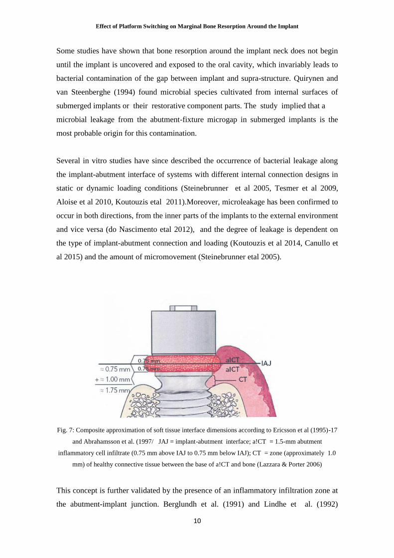
Fig. 7: Composite approximation of soft tissue interface dimensions according to Ericsson et al (1995)-17
and Abrahamsson et al. (1997/ JAJ = implant-abutment interface; a!CT = 1.5-mm abutment
inflammatory cell infiltrate (0.75 mm above IAJ to 0.75 mm below IAJ); CT = zone (approximately 1.0
mm) of healthy connective tissue between the base of a!CT and bone (Lazzara & Porter 2006)
This concept is further validated by the presence of an inflammatory infiltration zone at
the abutment-implant junction. Berglundh et al. (1991) and Lindhe et al. (1992)

Effect of Platform Switching on Marginal Bone Resorption Around the Implant
11
evaluated the microgap of the Branemark 2-stage implant and found that inflamed
connective tissue existed 0.5 mm above and below the abutment-implant connection
which resulted in 0.5 mm bone loss within 2 weeks after the abutment was connected to
the implant. Additionally, Berglundh and Lindhe (1996) and Ericsson et al. (1995)
observed in histologic sections of crestal bone and soft tissue that crestal bone is always
separated from the base of the abutment inflammatory zone by an approximate 1 mm
wide zone of healthy connective tissue, as depicted in Fig 7. This was further
demonstrated when radiologic investigations in animals and humans showed that the
first bone-to-implant contact is always established at a certain vertical distance apical to
the microgap, regardless of the initial vertical position of the microgap with respect to
the surrounding bone level (Astrand et al 2004, Cochran et al 2009, Weng et
al 2011)
Fig. 8: Radiographs of implant in dog model (left) immediately after implant placement and (right) 6
months later. Yellow dots = microgap; green dots= radiographic bone level (Weng etal 2011)
2.3.4. Plaque-induced Peri-implantitis
The 6th European Workshop on Periodontology (Lindhe & Meyle 2008)80 confirmed
that peri-implant diseases are infectious in nature. It described peri-implant mucositis as
an inflammatory lesion that resides in the mucosa, while peri-implantitis also affects the
supporting bone.
Effect of Platform Switching on Marginal Bone Resorption Around the Implant
12
The 7th European Workshop on Periodontology (Lang & Berglundh 2011)73 further
stated that peri-implantitis can be diagnosed by changes in the level of the crestal bone
in conjunction with bleeding on probing with or without concomitant deepening of peri
implant pockets, while pus can also be a common finding in peri-implantitis sites.
A correlation between plaque accumulation and progressive bone loss around implants
has already been reported in previous experimental studies (Schou et al 1993, Mombelli
1999) and clinical studies (Adell etal 1981) Recently, ligature-induced periodontal
breakdown around implants has helped further observe the cause-and-effect relationship
between bacterial load from plaque and peri-implant bone loss (Albouy etal 2012)
2.3.5. Excess Cement
Fixed dental restorations can be retained on implants either by screws or cementation. In
the case of cementation, excess cement left in the peri-implant sulcus has been shown to
cause a loss of biologic attachment (Wilson 2009, Korsch et al 2013 & 2014), leading to
bone resorption. The presence of excess cement promotes the formation of a biofilm
(Busscher etal 2010, Obst etal 2012), leading to inflammation in the periimplant
tissue (Wilson 2009, Korsch et al 2014 & 2015). This inflammation disappears after the
removal of the excess cement (Wilson 2009, Korsch etal 2015). The implant diameter
(Korsch et al 2013, Vindasiute et al 2013) and the depth of the cementation margin
(Santosa et al 2010) have been identified as predictors of excess cement. In the cases of
larger implant diameters and deeper cementation margins, significantly more excess
cement was found. According to Korsch et al. (2014), it must be assumed that the
complete avoidance of excess cement is clinically impossible.

Effect of Platform Switching on Marginal Bone Resorption Around the Implant
13
Fig. 9: Abutment with undetected excess cement after removal (Korsch et al. 2015/ 7
2.3.6. Occlusal Overload
Bone is a dynamic tissue that remodels remarkably in response to mechanical,
nutritional, or hormonal influences. It responds favorably to functional forces by
improving the quality of its structure and the bone-implant interface. It has been
suggested, however, that an over-function beyond the threshold of tolerance of the
structures supporting a successfully-osseointegrated implant could result in marginal
bone loss as well as a total loss of integration (lsidor 1996, Tawil 2008).
Most of the suggestions are, however, speculative in nature, due to the difficulty in
quantifying the magnitude and direction of physiological occlusal forces, as opposed to
what is defined as excessive (Isidor 2006). Thus, the impact of excessive loading on
dental implants, and whether this could cause or contribute to marginal bone loss or loss
of osseointegration, continues to be a point of controversy (Mattheos etal 2013).
Experimental animal studies have so far failed to show a clear role for excessive loading
in the loss of osseointegration (Chambrone et al 2010, Mattheos et al 2013). An animal
experiment conducted by Duyck et al. (2001) provided evidence of marginal bone
remodeling when the implant was excessively loaded, without leading to implant loss,
supporting earlier theories presented by Adell et al. (1981) and Esposito et al. (1998).
This was contradicted by more recent animal experiments that did not show loss of bone
or osseointegration when the implants were subjected to excessive force in the absence
of plaque. It was even reported that excess occlusal load increased bone to implant

Effect of Platform Switching on Marginal Bone Resorption Around the Implant
14
contact (Heitz-Mayfield etal 2004, Kozlovsky etal 2007) Chambrone et al.(2010)
remarked that the presence of excessive occlusal loading has lead to early, pre
osseointegration implant failures, but it has not shown to consistently compromise
osseointegration of successfully integrated dental implants when oral hygiene standards
were maintained. Recently, Mattheos et al. (2013) presented a human case report with
the aim of clarifying the influence of occlusal overload on osseointegrated implants.
The two cases indicated that the loss of osseointegration in the absence of plaque-
induced peri-implants inflammation is possible, although rarely observed in marginal
cases of compromised bone conditions.
The group further noted that the clinical manifestations in these cases were different to
these of peri-implantitis, as the occlusal loading did not result in marginal bone loss.
Finally, a recent systematic review conducted by Naert et al. (2012) noted that
randomized and/or controlled trials of treatment interventions of oral implants designed
to study overload are nearly lacking, making it difficult to reveal any solid relationship
between occlusal overload and marginal bone loss. The study does go on to conclude,
however, that the systematic review of included animal experimental data provided
evidence for a differential peri-implant bone tissue response to overload depending on
the mucosal health. Supra-occlusal contacts acting in an uninflamed peri-implant
environment did not negatively affect osseointegration and were even beneficial to the
net bone tissue. In contrast, supra-occlusal contacts in the presence of inflammation
significantly increased the presumed plaque-induced bone resorption.
2.3.7. Other Factors
Marginal bone levels around implants can also be influenced by other factors, such as
the implant surface roughness (Hermann etal 2011, Schwarz etal 2014), the proximity
between adjacent implants (Tarnow et al 2000) surgical trauma during implant
placement (Oh et al 2002), patients smoking habits (DeLuca & Zarb 2006,
Chrcanovic etal 2015) and diabetes (Accursi 2000, Chrcanovic etal 2014).
Effect of Platform Switching on Marginal Bone Resorption Around the Implant
15
2.3.7.1. Implant Surface Roughness
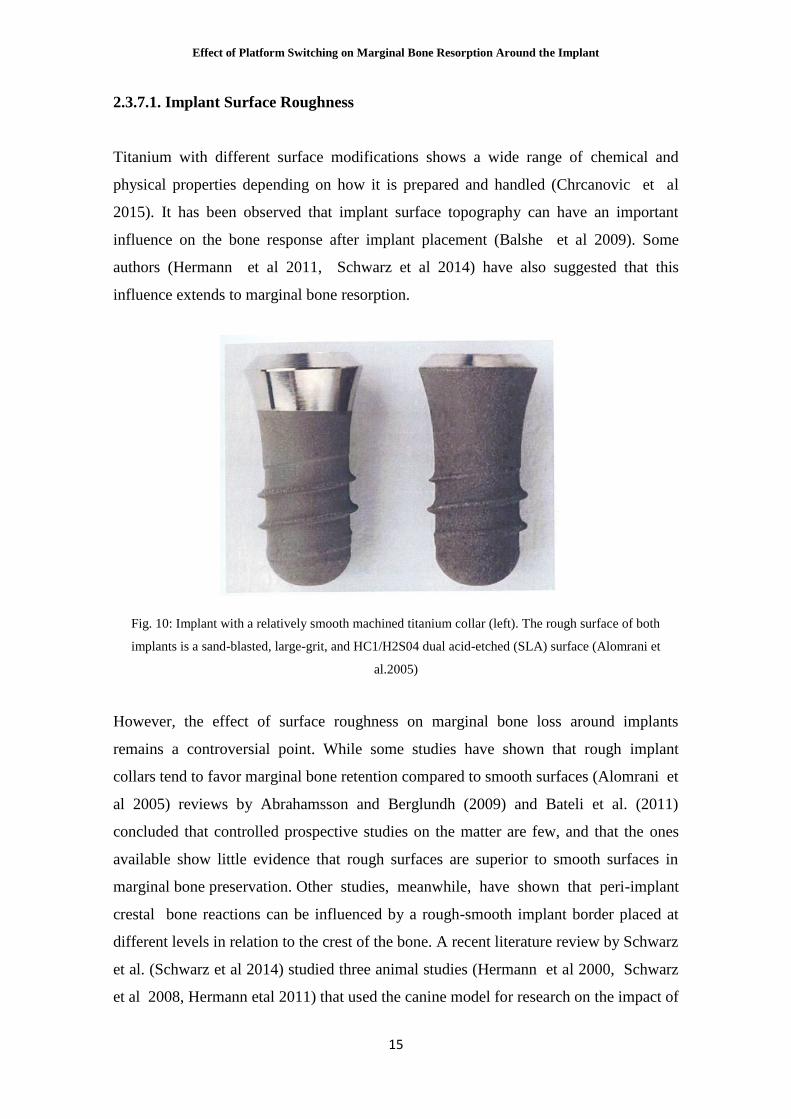
Titanium with different surface modifications shows a wide range of chemical and
physical properties depending on how it is prepared and handled (Chrcanovic et al
2015). It has been observed that implant surface topography can have an important
influence on the bone response after implant placement (Balshe et al 2009). Some
authors (Hermann et al 2011, Schwarz et al 2014) have also suggested that this
influence extends to marginal bone resorption.
Fig. 10: Implant with a relatively smooth machined titanium collar (left). The rough surface of both
implants is a sand-blasted, large-grit, and HC1/H2S04 dual acid-etched (SLA) surface (Alomrani et
al.2005)
However, the effect of surface roughness on marginal bone loss around implants
remains a controversial point. While some studies have shown that rough implant
collars tend to favor marginal bone retention compared to smooth surfaces (Alomrani et
al 2005) reviews by Abrahamsson and Berglundh (2009) and Bateli et al. (2011)
concluded that controlled prospective studies on the matter are few, and that the ones
available show little evidence that rough surfaces are superior to smooth surfaces in
marginal bone preservation. Other studies, meanwhile, have shown that peri-implant
crestal bone reactions can be influenced by a rough-smooth implant border placed at
different levels in relation to the crest of the bone. A recent literature review by Schwarz
et al. (Schwarz et al 2014) studied three animal studies (Hermann et al 2000, Schwarz
et al 2008, Hermann etal 2011) that used the canine model for research on the impact of

Effect of Platform Switching on Marginal Bone Resorption Around the Implant
16
the positioning of the machined collar on crestal bone level changes. The mean
difference between machined collars placed either above or below the bone crest
amounted to 0.835 mm, favoring an epicrestal positioning of the rough/smooth border.
2.3.7.2. Proximity between Implants
In a study conducted by Tarnow et al. (2000), a crestal bone loss of just 0.45 mm was
observed when the distance between two adjacent implants was 3 mm. This was in stark
contrast to the 1.04 mm of bone loss observed when the inter-implant distance was
smaller than 3 mm. A correlation between inter-implant distance and marginal bone loss
was thus established, considering a 3 mm distance to be a guideline for adjacent implant
placement.
Subsequent animal studies have emphasized the value of inter-implant distances of 3
mm from other perspectives, as well. Traini et al. (2008) placed implants at 2 and 3 mm
intervals in adult dogs. All the values on longitudinal collagen fiber, transverse collagen
fiber, marrow spaces, and mineral density that were produced by a 2 mm interval
showed significantly reduced values compared to those produced by a 3 mm interval.
The same authors (Traini et al. 2010) later performed an evaluation of the
vascularization level for de novo bone formation, contact osteogenesis, and bone
remodeling in groups of 2 and 3 mm distances in adult dogs. They observed better
vascularization in the latter group.
2.3.7.3. Surgical Trauma
Surgical trauma has been regarded as one of the most commonly suspected etiologies
proposed for early implant failure (Albreksson et al. 1986, Esposito et al. 1998,
Eriksson and Albrektsson 1984) reported that the critical temperature for implant site
preparation was 47°C for 1 minute or 40°C for 7 minutes, and that when the bone is
overheated, risk of implant failure is significantly increased. Wilderman et al. (1970)
reported that the mean horizontal bone loss after osseous surgery with periosteal

Effect of Platform Switching on Marginal Bone Resorption Around the Implant
17
elevation is approximately 0.8 mm. A more recent review conducted by Oh et
al. (2002), however, noted that the signs of bone loss from surgical trauma and
periosteal reflection are not commonly observed at implant stage 2 surgery in
successfully osseointegrated implants, adding that the pattern of bone loss in implants is
more likely to be vertical than horizontal. Thus, they concluded that the hypothesis
of the surgical causes of early implant bone loss remains to be determined.
2.3.7.4. Smoking Habit
De Luca and Zarb (2006) investigated the effects a smoking habit might have on
marginal bone loss. They observed that a positive smoking history was associated with
a higher rate of peri-implant bone loss, and that long-term heavy smokers could be at a
slightly higher risk of late implant failure and are susceptible to more marginal bone
loss over the long-term, irrespective of their smoking status at the time of implant
placement. A recent systematic review and meta-analysis conducted by Chrcanovic et
al. (2015) arrived at similar conclusions.
2.3.7.5. Diabetic Patients
With respect to marginal bone loss around implants in diabetic patients, another
systematic review and meta-analysis by Chrcanovic et al. (2014) found a significant
difference in favor of non-diabetic patients, with less marginal bone loss
observed compared to diabetic ones. However, it should be noted that the difference
was based on the only 2 publications (Accursi 2000, Tawil 2008) that were available.
2.4. Consequences of Marginal Bone Loss around Implants
The crestal bone supports the gingival architecture. Therefore, the stability of the
crestal bone is believed to be the key factor for maintaining stable soft tissue
dimensions over time (Vervaeke et al 2014). This has lead several authors (Albrektsson

Effect of Platform Switching on Marginal Bone Resorption Around the Implant
18
et al 1986, Papaspyridakos etal 2012) to consider the peri-implant bone level as a main
criterion to assess the success of dental implants.
Its importance in preserving the integrity of gingival margins and interdental
papillae means that marginal bone loss could compromise the final esthetic and
functional outcome of the implant, and thus contribute in the failure of the treatment.
Moreover, vertical peri-implant bone loss can alter the initial crown/implant ratio and
even invert it, creating an unfavorable situation that reduces the long-term
predictability of the restoration (Vela-Nebot etal 2008)
2.5. Proposed Solutions
Several new concepts have arisen to combat marginal bone loss. Roughened-
surface implants have proved to have a higher survival rate than machined-
surface implants, while different abutment shapes and connection types have also
shown promising results.
One concept that seems to be particularly efficient, however, is platform switching,
where the inward shifting of the connection microgap has been shown to significantly
reduce crestal bone remodeling and open up a host of new possibilities in implant
treatment.
Effect of Platform Switching on Marginal Bone Resorption Around the Implant
19
3. PLATFORM SWITCHING AS A SOLUTION TO EARLY CRESTAL
BONE LOSS AROUND IMPLANTS
3.1. Discovery
Historically, two-piece dental implant systems have been restored with prosthetic
components that locate the interface between the implant and the attached component
element at the outer edge of the implant platform. In 1991, Implant Innovations
introduced wide-diameter implants with matching wide-diameter platforms. During
that time, however, matching-diameter prosthetic components were not yet
commercially available, and many of the early 5.0- and 6.0-mm-wide implants
received standard diameter (4.1-mm) healing abutments and were restored with
standard-diameter (4.1-mm) prosthetic components (Lazzara & Porter 2006).
Long-term radiographic follow-up of these platform-switched, wide-diameter dental
implants demonstrated a smaller than expected vertical change in the peri-implant
crestal bone height than is typically observed around implants restored conventionally
with prosthetic components of matching diameters. This observation suggested that the
post restorative biologic process resulting in the loss of crestal bone height is altered
when the outer edge of the implant-abutment interface is horizontally repositioned
inwardly and away from the outer edge of the implant platform (Lazzara & Porter
2006). It led to the introduction of the concept of platform switching by Gardner in
2005 and Lazzara and Porter in 2006.
Several clinical reports (Vela-Nebot et al. 2006, Hiirzeler et al. 2007, Canullo &
Rasperini 2007) then demonstrated more favorable soft and hard tissue responses using
implants placed with platform switching compared to standard platform-matched
implants. Consequently, an increasing number of implant systems incorporated platform
switching into their designs as an innovative feature for preserving the peri-implant
bone (Atieh et al. 2010)

Effect of Platform Switching on Marginal Bone Resorption Around the Implant
20
3.2. Concept and Rationales
The concept of platform switching suggests the use of a smaller-diameter abutment or
supra-structure on a larger-diameter implant collar. This configuration results in a
circular horizontal step and the inward horizontal repositioning of the implant-abutment
junction (Gardner 2005, Lazzara & Porter 2006)
Fig. 11: Platform switching is demonstrated. A 0. 95-mm circumferential horizontal mismatch in
dimension is created when a 4.1-mm-diameter prosthetic UCLA abutment is placed on a 6.0mm diameter
implant with matching 6.0mm diameter platform (Lazzara & Porter 2006)
Several theories have been suggested to explain the potentiality of platform switching to
preserve peri-implant marginal bone (Annibali et al. 2012).
3.2.1. Biologic Rationale
A biologic rationale has been established to understand why there appears to be little or
no crestal bone remodeling following the placement of an implant with a
platform switched design. This rationale suggests that the inward positioning of

Effect of Platform Switching on Marginal Bone Resorption Around the Implant
21
the implant abutment junction (IAJ) influences the bone remodeling process in two
ways (Lazzara & Porter 2006).
First, the inward positioning of the implant-abutment interface exposes the
implant seating surface, thus creating an additional horizontal surface area. This
allows the biologic width to be formed horizontally, reducing the amount of crestal
bone resorption necessary to expose a minimum amount of implant surface to which the
soft tissue can attach (Lazzara & Porter 2006). Second, by repositioning the IAJ inward
and away from the outer edge of the implant and adjacent bone, the overall effect of the
abutment inflammatory cell infiltrate (ICT) on the surrounding tissue may be reduced,
thus decreasing the resorptive effect of the abutment ICT on the surrounding crestal
bone (Lazzara & Porter 2006).
It is also suggested that platform switching locates the inflammatory infiltrate within
an approximate :::; 90-degree confined area of exposure instead of a :S 180-degree
area of direct exposure to the surrounding hard and soft tissues, as depicted in Fig. 12.
As a consequence, the reduced exposure and confinement of the platform-switched
abutment ICT may also contribute in reducing its inflammatory effect (Lazzara & Porter
2006).
Fig. 12: Amount of exposure the abutment JCTwill have with the surrounding bone and soft tissue when
positioned at the outer edge of the implant (left). In contrast, the inward, horizontal re positioning of the
abutment JCT (right) will move the abutment JCT away from the crestal bone and into a more confined
area ( Lazzara & Porter 2006).

Effect of Platform Switching on Marginal Bone Resorption Around the Implant
22
These concepts were further supported by animal (Cochran et al. 2009, Farronato et al.
2012, Cochran et al. 20l3 and human (Luongo et al. 2008, Degidi et al. 2008, Canullo
et al. 2011) histological studies.
3.2.2. Biomechanical Rationale
Another theory, supported by finite element analysis, exammes the biomechanical
advantages of the platform switching configuration in terms of stress distribution in and
around the implant. It suggests that the platform switching design reduces the stress at
the one-implant interface and in the crestal region of cortical bone by shifting stress
away from the bone-implant interface toward the center of the implant (Maeda et al.
2007).
Fig. 13: Mixed chronic inflammatory cell infiltrate at the implant-abutment interface (abutment ICT)
over a study period of 24 weeks conducted by Becker et al. (2009). The abutment ICT seems to be
limited to an approximate 90-degree confined area of exposure in the platform-switched implant (right)
instead of a 180-degree area in the platform-matched implant (left).
Effect of Platform Switching on Marginal Bone Resorption Around the Implant
23
These concepts were further supported by animal (Cochran et al 2009, Farronato et al
2012, Cochran et al 2013) and human (Luongo et al 2008, Degidi et al 2008, Canullo
etal 2011) histological studies.
Fig. 14: Strain energy density distribution in the implant (Maeda et al 2007)
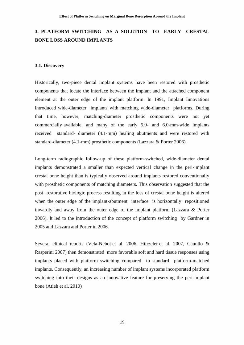
In a three-dimensional finite element analysis, Maeda et al. (2007) discovered a high
stress area around the periphery of the implant's top surface and along its lateral surface
as well as in the bone facing that area in the normal model, while this high stress area
shifted toward the center of the implant in the platform-switched model (Fig. 14). Also,
the strain energy in the normal model implant was more widely spread along its lateral
surface down toward the implant tip, while it was concentrated near the abutment-
implant interface area in the platform-switched model. Strain energy in the cortical
bone surface was higher in the normal model than in the platform-switched model.
These results were later supported by studies conducted by Chang et al. (2010), Tabata
et al. (2011) and Yang and Maeda (2013) who observed that Von Mises, maximum
(tensile), and minimum (compressive) principal stress were reduced in implants and
peri implant bone tissue when the platform switching concept was used. It was
suggested that wide-diameter implants had a large influence in reducing stress
values (Tabata et al. 2011)

Effect of Platform Switching on Marginal Bone Resorption Around the Implant
24
The decrease in stress values observed may provide a biomechanical explanation as to
why platform switching seems to reduce the expected post-restoration crestal bone
remodeling. The lower concentration of stress in the peri-implant bone tissue would
lead to less microdamage in the bone, resulting in minimized crestal bone loss
(Cardaropoli et al. 2006, Hiirzeler et al. 2007). The model can also contribute in
reducing shearing stress, which is most likely to cause disintegration (Sugiura et al.
2000). Another possible explanation lies in the distance between the bone surface and
the stress concentrated area on the implant surface. As microorganisms are likely to
move toward the high-energy area by the mechanism of interface micromovements, it is
advantageous to have a large distance between the stress concentration area and bone
surface (Assenza et al. 2003, Guindy et al. 2004)
3.2.3. Proof of Concept
Since its breakthrough into the professional conscwusness, the platform switching
concept has been continuously put to the test by a host of studies and experiments.
Today, the literature contains enough evidence to transcend platform switching from the
realm of theory to the realm of scientific truth.
Early studies showed encouraging results. In a randomized clinical trial, Canullo and
Rasperini (2007) studied 10 platform-switched implants during a follow-up period of
18 to 36 months and observed a smaller mean marginal bone loss than what was
reported by the literature for regular implants. A prospective study by Hiirzeler et
al. (2007) compared the marginal bone loss around 14 platform-switched implants to
that around 8 platform-matched implants and concluded that a platform-switched
configuration reduced peri-implant bone loss by a significant margin. Later comparative
studies (Prosper et al.2009, Vigolo & Givani 2009, Canullo et al. 2010, Canullo et al.
2012, Telleman et al. 2014, Guerra et al. 2014) continued to show better bone
preservation around platform-switched implants when compared to regular platform-
matched implants (Fig. 15, Fig. 16, Fig. 17).

Effect of Platform Switching on Marginal Bone Resorption Around the Implant
25
Fig. 15: Vigolo and Givani (2009) followed up 182 implants placed in the posterior region for 5 years. A
statistically significant difference was detected in crestal bone changes in wide-diameter implants
restored with matching wide-diameter prosthetic components (group A) and wide-diameter implants
restored with platform-switched prosthetic components (group B). After I 2 months of function, the group
B implants showed less bone loss than the group A implants. The data did not change during the
following 4 years of function.
Fig. 16: Canullo etal (2010) placed 80 posterior implants divided according to their platform diameter
into four groups: 3.8 mm (control group), 4.3 mm (test group 1), 4.8 mm (test group 2) and 5.5 mm (test
group 3). Radiographic bone height was measured at the time of implant placement and after 9, 15, 2 1
and 33 months. After 21 months, radiographic evaluation showed a mean bone loss of0.99 mm for test
group 1, 0.82mmfor test group 2 and 0.56 mm for test group 3. These values were significantly lower
compared with the control group (1.49 mm).

Effect of Platform Switching on Marginal Bone Resorption Around the Implant
26
Fig 17: In a study by Guerra et al (2014), 146 implants were radiographically observed: 74 platform
switched implants and 72 platform-matched The difference of mean marginal bone level change from
surgery to 12 months was significant between the two groups, with the platform-switched group showing
superior bone preservation and gain. Standardized peri-apical radiographs were taken before implant
placement (a), immediately post-surgery (b), before (c) and after abutment/crown placement (d) and at I
year post-loading (e).
In a recent systematic review and meta-analysis covering 28 publications reporting
on 1216 platform-switched implants and 1157 platform-matched implants, Chrcanovic
et al. (2015) concluded that platform-switched implants resulted in significantly
less marginal bone loss than platform-matched implants. However, the group
mentioned that the results of the review should be interpreted with caution due to
the presence of uncontrolled confounding factors in the included studies, most of them
with short follow up periods.
3.3. Design Specifications and Variations
The effects of platform switching are influenced by a number of factors. The following
modifications have been observed to play at least a small part in the bone-preserving
properties of platform switching.

Effect of Platform Switching on Marginal Bone Resorption Around the Implant
27
3.3.1. Extent of Platform Mismatch
While there does not seem to be a clear indication as to what the ideal extent of
implant/abutment mismatch is, some systematic reviews and meta-analyses
have suggested that an implant/abutment mismatch of at least 0.4 rnrn is more
beneficial for preserving marginal bone (Atieh et al. 2010, Annibali et al. 2012) Atieh et
al. (2010) also noted that the changes in marginal bone levels were more favorable
with increasing the extent of mismatch between implants and abutments. A similar
observation was made in a more recent review and meta-analysis conducted by
Chrcanovic et al. (2015), as demonstrated in Fig. 18.
Fig. 18: Scatter plot for the meta-regression with the association between the mean differences (in
millimeters) of the marginal bone loss between the two procedures (platform-switched vs. platform-
matched) and the mismatch (in millimeters) (Chrcanovic etal 2015)
Indeed, the effect of platform switching on marginal bone levels seems to be "dose
dependent." In a randomized control trial, Canullo et al. (2010/ 4 demonstrated that the
greatest platform-abutment mismatch resulted in the least marginal bone loss and
concluded that the degree of platform switching could have a significant influence on
peri-implant marginal bone remodeling (Fig. 19). It was speculated that these findings
could be attributed to a wider space for horizontal repositioning of the biological width
and/or a better distribution of loading stress at the bone/implant interface.
It has been suggested that the findings of reduced bone remodeling accompanying
a larger implant-abutment difference may be due to an increased implant diameter
Effect of Platform Switching on Marginal Bone Resorption Around the Implant
28
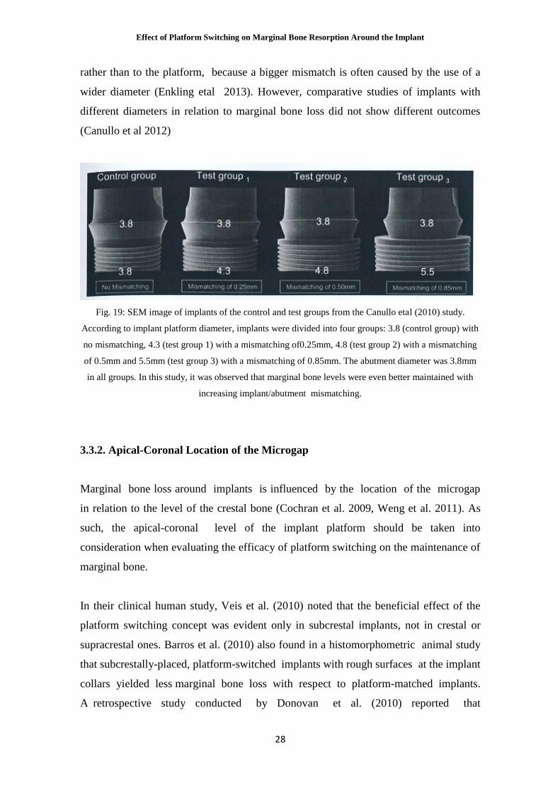
rather than to the platform, because a bigger mismatch is often caused by the use of a
wider diameter (Enkling etal 2013). However, comparative studies of implants with
different diameters in relation to marginal bone loss did not show different outcomes
(Canullo et al 2012)
Fig. 19: SEM image of implants of the control and test groups from the Canullo etal (2010) study.
According to implant platform diameter, implants were divided into four groups: 3.8 (control group) with
no mismatching, 4.3 (test group 1) with a mismatching of0.25mm, 4.8 (test group 2) with a mismatching
of 0.5mm and 5.5mm (test group 3) with a mismatching of 0.85mm. The abutment diameter was 3.8mm
in all groups. In this study, it was observed that marginal bone levels were even better maintained with
increasing implant/abutment mismatching.
3.3.2. Apical-Coronal Location of the Microgap
Marginal bone loss around implants is influenced by the location of the microgap
in relation to the level of the crestal bone (Cochran et al. 2009, Weng et al. 2011). As
such, the apical-coronal level of the implant platform should be taken into
consideration when evaluating the efficacy of platform switching on the maintenance of
marginal bone.
In their clinical human study, Veis et al. (2010) noted that the beneficial effect of the
platform switching concept was evident only in subcrestal implants, not in crestal or
supracrestal ones. Barros et al. (2010) also found in a histomorphometric animal study
that subcrestally-placed, platform-switched implants with rough surfaces at the implant
collars yielded less marginal bone loss with respect to platform-matched implants.
A retrospective study conducted by Donovan et al. (2010) reported that
Effect of Platform Switching on Marginal Bone Resorption Around the Implant
29
subcrestal placement of dental implants with a platform-switched Morse taper
connection resulted in minimal marginal bone loss and a high percentage of implants
with mineralized hard tissue on the implant platform. Similar results were found in a
randomized prospective clinical study conducted by Koutouzis et al. (20I4), where it
was reported that implants placed with the implant/abutment interface 1 or 2 mm apical
to the buccal aspect of the bone crest demonstrated less marginal bone loss apical to
the implant platform, and a greater percentage of implant surfaces showed bone on the
implant platform, compared to implants placed with the implant/abutment interface at
the level of the buccal aspect of the alveolar bone crest.
In a recent systematic review, however, Chrcanovic et al. (2015) noted that, as the
implant platform varied from study to study and this information was not always
provided, it may still be difficult to unequivocally interpret the available evidence about
implant/abutment interface placement in platform-switched implants.
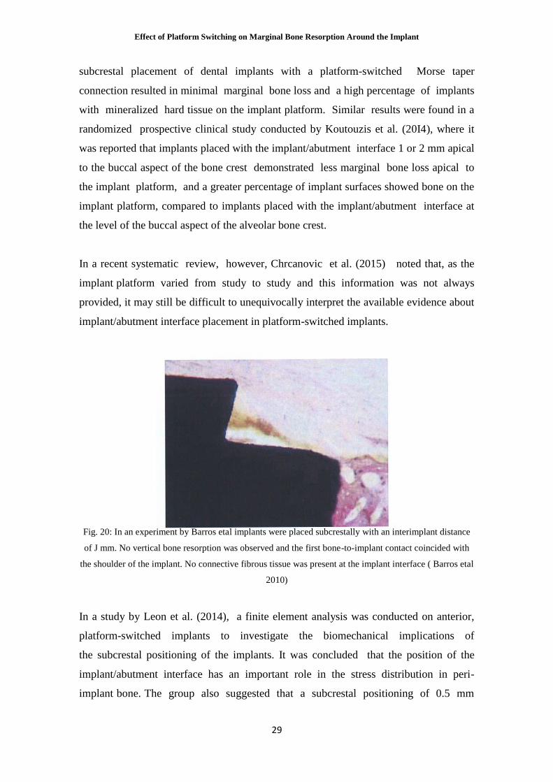
Fig. 20: In an experiment by Barros etal implants were placed subcrestally with an interimplant distance
of J mm. No vertical bone resorption was observed and the first bone-to-implant contact coincided with
the shoulder of the implant. No connective fibrous tissue was present at the implant interface ( Barros etal
2010)
In a study by Leon et al. (2014), a finite element analysis was conducted on anterior,
platform-switched implants to investigate the biomechanical implications of
the subcrestal positioning of the implants. It was concluded that the position of the
implant/abutment interface has an important role in the stress distribution in peri-
implant bone. The group also suggested that a subcrestal positioning of 0.5 mm
Effect of Platform Switching on Marginal Bone Resorption Around the Implant
30
might be appropriate to avoid the risk of overloading the implant, with the added
benefit of concealing the implant neck and establishing an adequate emergence profile.
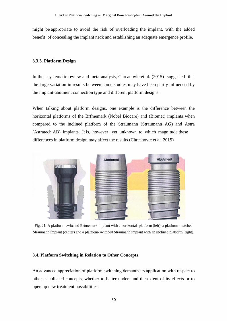
3.3.3. Platform Design
In their systematic review and meta-analysis, Chrcanovic et al. (2015) suggested that
the large variation in results between some studies may have been partly influenced by
the implant-abutment connection type and different platform designs.
When talking about platform designs, one example is the difference between the
horizontal platforms of the Brfmemark (Nobel Biocare) and (Biomet) implants when
compared to the inclined platform of the Straumann (Straumann AG) and Astra
(Astratech AB) implants. It is, however, yet unknown to which magnitude these
differences in platform design may affect the results (Chrcanovic et al. 2015)
Fig. 21: A platform-switched Brtmemark implant with a horizontal platform (left), a platform-matched
Straumann implant (center) and a platform-switched Straumann implant with an inclined platform (right).
3.4. Platform Switching in Relation to Other Concepts
An advanced appreciation of platform switching demands its application with respect to
other established concepts, whether to better understand the extent of its effects or to
open up new treatment possibilities.

Effect of Platform Switching on Marginal Bone Resorption Around the Implant
31
3.4.1 Marginal Bone Loss with Respect to Time
The results of a systematic review and meta-analysis conducted by Chrcanovic et al.
(2015) suggested that that there is an increase of the mean difference of marginal bone
loss between the platform-switched and platform-matched approaches with the increase
of the follow-up time (Fig X). The group stressed, however, that the existence of only a
few studies with long-term follow-ups is problematic to the conclusiveness of these
findings.
Fig. 22: Scatter plot for the meta-regression with the association between the mean differences (in
millimetres) of the marginal bone loss between the two procedures (platform-switched vs. platform
matched) and the follow-up time (in months) (Chrcanovic etal 2015).
3.4.2. Proximity to Natural Teeth
Esposito et al. (1993) evaluated implants with matching implant-abutment platforms
and reported increased bone loss to adjacent teeth as the horizontal tooth-implant
distance between the two structures decreased. Subsequent studies further validated
these findings, leading many authors to recommend a minimum of 1.5 to 2 mm between
the tooth and implant to avoid causing bone loss around them. This makes it impossible
to place a 4 mm diameter implant in a mesio-distal space of 7 mm (Vela et al 2012),
causing a potential hurdle to the treatment.

Effect of Platform Switching on Marginal Bone Resorption Around the Implant
32
Platform-switched implants seem to pose a solution to this. A study by Vela et
al. (2012) using platform-switched implants demonstrated a 0.37 mm mean bone
peak resorption at a mean tooth-implant distance of 0.9 mm. These values (Fig. 23)
were much lower than those found in the study conducted by Esposito et al, suggesting
that a tooth implant distance of 1 mm was sufficient to maintain the bone peak. Similar
findings were observed in a more recent study by Urdaneta et al. (2014).
Fig. 23: Means, standard deviations (SDs), and minimum and maximum measurements obtained for lTD
(distance between implant and tooth), HBR (horizontal bone resorption) and VBR (vertical bone
resorption) (Vela 2012)
3.4.3. Proximity to Other Platform-switched Implants
While it has been shown that a minimum of 3 mm must be kept between two adjacent
platform-matched implants (Tarnow etal 2000, Traini etal 2008 & 2010) thesame does
not appear to be the case when using platform-switched implants.
A histomorphometric animal study about the effect of interimplant distance on
crestal bone loss was conducted by Elian et al. (2011) • The group inserted platform-
switched internal connection implants with 2 and 3 mm intervals in Gottingen
minipigs, and reported no significant differences on inter-implant crestal bone
heights 2 months after the implantation. This was in agreement with previous animal
experiments on the matter (Novaes et al 2006, Barros et al 2010) Human studies also
seem to confirm these findings. Chang and Wennstrom (2010) analyzed peri-
implant bone change using radiographic evaluation for 5 years and reported that
the mean change of inter-implant crestal bone around close-proximity posterior
implants 1 year after the abutment connection was found to be -0.13 ± 0.34

Effect of Platform Switching on Marginal Bone Resorption Around the Implant
33
mm. The change after 5 years of connection reached -0.32 ± 0.6 mm. These values
compare favorably with those found in the Tarnow et al. (2000) experiment.
A more recent human trial conducted by Jo et al. (2014) was in accordance with these
findings: the inter-proximal distance with platform switched internal connection
implants did not show a significant influence on crestal bone loss (Fig. 24), and
the horizontal vertical marginal bone loss was found to be too small to result in an
overlapping loss of inter-implant crestal bone.
Fig. 24: Correlation between crestal bone loss and inter-implant distance from the Jo et at. (2014) study.
3.4.4. Disconnection and Reconnection of Platform-switched Abutments
In two piece implants, the abutment is typically disconnected several times during the
prosthetic phase of treatment. The disruption of the soft tissue that occurs each time the
implant components are disconnected and reconnected is thought to influence bone
resorption around the implant (Abrahamsson et al. 1997, Rodriguez et al. 2013).
Standard clinical protocols may require the removal of an abutment up to four times: for
implant-level impressions, try-in of the metal framework, try-in of the porcelain before
the final firing and glazing, and delivery of the definitive prosthesis (Rodriguez et al.
2013).

Effect of Platform Switching on Marginal Bone Resorption Around the Implant
34
In an animal study, Abrahamsson et al. (1997) showed that vertical peri-implant bone
resorption increased from 0.78 mm prior to any abutment disconnection to 1.49 mm
after five changes. A more recent animal study by Rodriguez et al. (2013) aimed to
confirm the resorptive effect of disconnecting and reconnecting abutments while
presenting platform switching as a potential solution. After four disconnections, the
vertical and horizontal bone resorption values for the platform-matched and
platform-switched implants were similar to the values normally found in similar studies
(Fig. 25).
Fig. 25: Horizontal and vertical peri-implant bone resorption on each implant site from the Rodriguez et
al. study. After four dislreconnections, vertical peri-implant bone resorption in matched implants was 1. 1
mm and horizontal was 0.98 mm, while peri-implant bone resorption in platform-switched implants was
0.24 mm horizontal and 0.40 mm vertical (Rodriguez etal 2013)
Considering the remaining results, however, Rodriguez et al. (2013) concluded that
implants with a platform-switched design show less peri-implant bone resorption during
the healing process and as their abutments are disconnected than do dis/reconnected
platform-matched implants. In addition, a single dis/reconnection of the platform

Effect of Platform Switching on Marginal Bone Resorption Around the Implant
35
matched abutments may generate a peri-implant bone resorption similar to that
produced by four dis/reconnections. On platform-switched implants, meanwhile, a
greater number of dis/reconnections generate more peri-implant bone resorption, but at
least two times, two weeks apart are needed to trigger statistically significant bone
resorption. These findings confirm the importance of reducing the number of abutment
dis/reconnections when attempting to minimize peri-implant bone resorption and
hinted at platform switching's superior contribution to that cause.
3.5. Advantages and Disadvantages of Platform Switching
3.5.1. Advantages
The esthetic replacement of teeth has become an important standard for implant
dentistry, but the ability to restore implants esthetically has been fraught with obstacles
and sometimes has not been attainable. Two main concerns remain the loss of implant
papilla and the exposition of the metal collar at the implant shoulder in the esthetic zone
(Leblebicioglu et al. 2007) Moreover, the creation of the biologic width can cause
vertical peri-implant bone loss that alters the initial crown/implant ratio and even inverts
it, creating an unfavorable situation that reduces the long-term predictability of the
restoration (Fig. 26) (Vela-Nebot et al. 2008).
Fig. 26: Variations in crown/implant ratio. (a) Initial situation without platform switching. (b) Peri-
implant bone loss with platform switching. (c) Peri-implant bone loss without platform switching (Vela-
Nebot etal 2008)

Effect of Platform Switching on Marginal Bone Resorption Around the Implant
36
Platform switching presents a clear potential to negate some of these obstacles. This is
achieved through better preservation of the marginal bone around platform-switched
implants compared to conventional platform-matched implants (Chrcanovic et al.
2015) and a better distribution of biomechanical forces (Yang & Maeda 2013).
Based on this, platform switching has a range of clinical benefits:
(1) Platform switching helps retain peri-implant crestal bone, thus providing better
support for the soft tissues. This is extremely important in anterior restorations, in which
preserving the buccal plate and maintaining the peri implant crestal bone determines
gingival aesthetics and the health of the implant-supported restorations (Fig 27) (Vela-
Nebot et al. 2008).
(2) Platform switching makes it possible to place implants at a closer proximity to other
implants and to natural teeth when the prosthetic guide requires it, while still preserving
the adjacent bone level. This allows for better functional and esthetic results in cases
where the mesio-distal space is limited (Rodriguez Cuirana et al. 2009, Vela et al.
2012)
(3) Platform switching permits a superior management of occlusal stress (Maeda et al.
2007, Yang & Maeda 2013), better protection of peri-implant soft tissues from
abutment dis/reconnection (Rodriguez et al. 2013) and its effect is stable with time
(Chrcanovic et al 2015), thus allowing for more predictability in the implant
treatment.
Fig. 27: Tooth II restored with a platform-switched implants after three years of follow-up (Vela-Nebot
et al 2008)

Effect of Platform Switching on Marginal Bone Resorption Around the Implant
37
3.5.2 Disadvantages
While platform switching has the biomechanical advantage of shifting the stress
concentration away from the bone-implant interface, it has been suggested that higher
stress occurred around the outside of the abutment and implant connection area,
possibly causing problems such as abutment screw deformation over the elastic limit
(Maeda et al. 2007). In addition to this, it has been reported that probing and achieving
an adequate prosthetic emergence profile can become slightly trickier due to the
unconventional profile of the platform-switched implant, but these claims have yet to
be substantiated by scientific evidence.
3.5.3. Indications
Taking the advantages and disadvantages of platform switching into consideration,
a number of possible indications can be presented:
(1) Anterior esthetic zone (Vela et al. 2012)
(2) Limited mesio-distal space (Vela et al. 2012)
(3) Implant-related interventions that require a careful management of occlusal
forces, such as sinus grafts (Rodriguez-Ciurana et al. 2009(4) Short implant (Rodriguez-
Ciurana et al. 2009)
(5) Oblique loading (Yang & Maeda 2013)

Effect of Platform Switching on Marginal Bone Resorption Around the Implant
38
4. CONCLUSION
Marginal bone loss around the implant neck has long been considered an unavoidable
obstacle in the way of an ideal implant restoration, a necessary evil to be borne despite
the clinician's best efforts to avoid it. Such has been the implantologist's powerlessness
in the face of it that a marginal bone resorption of 1.5 mm during the first year and 0.5
mm during every subsequent year became considered as a criterion of implant success
(Albrektsson et al. 1986l. Today, however, advancements in implant design and
individual case treatment planning provide ways to control marginal bone loss. The
concept of platform switching is one such advancement.
Platform switching is a new concept in implant design that promises a solution to peri
implant marginal bone loss. It revolves around the use of a smaller-diameter abutment
or supra-structure on a larger-diameter implant collar, resulting in a circular horizontal
step and the inward horizontal repositioning of the implant-abutment junction (Lazzara
& Porter 2006). On a biologic level, this achieves two things: a horizontal platform for
the biologic width to establish itself on and the repositioning of the abutment
inflammatory cell infiltrate (ICT) away from the surrounding bone, reducing its
resorptive effect (Lazzara & Porter 2006, Cochran et al. 2013). On a
biomechanicallevel, platformswitched implants yield better stress distribution along
the implant and reduce stress levels on the implant/bone interface (Maeda et al. 2007,
Yang & Maeda 2013). Several studies (Chrcanovic et al. 2015) have since confirmed
platform switching's potential to preserve marginal bone. Its effect has also been shown
to be influenced by the extent of the platform mismatch (Canullo et al. 2010) and
the apical-coronal location of the microgap (Leon et al. 2014) and has positive
implications on previous limitations such as the proximity of placement next to other
implants (Jo et al. 2014) and natural teeth (Vela et al. 2012) and the repeated
disconnections/reconnections of abutments (Rodriguez et al. 2013). It has been
confirmed that platform switching achieves a better preservation of marginal bone
(Chrcanovic etal 2015) and a superior management of occlusal stress (Maeda et al 2007,
Yang & Maeda 2013) compared to conventional implants. This brings with it several
clinical benefits. Platform switching helps retain peri-implant crestal bone, thus
providing better support for soft tissues, an extremely important criterion in
esthetic anterior restorations (Vela-Nebot et al. 2008) Platform switching also preserves

Effect of Platform Switching on Marginal Bone Resorption Around the Implant
39
bone levels when placing implants in close proximity to each other or to natural
teeth (Rodriguez-Ciurana et al. 2009, Vela et al 2012), improves the biomechanical
properties of implant-supported restorations (Vela-Nebot etal 2008) and protects the
peri-implant soft tissues (Rodriguez etal 2013).
Platform switching's impressive results bring with them an exciting revelation: marginal
bone loss is not as unavoidable as it has long been considered. The concept's
implications are as beneficial to implant treatment as marginal bone loss has been
detrimental, and its mastery and continuous refinement opens up a host of new
possibilities and opportunities in implant treatment. As in any other technology,
however, one must fully understand its underlying science to truly be able to wield it to
its full effect.

Effect of Platform Switching on Marginal Bone Resorption Around the Implant
40
5. REFERENCES
I. Abrahamsson I, Berglundh T. Effects of different implant surfaces and designs on
marginal bone-level alterations: a systematic review. Clin Orallmpl Res 2009; 20: 207-
215.
2. Abrahamsson I, Berglundh T, Lindhe J. The mucosal barrier following abuttnent
dis/reconnection. An experimemal study in dogs. J Clin Periodontol1997: 24: 568-572.
3. Abrahamsson I, Berglundh T, Wennstrom J, Lindhe J. The peri-implant
hard and soft tissue characteristics at different implant systems. A comparative study
in dogs. Clin Oral Implant Res 1996; 7:
212-219.
4. Accursi GE. Treatment outcomes with osseointegrated Branemark implants in
diabetic patients: a retrospective study (thesis). Toronto, ON: University of Toronto.
2000.
5. Adell R, Lekholm U, Rockier 8, Branemark PI. A 15-year study of
osseointegrated implants in the treatment of the edentulous jaw. Int J Oral Surg 1981 ;
10: 387-416.
6. Albouy JP, Abrahamsson I, Berglundh T. Spontaneous progression of
experimental peri-implantitis at implants with different surface characteristics. An
experimental study in dogs. J Clin Periodontol20 12; 39:
182-187.
7. Albrektsson T, Dahlin C, Jemt T, Sennerby L, Turri A, Wennerberg A. Is marginal
bone loss around oral implants the result of a provoked foreign body reaction? Clin
Implant Dent Relat Res 20 14; 16(2): 155-65.
8. Albrektsson T, Zarb G, Worthington P, Eriksson AR. The long-term efficacy of
currently used dental implants: a review and proposed criteria of success. lnt J Oral
Maxillofac Implants 1986; I: 11 -25.

Effect of Platform Switching on Marginal Bone Resorption Around the Implant
41
9. Aloise JP, Curcio R, Laporta MZ, Rossi L, da Silva AM, Rapoport A. Microbial
leakage through the implant-abutment interface of morse taper implants in vitro. Clin
Ora/Implants Res 201 0; 21 : 328-335.
I 0. Alomrani AN, Hermann JS, Jones AA, Buser D, Schoolfield J, Cochran DL. The
Effect of a Machined Collar on Coronal Hard Tissue Around Titanium Implants: A
Radiographic Study in the Canine Mandible. Int J Oral Maxi/lofac Implants 2005; 20:
677-686.
II . Annibali S, Bignozzi I, Cristalli MP, Graziani F, La Monaca G, Polimeni A.
Peri-implant marginal bone level: a systematic review and meta-analysis of
studies comparing platform switching versus conventionally restored implants. J
Clin Periodonto/2012 ; 39: 1097-1113.
12. Arora R, Narula SC, Sharma RK, Tewari S. Supracrestal Gingival Tissue:
Assessing Relation with Periodontal Biotypes in a Healthy Periodontium. lnt J
Periodontics Restorative Dent 2013; 33: 763-771.
13. Assenza 8, Scarano A, Petrone G, et al. Crestal bone remodeling in loaded and
unloaded implants and the microgap: A histologic study. Implant Dent 2003; 12: 235-
241.
14. Astrand P, Engquist B, Dahlgren S, Grondahl K, Engquist E, Feldmann H. Astra
Tech and Branemark system implants: A 5-year prospective study of marginal bone
reactions. Clin Oral Implants Res 2004; 15:
413-420.
15. Atieh MA, Ibrahim HM, Atieh AH. Platform switching for marginal bone
preservation around dental implants: a systematic review and meta-analysis. J
Periodonto/20 10; 81: 1350-1366.
16. Becker J, Ferrari D, Mihatovic I, Sahm N, Schaer A, Schwarz F. Stability of
crestal bone level at platform switched non-submerged titanium implants. A
histomorphometrical study in dogs. J Clin Periodonto/2009; 36: 532-539.

Effect of Platform Switching on Marginal Bone Resorption Around the Implant
42
17. Balshe AA, Assad DA, Eckert SE, Koka S, Weaver AL. A retrospective study
of the survival of smooth- and rough-surface dental implants. Int J Oral Maxillofac
Implants 2009; 24: 1113-1118.
18. Barros RRM, Novaes AB Jr., Muglia VA, Iezzi G, Piattelli A. Influence of
interimplant distances and placement depth on peri-implant bone remodeling of
adjacent and immediately loaded Morse cone connection implants: a
histomorphometric study in dogs. Clin Oral Imp! Res 201 0; 21: 371-378.
19. Bateli M, Att W, Strub JR. Implant Neck Configurations for Preservation of
Marginal Bone Level: A Systematic Review. Int J Oral Maxillofac Implants 2011; 26:
290-303.
20. Berglundh T, Abrahamsson I, Welander M, Lang NP, Lindhe J. Morphogenesis
of the peri-implant mucosa: an experimental study in dogs. Clin Oral Imp! Res 2007;
18: 1-8
21. Berglundh T, Lindhe J. Dimension of the periimplant mucosa. Biologic width
revisited. J Clin Periodonto/1996; 23: 971-973.
22. Berglundh T, Lindhe J, Ericsson I, Marinello CP, LiUenberg B, Thomsen P. The
soft tissue barrier at implants and teeth. Clin Oral Implants Res 1991; 2: 81-90.
23. Busscher HJ, Rinastiti M, Siswomihardjo W, van der Mei HC. Biofilm formation
on dental restorative and implant materials. J Dent Res 2010; 89: 657-665.
24. Canullo L, Fedele OR, Iannello G, Jepsen S. Platform switching and marginal
bone-level alterations: The results of a randomizedcontrolled trial. Clin Oral Implants
Res 2010; 21: 115-121.
25. Canullo L, Iannello 0, Penarrocha M, Garcia B. Impact of implant diameter on
bone level changes around platform switched implants: preliminary results of 18
months follow-up a prospective randomized match-paired controlled trial. Clin Oral
Imp! Res 2012; 23: 1142-1146.

Effect of Platform Switching on Marginal Bone Resorption Around the Implant
43
26. Canullo L, Pellegrini 0, Allievi C, Trombelli L, Annibali S, Dellavia C: Soft tissues
around long-term platfonn switching implant restorations: a histological human
evaluation. Preliminary results. J Clin Periodonto/20 11; 38: 86-94.
27. Canullo L, Penarrocha-Oltra D, Soldini C, Mazzocco F, Penarrocha M, Covani U.
Microbiological assessment of the implant-abutment interface in different connections:
cross-sectional study after 5 years of functional loading. Clin Oral Implants Res 2015;
26,426-434.
28. Canullo L, Rasperini G. Preservation of peri-implant soft and hard tissues using
platform switching of implants placed in immediate extraction sockets: A proof-of-
concept study with 12- to 36-month follow-up. lnt J Oral Maxillofac Implants 2007; 22:
995-1000.
29. Cardaropoli G, Lekholm U, Wennstrom JL. Tissue alterations at implant-
supported single-tooth replacements: A !-year prospective clinical study. Clin
Ora/Implants Res 2006; 17: 165-171.
30. Chambrone L, Chambrone LA, Lima LA. Effects of occlusal overload on peri-
implant tissue health: a systematic review of animal model studies. J Periodonto/2010 ;
81: 1367-1378.
31 . Chang CL, Chen CS, Hsu ML. Biomechanical effect of platform switching in
implant dentistry: A three-dimensional finite element analysis. lnt J Oral Maxillofac
Implants 20 I 0; 25: 295-304.
32. Chang M, Wennstrom JL. Bone alterations at implant-supported FOPs in relation to
inter-unit distances: A 5-year radiographic study. Clin Oral Implants Res 20 I 0; 21:
735-740.
33. Chrcanovic BR, Albrektsson T, Wennerberg A. Platform switch and dental
implants: A meta-analysis. J Dent2015;43: 629-646.
Effect of Platform Switching on Marginal Bone Resorption Around the Implant
44
34. Chrcanovic BR, Albrektsson T, Wennerberg A. Smoking and dental implants: A
systematic review and meta-analysis. J Dent 2015; 43:487-498.
35. Chrcanovic BR, Albrektsson T, Wennerberg A. Diabetes and Oral Implant Failure:
A Systematic Review. J Dent Res 2014; 93: 859-867.
36. Cochran DL, Bosshardt DO, Grize L, et al. Bone response to loaded implants with
non-matching implant-abutment dismeters in the canine mandible. J Periodonto/2009 ;
80: 609-617.
37. Cochran DL, Hermann JS, Schenk RK, Higginbottom FL, Buser D. Biologic width
around titanium implants. A histometric analysis of the implanto-gingival junction
around unloaded and loaded nonsubmerged implants in canine mandible. J
Periodonto/1997; 68: 186--198.
38. Cochran DL, Mau LP, Higginbottom FL, Wilson TO, Bosshardt DO, Schoolfield J,
et al. Soft and Hard Tissue Histologic Dimensions Around Dental Implants in the
Canine Restored with Smaller-Diameter Abutments: A Paradigm Shift in Peri-implant
Biology. lnt J Oral Maxillofac Implants 2013; 28:494-502.
39. De Luca S, Zarb G. The effect of smoking on osseointegrated dental implants. Part
II: Peri-implant bone loss. lnt J Prosthodont 2006; 19: 560-6.
40. De Rouck T, Eghbali R, Collys K, De Bruyn H, Cosyn J. The gingival biotype
revisited: transparency of the periodontal probe through the gingival margin as a
method to discriminate thin from thick gingiva. J Clin Periodonto/2009 ; 5: 428-433.
41. Degidi M, Iezzi G, Scarano A, Piattelli A. Immediately loaded titanium implant
with a tissue stabilizing/maintaining design ("beyond platform switch") retrieved from
man after 4 weeks: A histologicaland histomorphometrical evaluation. A case report.
Clin Oral Implants Res 2008; 19: 276--282.
Effect of Platform Switching on Marginal Bone Resorption Around the Implant
45
42. do Nascimento C, Miani PK, Pedrazzi V, Muller K, de Albuquerque Junior RF.
Bacterial leakage along the implant-abutment interface: culture and DNA Checkerboard
hybridization analyses. C/in Oral Imp! Res 2012; 23: 1168-1172.
43. Donovan R, Fetner A, Koutouzis T, Lundgren T. Crestal Bone Changes Around
Implants With Reduced Abutment Diameter Placed Non-Submerged and at Subcrestal
Positions: A 1-Year Radiographic Evaluation. J Periodontal 20 I 0; 81 : 428-434.
44. Duyck J, Ronold HJ, Van Oosterwyck H, Naert I, Vander SJ, Ellingsen JE. The
influence of static and dynamic loading on marginal bone reactions around
osseointegrated implants: an animal experimental study. Clin Ora/Implants Res 2001 ;
12: 207-218.
45. Elian N, Bloom M, Dard M, Cho SC, Trushkowsky RD, Tarnow D. Effect of
interimplant distance (2 and 3 mm) on the height of interim plant bone crest: A
histomorphometric evaluation. J Periodontal 2011; 82: 1749-1756.
46. Enkling N, Johren P, Katsoulis J, Bayer S, Jervoe-Storm PM, Mericske-Stem R, et
al. Influence of platform switching on bone-level alterations: a three-year randomized
clinical trial. J Dent Res 2013; 92: 139s-45s.
47. Ericsson I, Persson LG, Berglundh T, Marinello CP, Lindhe J, Klinge B. Different
types of inflammatory reactions in peri-implant soft tissues. J Clin Periodonto/1995; 22:
255-261.
48. Eriksson RA, Albrektsson T. The effect of heat on bone regeneration. J Oral
Maxillofac Surg 1984; 42: 701 -711.
49. Esposito M, Ekestubbe A, Grondahl K. Radiological evaluation of marginal bone
loss at tooth surfaces facing single Bn1nemark implants. Clin Ora/Implants Res 1993; 4:
151-157.

Effect of Platform Switching on Marginal Bone Resorption Around the Implant
46
50. Esposito M, Hirsch JM, Lekholm U, Thomsen P. Biological factors contributing to
failures of osseointegrated oral implants. (II). Etiopathogenesis. Eur J Oral Sci 1998; I
06: 721-764.
51. Farronato D, Santoro G, Canullo L, Botticelli D, Maiorana C, Lang NP.
Establishment of the epithelial attachment and connective tissue adaptation to implants
installed under the concept of "platform switching": a histologic study in minipigs. C/in
Ora/Imp! Res 2012; 23: 90-94.
52. Gardner OM. Platform switching as a means to achieving implant esthetics. N Y
State Dent J 2005;71 :34-7.
53. Gargiulo A,Wentz F, Orban B. Dimensions and relations of the dentogingival
junction in humans. J Periodonto/1961 ; 32: 261 -268.
54. Gottlieb B. Der Epithelansatz am Zahne. Deutsche monatschrift fiihr
Zahnheilkunde 1921 ; 39: 142-147.
55. Guerra F, Wagner W, Wiltfang J, Rocha S, Moergel M, Behrens E, Nicolau P.
Platform switch versus platform match in the posterior mandible - !-year results of a
multicenter randomized clinical trial. J Clin Periodonto/20 14; 41: 521 -529.
56. Guindy JS, Schiel H, Schmidle F, Wirz J. Corrosion at the marginal gap
of implant-supported superstructures and implant failure. lnt J Oral Maxillofac
Implants 2004; 19: 826---831 .
57. Hadjidakis DJ, Androulakis II. Bone remodeling. Ann NY Acad Sci 2006; 1092:
385-96.
58. Heitz-Mayfield LJ, Schmid B, Weigel C, Gerber S, Bosshardt DD, Jonsson,
J, Lang NP. Does excessive occlusal load affect osseointegration? An experimental
study in the dog. Clin Oral Implants Res 2004; 15:259-268.
Effect of Platform Switching on Marginal Bone Resorption Around the Implant
47
59. Hermann JS, Buser D, Schenk RK, Higginbottom FL, Cochran DL. Biologic
width around titanium imp lants. A physiologically formed and stable dimension
over time. Clin Oral Imp/ Res 2000; II: I -ll.
60. Hermann JS, Jones AA, Bakaeen LG, Buser D, Schoolfield JD, Cochran DL.
Influence of a machined collar on crestal bone changes around titanium implants:
a histometric study in the canine mandible. J Periodonto/2011; 82: 1329-1338.
61 . Hermann JS, Schoolfield JD, Schenk RK, Buser D, Cochran DL. Influence of
the size of the microgap on crestal bone changes around titanium implants. A
histometric evaluation of unloaded non-submerged implants in the canine mandible. J
Periodontal 2001 ; 72: 1372-1383.
62. HUrzeler M, Fickl S, Zuhr 0, Wachtel HC. Peri-implant bone level around
implants with platformswitched abutments: Preliminary data from a prospective
study. J Oral Maxillofac Surg 2007; 65 (7 suppl): 33-39.
63. Isidor F. Influence offorces on peri-implant bone. Clin Oral Implants Res 2006; 17:
8-18.
64. Isidor F. Loss of osseointegration caused by occlusal load of oral implants. A
clinical and radiographic study in monkeys. Clin Oral Implants Res 1996; 7: 143-152.
65. Jo DW, Yi YJ, Kwon MJ, Kim YK . Correlation Between Interimplant Distance
and Crestal Bone Loss in Internal Connection Implants with Platform Switching. Int
J Oral Maxillofac Implants 2014; 29: 296-302.
66. Korsch M, Obst U , Walther W. Cement-associated peri-implantitis: A
retrospective clinical observational study of fixed implant-supported restorations
using a methacrylate cement. Clin Oral Implants Res 20 14; 25: 797-802.
67. Korsch M, Robra BP, Walther W. Cement-Associated Signs of Inflammation:
Retrospective Ana l ysis of the Effect of Excess Cement on Peri-implant Tissue. lnt J
Prosthodont 2015; 28: 11-18.
Effect of Platform Switching on Marginal Bone Resorption Around the Implant
48
68. Korsch M, Robra BP, Walther W. Predictors of excess cement and tissue
response to fixed implant supported dentures after cementation. Clin Implant Dent
Relat Res 2013; doi 10.1111/ cid.l2122 [epub ahead of print].
69. Kosyfaki P, del Pilar Pinilla Martin M, Strub JR. Relationship between crowns
and the periodontium: al iterature update. Quintessence lnt 20I 0; 41: I 09-126.
70. Koutouzis T, Neiva R, Nair M, Nonhoff J, Lundgren T. Cone Beam
Computed Tomographic Evaluation of Implants with Platform-Switched Morse
Taper Connection with the Implant-Abutment Interface at Different Levels in
Relation to the Alveolar Crest. lnt J Oral Maxillofac Implants 2014; 29:1157-1163.
71. Koutouzis T, Wallet S, Calderon N, Lundgren T. Bacterial colonization of
the implant-abutment interface using an in vitro dynamic loading model. J
Periodonto/20 11 ; 82: 613-618.
72. Kozlovsky A, Tal H, Laufer BZ, Leshem R, Rohrer MD, Weinreb M, Artzi Z.
Impact of implant overloading on the peri-implant bone in inflamed and non-
inflamed peri-implant mucosa. Clin Oral Implants Res 2007; 18:601-610.
73. Lang NP, Berglundh T on Behalf of Working Group 4 of the Seventh European
Workshop on Periodontology: Periimplant diseases: where are we now? - Consensus of
the Seventh European Workshop on Periodontology. J Clin Periodonto/20 11 ; 38: 178-
181.
74. Laurell L, Lundgren D. Marginal bone level changes at dental implants after 5
years in function: a meta-analysis. Clin Implant Dent Relat Res 20 II ; 13: 19-28.
75. Lazzara RJ, Porter SS. Platform switching: A new concept in implant
dentistry for controlling postoperative crestal bone levels. lnt J Periodontics
Restorative Dent 2006; 26: 9-17.
76. Leblebicioglu B, Rawal S, Mariotti A. A review of the functional and esthetic
requirements for dental implants. J American Dental Association 2007; 138: 321-329.

Effect of Platform Switching on Marginal Bone Resorption Around the Implant
49
77. Leon J, Carrascosa A, Rodriguez X, Ruiz-Magaz V, Pascual A, Nart J. Finite
element analysis relative to the crestal position of a 3.0-mm-diameter implant. lnt J
Periodontics Restorative Dent 2014; 34: 381-387.
78. Lindhe J, Berglundh T, Ericsson I , Liljenberg B, Marinello C. Experimental
breakdown of peri-implant and periodontal tissues. Clin Ora/Implants Res 1992; 3: 9-
16.
79. Lindhe J, editors. Clinical Periodontology and Implant Dentistry . 51 ed. Blackwell,
2008.
80. Lindhe J, Meyle J. Peri-implant diseases: Consensus report of the Sixth
European Workshop on Periodontology. J Clin Periodonto/2008 ; 35: 282-285.
81. Linkevicius T, Aspe P, Grybauskas S, Puisys A. The influence of soft tissue
thickness on crestal bone changes around implants: a !-year prospective clinical st
udy. Int J Oral Maxillofac Implants 2009; 24:71 2-719.
82. Luongo R, Traini T, Guidone PC, Bianco G, Cocchetto R, Celletti R. Hard and soft
tissue responses to the platform-switching technique. lnt J Periodontics Restorative Dent
2008; 28: 551-557.
83. Maeda Y, Miura J, Taki I, Sogo M. Biomechanical analysis on platform
switching: Is there any biomechanical rationale? Clin Ora/Implants Res 2007; 18: 581 -
584.
84. Mattheos N, Schittek Janda M, Zampelis A, Chronopoulos V. Reversible, non-
plaque-induced loss of osseointegration of successfully loaded dental implants. Clin
Oral Implants Res 20 13; 24: 347-54.
85. McKinney RV, Stetlik DE, Koth DL. The biological response to the single crystal
sapphire endosteal dental implant. Scanning electron microscopic observations. J
Prosthet Dent 1984; 51: 372-379.
Effect of Platform Switching on Marginal Bone Resorption Around the Implant
50
86. Mombelli A. Prevention and therapy of peri-implantitis infections. In: Lang NP,
Karring T, Lindhe J, eds. Proceedings of the 3rd European Workshop on
Periodontology: Implant Dentistry. Berlin: Quintessence Publishing Co.; 1999:281-
303.
87. Naert I, Duyck J, Vandamme K. Occlusal overload and bone/implant loss. Clin Oral
Implants Res 2012; 23:95-107.
88. Novaes AB Jr, de Oliveira RR, Muglia VA, Papalexiou V, Taba M. The effects
of interim plant distances on papilla formation and crestal resorption in implants with
a morse cone connection and a platform switch: A histomorphometric study in dogs. J
Periodontol2006; 77: 1839-1849.
89. Obst U, Marten SM, Niessner C, Hartwig E, Korsch M, Walther W. Diversity of
patients microtlora on orthopaedic and dental implants. Int J ArtifOrgans 2012; 35: 727-
734.
90. Oh TJ, Yoon J, Misch CE, Wang HL. The causes of early implant bone loss: Myth
or science? J Periodontol2002; 73: 322-333.
91. Olsson M, Lindhe J, Marinello CP. On the relationship between crown form and
clinical features of the gingival in adolescents. J Clin Periodontol1993; 20: 570-577
92. Orban B, Kohler J. Die physiologische Zanhtl eischetasche, Epithelansatz und
Epitheltiefenwucherung. Zeitschrift fiir Stomatologie 1924; 22: 353.
93. Papaspyridakos P, Chen CJ, Singh M, Weber HP, Gallucci GO. Success Criteria in
Implant Dentistry: A Systematic Review. J Dent Res 20 12; 91: 242-248.
94. Pontes AEF, Ribeiro F, da Silva V, et al. Clinical and radiographic changes around
dental implants inserted in different levels in relation to the crestal bone, under different
restoration protocols, in the dog model. J Periodontol2008; 79: 486--494.

Effect of Platform Switching on Marginal Bone Resorption Around the Implant
51
95. Prosper L, Redaelli S, Pasi M, Zarone F, Radaelli G, Gherlone EF. A
randomized prospective multicenter trial evaluating the platform-switching technique
for the prevention of postrestorative crestal bone loss. lnt J Oral Maxillofac Implants
2009; 24: 299-308.
96. Quirynen M, Bollen CM, Eyssen H, van Steenberghe D. Microbial penetration
along the implant components of the Branemark system. An in vitro study. Clin Oral
Implants Res 1994; 5: 239--44.
97. Rodriguez X, Vela X, Mendez V, Segala M, Calvo-Guirado JL, Tarnow DP. The
effect of abutment dis/reconnections on peri-implant bone resorption: A radiologic
study of platform-switched and non platform-switched implants placed in animals. Clin
Orallmpl Res 2013; 24:305-311.
98. Rodriguez-Ciurana X, Vela-Nebot X, Segala-Torres M, Calvo-Guirado JL, Cambra
J, Mendez- Blanco V, Tarnow D. The effect of interimplant distance on the height of the
interimplant bone crest when using platform-switched implants. lnt J Periodontics
Restorative Dent 2009 29: 141 -151.


