
Animations from Wikipedia
Dr Katharine Dibb
Lecture 1 – Membrane Excitability
Lecture 2 – Cardiac Electrophysiology
Case studies 4 and 5
Learning objectives
•How is the heart beat generated?
•How does excitation propagate through the heart?
•What are the regional differences in the cardiac action potential?
•Effects of the autonomic nervous system

Membrane excitation
Cardiac Action
Potential
How membrane excitation links to other lectures
Initiation of Excitation
Contraction Coupling
L-type Ca current
Spread of excitation
through the heart
Cardiac ECG

The sino-atrial node and the origin of the heart beat
Monfredi et al. Pacing and Clinical Electrophysiology 33; 1392-1406
Sino-atrial node (pacemaker of the heart)
time (s)
0.0 0.5 1.0 1.5
-100
-50
0
50 Ventricular action potential
Resting membrane potential

mV
0
- 60
How do sino-atrial node cells beat spontaneously?
“Resting”
membrane potential
Threshold
potential Ca2+ influx
K+ efflux
Pacemaker potential

What ion channels are responsible for the pacemaker potential?
0 mV
- 60 mV
K+ efflux
Ca2+ influx
If ICa,T
ICa,L
Decay of the pacemaker potential:-
opening of inward currents closing of outward currents
If - ‘funny’ current – Na influx (slow)
Ca channels open (T-type and L-type)
K channels slowly close

Properties of the sino-atrial node (SAN) action potential
In comparison to the ventricular action potential Slow to rise fast Na channels slower Ca channels Small amplitude Slow conduction

How does excitation spread through the heart? - Structure
Cardiac muscle cells form a syncytium
Nucleus Intercalated disc

How does excitation spread through the heart? - Structure
Kanzaki Y et al. Circulation. 2010;122:1973-1974
50mm
Human ventricle
ID: intercalated disc
Professor Giorgio Gabella, Wellcome Images
Intercalated disc
Structural and electrical coupling between cells

The electrical coupling: Gap Junctions
Gap between two cells
Gap Junction
Channel formed by the gap junction
Connexin subunit Six
subunits form an ion
channel Closure limits
cell death after MI

How does excitation spread through the heart? - Currents
mV
0
- 80
+ + + + + + + +
+ + + + - - - -
- - - - - - - - - - - - - - - -
- - - - + + + + + + + +
Cell-to-cell spread of the cardiac action potential
Gap junction
Local circuit currents

mV
0
- 90
- - - - - - - - - - - - - - - -
- - - - + + + + - + + + +
+ + + + + + + +
+ + + + + + + +
Gap junction
Local circuit currents
Cell-to-cell spread of the cardiac action potential
Refractory
- - - - - - - -

The cardiac conduction system
Images from wikimedia
The conduction system delivers electrical excitation to the heart
SAN
AVN (delay)
Bundle of HIS
Left & right atria
(atria contract)
Left & right bundle
branches
Purkinje system
ventricular myocardium (ventricles contract)

Factors controlling conduction rate
The speed of conduction is fastest when:- Cells are wider – lower axial electrical resistance (purkinje fibre vs SAN) Action potentials are large and rapid to rise – generate large propagating currents (rate of ion influx)

The action potential of the sino-atrial node
0 mV
- 60 mV
Ca2+ influx
Conduction
velocity
0.05 m/s
Small diameter cells Action potential: slow and small
mV
0
- 90
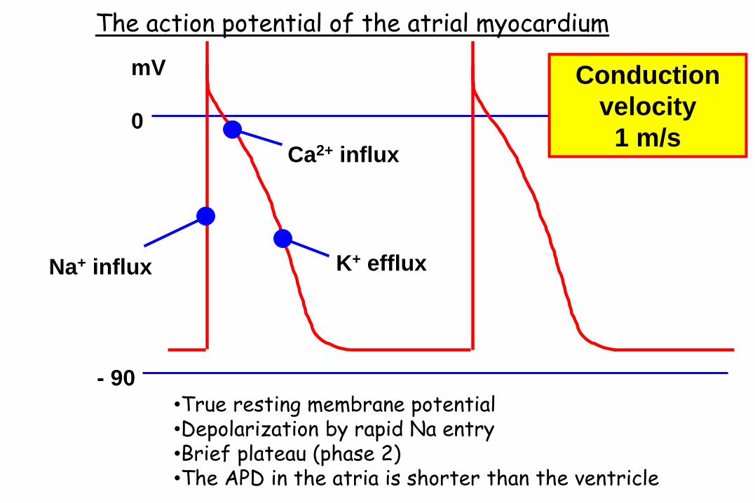
The action potential of the atrial myocardium
Na+ influx
Ca2+ influx
K+ efflux
Conduction
velocity
1 m/s
•True resting membrane potential •Depolarization by rapid Na entry •Brief plateau (phase 2) •The APD in the atria is shorter than the ventricle
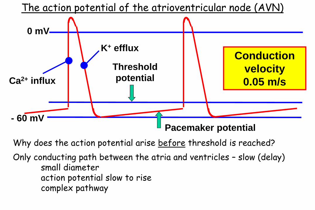
Threshold
potential
- 60 mV
0 mV
Pacemaker potential
Conduction
velocity
0.05 m/s Ca2+ influx
K+ efflux
Only conducting path between the atria and ventricles – slow (delay) small diameter action potential slow to rise complex pathway
The action potential of the atrioventricular node (AVN)
Why does the action potential arise before threshold is reached?

The action potential of the purkinje fibres
Threshold
potential
“Resting” membrane potential Pacemaker potential (weak)
Ca2+ influx
Na+ influx
K+ efflux
0 mV
-80 mV
Conduction
velocity
4 m/s
•Largest cells in the heart •Rapid depolarization •Refractory

Ca2+ influx
K+ efflux
Na+ influx
Resting membrane potential
The action potential of the ventricular myocyte
Conduction
velocity
1 m/s

Area of heart Cell diameter
(microns)
Fast Na+
channels
Gap junction
proteins
Speed of
impulse
conduction
SA Node < 10 (+) + 0.05 m/s
Atrial muscle 15 ++ ++ 1 m/s
AV Node < 10 (+) + 0.05 m/s
Purkinje
fibres
40 ++++ ++ 4 m/s
Ventricular
muscle
20 ++ ++ 1 m/s
Summary of the characteristics of cells in various regions

V1
V2
V1 – V2
0
ECG electrodes only pick up current if voltage difference points towards them
How does electrical activity relate to the ECG?
Depolarization wave travelling toward a positive electrode = positive deflection Depolarization wave travelling away from a positive electrode = negative deflection.
Repolarization wave travelling toward a positive electrode = negative deflection.
Repolarization wave travelling away from a positive electrode = positive deflection.
Perpendicular to an electrode = no deflection
Amplitude (V) is directly related to the mass of tissue

atrium
SA node
AV node
Purkinje fibre
ventricle
QRS T P
How does the action potential relate to the ECG?
Epi
Mid

Effects of the autonomic nervous system on the heart
SAN
AVN
Sympathetic chains
Vagus nerve (ACh)
Purkinje fibres
Autonomic nervous system
Heart rate
AVN conduction speed
AP duration
Sympathetic Parasympathetic (vagal nerves)
Heart rate
AVN conduction speed

The sympathetic nervous system
Stimulation of sympathetic nerve supply
Threshold potential
Pacemaker potential
Sinoatrial node
Tachycardia
Noradrenaline b-receptors cAMP depolarizing currents e.g. Funny &Ca current Deactivated repolarising (K) currents more quickly

The sympathetic nervous system
Atrioventricular node
Activates b-receptors in the AVN increased conduction speed
Atria and ventricle
Atrial and ventricular action potential is shortened due to increased repolarising (K) current

Effects of the autonomic nervous system on the heart
SAN
AVN
Sympathetic chains
Vagus nerve (ACh)
Purkinje fibres
Autonomic nervous system
Sympathetic Parasympathetic (vagal nerves)
Heart rate Heart rate
AVN conduction speed
AP duration
AVN conduction speed
N.B. The ventricular myocardium does not receive parasympathetic innervation

Threshold potential
Stimulation of vagus nerve Pacemaker potential
Bradycardia
The parasympathetic nervous system Sinoatrial node
ACh acts on muscarinic receptors
K+-channel opening - causes hyperpolarization
and a pacemaker potential of reduced slope.

The parasympathetic nervous system
Atrioventricular node
Reduces conduction through the AVN strong enough heart block
Autonomic nerves and the normal heart rate
Nerve fibres are always active pacemaker firing continuously modified
Parasympathetic predominates at rest atropine + propranolol = intrinsic rate (~105 beats per minute)

Learning objectives
•How is the heart beat generated?
•How does excitation propagate through the heart?
•What are the regional differences in the cardiac action potential?
•Effects of the autonomic nervous system


