
Doppler in Obstetrics
Farhan Hanif,MD
Maternal Fetal Medicine
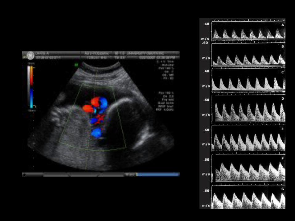
• Doppler assessment of the placental and fetal circulation is important tool screening for adverse pregnany outcomes
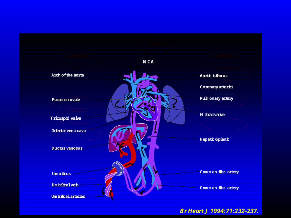
Arch of the aorta
Foramen ovale
Inferior vena cava
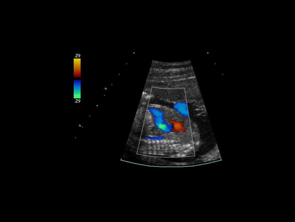
Ductus venosus
Umbilicus
Umbilical vein
Umbilical arteries
Aortic isthmus
Coronary arteries
Pulmonary artery
Hepatic/Splenic
Common iliac artery
Common iliac artery
Br Heart J 1994;71:232-237.
Tricuspid valveMitral valve
MCA

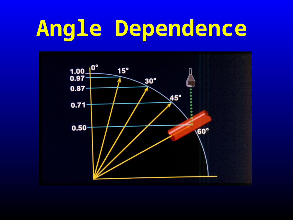
Angle Dependence
Rizzo et al. Ultrasound Obstet Gynecol 1996;7:401-410.
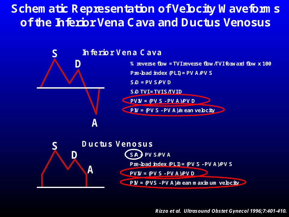
Schematic Representation of Velocity Waveformsof the Inferior Vena Cava and Ductus Venosus
SD
A
Inferior Vena Cava% reverse flow = TVI reverse flow/TVI forward flow x 100
Pre-load index (PLI) = PV A/PV S
S/D = PV S/PV D
S/D TVI = TVI S/TVI D
PVIV = (PV S - PV A)/PV D
PIV = (PV S - PV A)/mean velocity
Ductus VenosusSD
A
S/A = PV S/PV A
Pre-load index (PLI) = (PV S - PV A)/PV S
PVIV = (PV S - PV A)/PV D
PIV = (PVS - PV A)/mean maximum velocity
Doppler in IUGR
• EFW<10th %ile
• EFW <2SD above the mean
• EFW <5th %ile
• AC <5th %ile
• ACOG defines IUGR as EFW <10thile
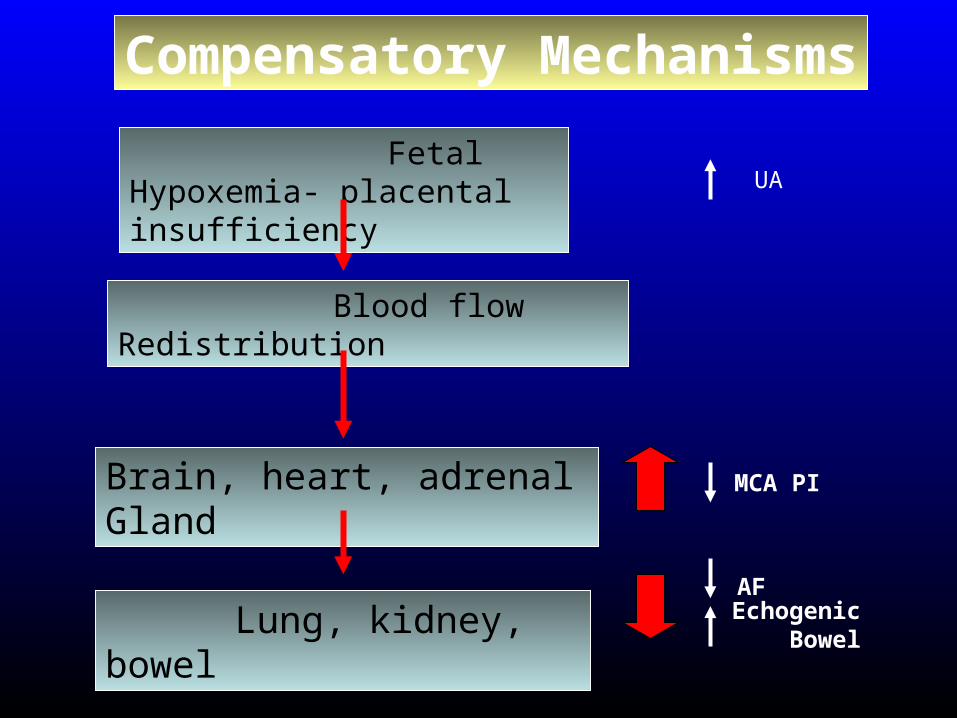
Compensatory Mechanisms
MCA PI
Echogenic Bowel
AF
Fetal Hypoxemia- placental insufficiency
Blood flow Redistribution
Brain, heart, adrenal Gland
Lung, kidney, bowel
UA
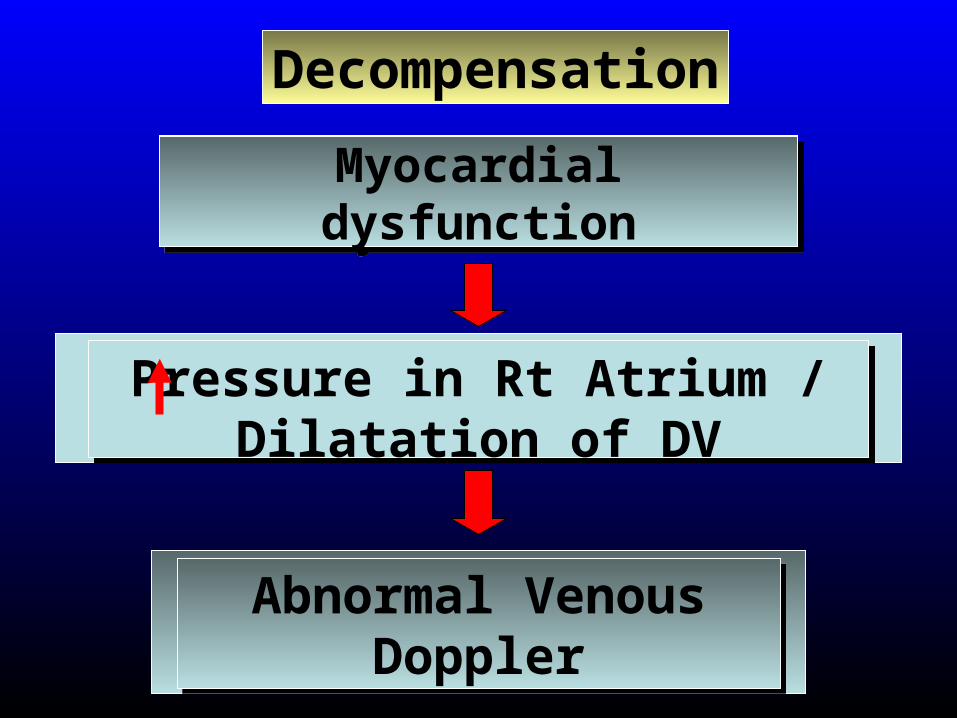
Abnormal Venous Doppler
Abnormal Venous Doppler
Myocardial dysfunctionMyocardial dysfunction
Pressure in Rt Atrium / Dilatation of DV
Pressure in Rt Atrium / Dilatation of DV
Decompensation
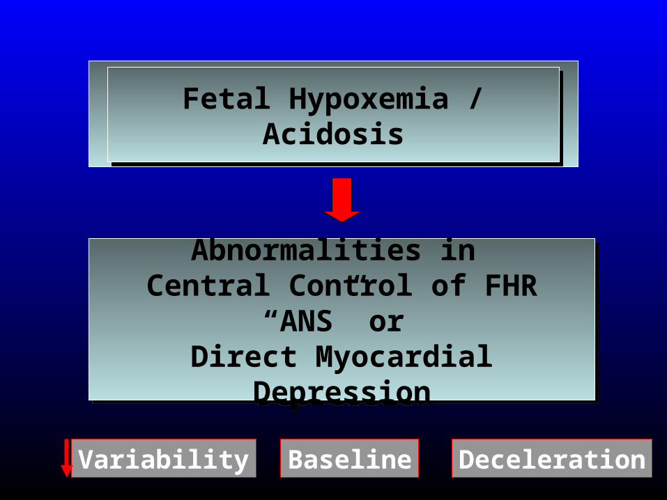
Fetal Hypoxemia / AcidosisFetal Hypoxemia / Acidosis
Abnormalities in Central Control of FHR
“ANS” or Direct Myocardial Depression
Abnormalities in Central Control of FHR
“ANS” or Direct Myocardial Depression
Variability Baseline Deceleration
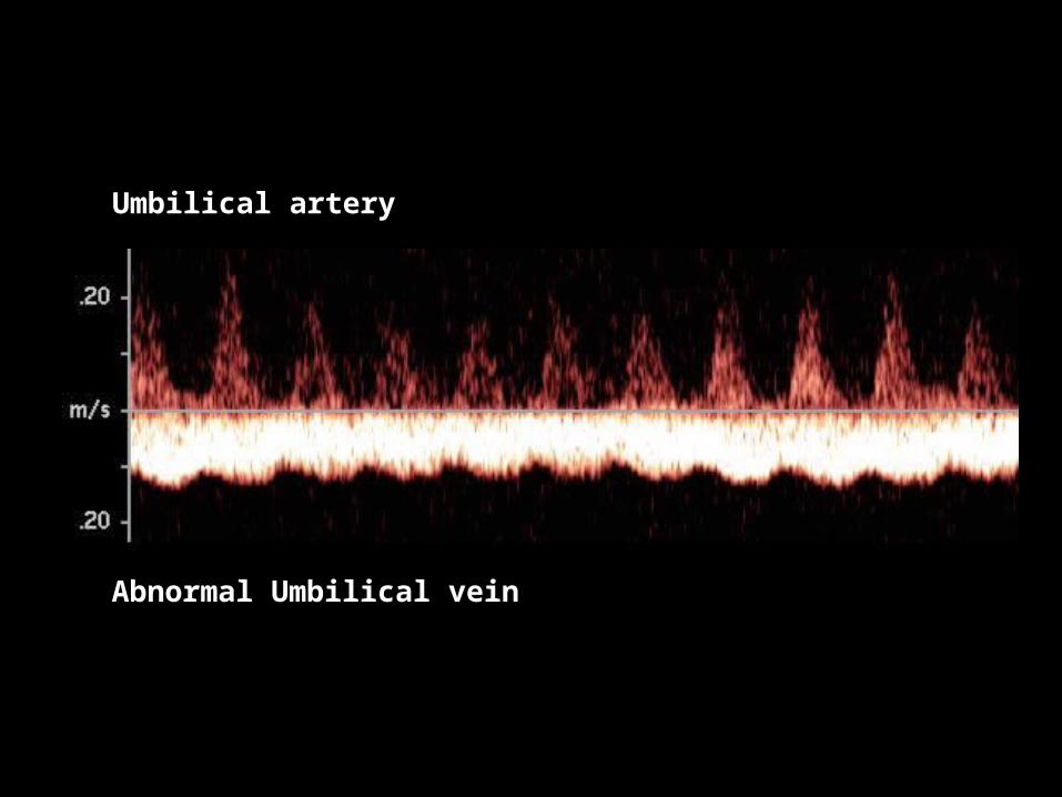
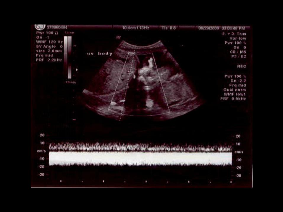
Umbilical artery
Abnormal Umbilical vein
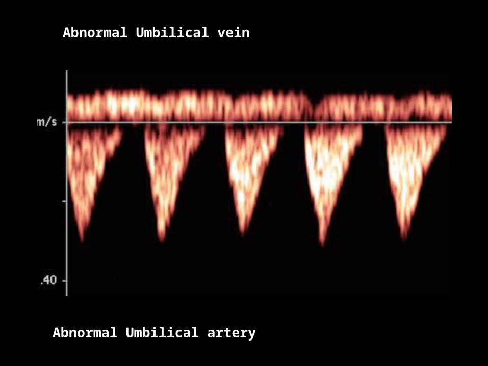
Abnormal Umbilical vein
Abnormal Umbilical artery
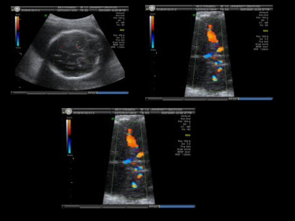
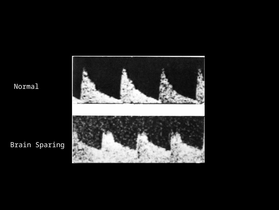
MCA waveformsA = Normal
B = “Brain sparing effect”
Normal
Brain Sparing
MCA Doppler In Anemia
• In Anemic fetuses, the PSV will inrease.
• Obtaining PSV at 0 degrees angle is important in anemic fetuses.
• Increase False positive rate after 34 weeks
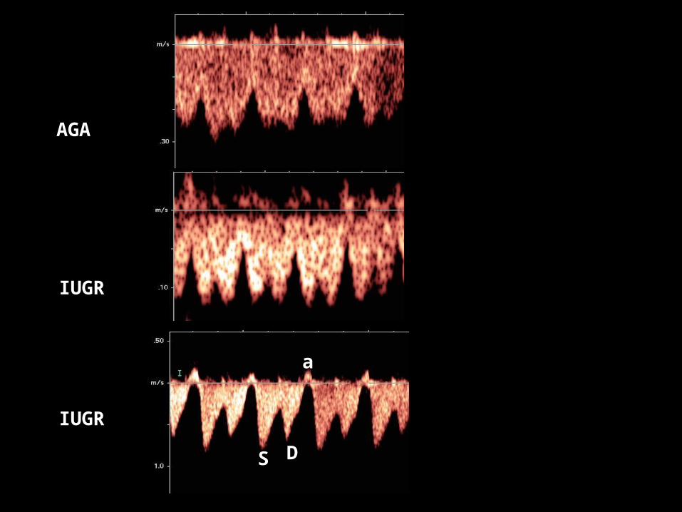
IUGR
AGA
IUGR
S D
a
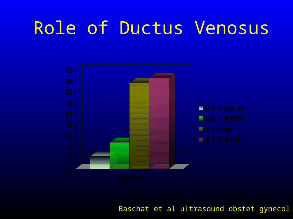
Role of Ductus Venosus
Baschat et al ultrasound obstet gynecol 2004
0
5
10
15
20
25
30
35
40
45
Groups
DV Normal
UA A/REDF
DV Abn
DV A/REDF
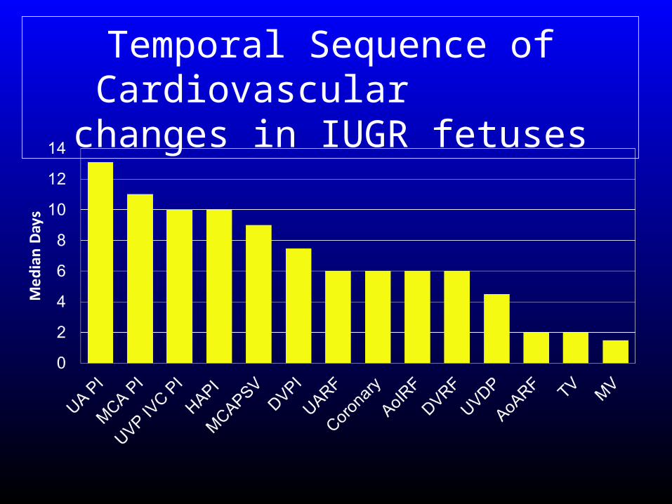
Temporal Sequence of Cardiovascular changes in
IUGR fetuses
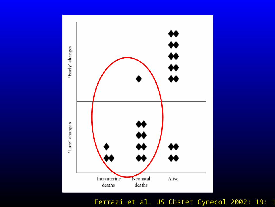
Ferrazi et al. US Obstet Gynecol 2002; 19: 140-6
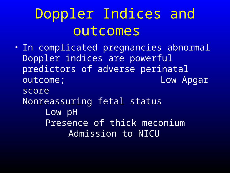
Doppler Indices and outcomes
• In complicated pregnancies abnormal Doppler indices are powerful predictors of adverse perinatal outcome;
Low Apgar scoreNonreassuring fetal status Low pHPresence of thick meconiumAdmission to NICU

Doppler Indices and outcomes
• Reduce perinatal death and unnecessary induction of labor in the preterm growth restricted fetus.
• A meta-analysis use of Doppler ultrasonography reduced the odds of perinatal death by 38 percent (95% CI 15-55)
Alfirevic Z et al Am J Obstet Gynecol 1995

Umbilical Artery
• Absence or reversal of end-diastolic flow in the umbilical artery is suggestive of poor fetal condition, whereas normal or slightly decreased umbilical Doppler flow is rarely associated with significant morbidity
Ott WJ J Ultrasound Med 2000

IUGR
Serial Growth Scan 4 weeks intervalDoppler UA and MCA every 1-2 weeksEvaluate MCA at term
Doppler UA and MCA
If Normal
Repeat Doppler in 1-2 weeks
If normal
?APFSConsider Delivery at 39 weeks
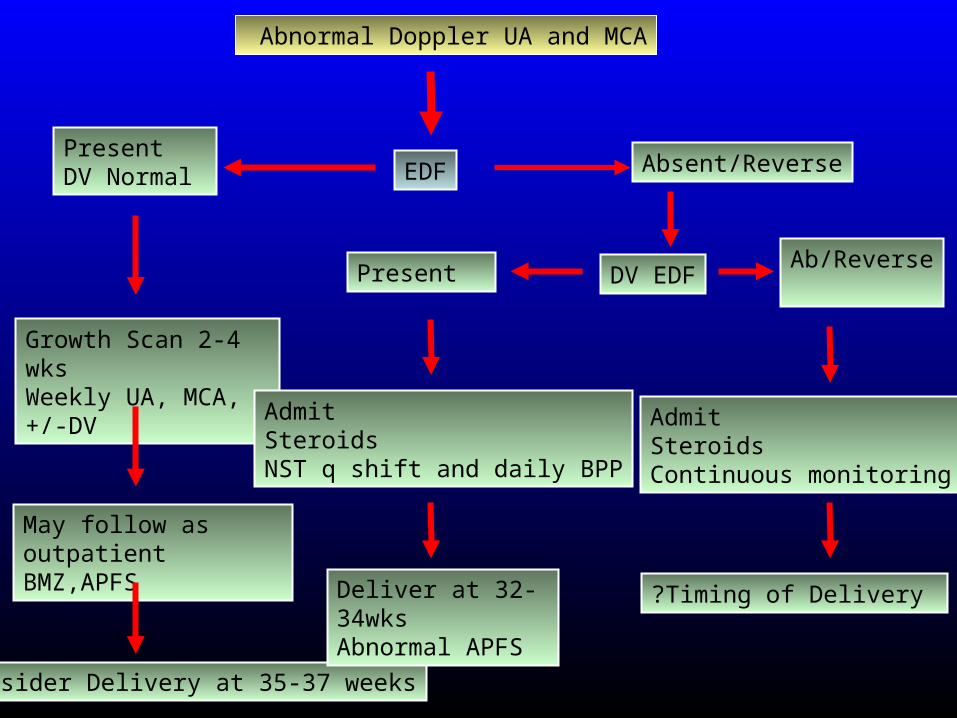
EDF
Abnormal Doppler UA and MCA
PresentDV Normal
Growth Scan 2-4 wksWeekly UA, MCA,+/-DV
May follow as outpatientBMZ,APFS
Consider Delivery at 35-37 weeks
Absent/Reverse
Admit Steroids NST q shift and daily BPP
Deliver at 32-34wks Abnormal APFS
DV EDFPresent Ab/Reverse
Admit Steroids Continuous monitoring
?Timing of Delivery
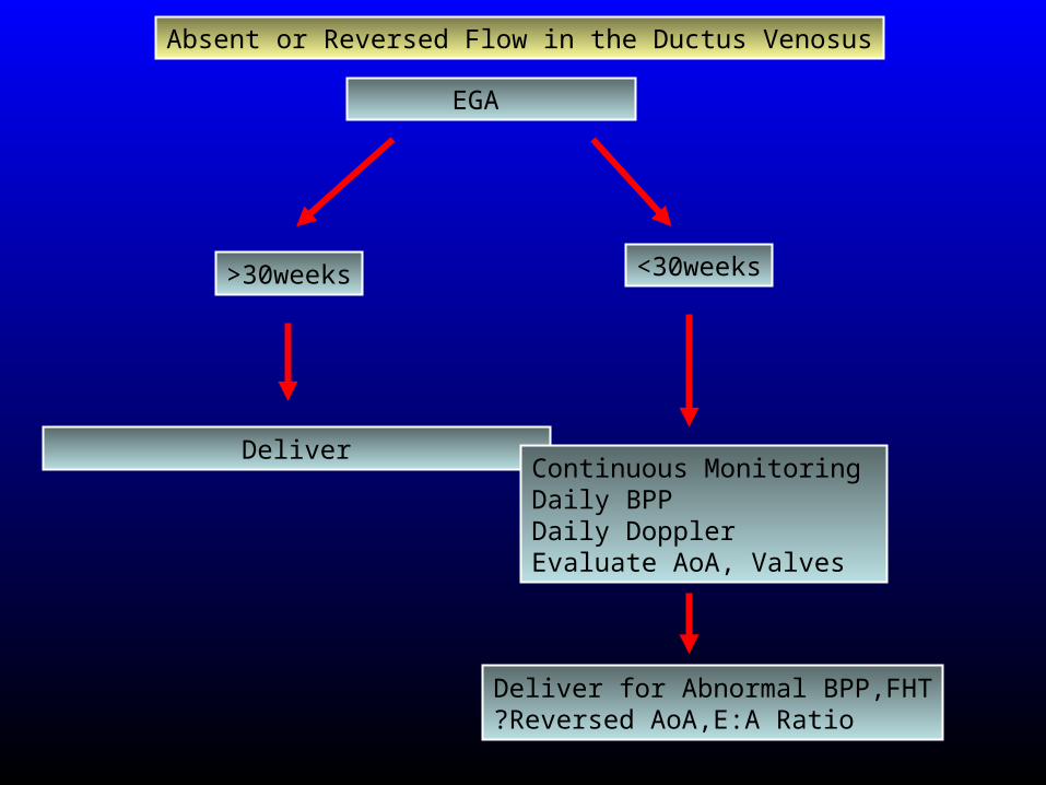
EGA
>30weeks
Deliver
<30weeks
Continuous Monitoring Daily BPPDaily DopplerEvaluate AoA, Valves
Deliver for Abnormal BPP,FHT?Reversed AoA,E:A Ratio
Absent or Reversed Flow in the Ductus Venosus

Doppler in AGA Fetuses
• Routine screening with dopplers in AGA fetuses is controversial
• However, abnormal UA identifies the fetuses at risk in uncomplicated pregnancies as
DM Ch HTNSLE
Maternal autoimmune Twins Postterm
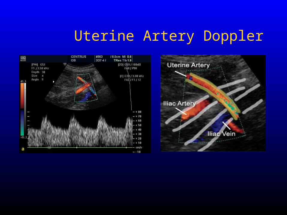
Uterine Artery Doppler
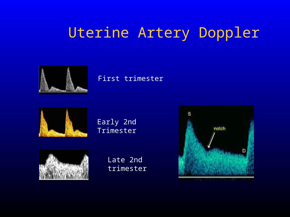
First trimester
Early 2nd Trimester
Late 2nd trimester
Uterine Artery Doppler

Outcome Sensitivity Specificity NPV
PE 78 95 99
IUGR <10 23 95 96
IUGR <3 36 96 92
Prediction of PE
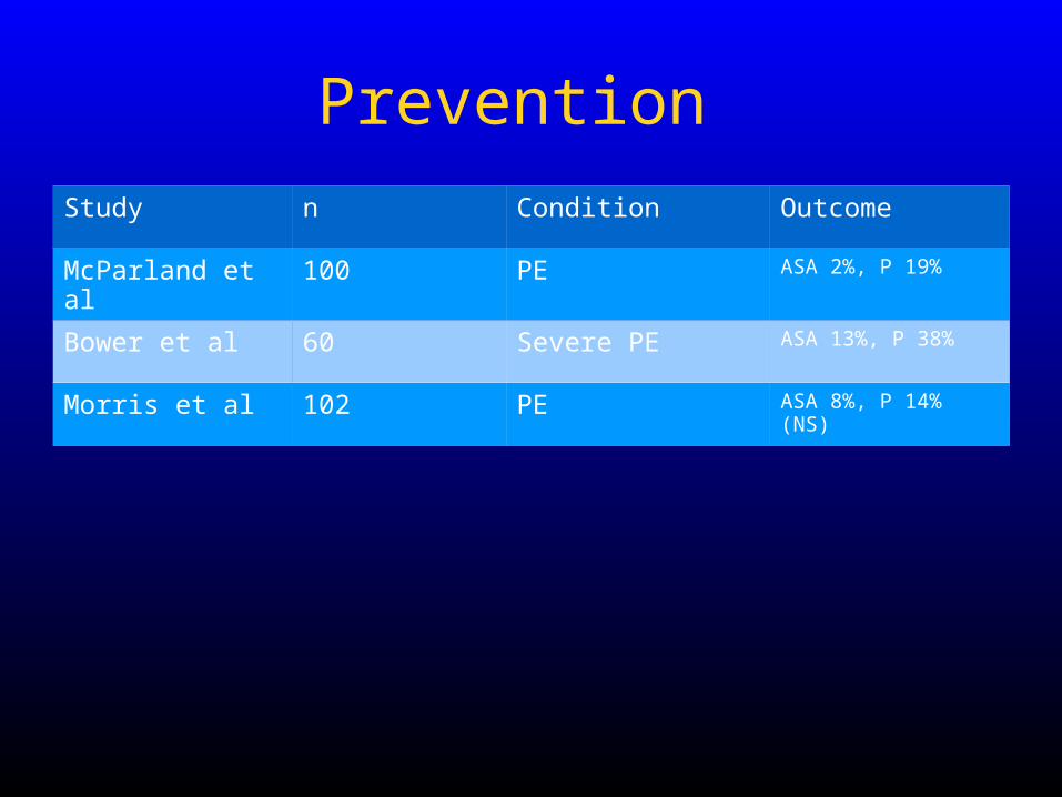
Study n Condition Outcome
McParland et al 100 PE ASA 2%, P 19%
Bower et al 60 Severe PE ASA 13%, P 38%
Morris et al 102 PE ASA 8%, P 14% (NS)
Prevention
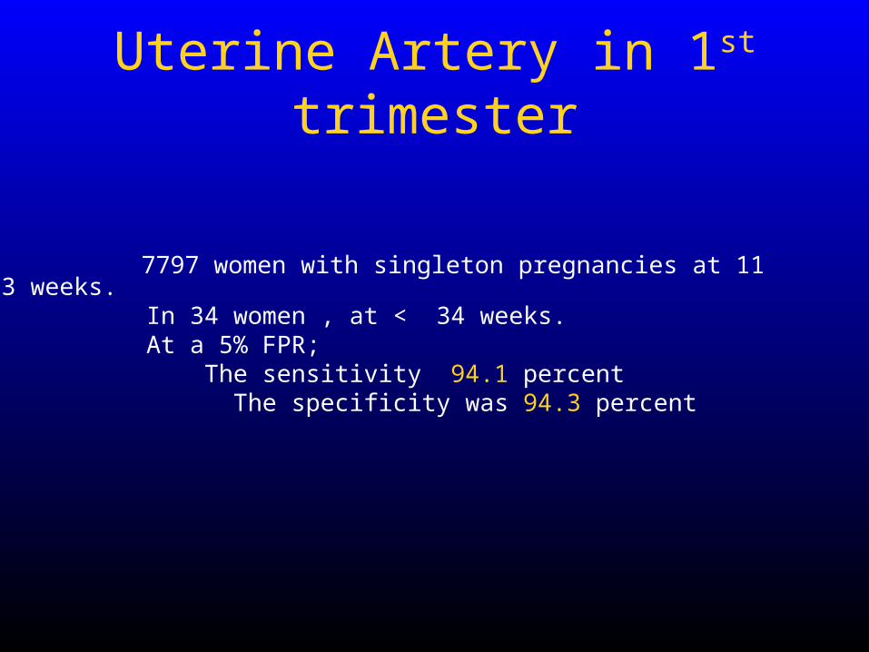
Uterine Artery in 1st trimester
7797 women with singleton pregnancies at 11 to 13 weeks. In 34 women , at < 34 weeks. At a 5% FPR; The sensitivity 94.1 percent The specificity was 94.3 percent
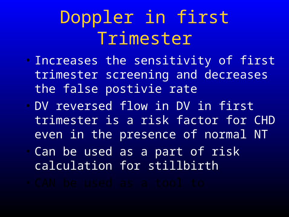
Doppler in first Trimester
• Increases the sensitivity of first trimester screening and decreases the false postivie rate
• DV reversed flow in DV in first trimester is a risk factor for CHD even in the presence of normal NT
• Can be used as a part of risk calculation for stillbirth
• CAN be used as a tool to


