
Diagnostic approach to hereditary renal hypouricemia
Ivan Sebesta
Institute of Inherited Metabolic Disorders, Institute of Medical Biochemistry and Laboratory Diagnostics, First Faculty of Medicine,
Charles University in Prague
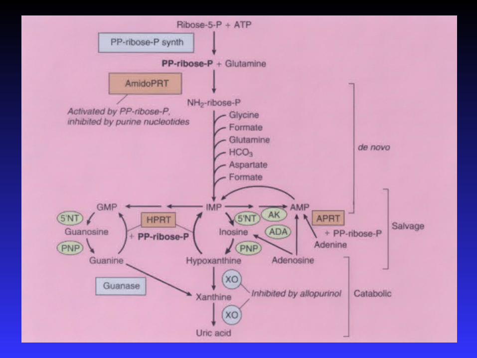

Introduction – hypouricemia
- hereditary renal hypouricemia
- hereditary xanthinuria
Characteristics of Czech patients
Problems of diagnosis
- incidence
- dg. flow charts


Hypoexcretion of urate
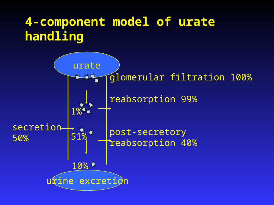
secretion50%
glomerular filtration 100%
reabsorption 99%
post-secretory reabsorption 40%
urine excretion
urate
4-component model of urate handling
1%
51%
10%
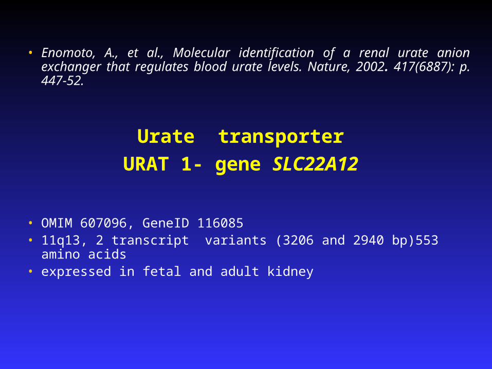
• Enomoto, A., et al., Molecular identification of a renal urate anion exchanger that regulates blood urate levels. Nature, 2002. 417(6887): p. 447-52.
• OMIM 607096, GeneID 116085• 11q13, 2 transcript variants (3206 and 2940 bp)553 amino acids• expressed in fetal and adult kidney
Urate transporter
URAT 1- gene SLC22A12

Lipkowitz MS.Curr Rheumatol Rep (2012) 14:179-188

Hypouricemia < 119 µmol/l (2 mg/dL)
it is important to distinguish :
primary genetic defect - hereditary xanthinuria
transport defect - primary renal hypouricemia (RHUC1, RHUC2)
secondary increased renal secretion (Fanconi sy.,Wilson´s disease) medication (allopurinol,salicylates )
severe liver disease
thyrotoxicosis, diabetes mellitus, acute respiratory sy.

Hereditary xanthinuria
xanthine oxidoreductase ( XO) deficiency type I
XO def. + aldehyde oxidase deficiency type II
molybdenum cofactor def. “ “ + sulfite oxidase def.
dg.markers: hypouricemia
high urinary concentration of xanthine
symptoms: cca 50% patients - hematuria,renal colic acute renal failure,
crystalluria,urolithiasis
th: low purine diet,high fluid intake
(alkalization of urine is of no value)

• new transport defect of uric acid
• biochemical markers– hypouricemia (SKM<120 μmol/l)
– increased excretion fraction of uric acid (EFKM >10% )
• clinical features
– urolithiasis– acute renal failure (exercise-induced)
RHUC 1 - URAT1 (SLC22A12 gene) RHUC 2 - GLUT 9 (SLC2A9 gene)
Hereditary renal hypouricemia

Hereditary renal hypouricemia mutation - gene SLC22A12 W258X- prevalent mutation
Enomoto, A., et al., Nature, 2002. 417(6887): p. 447-52. Ichida, K., et al., J Am Soc Nephrol, 2004.15:p.164-73.Iwai, N., et al., Kidney Int, 2004.66:935-44.Wakida, N., et al., J Clin Endocrinol Metab, 2005. 90:2169-74.
Sendai
Sapporo
TokyoOsakaKitakyushu

Institute of Inherited Metabolic Disorder, First Faculty of of Medicine, Charles University, Prague
( patients with HPRT def., FJHN, APRT def, ASL def., ADA def.)
Are disorders with hypouricemia also
in the Czech population ?

Investigation of unexplained hypouricemia
exclusion of secondary causes of hypouricemia !
1.assessment of uric acid - serum , urine
2.urinary purine metabolites
(+ allopur.loading test)
3.molecular genetic analysis
SLC22A12, SLC2A9
(in cooperation with Japan – SLC17A3, ABCC4, ABCG2 )
AU
0.00
0.02
0.04
0.06
0.08
0.10
0.12
0.14
Minutes
2.00 3.00 4.00 5.00 6.00 7.00 8.00 9.00 10.00 11.00 12.00 13.00 14.00
XO. def.HX Xanthine
UA
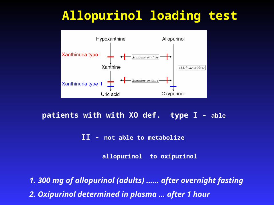
Allopurinol loading test
patients with with XO def. type I - able
II - not able to metabolize
allopurinol to oxipurinol
1. 300 mg of allopurinol (adults) …… after overnight fasting
2. Oxipurinol determined in plasma … after 1 hour
Ichida K et al (1997) J Clin Invest 99, 2391-97
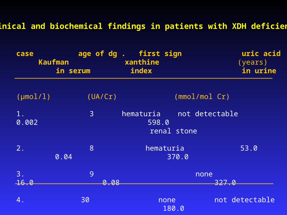
Clinical and biochemical findings in patients with XDH deficiency
case age of dg . first sign uric acid Kaufman xanthine (years) in serum index in urine
(µmol/l) (UA/Cr) (mmol/mol Cr)
1. 3 hematuria not detectable 0.002 598.0 renal stone
2. 8 hematuria 53.0 0.04 370.0
3. 9 none 16.0 0.08 327.0
4. 30 none not detectable 180.0
controls 120- 360 0.7 30.0

Clinical features and mutations (1-7th patients in SLC22A12 gene) and 8-9th patients in SLC2A9 gene
case sex age UA FEUA ARF uro- mutation
yrs μmol/l (%) lithiasis
1. f. 73 124 52.4 + - g. 8294-8302del
2. f. 39 58 53.4 + - g. 82948302del/ g.9184C/T
3. f. 53 78 60.3 - - g. 82948302del/ g.9184C/T
4. m. 35 63 43.0 - - g. 8145G/C g.9214G/A
5. f. 15 35 55.2 - - g. 8294-8302del g.9184C/T
6. m. 5 95 52.6 - + 1242-1250delGCTGGCAGG
7. m. 5 50. - - 1245-1253delGGCAGGGCT
8 f. 18 11 240.0 - - g. 43412_43413insC
9. m. 23 10 220.0 - - g. 43412_43413insC

Clinical features (two UK patients with acute renal failure-ARF) and mutations in SLC2A9 gene
case sex age UA FEUA Cr ARF mutation
yrs μmol/l (%) μmol/l
1. m 12 40 93.0 297 + p.G216R; p.N333S
2. m 14 58 53.4 202 + p.G216R
- further evidence … …..SLC2A9 is a causative gene in RHUC2
- supports the prediction….both URAT1 and GLUT9 are essential for UA reabsorption
.
Sebesta I. Adv Chronic Kidney Dis 2012,19(6):398-403 Stiburkova B ,Ichida K,Sebesta I. Mol Genet Metab.2011,102(4):1411-5

Renal hypouricemia -unrecognized disorder ?
absence of SLC22A12 gene mutations in Greek Caucasian Tzovaraz V. et.al. Scand J Clin lab Invest.2007;67:589-95
5 patients (Macedonia), 2 (UK) – RHUC1 (URAT 1)
Tesic V. et.al. Plos One. 2011;6(12):e28641
9
5
4

EARLY DIAGNOSIS of INBORN ERRORS OF METABOLISM
1. available methods
2. proper indication
screening
newborn
(PKU, hypothyreosis.etc.)
selective screening
- family history
-suspicious clinical signs
diagnostic guidelines

Dg. flow chart for unexplained hypouricemia (SUA: <120 μmol/l )
Evaluation of case history ( urolithiasis, seizures, immunodeficiency) Exclusion of secondary causes ( drugs /allopurinol/, Fanconi sy. etc.)
↓1. Estimation of EXCRETION FRACTION OF UA ↓
if high→ - mol.genet.analysis of URAT1, GLUT9
2. Urinary concentration of XANTHINE, S-SULFOCYSTEIN, THIOSULFATE
3. Urinary concentration of (DEOXY) GUANOSINE, (DEOXY) INOSINE ↓ if positive - assay of purine nucleoside phoshorylase (PNP) in ery.

Dg.protocol allows to differentiate
a) XANTHINURIA (def.XO) (lithiasis, 50% of the patients are asymptomatic)
b) COMBINED DEFICIENCY OF XO/SULPHITE OXIDASE (seizures in newborns, evaluation od UA could be the first step to diagnosis)
c) PURINE NUCLEOSIDE PHOSPHORYLASE (defect of T-cell
immunity)
c) HEREDITARY RENAL HYPOURICEMIA (lithiasis, high EF-UA)
d) Primary hypouricemia can be excluded ( ? new defect)

Diagnosis of hereditary renal hypouricemia
1. estimation of uric acid (UA) in serum
- if less then 120 µmo/l
2. estimation of excretion fraction of UA
- if high more than 10%
3. exclusion of other secondary causes
of hyperuricosuric hypouricemia
if excluded
4. molecular analysis of SLC22A and SLC2A9 genes

Conclusions
• hypouricemia → risk factor for kidney injury
→ indication for detailed purine metabolic investigation
• hypouricemia can be good diagnostic tool – enables to find asymptomatic patients
• available guidelines will help for early diagnosis of purine disorders with hypouricemia
Conclusions
• first patients with hereditary renal hypouricemia and xanthinuria were diagnosed in Czech population
• findings of a defect in the SLC2A9 gene provides further evidence that SLC2A9 is a causative gene in renal hypouricemia and support the prediction that normal function of both URAT1 and GLUT 9 are essential for normal uric reabsorption
• renal hypouricemia is still unrecognized disorder and probably not wide spread in Asia only


