
Developments in CE/MS analysis of ProteinsMike KniermanSr. Research ScientistEli Lilly and Co.

Introduction
♦ Career has heavily involved protein and peptide mass spectrometry
♦ Interested in top down characterization of proteins.
♦ Poor LC separations of larger proteins, carryover / plugging is a big problem
♦ I see many different types of samples from pure proteins, immunoprecipitations from biological samples, to cell lysates. As such I need a universal CE method to start with for all proteins.

Why Capillary Electrophoresis?
• Separation does not require proteins to absorb on a surface
• Very small amounts of material used in separation
• Can get very high resolution separations• At pH <3 all proteins should be positively charged
and migrate to the mass spectrometer. (Universal method)
• How to interface to the mass spectrometer?

Mass Spectrometry Overview
IonizationNeed to bring molecule from the liquid into the gas phase with a charge(s)
AnalysisNeed to separate the ions by mass
mass/charge
DetectionNeed to observe separated ions
Destructive or nondestructive
M+M
M+
M+ M+
M++
M+
M+
-

Dovichi CE-MS interface
• LRAP funded project to bring the technology to Lilly and develop CE/MS of proteins
Wojcik R, et al., Rapid Commun. Mass Spectrom. (2010) 24: 2554–2560.

Initial Implementation on Thermo orbitrap velos pro
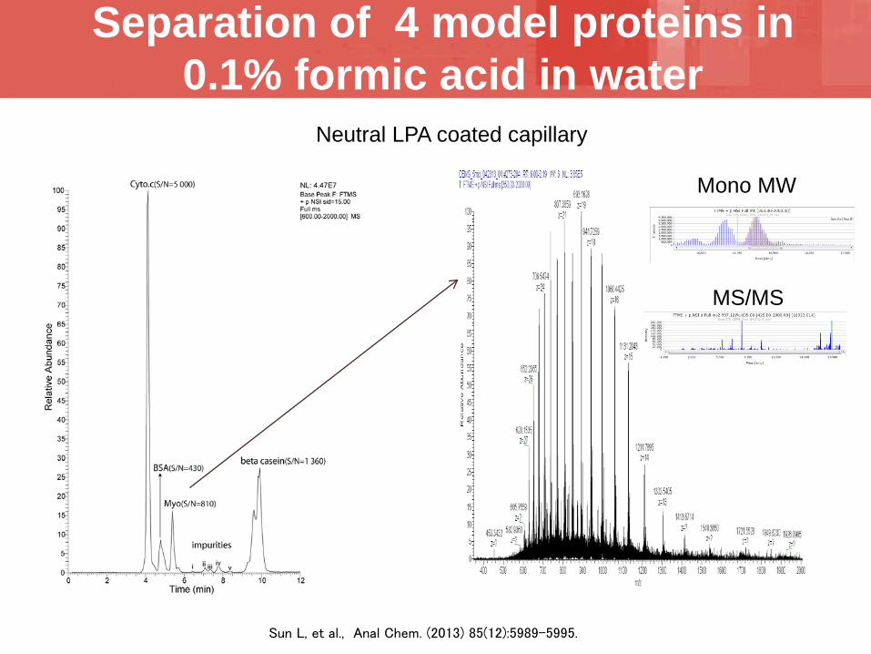
Separation of 4 model proteins in 0.1% formic acid in water
Sun L, et al., Anal Chem. (2013) 85(12):5989-5995.
MS/MS
Mono MW
Neutral LPA coated capillary

Operational issues
♦ Bubble formation in glass needle ♦ Very hard to flush out once formed
• Resulted in replacing the glass needle ♦ Contamination of sheath liquid
• If a large amount of protein was injected the excess would linger in the sheath and cause background issues.
• Long time to bleed out before next sample.♦ Tight space between CE and MS to adjust interface♦ Visualization of capillary placement and nanospray

Improvements
♦ Increase diameter of glass needle to 1.6 mm OD and 1.1 mm ID. • Allows use of standard HPLC fittings• Allows up to 3 x 360 um OD capillaries to be
placed in needle
♦ Second capillary is used to provide a constant back flush of sheath liquid at 20 ul/min.
Separation capillarySheath liquid capillary

Improvements
♦ Modified nanospray source for a better camera system
♦ Added remote electronic linear actuators for source adjustment.
♦ HPLC pump with degasser for sheath liquid helps prevent bubble formation
♦ Nanospray voltage always on. Keeping the glass needle spraying prevents from it from drying out and plugging.
♦ Neutral capillary with etched tip to get closer to nanospray tip. (Commercialized by CMP)

Innovation – CE divert valve
Separation capillarySheath liquid capillary
Analysis state
Divert state
Divert valves are common on LC/MS systems, help protect the MS system
Separation capillarySheath liquid capillary

CE divert valve Example - Diverted protein peak

CE divert valve - Uses
♦ Divert sample buffer peak away from nanosprayemitter
♦ Divert large peaks when looking for trace analytes
♦ Divert capillary flush from nanospray emitter• With this ability could you used non mass
spectrometry compatible buffers as a background electrolyte?
• This would allow CE/MS to leverage a large body of previous CE work on protein separations.
• Initial attempts were hampered by electrospray ion suppression in the buffers.

♦ Standard Agilent micro vials need a minimum of 10 µL for injecting on the CE system. The injection volume is 0.2-0.4% of the sample (~20-40 nl)
♦ Designed a vial insert with a sealed 10 µL pipette tip to reduce required volume to 2 µL resulting in a 5x sensitivity gain.(injecting 1-2% of the sample)
♦ The 2 µL volume allows for many injections.
Sample Volume Reduction

Insert use
2 uL mark
Use Eppendorf gel loading pipette tips to remove or mix reagents with sample
Then Spin to load into tip
Load 2 uL sample
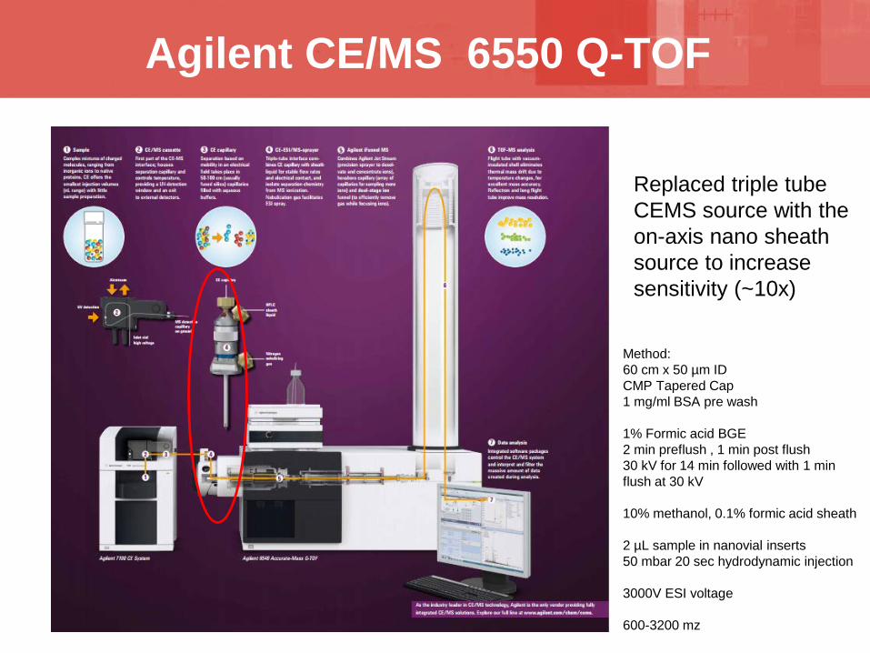
Agilent CE/MS 6550 Q-TOF
Replaced triple tube CEMS source with the on-axis nano sheath source to increase sensitivity (~10x)
Method:60 cm x 50 µm IDCMP Tapered Cap1 mg/ml BSA pre wash
1% Formic acid BGE2 min preflush , 1 min post flush30 kV for 14 min followed with 1 min flush at 30 kV
10% methanol, 0.1% formic acid sheath
2 µL sample in nanovial inserts50 mbar 20 sec hydrodynamic injection
3000V ESI voltage
600-3200 mz
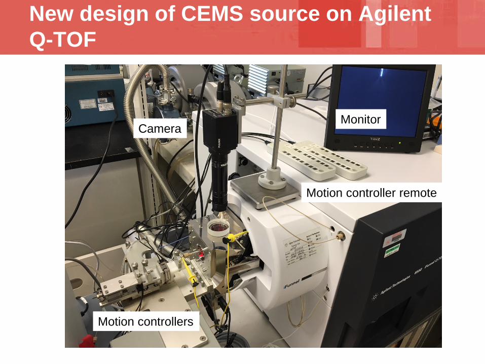
New design of CEMS source on Agilent Q-TOF
Motion controllers
CameraMonitor
Motion controller remote

Source close up
Sheath 360 um capillary Motion controllers
Vacuum Waste Line
SS HPLC tubingvery wide bore
HPLC unionCE 360 um capillary

CE/MS system
chiller
HPLC pump for sheath 6550 QTOF
CE
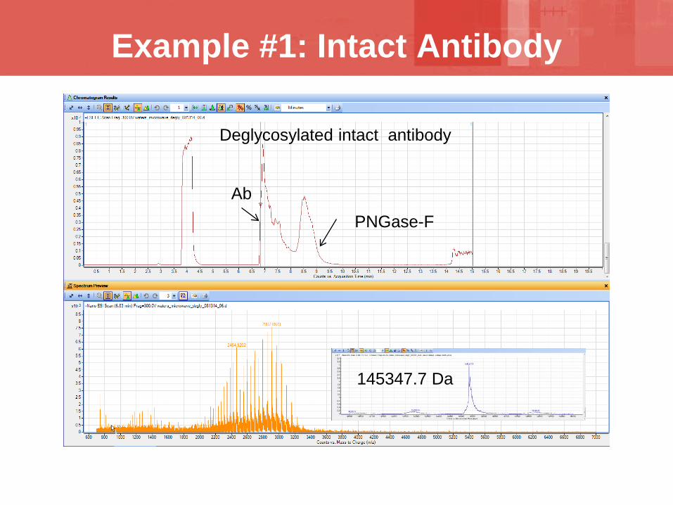
Example #1: Intact Antibody
Deglycosylated intact antibody
PNGase-FAb
145347.7 Da
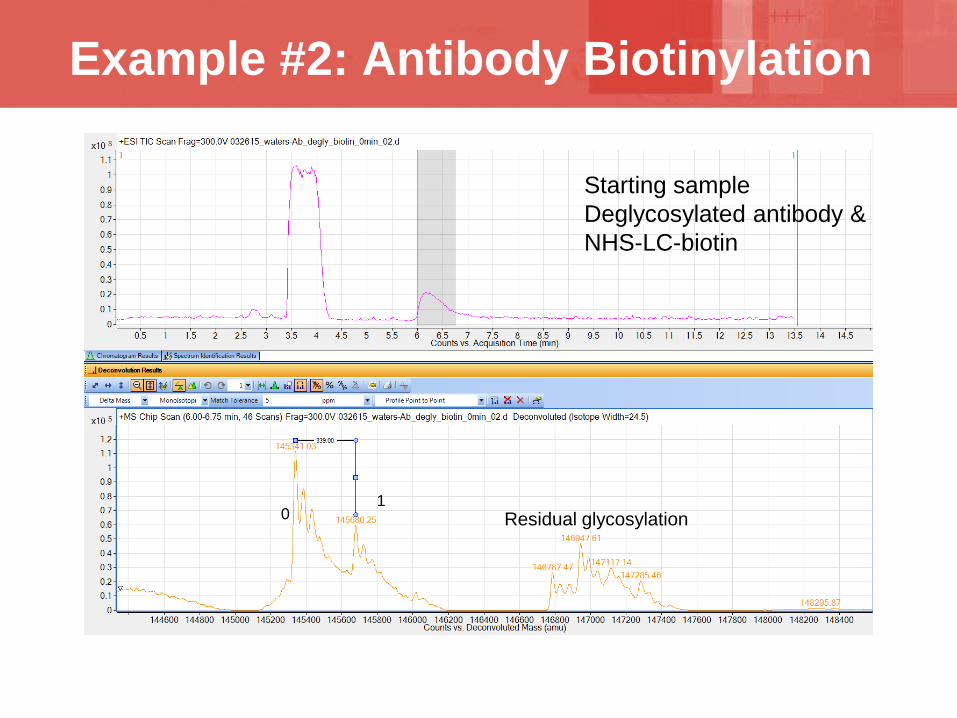
Example #2: Antibody Biotinylation
Residual glycosylation1
0
Starting sampleDeglycosylated antibody & NHS-LC-biotin

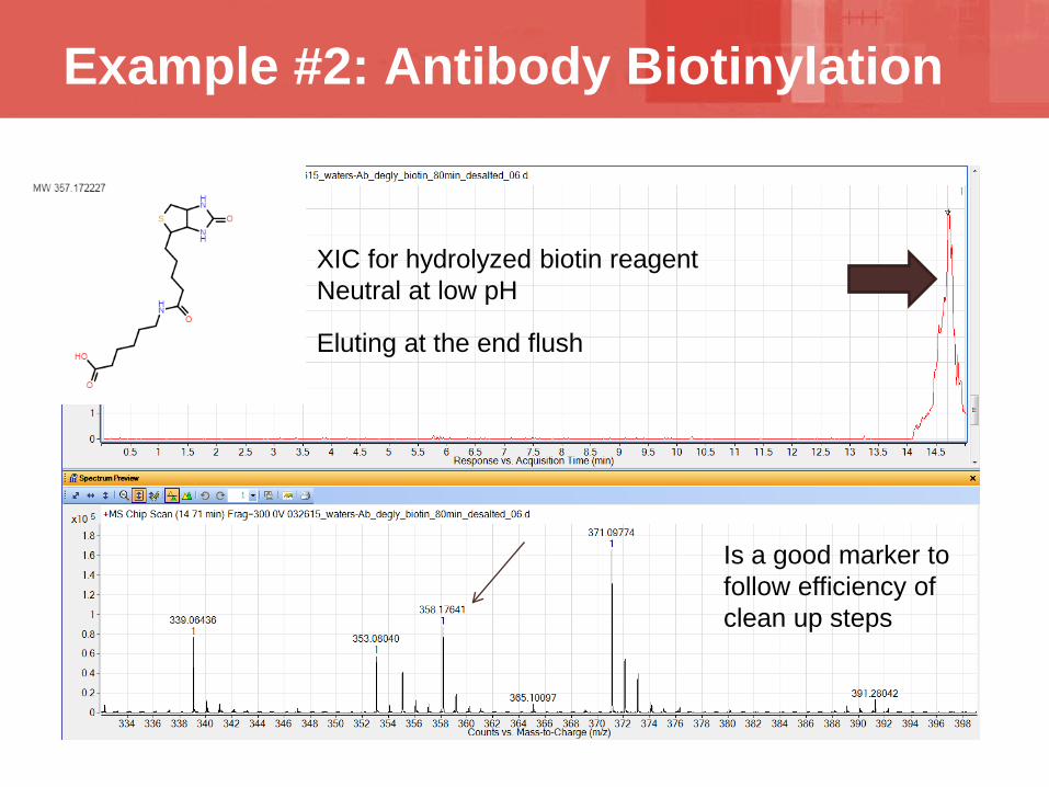
Example #2: Antibody Biotinylation
0
12 3
4
5
80 min reaction time

Example #2: Antibody Biotinylation
Light Chain
Heavy Chain1
2
12
00
Reduction of 80 min sample with DTT in vial
Example #2: Antibody Biotinylation
XIC for hydrolyzed biotin reagentNeutral at low pH
Eluting at the end flush
Is a good marker to follow efficiency of clean up steps

Example #3: Abeta 1-40 Quantitation
30 pg of abeta in vial
X axis is concentration of abeta, but only need 2uL in vial
1 ug/ml2 ng/2uL
Only 1/100 of the sample is actually injected into the capillary
Sensitivity

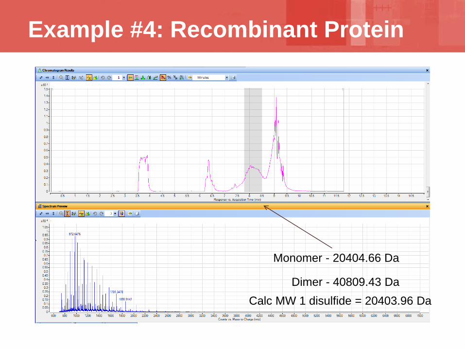
Example #4: Recombinant Protein
20406.06 Da
Calc MW no disulfide = 20405.96 Da
Expected fully reduced protein
Example #4: Recombinant Protein
Monomer - 20404.66 Da
Dimer - 40809.43 DaCalc MW 1 disulfide = 20403.96 Da

Example #4: Recombinant Protein
20404.12 Da
Calc MW 1 disulfide = 20403.96 Da

Example #4: Recombinant Protein
Both peaks are resulting from a single disulfide bond formation
Same mass
Coeluting dimer
Only by the combination of CE and MS can we quickly see this difference
Hydrodynamic effect

Example #4: Recombinant Protein
20406.50 Da
Calc MW no disulfide = 20405.96 Da
Reduction with DTT
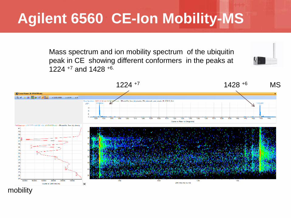
Agilent 6560 CE-Ion Mobility-MS
Mass spectrum and ion mobility spectrum of the ubiquitin peak in CE showing different conformers in the peaks at 1224 +7 and 1428 +6.
MS
mobility
1428 +61224 +7
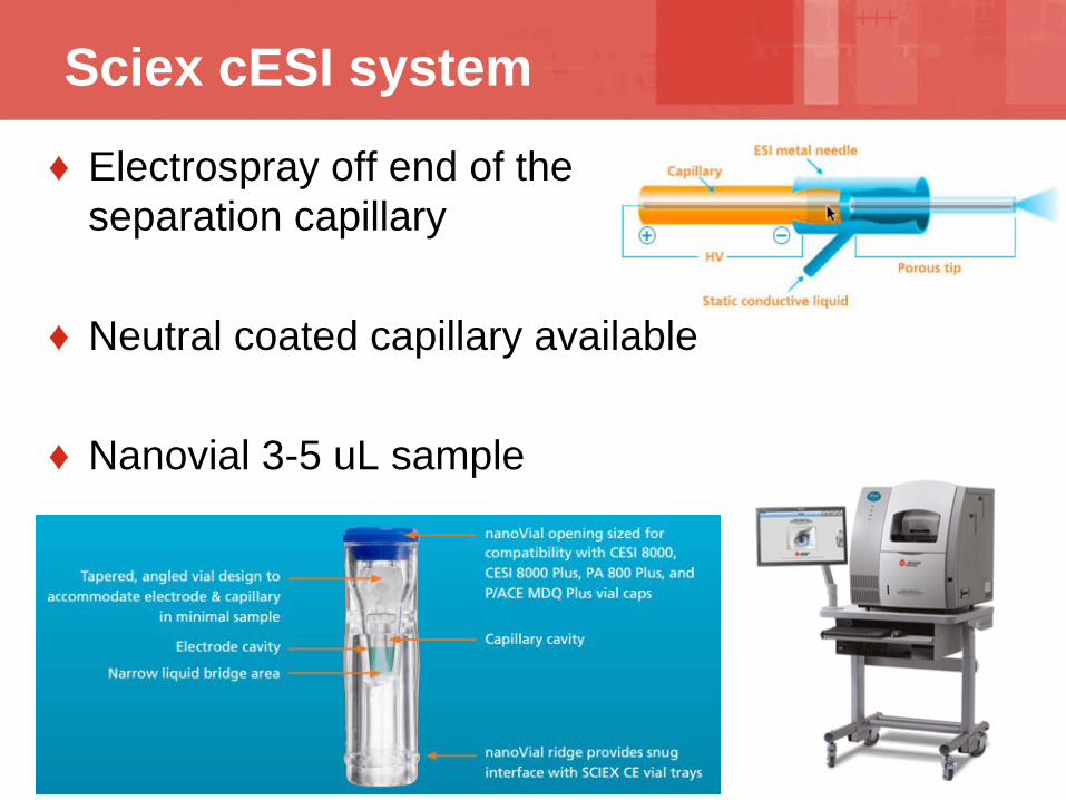
Sciex cESI system♦ Electrospray off end of the
separation capillary
♦ Neutral coated capillary available
♦ Nanovial 3-5 uL sample

New instrument in CEMS
♦ 908 Devices zipchip
• Based on technology out of Michael Ramsey’s lab at University of North Carolina – Chapel Hill
• Very fast CEMS runs- proteins in 3 min.• Quick setup

Zipchip Separation of 4 Proteins
RT: 0.00 - 3.03
0.0 0.2 0.4 0.6 0.8 1.0 1.2 1.4 1.6 1.8 2.0 2.2 2.4 2.6 2.8 3.0Time (min)
05
1015202530
35404550556065
707580859095
100
Rel
ativ
e A
bund
ance
2.11
1.86 1.98
2.43
1.811.26
2.370.990.68 0.94 1.070.860.65 2.701.57 2.86 2.951.500.53 1.360.310.05 2.551.700.08
NL:6.40E6TIC MS 082916_4mix_10k_1-5kV_lower_Ctrap_pressure_100mM_AmAc_08
Lysozyme
Ubiquitin Carbonic Anhydrase
Alpha-Synuclein
Isotachophoretic loading
Zipchip HR on Orbitrap velos pro
♦ CE/MS of proteins is sensitive♦ Low carry over of proteins observed for neutral
capillary♦ Very small amounts of sample are consumed for a
CE/MS run♦ CE/MS is amenable for intact and denatured
proteins♦ The nanosheath CE/MS system is very robust,
weeks of runtime with same nanospray needle and capillary
Summary

Eli Lilly & CompanyMegan LannanKyle FrederickJesus GutierrezJeff HillRobert SiegelRobert KonradLilly LRAP grant
University of Notre Dame Norman DovichiLiangliang Sun
AgilentGarrison BirchMark AlbrightMike HartzGeorge StaffordChris Klein
908 DevicesErin RedmanMike Goodwin
Acknowledgements


