Ebola Virus Detection in Oral Fluid Specimens • CID 2006:42 (1 June) • 1521
M A J O R A R T I C L E
Detection of Ebola Virus in Oral Fluid Specimensduring Outbreaks of Ebola Virus Hemorrhagic Feverin the Republic of Congo
Pierre Formenty,1 Eric Maurice Leroy,4 Alain Epelboin,2 Francois Libama,6 Marco Lenzi,1 Hinrich Sudeck,3
Philippe Yaba,4 Yokouide Allarangar,5 Paul Boumandouki,6 Virginot Blad Nkounkou,6 Christian Drosten,3
Allen Grolla,7 Heinz Feldmann,7 and Cathy Roth1
1World Health Organization, Department of Communicable Diseases Surveillance and Response, Geneva, Switzerland; 2Centre Nationalde la Recherche Scientifique et Museum National d’Histoire Naturelle, Paris, France; 3Bernhard-Nocht Institute for Tropical Medicine, Hamburg,Germany; 4Institut de Recherches pour le Developpement, U034, Centre International de Recherche Medicale de Franceville, Franceville,and 5World Health Organization, Regional Office, Libreville, Gabon; 6Ministere de la Sante et de la Population, Brazzaville, Republic of Congo;and 7Special Pathogens Program, National Microbiology Laboratory, Public Health Agency of Canada, Winnipeg, Manitoba, Canada
Background. Patients who have refused to provide blood samples has meant that there have been significantdelays in confirming outbreaks of Ebola virus hemorrhagic fever (EVHF). During the 2 EVHF outbreaks in theRepublic of Congo in 2003, we assessed the use of oral fluid specimens versus serum samples for laboratoryconfirmation of cases of EVHF.
Methods. Serum and oral fluid specimens were obtained from 24 patients with suspected Ebola and 10 healthycontrol subjects. Specimens were analyzed for immunoglobulin G antibodies by enzyme-linked immunosorbentassay (ELISA) and for Ebola virus by antigen detection ELISA and reverse-transcriptase polymerase chain reaction(RT-PCR). Oral fluid specimens were collected with a commercially available collection device.
Results. We failed to detect antibodies against Ebola in the oral fluid specimens obtained from patients whoseserum samples were seropositive. All patients with positive serum RT-PCR results also had positive results fortheir oral fluid specimens.
Conclusions. This study demonstrates the usefulness of oral fluid samples for the investigation of Ebolaoutbreaks, but further development in antibodies and antigen detection in oral fluid specimens is needed beforethese samples are used for filovirus surveillance activities in Africa.
Early detection and confirmation of viral hemorrhagic
fever outbreaks have frequently been hampered by the
difficulties in obtaining the appropriate clinical samples
from subjects with suspected Ebola and transporting
the samples [1–3]. In particular, the requirement for
blood or serum samples has often led to significant
delays in the notification and diagnosis of filovirus
hemorrhagic fever cases, because of the lack of available
sampling equipment, the weaknesses in the infrastruc-
Received 7 October 2005; accepted 21 January 2006; electronically published26 April 2006.
Reprints or correspondence: Dr. Pierre Formenty, Global Alert and ResponseTeam, Dept. of Communicable Diseases Surveillance and Response, World HealthOrganization, 20 Ave. Appia, CH-1211 Geneva 27, Switzerland ([email protected]).
Clinical Infectious Diseases 2006; 42:1521–6� 2006 by the Infectious Diseases Society of America. All rights reserved.1058-4838/2006/4211-0003$15.00
ture for communication and transportation, the ab-
sence of properly trained personnel, and the cultural
objections to the taking of blood or to other pre- or
postmortem invasive sampling. These conditions are
prevalent in many areas in Africa where outbreaks of
Ebola virus hemorrhagic fever (EVHF) and Marburg
virus hemorrhagic fever have occurred in the past
decade.
During an outbreak of EVHF in the Republic of
Congo in 2003 [4], strong cultural objections to col-
lection of blood or postmortem skin biopsy specimens
delayed the definitive diagnosis of the outbreak. On 28
January 2003, the Ministry of Health of the Republic
of Congo and the World Health Organization were
alerted of suspected cases of EVHF in the towns of
Mbomo and Kelle, which are situated in the Cuvette
Ouest region. They immediately sent a team to inves-
tigate the rumors and to collect initial clinical samples
at Northeastern U
niversity Libraries on O
ctober 16, 2014http://cid.oxfordjournals.org/
Dow
nloaded from
1522 • CID 2006:42 (1 June) • Formenty et al.
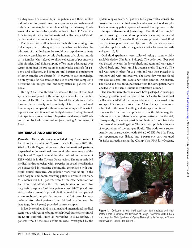
Figure 1. Collection of oral fluid specimens from subjects with sus-pected Ebola in Mbomo, the Republic of Congo, November 2003. (Photowas taken by Alain Epelboin of Centre National de la Recherche Scien-tifique/World Health Organization).
for diagnosis. For several days, the patients and their families
did not want to provide any tissue specimens for analysis, and
only 5 serum samples were obtained by 12 February. Ebola
virus infection was subsequently confirmed by ELISA and RT-
PCR testing at the Centre International de Recherche Medicale
de Franceville (Franceville, Gabon).
The reluctance to allow invasive methods of obtaining clin-
ical samples led to the query as to whether noninvasive ob-
tainment of oral fluid samples would be acceptable to patients
who were unwilling to permit obtainment of a blood sample
or to families who refused to allow collection of postmortem
skin biopsies. Oral fluid sampling offers many advantages over
serum sampling: the procedure is noninvasive and safe for pa-
tients and collectors, and some cultural barriers to obtainment
of other samples are absent [5]. However, to our knowledge,
no study thus far has assessed the use of oral fluid samples to
determine the antigen and antibody status in patients with
Ebola.
During 2 EVHF outbreaks, we assessed the use of oral fluid
specimens, compared with serum specimens, for the confir-
mation of EVHF. The main objective of the study was to de-
termine the sensitivity and specificity of tests that used oral
fluid samples, compared with use of serum samples. This article
reports virus detection and antibody detection findings for oral
fluid specimens collected from 24 patients with suspected Ebola
and from 10 healthy control subjects during 2 outbreaks of
Ebola.
MATERIALS AND METHODS
Patients. The study was conducted during 2 outbreaks of
EVHF in the Republic of Congo. In early February 2003, the
World Health Organization and other international partners
dispatched an international team to aid the government of the
Republic of Congo in containing the outbreak in the town of
Kelle, which is in the Cuvette Ouest region. The team included
medical anthropologists with expertise in social mobilization
who succeeded in restoring community compliance with out-
break-control measures. An isolation ward was set up in the
Kelle hospital and began receiving patients. From 18 February
to 6 March 2003, 11 patients who fit the case definition for
EVHF were admitted in the Kelle hospital isolation ward. For
diagnostic purposes, 9 of these patients (age, 28–75 years) pro-
vided verbal consent to provide both an oral fluid sample and
a venous blood sample. Serum and oral fluid samples were
collected from the 9 patients. Later, 10 healthy volunteer sub-
jects (age, 30–45 years) provided control samples.
In late November 2003, a national and international medical
team was deployed in Mbomo to help local authorities control
an EVHF outbreak. From 24 November to 8 December, 15
patients who fit the case definition were investigated by the
epidemiological team. All patients but 2 gave verbal consent to
provide both an oral fluid sample and a venous blood sample.
The 2 remaining patients provided an oral fluid specimen only.
Sample collection and processing. Oral fluid is a complex
fluid consisting of several components, including saliva and
crevicular fluid. Crevicular fluid is a component of oral fluid
that contains plasma-derived IgG and IgM, which transude
from the capillary beds in the gingival crevice between the teeth
and gums [6, 7].
Oral fluid specimens were collected using a commercially
available device (OraSure; Epitope). The collection fiber pad
was placed between the lower cheek and gum and was gently
rubbed back and forth, until it became moist (figure 1). The
pad was kept in place for 2–5 min and was then placed in a
transport vial with preservative. The same day, venous blood
was also collected into Vacutainer tubes (Becton Dickinson).
The blood and oral fluid specimens from the same patient were
labelled with the same unique identification number.
The samples were stored in a cool-box, packaged with a triple
packaging system, and transported to the Centre International
de Recherche Medicale de Franceville, where they arrived in an
average of 5 days after collection. All of the specimens were
subjected to the same handling and storage conditions.
When the oral fluid samples arrived in the laboratory, the
pads were dry, and there was no preservative left in the vial;
consequently, it was not possible to obtain any fluid from the
specimen after centrifugation. This was most probably because
of evaporation of the stopper liquid. The pads were subse-
quently put in suspension with 400 mL of PBS for 1 h. Then,
the supernatant was divided into 2 parts: one part was used
for RNA extraction using the QIamp Viral RNA kit (Qiagen),
at Northeastern U
niversity Libraries on O
ctober 16, 2014http://cid.oxfordjournals.org/
Dow
nloaded from

Ebola Virus Detection in Oral Fluid Specimens • CID 2006:42 (1 June) • 1523
Table 1. Results of analysis of oral fluid and serum specimens collected from patients with laboratory-confirmed Ebola in Kelle(February–March) and Mbomo (November–December), Republic of Congo, 2003.
Location,patient
Date ofdiseaseonset
Date thatsamples were
obtainedStatus
(date of outcome)aGingivalbleeding
Oral fluidspecimen result Serum sample result
IgG Ag RT-PCR IgG Ag RT-PCR
KelleK1 6 Feb 28 Feb Survivor No � � � 1:6400 � �
K2 6 Feb 28 Feb Survivor No � � � 1:1600 � �
K3 20 Feb 28 Feb Death (2 Mar) Yes � � + � 11:256 +K4 23 Feb 28 Feb Death (2 Mar) Yes � 11:256 + � 11:256 +K5 23 Feb 28 Feb Death (3 Mar) No � 11:64 + � 11:256 +K6 21 Feb 28 Feb Death (8 Mar) Yes � � + � 11:256 +K7 24 Feb 1 Mar Death (7 Mar) No � 11:4 + � 11:256 +K8 24 Feb 1 Mar Death (5 Mar) No � � + � 11:256 +K9 24 Feb 6 Mar Death (8 Mar) No � ND + � ND +K10 … 20 Mar Healthy subject No � � � � � �
K11 … 20 Mar Healthy subject No � � � � � �
K12 … 20 Mar Healthy subject No � � � � � �
K13 … 20 Mar Healthy subject No � � � � � �
K14 … 20 Mar Healthy subject No � � � � � �
K15 … 20 Mar Healthy subject No � � � � � �
K16 … 20 Mar Healthy subject No � � � � � �
K17 … 20 Mar Healthy subject No � � � � � �
K18 … 20 Mar Healthy subject No � � � � � �
K19 … 20 Mar Healthy subject No � � � � � �
MbomoM1 15 Nov 24 Nov Survivor No � 11:64 + � 11:256 +M2 20 Nov 24 Nov Death (29 Nov) No � 11:64 + ND ND NDM3 22 Nov 28 Nov Survivor No � 11:64 + ND ND NDM4 22 Nov 2 Dec Survivor No � � � 1:1600 � �
M5 16 Nov 2 Dec Survivor No � � � 1:400 � �
M6 15 Nov 3 Dec Survivor No � � � 1:400 � �
M7 15 Nov 5 Dec Survivor No � � � 1:1600 � �
M8 15 Nov 3 Dec Noncase … � � � � � �
M9 16 Nov 2 Dec Noncase … � � � � � �
M10 18 Nov 28 Nov Noncase … � � � � � �
M11 20 Nov 24 Nov Noncase … � � � � � �
M12 20 Nov 4 Dec Noncase … � � � � � �
M13 23 Nov 3 Dec Noncase … � � � � � �
M14 23 Nov 8 Dec Noncase … � � � � � �
M15 29 Nov 5 Dec Noncase … � � � � � �
NOTE. IgG antibodies against Ebola virus and Ebola virus antigens (Ag) were detected by ELISA. ND, not done; +, positive; �, negative.a Noncases were initially classified as suspected cases.
and the other part was incubated in water at 60�C for 1 h to
inactivate the virus and was then used for IgG detection and
antigen detection. The tests were performed with this fluid,
with a 1:10 dilution used for IgG detection and a 1:4 used
dilution for antigen detection. From the serum samples, we
used a dilution series of 1:100–1:6400 for the ELISA antibody
test and a dilution series of 1:4–1:256 for the antigen detection
test.
Laboratory tests. Both the oral fluid sample and the serum
sample were analyzed for IgG antibodies using the ELISA assay
and for Ebola virus using the antigen detection ELISA [8]. The
specimens were examined for Ebola RNA by RT-PCR using
primers derived from the L gene, as described elsewhere [9].
During the outbreak in Mbomo, we also detected Ebola RNA
in the samples using the SmartCycler Technology (Cepheid)
with L and NP gene-specific primers [10].
at Northeastern U
niversity Libraries on O
ctober 16, 2014http://cid.oxfordjournals.org/
Dow
nloaded from
1524 • CID 2006:42 (1 June) • Formenty et al.
Table 2. Detection of Ebola IgG antibodies in oral fluid speci-mens versus serum samples during 2 outbreaks of Ebola virushemorrhagic fever in the Republic of Congo, 2003.
Oral fluidsample results
Serum sample results
Positive Negative Total
Positive 0 0 0Negative 6 26 32
Total 6 26 32
NOTE. Data are no. of patients. The sensitivity of the IgG antibody testfor oral fluid samples was 0%, and the specificity was 100%; the positivepredictive value could not be calculated, and the negative predictive value was81%.
Table 3. Detection of Ebola antigen in oral fluid specimens ver-sus serum specimens during 2 outbreaks of Ebola virus hemor-rhagic fever in the Republic of Congo, 2003.
Oral fluidsample results
Serum sample results
Positive Negative Total
Positive 4 0 4Negative 3 24 27
Total 7 24 31
NOTE. Data are no. of patients. The sensitivity for detection of Ebola an-tigen for oral fluid samples was 57%, the specificity was 100%, the positivepredictive value was 100%, and the negative predictive value was 89%.
RESULTS
The results of the tests are presented in table 1. During the
February outbreak, all 9 suspected cases were confirmed to be
Ebola, whereas only 7 of the 15 suspected cases were confirmed
to be Ebola during the November outbreak. The 8 remaining
cases were classified as “noncases.”
Serologic testing for IgG antibodies. The results of tests
for the detection of IgG antibodies in oral fluid specimens
versus serum specimens are presented in table 2. Six patients
(K1, K2, M4, M5, M6, and M7), whose samples were obtained
10–22 days after the onset of symptoms, tested positive (at the
1:400–1:6400 dilution) for serum IgG antibodies against Ebola
virus but negative for IgG antibodies in oral fluid specimens.
We failed to detect IgG antibodies against Ebola in the oral
fluid specimens collected from these patients. The sensitivity
of the IgG antibody test for oral fluid samples was 0%, and
the specificity was 100%; the positive predictive value could
not be calculated, and the negative predictive value was 81%.
Antigen detection. Findings regarding antigen detection in
oral fluid and serum samples are presented in table 3. Seven
patients (K3, K4, K5, K6, K7, K8, and M1) had detectable
antigen in their serum samples, but we were able to detect
antigen in the oral fluid samples for only 4 of them. In addition,
all 7 patients positive serum test results had high titers (superior
to 1:256) of Ebola virus antigen, whereas the 4 oral fluid spec-
imens had low titers. For oral fluid samples, the sensitivity of
the antigen detection test was 57%, the specificity was 100%,
the positive predictive value was 100%, and the negative pre-
dictive value was 89%.
RT-PCR results. RT-PCR results for oral fluid and serum
samples are compared in table 4. For 8 seriously ill subjects
(K3, K4, K5, K6, K7, K8, K9, and M1) whose samples were
obtained 5–10 days after the onset of symptoms, the serum
samples all tested positive for Ebola virus RNA. Their oral fluid
samples were all positive for Ebola virus by RT-PCR. For oral
fluid specimens, the sensitivity of RT-PCR was 100%, the spec-
ificity was 100%, the positive predictive value was 100%, and
the negative predictive value was 100%.
Two patients who consented to provide an oral fluid sample
refused to provide a serum sample. Both patients tested positive
for Ebola virus by RT-PCR and antigen detection but negative
for Ebola-specific oral fluid IgG antibodies.
The PCR products derived from oral fluid and serum spec-
imens were identical. PCR products were analyzed to charac-
terize the incriminated strain. Analysis of the sequenced PCR
products (420 bp) showed only few synonymous substitutions,
compared with Mayinga-76 (DR Congo, 1976), Kikwit-95 (DR
Congo, 1995), and Gabon-94 sequences. This strain, therefore,
belongs to the species Zaire ebolavirus.
DISCUSSION
Oral fluid specimens have been used for the surveillance of
vaccine-preventable diseases, such as measles, mumps, and ru-
bella [5, 11], and for individual diagnosis of HIV infection [12]
by detecting antibodies against the target pathogens. During
this study, we failed to detect Ebola IgG antibodies in 6 oral
fluid samples, but numerous studies have testified to the suit-
ability of oral fluid samples as a substitute for serum samples
for the detection of specific antibodies to a variety of viral
infections. The specimen pads arrived in poor condition (with
small quantity of preservative fluid) at Centre International de
Recherche Medicale de Franceville, thus not allowing an op-
timal extraction of the oral fluid. But, in itself, this cannot
explain the total absence of antibody detected during this study.
Very low levels of antibodies against Ebola in the crevicular
fluid may be an explanation of the reported study. If future
attempts/studies should try to extract the oral fluid in the field
on the basis of the manufacturer’s instructions, IgG antibody
detection needs further development before being used for in-
dividual diagnoses or for seroepidemiological studies.
We were able to detect Ebola virus antigen only in 4 oral
fluid specimens obtained from 7 patients whose serum samples
were positive for Ebola virus antigen. The absence of antigen
detection in the oral mucosal transudate may partly be ex-
plained by the bad storage and transportation conditions of
the specimens. Studies of other RNA viruses have shown that
the manipulation of and the storage conditions for oral fluid
at Northeastern U
niversity Libraries on O
ctober 16, 2014http://cid.oxfordjournals.org/
Dow
nloaded from
Ebola Virus Detection in Oral Fluid Specimens • CID 2006:42 (1 June) • 1525
Table 4. Detection of Ebola RNA by RT-PCR of oral fluid spec-imens versus serum specimens during 2 outbreaks of Ebola virushemorrhagic fever in the Republic of Congo, 2003.
Oral fluid result
Serum sample result
Positive Negative Total
Positive 8 0 8Negative 0 24 24
Total 8 24 32
NOTE. Data are no. of patients. The sensitivity for detection of Ebola an-tigen for oral fluid samples was 100%, the specificity was 100%, the positivepredictive value was 100%, and the negative predictive value was 100%.
specimens can influence the stability of Ebola virus antigen
[13]. It is also possible that the transudation of the antigen into
the oral fluid is very low. Gingival bleeding was reported by 3
of the 7 antigen-positive patients; surprisingly, the results of
ELISA antigen detection test of the oral fluid specimens were
negative for 2 of them. Patients with blood in their oral fluid
should have been the most likely to have positive antigen test
results, which was not the case. Therefore, we cannot rule out
a technical problem with the antigen detection ELISA of oral
fluid samples. The assessment of the sensitivity (57%) and the
specificity (100%) of standard antigen detection testing of oral
fluid specimens for Ebola virus indicates that these samples
cannot be used for confirmation until additional studies have
been performed.
RT-PCR assays for the detection of Ebola virus yielded con-
sistent results with both oral fluid specimens and serum spec-
imens. RT-PCR of oral fluid samples confirmed all of the results
found with blood specimens. The assessment of sensitivity
(100%) and specificity (100%) of standard RT-PCR diagnostic
tools that use oral fluid samples for the detection of Ebola virus
indicates that these samples can be used for confirmation when
blood/serum collection is not possible.
Given the current diagnostic algorithm for laboratory-con-
firmed cases of Ebola that involve confirmation by antigen
detection, RT-PCR, or detection of IgM antibodies in the blood,
detection of Ebola virus by RT-PCR in oral fluid specimens
seems to be sufficiently reliable as a diagnostic tool in outbreak
investigations. Individual confirmation of isolated cases of
EVHF will be difficult to interpret with use of oral fluids, com-
pared with serum samples, but it will not be as critical for
confirmation of EVHF outbreaks in which several samples may
be collected and tested. Oral fluid samples obtained from pa-
tients with isolated cases that are to be used for diagnosis should
ideally be collected during the acute phase of illness, within the
first 10 days of the disease (table 1).
In Africa, and especially in Central Africa, where the last
epidemics of Ebola and Marburg infection occurred, blood is
one of the essential constituents of the “vital force.” According
to a number of autochtonous representations, it is an object
of greed, “devoured” by the man-eater sorcerers. Collection of
blood specimens, regardless of the volume, is very poorly ac-
cepted and is considered to decrease a person’s “vital force.”
Several times during the Ebola outbreaks in the Republic of
Congo in 2002 and 2003, physicians, nurses, and Red Cross
volunteers were verbally attacked and compared with mondenge
(i.e., bloodsuckers). In such regions, replacement of blood sam-
ples with oral fluid specimens appears to be a very positive
step. This sampling technique will cause less anxiety than would
the sampling of blood and will help to convince the community
of the exclusive viral etiology of the disease.
But in a system of “magic” thoughts that involve denials of
the viral responsibility for illness, it is indispensable to analyze
the oral fluid sampling from an anthropological point of view.
For local perceptions, the saliva is a vector of the “good” word
as well as the “bad” word. The partisan behavior of some social
groups (e.g., religious, social, economic, and political groups)
during previous Ebola epidemics [14] taught us that one may
want to persuade the populations that oral fluid specimens are
“a grip of the word” of the community. We should stay cautious
with regard to any sampling methods, and we should further
investigate the acceptability of oral fluid collection devices in
rural African populations.
Oral fluid sampling is safer than blood sampling, eliminates
the risk of disease transmission associated with needlestick in-
juries [15], and is easier and more economical. Oral sampling
offers a considerable compliance advantage to the patient and
seems to be a more culturally accepted procedure. This study
demonstrated that oral fluid specimens can be used as an al-
ternative sample for diagnosis of Ebola when patients refuse to
provide blood samples. However, use of oral fluid samples does
not permit for a wide range of biological investigations; thus,
it is still necessary to obtain blood samples when physicians
need to investigate the biological status of the patient (e.g., by
biochemical and hematological tests) or the immune response
of the patient, to optimize their treatment schemes.
Greater acceptability of diagnostic testing and the absence
of the need for cold chain and rapid transfer of samples improve
the prospects for the design of sensitive early-warning surveil-
lance systems for viral hemorrhagic fevers in areas with less
advanced technologically. Use of oral fluid samples has a wide
application for case management and outbreak control, but
laboratory techniques need further development.
Given that antibody- and antigen-detection techniques for
oral fluid specimens will improve in the future—using, for
example, an ELISA amplification system (e.g., Biotine-Strep-
tavidine [Dako])—these findings may change the approach to
surveillance and response to viral hemorrhagic fever, and they
may improve our ability to detect suspected cases early and to
design more-sensitive early-warning surveillance for these dis-
at Northeastern U
niversity Libraries on O
ctober 16, 2014http://cid.oxfordjournals.org/
Dow
nloaded from

1526 • CID 2006:42 (1 June) • Formenty et al.
eases, to permit more-rapid responses to disease and imple-
mentation of disease-control measures.
The use of oral fluid samples could make the earlier detection
of outbreaks much easier. We need to perform additional stud-
ies to clearly define the limitations and appropriate applications
of this technology with regard to the diagnosis of EVHF and
other viral hemorrhagic fevers.
Acknowledgments
We are very grateful to Dr. Alain Moka (Minister of Health of theRepublic of Congo) and Dr. Lamine-Cisse Sarr (the World Health Orga-nization representative in the Republic of Congo) for their constant en-couragement and support throughout the study. We thank Tom Ksiazeck(Special Pathogen Branch, Centers for Disease Control and Prevention),for kindly providing reagents for ELISA antigen detection tests, and DarylDick, for providing editorial help.
Potential conflicts of interest. All authors: no conflicts.
References
1. Nkoghe D, Formenty P, Leroy EM, et al. Plusieurs epidemies de fievrehemorragique a virus Ebola au Gabon, Octobre 2001 a Avril 2002. BullSoc Pathol Exot 2005; 98:224–9.
2. Boumandouki P, Formenty P, Epelboin A, et al. Prise en charge desmalades et des defunts lors de l’epidemie de fievre hemorragique avirus Ebola a Mbandza et Mbomo d’Octobre a Decembre 2003 auCongo. Bull Soc Pathol Exot 2005; 98:218–23.
3. World Health Organization. Outbreak(s) of Ebola haemorrhagic fever,Congo and Gabon, October 2001–July 2002. Wkly Epidemiol Rec2003; 78:217–24.
4. Formenty P, Libama F, Epelboin A, et al. La riposte a l’epidemie defievre hemorragique a virus Ebola en Republique du Congo, 2003: unenouvelle strategie? Medecine Tropicale 2003; 63:291–5.
5. Nokes DJ, Enquselassie F, Nigatu W, et al. Has oral fluid the potential
to replace serum for the evaluation of population immunity levels? Astudy of measles, rubella and hepatitis B in rural Ethiopia. Bull WorldHealth Organ 2001; 79:588–95.
6. Vyse AJ, Cohen BJ, Ramsay ME. A comparison of oral fluid collectiondevices for use in the surveillance of virus diseases in children. PublicHealth 2001; 115:201–7.
7. Jin L, Vyse A, Brown DW. The role of RT-PCR assay of oral fluid fordiagnosis and surveillance of measles, mumps and rubella. Bull WorldHealth Organ 2002; 80:76–7.
8. Ksiazek TG, Rollin PE, Williams AJ, et al. Clinical virology of Ebolahemorrhagic fever (EHF): virus, virus antigen, and IgG and IgM an-tibody findings among EHF patients in Kikwit, Democratic Republicof the Congo, 1995. J Infect Dis 1999; 179(Suppl 1):S177–87.
9. Leroy EM, Baize S, Lu C-Y, et al. Diagnosis of Ebola hemorrhagic feverby RT-PCR in an epidemic setting. J Med Virol 2000; 60:463–7.
10. Grolla A, Lucht A, Dick D, Strong J, Feldmann H. Laboratory diagnosisof Ebola and Marburg hemorrhagic fever. Bull Soc Pathol Exot 2005;98:205–9.
11. Thieme T, Yoshihara P, Piacentini S, Beller M. Clinical evaluation oforal fluid samples for diagnosis of viral hepatitis. J Clin Microbiol1992; 30:1076–9.
12. Granade TC, Phillips SK, Parekh B, et al. Detection of antibodies tohuman immunodeficiency virus type 1 in oral fluids: a large-scaleevaluation of immunoassay performance. Clin Diagn Lab Immunol1998; 5:171–5.
13. Roy KM, Bagg J, McCarron B. The effect of saliva specimen collection,handling and storage protocols on hepatitis C virus (HCV) RNA de-tection by PCR. Oral Dis 1999; 5:123–7.
14. Thomas JMC, Bahuchet S, Epelboin A, Furniss S. Encyclopedie despygmees Aka II: dictionnaire ethnographique Aka-Francais. In: AromS, Bahuchet S, Cloarec-Heiss F, et al., eds. Encyclopedie des pygmeesaka, techniques, langage et societe des chasseurs cueilleurs de la foretcentrafricaine. Paris: Selaf-Peeters, 2003.
15. Hutin YJF, Hauri AM, Armstrong GL. Use of injections in healthcaresettings worldwide, 2000: literature review and regional estimates. BMJ2003; 327:1075–80.
at Northeastern U
niversity Libraries on O
ctober 16, 2014http://cid.oxfordjournals.org/
Dow
nloaded from


