National Toxicology Program Toxicity Report Series
Number 50
NTP Technical Report on Toxicity Studies of
Cyclohexanone Oxime (CAS No. 100-64-1)
Administered by Drinking Water to B6C3F Mice1
Leo T. Burka, Ph.D., Study Scientist National Toxicology Program
Post Office Box 12233 Research Triangle Park, NC 27709
United States Department of Health and Human Services Public Health Service
National Institutes of Health

Note to the Reader
The National Toxicology Program (NTP) is made up of four charter agencies of the United States Department of Health and Human Services (DHHS):
the National Cancer Institute (NCI) of the National Institutes of Health; the National Institute of Environmental Health Sciences (NIEHS) of the National Institutes of Health; the National Center for Toxicological Research (NCTR) of the Food and Drug Administration; and the National Institute for Occupational Safety and Health (NIOSH) of the Centers for Disease Control.
In July 1981, the Carcinogenesis Bioassay Testing Program was transferred from NCI to NIEHS. NTP coordinates the relevant Public Health Service programs, staff, and resources that are concerned with basic and applied research and with biological assay development and validation.
NTP develops, evaluates, and disseminates scientific information about potentially toxic and hazardous chemicals. This knowledge is used for protecting the health of the American people and for the primary prevention of disease.
NTP designs and conducts studies to characterize and evaluate the toxicologic potential of selected chemicals in laboratory animals (usually two species, rats and mice). Chemicals selected for NTP toxicology studies are chosen primarily on the bases of human exposure, level of production, and chemical structure. Selection per se is not an indicator of a chemical's toxic potential. The interpretive conclusions presented in this Toxicity Report are based only on the results of these NTP studies. Extrapolation of these results to other species and quantitative risk analyses for humans require wider analyses beyond the purview of these studies.
The studies described in this toxicity study report were performed under the direction of NIEHS and were conducted in compliance with NTP laboratory health and safety requirements. These studies met or exceeded all applicable federal, state, and local health and safety regulations. Animal care and use were in accord and compliance with the Public Health Service Policy on Humane Care and Use of Animals.
Single copies of this report are available without charge, while supplies last, from the NTP Central Data Management (telephone number 919/541-3419).
NTP Central Data Management NIEHS
Post Office Box 12233 Research Triangle Park, NC 27709
National Toxicology Program Toxicity Report Series
Number 50
NTP Technical Report on Toxicity Studies of
Cyclohexanone Oxime (CAS No. 100-64-1)
Administered by Drinking Water to B6C3F Mice1
Leo T. Burka, Ph.D., Study Scientist National Toxicology Program
Post Office Box 12233 Research Triangle Park, NC 27709
NIH Publication 96-3934 April 1996
United States Department of Health and Human Services Public Health Service
National Institutes of Health
2 CYCLOHEXANONE OXIME, NTP TOXICITY REPORT NUMBER 50
CONTRIBUTORS This NTP report on the toxicity studies of cyclohexanone oxime is based primarily on 2-week studies that took place in January 1991 and 13-week studies that took place from April 1991 through July 1991.
National Toxicology Program Evaluated experiment, interpreted results, and reported findings
Leo T. Burka, Ph.D., Study Scientist Charles J. Alden, Ph.D. John R. Bucher, Ph.D. Robert E. Chapin, Ph.D. Michael R. Elwell, D.V.M., Ph.D. Charles D. Hébert, Ph.D. Joel Mahler, D.V.M. Gregory S. Travlos, D.V.M. Kristine L. Witt, M.S.
Oak Ridge Associated Universities
Microbiological Associates, Inc. Principal contributors
Martin L. Wenk, Ph.D., Principal Investigator
Harold J. Paulin, M.S. Lynda L. Pipin, D.V.M.
NTP Pathology Working Group Evaluated slides and prepared pathology report
John C. Seeley, D.V.M., Chair PATHCO, Inc.
Fumio Chatani, Ph.D. National Toxicology Program
Michael R. Elwell, D.V.M., Ph.D. National Toxicology Program
Ronald A. Herbert, D.V.M., Ph.D. National Toxicology Program
Joel Mahler, D.V.M. National Toxicology Program
Michael Pino, D.V.M., Ph.D. North Carolina State University
Kimimasa Takahashi, D.V.M., Ph.D. National Toxicology Program
Experimental Pathology Laboratories, Inc. Provided pathology quality assessment
William F. MacKenzie, M.S., D.V.M.
Environmental Health Research and Testing, Inc. Provided sperm motility and vaginal cytology evaluation
Teresa Cocanougher, B.A. Dushyant K. Gulati, Ph.D. Susan Russell, B.A.
Analytical Sciences, Inc. Provided statistical analyses
Steven Seilkop, M.S. Sarah Rosenblum, M.S.
Biotechnical Services, Inc. Provided toxicity report preparation
C. Michael Bailey, B.S. Pharm. Principal Investigator
Suzanne M. Swift, B.S.
3 CYCLOHEXANONE OXIME, NTP TOXICITY REPORT NUMBER 50
PEER REVIEW The draft report on the toxicity studies of cyclohexanone oxime was evaluated in March 1995 by the reviewers listed below. These reviewers serve as independent scientists, not as representatives of any institution, company, or governmental agency. In this capacity, reviewers determine if the design and conditions of these NTP studies are appropriate and ensure that the toxicity study report presents the experimental results and conclusions fully and clearly.
Robert E. Taylor, M.D., Ph.D. Rochelle W. Tyl, Ph.D. Department of Pharmacology Research Triangle Institute Howard University College of Medicine Research Triangle Park, NC Washington, DC
4 CYCLOHEXANONE OXIME, NTP TOXICITY REPORT NUMBER 50
TABLE OF CONTENTS
ABSTRACT . . . . . . . . . . . . . . . . . . . . . . . . . . . . . . . . . . . . . . . . . . . . . . . . . . . . . . . . . . . . . . . . . . . . . . . . . . . . . 5
INTRODUCTION . . . . . . . . . . . . . . . . . . . . . . . . . . . . . . . . . . . . . . . . . . . . . . . . . . . . . . . . . . . . . . . . . . . . . . . . . 7 Physical and Chemical Properties, Production, Use, and Exposure . . . . . . . . . . . . . . . . . . . . . . . . . . . . . 7 Disposition and Metabolism . . . . . . . . . . . . . . . . . . . . . . . . . . . . . . . . . . . . . . . . . . . . . . . . . . . . . . . . . . . 8 Toxicity . . . . . . . . . . . . . . . . . . . . . . . . . . . . . . . . . . . . . . . . . . . . . . . . . . . . . . . . . . . . . . . . . . . . . . . . . . . . 8 Study Rationale and Design . . . . . . . . . . . . . . . . . . . . . . . . . . . . . . . . . . . . . . . . . . . . . . . . . . . . . . . . . . . . 11
MATERIALS ANDMETHODS . . . . . . . . . . . . . . . . . . . . . . . . . . . . . . . . . . . . . . . . . . . . . . . . . . . . . . . . . . . . . . . 13 Procurement and Characterization of Cyclohexanone Oxime . . . . . . . . . . . . . . . . . . . . . . . . . . . . . . . . . . 13 Dose Formulations . . . . . . . . . . . . . . . . . . . . . . . . . . . . . . . . . . . . . . . . . . . . . . . . . . . . . . . . . . . . . . . . . . . 14 Toxicity Study Designs . . . . . . . . . . . . . . . . . . . . . . . . . . . . . . . . . . . . . . . . . . . . . . . . . . . . . . . . . . . . . . . 14 Genetic Toxicity . . . . . . . . . . . . . . . . . . . . . . . . . . . . . . . . . . . . . . . . . . . . . . . . . . . . . . . . . . . . . . . . . . . . . 19 Statistical Methods . . . . . . . . . . . . . . . . . . . . . . . . . . . . . . . . . . . . . . . . . . . . . . . . . . . . . . . . . . . . . . . . . . . 21 Quality Assurance . . . . . . . . . . . . . . . . . . . . . . . . . . . . . . . . . . . . . . . . . . . . . . . . . . . . . . . . . . . . . . . . . . . 23
RESULTS . . . . . . . . . . . . . . . . . . . . . . . . . . . . . . . . . . . . . . . . . . . . . . . . . . . . . . . . . . . . . . . . . . . . . . . . . . . . . . 25 2-Week Drinking Water Study in B6C3F Mice . . . . . . . . . . . . . . . . . . . . . . . . . . . . . . . . . . . . . . . . . . . . 251
13-Week Drinking Water Study in B6C3F Mice . . . . . . . . . . . . . . . . . . . . . . . . . . . . . . . . . . . . . . . . . . . 271
Genetic Toxicity . . . . . . . . . . . . . . . . . . . . . . . . . . . . . . . . . . . . . . . . . . . . . . . . . . . . . . . . . . . . . . . . . . . . . 32
DISCUSSION . . . . . . . . . . . . . . . . . . . . . . . . . . . . . . . . . . . . . . . . . . . . . . . . . . . . . . . . . . . . . . . . . . . . . . . . . . . . 33
REFERENCES . . . . . . . . . . . . . . . . . . . . . . . . . . . . . . . . . . . . . . . . . . . . . . . . . . . . . . . . . . . . . . . . . . . . . . . . . . . 37
APPENDIXES
Appendix A Summary of Nonneoplastic Lesions in Mice . . . . . . . . . . . . . . . . . . . . . . . . . . . . . . . . A-1
Appendix B Organ Weights and Organ-Weight-to-Body-Weight Ratios . . . . . . . . . . . . . . . . . . . . B-1
Appendix C Reproductive Tissue Evaluations and Estrous Cycle Characterization . . . . . . . . . . . . . . . . . . . . . . . . . . . . . . . . . . . . . . . C-1
Appendix D Genetic Toxicology . . . . . . . . . . . . . . . . . . . . . . . . . . . . . . . . . . . . . . . . . . . . . . . . . . .D-1
Appendix E Metabolism and Disposition Study . . . . . . . . . . . . . . . . . . . . . . . . . . . . . . . . . . . . . . . E-1

5 CYCLOHEXANONE OXIME, NTP TOXICITY REPORT NUMBER 50
ABSTRACT
Cyclohexanone Oxime
Molecular Formula C6H11NO CAS Number 100-64-1 Molecular Weight 113.16 Synonyms (Hydroxyimino)cyclohexane
Antioxidant D
Cyclohexanone oxime is used primarily as a captive intermediate in the synthesis of caprolactam for the
production of polycaprolactam (Nylon-6) fibers and plastics and also in a variety of industrial
applications. Cyclohexanone oxime was selected for study because of the potential for human exposure
and the interest in oximes as a chemical class. Toxicity studies of cyclohexanone oxime (approximately
99% pure) were carried out in male and female B6C3F 1 mice; the compound was administered in
drinking water for 2 weeks or 13 weeks. In addition, the genetic toxicity of cyclohexanone oxime was
evaluated by determining mutagenicity in Salmonella typhimurium and induction of chromosomal
aberrations in cultured Chinese hamster ovary cells in vitro, with and without S9 activation. The
frequency of micronucleated normochromatic erythrocytes in the bone marrow and peripheral blood of
mice from the 13-week study was also determined.
In the 2-week study, groups of five male and five female mice were given drinking water containing 0,
106, 312, 625, 1,250, or 2,500 ppm cyclohexanone oxime. No deaths occurred, and there was no
decrease in weight gain in any group. No gross lesions were observed; there were significant increases

6 CYCLOHEXANONE OXIME, NTP TOXICITY REPORT NUMBER 50
in relative spleen weights of males and females in the 2 ,500 ppm group and increases in the relative liver
weight of male mice exposed to 312 ppm or greater.
In the 13-week studies, groups of 10 male and 10 female mice were given drinking water containing 625,
1,250, 2,500, 5,000 or 10 ,000 ppm cyclohexanone oxime. Deaths occurred in the 10 ,000 ppm groups
and weight gain was depressed in males and females given 10,000 ppm and in females given 5,000 ppm.
There were significant increases in relative spleen weight at exposure levels of 5,000 and 10,000 ppm
and significant increases in the relative liver weights of males and females that received 10,000 ppm.
Microscopically, hematopoietic cell proliferation was observed in the spleen of males and females in
the 5,000 and 10,000 ppm groups. Centrilobular cell hypertrophy was observed in the liver of males in
the 2,500, 5,000, and 10 ,000 ppm groups and in females in the 5,000 and 10,000 ppm groups.
Olfactory epithelial degeneration was observed in all exposed groups.
Cyclohexanone oxime was mutagenic in Salmonella typhimurium strain TA1535 with S9 activation
only; results of mutagenicity testing of cyclohexanone oxime were negative in strains TA97, TA98, and
TA100, with and without S9. Cyclohexanone oxime gave equivocal results in a test for induction of
chromosomal aberrations in cultured Chinese hamster ovary cells without S9; with S9, results were
negative. In vivo, no induction of micronuclei was noted in erythrocytes of mice treated with
cyclohexanone oxime either for 13 weeks in drinking water or for 3 days by intraperitoneal injection.
In summary, the major targets of cyclohexanone oxime toxicity are the erythrocyte, spleen , liver, and
nasal epithelium. The no-effect level for erythrotoxicity is 2,500 ppm following 13 weeks of exposure.
The no-effect level for hematopoietic cell proliferation in the spleen is 2,500 ppm. The no-effect level
for hepatotoxicity is 1,250 ppm for males and 2,500 ppm for females following 13 weeks of exposure.
Some nasal olfactory epithelial degeneration was observed at all exposure levels; only at 625 ppm in
males was the incidence of this lesion not significantly different from that in the controls.
Cyclohexanone oxime was mutagenic only in Salmonella typhimurium strain TA1535 with S9
activation.

7 CYCLOHEXANONE OXIME, NTP TOXICITY REPORT NUMBER 50
INTRODUCTION
Physical and Chemical Properties, Production, Use, and Exposure
Cyclohexanone oxime, a white crystalline solid, has a molecular weight of 113.16 and a melting
point of 90 C. It is soluble in water and ethanol (Lide, 1992). Cyclohexanone oxime is produced
by the condensation of cyclohexanone with hydroxylamine sulfate or hydroxylamine phosphate
(Fisher and Cresentini, 1985). It was estimated that 100,000 to 500,000 tons of cyclohexanone
oxime were produced by the United States in 1977. In 1976, 15.4 tons of cyclohexanone oxime
were imported into the U.S.; more recent import or production figures are not available.
Cyclohexanone oxime is used in a wide variety of industrial applications. Primarily, it is used as
a captive intermediate in the synthesis of caprolactam, which is polymerized in the production of
polycaprolactam (Nylon-6) fibers and plastics (Fisher and Cresentini, 1985; NCI, 1985). The
annual U.S. caprolactam production is over 500,000 tons (NCI, 1985). Approximately 90% of the
monomer is used to produce fibers for clothing, carpets, home furnishings, and tire cording. The
remaining 10% is used to produce nylon resins for food packaging film, extrusion compounds for
bristle filaments and wire coatings, and molded plastics for automobiles and appliances (NCI,
1985). Cyclohexanone oxime is also thought to be an intermediate in the oxidative metabolism
of sodium cyclamate, an artificial sweetener (Unger and McMahon, 1981).
Because of the large production volume of cyclohexanone oxime and the widespread applications
of cyclohexanone oxime-derived products, a large population is potentially exposed to this
chemical. No exposure limits have been set for cyclohexanone oxime (OSHA, 1983).
Hematologic disorders have been reported in humans exposed to cyclohexanone oxime, and
dermatitis and skin sensitization may also be potential effects of occupational exposure (Finkel,
1983). Further study of exposure to this oxime is warranted due to the potential association.
8 CYCLOHEXANONE OXIME, NTP TOXICITY REPORT NUMBER 50
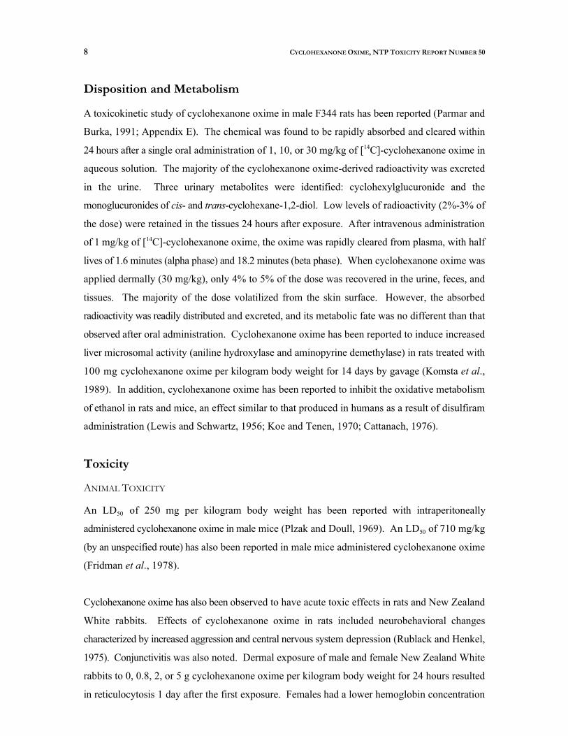
Disposition and Metabolism
A toxicokinetic study of cyclohexanone oxime in male F344 rats has been reported (Parmar and
Burka, 1991; Appendix E). The chemical was found to be rapidly absorbed and cleared within 1424 hours after a single oral administration of 1, 10, or 30 mg/kg of [ C]-cyclohexanone oxime in
aqueous solution. The majority of the cyclohexanone oxime-derived radioactivity was excreted
in the urine. Three urinary metabolites were identified: cyclohexylglucuronide and the
monoglucuronides of cis- and trans-cyclohexane-1,2-diol. Low levels of radioactivity (2%-3% of
the dose) were retained in the tissues 24 hours after exposure. After intravenous administration 14of 1 mg/kg of [ C]-cyclohexanone oxime, the oxime was rapidly cleared from plasma, with half
lives of 1.6 minutes (alpha phase) and 18.2 minutes (beta phase). When cyclohexanone oxime was
applied dermally (30 mg/kg), only 4% to 5% of the dose was recovered in the urine, feces, and
tissues. The majority of the dose volatilized from the skin surface. However, the absorbed
radioactivity was readily distributed and excreted, and its metabolic fate was no different than that
observed after oral administration. Cyclohexanone oxime has been reported to induce increased
liver microsomal activity (aniline hydroxylase and aminopyrine demethylase) in rats treated with
100 mg cyclohexanone oxime per kilogram body weight for 14 days by gavage (Komsta et al.,
1989). In addition, cyclohexanone oxime has been reported to inhibit the oxidative metabolism
of ethanol in rats and mice, an effect similar to that produced in humans as a result of disulfiram
administration (Lewis and Schwartz, 1956; Koe and Tenen, 1970; Cattanach, 1976).
Toxicity
ANIMAL TOXICITY
An LD50 of 250 mg per kilogram body weight has been reported with intraperitoneally
administered cyclohexanone oxime in male mice (Plzak and Doull, 1969). An LD50 of 710 mg/kg
(by an unspecified route) has also been reported in male mice administered cyclohexanone oxime
(Fridman et al., 1978).
Cyclohexanone oxime has also been observed to have acute toxic effects in rats and New Zealand
White rabbits. Effects of cyclohexanone oxime in rats included neurobehavioral changes
characterized by increased aggression and central nervous system depression (Rublack and Henkel,
1975). Conjunctivitis was also noted. Dermal exposure of male and female New Zealand White
rabbits to 0, 0.8, 2, or 5 g cyclohexanone oxime per kilogram body weight for 24 hours resulted
in reticulocytosis 1 day after the first exposure. Females had a lower hemoglobin concentration

9 CYCLOHEXANONE OXIME, NTP TOXICITY REPORT NUMBER 50
after 7 days exposure to 5 g/kg (Gad et al., 1985). Oral administration of 0.1 or 1.0 g/kg to rabbits
resulted in lower erythrocyte counts and elevated methemoglobin levels (Rublack and Henkel,
1975).
Two-week gavage studies in male and female Sprague-Dawley rats (Komsta et al., 1989) and
F344 rats (Derelanko et al., 1985) revealed dose-related erythroid hyperplasia in the spleen and
bone marrow. Sprague-Dawley rats that received 1, 10, or 100 mg cyclohexanone oxime per kg
body weight for 2 weeks had hematologic differences including lower erythrocyte counts, higher
platelet counts, lower hemoglobin concentrations and hematocrit levels, and greater mean red cell
hemoglobin and mean red cell volume values than the control values. Bone marrow smears
indicated lower myeloid, lymphocyte, and monocyte counts concomitant with elevated erythroid
counts. There was also general splenic enlargement with hematopoietic cell proliferation.
Male and female F344 rats that received 10, 25, 75, 150, or 300 mg cyclohexanone oxime per
kilogram body weight by gavage for 2 weeks had hematologic changes similar to those of the
Sprague-Dawley rats (Derelanko et al., 1985). Observations included a dose-related decrease in
erythrocyte counts with concomitant increases in the numbers of circulating nucleated erythrocytes
and reticulocytes and reduced hematocrit levels and hemoglobin concentrations. Methemoglobin
concentrations, measured at the highest dose, were significantly elevated. The rats were observed
for another 2 weeks without dose administration. By Day 28, hematologic values in females had
returned to normal and males displayed only slightly depressed erythrocyte counts and mildly
elevated reticulocyte counts. No significant effects on body weights and no clinical signs of toxicity
were noted in males or females. Splenomegaly and hepatomegaly were observed in male and
female mice on Day 14 and Day 28. The hematology results suggested that the hematotoxic effects
of cyclohexanone oxime administration were reversible following cessation of exposure. The
authors theorized that cyclohexanone oxime induces oxidative damage to the erythrocyte resulting
in hemolytic anemia compensated by increased erythropoiesis.
Results of a 13-week gavage study with male and fem?ale F344 rats were similar to those of the
2-week studies, with evidence of splenomegaly and erythroid hyperplasia in the spleen and bone
marrow (Gad et al., 1985). Rats received doses of 0, 0.25, 2.5, and 25 mg cyclohexanone oxime
per kilogram body weight five times a week for 13 weeks. All males survived to the end of the
study; three of 20 females in the 25 mg/kg group died before the end of the study. Males were
observed with clinical signs of toxicity that included persistent red nasal discharge (at 25 mg/kg

10 CYCLOHEXANONE OXIME, NTP TOXICITY REPORT NUMBER 50
only), chromodacryorrhea and swollen conjunctiva (at 2.5 and 25 mg/kg), and corneal opacity (at
all dose levels). No significant effects on body weight or feed consumption were observed in
males or females. Hematologic differences similar to those seen in the 2-week study were noted.
Dose-related anisocytosis, poikilocytosis, elevated osmotic red blood cell fragility, and a greater
incidence of Howell-Jolly bodies were observed. Splenomegaly was noted at necropsy, and
histopathologic examination showed erythroid hyperplasia in the bone marrow and spleen and
increased hemosiderin pigment deposition in the spleen (Gad et al., 1985).
3In an inhalation study, rats were administered 0.03, 0.1, or 1 mg cyclohexanone oxime per m for
6 to 10 weeks (Tsulaya et al., 1975). Reduced erythrocyte count and blood cholinesterase activity 3 3were observed at exposure levels of 0.1 and 1 mg/m . At necropsy the rats in the 0.1 mg/m group
displayed degenerative differences in the parenchymatous organs and desquamation of bronchial 3epithelium. No adverse effects were noted in study animals at the 0.03 mg/m exposure level.
GENETIC TOXICITY
Negative results were obtained in mutagenicity tests with several strains of Salmonella
typhimurium, with and without metabolic activation (Araki et al., 1986; Rogers-Back et al., 1988)
and with Escherichia coli strain WP2 (Araki et al., 1986). In addition, no increase in the frequency
of sex-linked recessive lethal mutations was observed in germ cells of male Drosophila
melanogaster administered cyclohexanone oxime (8.8 mM) by feeding (Vogel and Chandler,
1974). The only mutagenic activity reported for cyclohexanone oxime was noted in L5178Y
mouse lymphoma cells treated in the absence of S9 activation; the addition of rat liver S9
eliminated the mutagenic effect (Rogers-Back et al., 1988).

11 CYCLOHEXANONE OXIME, NTP TOXICITY REPORT NUMBER 50
Study Rationale and Design
The oximes, as a chemical class, are produced in relatively large quantities and are currently used
in a wide variety of industrial applications. There is growing interest in using oximes as magnetic
tape binders, photochemical additives, biocides for water and waste treatment, ingredients in
cosmetics, and sweeteners. Despite the potential for wide-ranging occupational exposure, little is
known regarding the potential toxicity of oximes. Carcinogenic effects associated with the cyclic
oxime p-quinone dioxime (NCI, 1979) and with the aliphatic oxime, acetoxime (Mirvish et al.,
1982), have made oximes high-priority chemicals for toxicity testing. Cyclohexanone oxime was
selected by the NTP as a representative alicyclic oxime for toxicity testing in a 2-week range-
finding study and a 13-week subchronic study in B6C3F mice. The subacute (Derelanko et al.,1
1985) and subchronic (Gad et al., 1985) oral toxicity of cyclohexanone oxime in F344 rats has
been reported, and studies in this species were not repeated.
Cyclohexanone oxime was administered by dosed water to male and female B6C3F mice for 21
weeks at concentrations of 0,106, 312, 625, 1,250, and 2,500 ppm and for 13 weeks at
concentrations of 0, 625, 1,250, 2,500, 5,000, and 10,000 ppm. Information gained from these
studies may be used to set exposure levels for 2-year chronic toxicity and carcinogenicity studies
in B6C3F mice.1

12 CYCLOHEXANONE OXIME, NTP TOXICITY REPORT NUMBER 50

13 CYCLOHEXANONE OXIME, NTP TOXICITY REPORT NUMBER 50
13 CYCLOHEXANONE OXIME, NTP TOXICITY REPORT NUMBER 50
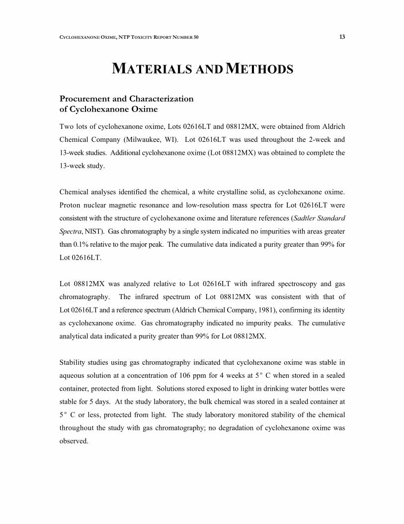
MATERIALS AND METHODS
Procurement and Characterization of Cyclohexanone Oxime
Two lots of cyclohexanone oxime, Lots 02616LT and 08812MX, were obtained from Aldrich
Chemical Company (Milwaukee, WI). Lot 02616LT was used throughout the 2-week and
13-week studies. Additional cyclohexanone oxime (Lot 08812MX) was obtained to complete the
13-week study.
Chemical analyses identified the chemical, a white crystalline solid, as cyclohexanone oxime.
Proton nuclear magnetic resonance and low-resolution mass spectra for Lot 02616LT were
consistent with the structure of cyclohexanone oxime and literature references (Sadtler Standard
Spectra, NIST). Gas chromatography by a single system indicated no impurities with areas greater
than 0.1% relative to the major peak. The cumulative data indicated a purity greater than 99% for
Lot 02616LT.
Lot 08812MX was analyzed relative to Lot 02616LT with infrared spectroscopy and gas
chromatography. The infrared spectrum of Lot 08812MX was consistent with that of
Lot 02616LT and a reference spectrum (Aldrich Chemical Company, 1981), confirming its identity
as cyclohexanone oxime. Gas chromatography indicated no impurity peaks. The cumulative
analytical data indicated a purity greater than 99% for Lot 08812MX.
Stability studies using gas chromatography indicated that cyclohexanone oxime was stable in
aqueous solution at a concentration of 106 ppm for 4 weeks at 5 C when stored in a sealed
container, protected from light. Solutions stored exposed to light in drinking water bottles were
stable for 5 days. At the study laboratory, the bulk chemical was stored in a sealed container at
C or less, protected from light. The study laboratory monitored stability of the chemical
throughout the study with gas chromatography; no degradation of cyclohexanone oxime was
observed.
5
14 CYCLOHEXANONE OXIME, NTP TOXICITY REPORT NUMBER 50
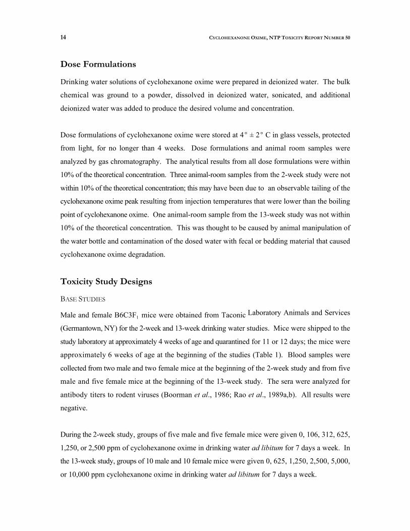
Dose Formulations
Drinking water solutions of cyclohexanone oxime were prepared in deionized water. The bulk
chemical was ground to a powder, dissolved in deionized water, sonicated, and additional
deionized water was added to produce the desired volume and concentration.
Dose formulations of cyclohexanone oxime were stored at 4 ± 2 C in glass vessels, protected
from light, for no longer than 4 weeks. Dose formulations and animal room samples were
analyzed by gas chromatography. The analytical results from all dose formulations were within
10% of the theoretical concentration. Three animal-room samples from the 2-week study were not
within 10% of the theoretical concentration; this may have been due to an observable tailing of the
cyclohexanone oxime peak resulting from injection temperatures that were lower than the boiling
point of cyclohexanone oxime. One animal-room sample from the 13-week study was not within
10% of the theoretical concentration. This was thought to be caused by animal manipulation of
the water bottle and contamination of the dosed water with fecal or bedding material that caused
cyclohexanone oxime degradation.
Toxicity Study Designs
BASE STUDIES
Laboratory Animals and Services Male and female B6C3F1 mice were obtained from Taconic
(Germantown, NY) for the 2-week and 13-week drinking water studies. Mice were shipped to the
study laboratory at approximately 4 weeks of age and quarantined for 11 or 12 days; the mice were
approximately 6 weeks of age at the beginning of the studies (Table 1). Blood samples were
collected from two male and two female mice at the beginning of the 2-week study and from five
male and five female mice at the beginning of the 13-week study. The sera were analyzed for
antibody titers to rodent viruses (Boorman et al., 1986; Rao et al., 1989a,b). All results were
negative.
During the 2-week study, groups of five male and five female mice were given 0, 106, 312, 625,
1,250, or 2,500 ppm of cyclohexanone oxime in drinking water ad libitum for 7 days a week. In
the 13-week study, groups of 10 male and 10 female mice were given 0, 625, 1,250, 2,500, 5,000,
or 10,000 ppm cyclohexanone oxime in drinking water ad libitum for 7 days a week.

15
3
CYCLOHEXANONE OXIME, NTP TOXICITY REPORT NUMBER 50
For all studies, mice were housed in individual cages. NIH-07 Open Formula Diet (Zeigler
Brothers, Inc., Gardners, PA) in pellet form was available ad libitum. Animal rooms were
maintained with 12 hours of fluorescent light per day. The temperature was maintained at 72 ±
F and relative humidity at 50% ± 15%, with at least 10 room air changes per hour.
Necropsies were performed on all animals in the 2-week and 13-week studies. In the 2-week
study, the liver and spleen were weighed. In the 13-week study, the heart, right kidney, liver, lungs,
spleen, right testis, and thymus of each animal were weighed. Organs and tissues were examined
for gross lesions and fixed in 10% neutral buffered formalin. Tissues to be examined
microscopically were trimmed, embedded in paraffin, sectioned, and stained with hematoxylin and
eosin. In the 2-week study only gross lesions were examined. In the 13-week study, complete
histopathologic examinations were performed on all animals. For all paired organs (i.e., kidney,
ovary, adrenal gland), samples from each organ were examined. Tissues examined microscopically
are listed in Table 1.
Upon completion of the laboratory pathologist's histopathologic evaluation, the slides, paraffin
blocks, and residual wet tissues were sent to the NTP Archives for inventory, slide/block match,
and wet tissue audit. The slides, individual animal data records, and pathology tables were sent
to an independent pathology laboratory where quality assessment was performed. Results were
reviewed and evaluated by the NTP Pathology Working Group (PWG); the final diagnoses
represent a consensus of contractor pathologists and the PWG. Details of these review procedures
have been described by Maronpot and Boorman (1982) and Boorman et al. (1985).
SUPPLEMENTAL EVALUATIONS
Sperm Motility and Vaginal Cytology in Mice
Sperm motility and vaginal cytology evaluations were performed on 13-week study mice (10 males
and 10 females) from the 0, 1,250, 2,500, and 5,000 ppm groups at the end of the study. The
parameters that were evaluated are listed in Table 1. Methods were those described in the NTP
statement of work (1987). Briefly, for 12 consecutive days prior to the end of the study, the
vaginal vaults were moistened with saline, if necessary, and vaginal lavage samples were taken
from 10 females per group and spread on a glass slide, dried, and stained. Relative number of
leukocytes, nucleated epithelial cells, and large squamous epithelial cells were determined and used
to ascertain estrous cycle stage (i.e., diestrus, proestrus, estrus, and metestrus).

16 CYCLOHEXANONE OXIME, NTP TOXICITY REPORT NUMBER 50
Sperm motility was evaluated at necropsy in the following manner. The left epididymis was
isolated and weighed. The cauda epididymis was removed from the epididymis body (corpus
epididymis) and weighed. Modified Tyrode's buffer was applied to slides and a small incision was
made at the distal border of the cauda epididymis. The sperm effluxing from the incision were
dispersed in the buffer on the slides. The numbers of motile and nonmotile spermatozoa were
counted for five fields per slide by two observers.
Following completion of sperm motility estimates, each left cauda epididymis was placed in
buffered saline solution. Caudae were finely minced and the tissue was incubated in a saline
solution and then heat fixed at 65 C. Sperm density was then determined microscopically with
the aid of a hemacytometer. To quantify spermatogenesis, testicular spermatid head count was
determined by removing the tunica albuginea and homogenizing the left testis in phosphate
buffered saline containing 10% dimethyl sulfoxide. Homogenization-resistant spermatid nuclei
were counted with a hemacytometer.

17 CYCLOHEXANONE OXIME, NTP TOXICITY REPORT NUMBER 50
TABLE 1 Experimental Design and Materials and Methods in the 2-Week and 13-Week Drinking Water Studies of Cyclohexanone Oxime
2-Week Study 13-Week Study
EXPERIMENTAL DESIGN
Study Laboratory Microbiological Associates, Inc. (Bethesda, MD)
Strain and Species B6C3F mice 1
Animal Source Taconic Farms (Germantown, NY)
Size of Study Groups Five males and five females
Exposure Concentrations 0, 106, 312, 625, 1,250, or 2,500 ppm in drinking water ad libitum for 2 weeks
Date of First Exposure 8 January 1991
Date of Last Exposure 22 January 1991
Date of Necropsy 22 January 1991
Type and Frequency of Observation Observed twice daily for mortality/moribundity andclinical signs of toxicity. Clinical observations and individual body weights were recorded on Days 1 and 8and at the end of the study. Water consumption wasrecorded once weekly.
Necropsy Necropsies were performed on all animals. The liver and spleen were weighed at necropsy.
Same as 2-week study
Same as 2-week study
Same as 2-week study
10 males and 10 females
0, 625, 1,250, 2,500, 5,000, or 10,000 ppm in drinking water ad libitum for 13 weeks
29 April 1991
29-30 July 1991
29-30 July 1991
Observed twice daily for mortality/moribundity andclinical signs of toxicity. Clinical observations were recorded weekly. Individual body weights were recordedat the start of the study, weekly thereafter, and at the endof the study. Water consumption was recorded twiceweekly.
Necropsies were performed on all animals in the basestudy. The heart, right kidney, liver, lungs, spleen, righttestis, and thymus were weighed at necropsy.
18 CYCLOHEXANONE OXIME, NTP TOXICITY REPORT NUMBER 50
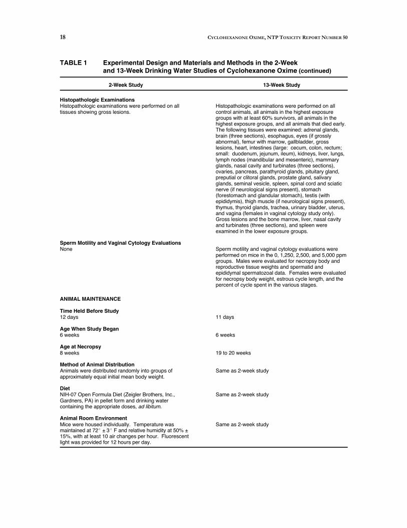
TABLE 1 Experimental Design and Materials and Methods in the 2-Week and 13-Week Drinking Water Studies of Cyclohexanone Oxime (continued)
2-Week Study 13-Week Study
Histopathologic Examinations Histopathologic examinations were performed on alltissues showing gross lesions.
Sperm Motility and Vaginal Cytology Evaluations None
ANIMAL MAINTENANCE
Time Held Before Study 12 days
Age When Study Began 6 weeks
Age at Necropsy 8 weeks
Method of Animal Distribution Animals were distributed randomly into groups ofapproximately equal initial mean body weight.
Diet NIH-07 Open Formula Diet (Zeigler Brothers, Inc.,Gardners, PA) in pellet form and drinking watercontaining the appropriate doses, ad libitum.
Animal Room Environment Mice were housed individually. Temperature wasmaintained at 72 ± 3 F and relative humidity at 50% ±15%, with at least 10 air changes per hour. Fluorescent light was provided for 12 hours per day.
Histopathologic examinations were performed on allcontrol animals, all animals in the highest exposuregroups with at least 60% survivors, all animals in thehighest exposure groups, and all animals that died early.The following tissues were examined: adrenal glands,brain (three sections), esophagus, eyes (if grosslyabnormal), femur with marrow, gallbladder, grosslesions, heart, intestines (large: cecum, colon, rectum; small: duodenum, jejunum, ileum), kidneys, liver, lungs,lymph nodes (mandibular and mesenteric), mammaryglands, nasal cavity and turbinates (three sections),ovaries, pancreas, parathyroid glands, pituitary gland,preputial or clitoral glands, prostate gland, salivaryglands, seminal vesicle, spleen, spinal cord and sciaticnerve (if neurological signs present), stomach(forestomach and glandular stomach), testis (withepididymis), thigh muscle (if neurological signs present),thymus, thyroid glands, trachea, urinary bladder, uterus,and vagina (females in vaginal cytology study only).Gross lesions and the bone marrow, liver, nasal cavityand turbinates (three sections), and spleen wereexamined in the lower exposure groups.
Sperm motility and vaginal cytology evaluations wereperformed on mice in the 0, 1,250, 2,500, and 5,000 ppmgroups. Males were evaluated for necropsy body andreproductive tissue weights and spermatid andepididymal spermatozoal data. Females were evaluated for necropsy body weight, estrous cycle length, and thepercent of cycle spent in the various stages.
11 days
6 weeks
19 to 20 weeks
Same as 2-week study
Same as 2-week study
Same as 2-week study
19 CYCLOHEXANONE OXIME, NTP TOXICITY REPORT NUMBER 50
Genetic Toxicity
SALMONELLA TYPHIMURIUM MUTAGENICITY TEST PROTOCOL
Testing was performed as reported by Zeiger et al. (1992). Cyclohexanone oxime was sent to the
laboratory as a coded aliquot and was incubated with the Salmonella typhimurium tester strains
(TA97, TA98, TA100, and TA1535) either in buffer or S9 mix (metabolic activation enzymes and
cofactors from Aroclor 1254-induced male Sprague-Dawley rat or Syrian hamster liver) for 20
minutes at 37 C. Top agar supplemented with l-histidine and d-biotin was added, and the
contents of the tubes were mixed and poured onto the surfaces of minimal glucose agar plates.
Histidine-independent mutant colonies arising on these plates were counted following incubation
for 2 days at 37 C.
Each trial consisted of triplicate plates of concurrent positive and negative controls and of at least
five doses of cyclohexanone oxime. The high dose was limited by toxicity to 6,666 µg/plate. All
positive assays were repeated under the conditions that elicited a positive response, and all negative
assays were repeated.
CHINESE HAMSTER OVARY CELL CHROMOSOMAL ABERRATION TEST PROTOCOL
Testing was performed as reported by Galloway et al. (1987). Cyclohexanone oxime was sent to
the laboratory as a coded aliquot. It was tested in cultured Chinese hamster ovary (CHO) cells for
induction of chromosomal aberrations (Abs) both in the presence and absence of Aroclor
1254-induced male Sprague-Dawley rat liver S9 and cofactor mix. Each test consisted of
concurrent solvent and positive controls and of at least three doses of cyclohexanone oxime. In
the absence of toxicity, 5 mg/mL was selected as the high dose. Doses in the second trial without
S9 were selected to bracket the dose that gave a positive response in the first trial without S9. A
single flask per dose was used, and tests yielding equivocal or positive results were repeated.
In the Abs test without S9, cells were incubated in McCoy's 5A medium with cyclohexanone oxime
for 10 hours; Colcemid was added and incubation continued for 2 hours. The cells were then
harvested by mitotic shake-off, fixed, and stained with Giemsa. For the Abs test with S9, cells
were treated with cyclohexanone oxime and S9 for 2 hours, after which the treatment medium was
removed and the cells incubated for 10 hours in fresh medium, with Colcemid present for the final
2 hours. Cells were harvested in the same manner as for the treatment without S9.
20 CYCLOHEXANONE OXIME, NTP TOXICITY REPORT NUMBER 50
Cells were selected for scoring on the basis of good morphology and completeness of karyotype
(21 ± 2 chromosomes). All slides were scored blind and those from a single test were read by the
same person. Two hundred first-division metaphase cells were scored at each dose level. Classes
of aberrations included simple (breaks and terminal deletions), complex (rearrangements and
translocations), and other (pulverized cells, despiralized chromosomes, and cells containing 10 or
more aberrations).
BONE MARROW MICRONUCLEUS TEST PROTOCOL
Preliminary range finding studies were performed. Factors affecting dose selection included
chemical solubility, toxicity, and the extent of cell cycle delay induced by the chemical exposure.
Based on these studies, male mice to be tested for bone marrow micronuclei were injected
intraperitoneally three times at 24-hour intervals with cyclohexanone oxime dissolved in corn oil.
The total dosing volume was 0.4 mL. Solvent control animals were injected with 0.4 mL corn oil
only. The positive control mice received injections of cyclophosphamide. Twenty-four hours after
the third injection, the mice were killed and smears of the bone marrow cells obtained from the
femurs were prepared. Air-dried smears were fixed and stained; 2,000 polychromatic erythrocytes
were scored for frequency of micronucleated cells in each of five animals per dose group.
PERIPHERAL BLOOD MICRONUCLEUS TEST PROTOCOL
A detailed discussion of this assay is presented in MacGregor et al. (1990). At the end of the 13
week toxicity study, peripheral blood samples were obtained from male and female mice, and
smears were immediately prepared and fixed in absolute methanol. The methanol-fixed slides were
later stained with a chromatin-specific fluorescent dye (acridine orange) and coded. Two thousand
normochromatic erythrocytes were scanned in each of five mice per exposure group. The criteria
of Schmid (1976) were used in defining micronuclei. The results were tabulated as the mean of
the pooled results from all animals within a treatment group plus or minus the standard error of
the mean.
21 CYCLOHEXANONE OXIME, NTP TOXICITY REPORT NUMBER 50
Statistical Methods
ANALYSIS AND CALCULATION OF NONNEOPLASTIC LESION INCIDENCES
The incidences of nonneoplastic lesions presented in Tables A1 and A2 are given as the number
of animals bearing such lesions at a specific site and the number of animals with that site examined
microscopically. For lesions detected in the 13-week study, the Fisher exact test, a procedure
based on the overall proportion of affected animals, was used (Gart et al., 1979).
ANALYSIS OF CONTINUOUS VARIABLES
Two approaches were employed to assess the significance of pairwise comparisons between
exposed and control groups in the analysis of continuous variables. Organ and body weight data,
which have approximately normal distributions, were analyzed with the parametric multiple
comparisons procedures of Williams (1971, 1972) and Dunnett (1955). Spermatid and epididymal
spermatozoal data, which typically have skewed distributions, were analyzed with the
nonparametric multiple comparison methods of Shirley (1977) and Dunn (1964). Jonckheere's test
(Jonckheere, 1954) was used to assess the significance of exposure-response trends and to
determine whether a trend-sensitive test (Williams' or Shirley's test) was more appropriate for
pairwise comparisons than a test that does not assume monotonic exposure response (Dunnett's
or Dunn's test). Trend-sensitive tests were used when Jonckheere's test was significant at a P-value
less than 0.01. Prior to analysis, extreme values identified by the outlier test of Dixon and Massey
(1951) were examined by NTP personnel and implausible values were eliminated from the
analysis.
ANALYSIS OF VAGINAL CYTOLOGY DATA
Because the data are proportions (the proportion of the observation period that an animal was in
a given estrous stage), an arcsine transformation was used to bring the data into closer
conformance with normality assumptions. Treatment effects were investigated by applying a
multivariate analysis of variance (Morrison, 1976) to the transformed data to test for simultaneous
equality of measurements across exposure levels.
22 CYCLOHEXANONE OXIME, NTP TOXICITY REPORT NUMBER 50
ANALYSIS OF MUTAGENICITY IN SALMONELLA TYPHIMURIUM
A positive response in the Salmonella typhimurium assay is defined as a reproducible, dose-related
increase in histidine-independent (revertant) colonies in any one strain/activation combination. An
equivocal response is defined as an increase in revertants that is not dose related, not reproducible,
or not of sufficient magnitude to support a determination of mutagenicity. A negative response is
obtained when no increase in revertant colonies is observed following chemical treatment. There
was no minimum percentage or fold-increase required for a chemical to be judged positive or
weakly positive.
ANALYSIS OF CHINESE HAMSTER OVARY CELL CHROMOSOMAL ABERRATION DATA
Chromosomal aberration data are presented as percentages of cells with aberrations. To arrive at
a statistical call for a trial, analyses were conducted on both the dose-response curve and individual
dose points (Galloway et al., 1987). For a single trial, a statistically significant (P<0.05) difference
for one dose point and a significant trend (P<0.015) was considered weak evidence for a positive
response; significant differences for two or more doses indicated the trial was positive. A positive
trend in the absence of a statistically significant increase at any one dose point or a significant
increase at a single dose point in the absence of a positive trend led to an equivocal call.
Ultimately, the trial calls were based on a consideration of the statistical analyses as well as the
biological information available to the reviewers.
ANALYSIS OF PERIPHERAL BLOOD AND BONE MARROW MICRONUCLEUS DATA
The results were tabulated as the mean of the pooled results from all animals within a treatment
group, plus or minus the standard error of the mean. The frequency of micronucleated cells among
normochromatic erythrocytes was analyzed by a statistical software package that tested for
increasing trend over exposure groups with a one-tailed Cochran-Armitage trend test, followed by
pairwise comparisons between each exposure group and the control group (Margolin et al., 1990).
In the presence of excess binomial variation, as detected by a binomial dispersion test, the binomial
variance of the Cochran-Armitage test was adjusted upward in proportion to the excess variation.
In the micronucleus test, an individual trial was considered positive if the trend test P-value was
less than or equal to 0.025 or the P-value for any single exposure group was less than or equal to
0.025 divided by the number of exposure groups. A final call of positive for micronucleus
induction is preferably based on reproducible positive trials (as noted above). Ultimately, the final
23 CYCLOHEXANONE OXIME, NTP TOXICITY REPORT NUMBER 50
call was determined by the scientific staff after considering the results of statistical analyses,
reproducibility of any effects observed, and the magnitudes of those effects.
Quality Assurance
The animal studies of cyclohexanone oxime were performed in compliance with U.S. Food and
Drug Administration Good Laboratory Practices regulations (21 CFR, Part 58). The Quality
Assurance Unit of Microbiological Associates, Inc. performed audits and inspections of protocols,
procedures, data, and reports throughout the course of the studies.

24 CYCLOHEXANONE OXIME, NTP TOXICITY REPORT NUMBER 50

25 CYCLOHEXANONE OXIME, NTP TOXICITY REPORT NUMBER 50

25 CYCLOHEXANONE OXIME, NTP TOXICITY REPORT NUMBER 50
RESULTS
2-Week Drinking Water Study in B6C3F Mice 1
All mice survived until the end of the study (Table 2). Males in the 2,500 ppm group had lower
final mean body weights than the controls. The final mean body weights for females in the 625
ppm group were greater than the control value. No clinical signs of toxicity were noted in male
mice. One female in the control group and one in the 106 ppm group were observed to be thin.
Average daily water consumption was greater than or equal to that of the controls for both males
and females at all exposure levels except 2,500 ppm; at this concentration, males and females had
lower average water consumption than the controls.
TABLE 2 Survival, Weight Gain, Water Consumption, and Compound Consumption of B6C3F Mice in the 2-Week Drinking Water Study 1
of Cyclohexanone Oxime
Final Weight Water Compound Concentration Mean Body Weight (grams) Relative to Consumption Consumption
2(ppm) Survival1 Initial Final Change Controls (%) (g/day) (mg/kg/day)
MALE
0 106 312 625
1,250 2,500
FEMALE
0 106 312 625
1,250 2,500
5/5 5/5 5/5 5/5 5/5 5/5
5/5 5/5 5/5 5/5 5/5 5/5
25.5 29.6 4.1 5.3 25.2 28.6 3.4 97 6.8 27 25.0 28.5 3.4 96 5.7 69 25.4 28.9 3.5 98 6.0 141 25.4 28.5 3.1 96 6.6 316 25.8 28.1 2.4 95 4.6 439
19.4 23.1 3.7 6.5 19.4 22.9 3.5 99 7.3 38 19.8 23.6 3.8 102 6.5 98 19.5 24.3 4.8 105 6.7 203 19.8 23.3 3.6 101 7.0 416 19.7 22.5 2.8 97 3.7 441
1 Number surviving at 15 days/number of animals per group.2 (Exposure group mean/control group mean) × 100.
26 CYCLOHEXANONE OXIME, NTP TOXICITY REPORT NUMBER 50
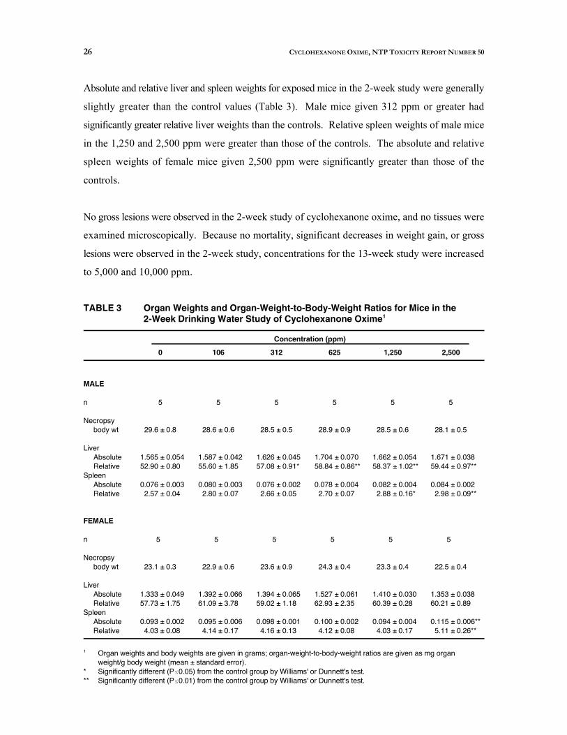
Absolute and relative liver and spleen weights for exposed mice in the 2-week study were generally
slightly greater than the control values (Table 3). Male mice given 312 ppm or greater had
significantly greater relative liver weights than the controls. Relative spleen weights of male mice
in the 1,250 and 2,500 ppm were greater than those of the controls. The absolute and relative
spleen weights of female mice given 2,500 ppm were significantly greater than those of the
controls.
No gross lesions were observed in the 2-week study of cyclohexanone oxime, and no tissues were
examined microscopically. Because no mortality, significant decreases in weight gain, or gross
lesions were observed in the 2-week study, concentrations for the 13-week study were increased
to 5,000 and 10,000 ppm.
TABLE 3 Organ Weights and Organ-Weight-to-Body-Weight Ratios for Mice in the 2-Week Drinking Water Study of Cyclohexanone Oxime1
Concentration (ppm)
0 106 312 625 1,250 2,500
MALE
n 5 5 5 5 5 5
Necropsy body wt 29.6 ± 0.8 28.6 ± 0.6 28.5 ± 0.5 28.9 ± 0.9 28.5 ± 0.6 28.1 ± 0.5
Liver Absolute 1.565 ± 0.054 1.587 ± 0.042 1.626 ± 0.045 1.704 ± 0.070 1.662 ± 0.054 1.671 ± 0.038 Relative 52.90 ± 0.80 55.60 ± 1.85 57.08 ± 0.91* 58.84 ± 0.86** 58.37 ± 1.02** 59.44 ± 0.97**
Spleen Absolute 0.076 ± 0.003 0.080 ± 0.003 0.076 ± 0.002 0.078 ± 0.004 0.082 ± 0.004 0.084 ± 0.002 Relative 2.57 ± 0.04 2.80 ± 0.07 2.66 ± 0.05 2.70 ± 0.07 2.88 ± 0.16* 2.98 ± 0.09**
FEMALE
n 5 5 5 5 5 5
Necropsy body wt 23.1 ± 0.3 22.9 ± 0.6 23.6 ± 0.9 24.3 ± 0.4 23.3 ± 0.4 22.5 ± 0.4
Liver Absolute 1.333 ± 0.049 1.392 ± 0.066 1.394 ± 0.065 1.527 ± 0.061 1.410 ± 0.030 1.353 ± 0.038 Relative 57.73 ± 1.75 61.09 ± 3.78 59.02 ± 1.18 62.93 ± 2.35 60.39 ± 0.28 60.21 ± 0.89
Spleen Absolute 0.093 ± 0.002 0.095 ± 0.006 0.098 ± 0.001 0.100 ± 0.002 0.094 ± 0.004 0.115 ± 0.006** Relative 4.03 ± 0.08 4.14 ± 0.17 4.16 ± 0.13 4.12 ± 0.08 4.03 ± 0.17 5.11 ± 0.26**
Organ weights and body weights are given in grams; organ-weight-to-body-weight ratios are given as mg organ weight/g body weight (mean ± standard error).
* Significantly different (P 0.05) from the control group by Williams' or Dunnett's test. ** Significantly different (P 0.01) from the control group by Williams' or Dunnett's test.
1

27 CYCLOHEXANONE OXIME, NTP TOXICITY REPORT NUMBER 50
13-Week Drinking Water Study in B6C3F Mice1
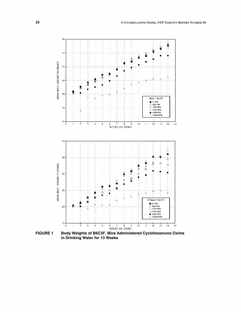
Five male and five female 10,000 ppm mice were killed moribund in the third week of the study
(Table 4). All other mice survived until the end of the study. The final mean body weights of
males in the 5,000 and 10,000 ppm groups were less than those of the controls (Figure 1).
Females in the 2,500, 5,000, and 10,000 ppm groups also had lower final mean body weights than
the controls. Clinical signs of toxicity were observed in males from the higher exposure groups.
At exposure levels of 2,500 and 5,000 ppm, one male in each group was observed to be
hyperactive. All males in the 10,000 ppm group appeared thin, one male was observed to have
abnormal posture, and four males had ruffled fur. Clinical signs of toxicity in females were
restricted to the 10,000 ppm group; 9 of 10 females were observed to be thin, and two females had
abnormal posture and ruffled fur. One female in the 10,000 ppm group was observed to be
hyperactive. Lethargy, abnormal breathing, and ataxia were also observed. Average water
consumption by males and females decreased with increasing exposure.
TABLE 4 Survival, Weight Gain, Water Consumption, and Compound Consumption of B6C3F Mice in the 13-Week Drinking Water Study 1
of Cyclohexanone Oxime
Concentration (ppm) Survival1
Mean Initial
Body Weight (grams) Final Change
Final Weight Relative to
Controls (%) 2
Water Consumption
(g/day)
Compound Consumption (mg/kg/day)
MALE
0 10/10 26.0 41.5 15.5 5.2 625 10/10 25.2 42.3 17.1 102 5.0 96
1,250 10/10 25.5 41.5 15.9 100 4.8 181 2,500 10/10 25.8 41.7 15.9 101 3.8 287 5,000
10,000 10/10 5/103
26.1 25.6
39.0 30.6
12.9 4.3
94 74
3.6 3.0
573 1,152
FEMALE
0 10/10 20.4 35.1 14.7 5.8 625 10/10 20.3 35.5 15.2 101 5.6 132
1,250 10/10 20.3 35.3 15.0 100 5.2 239 2,500 10/10 20.0 33.2 13.2 95 4.2 403 5,000
10,000 10/10 5/103
20.4 20.5
30.7 25.0
10.3 3.9
87 71
3.4 2.9
681 1,350
1 Number surviving at 13 weeks/number of animals per group.2 (Exposure group mean/control group mean) × 100.3 Week of death: 3.
28 CYCLOHEXANONE OXIME, NTP TOXICITY REPORT NUMBER 50
FIGURE 1 Body Weights of B6C3F1 Mice Administered Cyclohexanone Oxime in Drinking Water for 13 Weeks

29 CYCLOHEXANONE OXIME, NTP TOXICITY REPORT NUMBER 50
Spleen weights of male and female mice given 5,000 or 10,000 ppm cyclohexanone oxime were
significantly greater than those of the controls (Table 5 and Appendix B). Absolute liver weights
of males and females given 10,000 ppm were lower than those of the controls. Relative liver
weights of males given 10,000 ppm and females given 5,000 or 10,000 ppm were significantly
greater than the controls. Other differences in organ weights were considered secondary to body
weight differences. Kidney weight increases may have been due to decreased water consumption.
TABLE 5 Organ Weights and Organ-Weight-to-Body-Weight Ratios for Mice in the 13-Week Drinking Water Study of Cyclohexanone Oxime1
Concentration (ppm)
0 625 1,250 2,500 5,000 10,000
MALE
n 10 10 10 10 10 5
Necropsy body wt 43.5 ± 1.1 43.6 ± 0.8 43.2 ± 0.9 42.1 ± 1.0 39.1 ± 0.9** 31.1 ± 1.0**
Right kidney Absolute 0.310 ± 0.009 0.304 ± 0.006 0.311 ± 0.009 0.322 ± 0.011 0.322 ± 0.004 0.297 ± 0.010 Relative 7.13 ± 0.16 6.98 ± 0.22 7.20 ± 0.13 7.64 ± 0.10* 8.27 ± 0.17** 9.56 ± 0.26**
Liver Absolute 2.010 ± 0.129 2.088 ± 0.046 1.970 ± 0.086 2.046 ± 0.101 1.905 ± 0.056 1.594 ± 0.060* Relative 45.97 ± 1.96 47.87 ± 0.74 45.48 ± 1.08 48.38 ± 1.27 48.76 ± 0.87 51.18 ± 1.01*
Spleen Absolute 0.079 ± 0.003 0.081 ± 0.003 0.077 ± 0.003 0.077 ± 0.003 0.106 ± 0.003** 0.145 ± 0.008** Relative 1.82 ± 0.06 1.86 ± 0.06 1.79 ± 0.04 1.82 ± 0.06 2.73 ± 0.09** 4.64 ± 0.15**
FEMALE
n 10 10 10 10 10 5
Necropsy body wt 35.6 ± 1.2 36.8 ± 0.9 34.3 ± 1.4 32.5 ± 1.0 30.4 ± 1.1** 24.2 ± 1.2**
Right kidney Absolute 0.209 ± 0.006 0.214 ± 0.004 0.211 ± 0.004 0.217 ± 0.005 0.225 ± 0.005 0.205 ± 0.008 Relative 5.90 ± 0.18 5.84 ± 0.13 6.21 ± 0.21 6.70 ± 0.20** 7.44 ± 0.17** 8.52 ± 0.22**
Liver Absolute 1.510 ± 0.052 1.623 ± 0.040 1.446 ± 0.032 1.380 ± 0.033 1.396 ± 0.053 1.172 ± 0.048** Relative 42.64 ± 1.28 44.13 ± 0.56 42.52 ± 1.14 42.61 ± 0.94 45.92 ± 0.79* 48.72 ± 1.48**
Spleen Absolute 0.092 ± 0.002 0.099 ± 0.003 0.098 ± 0.005 0.097 ± 0.004 0.151 ± 0.008** 0.156 ± 0.016** Relative 2.60 ± 0.09 2.69 ± 0.11 2.87 ± 0.14 3.00 ± 0.12 4.97 ± 0.21** 6.41 ± 0.48**
Organ weights and body weights are given in grams; organ-weight-to-body-weight ratios are given as mg organ weight/g body weight (mean ± standard error).
* Significantly different (P 0.05) from the control group by Williams' or Dunnett's test. ** Significantly different (P 0.01) from the control group by Williams' or Dunnett's test.
1

30 CYCLOHEXANONE OXIME, NTP TOXICITY REPORT NUMBER 50
There were no significant differences in sperm motility or vaginal cytology parameters between
exposed and control males and females (Tables C1 and C2).
At necropsy, no gross findings other than the thin carcasses of mice from the highest exposure
(10,000 ppm) groups were attributed to compound exposure. Microscopically, treatment-related
effects were present in the spleen, bone marrow, liver, and nasal cavity.
In the spleen, increased hematopoietic cell proliferation was associated with exposure to
cyclohexanone oxime in both male and female mice and correlated with increased spleen weights.
This change, minimal to moderate in severity, was characterized by an increased amount of
hematopoietic cells, primarily of the erythroid series, in the splenic red pulp of treated mice relative
to that seen in the controls (Table 6). In cases of mild to moderate severity, an increase in the
amount of hemosiderin pigment accompanied the increased hematopoiesis. In females, the
incidence and severity of this effect was dose dependent at concentrations of 1,250 ppm and above;
in males the lesion was present only at the two highest exposure levels (5,000 and 10,000 ppm).
At 10,000 ppm, splenic hematopoietic cell proliferation was present only in those mice which
survived to study termination.
A treatment-related increase in hematopoietic proliferation in the bone marrow, diagnosed as
"hyperplasia," occurred much less frequently than in the spleen; this change was present in only
three of the surviving males from the 10,000 ppm group (Table 6). Depletion of bone marrow
cells attributable to moribund condition was seen in a single early death female from the 10,000
ppm group.
In the liver, exposure to cyclohexanone oxime was associated with hypertrophy of centrilobular
hepatocytes, which correlated with increased relative liver weights (Table 6). Affected cells had
increased amounts of cytoplasm which frequently had an eosinophilic "ground glass" appearance.
Nuclei of affected centrilobular hepatocytes also tended to be larger than those of the periportal
cells. In males, the incidence and severity of this change was dose dependent at concentrations of
1,250 ppm and greater; in females, the lesion was present only at the two highest exposure levels
(5,000 and 10,000 ppm). At the highest exposure level (10,000 ppm), centrilobular hypertrophy
was present in only those mice that survived to study termination; in those that did not survive,
shrinkage of hepatic cords and loss of cytoplasmic volume consistent with glycogen depletion was
observed.
31 CYCLOHEXANONE OXIME, NTP TOXICITY REPORT NUMBER 50
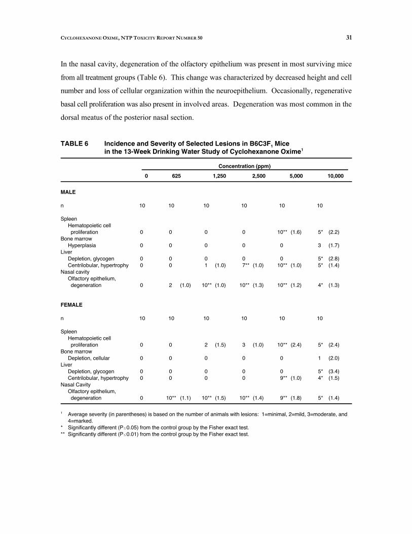
In the nasal cavity, degeneration of the olfactory epithelium was present in most surviving mice
from all treatment groups (Table 6). This change was characterized by decreased height and cell
number and loss of cellular organization within the neuroepithelium. Occasionally, regenerative
basal cell proliferation was also present in involved areas. Degeneration was most common in the
dorsal meatus of the posterior nasal section.
TABLE 6 Incidence and Severity of Selected Lesions in B6C3F Mice1
in the 13-Week Drinking Water Study of Cyclohexanone Oxime1
Concentration (ppm)
0 625 1,250 2,500 5,000 10,000
MALE
n 10 10 10 10 10 10
Spleen Hematopoietic cell proliferation
Bone marrow Hyperplasia
Liver Depletion, glycogen Centrilobular, hypertrophy
Nasal cavity Olfactory epithelium, degeneration
0
0
0 0
0
0
0
0 0
2 (1.0)
0
0
0 1 (1.0)
10** (1.0)
0
0
0 7** (1.0)
10** (1.3)
10** (1.6)
0
0 10** (1.0)
10** (1.2)
5*
3
5* 5*
4*
(2.2)
(1.7)
(2.8) (1.4)
(1.3)
FEMALE
n 10 10 10 10 10 10
Spleen Hematopoietic cell proliferation
Bone marrow Depletion, cellular
Liver Depletion, glycogen Centrilobular, hypertrophy
Nasal Cavity Olfactory epithelium, degeneration
0
0
0 0
0
0
0
0 0
10** (1.1)
2 (1.5)
0
0 0
10** (1.5)
3 (1.0)
0
0 0
10** (1.4)
10** (2.4)
0
0 9** (1.0)
9** (1.8)
5*
1
5* 4*
5*
(2.4)
(2.0)
(3.4) (1.5)
(1.4)
Average severity (in parentheses) is based on the number of animals with lesions: 1=minimal, 2=mild, 3=moderate, and 4=marked.
* Significantly different (P 0.05) from the control group by the Fisher exact test. ** Significantly different (P 0.01) from the control group by the Fisher exact test.
1
32 CYCLOHEXANONE OXIME, NTP TOXICITY REPORT NUMBER 50
Other than glycogen depletion in the liver, no microscopic changes were consistently observed in
the mice that died prior to study termination. The specific cause of death could not be determined
from these studies, but most were attributed to systemic toxicity and generalized debilitation.
Genetic Toxicity
Cyclohexanone oxime was tested for mutagenicity in four strains of Salmonella typhimurium in
a preincubation protocol with and without induced rat or hamster liver S9 (Table D1). Positive
results were obtained only in strain TA1535 in the presence of 5%, 10%, or 30% hamster S9;
negative results were obtained in strain TA1535 with rat liver S9 and in the absence of S9. Results
of mutagenicity testing of cyclohexanone oxime were negative in strains TA97, TA98, and
TA100, with or without S9.
No clear indication of mutagenic activity was observed in mammalian cell cytogenicity tests with
cyclohexanone oxime. Equivocal results were obtained in a test for induction of chromosomal
aberrations in cultured Chinese hamster ovary (CHO) cells in the absence of S9 (Table D2). In
each of two trials conducted without S9, a significant increase in aberrations was observed in one
of three dose groups tested, but the responses were not related to exposure and results of the trend
tests were negative. No induction of aberrations was noted in CHO cells treated with
cyclohexanone oxime in the presence of S9. Two in vivo micronucleus tests were performed with
cyclohexanone oxime in mice. Both tests had negative results. In the first test, cyclohexanone
oxime was administered by intraperitoneal injection (400 to 1,000 mg/kg) to male mice three times
at 24-hour intervals; no increase in the frequency of micronucleated polychromatic erythrocytes
was observed in bone marrow preparations obtained 24 hours after the final injection (Table D3).
In the second test, male and female mice were administered cyclohexanone oxime (625 to
10,000 ppm) in drinking water for 13 weeks; no increase in the frequency of micronucleated
normochromatic erythrocytes was observed in peripheral blood smears of treated mice (Table D4).

33 CYCLOHEXANONE OXIME, NTP TOXICITY REPORT NUMBER 50

33 CYCLOHEXANONE OXIME, NTP TOXICITY REPORT NUMBER 50
DISCUSSION
Oximes are a class of chemicals that are produced in relatively large quantities and are used in a
variety of industrial applications. Despite the potential for widespread exposure, little is known
regarding the potential toxicity of oximes. Cyclohexanone oxime, an important intermediate in the
production of Nylon-6, was chosen by the NTP as a representative alicyclic oxime for toxicity
testing. The NTP has conducted a 2-week range-finding study and a 13-week subchronic study
in B6C3F mice, as well as genetic toxicology and chemical disposition studies.1
All mice survived to the end of the 2-week study. Final mean body weights were between 95%
and 105% of those of the controls. No clinical signs of toxicity were observed in male mice; two
female mice, one of which was a control, appeared thin. Decreased water consumption was
observed for males and females in the 2,500 ppm groups (the highest exposure groups). Slightly
increased relative liver weights were observed in male mice given 312 ppm or greater; no
significant organ weight effect was observed in female mice at any exposure level. Increased
relative spleen weights were observed in male mice in the 1,250 and 2,500 ppm groups and in
female mice in the 2,500 ppm group. This is in contrast to the F344 rat study of Derelanko et al.
(1985), where splenomegaly occurred at a lower exposure than hepatomegaly. Hepatomegaly in
male rats was observed at a lower exposure concentration than in female rats; this was similar to
the response observed in the 2-week study in mice.
Exposure levels were increased in the 13-week study because only relatively minor effects were
observed in the 2-week study. In the 13-week study, five males and five females were killed
moribund in the 10,000 ppm group; final mean body weights were reduced in males in the 5,000
and 10,000 ppm groups and in females in the 2,500, 5,000, and 10,000 ppm groups. Spleen
weights of males and females were elevated in the 5,000 and 10,000 ppm groups; however, in
contrast to the 2-week study, spleen weights of males and females in the 1,250 and 2,500 ppm
groups were similar to those of the controls. In general, relatively more cyclohexanone oxime was
consumed at a given exposure level in the 2-week study compared to the 13-week study. For
example, males that received 1,250 ppm in the 2-week study consumed an average of
approximately 316 mg/kg daily; in contrast, in the 13-week study, males that received 1,250 ppm
consumed an average of only 181 mg/kg daily. This difference may be due in part to the normal
pattern of water and feed consumption as a function of body weight as young animals grow. The

34 CYCLOHEXANONE OXIME, NTP TOXICITY REPORT NUMBER 50
spleen effect in the 2-week study may also be a consequence of the destruction of susceptible
erythrocytes; as these cells are replaced with less susceptible cells, the spleen is able to recover.
Gad et al. (1985) observed a decrease in erythrocyte fragility following 13 weeks of treatment in
the rat study. This was considered to be due to the cell population being selected for younger cells
that were more resistant to toxic effects.
Increased spleen weights were correlated microscopically with increased hematopoietic cell
proliferation in the spleen. Hematopoietic cell proliferation (hyperplasia) was also observed in the
bone marrow. Similar observations were made in the F344 rat studies (Derelanko et al., 1985;
Gad et al., 1985). These observations are consistent with a cyclohexanone oxime-mediated
destruction of erythrocytes. It has been proposed that erythrocyte destruction results from reaction
of the hemolytic agent with hemoglobin, generating radical species which result in peroxidation
of the cellular membrane which in turn damages the erythrocytes and leads to their eventual
destruction and removal (Gad et al., 1985).
In the metabolism study of cyclohexanone oxime, it was observed that the oxime rapidly
hydrolyzes to cyclohexanone and, presumably, hydroxylamine. Hydroxylamine (2,600 ppm
hydroxylamine sulfate in the drinking water for 52 weeks) causes hematologic effects, such as
methemoglobinemia and splenomegaly in mice (Yamamoto et al., 1967; Gross, 1985), similar to
those observed after exposure to cyclohexanone oxime. Thus, it is not clear whether the
erythrotoxicity of cyclohexanone oxime results from the parent compound or hydroxylamine, the
hydrolysis product.
In a 2-year study, male and female rats were given 3,300 or 6,500 ppm, male mice were given
6,500 or 13,000 ppm, and female mice were given 6,500, 13,000, or 25,000 ppm cyclohexanone
in drinking water (Lijinsky and Kovatch, 1986). No mention is made of splenomegaly or
hematotoxicity in that study. There is, however, a report (Koeferl et al., 1981) of hemosiderin
deposits and extramedullary hematopoiesis in the spleen of beagle dogs repeatedly administered
cyclohexanone intravenously (284 mg/kg per day administered as a 6% solution, either 5 or
75 mL/min). There was no increase in absolute or relative spleen weight in this study. Because
cyclohexanone has not been reported to be hematotoxic to other species or by other routes of
administration, it is possible that the effect in dogs was due to a relatively high concentration of
good lipid solvent disrupting the erythrocyte membrane. It seems far more likely that the
35 CYCLOHEXANONE OXIME, NTP TOXICITY REPORT NUMBER 50
erythrotoxicity of cyclohexanone oxime is due to the parent or hydroxylamine rather than due to
cyclohexanone or a cyclohexanone-derived metabolite.
Cyclohexanone oxime's effect on liver weight was also observed at much lower exposures in the
2-week study compared to the 13-week study. Relative liver weights of males given 10,000 ppm
and females given 5,000 and 10,000 ppm were greater than those of the controls. In the 2-week
study, increased liver weights were observed in males given 312 ppm or greater. Again, this may
be due in part to lower actual consumption of cyclohexanone oxime at the same exposure level and
also to compensation by the liver to the longer exposure.
Microscopic examination of the liver revealed hypertrophy of centrilobular hepatocytes which
correlated with increased liver weights. While hepatomegaly was observed in the F344 rat studies,
and the liver was examined histopathologically, no pathologic findings were reported (Derelanko
et al., 1985; Gad et al., 1985). Chemical-related centrilobular hypertrophy is presumed to be due
to induction of xenobiotic metabolizing enzymes and proliferation of smooth endoplasmic
reticulum. The centrilobular region is where certain xenobiotic metabolizing enzymes are
concentrated, and inducers sometimes result in organelle and cell proliferation in this region (Baron
and Kawabata, 1983). There are apparently no studies of enzyme induction by cyclohexanone
oxime in mice. However, some cyclohexanone oxime metabolites in rats appear to require
cytochrome P450-mediated oxidation (Parmar and Burka, 1991), and cyclohexanone oxime has
been reported to induce liver microsomal activity in the rat (Komsta et al., 1989). In contrast,
cyclohexanone apparently does not induce cytochrome P450 (Gupta et al., 1979).
The only other microscopically observed, treatment-related effect was in the nasal cavity, where
degeneration of the olfactory epithelium was present in most surviving mice in all treatment
groups. This lesion was not noted in the rat studies, although it may not have been looked for
(Derelanko et al., 1985; Gad et al., 1985). Likewise, this lesion was not noted in the toxicity
studies of cyclohexanone in mice, even when exposure was by inhalation (Gupta et al., 1979).
Olfactory toxicity following systemic exposure has been observed with numerous chemicals and
has been related to site-specific metabolism in this tissue for some chemicals (Gaskell, 1990).
Studies in rabbits have demonstrated that cytochrome P450 is present in nasal tissue and can be
induced by exposure to xenobiotics (Ding and Coon, 1990).

36 CYCLOHEXANONE OXIME, NTP TOXICITY REPORT NUMBER 50
In parallel to the studies on cyclohexanone oxime, 2-week and 13-week studies were also
performed with both F344/N rats and B6C3F mice on an acyclic oxime analogue, methyl ethyl1
ketoxime. In mice, the two chemicals exhibited very similar effects. Exposure to similar
concentrations of either chemical resulted in similarly increased liver and spleen weights. The only
marked difference in this parameter occurred at the 10,000 ppm concentration, where the relative
spleen weight following exposure to methyl ethyl ketoxime was approximately 14 mg per gram
body weight, compared to 4 to 6 mg per gram body weight for the same cyclohexanone oxime
exposure. Lesions were observed in the liver, spleen, and nasal epithelium for both chemicals.
Hyperplasia of the urinary bladder transitional epithelium was observed in the methyl ethyl
ketoxime study but not in the cyclohexanone oxime study.
In summary, the major targets of cyclohexanone oxime toxicity are the erythrocyte, the liver, and
nasal epithelium. The no-effect level for erythrotoxicity is 2,500 ppm following 13 weeks of
exposure. The no-effect level for hematopoietic cell proliferation in the spleen is 2,500 ppm. The
no-effect level for hepatotoxicity is 1,250 ppm for males and 2,500 ppm for females following 13
weeks of exposure. Some nasal olfactory epithelial degeneration was observed at all exposure
levels; only at 625 ppm in males was the incidence of this lesion not significantly different from that
in the controls. Cyclohexanone oxime was mutagenic only with Salmonella typhimurium strain
TA1535 with S9 activation.

37 CYCLOHEXANONE OXIME, NTP TOXICITY REPORT NUMBER 50
REFERENCES
ALDRICH CHEMICAL COMPANY (1981). The Aldrich Library of Infrared Spectra, 3rd ed.,
Spectrum C 10220-2. Milwaukee, WI.
ARAKI, A., TAKAHASHI, F., AND MATSUSHIMA, T. (1986). Mutagenicities of oxime compounds in
S. typhimurium TA98, TA100, TA2637 and E. coli WP2 uvrA/pKM101. Mutat. Res. 164,
263.
BARON, J., AND KAWABATA, T. T. (1983). Intratissue distribution of activating and detoxicating
enzymes. In Biological Basis of Detoxication (J. Caldwell and W. B. Jakoby, Eds.), pp.
105-135. Academic Press, New York.
BOORMAN, G. A., MONTGOMERY, C. A., JR., EUSTIS, S. L., WOLFE, M. J., MCCONNELL, E. E., AND
HARDISTY, J. F. (1985). Quality assurance in pathology for rodent carcinogenicity studies. In
Handbook of Carcinogen Testing (H. A. Milman and E. K. Weisburger, Eds.), pp. 345-357.
Noyes Publications, Park Ridge, NJ.
BOORMAN, G. A., HICKMAN, R. L., DAVIS, G. W., RHODES, L. S., WHITE, N. W., GRIFFIN, T. A.,
MAYO, J., AND HAMM, T. E., JR. (1986). Serological titers to murine viruses in 90-day and
2-year studies. In Complications of Viral and Mycoplasmal Infections in Rodents
to Toxicology Research and Testing (T. E. Hamm, Jr., Ed.), pp. 11-23. Hemisphere
Publishing Co., New York.
CATTANACH, B. M. (1976). Mutagenicity of cyclamates and their metabolites. Mutat. Res. 39, 1
28.
CODE OF FEDERAL REGULATIONS (CFR) 21, Part 58. Good Laboratory Practice for Nonclinical
Laboratory Studies.
DERELANKO, M. J., GAD, S. C., POWERS, W. J., MULDER, S., GAVIGAN, F., AND BABICH, P. C.
(1985). Toxicity of cyclohexanone oxime. I. Hematotoxicity following subacute exposure in
rats. Fundam. Appl. Toxicol. 5, 117-127.

38 CYCLOHEXANONE OXIME, NTP TOXICITY REPORT NUMBER 50
DING, X., AND COON, M. J. (1990). Induction of cytochrome P-450 isozyme 3a (P-450IIE1) in
rabbit olfactory mucosa by ethanol and acetone. Drug Metab. Dispos. 18, 742-745.
DIXON, W. J., AND MASSEY, F. J., JR. (1951). Introduction to Statistical Analysis, 1st ed., pp. 145
147. McGraw-Hill Book Company, New York.
DUNN, O. J. (1964). Multiple comparisons using rank sums. Technometrics 6, 241-252.
DUNNETT, C. W. (1955). A multiple comparison procedure for comparing several treatments with
a control. J. Am. Stat. Assoc. 50, 1096-1121.
FINKEL, A. J. (1983). Hamilton and Hardy's Industrial Toxicology, 4th ed. John Wright PSG,
Boston, MA.
FISHER, W. B., AND CRESENTINI, L. (1985). Caprolactam. In Kirk-Othmer Concise Encyclopedia
of Chemical Technology, p. 920. John Wiley and Sons, New York.
FRIDMAN, A. L., ZALESOV, V. S., DOLBILKIN, K. V., KON'SHINA, L. O., SIVKOVA, M. P., AND
MOISEEV, I. K. (1978). Study of antispasmodic and bacteriostatic activities of oximes. Pharm.
Chem. J. 12, 227-230.
GAD, S. C., DERELANKO, M. J., POWERS, W. J., MULDER, S., GAVIGAN, F., AND BABICH, P. C.
(1985). Toxicity of cyclohexanone oxime. II. Acute dermal and subchronic oral studies.
Fundam. Appl. Toxicol. 5, 128-136.
GALLOWAY, S. M., ARMSTRONG, M. J., REUBEN, C., COLMAN, S., BROWN, B., CANNON, C., BLOOM,
A. D., NAKAMURA, F., AHMED, M., DUK, S., RIMPO, J., MARGOLIN, B. H., RESNICK, M. A.,
ANDERSON, B., AND ZEIGER, E. (1987). Chromosome aberrations and sister chromatid
exchanges in Chinese hamster ovary cells: Evaluations of 108 chemicals. Environ. Mol.
Mutagen. 10 (Suppl. 10), 1-175.
GART, J. J., CHU, K. C., AND TARONE, R. E. (1979). Statistical issues in interpretation of chronic
bioassay tests for carcinogenicity. J. Natl. Cancer Inst. 62, 957-974.

39 CYCLOHEXANONE OXIME, NTP TOXICITY REPORT NUMBER 50
GASKELL, B. A. (1990). Nonneoplastic changes in the olfactory epithelium)experimental studies.
Environ. Health Perspect. 85, 275-289.
GROSS, P. (1985). Biological activity of hydroxylamine: A review. CRC Crit. Rev. Toxicol. 14,
87-99.
GUPTA, P. K., LAWRENCE, W. H., TURNER, J. E., AND AUTIAN, J. (1979). Toxicological aspects of
cyclohexanone. Toxicol. Appl. Pharmacol. 49, 525-533.
JONCKHEERE, A. R. (1954). A distribution-free k-sample test against ordered alternatives.
Biometrika 41, 133-145.
KOE, B. K., AND TENEN, S. S. (1970). Inhibiting action of n-butyraldoxime on ethanol metabolism
and on natural ethanol preference of C57BL mice. J. Pharm. Exp. Ther. 174, 434-449.
KOEFERL, M. T., MILLER, T. R., FISHER, J. D., MARTIS, L., GARVIN, P. J., AND DORNER, J. L. (1981).
Influence of concentration and rate of intravenous administration on the toxicity of
cyclohexanone in beagle dogs. Toxicol. Appl. Pharmacol. 59, 215-229.
KOMSTA, E., SECOURS, V. E., CHU, I., VALLI, V. E., MORRIS, R., HARRISON, J., BARANOWSKI, E.,
AND VILLENEUVE, D. C. (1989). Short-term toxicity of nine industrial chemicals. Bull.
Environ. Contam. Toxicol. 43, 87-94.
LEWIS, W., AND SCHWARTZ, L. (1956). The occupational disease no one talked about. A. M. A.
Arch. Ind. Health 13, 628-631.
LIDE, D. R. (ED.) (1992). Handbook of Chemistry and Physics, 73rd ed., p. 3)199. CRC Press,
Boca Raton, FL.
LIJINSKY, W., AND KOVATCH, R. M. (1986). Chronic toxicity study of cyclohexanone in rats and
mice. JNCI 77, 941-949.
40 CYCLOHEXANONE OXIME, NTP TOXICITY REPORT NUMBER 50
MACGREGOR, J. T., WEHR, C. M., HENIKA, P. R., AND SHELBY, M. D. (1990). The in vivo
erythrocyte micronucleus test: Measurement at steady state increases assay efficiency and
permits integration with toxicity studies. Fundam. Appl. Toxicol. 14, 513-522.
MARGOLIN, B. H., RISKO, K. J., FROME, E. L., AND TICE, R. R. (1990). A general purpose statistical
analysis program for micronucleus assay data. Appendix 2: Micronucleus data management
and analysis version 1.4a. Integrated Laboratory Systems, Research Triangle Park, NC.
MARONPOT, R. R., AND BOORMAN, G. A. (1982). Interpretation of rodent hepatocellular
proliferative alterations and hepatocellular tumors in chemical safety assessment. Toxicol.
Pathol. 10, 71-80.
MIRVISH, S. S., SALMASI, S., AND RUNGE, R. G. (1982). Carcinogenicity test of acetoxime in MRC-
Wistar rats. JNCI 69, 961-962.
MORRISON, D. F. (1976). Multivariate Statistical Methods, 2nd ed., pp. 170-179. McGraw-Hill
Book Company, New York.
NATIONAL CANCER INSTITUTE (NCI) (1979). Bioassay of p-Quinone Dioxime for Possible
Carcinogenicity (CAS No. 105-11-3). Technical Report Series No. 179. NIH Publication No.
79-1735. U.S. Department of Health, Education, and Welfare, Public Health Service, National
Institutes of Health, Bethesda, MD.
NATIONAL CANCER INSTITUTE (NCI) (1985). Summary of Data for Chemical Selection:
Cyclohexanone Oxime. Prepared for NCI by Tracor Jitco/Technical Resources under contract
NO1-CP-41003.
NATIONAL TOXICOLOGY PROGRAM (NTP) (1987). Technical Protocol for Sperm Morphology and
Vaginal Cytology Evaluations in Toxicity Testing for Rats and Mice, 10/31/82 version
(updated December 1987). Research Triangle Park, NC.
NIST STANDARD REFERENCE DATABASE, NBS/EPA/MSDC Mass Spectral Database PC version
(database 1-A), National Institute of Standards and Technology, Gaithersburg, MD.
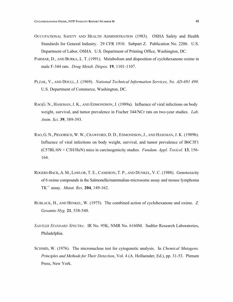
41 CYCLOHEXANONE OXIME, NTP TOXICITY REPORT NUMBER 50
OCCUPATIONAL SAFETY AND HEALTH ADMINISTRATION (1983). OSHA Safety and Health
Standards for General Industry. 29 CFR 1910. Subpart Z. Publication No. 2206. U.S.
Department of Labor, OSHA. U.S. Department of Printing Office, Washington, DC.
PARMAR, D., AND BURKA, L. T. (1991). Metabolism and disposition of cyclohexanone oxime in
male F-344 rats. Drug Metab. Dispos. 19, 1101-1107.
PLZAK, V., AND DOULL, J. (1969). National Technical Information Services, No. AD-691 490.
U.S. Department of Commerce, Washington, DC.
RAO,G. N., HASEMAN, J. K., AND EDMONDSON, J. (1989a). Influence of viral infections on body
weight, survival, and tumor prevalence in Fischer 344/NCr rats on two-year studies. Lab.
Anim. Sci. 39, 389-393.
RAO, G. N., PIEGORSCH, W. W., CRAWFORD, D. D., EDMONDSON, J., AND HASEMAN, J. K. (1989b).
Influence of viral infections on body weight, survival, and tumor prevalence of B6C3F1
(C57BL/6N × C3H/HeN) mice in carcinogenicity studies. Fundam. Appl. Toxicol. 13, 156
164.
ROGERS-BACK, A. M., LAWLOR, T. E., CAMERON, T. P., AND DUNKEL, V. C. (1988). Genotoxicity
of 6 oxime compounds in the Salmonella/mammalian-microsome assay and mouse lymphoma
TK+/ assay. Mutat. Res. 204, 149-162.
RUBLACK, H., AND HENKEL, W. (1975). The combined action of cyclohexanone and oxime. Z.
Gesamte Hyg. 21, 538-540.
SADTLER STANDARD SPECTRA. IR No. 95K; NMR No. 6160M. Sadtler Research Laboratories,
Philadelphia.
SCHMID, W. (1976). The micronucleus test for cytogenetic analysis. In Chemical Mutagens.
Principles and Methods for Their Detection, Vol. 4 (A. Hollaender, Ed.), pp. 31-53. Plenum
Press, New York.
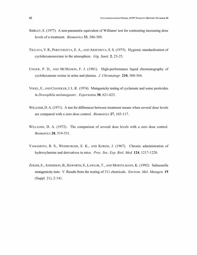
42 CYCLOHEXANONE OXIME, NTP TOXICITY REPORT NUMBER 50
SHIRLEY, E. (1977). A non-parametric equivalent of Williams' test for contrasting increasing dose
levels of a treatment. Biometrics 33, 386-389.
TSULAYA, V. R., PEREVERZEVA, E. A., AND ARSENIEVA, S. S. (1975). Hygienic standardization of
cyclohexanonoxime in the atmosphere. Gig. Sanit. 2, 23-25.
UNGER, P. D., AND MCMAHON, F. J. (1981). High-performance liquid chromatography of
cyclohexanone oxime in urine and plasma. J. Chromatogr. 210, 360-364.
VOGEL, E., AND CHANDLER, J. L. R. (1974). Mutagenicity testing of cyclamate and some pesticides
in Drosophila melanogaster. Experientia 30, 621-623.
WILLIAMS, D. A. (1971). A test for differences between treatment means when several dose levels
are compared with a zero dose control. Biometrics 27, 103-117.
WILLIAMS, D. A. (1972). The comparison of several dose levels with a zero dose control.
Biometrics 28, 519-531.
YAMAMOTO, R. S., WEISBURGER, E. K., AND KORZIS, J. (1967). Chronic administration of
hydroxylamine and derivatives in mice. Proc. Soc. Exp. Biol. Med. 124, 1217-1220.
ZEIGER, E., ANDERSON, B., HAWORTH, S., LAWLOR, T., AND MORTELMANS, K. (1992). Salmonella
mutagenicity tests: V. Results from the testing of 311 chemicals. Environ. Mol. Mutagen. 19
(Suppl. 21), 2-141.

A-1 CYCLOHEXANONE OXIME, NTP TOXICITY REPORT NUMBER 50
APPENDIX A
Summary of Nonneoplastic Lesions in Mice Table A1 Summary of the Incidence of Nonneoplastic Lesions
in Male B6C3F Mice in the 13-Week Drinking Water Study 1
of Cyclohexanone Oxime . . . . . . . . . . . . . . . . . . . . . . . . . . . . . . . . . . . . . . . . . . . . . . . . . . . . . . . . . . . . . . . . . . . . . . A-2
Table A2 Summary of the Incidence of Nonneoplastic Lesions in Female B6C3F Mice in the 13-Week Drinking Water Study 1
of Cyclohexanone Oxime . . . . . . . . . . . . . . . . . . . . . . . . . . . . . . . . . . . . . . . . . . . . . . . . . . . . . . . . . . . . . . . . . . . . . . A-4

A-2 CYCLOHEXANONE OXIME, NTP TOXICITY REPORT NUMBER 50
TABLE A1 Summary of the Incidence of Nonneoplastic Lesions in Male B6C3F Mice1
in the 13-Week Drinking Water Study of Cyclohexanone Oxime1
0 ppm 625 ppm 1,250 ppm 2,500 ppm 5,000 ppm 10,000 ppm
Disposition Summary Animals initially in study Early deaths
Moribund Survivors
Terminal sacrifice
10
10
10
10
10
10
10
10
10
10
10
5
5
Animals examined microscopically 10 10 10 10 10 10
Alimentary System Liver
Depletion glycogen
Hemorrhage Inflammation Centrilobular, hypertrophy Centrilobular, cytoplasmic vacuolization
Pancreas Inflammation
(10) (10) (10) (10) (10) (10) 5 ( 5 0 % )
1 (10%) 1 (10%)
1 (10%) 7 (70%) 10 (100%) 5 (50%)
1 (10%) 3 (30%) 4 (40%) 2 (20%) (10) (10) (10)
1 (10%)
Cardiovascular System None
Endocrine System None
General Body System None
Genital System None
Hematopoietic System Bone marrow
Hyperplasia Lymph node, mandibular
Hemorrhage
Spleen Hematopoietic cell proliferation
Red pulp, cellular depletion Thymus
Atrophy Hemorrhage
(10) (10) (10) (10) (10)
(10) 3 (30%)
(10) 2 (20%)
(10) (10) (10) (10) (10) 10 (100%)
(10) (10)
2 (20%)
(10) 3 (30%)
(10) 1 ( 1 0 % )
(10) 5 ( 5 0 % )
4 (40%) (9)
4 (44%)
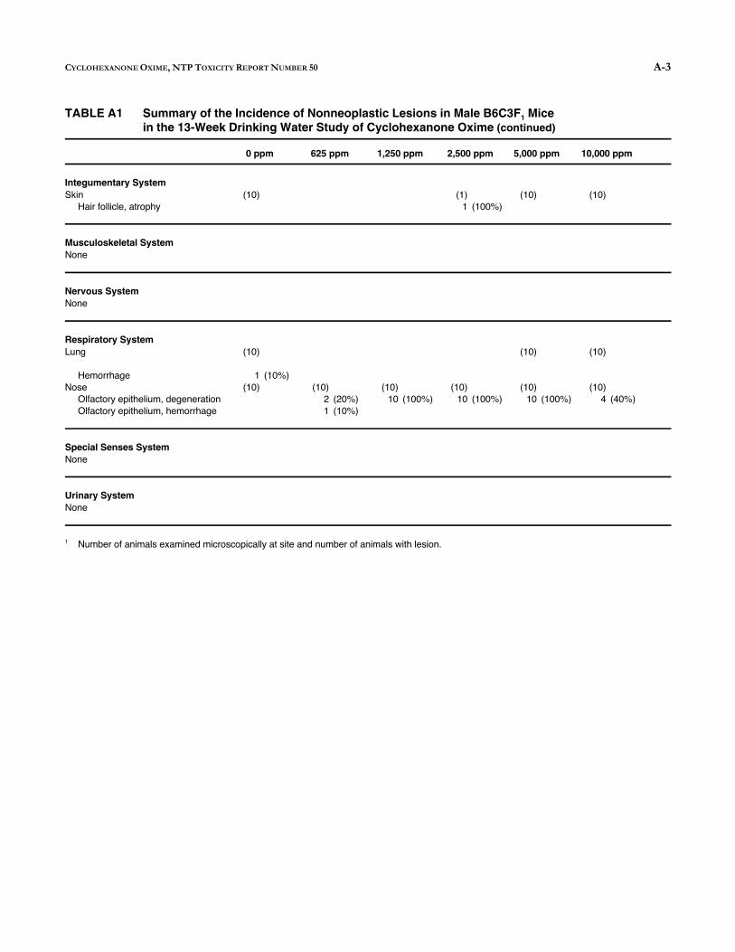
A-3
1
CYCLOHEXANONE OXIME, NTP TOXICITY REPORT NUMBER 50
TABLE A1 Summary of the Incidence of Nonneoplastic Lesions in Male B6C3F Mice1
in the 13-Week Drinking Water Study of Cyclohexanone Oxime (continued)
0 ppm 625 ppm 1,250 ppm 2,500 ppm 5,000 ppm 10,000 ppm
Integumentary System Skin
Hair follicle, atrophy (10) (1)
1 (100%) (10) (10)
Musculoskeletal System None
Nervous System None
Respiratory System Lung (10) (10) (10)
Hemorrhage Nose
Olfactory epithelium, degeneration Olfactory epithelium, hemorrhage
1 (10%) (10) (10)
2 (20%) 1 (10%)
(10) 10 (100%)
(10) 10 (100%)
(10) 10 (100%)
(10) 4 (40%)
Special Senses System None
Urinary System None
Number of animals examined microscopically at site and number of animals with lesion.
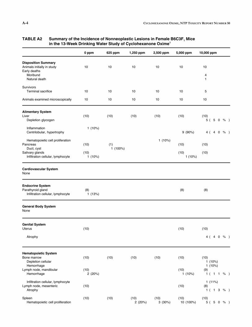
A-4 CYCLOHEXANONE OXIME, NTP TOXICITY REPORT NUMBER 50
TABLE A2 Summary of the Incidence of Nonneoplastic Lesions in Female B6C3F Mice1
in the 13-Week Drinking Water Study of Cyclohexanone Oxime1
0 ppm 625 ppm 1,250 ppm 2,500 ppm 5,000 ppm 10,000 ppm
Disposition Summary Animals initially in study Early deaths
Moribund Natural death
Survivors Terminal sacrifice
Animals examined microscopically
10
10
10
10
10
10
10
10
10
10
10
10
10
10
10
10
4 1
5
10
Alimentary System Liver
Depletion glycogen
Inflammation Centrilobular, hypertrophy
Hematopoietic cell proliferation Pancreas
Duct, cyst Salivary glands
Infiltration cellular, lymphocyte
(10)
1 (10%)
(10)
(10) 1 (10%)
(10)
(1) 1 (100%)
(10) (10)
1 (10%)
(10)
9 (90%)
(10)
(10) 1 (10%)
(10) 5 ( 5 0 %
4 ( 4 0 %
(10)
(10)
)
)
Cardiovascular System None
Endocrine System Parathyroid gland
Infiltration cellular, lymphocyte (8)
1 (13%) (8) (8)
General Body System None
Genital System Uterus (10) (10) (10)
Atrophy 4 ( 4 0 % )
Hematopoietic System Bone marrow
Depletion cellular Hemorrhage
Lymph node, mandibular Hemorrhage
(10)
(10) 2 (20%)
(10) (10) (10) (10)
(10) 1 (10%)
(10) 1 (10%) 1 (10%)
(9) 1 ( 1 1 % )
Infiltration cellular, lymphocyte Lymph node, mesenteric
Atrophy (10) (10)
1 (11%) (8)
1 ( 1 3 % )
Spleen Hematopoietic cell proliferation
(10) (10) (10) 2 (20%)
(10) 3 (30%)
(10) 10 (100%)
(10) 5 ( 5 0 % )

CYCLOHEXANONE OXIME, NTP TOXICITY REPORT NUMBER 50 A-5
Lymphoid follicle, depletion 2 ( 2 0 % )
Red pulp, cellular depletion 1 (10%)
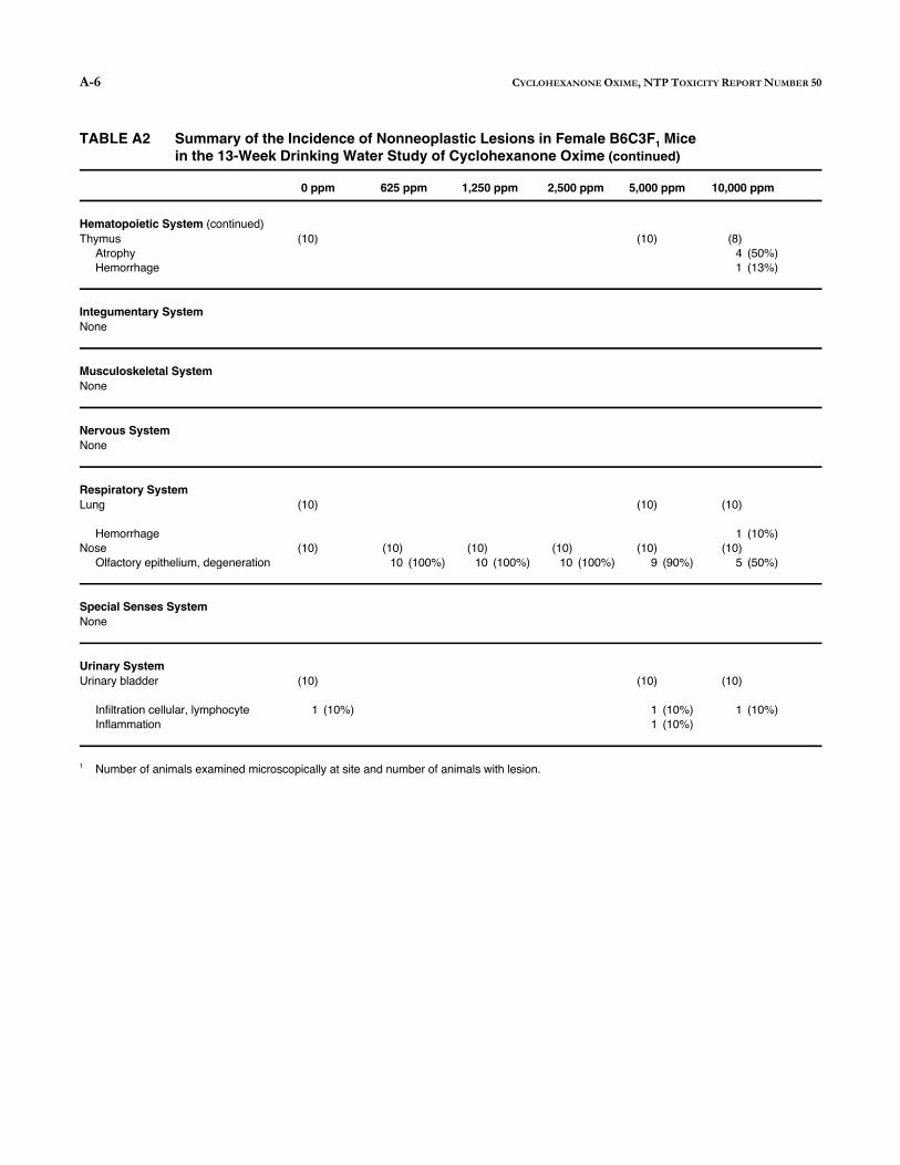
A-6
1
CYCLOHEXANONE OXIME, NTP TOXICITY REPORT NUMBER 50
TABLE A2 Summary of the Incidence of Nonneoplastic Lesions in Female B6C3F Mice1
in the 13-Week Drinking Water Study of Cyclohexanone Oxime (continued)
0 ppm 625 ppm 1,250 ppm 2,500 ppm 5,000 ppm 10,000 ppm
Hematopoietic System (continued) Thymus
Atrophy Hemorrhage
(10) (10) (8) 4 (50%) 1 (13%)
Integumentary System None
Musculoskeletal System None
Nervous System None
Respiratory System Lung (10) (10) (10)
Hemorrhage Nose
Olfactory epithelium, degeneration (10) (10)
10 (100%) (10)
10 (100%) (10)
10 (100%) (10)
9 (90%)
1 (10%) (10)
5 (50%)
Special Senses System None
Urinary System Urinary bladder (10) (10) (10)
Infiltration cellular, lymphocyte Inflammation
1 (10%) 1 (10%) 1 (10%)
1 (10%)
Number of animals examined microscopically at site and number of animals with lesion.

A-7 CYCLOHEXANONE OXIME, NTP TOXICITY REPORT NUMBER 50

A-8 CYCLOHEXANONE OXIME, NTP TOXICITY REPORT NUMBER 50

B-1 CYCLOHEXANONE OXIME, NTP TOXICITY REPORT NUMBER 50
APPENDIX B
Organ Weights and Organ-Weight-to-Body-Weight Ratios
Table B Organ Weights and Organ-Weight-to-Body-Weight Ratios for B6C3F Mice1
in the 13-Week Drinking Water Study of Cyclohexanone Oxime . . . . . . . . . . . . . . . . . . . . . . . . . . . . . . . . . . . . . . B-2

B-2 CYCLOHEXANONE OXIME, NTP TOXICITY REPORT NUMBER 50
TABLE B Organ Weights and Organ-Weight-to-Body-Weight Ratios for B6C3F Mice 1
in the 13-Week Drinking Water Study of Cyclohexanone Oxime1
0 ppm 625 ppm 1,250 ppm 2,500 ppm 5,000 ppm 10,000 ppm
n 10 10 10 10 10 5
MALE
Necropsy body wt 43.5 ± 1.1 43.6 ± 0.8 43.2 ± 0.9 42.1 ± 1.0 39.1 ± 0.9** 31.1 ± 1.0**
Heart Absolute 0.160 ± 0.005 0.173 ± 0.006 0.166 ± 0.005 0.177 ± 0.007 0.171 ± 0.005 0.156 ± 0.005 Relative 3.69 ± 0.07 3.96 ± 0.11 3.84 ± 0.07 4.21 ± 0.12** 4.39 ± 0.10** 5.01 ± 0.09**
Right kidney Absolute 0.310 ± 0.009 0.304 ± 0.006 0.311 ± 0.009 0.322 ± 0.011 0.322 ± 0.004 0.297 ± 0.010 Relative 7.13 ± 0.16 6.98 ± 0.22 7.20 ± 0.13 7.64 ± 0.10* 8.27 ± 0.17** 9.56 ± 0.26**
Liver Absolute 2.010 ± 0.129 2.088 ± 0.046 1.970 ± 0.086 2.046 ± 0.101 1.905 ± 0.056 1.594 ± 0.060* Relative 45.97 ± 1.96 47.87 ± 0.74 45.48 ± 1.08 48.38 ± 1.27 48.76 ± 0.87 51.18 ± 1.01*
Lungs Absolute 0.298 ± 0.016 0.308 ± 0.018 0.304 ± 0.011 0.303 ± 0.007 0.320 ± 0.007 0.314 ± 0.006 Relative 6.88 ± 0.37 7.03 ± 0.37 7.06 ± 0.22 7.21 ± 0.15 8.22 ± 0.18** 10.09 ± 0.22**
Spleen Absolute 0.079 ± 0.003 0.081 ± 0.003 0.077 ± 0.003 0.077 ± 0.003 0.106 ± 0.003** 0.145 ± 0.008** Relative 1.82 ± 0.06 1.86 ± 0.06 1.79 ± 0.04 1.82 ± 0.06 2.73 ± 0.09** 4.64 ± 0.15**
Right testis Absolute 0.120 ± 0.004 0.128 ± 0.004 0.127 ± 0.002 0.128 ± 0.003 0.128 ± 0.002 0.120 ± 0.001 Relative 2.78 ± 0.10 2.94 ± 0.06 2.95 ± 0.07 3.05 ± 0.08* 3.29 ± 0.10** 3.86 ± 0.11**
Thymus Absolute 0.070 ± 0.009 0.069 ± 0.007 0.074 ± 0.006 0.066 ± 0.005 0.056 ± 0.006 0.047 ± 0.009 Relative 1.60 ± 0.19 1.58 ± 0.15 1.71 ± 0.14 1.59 ± 0.14 1.43 ± 0.15 1.49 ± 0.23
FEMALE
Necropsy body wt 35.6 ± 1.2 36.8 ± 0.9 34.3 ± 1.4 32.5 ± 1.0 30.4 ± 1.1** 24.2 ± 1.2**
Heart Absolute 0.137 ± 0.003 0.134 ± 0.002 0.144 ± 0.004 0.133 ± 0.003 0.137 ± 0.007 0.122 ± 0.008 Relative 3.87 ± 0.12 3.64 ± 0.07 4.26 ± 0.18 4.14 ± 0.16 4.52 ± 0.17** 5.04 ± 0.14**
Right kidney Absolute 0.209 ± 0.006 0.214 ± 0.004 0.211 ± 0.004 0.217 ± 0.005 0.225 ± 0.005 0.205 ± 0.008 Relative 5.90 ± 0.18 5.84 ± 0.13 6.21 ± 0.21 6.70 ± 0.20** 7.44 ± 0.17** 8.52 ± 0.22**
Liver Absolute 1.510 ± 0.052 1.623 ± 0.040 1.446 ± 0.032 1.380 ± 0.033 1.396 ± 0.053 1.172 ± 0.048** Relative 42.64 ± 1.28 44.13 ± 0.56 42.52 ± 1.14 42.61 ± 0.94 45.92 ± 0.79* 48.72 ± 1.48**
Lungs Absolute 0.283 ± 0.008 0.269 ± 0.017 0.311 ± 0.010 0.301 ± 0.012 0.288 ± 0.014 0.278 ± 0.014 Relative 8.07 ± 0.39 7.34 ± 0.46 9.17 ± 0.41 9.27 ± 0.34 9.53 ± 0.44* 11.61 ± 0.68**
Spleen Absolute 0.092 ± 0.002 0.099 ± 0.003 0.098 ± 0.005 0.097 ± 0.004 0.151 ± 0.008** 0.156 ± 0.016** Relative 2.60 ± 0.09 2.69 ± 0.11 2.87 ± 0.14 3.00 ± 0.12 4.97 ± 0.21** 6.41 ± 0.48**
Thymus Absolute 0.071 ± 0.005 0.066 ± 0.004 0.067 ± 0.005 0.065 ± 0.003 0.059 ± 0.005 0.057 ± 0.003 Relative 2.00 ± 0.11 1.80 ± 0.10 1.93 ± 0.08 2.03 ± 0.12 1.92 ± 0.11 2.41 ± 0.22
Organ weights and body weights are given in grams; organ-weight-to-body-weight ratios are given as mg organ weight/g body weight (mean ± standard error).
* Significantly different (P 0.05) from the control group by Williams' or Dunnett's test. ** Significantly different (P 0.01) from the control group by Williams' or Dunnett's test.
1
C-1 CYCLOHEXANONE OXIME, NTP TOXICITY REPORT NUMBER 50
APPENDIX C
Reproductive Tissue Evaluations and Estrous Cycle Characterization
Table C1
Table C2
Summary of Reproductive Tissue Evaluations in Male B6C3F Mice 1
in the 13-Week Drinking Water Study of Cyclohexanone Oxime . . . . . . . . . . . . . . . . . . . . . . . . . . . . . . . . . . . . . .
Summary of Estrous Cycle Characterization in Female B6C3F Mice1
in the 13-Week Drinking Water Study of Cyclohexanone Oxime . . . . . . . . . . . . . . . . . . . . . . . . . . . . . . . . . . . . . .
C-2
C-2

C-2
1
1
CYCLOHEXANONE OXIME, NTP TOXICITY REPORT NUMBER 50
TABLE C1 Summary of Reproductive Tissue Evaluations in Male B6C3F Mice 1
in the 13-Week Drinking Water Study of Cyclohexanone Oxime1
Study Parameters 0 ppm 1,250 ppm 2,500 ppm 5,000 ppm
n 10 10 10 10
Weights (g) Necropsy body weight 43.5 ± 1.1 43.2 ± 0.9 42.1 ± 1.0 39.1 ± 0.9** Left epididymis 0.054 ± 0.002 0.051 ± 0.001 0.053 ± 0.001 0.053 ± 0.001 Left cauda epididymis 0.019 ± 0.001 0.020 ± 0.001 0.021 ± 0.001 0.019 ± 0.001 Left testis 0.120 ± 0.003 0.123 ± 0.003 0.122 ± 0.003 0.125 ± 0.002
Spermatid measurements 7Spermatid heads (10 /g testis) 20.34 ± 0.50 20.24 ± 0.40 19.40 ± 0.61 20.50 ± 0.35 7Spermatid heads (10 /testis) 2.43 ± 0.05 2.49 ± 0.05 2.35 ± 0.04 2.57 ± 0.05
Spermatid count-4 (mean/10 mL suspension) 76.05 ± 1.56 77.80 ± 1.42 73.33 ± 1.31 80.33 ± 1.45
Epididymal spermatozoal measurements Motility (%) 87.89 ± 0.59 88.75 ± 0.58 87.80 ± 0.36 86.93 ± 0.50 Concentration
6 (10 /g cauda epididymal tissue) 755 ± 31 716 ± 67 647 ± 36 668 ± 44
Data are presented as mean ± standard error. Differences from the control group for spermatid and epididymal spermatozoal measurements are not significant by Dunn's test.
** Significantly different (P 0.01) from the control group by Shirley's test.
TABLE C2 Summary of Estrous Cycle Characterization in Female B6C3F Mice1
in the 13-Week Drinking Water Study of Cyclohexanone Oxime1
Study Parameters 0 ppm 1,250 ppm 2,500 ppm 5,000 ppm
n 10 10 10 10
Necropsy body weight (g) 35.6 ± 1.2 34.3 ± 1.4 32.5 ± 1.0 30.4 ± 1.1** Estrous cycle length (days) 4.90 ± 0.26 4.90 ± 0.37 4.70 ± 0.13 4.60 ± 0.15 Estrous stages (% of cycle)
Diestrus 33.3 29.2 25.0 26.7 Proestrus 15.8 17.5 20.0 17.5 Estrus 31.7 34.2 33.3 33.3 Metestrus 19.2 19.2 21.7 22.5
Necropsy body weight and estrous cycle length data are presented as mean ± standard error. Differences from the control group for estrous cycle length are not significant by Dunn's test. By multivariate analysis of variance, exposed groups did not differ significantly from the control group in the relative length of time spent in the estrous stages.
** Significantly different (P 0.01) from the control group by Shirley's test.

D-1 CYCLOHEXANONE OXIME, NTP TOXICITY REPORT NUMBER 50
APPENDIX D
Genetic Toxicology
Table D1 Mutagenicity of Cyclohexanone Oxime inSalmonella typhimurium . . . . . . . . . . . . . . . . . . . . . . . . . . . . . . . . . . . . . . . D-2
Table D2 Induction of Chromosomal Aberrations in Chinese Hamster Ovary Cells by Cyclohexanone Oxime . . . . . . . . . . . . . . . . . . . . . . . . . . . . . . . . . . . . . . . . . . . . . . . . . . . . . . . . . . . . . . . . . . . . . . . D-4
Table D3 Induction of Micronuclei in Bone Marrow Erythrocytes of Male B6C3F Mice 1
by Cyclohexanone Oxime . . . . . . . . . . . . . . . . . . . . . . . . . . . . . . . . . . . . . . . . . . . . . . . . . . . . . . . . . . . . . . . . . . . . . . . D-5
Table D4 Frequency of Micronulei in Peripheral Blood of Male and Female B6C3F Mice Exposed to Cyclohexanone Oxime 1
in Drinking Water . . . . . . . . . . . . . . . . . . . . . . . . . . . . . . . . . . . . . . . . . . . . . . . . . . . . . . . . . . . . . . . . . . . . . . . . . . . . . D-6

D-2 CYCLOHEXANONE OXIME, NTP TOXICITY REPORT NUMBER 50
TABLE D1 Mutagenicity of Cyclohexanone Oxime in Salmonella typhimurium1
Dose Revertants/plate2
Strain (µg/plate) -S9 +30% hamster S9 +30% rat S9
TA97 0 118 ± 11.4 159 ± 5.5 195 ± 4.0 10 35 ± 4.0 33 135 ± 8.1 168 ± 23.4 208 ± 5.0
100 153 ± 3.5 158 ± 4.7 175 ± 5.7 333 130 ± 9.5 177 ± 12.0 208 ± 5.7
1,000 128 ± 10.7 156 ± 4.0 198 ± 15.4 3,333 69 ± 2.4 163 ± 11.4 179 ± 7.8
Trial summary Negative Negative Negative Positive control3 350 ± 12.8 383 ± 11.5 357 ± 17.9
TA98 0 35 ± 2.3 35 ± 7.5 49 ± 5.7 33 40 ± 4.6 45 ± 2.2 38 ± 2.7
100 39 ± 6.7 43 ± 2.9 37 ± 2.2 333 35 ± 2.2 37 ± 4.8 36 ± 2.5
1,000 31 ± 6.9 55 ± 3.3 21 ± 2.4 3,333 34 ± 7.0 21 ± 4.3
Trial summary Negative Negative Negative Positive control 466 ± 19.4 435 ± 28.9 151 ± 19.4
TA100 0 149 ± 5.0 145 ± 4.1 159 ± 5.4 10 145 ± 2.3 33 155 ± 10.9 176 ± 7.3 155 ± 20.9
100 118 ± 3.1 135 ± 4.9 170 ± 7.7 333 135 ± 4.0 149 ± 5.4 138 ± 20.6
1,000 115 ± 11.4 157 ± 11.3 148 ± 5.2 3,333 127 ± 7.5 122 ± 19.7
Trial summary Negative Negative Negative Positive control 764 ± 25.4 557 ± 16.2 406 ± 10.5

D-3 CYCLOHEXANONE OXIME, NTP TOXICITY REPORT NUMBER 50
TABLE D1 Mutagenicity of Cyclohexanone Oxime in Salmonella typhimurium (continued)
Revertants/plate
Strain Dose -S9 +hamster S9 (µg/plate) Trial 1 Trial 2 5% 5% 10% 10% 10%
TA1535 0 3
10 33
100 333 666
1,000 1,666 3,333 6,666
10 ±
18 ± 19 ± 18 ±
17 ±
6 ±
1.7
5.8 1.5 4.4
4.2
2.6
8 ± 8 ± 7 ± 7 ± 6 ± 8 ±
2.2 1.2 0.9 0.3 1.5 1.5
8 ±
11 ± 9 ±
10 ± 8 ± 3 ± 0 ±
1.0
2.2 1.5 2.6 2.1 1.0 0.0
11 ±
22 ± 33 ± 33 ± 37 ± 30 ± 1 ±
2.2
1.7 1.7 3.3 1.5 3.4 1.04
10 ±
12 ± 8 ±
10 ± 13 ± 11 ± 5 ± 1 ±
3.2
1.2 2.5 0.7 3.7 0.6 1.0 0.9
12 ±
23 ± 52 ± 58 ± 59 ±
104 ±
2.2
2.3 1.9 5.4 4.5 4.2
10 ±
14 ± 27 ± 48 ± 63 ± 56 ± 0 ±
2.7
3.8 3.6 2.2 3.5 7.3 0.04
Trial summary Positive control
Equivocal 826 ± 29.9
Negative 1,186 ± 56.2
Negative 95 ± 13.9
Positive 102 ± 7.4
Negative 69 ± 3.5
Positive 63 ± 1.9
Positive 73 ± 4.0
TA1535 (continued) +hamster S9 (continued) +rat S9
30% 30% 30% 30%
0 33
100 333
11 ± 0.3 11 ± 1.5 5 ± 0.3 6 ± 0.3
10 ± 2.9
9 ± 2.7
10 ± 0.7
14 ± 1.7
14 ± 2.4 10 ± 0.6 10 ± 1.8 12 ± 2.3
666 1,000 1,666 3,333 6,666
17 ± 1.0
31 ± 1.2
10 ± 3.6 11 ± 1.3 21 ± 5.2 17 ± 2.5 5 ± 1.2
25 ± 1.5 37 ± 1.2 53 ± 2.6 72 ± 5.0 9 ± 1.74
17 ± 1.3
16 ± 2.6
Trial summary Positive control
Equivocal 279 ± 10.5
Equivocal 141 ± 14.0
Positive 75 ± 7.0
Negative 51 ± 5.6
1 Study performed at SRI, Inc. The detailed protocol and these data are presented in Zeiger et al. (1992); 0 µg/plate is the solvent control.2 Revertants are presented as mean ± standard error from three plates.3 The positive controls in the absence of metabolic activation were 4-nitro-o-phenylenediamine (TA98), sodium azide (TA100 and TA1535), and
9-aminoacridine (TA97). The positive control for metabolic activation with all strains was 2-aminoanthracene. 4 Slight toxicity.

D-4 CYCLOHEXANONE OXIME, NTP TOXICITY REPORT NUMBER 50
TABLE D2 Induction of Chromosomal Aberrations in Chinese Hamster Ovary Cells by Cyclohexanone Oxime1
Dose (µg/mL)
Total Cells
-S9
No. of Abs
Abs/ Cell
Cells with Abs (%)
Dose (µg/mL)
Total Cells
+S9
No. of Abs
Abs/ Cell
Cells with Abs (%)
Trial 1 — Harvest time: 12 hours Summary: Equivocal
Trial 1 — Harvest time: 12 hours Summary: Negative
Dimethylsulfoxide 200 2 0.01 1.0
Dimethylsulfoxide 200 1 0.01 0.5
Mitomycin-C 0.4 25 10 0.40 36.0
Cyclophosphamide 20 25 14 0.56 44.0
Cyclohexanone oxime 1,081 200 2,325 200 5,000 2002
10 4 1
0.05 0.02 0.01
4.5* 1.5 0.5
Cyclohexanone oxime 1,081 200 2,325 200 5,000 2002
1 0 3
0.01 0.00 0.02
0.5 0.0 1.5
P=0.853 3 P=0.158
Trial 2 — Harvest time: 12 hours Summary: Equivocal
Dimethylsulfoxide 200 2 0.01 0.5
Mitomycin-C 0.4 25 16 0.64 32.0
Cyclohexanone oxime 500 200
1,000 200 2,000 200
3 7 3
0.02 0.04 0.02
1.5 3.5* 0.5
P=0.301
1 Study performed at Sitek Research Laboratories. A detailed description of the protocol is presented in Galloway et al. (1987). Abs = aberrations.
2 Precipitate formed at this dose level.3 Significance of percent cells with aberrations tested by the linear regression trend test versus log of the dose. * Positive (P<0.05).
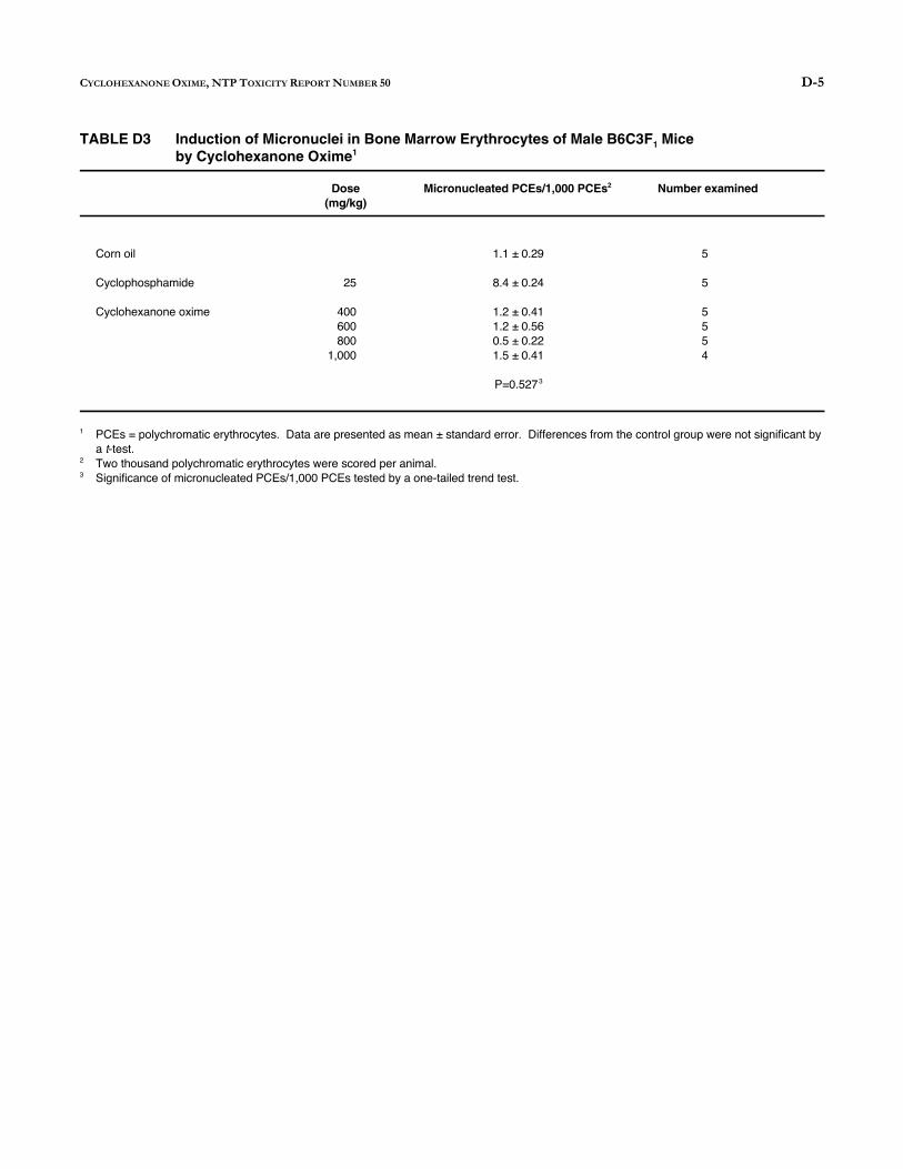
D-5 CYCLOHEXANONE OXIME, NTP TOXICITY REPORT NUMBER 50
TABLE D3 Induction of Micronuclei in Bone Marrow Erythrocytes of Male B6C3F Mice 1
by Cyclohexanone Oxime1
Dose Micronucleated PCEs/1,000 PCEs2 Number examined (mg/kg)
Corn oil 1.1 ± 0.29 5
Cyclophosphamide 25 8.4 ± 0.24 5
Cyclohexanone oxime 400 1.2 ± 0.41 5 600 1.2 ± 0.56 5 800 0.5 ± 0.22 5
1,000 1.5 ± 0.41 4
P=0.5273
1 PCEs = polychromatic erythrocytes. Data are presented as mean ± standard error. Differences from the control group were not significant by a t-test.
2 Two thousand polychromatic erythrocytes were scored per animal.3 Significance of micronucleated PCEs/1,000 PCEs tested by a one-tailed trend test.

D-6 CYCLOHEXANONE OXIME, NTP TOXICITY REPORT NUMBER 50
TABLE D4 Frequency of Micronuclei in Peripheral Blood of Male and Female B6C3F Mice Exposed to Cyclohexanone Oxime 1
in Drinking Water1
Concentration Micronucleated NCEs/1,000 NCEs2 Number examined (ppm)
MALE 0 4.3 ± 0.3 5 625 3.9 ± 0.9 5
1,250 3.6 ± 0.2 5 2,500 3.6 ± 0.7 5 5,000 1.1 ± 0.3 5
10,000 1.2 ± 0.5 5
P=1.0003
FEMALE 0 2.9 ± 0.6 5 625 3.2 ± 0.7 5
1,250 1.8 ± 0.5 5 2,500 2.4 ± 0.7 5 5,000 0.7 ± 0.3 5
10,000 0.9 ± 0.2 5
P=1.000
1 A detailed description of the protocol is found in MacGregor et al. (1990). Data are presented as mean ± standard error. 2 NCEs = normochromatic erythrocytes. Two thousand normochromatic erythrocytes were scored per animal. 3 Significance of micronucleated NCEs/1,000 NCEs tested by a one-tailed trend test.
E-1 CYCLOHEXANONE OXIME, NTP TOXICITY REPORT NUMBER 50
APPENDIX E
Metabolism and Disposition Study
Metabolism and Disposition of Cyclohexanone Oxime in Male F-344 Rats
D. PARMAR AND L. T. BURKA
Drug Metabolism and Disposition 19, 1101-1107 (1991)
Reproduced with permission of The American Society for Pharmacology and Experimental Therapeutics

0090-9556/91/1906-1101$03.00/0 DRUG METABOLISM AND DISPOSITION VoL 19, No.6 Copyright @:- 1991 by The American Society for Pharmaeology and Experimental Therapeutics Printed in U.S.A.
METABOLISM AND DISPOSITION OF CYCLOHEXANONE OXIME IN MALE F-344 RATS
DEVENDRA PARMAR AND LEO T. BURKA
National Toxicology Program, National Institute of Environmental Health Sciences
(Received March 26, 1991; accepted August 6, 1991)
ABSTRACT:
Cyclohexanone oxime (CHOX), an intermediate used in the synthesis of Polycaprolactam/Nylon, was found to be rapidly absorbed and cleared from the body within.24 hours after a single oral administra· lion of 1, 10 and 30 mg/kg of (14C]·CHOX to the adult male Fischer rats. The majority of the CHOX derived radioactivity (65-90% ofthe dose) was excreted in the urine. Elimination in the feces accounted for 5-10% of the dose and very low levels of radioactivity (2-3%) were retained in the tissues 24 hours after exposure. After iv a(!min· istration of 1 mgfkg of (14C]·CHOX, the oxime was rapidly cieared from plasma with half-lives. of 1.6 (alpha phase) and 18.2 min (beta phase). However, when CHOX was applied dermally (30 mgfkg), only about 4-5% of the dose was recovered. in urine, feces and the
Oxirnes have a wide variety of applications, as chemical and pesticide intermediates; blocking agents in the polymer industry; chelators in the metal industry; drugs; antioxidants; anti-knock agents in fuels; and in dye synthesis. CHOX' is almost exclusively used as a captive intermediate for the production of caprolactam, which, in tum, is used to form polycaprolactarn or Nylon 6, a fiber used in clothing, carpeting, horne furnishings, and similar products.
Although there are no reports of occupational exposure to CHOX in humans, the large volume ofCHOX produced yearly necessitates a thorough understanding of the toxicity of this compound. Studies in laboratory animals have shown that CHOX produces toxic lesions in the hematopoietic system (l-3).c Systemically absorbed CHOX is reported to induce oxidative damage to the· erythrocytic· population in animals resulting in hemolytic anemia accompanied by increased erythropoiesis. Oral administration of CHOX for 2 weeks produced a dose-related decrease in the erythrocyte number, Hb, and hematocrit with an accompanying increase in teticulocytes and circulating nucleated erythrocytes in male and female rats. Methemoglobin levels were also significantly elevated in both sexes. Splenomegaly and hepatomegaly were also observed and the effects persisted for up to 2 weeks after exposure. Histopathological examination revealed compound-related effects in the spleen and bone marrow of male and female rats (I). Similar hernatotoxic effects were reported when animals were treated subchronically for .13 weeks by gavage (2). Gad et al. (2) suggested that significant amounts of CHOX, sufficientto elicit toxic manifestations, may be absorbed through the skin. They reported that dermal application of 0.8to 5 g/kg
'Abbreviations used are: CHOX, cyclohexanone oxime; b.w., body Weight. • Ref. 3, per citation in refs. 1 and 2.
Send reprint requests to: Or. Leo T. Burka, NIEHS, P.O. Box 12233, Research Triangle Park, NC 27709.
tissues. The majority of the dose volatilized from the skin surface. However, the absorbed oxime was readily distributed and excreted, and its metabolic late was no different than observed after oral administrations. HPLC analysis of urine showed that the majority of the radioactivity excreted was in the form of three metabolites, cyclohexylglucuronide and 'the monoglucuronides of cis· and transcyclohexane-1,2-diol./n vitro studies showed that these metabolites arise primarily by hydrolysis of the oxime to cyclohexanone which is then reduced to cyclohexanol and eliminated as the glucuronide conjugate. The cyclohexanol, in tum could be metabolized to cis· and trans·cyclohexane"1,2·diols, which are excreted as their monoglucuronides.
of CHOX increased reticulocyte counts 24 hr after e:-.posure in male rabbits, while the Hb levels in females were depressed (2). Rats exposed by inhalation to 0.03, 0.1, and I rng/rn' ofCHOX for 6 and !0 weeks also exhibited decreased erythrocyte counts and increased levels of methemoglobin. Degenerative changes in the parenchymatous organs and desquamation of bronchial epithelium were observed in the high dose group (3).
CHOX was reported to be mutagenic in mouse lymphoma cells without exogenous metabolic activation. However, no mutagenic activity was observed in the presence of S-9 activation, suggesting that the chemical may be deactivated by the enzymes in S-9 (4). CHOX was non-mutagenic in Salmonella typhimurium and in Escherichia coli with or without metabolic activation (4, 5). CHOX also failed to induce sex-linked recessive mutations in Drosophila (6, 7).
At present, little information is available on the disposition and metabolism of CHOX, except that some oximes, including CHOX, are reported to inhibit the oxidative metabolism of ethanol (8, 9). Additional interest has been generated in the toxicity ofCHOX since there are reports suggesting that CHOX is formed by the oxidative metabolism .of sodium cyclamate, an artificial sweetener (1 0). The present study was designed to provide greater insight into the fate of CHOX by studying .its absorption, tissue distribution, metabolism, and excretion in adult.rnale Fischer rats.
Materials and Methods
Chemicals. Cyclohexanone. oxime P ·"C]. specific actiYity 6.85 mCi/ mmol. was synthesized by NEN Research Products (Boswn. MA). Radiochemical purity was determined by HPLC to be greater than 99%. Unlabeled CHOX, cyclohexanol, cyclohexanone, and 1rans· and ciscyclohexane·l ,2-diol were purchased from Aldrich Chemical Co. (Milwaukee, WI). Unlabeled CHOX was recrystallized from hexane before use. NADPH, NADP, nicotinamide. ;'i-glucuronidase (tYpe B-l ). and glucose 6-phosphate were obtained from Sigma Chemical Co. (St. Louis,
1101
E-2 CYCLOHEXANONE OXIME, NTP TOXICITY REPORT NUMBER 50

1102 PARMAR AND BURKA
MO). All other chemicals were of the highest purity commercially available.
Apparatus. HPLC. The HPLC svstem used consisted of Waters Associates (Milford, MA) pumps, aut~mated gradient controller, and tunable absorbance detector. A Flow-One radioactive flow detector (Radiomatic Instruments Co, Tampa, FL) was used for analysis and a Beckman 171 radioisotope detector (Beckman Instruments, Fullenon, CA) was used for isolation of metabolites. A 25 em x 4.6 mm Microsorb C-18 column (Rainin Instrument Co., Worburn, MA) was used for both analysis and isolation.
Gas Chromatography. GC was performed on a Hewlett-Packard (Avondale, PA) model5880-A gas chromatograph equipped with a flame ionization detector, a Grab-type splitless injector, and a :5-m fused silica ·capillary column having 0.22 mm i.d. and a 0.25-i<m film thickness of BPlO (Bodman Chemicals, Doraville, GA). The oven temperature was programmed to rise from 35"C to IlO'C at a rate of 7 .5'/min for the analysis of cyclohexanol and cyclohexanone and from 35'C to 200'C for cis- and trans-cyclohexane-1 ,2-diol. The injector and detector temperatures were 250'C and 275'C, respectively.
'H NMR Spectra. These were recorded ori a General Electric (Fremont, CA) GN-500 NMR spectrometer using 0 20 as solvent. The chemical shifts reponed are in ppm relative to tetramethylsilane as the external standard.
Mass Spectra. Mass spectra were recorded on a Concept I SQ hybrid mass spectrometer (Kratos Analytical, Manchester, UK). This instrument utilizes a Hewlett-Packard model5890 gas chromatograph. A J&W DB-5 capillary column, 0.25 mm i.d. x 60 m, with a 0.25-l'm film thickness (J&W Scientific, Fulsom, CA) was used for analysis of in vivo metabolites; a similar 30-m column was used for analysis of the in \'itro metabolites.
Animals. Male Fischer 344 rats, 8-10 weeks old (200-250 g), obtained from Charles River Breeding Laboratories (Raleigh, NC), were used throughout the study. All animals received rat chow and water ad libitum.
Disposition Studies. Rats were housed in individual glass metabolic cages allowing for separate collection of urine, feces, C02, and other volatile materials. For oral administration the dosing solutions were prepared in distilled water. Radiolabeled CHOX was diluted as needed with unlabeled CHOX to administer approximately 50 I'Ci/kg body weight (b.w.). The oral doses administered were 1, 10, and 30 mg/kg b.w. in 5 ml/kg of dosing solution, and the animals were sacrificed 24 hr later. A group of animals was also sacrificed 6 hr after oral exposure to 30 mg/kg of CHOX. A dermal dose of CHOX, 30 mg/kg, was dissolved in acetone and applied at 0.2 ml/kg to the interscapular area. Radiolabeled CHOX was diluted as needed with unlabeled CHOX to administer 50 I'Ci/kg to each animal. The interscapular area was clipped 24 hr before dosing, At the time of dosing each animal was anesthetized with I ml/kg of Ketamine. The dermal dose was applied with a microliter syringe with a blunt needle in a circular area of I em'. This area was then covered with a perforated metal tissue capsule (Lipshaw, Detroit, Ml) to prevent removal or ingestion of the dermal dose by grooming. The capsule was glued directly to the skin with cyanoacrylate glue and remained in place throughout the course of the study. The animals were kept in individual glass metabolism cages and sacrificed 24 hr after the treatment except for one group which was sacrificed 3-5 min after dosing.
Three animals were used in each group and the animals were treated between 9:00 and 11 :00 a.m. Following sacrifice, animals were immediately dissected and the tissues were removed, weighed, and then stored at -20'C until assayed. The radioactivity in each tissue was analyzed by combustion of triplicate 50- to 100-mg samples to "C02 in a Packard Tricarb sample oxidizer (Packard Instrument Co., Downers Grove, IL). The ••co, was then counted in a Beckman model LS-9800 liquid scintillation counter. Body composition estimates for blood and muscle were 8 and 50%, respectively (11 ), and II and 16% for adipose tissue and skin, respectively (12). All other tissues were determined gravimetrically. Excretion of CHOX-derived radioactivity was determined by complete collection of the urine and feces for 24 hr after exposure. Urine samples (0.02-0.05 ml) were counted directly without combustion. Fecal samples were air-dried, weighed, and ground to a fine powder. Triplicate
samples were oxidized and the radioactivity was determined as described above. For the group sacrificed immediately after dermal administration, the application site was excised, washed with acetone two to three times, and cut into several pieces. Radioactivity in the washings was determined by adding 0.05-ml aliquots to scintillation fluid and counting. The radioactivity retained in the skin after acetone extraction was determined by combustion.
Radioactivity excreted in the urine was subjected to enzymatic hydrolysis by !l-glucuronidase to free possible glucuronide conjugates. The incubation mixture contained I 00-200 11g of CHOX-derived material and 2000 units of !l-glucuronidase in 0.1 M sodium acetate buffer, pH · 5.0. The reaction mixture was incubated at 37'C for 15 hr and then analyzed by GC or HPLC. Similar incubation mixtures without enzyme served as controls.
Collection of CO, and Volatiles. After oral or dermal administration of 14C-CHOX, rats were placed in Metabowl-Mark III glass metabolism cages (Jencons, Heme! Hempstead, Herfordshire, UK) maintained at an air flow of 0.4-0.5 liter/min. Total air flow through the cage was first passed through an ethanol (200 ml) trap for the collection of volatiles and then through 200 ml of a 7:3 (v /v) mixture of 2-methoxyethanol/ ethanolamine for '4C02 collection. The percentage of the total dose of 14C-CHOX eliminated as 14CO, and volatiles was determined by counting triplicate 1-ml aliquots of each trapping solution.
For the dermal studies, activated charcoal filters (SKC Inc., Eighty Four, PA) were used in place of ethanol for trapping the expired volatiles. The expired volatiles absorbed by the charcoal traps were extracted with 1 ml of acetonitrile from each section of the filter (sample zone with 200 mg of activated charcoal and back-up zone with 100 mg), and radioactivity was counted in a 20-1'1 aliquot. Following extraction, the activated charcoal was also analyzed by combustion of triplicate 25-mg samples and the radioactivity was determined as described earlier.
Pharmacokinetic Studies. An iv dose of 1 mg/kg CHOX dissolved in I ml/kg normal saline was used. Radiolabeled CHOX was diluted with unlabeled CHOX to administer 50 I'Ci/kg to each animal. Male rats weighing 200-230 g were anesthetized with I ml/kg of Ketamine. A silicone tube was installed in the jugular vein and exteriorized through the skin at the back of the neck. Following a 24-hr recovery period, an iv dose was injected through the tubing. At specified times after dosing, known amounts of blood were withdrawn. The blood was immediately centrifuged and the plasma stored frozen at - 20'C until analysis. The concentration of unmetabolized oxime was determined in the plasma samples by HPLC using an isocratic system of 90: I 0 water/acetonitrile at a flow rate of 1.5 ml/min for 20 min and a Flow-one radioactivity detector. Pharmacokinetic parameters were calculated using NONLIN (Statistical Consultants, Inc., Lexington, KY). Initial parameters were obtained graphically. The data were weighted by the reciprocal of th~, concentration.
Metabolism of 14C-CHOX In Vivo. Metabolism was studied by determining the relative amounts of CHOX metabolites in urine and liver extracts. Urine samples were collected at intervals of 0-2, 2-4, 4-6, 6-8, 8-12, and 12.,.24 hr after treatment. These samples were acidified with I 0 l'l of glacial acetic acid per ml and centrifuged at low speed in a clinical centrifuge. The supernatants were then filtered through type HV 0.45-l'm filter units (Millipore Corp., Bedford, CA) and analyzed by HPLC. Livers from animals sacrificed 6 hr after treatment were homogenized in acetonitrile and centrifuged, and the supernatant was filtered before HPLC analvsis. HPLC analvsis was carried out with a C-18 column using a line~r gradient of99.9:0.I water/acetic acid to 74.9:25:0.1 water/acetonitrile/acetic acid in 28 min at a flow rate of 1.5 ml/min.
Isolation and Identification of the Metabolites. The three major metabolites in urine were isolated isocratically using a C-18 column with 89.9:10:0.1 water/acetonitrile/trifluoroactetic acid at a flow rate of 1.5 ml/min. The radioactive peaks, M-1, M-2, and M-3, were collected and freeze-dried. Peaks M-1 and M-2 were funber purified isocratically using 98.9: I :0.1 water/acetonitrile/trifluoroacetic acid. NMR spectra of the isolated metabolites were obtained:
M-1, 'H NMR, 1.1-1.4 (m, 4H, 2x CH,), 1.65 (br s, 2H, CH,), 1.94 (br
E-3 CYCLOHEXANONE OXIME, NTP TOXICITY REPORT NUMBER 50
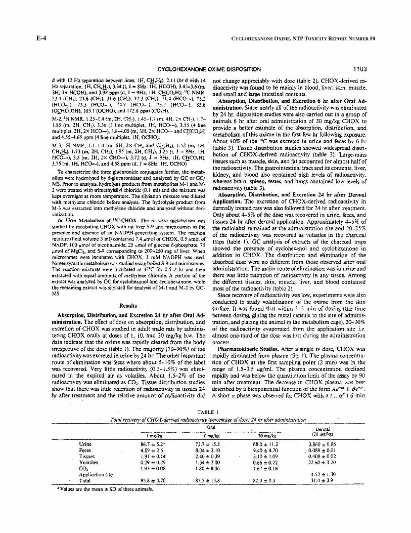
d with 12Hz separation between lines, IH, C!!,Hn). 2.11 (br d with 14 not change appreciably with dose (table 2). CHOX-derived ra dioactivity was found to be mainly in blood, liver, skin, muscle, and small and large intestinal contents. Absorption, Distribution, and Excretion 6 hr after Oral Ad·
ministration. Since nearly all of the radioactivity was eliminated by 24 hr, disposition studies were also carried out in a group of animals 6 hr after oral administration of 30 mg/kg CHOX to
provide a better estimate of the absorption, distribution, and
metabolism of this oxime in the first few hr following exposure. About 40% of the 14C was excreted in urine and feces by 6 hr
(table 3). Tissue distribution studies showed widespread distri
bution of CHOX-derived radioactivity (table 3). Large-mass tissues such as muscle, skin, and fat accounted for almost half of the radioactivity. The gastrointestinal tract and its contents, liver, kidney, and blood also contained high levels of radioactivity,
whereas brain, spleen, testes, and lungs contained low levels of radioactivity (table 3).
Absorption, Distribution, and Excretion 24 hr after Dermal Application. The excretion of CHOX-derived radioactivity in dermally treated rats was also followed for 24 hr after treatment. Only about 4-5% of the dose was recovered in urine, feces, and
tissues 24 hr after dermal application. Approximately 4-5% of the radiolabe! remained at the administration site and 20-25% of the radioactivity was recovered as volatiles in the charcoal traps (table ! ). GC analysis of extracts of the charcoal traps
showed the presence of cyclohexano! and cyclohexanone in addition to CHOX. The distribution and elimination of the absorbed dose were no different from those observed after oral
administration. The major route of elimination was in urine and there was little retention of radioactivity in any tissue. Among
the different tissues, skin, muscle, liver, and blood contained most of the radioactivity (table 2).
Since recovery of radioactivity was low, experiments were also conducted to study volatilization of the oxime from the skin surface. It was found that within 3-5 min of dosing (the time between dosing, gluing the metal capsule to the site of adminis
tration,.and placing the animal in the metabolism cage), 20-30% of the radioactivity evaporated from the application site i.e.
almost one-third of the dose was lost during the administration process.
Pharmacokinetic Studies. After a single iv dose, CHOX was rapidly eliminated from plasma (fig. !). The plasma concentra
tion of CHOX at the first sampling point (2 min) was in the range of 1.5-3.5 J.Lg/ml. The plasma concentration declined
rapidly and was below the quantitation limit of the assay by 90 min after treatment. The decrease in CHOX plasma was best
described by·a biexponential function of the form Ae-"' +Be-·". A short a phase was observed for CHOX with a !, 10 of 1.6 min
Hz separation, I H, CH,H,), 3.34 (t, J = 8Hz, I H. HCOH), 3.4:i-3.6 (m,3H. 3x HCOH), and 3.98 ppm (d, J = 9Hz, I H, C!!C02H); "C NMR,23.4 (CH,). 23.6 (CH,), 31.6 (CH,), 32.3 (CH,). 71.4 (HCO-), 73.2(HCO-), 73.5 (HCO-), 74.7 (HCO-),. 75.2 (HCO-), 85.8(O£HC02H), 103.1 (OCHO), and 172.8 ppm (CO,H).
M-2, 'H NMR, 1.25-1.4 (m, 2H. CH:). 1.45-!.7 (m, 4H, 2x CH,), 1.7-1.85 (m, 2H, CH,), 3.36 (3 line multiplet, I H, HCO-), 3.55 (4 linemultiplet, 2H, 2x HCO-), 3.8-4.05 (m, 3H, 2x HCO- and C!:!CO,H)and 4.55-4.65 ppm (41ine multiplet, IH, OCHO).
M-3, 'H NMR, 1.1-1.4 (m, SH. 2>< CH, and C!!,H.), 1.52 (m, IH,CH,Hn). 1.73 (m, 2H. CH,), 1.95 (m, 2H, CH,), 3.25 (t, J = 8Hz, lH,HC0:....), 3.5 (rn, 2H, 2x CHO-), 3.72 (d, J =9Hz, IH, C!:!CO,H),3.75 (m, IH, HCO-), and 4.58 ppm (d, J =8Hz, IH, OCHO)
To characterize the three glucuronide conjugates further, the metabolites were hydrolysed by ~glucuronidase and analyzed by GC or GC/MS. Prior to analysis, hydrolysis products from metabolites M- I and M-2 were treated with trimethylsilyl chloride (0,1 ml) and the mixture waskept overnight at room temperature. The silylation mixture was dilutedwith methylene chloride before analysis. The hydrolysis product from M-3 was extracted into methylene chloride and analyzed without derivatization.
In Jlitro Metabolism of .. C-CHOX. The in vitro metabolism wasstudied by incubating CHOX with rat liver S-9 and microsomes in thepresence and absence of an NADPH-generating system. The reactionmixture (final volume 3 ml) contained 7.4 11mol ofCHOX, 0.5 ~tmol ofNADP, 100 ~tmol of nicotinamide, 25 ~tmol of glucose 6-phosphate, 75 ,umol of MgCh, and S-9 corresponding to 200-250 mg of liver. Whenmicrosomes were incubated with CHOX, l mM NADPH was used.Nonenzymatic metabolism was studied using boiled S-9 and microsomes. The reaction mixtures were incubated at 37'C for 0.5-2 hr and thenextracted with equal amounts of methylene chloride. A portion of theextract was analyzed by GC for cyclohexanol and cyclohexanone, while the remaining extract was silylated for analysis of M-1 and M-2 by GCMS.
Results
Absorption. Distribution, and Excretion 24 hr after Oral Administration. The effect of dose on absorption, distribution, andexcretion of CHOX was studied in adult male rats by administering CHOX orally at doses of l, 10, and 30 mg/kg b.w. Thedata indicate that the oxime was rapidly cleared from the body irrespective of the dose (table I). The majority (70;..90%) of theradioactivity was excreted in urine by 24 hr. The other importantroute of elimination was feces where about 5-l 0% of the label was recovered. Very little radioactivity (0.3-1.5%) was eliminated in the expired air as volatiles. About 1.5-2% of theradioactivity was eliminated as CO,. Tissue distribution studiesshow that there was little retention of radioactivity in tissues 24 hr after treatment and the relative amount of radioactivity did
TABLE 1
Total recovery ofCHOX-derived radioactivity (percentage of dose) 24 hr after administration Oral Dennal
(30 mgjkg) ! mgjkg lO mgjkg 30 mg/kg
Urine 86.7 ± 5.2• 73.7 :t 15.5 68.0 ± 11.3 3.860:!: 0.86 Feces 4.89 ± 2.6 8.04 ± 2.10 9.40:!: 4.70 0.089 ±0.01 Tissues 1.91±0.14 2.40 :!:0.39 3.!0 ± !.09 0,408 ±0.02 Volatiles 0.29 ±0.29 1.54 ± 2.00 0.66 ± 0.22 22.60 ± 3.20 C02 1.93:!: 0.08 1.80 :t 0.06 1.67 ± 0.16 Application site 4.52:!: 1.30 Total 95.8:!: 3.70 87.5 ± 15.8 82.9 ± 9.3 31.4:!: 3.9
• Values are the mean ± SD of three animals.
E-4 CYCLOHEXANONE OXIME, NTP TOXICITY REPORT NUMBER 50
CYCLOHEXANONE OXIME DISPOSITION 1103
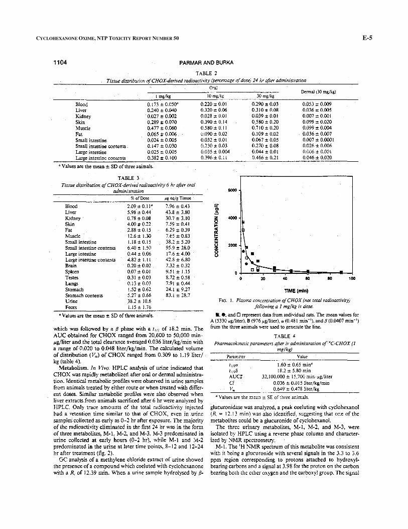
1104 PARMAR AND BURKA
TABLE 2 Tissue distribution ofCHOX-derived radioactivity (percentage of dose) 24 hr after administrmion
Oral Dermal ( 30 mg/kg)
I mg/kg 10 mg/kg 30 mg/kg
Blood 0.173 ± 0.050" 0.220 ± O.Ql 0.290 ± 0.03 0.053 ± 0.009 Liver 0.240 ± 0.040 0.320 ± 0.06 0.310 ± 0.08 0.036 ± 0.005 Kidney 0.027 ± 0.002 0.028 ± O.Ql 0.039 ± 0.01 0.007 ± 0.001 Skin 0.289 ± 0.070 0.390 ± 0.14 0.580 ± 0.20 0.099 ± 0.020 Muscle 0.4 77 ± 0.060 0.580 ± 0.11 0.710 ± 0.20 0.099 ± 0.004 Fat 0.065 ± 0.006 0.090 ± 0.02 0.109 ± 0.02 0.036 ± 0.007 Small intestine 0.024 ± 0.005 0.032 ± 0.01 0.067 ± 0.05 0.007 ± 0.0001 Small intestine contents 0.147 ± 0.030 0.230 ± O.D3 0.270 ± 0.08 0.026 ± 0.006 Large intestine O.D25 ± 0.005 O.D35 ± 0.004 0.044 ± 0.01 0.006 ± 0.00 I Large intestine contents 0.382 ± 0.100 0.396 ± 0.11 0.466 ± 0.21 0.046 ± 0.020
"Values are the mean± SD of three animals.
TABLE 3 Tissue distribution of CHOX-derived radioactivity 6 hr after oral
administrarion %of Dose I'S eqjg Tissue
Blood 2.09 ± 0.11" 7.96 ± 0.43 Liver 5.98. ± 0.44 43.8 ± 3.80 Kidney 0.78 ± 0.08 30.7 ± 3.10 Skin 4.00 ± 0.22 7.59 ± 0.41 Fat 2.88 ± 0.15 6.29 ± 0.39 Muscle 12.6 ± 1.30 7.65 ± 0.83 Small intestine 1.18 ± 0.15 38.2 ± 5.20 Small intestine contents 6.40 ± 1.50 95.9 ± 28.0 Large intestine 0.44 ± 0.06 17.6 ± 4.00 Large intestine contents 4.82 ± 1.11 42.8 ± 6.80 Brain 0.20 ± 0.02 7.32 ± 0.32 Spleen 0.07 ± O.Ql 9.51 ± 1.15 Testes 0.31 ± 0.03 8.72 ± 0.58 Lungs 0.13 ± O.D3 7.91 ± 0.44 Stomach 1.52 ± 0.62 24.1 ± 9.27 Stomach contents 5.27 ± 0.66 83.1 ± 28.7 Urine 38.2 ± 10.6 Feces 1.15± 1.76
"Values are the mean ± SD of three animals.
which was followed by a fJ phase with a t, 1, of 18.2 min. The AUC obtained for CHOX ranged from 20,600 to 50,000 min· l'g/liter and the total clearance averaged 0.036literfkg/min with a range of 0.020 to 0.048 liter/kg/min. The calculated volume of distribution {V") of CHOX ranged from 0.309 to 1.19 liter/ kg (table 4).
Metalmlism. In Vivo. HPLC analysis of urine indicated that CHOX was rapidly metabolized after oral or dermal administration. Identical metabolic profiles were observed in urine samples from animals treated by either route or when treated with different doses. Similar metabolic prof!les were also observed when liver extracts from animals sacrificed after 6 hr were analyzed by HPLC. Only trace amounts of the total radioactivity injected had a retention time similar to that of CHOX, even in urine samples collected as early as 0-2 hr after exposure. The majority of the radioactivity eliminated in the first 24 hr was in the form of three metabolites, M-1, M-2, and M-3. M-3 predominated in urine collected at early hours (0-2 hr), while M-1 and M-2 predominated in the urine at later time points, 8-12 and 12-24 hr after treatment (fig. 2).
GC analysis of a methylene chloride extract of urine showed the presence of a compound which coeluted with cyclohexanone with a R, of 12.39 min. When a urine sample hydrolysed by {J-
6000
z 4000
~ ~ w ~ 2000
8
20 40 80 80 100
TIME (min)
FIG. I. Plasma concemratiori ofCHOX (not total radioactivity) following a 1 mgfkg ir dose.
II, e, and 0 represent data from individual rats. The mean values for A (5330 l'g/liter), B (976~tg/liter), a (0.481 min"'), and {3 (0.0407 min"') from the three animals were used to generate the line.
TABLE 4
Pharmacokinetic parameters after iv administrazion of"C-CHOX (1 mgfkg)
Parameter Value
1112a 1.60 ± 0.65 min" zl/,/3 18.2 ± 5.80 min AUCo 32,100.000 ± 15,700 min-.ug/liter Cl 0.036 ± O.Dl5 liter/kg/min VD 0.649 ± 0.478 liter/kg
a Values are the mean ± SE of three animals.
glucuronidase was analyzed, a peak coeluting with cyclohexanol (R, = 12.13 min) was also identified, suggesting that one of the metabolites could be a glucuronide of cyclohexanol.
The three urinary metabolites, M-1, M-2, and M-3, were isolated by HPLC using a reverse phase column and characterized by NMR spectrometry.
M-1. The 'H NMR spectrum of this metabolite was consistent with it being a glucuronide with several signals in the 3.3 to 3.6 ppm region corresponding to protons attached to hydroxylbearing carbons and a signal at 3.98 for the proton on the carbon bearing both the ether oxygen and the carboxyl group. The signal
E-5 CYCLOHEXANONE OXIME, NTP TOXICITY REPORT NUMBER 50
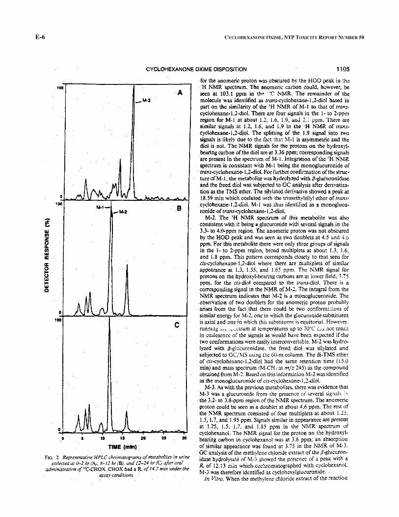
CYCLOHEXANONE OXIME DISPOSITION 1105
IGO A
-M·3
0
IGO M-1- B
-M-2 -! w rn z 0 ~ rn w a: a: 0 ti w 1-w Q
0
100 c
0
0 30
11ME (min)
FIG. 2. Repr~sentative HPLC chromatograms of metabolites in urine collected at 0-2 hr(A). 8-12 hr (B). and 12-24 hr (C) after oral
administration of'4C-CHOX. CHOX hadaR, of 14.7 min under the assay conditions.
for the anomeric proton was obscured by the HOD peak in the 'H NMR spectrum. The anomeric carbon could, however, be seen at 103.1 ppm in the ';C NMR. The remainder of the molecule· was identified as trans-cyclohexane-1 ,2-diol based in part on the similarity of the 'H NMR of M-1 to that of transcyclohexane-1 ,2-diol. There are four signals in .the I· to 2-ppm region for M~l at about 1.2, 1.6, 1.9, anJ 2. ~ ppm. There are similar signals at 1.2, 1.6, and 1.9jn the 'H NMR of transcyclohexane-1,2~iol. The splitting of the 1.9 signal into two signals is likely due to the fact that ~-1 is asymmetric and the diol is not. The NMR signals for the protons on the hydroxylbearing carbon of the diol are at 3.36ppm; corresponding signals are present in the spectrum ofM-1. Integration of the 'H NMR spectrum is consistant with M-1 being the monoglucuronide of trans-cyclohexane-1 ,2-diol. For further confirmation of the structure ofM-1, the metabolite was hydrolyzed with ,8-glucuronidase and the freed diol was subjected to GC analysis after derivatization as the TMS ether. The silylated derivative showed a peak at 18.59 min which coeluted with the trimethylsilyl ether of transcyclohexane-1 ,2-diol. M-1 was .thus identified as. a monoglucu-. ronide of trans-cyclohexane, I ,2~iol.
M-2. The 'H NMR spectrum of this metabolite was also consistent with it being a glucuronide with several signals in the 3.3· to 4.0-ppm region. The anomeric proton was not obscured by the HOD peak and was seen as two doublets at 4.5 and 4.6 ppm. For this metabolite there were only three groups of signals in the I- to 2-ppm region, broad multiplets at about 1.3, 1.6, and 1.8 ppm. This pattern corresponds closely to that seen for cis-cyclohexane-1 ,2-diol where there are multiplets of similar appearance at 1.3, 1.55, and !.65 ppm. The NMR signal for protons on the hydroxyl-bearing carbQns are at lower field, 3. 7 5 ppm, for the cis-dial compared to the trans-diol. There is a corresponding signal in the NMR ofM"2. The integral from the NMR spectrum indicates that M-2 is a monoglucuronide. The obserVation of two doublets for the anomeric proton probably arises from the fact that there could be two conforrna:ions of similar energy for M-2, one in which the glucuronide substituent is axialand one in which this substituent is equitorial. However. running ,;,~ """~trum at temperatures up to 70"C d.u. not result in coalesence of the signals as would have been expected if the two conformations were easily interconvertable. M-2 was hydrolyzed with ;3-glucuronidase. the freed diol was silylated and subjected to GC/MS using the 60-m column. The di-TMS ether of cis-cyclohexane-1 ,2-diol had the same retention time ( 15.0 min) and mass spectrum (M-CH, at m/z 245) as the compound obtained from M-2. Based on this information M·2 was identified as the monoglucuronide of cis-cyclohexane-1 ,2-diol.
M-3. As with the previous metabolites, there was evidence that M-3 was a glucuronide from the presence ofseveral signals ':1
the 3.2- to 3.8-ppm region of the NMR spectrum. The anomeric proton could be seen as a doublet at about 4.6 ppm. The rest of the NMR spectrum consisted of four multiplets at about 1.25, 1.5, 1.7, and !.95 ppm. Signals similar in appearance are present in L25, 1.5, 1.7, and 1.85 ppm in the NMR spectrum of cyclohexanol. The NMR signal for the proton on the hydroxylbearing carbon in cyclohexanol was at 3.6 ppm; an absorption of similar appearance was found at 3.75 in the NMR of M-3. GC analysis of the methylene chloride extract of the ,a-glucuronidase hydrolysate of M-3 showed the presence of a peak with a R, of 12.13 min which cochromatographed with cyclohexanoL M-3 wastherefore identified as cyclohexylglucuronide.
In Vitro. When the methylene chloride extract of the reaction
E-6 CYCLOHEXANONE OXIME, NTP TOXICITY REPORT NUMBER 50


