© 2020 Cue Biopharma. All rights reserved.Presented at The Society for Immunotherapy of Cancer’s 35th Anniversary Annual Meeting (SITC 2020)
CUE-100 series Immuno-STATs from concept to the clinic: Leveraging protein engineering to stimulate and selectively deliver affinity-attenuated IL-2 to antigen-specific T cellsHisted A., Girgis N., Moreta M., Soriano J., Witt L., Merazga Z., Diaz F., Zhao F., Kemp M., Ruthardt P., Thapa D., Suri A., Seidel R., Pienta K., Simcox. M., Quayle S., Ross J., Cemerski S.
Background
PD Biomarkers in CUE-101 Clinical Trial
#553
CUE-101 is currently being tested in an open-label, 2-part clinical Phase I study to characterize thesafety, tolerability, PK, PD, immunogenicity, and preliminary antitumor activity as monotherapy in HLA-A*0201-positive patients with HPV16+ recurrent/metastatic HNSCC tumors. Assessment of the potentialto generate an antitumor immune response with CUE-101 treatment is an important secondary objectiveof the trial. Blood samples are collected prior to dosing and at several different time points followingCUE-101 administration. Preliminary data from PBMC immunophenotyping and tetramer staining showearly signals of expansion of HPV-16 E711-20-specific CD8+ T cells.
Activation of T cells requires a specific peptide/HLA (human leukocyte antigen) signal presented by aninteracting immune or target cell along with engagement of co-stimulatory molecules or cytokinereceptors. Cue Biopharma has developed a proprietary biologics platform, termed Immuno-STAT™(Selective Targeting and Alteration of T cells), wherein a singular protein framework incorporatespeptide/HLA complexes and co-stimulatory, co-inhibitory or cytokine molecules. The CUE-100 seriesImmuno-STATs selectively deliver rationally engineered IL-2 molecules (IL-2 variants) to antigen-specificT cells. The IL-2 variants in the CUE-100 series Immuno-STATs contain mutations that attenuate bindingto IL-2 receptors alpha and beta, which minimizes activation of regulatory T cells (Tregs) and theirrelevant non-antigen-specific T cell repertoire. We have demonstrated that CUE-100 series Immuno-STATs specific for different antigenic peptides (from HPV16, WT1, MART-1, CMV, FLU virus, and HIV)induce expansion of functional, oligoclonal, antigen-specific repertoires from human PBMCs. The leadclinical candidate CUE-101, presenting the E711-20 peptide from HPV-16 in the context of HLA-A*02:01, iscurrently being tested in a Phase 1 clinical trial in recurrent/metastatic head and neck cancer patientswith evidence of dose-proportional PK, early pharmacodynamic effects and signals of clinical activity.
Cue Biopharma’s Immuno-STAT Platform
The intimate interactions between the T cells and APCs occur within a molecular interface known as theimmunological or immune synapse. The immune synapse allows controlled engagement and selectiveactivation of T cells through the presentation of two key distinct signals: Signal 1, TCR engagement bythe pMHC; and Signal 2, co-stimulatory, co-inhibitory or cytokine signals. Through rational proteinengineering, we have developed a proprietary class of biologics termed Immuno-STATs that induce andmodulate T cell activity via delivery of the distinct signals provided naturally to T cells within the immunesynapse. We accomplish this by the co-engineering of a TCR targeting pMHC with co-stimulatory, co-inhibitory or cytokine signaling molecules in a singular biologic on an Fc framework.
CUE-100 Series Immuno-STATs
Signal 2
Peptide
MHC alpha chain
Beta-2 microglobulin
Fc
Through structure-based protein engineering, CUE-100 series molecules selectively deliver IL-2 to tumorassociated antigen (TAA)-specific T cells via binding of the pHLA complex to TAA-specific TCRs. Toincrease selectivity for target T cells and to minimize the potential for toxicity mediated through global IL-2-driven activation of IL-2 receptor (IL-2R) expressing cells, two point mutations (H16A and F42A) wereintroduced into the IL-2 sequences of CUE-100 series Immuno-STATs. These mutations were previouslydemonstrated to reduce IL-2 interaction with the IL-2Rα and IL-2Rβ chains, respectively. The bindingaffinity of a double mutant for human IL-2Rα and IL-2Rβ was decreased 110-fold and 3-fold, respectively,compared to wildtype IL-2 binding, predominantly due to a faster off-rate for each of these interactions.Functional attenuation of the mutant IL-2 components of a CUE-100 series Immuno-STAT (CUE-101)was also demonstrated in a CTLL-2 proliferation assay, where CUE-101-induced proliferation wasreduced ~2,600-fold relative to recombinant human IL-2 (rhIL-2). As a reminder, a slightly modified rhIL-2is commercially available as aldesleukin (Proleukin®).
CUE-101 and CUE-102/A02 for Patients with HPV16-Associated and WT1-Associated Malignancies:
Design, Manufacturability Assessment and Biophysical Characterization
CUE-100 Series Immuno-STATs Induce Expansion of Human Antigen-Specific T Cells
CUE-100 Series Immuno-STATs specific for:CMV pp65495-503Influenza MP158-66 KRAS G12V7-16HPV16 E711-20 WT137-45MART-126-35
tetramer-PE
tetra
mer
-APC
tetramer-PE
tetra
mer
-APC
Conclusions
tetramer-PE
tetra
mer
-APC
tetramer-PE
tetra
mer
-APC
tetramer-PE
tetra
mer
-APC
tetramer-PE
tetra
mer
-APC
CUE-101 and CUE-102/A02 vs Wild-Type IL-2:CUE’s Attenuated IL-2 Mitigates the Risk Associated
with Systemic IL-2 Activation
CUE-101 and CUE-102/A02 Expand Antigen-Specific Polyfunctional Effector CD8+ T Cells
CUE-101 and CUE-102/A02 Selectively Expand Antigen-Specific CD8+ T Cells from Healthy Human
PBMCs
CUE-100 Series Signal 2: Affinity Attenuated IL-2 Variant
• Abrogated binding to IL-2Rα• Reduced binding to IL-2Rβ• Four affinity attenuated IL-2 units per Immuno-STAT
Maintains ability of IL-2 to stimulate antigen-specific T cells while reducing undesired effects of IL-2
A)
B) CUE-100 series Immuno-STAT Wildtype IL-2
= <
A) Healthy donor PBMCs were stimulated for 10 days with CUE-101 or CUE-102/A02 Immuno-STAT inImmunoCultTM media. Peptide-specific CD8+ T cells were detected by flow cytometry upon staining withHPV16 E711-20 and WT137-45-specific tetramers. Unstimulated cells were used as negative control.
B) Healthy donor PBMCs were primed for 10 days with WT137-45 peptide in the presence of rhIL-2. CD8+
T cells were then enriched by magnetic separation and restimulated with the CUE-102/A02 Immuno-STAT in ImmunoCultTM media in the presence of mitomycin C-treated autologous PBMCs for 8 days.Peptide-specific CD8+ T cells were detected by flow cytometry upon staining with WT137-45-specifictetramers. Unstimulated cells were used as negative control.
A) B)
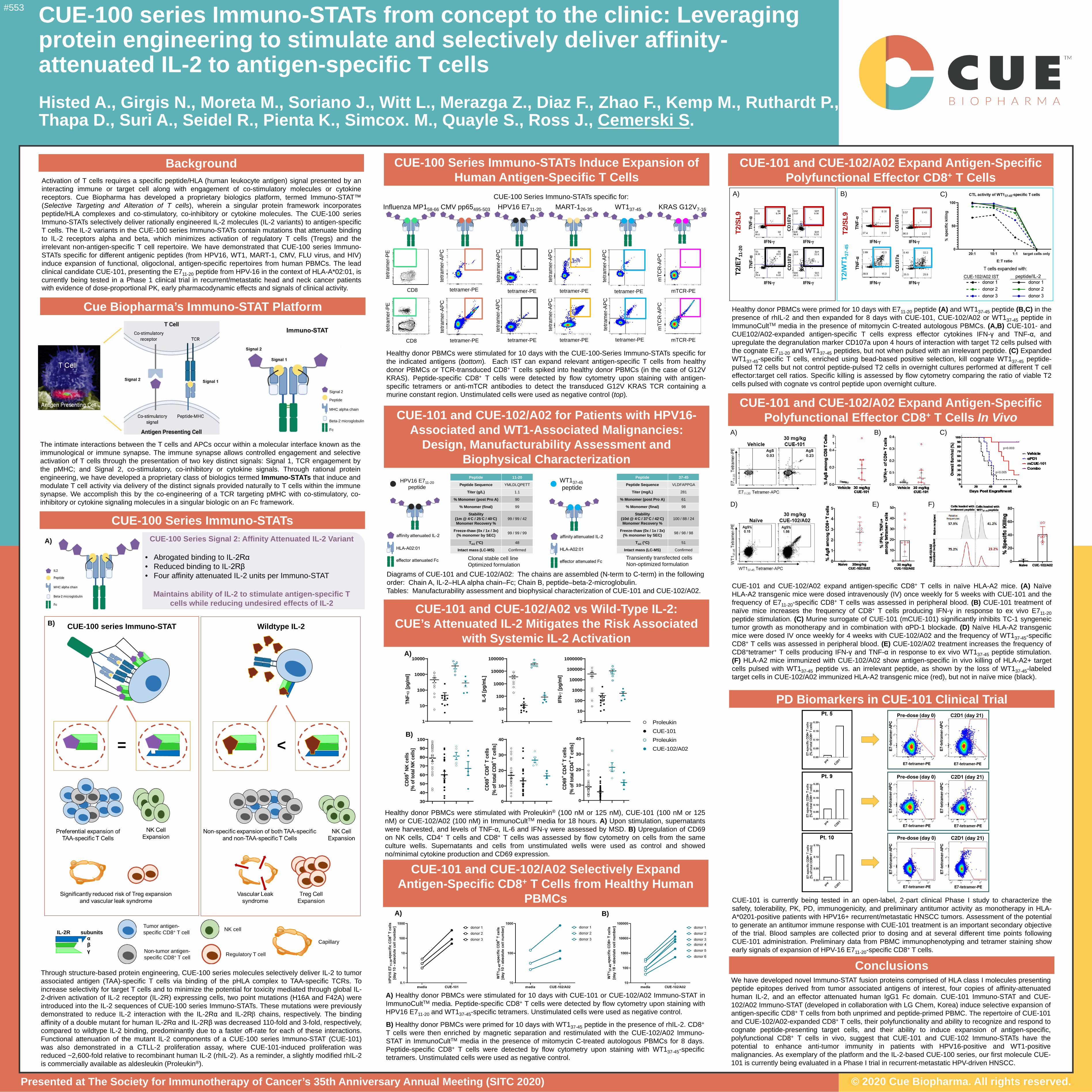
Healthy donor PBMCs were stimulated with Proleukin® (100 nM or 125 nM), CUE-101 (100 nM or 125nM) or CUE-102/A02 (100 nM) in ImmunoCultTM media for 18 hours. A) Upon stimulation, supernatantswere harvested, and levels of TNF-α, IL-6 and IFN-γ were assessed by MSD. B) Upregulation of CD69on NK cells, CD4+ T cells and CD8+ T cells was assessed by flow cytometry on cells from the sameculture wells. Supernatants and cells from unstimulated wells were used as control and showedno/minimal cytokine production and CD69 expression.
Healthy donor PBMCs were primed for 10 days with E711-20 peptide (A) and WT137-45 peptide (B,C) in thepresence of rhIL-2 and then expanded for 8 days with CUE-101, CUE-102/A02 or WT137-45 peptide inImmunoCultTM media in the presence of mitomycin C-treated autologous PBMCs. (A,B) CUE-101- andCUE102/A02-expanded antigen-specific T cells express effector cytokines IFN-γ and TNF-α, andupregulate the degranulation marker CD107a upon 4 hours of interaction with target T2 cells pulsed withthe cognate E711-20 and WT137-45 peptides, but not when pulsed with an irrelevant peptide. (C) ExpandedWT137-45-specific T cells, enriched using bead-based positive selection, kill cognate WT137-45 peptide-pulsed T2 cells but not control peptide-pulsed T2 cells in overnight cultures performed at different T celleffector:target cell ratios. Specific killing is assessed by flow cytometry comparing the ratio of viable T2cells pulsed with cognate vs control peptide upon overnight culture.
Diagrams of CUE-101 and CUE-102/A02: The chains are assembled (N-term to C-term) in the following order: Chain A, IL-2–HLA alpha chain–Fc; Chain B, peptide–beta-2-microglobulin. Tables: Manufacturability assessment and biophysical characterization of CUE-101 and CUE-102/A02.
HPV16 E711-20 peptide
WT137-45 peptide
We have developed novel Immuno-STAT fusion proteins comprised of HLA class I molecules presentingpeptide epitopes derived from tumor associated antigens of interest, four copies of affinity-attenuatedhuman IL-2, and an effector attenuated human IgG1 Fc domain. CUE-101 Immuno-STAT and CUE-102/A02 Immuno-STAT (developed in collaboration with LG Chem, Korea) induce selective expansion ofantigen-specific CD8+ T cells from both unprimed and peptide-primed PBMC. The repertoire of CUE-101and CUE-102/A02-expanded CD8+ T cells, their polyfunctionality and ability to recognize and respond tocognate peptide-presenting target cells, and their ability to induce expansion of antigen-specific,polyfunctional CD8+ T cells in vivo, suggest that CUE-101 and CUE-102 Immuno-STATs have thepotential to enhance anti-tumor immunity in patients with HPV16-positive and WT1-positivemalignancies. As exemplary of the platform and the IL-2-based CUE-100 series, our first molecule CUE-101 is currently being evaluated in a Phase I trial in recurrent-metastatic HPV-driven HNSCC.
mTCR-PE
mTC
R-A
PC
mTCR-PE
mTC
R-A
PC
tetramer-PE
tetra
mer
-APC
tetramer-PE
tetra
mer
-APC
Healthy donor PBMCs were stimulated for 10 days with the CUE-100-Series Immuno-STATs specific forthe indicated antigens (bottom). Each IST can expand relevant antigen-specific T cells from healthydonor PBMCs or TCR-transduced CD8+ T cells spiked into healthy donor PBMCs (in the case of G12VKRAS). Peptide-specific CD8+ T cells were detected by flow cytometry upon staining with antigen-specific tetramers or anti-mTCR antibodies to detect the transduced G12V KRAS TCR containing amurine constant region. Unstimulated cells were used as negative control (top).
IL-2R subunitsαβγ
Tumor antigen-specific CD8+ T cell
Non-tumor antigen-specific CD8+ T cell
NK cell
Regulatory T cell
Capillary
CD8
tetra
mer
-PE
CD8
tetra
mer
-PE
CUE-101 and CUE-102/A02 expand antigen-specific CD8+ T cells in naïve HLA-A2 mice. (A) NaïveHLA-A2 transgenic mice were dosed intravenously (IV) once weekly for 5 weeks with CUE-101 and thefrequency of E711-20-specific CD8+ T cells was assessed in peripheral blood. (B) CUE-101 treatment ofnaïve mice increases the frequency of CD8+ T cells producing IFN-γ in response to ex vivo E711-20peptide stimulation. (C) Murine surrogate of CUE-101 (mCUE-101) significantly inhibits TC-1 syngeneictumor growth as monotherapy and in combination with αPD-1 blockade. (D) Naïve HLA-A2 transgenicmice were dosed IV once weekly for 4 weeks with CUE-102/A02 and the frequency of WT137-45-specificCD8+ T cells was assessed in peripheral blood. (E) CUE-102/A02 treatment increases the frequency ofCD8+tetramer+ T cells producing IFN-γ and TNF-α in response to ex vivo WT137-45 peptide stimulation.(F) HLA-A2 mice immunized with CUE-102/A02 show antigen-specific in vivo killing of HLA-A2+ targetcells pulsed with WT137-45 peptide vs. an irrelevant peptide, as shown by the loss of WT137-45-labeledtarget cells in CUE-102/A02 immunized HLA-A2 transgenic mice (red), but not in naïve mice (black).
CUE-101 and CUE-102/A02 Expand Antigen-Specific Polyfunctional Effector CD8+ T Cells In Vivo
A)
p=0.003
p=0.005
B) C)
D) E) F)
Peptide 11-20
Peptide Sequence YMLDLQPETT
Titer (g/L) 1.1
% Monomer (post Pro A) 90
% Monomer (final) 99
Stability (1m @ 4◦C / 25◦C / 40◦C)Monomer Recovery %
99 / 99 / 42
Freeze-thaw (0x / 1x / 3x)(% monomer by SEC) 99 / 99 / 99
Tm1 (°C) 48
Intact mass (LC-MS) Confirmed
Clonal stable cell lineOptimized formulation
Peptide 37-45
Peptide Sequence VLDFAPPGA
Titer (mg/L) 281
% Monomer (post Pro A) 61
% Monomer (final) 98
Stability (10d @ 4◦C / 37◦C / 42◦C)Monomer Recovery %
100 / 88 / 24
Freeze-thaw (0x / 1x / 3x)(% monomer by SEC) 98 / 98 / 98
Tm1 (°C) 51
Intact mass (LC-MS) Confirmed
Transiently transfected cellsNon-optimized formulation
T2/E
7 11-
20T2
/SL9
A)
IFN-γ
TNF-α
CD
107a
IFN-γ
TNF-α
CD
107a
B) C)
T2/W
T137
-45
T2/S
L9
CD
107a
CD
107a
TNF-α
TNF-α
IFN-γ IFN-γ
IFN-γ IFN-γ
IFN-γ IFN-γ
ProleukinCUE-101ProleukinCUE-102/A02
30
40
50
60
70
80
90
100
CD69
+ NK
cells
[% o
f tot
al N
K ce
lls]
0
10
20
30
40
CD69
+ CD8
+ T c
ells
[% o
f tot
al C
D8+ T
cel
ls]
1
10
100
1000
10000
100000
1000000
IFN-γ [
pg/m
l]
1
10
100
1000
10000
TNF-α
[pg/
ml]
1
10
100
1000
10000
100000
IL-6
[pg/
mL]
A)
B)
0
10
20
30
40
CD69
+ CD4
+ T c
ells
[% o
f tot
al C
D4+ T
cel
ls]


