

CPR FACTS
In the hospital setting, among participating centers in the Get With The Guidelines-Resuscitation
quality improvement program, the median hospital survival rate from adult cardiac arrest is 18%
(interquartile range, 12%–22%) and from pediatric cardiac arrest, it is 36% (interquartile range,
33%–49%).
Circulation. 2013;128:417-435

CPR FACTS
• In a hospital setting, survival is >20% if the arrest occurs between the hours of 7 am and 11 pm
but only 15% if the arrest occurs between 11 pm and 7 am.
• There is significant variability with regard to location, with 9% survival at night in unmonitored
settings compared with nearly 37% survival in operating room/post anesthesia care unit
locations during the day.
Circulation. 2013;128:417-435

CPR FACTS
• Patient survival is linked to quality of cardiopulmonary resuscitation (CPR).
• When rescuers compress at a depth of <38 mm, survival-to-discharge rates after out-of-hospital
arrest are reduced by 30%.
• Similarly, when rescuers compress too slowly, return of spontaneous circulation (ROSC) after in-
hospital cardiac arrest falls from 72% to 42%.
Circulation. 2013;128:417-435

SURVIVAL AFTER IN-HOSPITAL CARDIAC ARREST
Girotra, NEJM 2012

SURVIVAL AFTER IN-HOSPITAL CARDIAC ARREST
Girotra, NEJM 2012

SCENARIO #1
• You respond to a code blue for a patient in 4 Jones rehabilitation unit.
• On arrival you find the patient in the corner of the room in a vail bed, pulseless
• What do you do next?

WHAT DO YOU DO?
A. Freak out
B. Tear open the vail bed with Hulk-like strength
C. Unzip the vail bed and start chest compressions
D. Yell at the 43 nurses in the room to get the crash cart

ACLS Cardiac Arrest Algorithm.
Neumar R W et al. Circulation 2010;122:S729-S767
Copyright © American Heart Association

ACLS Cardiac Arrest Circular Algorithm.
Neumar R W et al. Circulation 2010;122:S729-S767
Copyright © American Heart Association

The universal algorith
Hazinski M F et al. Circulation. 2010;122:S250-S275
Copyright © American Heart Association, Inc. All rights reserved.

ORIGINS OF CPR

INTERACTION OF DIFFERENT FACTORS
• Age
• Gender/Race/Ethnicity
• Morbidity
• First Monitored Rhythm
• Event Intervals
• Event Duration
• Hospital Location
• Time of Day

SCENARIO #1 (CONT.)
• You indeed tear open the vail bed and start compressions
• You yell at the 43 nurses standing around
• The crash cart is opened
• The cardiology fellow is placing a line
• You are doing chest compressions
• No one is bagging the patient

SCENARIO #1 (CONT.)
• Others finally come to your aid and good quality chest compressions are being done
• The patient is asystole when hooked up to the crash cart monitor
• A femoral central line is secured and IV medications are being given as well as IVF
• You attempt to bag the patient but you are getting very weak chest rise
• And the bed is stuck in the down position
• You get down on the floor and attempt intubation but are unable to intubate the patient after 2 attempts
• Anesthesia is on holiday and are unable to assist you
• What do you do to obtain an airway?

WHAT DO YOU DO TO OBTAIN AN AIRWAY?
A. Intubate the patient with GlideScope
B. Place an LMA
C. Emergent surgical airway
D. Bag the patient with an oral airway

DIFFICULT AIRWAY ALGORITHM
Plan A: Direct Laryngoscopy
Plan B: GlideScope
Plan C: Fiberoptic Intubation
Plan D: Intubate through LMA
Bailout: Ventilate through LMA and call for help
Plan Last: Emergent Surgical Airway

SCENARIO #2
• You are called to see a patient that is sent from MIMU to MICU by rapid response
• On arrival, the patient is awake and delirious
• HR 40, BP 80/42, sPo2 94%
• What do you do next?

APPROACH TO BRADYCARDIACauses
• Intrinsic
• Sinus node dysfunction
• Athletic heart
• Inferior MI
• Surgery
• Collagen-vascular disease
• Infiltrative disease
• Extrinsic
• Vagal-mediated
• Hypothermia
• Metabolic acidosis
• Hypoxia
• Electrolyte disorders
• Sepsis
• Increased ICP
• Medications
Treatments
• Is the patient symptomatic?
• Remove medications causing bradycardia
• Correct metabolic disturbances
• Avoid triggers causing vagal-mediated
reaction
• Medical intervention
• Atropine
• Epinephrine
• Dopamine
• Isoproterenol
• Glucagon
• Temporary/permanent pacing
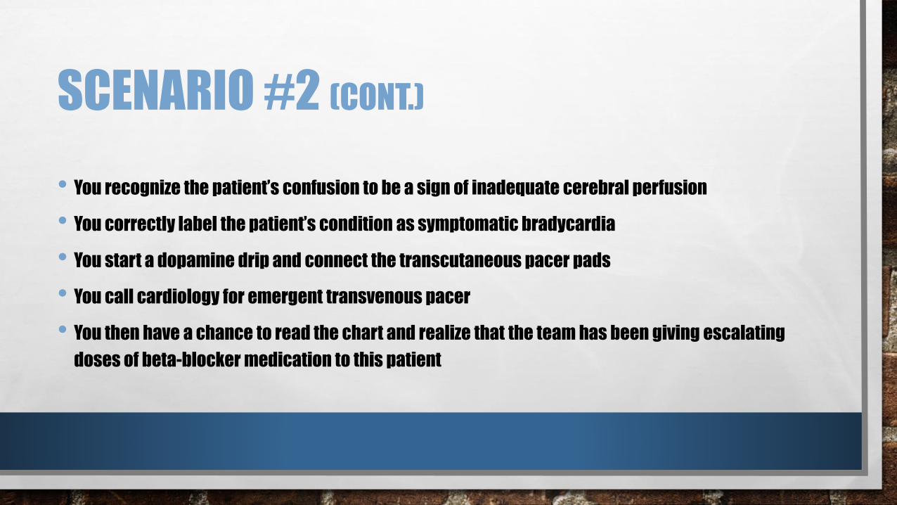
SCENARIO #2 (CONT.)
• You recognize the patient’s confusion to be a sign of inadequate cerebral perfusion
• You correctly label the patient’s condition as symptomatic bradycardia
• You start a dopamine drip and connect the transcutaneous pacer pads
• You call cardiology for emergent transvenous pacer
• You then have a chance to read the chart and realize that the team has been giving escalating
doses of beta-blocker medication to this patient

APPROACH TO CHANGE IN MENTAL STATUS
Questions to answer:
Is my patient having a stroke?
When in doubt/if patient has focal deficits, get a STAT noncontrast Head CT.
Is my patient having an MI?
Consider EKG, cardiac enzymes
Does my patient have sepsis?
Does your patient need IVF bolus for hypotension?
Does your patient need IV antibiotics urgently?

DEFINITIONS OF IMPAIRED CONSCIOUSNESS
Drowsiness
State of impaired awareness associated with desire or inclination to sleep
Stupor
State of impaired consciousness where the individual shows markedly diminished
reactivity to environmental stimuli
Comatose
State of profound unconsciousness where one cannot be aroused
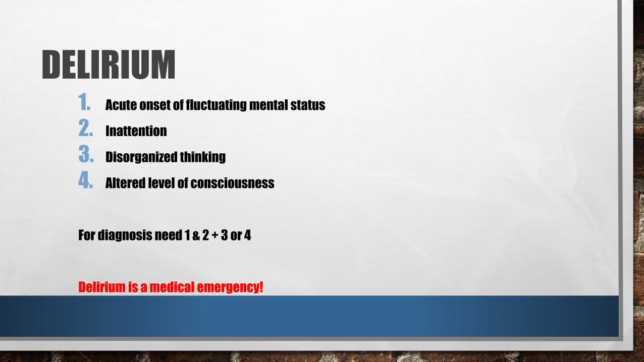
DELIRIUM1. Acute onset of fluctuating mental status
2. Inattention
3. Disorganized thinking
4. Altered level of consciousness
For diagnosis need 1 & 2 + 3 or 4
Delirium is a medical emergency!

CLUES IN ASSOCIATIONS Altered mental status + Diabetes
Think of oral hypoglycemics, get a finger stick!
Altered mental status + Fever
Think meningitis/encephalitis/UTI
Altered mental status + Hypotension
Think sepsis or inferior MI
Altered mental status + Dyspnea
Think pneumonia or MI/CHF
Altered mental status + Hemparesis or Dysarthria
Think stroke
Altered mental status + Failure to thrive
Think hyponatremia

SCENARIO #3
• You respond to code blue on 3 cullen
• On arrival to the room, you notice the patient is a 20 yr old white man
• He is found half way between the bathroom and the bed
• He is pulseless
• What do you do?

WHAT DO YOU DO?
A. Put him back in bed
B. Code him on the floor

SCENARIO #3 (CONT.)
• You call for help and the cavalry arrives
• You place him into bed
• Chest compressions are started
• A sinus brady rhythm is showing on the monitor, but he is pulseless

PEA DIFFERENTIAL DX
H’s
• Hypovolemia
• Hypoxia
• Hydrogen ion (acidosis)
• Hyper/hypokalemia
• Hypoglycemia
• Hypothermia
T’s
• Tablets/Toxins
• Tamponade (cardiac)
• Tension pneumothorax
• Thrombosis (coronary)
• Thrombosis (pulmonary)
• Trauma
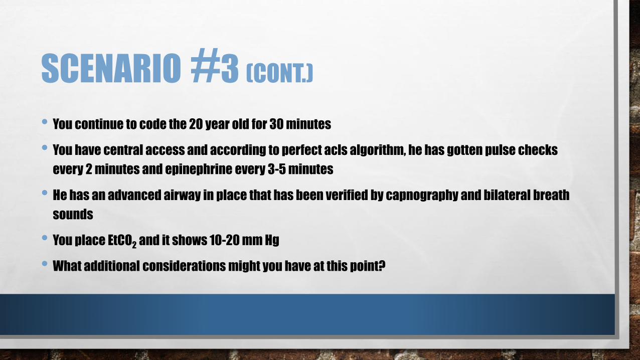
SCENARIO #3 (CONT.)
• You continue to code the 20 year old for 30 minutes
• You have central access and according to perfect acls algorithm, he has gotten pulse checks
every 2 minutes and epinephrine every 3-5 minutes
• He has an advanced airway in place that has been verified by capnography and bilateral breath
sounds
• You place EtCO2 and it shows 10-20 mm Hg
• What additional considerations might you have at this point?

PREDICTORS OF SURVIVAL- ETCO2?
Levine, NEJM 1997

WHO SHOULD GET E-CPR?
• Young patients
• Reversible cause
• Early initiation
• Good quality CPR
• Make sure ECMO is available

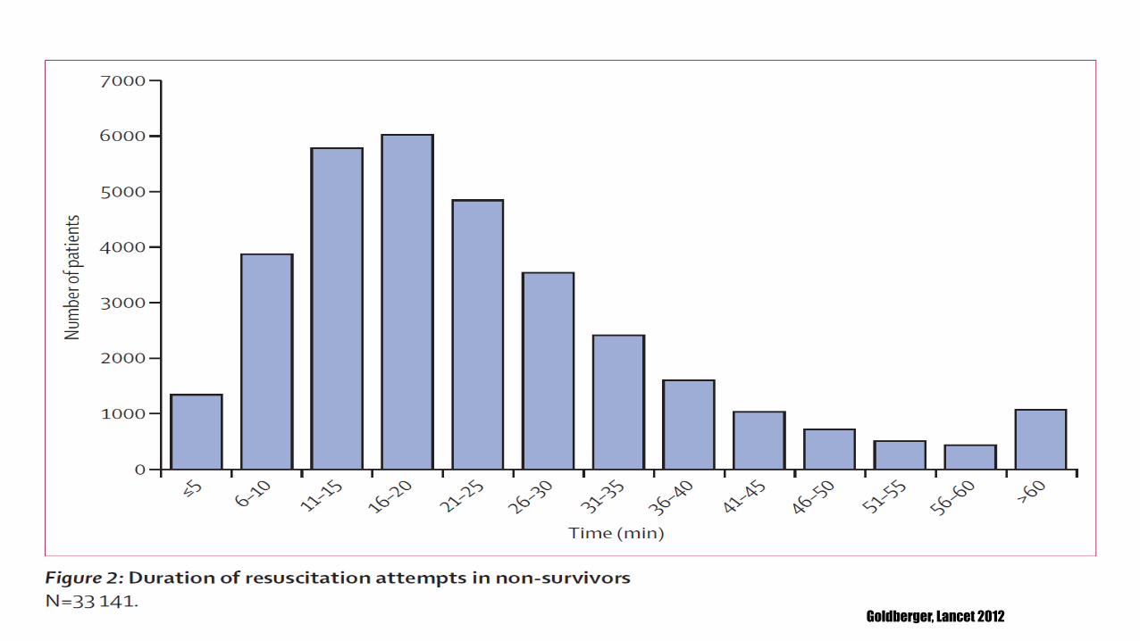
HOW MUCH TIME SHOULD YOU BE
CODED?Between 2000 and 2008, 64,339
patients with cardiac arrests at 435 US
hospitals within the Get With The
Guidelines—Resuscitation registry.
Goldberger, Lancet 2012
Goldberger, Lancet 2012

Goldberger, Lancet 2012

Goldberger, Lancet 2012

SCENARIO #4
• You are in the CCU
• You are a budding cardiologist
• You are seeing a 75 year old man with some hypoxemia on nasal cannula and obtaining a history
• He has atrial fibrillation on the monitor and you hear a harsh 3/6 SEM at the LUSB
• As you sit him up in bed, he becomes unresponsive
• On the monitor you see…


WHAT DO YOU DO?
A. Call a code
B. Push lidocaine
C. Start amiodarone
D. Give metoprolol
E. Pass out

SCENARIO #5
• You are minding your own business walking through 3C at night
• You have just finished a wonderful LBJ cafeteria meal
• You are checking on a middle-aged man that your co-resident admitted earlier in the day
• His history is unfamiliar to you but you think he has cancer and you heard the nurse say
something about fever
• You notice his heart rate is 110 on the monitor, his BP 90/40, his SpO2 92% on nasal cannula and
for some reason, the respiration monitor is picking up and says 30 bpm

YOU ARE WHICH OF THE FOLLOWING?
A. Not interested, you are already having a long day
B. Curious about the chemotherapy regimen that he is on
C. Too busy watching the world cup
D. Curious, but not enough to examine him
E. Concerned enough to call a rapid response

WHEN TO CONSIDER RAPID RESPONSE
• When the patient is hypotensive and not responsive to 2L IVF
• When patient has an unstable tachyarrhythmia
• When the patient is tachypneic and not readily responding to conservative measures
• When the patient is obtunded
• If you require NIPPV for rescue
• When the patient’s vital signs are deteriorating
• Bottom line: better to call rapid response before the ‘code blue’

SIRS CRITERIA
• Temperature < 36° C or > 38° C
• Heart Rate > 90 bpm
• Respiratory Rate > 20 breaths/MIN or PaCO2 < 32 mmHg
• White Blood Cell Count > 12,000 or < 4,000 cells/mm3 or > 10% bands

SHOCK
• Cardiogenic shock - a major component of the the mortality associated with cardiovascular
disease (the #1 cause of U.S. deaths)
• Hypovolemic shock - the major contributor to early mortality from trauma (the #1 cause of death
in those < 45 years of age)
• Septic shock - the most common cause of death in American ICUs (the 13th leading cause of
death overall in US)

Question 1: Is this patient in shock?
*Are there signs of end-organ hypoperfusion?
• Altered mental status/obtundation
• AKI manifested by oliguria
• Lactic acidosis
• Cool skin/extremities
• Decreased mean blood pressure
• Tachycardia
Question 2: If the patient is in shock, do they need to be intubated?
Question 3: Is the patient’s cardiac output adequate?
APPROACH TO HYPOTENSION

APPROACH TO HYPOTENSIONHypotension + Reduced Cardiac Output
Signs:
Narrow pulse pressure
Cool extremities/ delayed capillary refill (>3 sec)
Differential diagnosis:
Hypovolemic Shock
Cardiogenic Shock
Obstructive Shock
Possible Causes:
Hypovolemic Shock
Volume depletion/dehydration
Hemorrhage
Cardiogenic Shock
Myocardial Ischemia
Valvular lesions
Obstructive Shock
Acute Pulmonary Embolus
Pericardial Tamponade
Hypotension + Increased Cardiac Output
Signs:
Widened pulse pressure
Warm extremities/ bounding pulses
Differential diagnosis: You can infer from this situation
that the increased cardiac output with hypotension is
due to reduced SVR = DISTRIBUTIVE SHOCK
Possible Causes:
Sepsis/Septic Shock
Liver failure
Pancreatitis
Burns/Trauma
Anaphylaxis
Thyrotoxicosis
Neurogenic Shock

RESPIRATORY FAILUREIs the patient appropriate for NIPPV (Noninvasive Positive Pressure Ventilation a.k.a.
CPAP or BiPap® )?
COPD exacerbation
Cardiogenic pulmonary edema
Hypoxemic respiratory failure in immunosuppressed patients
Hypoxemic respiratory failure in post-thoracotomy patients
End of life palliative respiratory failure

Hemodynamic
Instability
Aspiration
Risk
Ineffective Therapy/
Delay in TherapyFacial Anatomy
Concerns
• Shock
• Cardiac arrest
• Coma/altered mentation
• Inability to protect airway
• Vomiting/bowel
obstruction
• Recent upper
GI surgery
• Life threatening
hypoxemia
• Severe pneumonia
• Pneumothorax
• Facial/upper airway
surgery
• Facial burns/trauma
• Fixed upper airway
obstruction
• Copious secretions
Meduri, Clin Chest Med 1996
Gupta, Respiratory Care 2013

NEWEST RECOMMENDATIONSHigh-quality CPR should be recognized as the foundation on which all other resuscitative efforts are
built. Target CPR performance metrics include:
a. CCF >80% (proportion of code that chest compressions are ongoing)
b. Compression rate of 100–120/min
c. Compression depth of ≥50 mm in adults with no residual leaning
i. (At least one third the anterior-posterior dimension of the chest in infants and
children)
d. Avoid excessive ventilation
i. (Only minimal chest rise and a rate of <12 breaths/min)Circulation. 2013;128:417-435

QUALITY IMPROVEMENT
• Simplify CPR
• 15:230:2Continuous Chest Compressions
• “Hands Only” for Adults
• Conventional CPR for Children
• Quality CPR
• De-emphasis of ACLS Drugs
• Minimize interruptions in Chest Compressions and Compression-Shock interval
• Organized Post-Cardiac Arrest Care

“A-B-C” TO “C-A-B”
• Early onset of chest compressions (30 sec to 18 sec)
• Early chest compressions Early defibrillation
• Increase likelihood of bystander CPR with emphasis on chest compressions
• “It is reasonable for healthcare providers to tailor the sequence of rescue actions to the most
likely cause of arrest.”

AIRWAY MANAGEMENT
• Class I recommendation for adults: use of quantitative waveform capnography for confirmation
and monitoring of endotracheal tube placement.
• The use of supraglottic advanced airways continues to be supported as an alternative to
endotracheal intubation for airway management during CPR.
• The routine use of cricoid pressure during airway management of patients in cardiac arrest is
no longer recommended.


