
Journal Pre-proof
COVID-19: Pathophysiology, diagnosis, complications and Investigationaltherapeutics
Samy A. Azer, PH.D, M.D., M.Ed.,F.A.C.G., M.P.H.
PII: S2052-2975(20)30090-1
DOI: https://doi.org/10.1016/j.nmni.2020.100738
Reference: NMNI 100738
To appear in: New Microbes and New Infections
Received Date: 9 May 2020
Revised Date: 26 June 2020
Accepted Date: 29 July 2020
Please cite this article as: Azer SA, COVID-19: Pathophysiology, diagnosis, complicationsand Investigational therapeutics, New Microbes and New Infections, https://doi.org/10.1016/j.nmni.2020.100738.
This is a PDF file of an article that has undergone enhancements after acceptance, such as the additionof a cover page and metadata, and formatting for readability, but it is not yet the definitive version ofrecord. This version will undergo additional copyediting, typesetting and review before it is publishedin its final form, but we are providing this version to give early visibility of the article. Please note that,during the production process, errors may be discovered which could affect the content, and all legaldisclaimers that apply to the journal pertain.
© 2020 Published by Elsevier Ltd.
COVID-19: Pathophysiology, Diagnosis, Complications and Investigational Therapeutics Samy A Azer, PH.D, M.D., M.Ed., F.A.C.G., M.P.H.
Professor of Medical Education and Gastroenterology Consultant, King Saud University
College of Medicine, Riyadh 11461, Saudi Arabia.
Correspondence to: Samy A Azer, PH.D, M.D., King Khalid Hospital, College of
Medicine, King Saud University, P. O. Box 2925, Riyadh 11461, Saudi Arabia.
Tel:+966542307075; Email: [email protected]
Keywords: COVID-19; Overview, Pathophysiology, Diagnosis, Complications
Acknowledgement
This work was funded by the College of Medicine Research Center, Deanship of Scientific
Research, King Saud University, Riyadh, Saudi Arabia.
Journ
al Pre-
proof
1
COVID-19: Pathophysiology, Diagnosis, Complications and Investigational Therapeutics
Journ
al Pre-
proof
2
Abstract
The novel coronavirus (COVID-19) outbreak started early in December 2019 in the Hubei
province and its capital Wuhan of the People’s Republic of China and caused a global
pandemic. The number of patients confined to this disease has exceeded nine million in more
than 215 countries, and the number who died is over 480,600 (up to 25 June 2020).
Coronaviruses were identified in the 1960s and recently identified to cause the Middle East
Respiratory Syndrome (MERS-CoV) in 2012 and severe acute respiratory syndrome (SARS)
in 2003. The current severe acute respiratory syndrome coronavirus-2 (SARS-CoV-2) is the
most recently identified. Patients with COVID-19 may be asymptomatic.
Typical symptoms including fever, dry cough, and shortness of breath. Gastrointestinal
symptoms such as nausea, vomiting, abdominal pain and diarrhea, have been reported—
neurologically related symptoms, particularly anosmia, hyposmia, and dysgeusia, have also
been reported. Physical examination may reveal a fever in over 44% of patients (and could be
documented in over 88% of patients after admission), increased respiratory rate, acute
respiratory disease, and maybe decreased consciousness, agitation, and confusion. This
article aims at presenting an up-to-date review on the pathogenesis, diagnosis and
complications of COVID-19 infection. Currently, no therapeutics have been found to be
effective. Investigational therapeutics are briefly discussed.
Keywords: COVID-19; Overview, Pathophysiology, Diagnosis, Complications
Journ
al Pre-
proof
3
Introduction On 31 December 2019, the Chinese authorities reported to the World Health Organisation
(WHO) an emerging of a novice coronavirus, currently the virus is known as SARS-CoV-2
and the disease name is coronavirus-19 disease (COVID-19), that has emerged in patients
from Wuhan city, Hubel Province [1]. This virus has a higher degree of lethality than other
endemic viruses, and is more lethal to humans compared to earlier emerging outbreaks of
SARS-CoV-1, in 2003, and the Middle East Respiratory Syndrome coronavirus (MERS-
CoV) in 2012. Both SARS-CoV-1 and MERS-CoV have common ancestry with viruses
found in bats. Both have intermediate hosts for transmission being palm civets and
dromedary camels for SARS-CoV-1, and MERS-CoV, respectively. However, there is no
strong evidence for the intermediate host yet.
The current pandemic is caused by SARS-CoV-2. It shares with the earlier two
coronaviruses the features of the Coronaviridae family. Coronavirus have large (~30-kb)
single-stranded, positive-sense RNA genomes and it is roughly 80% identical with other
coronaviruses at a nucleotide level. The virus closely related (share 90% of nucleotide
structure) to SARS-CoV-2 is RaTG13-2013 which was identified in bats [2]. The complete
genome of the severe acute respiratory syndrome coronavirus 2 isolated from Wuhan Hu-1 is
available at (https://www.ncbi.nlm. nih.gov/nuccore/NC_045512). Genetic epidemiology of
HCV-19 and submitted data since December 2019 are available from GISAID database (https
://www.gisaid.org/). The SARS-CoV-2 is composed of at least 11 ORFs (Open Reading
Frames) with the full length of 29,903 bp. Four major structural protein-coding genes have
been identified in the coronaviruses –Spike protein (S), Envelop protein (E), Membrane
protein (M), and Nucleocapsid protein (N) [3]. The spike protein of SARS-CoV-2 utilizes
angiotensin-converting enzyme (ACE2) as its cell surface receptor and utilization influences
the tropism of the virus.
Journ
al Pre-
proof

4
The COVID-19 infects people of all ages. However, there are two main groups at a higher
risk of developing severe disease including older people and people with underlying
comorbidities such as diabetes mellitus, hypertension, cardiorespiratory disorders, chronic
liver diseases, and renal failure. Patients with cancer and those on immunosuppressive
medication as well as pregnant women are also be at a higher risk of developing severe
disease when infected. [4].
Pathophysiology Transmission of infection
The transmission of infection is mainly person-to-person through respiratory droplets. Fecal
oral route is possible. The presence of the virus has been confirmed in sputum, pharyngeal
swabs and feces [5]. Vertical transmission of SARS-CoV-2 has been reported [6] and
confirmed by positive nasopharyngeal swab for COVID-19. The median incubation period of
COVID-19 is 5.2 days (most patients will develop symptoms in 11.5 to 15.5 days).
Therefore, it has been recommended to quarantine those exposed to infection (post-exposure)
for 14 days.
Pathogenesis mechanisms
The SARS-Co-2 infection enters the host cells through the S spike protein by binding to
ACE2 for internalisation, and aided by TMPRSS2 protease. The high infectivity of the virus
is related to mutations in the receptor binding domain and acquisition of a furan cleavage site
in the S spike protein. The virus interaction with ACE2 may down regulate the anti-
inflammatory function and heightened angiotensin II effects in predisposed patients [7]. With
the challenge we face with COVID-19, there has been advocate for the use and the cessation
of AT1R blockers and ACE inhibitors during the treatment of COVID-19 in hypertensive
Journ
al Pre-
proof
5
patients. Currently the recommendation of the Council on Hypertension of the European
Society of Cardiology is that patients should continue their antihypertensive treatment with
no changes because we do not have evidence supporting its cessation [8]. However, further
research is needed to give more evidence to these questions.
The invasion of the virus to the lung cells, myocytes and endothelial cells of the vascular
system results in inflammatory changes including oedema, degeneration and necrotic
changes. These changes are mainly related to pro-inflammatory cytokines including
interleukins Il-6, IL-10, and TNF-œ, granulocyte colony stimulating factor, monocyte
chemoattractant protein I, macrophage inflammatory protein 1 œ, and increased expression of
programmed cell death marker-1 (PD-1) and T cell immunoglobulin and mucin domain 3
(Tim-3) [9]. These changes contribute to lung injury pathogenesis, hypoxia-related myocyte
injury, body immune response, and increased damage of myocardial cells, intestinal and
cardiopulmonary changes.
Infection with SARS-CoV-2 has been also shown to cause hypoxaemia. These changes lead
to the accumulation of oxygen free radicals, changes in intracellular pH, accumulation of
lactic acid, electrolyte changes and further cellular damage.
Body systems and organs affected
The respiratory system is the primary system affected in SARS-CoV-2, and multiple
infiltrates of both lungs may be present. Real-time reverse transcription polymerase (RT-
qPCR) amplification of SARS-CoV-2 virus nucleic acid of nasopharyngeal swabs or sputum
is needed to confirm the diagnosis. However, the test may be negative in early days of
presentation. Clinical picture, including shortness of breath, increased respiratory rate,
decreased oxygen saturation, and raised C-reactive protein are non -specific. Other tests such
as IgG and IgM antibodies against SARS-CoV-2, CD4+ and CD8+ should be ordered. Both
Journ
al Pre-
proof

6
CD4+ and CD8+ are substantially lowered in SARA-CoV-2. The pathology of the lungs
shows microscopic bilateral diffuse alveolar damages, cellular fibromyxoid infiltrates,
interstitial mononuclear inflammatory infiltrates with lymphocytes domination [10].
The cardiovascular system is usually involved in COVID-19 infection. Biomarkers such as
elevated highly sensitive Troponin-T, natriuretic peptides and IL-6 are prognostic and their
progressive rise are associated with poor outcomes. The inflammation of the vascular system
results in these changes 1) Diffuse microangiopathic thrombi, 2) Inflammation of cardiac
muscle (myocarditis), 3) Cardiac arrhythmias, heart failure, acute coronary syndrome. These
cardiovascular complications may cause death [11,12]. The lymphocytopenia observed
during the infection, potentially involves the CD4+ and some CD8+ T cells. These changes
disturb the innate and acquired immune responses causing delayed viral clearance, and hyper-
stimulated macrophages and neutrophils. The Notch signalling is known to be a major
regulator of cardiovascular function and it is also implicated in several biological processes
mediating viral infections. Recently it was debated that targeting the Notch signalling to
prevent SARS-CoV-2 infection and interfering with the progression of COVID-19-
associated heart and lungs disease pathogenesis [13].
The reported gastrointestinal manifestations of COVID-19 include diarrhea, nausea, vomiting
and abdominal pain. Studies indicated that SARS-CoV-2 RNA been isolated stool specimen
and swabs taken from the anus/rectum [14]. The ACE2 has been found expressed in the
epithelial cells of the gastrointestinal tract suggesting that the virus entry through the ACE2
receptors and its replication causing inflammatory changes and patient’s symptoms. The
SARS-CoV-2 also causes liver injury manifested by elevated serum alanine aminotransferase
(ALT) and aspartate aminotransferase (AST) [15]. Mild elevation of serum bilirubin and
Journ
al Pre-
proof

7
gamma-glutamyl transpeptidase (GGT) have also been reported in some patients with
COVID-19 infection [16]. In most cases the liver injury was transient and mild. However,
severe liver dysfunction/injury has been reported in patients with severe disease. High levels
of ALT of over 7500 U/L has been reported in a Chinese study [17]. Microscopically,
microvesicular steatosis of the liver and mild lobular injury has been found in COVID-19
infected patents [16]. It is not clear whether the observed SARS-CoV-2-associated liver
injury is cause by direct viral injury or related to hepatoxic drugs, coexisting systemic
inflammatory changes, sepsis, respiratory distress syndrome-induced hypoxia, and multiple
organ failure [18].
There is clinical evidence that the SARS-CoV-19 has potential neuropathic properties.
Several neurologic related symptoms have been reported including headaches, dizziness,
seizure, decreased level of consciousness, acute hemorrhagic necrotizing encephalopathy
[19], agitation, and confusion.
Patients with co-morbidities
In patients with type 2 diabetes mellitus who are infected with COVID-19, it is important to
remember that two receptor proteins ACE-2 and dipeptidyl peptidase-4 (DPP-4) are
established in the pathogenesis of COVID-19 infection. These two receptors are also
involved transducers in normal physiological processes including metabolic signals
regulating glucose homeostasis, renal and cardiovascular physiology, and pathways
regulating inflammation.
Journ
al Pre-
proof

8
History and Physical
History and physical examination are extremely important for the diagnosis of COVID-19
infection. Common related symptoms are (i) A fever (in 44% of patients on presentation and
up to 88% of admitted patients), (ii) Dry cough, (iii) Shortness of breath, may be severe and
progressive particularly when the patient develops pneumonia, (iv) Myalgia and tiredness, (v)
Sore throat, and (vi) Nausea, vomiting and diarrhea. [20].
Patients may have neurologically related symptoms including (i) Acute cerebrovascular
disease, (ii) Headaches, (iii) Dizziness, (iv) Seizure, (v) Decreased level of consciousness,
(vi) encephalopathy, and (vii) agitation, confusion [40]. Recently, anosmia, hyposmia and
dysgeusia have been reported [21]. Physical signs include (i) Raised body temperature, (ii)
Increased respiratory rate, (iii) Decreased oxygen saturation, (iv) Auscultation of the lungs
may be normal or show crackles, and (v) Signs of heart failure, cardiac arrhythmias,
myocarditis, acute coronary syndrome, shock and death may occur.
Evaluation In patients with a clinical evidence of COVID-19 infection, laboratory tests may reveal (i)
Lymphocytopenia, (ii) Thrombocytopenia, (iii) Elevated liver transaminases, (iv) Elevated C-
reactive protein, and erythrocyte sedimentation rate, (v) Elevated serum lactate
dehydrogenase, and (vi) Decreased or normal serum albumin. Elevated serum Troponin-T
may be present, indicating liver injury. The following tests are used in patients presenting
with symptoms suggestive of COVID-19 infection:
Viral testing
This is the RT-qPCR test, used for qualitative detection of the nucleic acid for SARS-CoV-2.
Swabs are usually taken from nasal, nasopharyngeal, oropharyngeal, sputum, or lower
Journ
al Pre-
proof
9
respiratory tract aspirates or wash. Positive tests indicate the presence of SARS-CoV-2 RNA
and together with the clinical picture support the diagnosis., Negative test results do not
preclude SARS-CoV-2 infection and shall be interpreted in light of the clinical picture and
epidemiological information [22].
Serology
Serology testing for SARS-CoV-2 is now available. The test can assess prior exposure to
virus and cannot be used in the diagnosis of current infection. Cross reactivity with other
human coronaviruses may occur. The serology test is particularly useful (i) When the viral
test is not available. Using the serology test together with the clinical picture could guide in
decision making, (ii) Patients presenting with late complications of the disease and physicians
need to take immediate decisions (the viral test takes more time to get the results), (iii) In
some patients the viral shedding is reduced making RT-qPCR falsely negative. The serology
test can detect IgM, and IgG antibodies against SARS-CoV-2 in the serum, plasma, and
whole blood [23].
Rapid antigen testing
This test is a monoclonal antibody test against the SARS-CoV-2 nucleocapsid (N) protein.
This protein is abnormally expressed in infected cells. Monoclonal antibodies specifically
directed against N protein, and by using enzyme-linked immunosorbent assay (ELISA) it is
possible to detect SARS-CoV-2. The test has a reported sensitivity of 84.1% and a specificity
of 98.5%. No cross-reaction with human and animal coronaviruses in the assay were
reported. There are no reports about applying this test yet on SARS-CoV-2 [24].
Journ
al Pre-
proof

10
Ultrasonography
Whole body point of care ultrasound has been used in patients with COVID-19. Ultrasound
is considered an essential modality in the intensive care unit (ICU) and the wards in these
patients to guide the treatment in patients with cardiorespiratory failure. The current
recommendations are to extend its use to multisystem and the whole-body sonography-
thoracic, cardiac, abdomen, and deep venous thrombosis [25].
Computed tomography (CT) scan of the chest
Earlier studies during the outbreak in China suggested that CT chest imaging together with
clinical presentation, pneumonia patients with and without SARS-CoV-2 can be
differentiated. The authors propose that radiological images and clinical features can form
excellent diagnostic tools of COVID-19 [26].
Predictors of severe disease may include (i) high viral load, (ii) Elevated neutrophil
lymphocyte ratio (NLR), (iii) CT chest changes and extend of lesion, (iv) patient age, and (v)
presence of comorbidities [27]. Elevated age, and NLR are reported to be independent
biomarkers for poor clinical outcomes [28].
Complications
The age and sex have been shown to affect the severity of complications of COVID-19. The
rates of hospitalization and death are less than 0.1% in children and increased to 10% or more
in older patients. Men are more likely to develop severe complications compared to women
as a consequence of SARS-CoV-2 infection [29]. Patients with cancer and solid organ
transplant recipients are at increased risk of severe COVID-19 complications because of
immunosuppressant status.
The main complications reported in patients with SARS-CoV-2 may include:
Journ
al Pre-
proof

11
• Coagulopathy, mainly disseminated intravascular coagulation, venous
thromboembolism, elevated D-dimer, and prolonged prothrombin time.
• Laryngeal oedema, and laryngitis in critically ill patients with COVID-19.
• Necrotizing pneumonia as a result of superinfection caused by Panton Valentine
leukocidin-secreting Staphylococcus aureus infection. This super infection is usually
fatal [30].
• Cardiovascular complications including (i) Acute pericarditis, (ii) Left ventricular
dysfunction, (iii) Acute myocardial injury (associated with increased serum troponin),
and (iv) New or worsening arrhythmias, (v) New or worsening heart failure.
• Acute respiratory failure. Approximately 5% of COVID-19 patients require to be
admitted to intensive care unit because they develop severe disease complicated with
acute respiratory distress syndrome [ARDS] [31].
• Sepsis, septic shock and multiple organ failure.
• Patients with COVID-19 infection are at a higher risk of death, particularly (i) males
with severe disease, (ii) presence of heart injury and cardiac complications, (iii)
hyperglycemia, and (iv) patients on high dose of corticosteroids [32].
• Ventilation-associated pneumonia may occur in up to 30% of patients requiring
intensive mechanical ventilation.
• Massive pulmonary embolism complicated with acute right-sided heart failure [33].
Therapeutics
Currently, there is no vaccine or specific anti-viral therapy for SARS-CoV-2 infection. The
management is based on preventive measures and symptomatic treatment of infected people.
The guidelines of the Centers for Disease Control and Prevention for clinicians on
investigational therapeutics for patients with COVID-19 (updated April 25,2020) indicates
Journ
al Pre-
proof

12
that there are no drugs or therapeutics potentially approved by the US Food and Drug
Administration to prevent or treat COVID-19. The current recommendations include
infection prevention, and control measurements and supportive treatment of COVID-19
complications [34]. Because of rapid spread of SARS-CoV-2, anti-HIV and anti-HCV
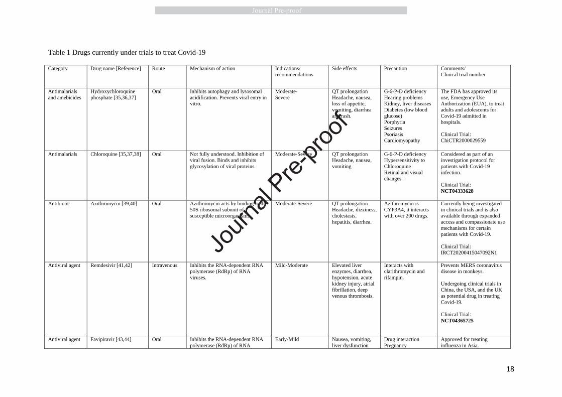
medications have been tried in cases admitted to ICU with severe pneumonia. Table 1
summarized these drugs- possible mechanisms of action, side effects, precautions and
recommendations. Also, shows ongoing registered clinical trials.
Summary
The COVID-19 pandemic represents the most significant public health crisis we face since
the pandemic influenza outbreak of 1918. To date (25 June 2020), over nine million people
infected, 480,600 deaths and over five million recovered. The outbreak originated in China,
but the more significant numbers of infections and deaths are reported from Europe and the
United States. SARS-CoV-2 belongs to ß-coronavirus, which is highly identical to bat
coronavirus. The virus uses the ACE2 receptor for cell entry and causing pathophysiological
changes of the respiratory, cardiovascular, gastrointestinal and nervous systems. Human-to-
human transmission is evident with a reproduction number ranging from 2.24 to 3.58,
indicating higher transmission. Clinical symptoms include fever, cough, and shortness of
breath. Symptoms related to the gastrointestinal, cardiac and nervous system have also been
reported. Patients at a higher risk of infection include the elderly, patients with comorbidity,
and immunocompromised. Currently, no specific therapeutics have been competent to
prevent or treat COVID-19. Several drugs have been tried, including antimalarials, antiviral
agents, immunomodulators, and plasma neutralizing antibodies transfusion. These
therapeutics are currently investigated in clinical trials.
Journ
al Pre-
proof

13
Acknowledgement
This work was funded by the College of Medicine Research Center, Deanship of Scientific
Research, King Saud University, Riyadh, Saudi Arabia.
Conflict of interest The author declares no financial or relationships that can be considered a conflict of interest.
Journ
al Pre-
proof

14
References
1. Sohrabi C, Alsafi Z, O'Neill N, et al. World Health Organization declares global emergency: A review of the 2019 novel coronavirus (COVID-19) [published correction appears in Int J Surg. 2020 Apr 15;:]. Int J Surg. 2020;76:71‐76. doi:10.1016/j.ijsu.2020.02.034
2. Zhang T, Wu Q, Zhang Z. Probable Pangolin Origin of SARS-CoV-2 Associated with the COVID-19 Outbreak [published correction appears in Curr Biol. 2020 Apr 20;30(8):1578]. Curr Biol. 2020;30(7):1346‐1351.e2. doi:10.1016/j.cub.2020.03.022
3. Brian DA, Baric RS. Coronavirus genome structure and replication. Curr Top Microbiol Immunol. 2005;287:1‐30. doi:10.1007/3-540-26765-4_1
4. Wang B, Li R, Lu Z, Huang Y. Does comorbidity increase the risk of patients with COVID-19: evidence from meta-analysis. Aging (Albany NY). 2020;12(7):6049‐6057. doi:10.18632/aging.103000.
5. D'Amico F, Baumgart DC, Danese S, Peyrin-Biroulet L. Diarrhea during COVID-19 infection: pathogenesis, epidemiology, prevention and management [published online ahead of print, 2020 Apr 8]. Clin Gastroenterol Hepatol. 2020;S1542-3565(20)30481-X. doi:10.1016/j.cgh.2020.04.001
6. Li M, Chen L, Zhang J, Xiong C, Li X. The SARS-CoV-2 receptor ACE2 expression of maternal-fetal interface and fetal organs by single-cell transcriptome study. PLoS One. 2020;15(4):e0230295. Published 2020 Apr 16. doi:10.1371/journal.pone.0230295.
7. Li W, Moore MJ, Vasilieva N, et al. Angiotensin-converting enzyme 2: A functional receptor for SARS coronavirus. Nature. 2003;426:450-454. doi:10.1007/s00018-004-4242-5
8. European Society of Cardiology. Position statement of the ESC Council on Hypertension on ACE-inhibitors and angiotensin receptor blockers. Published March 13, 2020. Accessed March 20, 2020. https://www.escardio.org/Councils/Council-on-Hypertension-(CHT)/News/position-statement-of-the-esc-council-on-hypertension-on-ace-inhibitors-and-ang
9. Chiappelli F, Khakshooy A, Greenberg G. CoViD-19 Immunopathology and Immunotherapy. Bioinformation. 2020;16(3):219–222. Published 2020 Mar 31. doi:10.6026/97320630016219.
10. Tian S, Hu W, Niu L, Liu H, Xu H, Xiao SY. Pulmonary Pathology of Early-Phase 2019 Novel Coronavirus (COVID-19) Pneumonia in Two Patients With Lung Cancer. J Thorac Oncol. 2020;15(5):700‐704. doi:10.1016/j.jtho.2020.02.010
11. Liu PP, Blet A, Smyth D, Li H. The Science Underlying COVID-19: Implications for the Cardiovascular System [published online ahead of print, 2020 Apr 15]. Circulation. 2020;10.1161/CIRCULATIONAHA.120.047549. doi:10.1161/CIRCULATIONAHA.120.047549.
12. Bansal M. Cardiovascular disease and COVID-19. Diabetes Metab Syndr. 2020;14(3):247‐250. doi:10.1016/j.dsx.2020.03.013
13. Rizzo P, Vieceli Dalla Sega F, Fortini F, Marracino L, Rapezzi C, Ferrari R. COVID-19 in the heart and the lungs: could we "Notch" the inflammatory storm?. Basic Res Cardiol. 2020;115(3):31. Published 2020 Apr 9. doi:10.1007/s00395-020-0791-5.
Journ
al Pre-
proof

15
14. Xu Y, Li X, Zhu B, et al. Characteristics of pediatric SARS-CoV-2 infection and potential evidence for persistent fecal viral shedding. Nat Med. 2020;26(4):502‐505. doi:10.1038/s41591-020-0817-4.
15. Zhao D, Yao F, Wang L, et al. A comparative study on the clinical features of COVID-19 pneumonia to other pneumonias [published online ahead of print, 2020 Mar 12]. Clin Infect Dis. 2020;ciaa247. doi:10.1093/cid/ciaa247
16. Xu L, Liu J, Lu M, Yang D, Zheng X. Liver injury during highly pathogenic human coronavirus infections [published online ahead of print, 2020 Mar 14]. Liver Int. 2020;10.1111/liv.14435. doi:10.1111/liv.14435
17. Chen N, Zhou M, Dong X et al. Epidemiological and clinical characteristics of 99 cases of 2019 novel coronavirus pneumonia in Wuhan, China: a descriptive study. Lancet 2020; 395: 507–13.
18. Feng G, Zheng KI, Yan QQ, et al. COVID-19 and Liver Dysfunction: Current Insights and Emergent Therapeutic Strategies. J Clin Transl Hepatol. 2020;8(1):18–24. doi:10.14218/JCTH.2020.00018
19. Poyiadji N, Shahin G, Noujaim D, Stone M, Patel S, Griffith B. COVID-19–associated Acute Hemorrhagic Necrotizing Encephalopathy: CT and MRI Features. Radiology. 2020. https://doi.org/10.1148/radiol.2020201222.
20. Zhu J, Ji P, Pang J, et al. Clinical characteristics of 3,062 COVID-19 patients: a meta-analysis [published online ahead of print, 2020 Apr 15]. J Med Virol. 2020;10.1002/jmv.25884. doi:10.1002/jmv.25884.
21. Lechien JR, Chiesa-Estomba CM, De Siati DR, et al. Olfactory and gustatory dysfunctions as a clinical presentation of mild-to-moderate forms of the coronavirus disease (COVID-19): a multicenter European study [published online ahead of print, 2020 Apr 6]. Eur Arch Otorhinolaryngol. 2020;1‐11. doi:10.1007/s00405-020-05965-1
22. Hong KH, Lee SW, Kim TS, et al. Guidelines for Laboratory Diagnosis of Coronavirus Disease 2019 (COVID-19) in Korea. Ann Lab Med. 2020;40(5):351‐360. doi:10.3343/alm.2020.40.5.351
23. Loeffelholz MJ, Tang YW. Laboratory diagnosis of emerging human coronavirus infections - the state of the art. Emerg Microbes Infect. 2020;9(1):747‐756. doi:10.1080/22221751.2020.1745095
24. Che XY, Qiu LW, Pan YX, et al. Sensitive and specific monoclonal antibody-based capture enzyme immunoassay for detection of nucleocapsid antigen in sera from patients with severe acute respiratory syndrome. J Clin Microbiol. 2004;42(6):2629–2635. doi:10.1128/JCM.42.6.2629-2635.2004
25. Sikachi R, Agrawal A. Whole body point of care ultrasound for COVID-19: a multi-system approach to a multi-system disease [published online ahead of print, 2020 Apr 16]. Anaesthesia. 2020;10.1111/anae.15087. doi:10.1111/anae.15087
26. Chen X, Tang Y, Mo Y, et al. A diagnostic model for coronavirus disease 2019 (COVID-19) based on radiological semantic and clinical features: a multi-center study [published online ahead of print, 2020 Apr 16]. Eur Radiol. 2020;10.1007/s00330-020-06829-2. doi:10.1007/s00330-020-06829-2.
27. Xia XY, Wu J, Liu HL, Xia H, Jia B, Huang WX. Epidemiological and initial clinical characteristics of patients with family aggregation of COVID-19 [published online
Journ
al Pre-
proof

16
ahead of print, 2020 Apr 12]. J Clin Virol. 2020;127:104360. doi:10.1016/j.jcv.2020.104360
28. Yang AP, Liu JP, Tao WQ, Li HM. The diagnostic and predictive role of NLR, d-NLR and PLR in COVID-19 patients [published online ahead of print, 2020 Apr 13]. Int Immunopharmacol. 2020;84:106504. doi:10.1016/j.intimp.2020.106504
29. Promislow DEL. A geroscience perspective on COVID-19 mortality [published online ahead of print, 2020 Apr 17]. J Gerontol A Biol Sci Med Sci. 2020;glaa094. doi:10.1093/gerona/glaa094
30. Duployez C, Le Guern R, Tinez C, et al. Panton-Valentine Leukocidin-Secreting Staphylococcus aureus Pneumonia Complicating COVID-19 [published online ahead of print, 2020 Apr 16]. Emerg Infect Dis. 2020;26(8):10.3201/eid2608.201413. doi:10.3201/eid2608.201413
31. Kluge S, Janssens U, Welte T, Weber-Carstens S, Marx G, Karagiannidis C. German recommendations for critically ill patients with COVID-19 [published online ahead of print, 2020 Apr 14]. Empfehlungen zur intensivmedizinischen Therapie von Patienten mit COVID-19 [published online ahead of print, 2020 Apr 14]. Med Klin Intensivmed Notfmed. 2020;1‐4. doi:10.1007/s00063-020-00689-w
32. Li X, Xu S, Yu M, et al. Risk factors for severity and mortality in adult COVID-19 inpatients in Wuhan [published online ahead of print, 2020 Apr 12]. J Allergy Clin Immunol. 2020;S0091-6749(20)30495-4. doi:10.1016/j.jaci.2020.04.006
33. Ullah W, Saeed R, Sarwar U, Patel R, Fischman DL. COVID-19 complicated by Acute Pulmonary Embolism and Right-Sided Heart Failure [published online ahead of print, 2020 Apr 17]. JACC Case Rep. 2020;10.1016/j.jaccas.2020.04.008. doi:10.1016/j.jaccas.2020.04.008.
34. The Centers for Disease Control and Prevention (CDC). Information for Clinicians on Investigational Therapeutics for Patients with COVID-19. Available at: https://www.cdc.gov/coronavirus/2019-ncov/hcp/therapeutic-options.html
35. Ferner RE, Aronson JK. Chloroquine and hydroxychloroquine in covid-19. BMJ. 2020;369:m1432. Published 2020 Apr 8. doi:10.1136/bmj.m1432.
36. Touret F, de Lamballerie X. Of chloroquine and COVID-19. Antiviral Res. 2020;177:104762. doi:10.1016/j.antiviral.2020.104762
37. Yazdany J, Kim AHJ. Use of Hydroxychloroquine and Chloroquine During the COVID-19 Pandemic: What Every Clinician Should Know [published online ahead of print, 2020 Mar 31]. Ann Intern Med. 2020;M20-1334. doi:10.7326/M20-1334
38. Cortegiani A, Ingoglia G, Ippolito M, Giarratano A, Einav S. A systematic review on the efficacy and safety of chloroquine for the treatment of COVID-19 [published online ahead of print, 2020 Mar 10]. J Crit Care. 2020;S0883-9441(20)30390-7. doi:10.1016/j.jcrc.2020.03.005
39. Damle B, Vourvahis M, Wang E, Leaney J, Corrigan B. Clinical Pharmacology Perspectives on the Antiviral Activity of Azithromycin and Use in COVID-19 [published online ahead of print, 2020 Apr 17]. Clin Pharmacol Ther. 2020;10.1002/cpt.1857. doi:10.1002/cpt.1857.
40. Gautret P, Lagier JC, Parola P, et al. Clinical and microbiological effect of a combination of hydroxychloroquine and azithromycin in 80 COVID-19 patients with at least a six-day follow up: A pilot observational study [published online ahead of
Journ
al Pre-
proof

17
print, 2020 Apr 11]. Travel Med Infect Dis. 2020;101663. doi:10.1016/j.tmaid.2020.101663
41. Grein J, Ohmagari N, Shin D, et al. Compassionate Use of Remdesivir for Patients with Severe Covid-19 [published online ahead of print, 2020 Apr 10]. N Engl J Med. 2020;NEJMoa2007016. doi:10.1056/NEJMoa2007016
42. Cao YC, Deng QX, Dai SX. Remdesivir for severe acute respiratory syndrome coronavirus 2 causing COVID-19: An evaluation of the evidence [published online ahead of print, 2020 Apr 2]. Travel Med Infect Dis. 2020;101647. doi:10.1016/j.tmaid.2020.101647
43. Lu CC, Chen MY, Chang YL. Potential therapeutic agents against COVID-19: What we know so far [published online ahead of print, 2020 Apr 1]. J Chin Med Assoc. 2020;10.1097/JCMA.0000000000000318. doi:10.1097/JCMA.0000000000000318
44. Furuta Y, Komeno T, Nakamura T. Favipiravir (T-705), a broad spectrum inhibitor of viral RNA polymerase. Proc Jpn Acad Ser B Phys Biol Sci. 2017;93(7):449–463. doi:10.2183/pjab.93.027
45. Cao B, Wang Y, Wen D, et al. A Trial of Lopinavir-Ritonavir in Adults Hospitalized with Severe Covid-19 [published online ahead of print, 2020 Mar 18]. N Engl J Med. 2020;NEJMoa2001282. doi:10.1056/NEJMoa2001282
46. Ahmad A, Rehman MU, Alkharfy KM. An alternative approach to minimize the risk of coronavirus (Covid-19) and similar infections. Eur Rev Med Pharmacol Sci. 2020;24(7):4030–4034. doi:10.26355/eurrev_202004_20873
47. Luo P, Liu Y, Qiu L, Liu X, Liu D, Li J. Tocilizumab treatment in COVID-19: A single center experience [published online ahead of print, 2020 Apr 6]. J Med Virol. 2020;10.1002/jmv.25801. doi:10.1002/jmv.25801
48. Siltuximab. In: Drugs and Lactation Database (LactMed). Bethesda (MD): National Library of Medicine (US); 2006.
49. Siltuximab. In: LiverTox: Clinical and Research Information on Drug-Induced Liver Injury. Bethesda (MD): National Institute of Diabetes and Digestive and Kidney Diseases; 2012.
50. Bloch EM, Shoham S, Casadevall A, et al. Deployment of convalescent plasma for the prevention and treatment of COVID-19 [published online ahead of print, 2020 Apr 7]. J Clin Invest. 2020;138745. doi:10.1172/JCI138745
Journ
al Pre-
proof
18
Table 1 Drugs currently under trials to treat Covid-19 Category Drug name [Reference] Route Mechanism of action Indications/
recommendations Side effects Precaution Comments/
Clinical trial number
Antimalarials and amebicides
Hydroxychloroquine phosphate [35,36,37]
Oral Inhibits autophagy and lysosomal acidification. Prevents viral entry in vitro.
Moderate- Severe
QT prolongation Headache, nausea, loss of appetite, vomiting, diarrhea and rash.
G-6-P-D deficiency Hearing problems Kidney, liver diseases Diabetes (low blood glucose) Porphyria Seizures Psoriasis Cardiomyopathy
The FDA has approved its use, Emergency Use Authorization (EUA), to treat adults and adolescents for Covid-19 admitted in hospitals. Clinical Trial: ChiCTR2000029559
Antimalarials Chloroquine [35,37,38] Oral Not fully understood. Inhibition of viral fusion. Binds and inhibits glycosylation of viral proteins.
Moderate-Severe
QT prolongation Headache, nausea, vomiting
G-6-P-D deficiency Hypersensitivity to Chloroquine Retinal and visual changes.
Considered as part of an investigation protocol for patients with Covid-19 infection. Clinical Trial: NCT04333628
Antibiotic Azithromycin [39,40]
Oral Azithromycin acts by binding to the 50S ribosomal subunit of susceptible microorganisms
Moderate-Severe
QT prolongation Headache, dizziness, cholestasis, hepatitis, diarrhea.
Azithromycin is CYP3A4, it interacts with over 200 drugs.
Currently being investigated in clinical trials and is also available through expanded access and compassionate use mechanisms for certain patients with Covid-19. Clinical Trial: IRCT20200415047092N1
Antiviral agent Remdesivir [41,42] Intravenous Inhibits the RNA-dependent RNA polymerase (RdRp) of RNA viruses.
Mild-Moderate Elevated liver enzymes, diarrhea, hypotension, acute kidney injury, atrial fibrillation, deep venous thrombosis.
Interacts with clarithromycin and rifampin.
Prevents MERS coronavirus disease in monkeys. Undergoing clinical trials in China, the USA, and the UK as potential drug in treating Covid-19. Clinical Trial: NCT04365725
Antiviral agent Favipiravir [43,44]
Oral Inhibits the RNA-dependent RNA polymerase (RdRp) of RNA
Early-Mild Nausea, vomiting, liver dysfunction
Drug interaction Pregnancy
Approved for treating influenza in Asia.
Journ
al Pre-
proof

19
viruses. It is an inhibitor of viral RNA-dependent RNA polymerase, causing chain termination and preventing RNA elongation.
Clinical Trial: NCT04358549
Lopinavir, is a human immunodeficiency virus (HIV) type 1
Lopinavir/ritonavir [45] (Ritonavir is combined with lopinavir to increase its plasma half- life through the inhibition of cytochrome P450)
Oral Aspartate protease inhibitor. Lopinavir binds to the site of HIV-1 protease activity and inhibits the cleavage of viral Gag-Pol polyprotein precursors and hence interfering with HIV infection.
Moderate-Severe
Anorexia, nausea, abdominal discomfort, diarrhea, acute gastritis, liver dysfunction, thrombocytopenia, and skin eruptions.
Drug interactions- it is a CYP3A4 substrate.
No benefit was observed with lopinavir–ritonavir treatment beyond standard care (Cao B et al, 2020). Clinical Trial: NCT04307693
Antiprotozoal agent
Nitazoxanide [46]
Oral Disturbs metabolism in anaerobic microbes and inhibits viral transcription factor.
Moderate-Severe Nausea, vomiting, abdominal pain, headache, dizziness, skin rash.
Hypersensitivity to nitazoxanide.
Nitazoxanide/Azithromycin has been tried in combination to treat Covid-19. A clinical trial – hydroxychloroquine vs nitazoxanide is currently investigated. Clinical Trials: NCT04361318; NCT04360356; NCT04343248; NCT04351347; NCT04348409; NCT04341493.
Immunomodulator (monoclonal antibody)
Tocilizumab [47]
intravenous A monoclonal antibody; blocks interleukin-6 (IL-6) receptor and inhibit Il-6 pathway.
Severe Nasopharyngitis, headache, hypertension, elevated ALT, Rash, dizziness, leukopenia, liver injury.
Thrombocytopenia Neutropenia Acute liver injury Renal failure
Considered as part of an investigation protocol for patients with Covid-19 infection. Clinical Trial: NCT04356937
Immunomodulator (monoclonal antibody)
Sarilumab [43] Subcutaneous A monoclonal antibody that blocks interleukin-6 (IL-6) receptor and inhibit IL-6 pathway.
Moderate-Severe Allergy Thrombocytopenia Neutropenia Elevated liver transaminases.
Allergy to Sarilumab Platelets <15,000/m3
Elevated alanine transaminases >5 times upper normal limit.
Clinical trials on patients infected with Covid-19 and complicated with pneumonia requiring ventilatory support. Clinical Trials: NCT04359901; NCT04357808.
Journ
al Pre-
proof
20
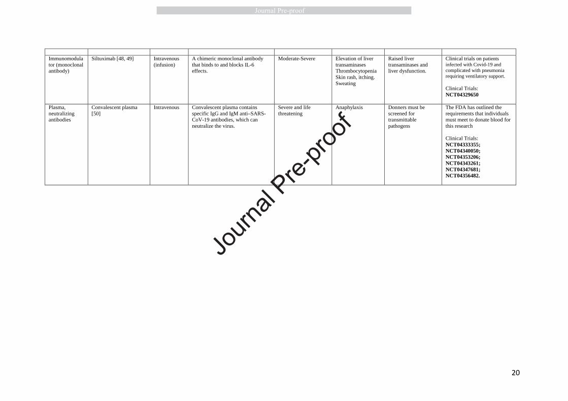
Immunomodulator (monoclonal antibody)
Siltuximab [48, 49] Intravenous (infusion)
A chimeric monoclonal antibody that binds to and blocks IL-6 effects.
Moderate-Severe
Elevation of liver transaminases Thrombocytopenia Skin rash, itching. Sweating
Raised liver transaminases and liver dysfunction.
Clinical trials on patients infected with Covid-19 and complicated with pneumonia requiring ventilatory support. Clinical Trials: NCT04329650
Plasma, neutralizing antibodies
Convalescent plasma [50]
Intravenous Convalescent plasma contains specific IgG and IgM anti–SARS-CoV-19 antibodies, which can neutralize the virus.
Severe and life threatening
Anaphylaxis Donners must be screened for transmittable pathogens
The FDA has outlined the requirements that individuals must meet to donate blood for this research Clinical Trials: NCT04333355; NCT04340050; NCT04353206; NCT04343261; NCT04347681; NCT04356482.
Journ
al Pre-
proof


