
Comparison of sample types and location of skin biopsies for BVDV detection using antigen capture
ELISA Testing
Dr. Brian Vander LeyBVD Symposium January 26, 2009
Phoenix, Arizona

Introduction
• We are looking for ways to screen for PI animals more effectively– Sample choices– Assay evaluation– Assay development

Overview
• 2 Projects– Sample project
• 40 PIs• Several different samples evaluated
– Hide project• 3 PIs• Involved systematic evaluation of whole hides for BVDV
antigen
Sample Project
• 40 PI calves in 2 stocker operations in Oklahoma– Identified by ACE– Segregated from non-PI herdmates

Skin Samples
• 3 ear notches– IHC– ACE– One was archived for
future use
• 1 tail fold biopsyAbove: Ear notcher with ear notch. Below: tail fold
Photo from: www.drugs.com/vet/images/1136000a.png

Swab Samples
• Nasal• Oral• Conjuctival• Vaginal/Preputial• Rectal
Photo from: www.puritanmedproducts.com
Collecting an oral swab

Sample Processing
• Skin Samples– Placed in dry tubes and frozen until testing – Incubated in 1 mL PBS for 24 hours at 4°C prior to
using ACE
• Swab Samples– Collected and place immediately in 1 mL PBS then
frozen – Thawed and allowed to incubate for 24 hours 4°C
prior to ACE
Results
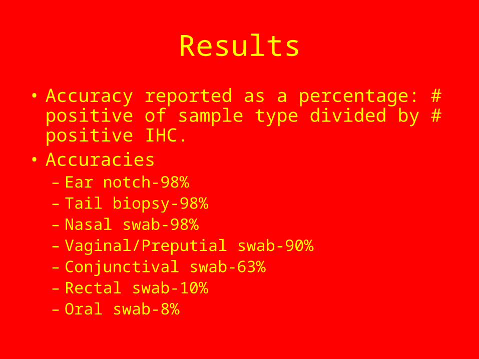
• Accuracy reported as a percentage: # positive of sample type divided by # positive IHC.
• Accuracies– Ear notch-98%– Tail biopsy-98%– Nasal swab-98%– Vaginal/Preputial swab-90%– Conjunctival swab-63%– Rectal swab-10%– Oral swab-8%
Sample Conclusions
• Nasal swabs could be a good sample for detecting PI cattle.– Simple– Non-invasive
• Other samples could also be good, but would pose some challenges
• More work would need to be done to validate any of these samples

Hide Project
• 3 PIs selected based on previous diagnosis as PI by ACE
• Each animal was euthanized and the skin was removed in its entirety
A complete hide
Hide Project
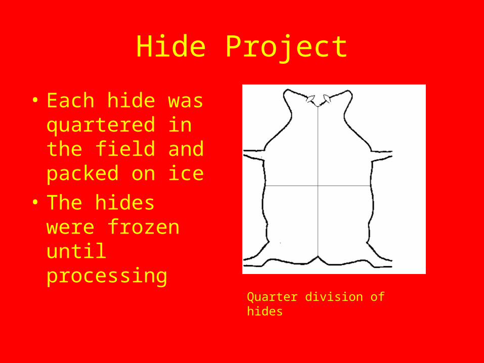
• Each hide was quartered in the field and packed on ice
• The hides were frozen until processing
Quarter division of hides

Hide Processing
• Each quarter was thawed and divided into 10 cm by 10 cm sections
• A 4 mm punch biopsy was taken from each section and placed in a dry microcentrifuge tube.
Hide section schematic
Sample Evaluation
• All samples were incubated in 1 mL PBS at 4°C for the same amount of time.
• All steps were performed the same way on each plate and in sequential order from plate 1 to plate 7.
• Multiple controls were used in each plate– Kit positive control– Kit negative control– Titered BVDV infected cell culture– Titered BVDV infected cell culture diluted 1:10 in PBS
Results
• All sections were positive by ACE• No significant differences in S/P ratios were
found– Between one particular quarter in all three hides– Between quarters on an individual– Between animals

Conclusions
• Large amounts of sample material are available for evaluation/development of diagnostic assays aimed at detecting PI cattle.
• The whole hide can be utilized as a source of samples for running the ACE.
Overall Conclusions
• BVDV Antigen can be recovered from a variety of places.– Many swabs– Essentially any haired skin sample
• There are large amounts of material that can be harvested from an individual animal for evaluation or development of diagnostic assays, especially those that use skin.
• More research needs to be done to validate any of the samples.

References
• Idexx Herdchek® Bovine Virus Diarrhea Antigen Test Kit. U.S. Vet. License No. 313. 2007.

Questions?
Thank You


