
Clinical Decision Support (CDS) Content and Health Level 7 (HL7)-
Compliant Knowledge Artifacts (KNARTs)
Eye Care Clinical Content White Paper
Department of Veterans Affairs (VA)
Knowledge Based Systems (KBS)
Office of Informatics and Information Governance (OIIG)
Clinical Decision Support (CDS)
Version 2.0
September 2018

Revision History
Date Revision Description Author
2018-06-18 1.0 CDS KNRT Eye Care Clinical Content White Paper final version as delivered by contractor team
B3 Group Inc.; Cognitive Medical Systems, Inc.
VA Subject Matter Expert team;
VA CDS Team
2018-09-27 2.0 Add “Revision History” Table to document
Revise first paragraph of Section 1.1 “Clinical Context”
Diane Montella, MD, Apurva Desai, VA KBS Team
Clinical Decision Support (CDS) Content and Health Level 7 (HL7)-Compliant Knowledge Artifacts (KNARTs): Eye Care Clinical Content White Paper
by Department of Veterans Affairs (VA)
Publication date June 2018
Copyright © 2018 B3 Group, Inc.
Copyright © 2018 Cognitive Medical Systems, Inc
B3 Group, Inc. NOTICE OF GOVERNMENT COPYRIGHT LICENSE AND UNLIMITED RIGHTS LICENSE
Licensed under the Apache License, Version 2.0 (the "License"); you may not use this file except in compliance
with the License.
You may obtain a copy of the License at http://www.apache.org/licenses/LICENSE-2.0
Unless required by applicable law or agreed to in writing, software distributed under the License is distributed
on an "AS IS" BASIS, WITHOUT WARRANTIES OR CONDITIONS OF ANY KIND, either express or
implied. See the License for the specific language governing permissions and limitations under the License.
Portions of this content are derivative works from content produced by Cognitive Medical Systems, Inc.
licensed under the Apache License, Version 2.0.
Additional portions of this content are derivative works from content contributed by Motive Medical
Intelligence Inc., under Creative Commons Attribution-ShareAlike 4.0.
Contributions from 2013-2018 were performed either by US Government employees, or under US Veterans
Health Administration contracts.
US Veterans Health Administration contributions by government employees are work of the U.S. Government
and are not subject to copyright protection in the United States. Portions contributed by government employees
are USGovWork (17USC §105). Not subject to copyright.
See: https://www.usa.gov/government-works
Contribution by contractors to the US Veterans Health Administration during this period are contractually
contributed under the Apache License, Version 2.0 and US Government sponsorship is acknowledged under
Contract VA118-16-D-1008, Task Order VA11817F10080007.
2018Cognitive Medical Systems, Inc.
Cognitive Medical Systems, Inc.
Licensed under the Apache License, Version 2.0 (the "License"); you may not use this file except in compliance
with the License.
You may obtain a copy of the License at http://www.apache.org/licenses/LICENSE-2.0
Unless required by applicable law or agreed to in writing, software distributed under the License is distributed
on an "AS IS" BASIS, WITHOUT WARRANTIES OR CONDITIONS OF ANY KIND, either express or
implied. See the License for the specific language governing permissions and limitations under the License.
This and related content produced by Cognitive Medical Systems, Inc. licensed under the Apache License,
Version 2.0 is available at: https://bitbucket.org/cogmedsys/hl7-kas-examples
Additional portions of this content are derivative works from content contributed by Motive Medical
Intelligence Inc., under Creative Commons Attribution-ShareAlike 4.0. https://bitbucket.org/cogmedsys/kas-
source-material
Contributions from 2013-2018 were performed either by US Government employees, or under US Veterans
Health Administration contracts.
US Veterans Health Administration contributions by government employees are work of the U.S. Government
and are not subject to copyright protection in the United States. Portions contributed by government employees
are USGovWork (17USC §105). Not subject to copyright. See: https://www.usa.gov/government-works
Contribution by contractors to the US Veterans Health Administration during this period are contractually
contributed under the Apache License, Version 2.0 and US Government sponsorship is acknowledged under
Contract VA118-16-D-1008-0007.
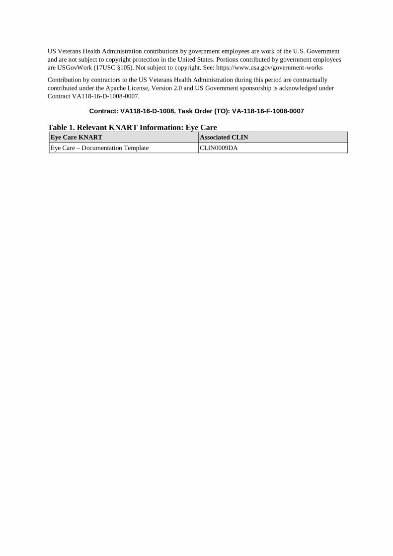
Contract: VA118-16-D-1008, Task Order (TO): VA-118-16-F-1008-0007
Table 1. Relevant KNART Information: Eye Care
Eye Care KNART Associated CLIN
Eye Care – Documentation Template CLIN0009DA

Table of Contents Table of Contents ..................................................................................................................................................... v
VA Subject Matter Expert (SME) Panel .............................................................................................................. viii
Introduction .............................................................................................................................................................ix
Conventions Used .................................................................................................................................................. 10
Chapter 1 - Eye Care .............................................................................................................................................. 11
Section 1.1 - Clinical Context ............................................................................................................................ 11
Section 1.2 - Knowledge Artifacts ..................................................................................................................... 11
Chapter 2 - Documentation Template: Eye Care ................................................................................................... 12
Section 2.1 - Knowledge Narrative .................................................................................................................... 12
Section 2.2 - Chief Complaint/Reason for Visit ................................................................................................ 12
Section 2.3 - History of Present Illness .............................................................................................................. 13
Section 2.4 - Past Ocular History ....................................................................................................................... 14
Section 2.4.1. - Eye Conditions .......................................................................................................................... 14
Section 2.5 - Past Ocular Procedures ................................................................................................................. 25
Section 2.6 - Family History of Ocular Conditions ........................................................................................... 77
Section 2.7 - Pertinent Systematic Medical History .......................................................................................... 82
Section 2.8 - Pertinent Social History ................................................................................................................ 84
Section 2.8.1. - Smoking and Substance Use ..................................................................................................... 84
Section 2.9 - Medications ................................................................................................................................... 85
Section 2.10 - Allergies ...................................................................................................................................... 85
Section 2.11 - Ocular Exam ............................................................................................................................... 85
Section 2.11.1. - Visual Acuity .......................................................................................................................... 86
Section 2.11.1.1. - Distance Vision .................................................................................................................... 86
Section 2.11.1.2 - Near Vision ........................................................................................................................... 92
Section 2.11.2. - Lensometry ............................................................................................................................. 95
Section 2.11.3. - Refraction................................................................................................................................ 97
Section 2.11.3.1 - Distance Visual Acuity with Refraction ............................................................................... 98
Section 2.11.3.2. - Near Visual Acuity with Refraction .................................................................................. 102
Section 2.11.4. - Intraocular Pressure Measurement........................................................................................ 105
Section 2.11.5. - External Eye Exam ............................................................................................................... 106
Section 2.11.6. - Slit Lamp Exam .................................................................................................................... 110
Section 2.11.7. - Fundoscopic Exam ................................................................................................................ 112
Section 2.12 - Special Testing .......................................................................................................................... 114
Section 2.12.1. - Color Vision Testing ............................................................................................................ 115
Section 2.12.2. - Stereo Vision Testing............................................................................................................ 115
Section 2.12.3. - Amsler Grid Testing ............................................................................................................. 116
Section 2.12.4. - A-Scan Ultrasound ................................................................................................................ 117
Section 2.12.5. - Optical Biometry................................................................................................................... 118

Section 2.12.6. - Anterior Segment Optical Coherence Tomography (OCT) .................................................. 118
Section 2.12.7. - B-Scan Ultrasound ................................................................................................................ 118
Section 2.12.8. - Confrontation Visual Field Testing ...................................................................................... 119
Section 2.12.9. - Corneal Topography/Tomography ....................................................................................... 119
Section 2.12.10. - Gonioscopy ......................................................................................................................... 120
Section 2.12.11. - Intraocular Lens (IOL) Calculations ................................................................................... 125
Section 2.12.12. - Keratometry ........................................................................................................................ 126
Section 2.12.13. - Optic Nerve Analysis .......................................................................................................... 127
Section 2.12.14. - Pachymetry ......................................................................................................................... 128
Section 2.12.15. - Perimetry ............................................................................................................................. 128
Section 2.12.16. - Retinal Imaging................................................................................................................... 130
Section 2.13 - Procedure Notes ........................................................................................................................ 130
Section 2.14 - Assessment and Plan ................................................................................................................. 131
Section 2.14.1. - Assessment............................................................................................................................ 131
Section 2.14.2. - Plan ....................................................................................................................................... 131
Section 2.14.2.1. - Vision Correction Prescription Glasses ............................................................................. 131
Section 2.14.2.2. - Vision Correction Prescription – Contact Lenses .............................................................. 132
Section 2.14.2.3. - Eye Prosthetic Devices ...................................................................................................... 133
Section 2.14.2.4. - Laboratory Tests ................................................................................................................ 133
Section 2.14.2.5. - Imaging Studies ................................................................................................................. 134
Section 2.14.2.6. - Eye Clinic Ancillary Study and Procedure Orders ............................................................ 134
Section 2.14.2.7. - Medications ........................................................................................................................ 136
Section 2.14.2.8. - Patient Education and Instructions .................................................................................... 139
Section 2.14.3. - Consultation Requests........................................................................................................... 140
Section 2.14.4. - Surgery Requests .................................................................................................................. 140
Section 2.14.5. - Return for Follow-Up ........................................................................................................... 140
Bibliography/Evidence ......................................................................................................................................... 142
Appendix A - Existing Sample VA Artifacts ....................................................................................................... 144
Appendix B: Basic Laboratory Panel Definition ................................................................................................. 145
Appendix C: Acronyms/Abbreviations ................................................................................................................ 146

List of Figures
Figure 1 – Motility and Alignment Document Tool ............................................................................................ 142

VA Subject Matter Expert (SME) Panel
Name Title Project Role
Linda Wedemeyer, MD Ophthalmologist/Physician
Informatician
Knowledge Based Systems, Office
of Health Informatics
Los Angeles, CA
SME, Primary
Mary Lynch, MD Ophthalmologist
Atlanta VA Medical Center
(VAMC)
Atlanta, GA
SME, Secondary
Rex Ballinger, OD Optometrist
Baltimore VAMC
Baltimore, MD
SME
Thomas Garrido, OD Optometrist
Scott AFB, IL SME
David Eliason, MD Ophthalmologist
Deputy Director, Vision Center of
Excellence
Martinsburg VAMC
Martinsburg, WV
SME
Kristin Biggerstaff, OD Ophthalmologist
Glaucoma Specialist
Michael E. DeBakey VAMC
Houston, TX
SME
Matthew Cordes, OD Optometrist
Chief of Optometry
The Villages VA OPC
The Villages, FL
SME

Introduction The VA is committed to improving the ability of clinicians to provide care for patients while increasing quality,
safety, and efficiency. Recognizing the importance of standardizing clinical knowledge in support of this goal,
VA is applying the Health Level 7 (HL7) Knowledge Artifact Specification for a wide range of VA clinical use
cases. Knowledge Artifacts, referred to as (KNARTs), enable the structuring and encoding of clinical
knowledge so the knowledge can be integrated with electronic health records to enable clinical decision support.
The purpose of this Clinical Content White Paper (CCWP) is to capture the clinical context and intent of
KNART use cases in sufficient detail to provide the KNART authoring team with the clinical source material to
construct the corresponding knowledge artifacts using the HL7 Knowledge Artifact Specification. This paper
has been developed using material from a variety of sources: VA artifacts, clinical practice guidelines, evidence
in the body of medical literature, and clinical expertise. After reviewing these sources, the material has been
synthesized and harmonized under the guidance of VA subject matter experts to reflect clinical intent for this
use case.
Unless otherwise noted, items within this white paper (e.g., documentation template fields, orderable items, etc.)
are chosen to reflect the clinical intent at the time of creation. To provide an exhaustive list of all possible items
and their variations is beyond the scope of this work.

Conventions Used Conventions used within the knowledge artifact descriptions include:
<obtain>: Indicates a prompt to obtain the information listed
• If possible, the requested information should be obtained from the underlying system(s). Otherwise,
prompting the user for information may be required
• The technical and clinical notes associated with a section should be consulted for specific constraints on the
information (e.g., time-frame, patient interview, etc.)
• Default Values: Unless otherwise noted, <obtain> indicates to obtain the most recent observation. It is
recognized that this default time-frame value may be altered by future implementations
[...]: Square brackets enclose explanatory text that indicates some action on the part of the clinical user, or general
guidance to the clinical or technical teams. Examples include, but are not limited to:
[Begin ...], [End ...]: Indicates the start and end of specific areas to clearly delineate them for technical purposes.
[Activate ...]: Initiates another knowledge artifact or knowledge artifact section.
[Section Prompt: ...]: If this section is applicable, then the following prompt should be displayed to the user.
[Section Selection Behavior: ...]: Indicates technical constraints or considerations for the selection of items within
the section.
[Attach: ...]: Indicates that the specified item should be attached to the documentation template if available.
[Link: ...]: Indicates that rather than attaching an item, a link should be included in the documentation template.
[Clinical Comment: ...]: Indicates clinical rationale or guidance.
[Technical Note: ...]: Indicates technical considerations or notes.
[If ...]: Indicates the beginning of a conditional section.
[Else, ...]: Indicates the beginning of the alternative branch of a conditional section.
[End if ...]: Indicates the end of a conditional section.
[Check box]: Indicates items that should be selected based upon the section selection behavior.

Chapter 1 - Eye Care
Section 1.1 - Clinical Context [Begin Clinical Context. Text in the Clinical Context section appears here as reference information for the reader of
this white paper, and is not intended to appear in a clinically implemented knowledge artifact.
The Veterans Health Administration seeks to standardize eye care data output from routine clinical documentation in
support of quality clinical care delivery, performance improvement, interoperability across health care platforms,
and clinical research activities. Prototype work has been done in both the DoD/VA Vision Center of Excellence
(Optimal Vision Care Prototype) and the VA (Eye Care SmartForm) to define eye care documentation requirements
and to promote standardized data output; however, that prototype work has not been implemented. This draft
documentation template content goes a step farther than earlier projects by conforming to HL7 standards for clinical
decision support, furthering the aim of an easily shareable tool with shareable data output from routine clinical
documentation.
Table 1.1. Clinical Context Domains
Target User Provider in an Ophthalmology or Optometry clinic
Patient Adult outpatient
Priority Routine
Specialty Eye Care
Location Outpatient
[End Clinical Context.]
Section 1.2 - Knowledge Artifacts [Begin Knowledge Artifacts.]
This Clinical Content White Paper includes one knowledge artifact, a documentation template as defined by the HL7
CDS KNART specification.
This section describes the single CDS KNART that is included in the Eye Care group:
• A Documentation Template: Eye Care KNART
o Documents information recorded during an eye exam.
o Includes logic for appropriate display of documentation sections.
[End Knowledge Artifacts.]
Chapter 2 - Documentation Template: Eye Care [Begin Eye Care Documentation Template.]
Section 2.1 - Knowledge Narrative [Begin Knowledge Narrative. Text in the Knowledge Narrative section appears here as reference information for
the reader of this white paper, and is not intended to appear in a clinically implemented knowledge artifact.]
Maintaining vision is an important part of maintaining functional independence. In addition to loss of vision, eye
diseases can cause significant symptoms and may represent manifestations of systemic conditions. As a result, eye
care is a key component of medical care. The development of standardized, structured data elements has contributed
to improvements in patient care, performance improvement, reporting, and research (Eleu Pacific Partners, March
2015). However, such documentation practices have not been consistently implemented. The development of a
standardized, structured, comprehensive eye care documentation template that can be used across the VA system has
the potential to lead to better integration of care, further performance improvement, ease reporting, and enable
enhanced research capabilities.
[End Knowledge Narrative.]
Section 2.2 - Chief Complaint/Reason for Visit [Begin Chief Complaint/Reason for Visit.]
[Section Prompt: Is this a returning patient?]
[Section Selection Behavior: Select one, required.]
Yes
[Technical Note: Auto populate prior visit date (s).]
<obtain> Prior visit date (s)
No
[Section Prompt: Chief Compliant/Reason for Visit]
[Section Selection Behavior: Select any. One selection is required.]
Routine Eye Exam
Eye Symptoms
Blurred Vision
Both Eyes
Right Eye
Left Eye
Eye Pain

Both Eyes
Right Eye
Left Eye
Other
Both Eyes
Right Eye
Left Eye
<obtain> Details
Follow up Visit
Glaucoma Suspect
Glaucoma
Diabetic Retinopathy
Cataract
Macular Degeneration
Other
<obtain> Details
Post-Op Visit
<obtain> Procedure
<obtain> Procedure Date
Both Eyes
Right Eye
Left Eye
<obtain> Additional Information
[End Chief Complaint/Reason for Visit.]
Section 2.3 - History of Present Illness [Begin History of Present Illness.]
<obtain> Symptoms
<obtain> Duration of symptoms
<obtain> Severity
<obtain> Laterality
<obtain> Other details

[End History of Present Illness.]
Section 2.4 - Past Ocular History [Begin Past Ocular History.]
[Technical Note: Provide link to Most Recent Ophthalmology visit note.]
Section 2.4.1. - Eye Conditions
[Begin Eye Conditions.]
[Technical Note: Pre-populate from the electronic medical record history if data available. Allow all data to be
updated.]
[Section Prompt: Eye Conditions.]
[Section Selection Behavior: Select any or none. Optional.]
Age-Related Macular Degeneration (AMD)
[Technical Note: Link to Age-Related Eye Disease Study (AREDS) AMD Category definitions
https://www.aao.org/preferred-practice-pattern/age-related-macular-degeneration-ppp-2015.]
[Section Prompt: the following AMD classification system is adapted from American Academy of Ophthalmology’s
2015 Age-Related Macular Degeneration Preferred Practice Pattern Guidelines (American Academy of
Ophthalmology Retina/Vitreous Panel, 2015).]
[Technical Note: Allow user to hover over the link and reveal an informational flyout box with the classification
outlined below.]
Age-related macular degeneration (AMD) is a disorder of the macula characterized by one or more of the following
(for specific terms, see Glossary):
• Presence of at least intermediate-size drusen (63 µm or larger in diameter)
• Retinal pigment epithelium (RPE) abnormalities such as hypopigmentation or hyperpigmentation
• Reticular pseudodrusen4
• Presence of any of the following features: geographic atrophy of the RPE, choroidal neovascularization
(exudative, wet), polypoidal choroidal vasculopathy, or retinal angiomatous proliferation.
• There are a number of classifications of AMD in the literature. This Preferred Practice Pattern uses the
classification of the Age-Related Eye Disease Study (AREDS) and a more recent clinical classification5to
define the early and intermediate stages of AMD since current treatment recommendations are based on
these classifications. The AREDS was a prospective multicenter randomized clinical trial conducted
between 1992 and 2006 designed to assess the natural course and risk factors for age-related cataract and
AMD. The effects of antioxidant vitamins and minerals on these two ocular conditions were studied.
The classification of AMD from the AREDS is as follows:6
• No AMD (AREDS category 1) represented the control group; it is characterized by no or few small drusen
(<63 µm in diameter).
• Early AMD (AREDS category 2) is characterized by a combination of multiple small drusen, few
intermediate drusen (63–124 µm in diameter), or mild RPE abnormalities.
• Intermediate AMD (AREDS category 3) is characterized by any of the following features:
o Numerous intermediate drusen
o At least one large druse (125 µm or larger in diameter)
o Geographic atrophy (a sharply demarcated, usually round or oval, area of atrophy of the RPE not
involving the center of the fovea)
• Advanced AMD (AREDS category 4) is characterized by one or more of the following (in the absence of
other causes) in one eye:

o Geographic atrophy of the RPE involving the foveal center
o Neovascular maculopathy that includes the following:
o Choroidal neovascularization (CNV) defined as pathologic angiogenesis originating from the
choroidal vasculature that extends through a defect in Bruch’s membrane
o Serous and/or hemorrhagic detachment of the neurosensory retina or RPE
Retinal hard exudates (a secondary phenomenon resulting from chronic intravascular leakage)
o Subretinal and sub-RPE fibrovascular proliferation
o Disciform scar (subretinal fibrosis)
Early AMD
Right Eye
Left Eye
<obtain> Description
Intermediate AMD
Right Eye
Left Eye
<obtain> Description
Advanced AMD: Neovascular
Right Eye
Left Eye
<obtain> Description
Advanced AMD: Geographic Atrophy
Right Eye
Left Eye
<obtain> Description
Amblyopia
Both Eyes
Right Eye
Left Eye
<obtain> Description
Blepharitis
<obtain> Description
Blindness
Both Eyes

Right Eye
Left Eye
<obtain> Description
Cataract
Both Eyes
Right Eye
Left Eye
<obtain> Description
Chalazion
Both Eyes
Right Eye
Left Eye
<obtain> Description
Corneal Guttata
Both Eyes
Right Eye
Left Eye
<obtain> Description
Cystoid Macular Edema
Both Eyes
Right Eye
Left Eye
<obtain> Description
Diabetes Mellitus without Retinopathy
Both Eyes
Right Eye
Left Eye
<obtain> Description
Diabetic Retinopathy

[Technical Note: If Diabetic Retinopathy is selected, provide a link https://www.aao.org/preferred-practice-
pattern/diabetic-retinopathy-ppp-updated-2017]
[Technical Note: Allow user to hover over the link and reveal an informational flyout box with the classification
outlined below.]
● No apparent retinopathy: No abnormalities
● Mild Nonproliferative Retinopathy (NPDR): Microaneurysms only
● Moderate NPDR: More than just microaneurysms but less than severe NPD
● Severe NPDR: Any of the following and no signs of proliferative retinopathy:
○ Severe intraretinal hemorrhages and microaneurysms in each of the four quadrants
○ Definite venous bleeding in two or more quadrants
○ Moderate IRMA in one or more quadrants
● Proliferative Retinopathy (PDR): One or both of the following.
○ Neovascularization
○ Vitreous/preretinal hemorrhage
● High-Risk PDR: The presence of at least three of the following findings:
○ Neovascularization (at any location)
○ Neovascularization at the optic disc
○ Severe neovascularization:
■ New vessels within one-disc diameter of the optic nerve head that are larger than one-
quarter to one-third disc area in size
■ New vessels elsewhere that are at least one-half disc area in size
○ Vitreous or preretinal hemorrhage
● Clinically Significant Macular Edema is defined by the ETDRS to include any of the following features:
○ Thickening of the retina at or within 500 um of the center of the macula
○ Hard exudates at or within 500 um of the center of the macula, when associated with adjacent
retinal thickening.
○ A zone or zones of retinal thickening one disc area or larger, where any portion of the thickening
is within one disc diameter of the center of the macula.
[Section Prompt: Diabetic retinopathy definitions are adapted from American Academy of Ophthalmology’s
Diabetic Retinopathy Preferred Practice Pattern, updated 2017 (American Academy of Ophthalmology
Retina/Vitreous Panel, 2016).]
Mild Nonproliferative Diabetic Retinopathy
Right Eye
Left Eye
<obtain> Description
Moderate Nonproliferative Diabetic Retinopathy
Right Eye
Left Eye
<obtain> Description
Severe Nonproliferative Diabetic Retinopathy
Right Eye

Left Eye
<obtain> Description
Non-High-Risk Proliferative Diabetic Retinopathy
Right Eye
Left Eye
<obtain> Description
High Risk Proliferative Diabetic Retinopathy
Right Eye
Left Eye
<obtain> Description
Clinically Significant Diabetic Macular Edema
Right Eye
Left Eye
<obtain> Description
Dry Eye
Both Eyes
Right Eye
Left Eye
<obtain> Description
Endophthalmitis
Both Eyes
Right Eye
Left Eye
<obtain> Description
Epiretinal Membrane
Both Eyes
Right Eye
Left Eye
<obtain> Description
Eye Trauma

Both Eyes
Right Eye
Left Eye
Nature of Injury
Blunt
<obtain> Description
Penetrating
<obtain> Description
Chemical
<obtain> Description
Optic Nerve
<obtain> Description
Other
<obtain> Description
[Section Prompt: Date of Injury]
On
<obtain> Date
About
<obtain> Number of Increments Ago
Days ago
Weeks ago
Months ago
Years ago
Around
January
February
March
April
May
June

July
August
September
October
November
December
Year
<obtain> Year
During
Winter of
Spring of
Summer of
Autumn of
Year
<obtain> Year
Unknown
[Section Prompt: Add another injury?]
[Technical Note: If “yes” is selected below, present the questions above again.]
Yes
Fuchs Endothelial Corneal Dystrophy
Both Eyes
Right Eye
Left Eye
<obtain> Description
Glaucoma Suspect
[Technical Note: If glaucoma suspect is selected, provide a link https://www.aao.org/preferred-practice-
pattern/primary-open-angle-glaucoma-suspect-ppp-update-201]
Both Eyes
Right Eye
Left Eye
<obtain> Description

Physiologic Cupping/Asymmetry
Both Eyes
Right Eye
Left Eye
<obtain> Description
Ocular Hypertension
Both Eyes
Right Eye
Left Eye
<obtain> Description
Glaucoma
[Technical Note: If glaucoma is selected, provide a link https://www.aao.org/preferred-practice-pattern/primary-
open-angle-glaucoma-ppp-update-2018]
[Section Prompt: Type of Glaucoma]
Open-Angle Glaucoma (OAG)
Right Eye
Left Eye
Narrow Angle Glaucoma (NAG)
Right Eye
Left Eye
Secondary Glaucoma
<obtain> Description
[Section Prompt: Disease definition, clinical findings, and severity categories derived from the American Academy
of Ophthalmology’s Preferred Pattern for Primary Open Angle Glaucoma.]
[Technical Note: Please make the following guidance viewable via “hover to discover”]
DISEASE DEFINITION
Primary open-angle glaucoma (POAG) is a chronic, progressive optic neuropathy in adults in which there is a
characteristic acquired atrophy of the optic nerve and loss of retinal ganglion cells and their axons. This condition is
associated with an open anterior chamber angle by gonioscopy.
CLINICAL FINDINGS CHARACTERISTIC OF PRIMARY OPEN-ANGLE GLAUCOMA
Primary open-angle glaucoma (POAG) is a chronic, progressive optic neuropathy in adults in which there is a
characteristic acquired atrophy of the optic nerve and loss of retinal ganglion cells and their axons. This condition is
associated with an open anterior chamber angle by gonioscopy.
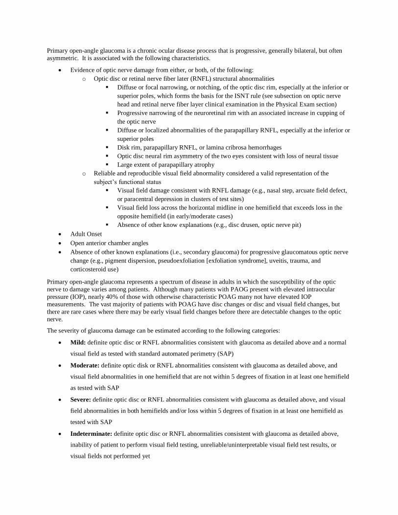
Primary open-angle glaucoma is a chronic ocular disease process that is progressive, generally bilateral, but often
asymmetric. It is associated with the following characteristics.
• Evidence of optic nerve damage from either, or both, of the following:
o Optic disc or retinal nerve fiber later (RNFL) structural abnormalities
▪ Diffuse or focal narrowing, or notching, of the optic disc rim, especially at the inferior or
superior poles, which forms the basis for the ISNT rule (see subsection on optic nerve
head and retinal nerve fiber layer clinical examination in the Physical Exam section)
▪ Progressive narrowing of the neuroretinal rim with an associated increase in cupping of
the optic nerve
▪ Diffuse or localized abnormalities of the parapapillary RNFL, especially at the inferior or
superior poles
▪ Disk rim, parapapillary RNFL, or lamina cribrosa hemorrhages
▪ Optic disc neural rim asymmetry of the two eyes consistent with loss of neural tissue
▪ Large extent of parapapillary atrophy
o Reliable and reproducible visual field abnormality considered a valid representation of the
subject’s functional status
▪ Visual field damage consistent with RNFL damage (e.g., nasal step, arcuate field defect,
or paracentral depression in clusters of test sites)
▪ Visual field loss across the horizontal midline in one hemifield that exceeds loss in the
opposite hemifield (in early/moderate cases)
▪ Absence of other know explanations (e.g., disc drusen, optic nerve pit)
• Adult Onset
• Open anterior chamber angles
• Absence of other known explanations (i.e., secondary glaucoma) for progressive glaucomatous optic nerve
change (e.g., pigment dispersion, pseudoexfoliation [exfoliation syndrome], uveitis, trauma, and
corticosteroid use)
Primary open-angle glaucoma represents a spectrum of disease in adults in which the susceptibility of the optic
nerve to damage varies among patients. Although many patients with PAOG present with elevated intraocular
pressure (IOP), nearly 40% of those with otherwise characteristic POAG many not have elevated IOP
measurements. The vast majority of patients with POAG have disc changes or disc and visual field changes, but
there are rare cases where there may be early visual field changes before there are detectable changes to the optic
nerve.
The severity of glaucoma damage can be estimated according to the following categories:
• Mild: definite optic disc or RNFL abnormalities consistent with glaucoma as detailed above and a normal
visual field as tested with standard automated perimetry (SAP)
• Moderate: definite optic disk or RNFL abnormalities consistent with glaucoma as detailed above, and
visual field abnormalities in one hemifield that are not within 5 degrees of fixation in at least one hemifield
as tested with SAP
• Severe: definite optic disc or RNFL abnormalities consistent with glaucoma as detailed above, and visual
field abnormalities in both hemifields and/or loss within 5 degrees of fixation in at least one hemifield as
tested with SAP
• Indeterminate: definite optic disc or RNFL abnormalities consistent with glaucoma as detailed above,
inability of patient to perform visual field testing, unreliable/uninterpretable visual field test results, or
visual fields not performed yet

[Section Prompt: For assistance with glaucoma billing codes: ICD-10 Glaucoma Staging Definitions, American
Academy of Ophthalmology website: https://www.aao.org/practice-management/news-detail/icd-10-glaucoma-
staging-definitions]
Mild Glaucoma
Both Eyes
Right Eye
Left Eye
<obtain> Description
Moderate Glaucoma
Both Eyes
Right Eye
Left Eye
<obtain> Description
Severe Glaucoma
Both Eyes
Right Eye
Left Eye
<obtain> Description
Indeterminate Glaucoma
Both Eyes
Right Eye
Left Eye
<obtain> Description
<obtain> Target Intraocular Pressure Right Eye (mm Hg)
<obtain> Target Intraocular Pressure Left Eye (mm Hg)
Macular Hole
Both Eyes
Right Eye
Left Eye
<obtain> Description
Nonglaucomatous Visual Field Defects

Both Eyes
Right Eye
Left Eye
<obtain> Description
Optic Atrophy
Both Eyes
Right Eye
Left Eye
<obtain> Description
Pseudophakia
Both Eyes
Right Eye
Left Eye
<obtain> Description
Pterygium
Both Eyes
Right Eye
Left Eye
<obtain> Description
Retinal Detachment
Both Eyes
Right Eye
Left Eye
<obtain> Description
Retinal Tear or Hole
Both Eyes
Right Eye
Left Eye
<obtain> Description
Strabismus

Both Eyes
Right Eye
Left Eye
<obtain> Description
Other
Both Eyes
Right Eye
Left Eye
<obtain> Description
<obtain> Additional Details
[End Conditions.]
[End Past Ocular History.]
Section 2.5 - Past Ocular Procedures [Begin Past Ocular Procedures.]
[Technical Note: Pre-populate fields based on history. Allow all fields to be updated. Allow user to enter multiple
procedures by selecting “Yes” for “Add Another [Procedure Name].”]
[Section Prompt: Past Ocular Procedures]
[Section Selection Behavior: One or more. Optional]
Cataract Surgery
Corneal Surgery
Refractive Surgery
YAG (Yttrium Aluminum Garnet) Laser Capsulotomy
Cyclodestruction
Goniotomy
Laser Iridotomy
Laser Trabeculoplasty
Minimally Invasive Glaucoma Surgery
Trabeculectomy
Tube Shunt
Focal Macular Laser Retina
Intravitreal Injection

Panretinal Photocoagulation
Posterior Vitrectomy
Retinal Detachment Repair (Scleral Buckle)
Retinopexy
Blepharoplasty
Enucleation
Ptosis Repair
Strabismus Surgery
[Technical Note: Activate the associated sub-sections associated with the “Past Ocular Procedures” checkboxes
selected.]
Cataract Surgery
Right Eye
No Complications
Complications
Anterior Vitrectomy
Vitreous Loss
Dropped Lens
<obtain> Description
[Section Prompt: Lens Type]
Standard Lens (Monofocal Lens)
Multifocal Lens
Toric Lens
Accommodating Lens
Section Prompt: Date of Surgery]
On
<obtain> Month/Day/Year
About
<obtain> Number of Increments Ago
Days ago
Weeks ago
Months ago

Years ago
Around
January
February
March
April
May
June
July
August
September
October
November
December
<obtain> Year
During
Winter of
Spring of
Summer of
Autumn of
<obtain> Year
Unknown
<obtain> Additional Details
Left Eye
No Complications
Complications
Anterior Vitrectomy
Vitreous Loss
Dropped Lens
<obtain> Description

[Section Prompt: Lens Type]
Standard Lens (Monofocal Lens)
Multifocal Lens
Toric Lens
Accommodating Lens
[Section Prompt: Date of Surgery]
On
<obtain> Month/Day/Year
About
<obtain> Number of Increments Ago
Days ago
Weeks ago
Months ago
Years ago
Around
January
February
March
April
May
June
July
August
September
October
November
December
<obtain> Year
During
Winter of

Spring of
Summer of
Autumn of
<obtain> Year
Unknown
<obtain> Additional Details
[Section Prompt: Add Another Cataract Surgery?]
Yes
Corneal Surgery
Right Eye
Descemet's Stripping Endothelial Keratoplasty (DSEK)
Descemet Stripping Automated Endothelial Keratoplasty (DSAEK)
Penetrating keratoplasty
Lamellar keratoplasty
[Section Prompt: Date of Surgery]
On
<obtain> Month/Day/Year
About
<obtain> Number of Increments Ago
Days ago
Weeks ago
Months ago
Years ago
Around
January
February
March
April
May
June

July
August
September
October
November
December
<obtain> Year
During
Winter of
Spring of
Summer of
Autumn of
<obtain> Year
Unknown
<obtain> Additional Details
Left Eye
Descemet's Stripping Endothelial Keratoplasty (DSEK)
Descemet Stripping Automated Endothelial Keratoplasty (DSAEK)
Penetrating keratoplasty
Lamellar keratoplasty
[Section Prompt: Date of Surgery]
On
<obtain> Month/Day/Year
About
<obtain> Number of Increments Ago
Days ago
Weeks ago
Months ago
Years ago
Around

January
February
March
April
May
June
July
August
September
October
November
December
<obtain> Year
During
Winter of
Spring of
Summer of
Autumn of
<obtain> Year
Unknown
<obtain> Additional Details
[Section Prompt: Add Another Corneal Surgery?]
Yes
Refractive Surgery
Right Eye
LASIK (Laser-Assisted in Situ Keratomileusis)
PRK (Photorefractive Keratectomy)
LRI (Limbal Relaxing Incisions)
[Section Prompt: Date of Surgery]
On
<obtain> Month/Day/Year
About
<obtain> Number of Increments Ago
Days ago
Weeks ago
Months ago
Years ago
Around
January
February
March
April
May
June
July
August
September
October
November
December
<obtain> Year
During
Winter of
Spring of
Summer of
Autumn of
<obtain> Year
Unknown
<obtain> Additional Details
Left Eye

LASIK (Laser-Assisted in Situ Keratomileusis)
PRK (Photorefractive Keratectomy)
LRI (Limbal Relaxing Incisions)
[Section Prompt: Date of Surgery]
On
<obtain> Month/Day/Year
About
<obtain> Number of Increments Ago
Days ago
Weeks ago
Months ago
Years ago
Around
January
February
March
April
May
June
July
August
September
October
November
December
<obtain> Year
During
Winter of
Spring of
Summer of

Autumn of
<obtain> Year
Unknown
<obtain> Additional Details
[Section Prompt: Add Another Refractive Surgery?]
Yes
YAG (Yttrium Aluminum Garnet) Laser Capsulotomy
Right Eye
[Section Prompt: Date of Procedure]
On
<obtain> Month/Day/Year
About
<obtain> Number of Increments Ago
Days ago
Weeks ago
Months ago
Years ago
Around
January
February
March
April
May
June
July
August
September
October
November
December
<obtain> Year
During
Winter of
Spring of
Summer of
Autumn of
<obtain> Year
Unknown
<obtain> Additional Details
Left Eye
[Section Prompt: Date of Procedure]
On
<obtain> Month/Day/Year
About
<obtain> Number of Increments Ago
Days ago
Weeks ago
Months ago
Years ago
Around
January
February
March
April
May
June
July
August
September

October
November
December
<obtain> Year
During
Winter of
Spring of
Summer of
Autumn of
<obtain> Year
Unknown
<obtain> Additional Details
[Section Prompt: Add Another YAG Laser Capsulotomy?]
Yes
Cyclodestruction
Right Eye
[Section Prompt: Date of Surgery]
On
<obtain> Month/Day/Year
About
<obtain> Number of Increments Ago
Days ago
Weeks ago
Months ago
Years ago
Around
January
February
March
April

May
June
July
August
September
October
November
December
<obtain> Year
During
Winter of
Spring of
Summer of
Autumn of
<obtain> Year
Unknown
<obtain> Additional Details
Left Eye
[Section Prompt: Date of Surgery]
On
<obtain> Month/Day/Year
About
<obtain> Number of Increments Ago
Days ago
Weeks ago
Months ago
Years ago
Around
January
February

March
April
May
June
July
August
September
October
November
December
<obtain> Year
During
Winter of
Spring of
Summer of
Autumn of
<obtain> Year
Unknown
<obtain> Additional Details
[Section Prompt: Add Another Cyclodestruction?]
Yes
Goniotomy
Right Eye
[Section Prompt: Date of Surgery]
On
<obtain> Month/Day/Year
About
<obtain> Number of Increments Ago
Days ago
Weeks ago

Months ago
Years ago
Around
January
February
March
April
May
June
July
August
September
October
November
December
<obtain> Year
During
Winter of
Spring of
Summer of
Autumn of
<obtain> Year
Unknown
Left Eye
[Section Prompt: Date of Surgery]
On
<obtain> Month/Day/Year
About
<obtain> Number of Increments Ago
Days ago

Weeks ago
Months ago
Years ago
Around
January
February
March
April
May
June
July
August
September
October
November
December
<obtain> Year
During
Winter of
Spring of
Summer of
Autumn of
<obtain> Year
Unknown
<obtain> Additional Details
[Section Prompt: Add Another Goniotomy?]
Yes
Laser Iridotomy
Right Eye
[Section Prompt: Date of Procedure]

On
<obtain> Month/Day/Year
About
<obtain> Number of Increments Ago
Days ago
Weeks ago
Months ago
Years ago
Around
January
February
March
April
May
June
July
August
September
October
November
December
<obtain> Year
During
Winter of
Spring of
Summer of
Autumn of
<obtain> Year
Unknown
Left Eye

[Section Prompt: Date of Procedure]
On
<obtain> Month/Day/Year
About
<obtain> Number of Increments Ago
Days ago
Weeks ago
Months ago
Years ago
Around
January
February
March
April
May
June
July
August
September
October
November
December
<obtain> Year
During
Winter of
Spring of
Summer of
Autumn of
<obtain> Year
Unknown

<obtain> Additional Details
[Section Prompt: Add Another Iridotomy?]
Yes
Laser Trabeculoplasty
Right Eye
[Section Prompt: Date of Procedure]
On
<obtain> Month/Day/Year
About
<obtain> Number of Increments Ago
Days ago
Weeks ago
Months ago
Years ago
Around
January
February
March
April
May
June
July
August
September
October
November
December
<obtain> Year
During
Winter of

Spring of
Summer of
Autumn of
<obtain> Year
Unknown
Left Eye
[Section Prompt: Date of Procedure]
On
<obtain> Month/Day/Year
About
<obtain> Number of Increments Ago
Days ago
Weeks ago
Months ago
Years ago
Around
January
February
March
April
May
June
July
August
September
October
November
December
<obtain> Year
During

Winter of
Spring of
Summer of
Autumn of
<obtain> Year
Unknown
<obtain> Additional Details
[Section Prompt: Add Another Laser Trabeculoplasty?]
Yes
Minimally Invasive Glaucoma Surgery
Right Eye
iStent
CyPass
Trabectome
TRAB360 Trabeculotomy
Xen Gel Stent
[Section Prompt: Date of Surgery]
On
<obtain> Month/Day/Year
About
<obtain> Number of Increments Ago
Days ago
Weeks ago
Months ago
Years ago
Around
January
February
March
April

May
June
July
August
September
October
November
December
<obtain> Year
During
Winter of
Spring of
Summer of
Autumn of
<obtain> Year
Unknown
Left Eye
iStent
CyPass
Trabectome
TRAB360 Trabeculotomy
Xen Gel Stent
[Section Prompt: Date of Surgery]
On
<obtain> Month/Day/Year
About
<obtain> Number of Increments Ago
Days ago
Weeks ago
Months ago

Years ago
Around
January
February
March
April
May
June
July
August
September
October
November
December
<obtain> Year
During
Winter of
Spring of
Summer of
Autumn of
<obtain> Year
Unknown
<obtain> Additional Details
[Section Prompt: Add Another Minimally Invasive Glaucoma Surgery?]
Yes
Trabeculectomy
Right Eye
Mitomycin C
Yes
No

[Section Prompt: Date of Surgery]
On
<obtain> Month/Day/Year
About
<obtain> Number of Increments Ago
Days ago
Weeks ago
Months ago
Years ago
Around
January
February
March
April
May
June
July
August
September
October
November
December
<obtain> Year
During
Winter of
Spring of
Summer of
Autumn of
<obtain> Year
Unknown

Left Eye
Mitomycin C
Yes
No
[Section Prompt: Date of Surgery]
On
<obtain> Month/Day/Year
About
<obtain> Number of Increments Ago
Days ago
Weeks ago
Months ago
Years ago
Around
January
February
March
April
May
June
July
August
September
October
November
December
<obtain> Year
During
Winter of
Spring of

Summer of
Autumn of
<obtain> Year
Unknown
<obtain> Additional Details
[Section Prompt: Add Another Trabeculectomy?]
Yes
Tube Shunt
Right Eye
[Section Prompt: Date of Surgery]
On
<obtain> Month/Day/Year
About
<obtain> Number of Increments Ago
Days ago
Weeks ago
Months ago
Years ago
Around
January
February
March
April
May
June
July
August
September
October
November

December
<obtain> Year
During
Winter of
Spring of
Summer of
Autumn of
<obtain> Year
Unknown
Left Eye
[Section Prompt: Date of Surgery]
On
<obtain> Month/Day/Year
About
<obtain> Number of Increments Ago
Days ago
Weeks ago
Months ago
Years ago
Around
January
February
March
April
May
June
July
August
September
October
November
December
<obtain> Year
During
Winter of
Spring of
Summer of
Autumn of
<obtain> Year
Unknown
<obtain> Additional Details
[Section Prompt: Add Another Tube Shunt?]
Yes
Focal Macular Laser
Right Eye
[Section Prompt: Date of Procedure]
On
<obtain> Month/Day/Year
About
<obtain> Number of Increments Ago
Days ago
Weeks ago
Months ago
Years ago
Around
January
February
March
April
May
June
July
August
September
October
November
December
<obtain> Year
During
Winter of
Spring of
Summer of
Autumn of
<obtain> Year
Unknown
Left Eye
[Section Prompt: Date of Procedure]
On
<obtain> Month/Day/Year
About
<obtain> Number of Increments Ago
Days ago
Weeks ago
Months ago
Years ago
Around
January
February
March
April

May
June
July
August
September
October
November
December
<obtain> Year
During
Winter of
Spring of
Summer of
Autumn of
<obtain> Year
Unknown
<obtain> Additional Details
[Section Prompt: Add Another Focal Macular Laser?]
Yes
Intravitreal Injection
Right Eye
[Section Prompt: Agent]
[Section Selection Behavior: Select one. Required]
Bevacizumab
Ranibizumab
Aflibercept
Other
<Obtain> details
[Section Prompt: Date of Procedure]
On

<obtain> Month/Day/Year
About
<obtain> Number of Increments Ago
Days ago
Weeks ago
Months ago
Years ago
Around
January
February
March
April
May
June
July
August
September
October
November
December
<obtain> Year
During
Winter of
Spring of
Summer of
Autumn of
<obtain> Year
Unknown
Left Eye
Agent
Bevacizumab
Ranibizumab
Aflibercept
<obtain> details
[Section Prompt: Date of Procedure]
On
<obtain> Month/Day/Year
About
<obtain> Number of Increments Ago
Days ago
Weeks ago
Months ago
Years ago
Around
January
February
March
April
May
June
July
August
September
October
November
December
<obtain> Year
During
Winter of
Spring of

Summer of
Autumn of
<obtain> Year
Unknown
<obtain> Additional Details
[Section Prompt: Add Another Intravitreal Injection?]
Yes
Panretinal Photocoagulation
Right Eye
<obtain> Number of Laser Spots
[Section Prompt: Date of Procedure]
On
<obtain> Month/Day/Year
About
<obtain> Number of Increments Ago
Days ago
Weeks ago
Months ago
Years ago
Around
January
February
March
April
May
June
July
August
September
October

November
December
<obtain> Year
During
Winter of
Spring of
Summer of
Autumn of
<obtain> Year
Unknown
Left Eye
<obtain> Number of Laser Spots
[Section Prompt: Date of Procedure]
On
<obtain> Month/Day/Year
About
<obtain> Number of Increments Ago
Days ago
Weeks ago
Months ago
Years ago
Around
January
February
March
April
May
June
July
August

September
October
November
December
<obtain> Year
During
Winter of
Spring of
Summer of
Autumn of
<obtain> Year
Unknown
<obtain> Additional Details
[Section Prompt: Add Another Panretinal Photocoagulation?]
Yes
Posterior Vitrectomy
Right Eye
[Section Prompt: Date of Surgery]
On
<obtain> Month/Day/Year
About
<obtain> Number of Increments Ago
Days ago
Weeks ago
Months ago
Years ago
Around
January
February
March
April
May
June
July
August
September
October
November
December
<obtain> Year
During
Winter of
Spring of
Summer of
Autumn of
<obtain> Year
Unknown
Left Eye
[Section Prompt: Date of Surgery]
On
<obtain> Month/Day/Year
About
<obtain> Number of Increments Ago
Days ago
Weeks ago
Months ago
Years ago
Around
January
February

March
April
May
June
July
August
September
October
November
December
<obtain> Year
During
Winter of
Spring of
Summer of
Autumn of
<obtain> Year
Unknown
<obtain> Additional Details
[Section Prompt: Add Another Posterior Vitrectomy?]
Yes
Retinal Detachment Repair (Scleral Buckle)
Right Eye
[Section Prompt: Date of Surgery]
On
<obtain> Month/Day/Year
About
<obtain> Number of Increments Ago
Days ago
Weeks ago

Months ago
Years ago
Around
January
February
March
April
May
June
July
August
September
October
November
December
<obtain> Year
During
Winter of
Spring of
Summer of
Autumn of
<obtain> Year
Unknown
Left Eye
[Section Prompt: Date of Surgery]
On
<obtain> Month/Day/Year
About
<obtain> Number of Increments Ago
Days ago

Weeks ago
Months ago
Years ago
Around
January
February
March
April
May
June
July
August
September
October
November
December
<obtain> Year
During
Winter of
Spring of
Summer of
Autumn of
<obtain> Year
Unknown
<obtain> Additional Details
[Section Prompt: Add Another Retinal Detachment Repair?]
Yes
Retinopexy
Right Eye
[Section Prompt: Date of Procedure]

On
<obtain> Month/Day/Year
About
<obtain> Number of Increments Ago
Days ago
Weeks ago
Months ago
Years ago
Around
January
February
March
April
May
June
July
August
September
October
November
December
<obtain> Year
During
Winter of
Spring of
Summer of
Autumn of
<obtain> Year
Unknown
Left Eye

[Section Prompt: Date of Procedure]
On
<obtain> Month/Day/Year
About
<obtain> Number of Increments Ago
Days ago
Weeks ago
Months ago
Years ago
Around
January
February
March
April
May
June
July
August
September
October
November
December
<obtain> Year
During
Winter of
Spring of
Summer of
Autumn of
<obtain> Year
Unknown

<obtain> Additional Details
[Section Prompt: Add Another Retinopexy?]
Yes
Blepharoplasty
Right Eye
[Section Prompt: Date of Surgery]
On
<obtain> Month/Day/Year
About
<obtain> Number of Increments Ago
Days ago
Weeks ago
Months ago
Years ago
Around
January
February
March
April
May
June
July
August
September
October
November
December
<obtain> Year
During
Winter of
Spring of
Summer of
Autumn of
<obtain> Year
Unknown
Left Eye
[Section Prompt: Date of Surgery]
On
<obtain> Month/Day/Year
About
<obtain> Number of Increments Ago
Days ago
Weeks ago
Months ago
Years ago
Around
January
February
March
April
May
June
July
August
September
October
November
December
<obtain> Year
During

Winter of
Spring of
Summer of
Autumn of
<obtain> Year
Unknown
<obtain> Additional Details
[Section Prompt: Add Another Blepharoplasty?]
Yes
Enucleation
Right Eye
[Section Prompt: Date of Surgery]
On
<obtain> Month/Day/Year
About
<obtain> Number of Increments Ago
Days ago
Weeks ago
Months ago
Years ago
Around
January
February
March
April
May
June
July
August
September

October
November
December
<obtain> Year
During
Winter of
Spring of
Summer of
Autumn of
<obtain> Year
Unknown
Left Eye
[Section Prompt: Date of Surgery]
On
<obtain> Month/Day/Year
About
<obtain> Number of Increments Ago
Days ago
Weeks ago
Months ago
Years ago
Around
January
February
March
April
May
June
July
August

September
October
November
December
<obtain> Year
During
Winter of
Spring of
Summer of
Autumn of
<obtain> Year
Unknown
<obtain> Additional Details
[Section Prompt: Add Another Enucleation?]
Yes
Ptosis Repair
Right Eye
[Section Prompt: Date of Surgery]
On
<obtain> Month/Day/Year
About
<obtain> Number of Increments Ago
Days ago
Weeks ago
Months ago
Years ago
Around
January
February
March

April
May
June
July
August
September
October
November
December
<obtain> Year
During
Winter of
Spring of
Summer of
Autumn of
<obtain> Year
Unknown
Left Eye
[Section Prompt: Date of Surgery]
On
<obtain> Month/Day/Year
About
<obtain> Number of Increments Ago
Days ago
Weeks ago
Months ago
Years ago
Around
January
February

March
April
May
June
July
August
September
October
November
December
<obtain> Year
During
Winter of
Spring of
Summer of
Autumn of
<obtain> Year
Unknown
<obtain> Additional Details
[Section Prompt: Add Another Ptosis Repair?]
Yes
Strabismus Surgery
Right Eye
[Section Prompt: Date of Surgery]
On
<obtain> Month/Day/Year
About
<obtain> Number of Increments Ago
Days ago
Weeks ago

Months ago
Years ago
Around
January
February
March
April
May
June
July
August
September
October
November
December
<obtain> Year
During
Winter of
Spring of
Summer of
Autumn of
<obtain> Year
Unknown
Left Eye
[Section Prompt: Date of Surgery]
On
<obtain> Month/Day/Year
About
<obtain> Number of Increments Ago
Days ago

Weeks ago
Months ago
Years ago
Around
January
February
March
April
May
June
July
August
September
October
November
December
<obtain> Year
During
Winter of
Spring of
Summer of
Autumn of
<obtain> Year
Unknown
<obtain> Additional Details
[Section Prompt: Add Another Strabismus Surgery?]
Yes
Other Surgery/Procedure
<obtain> Description
Right Eye
[Section Prompt: Date of Surgery]
On
<obtain> Month/Day/Year
About
<obtain> Number of Increments Ago
Days ago
Weeks ago
Months ago
Years ago
Around
January
February
March
April
May
June
July
August
September
October
November
December
<obtain> Year
During
Winter of
Spring of
Summer of
Autumn of
<obtain> Year
Unknown

Left Eye
[Section Prompt: Date of Surgery]
On
<obtain> Month/Day/Year
About
<obtain> Number of Increments Ago
Days ago
Weeks ago
Months ago
Years ago
Around
January
February
March
April
May
June
July
August
September
October
November
December
<obtain> Year
During
Winter of
Spring of
Summer of
Autumn of
<obtain> Year

Unknown
<obtain> Additional Details
[Section Prompt: Add Another Other Surgery/Procedure?]
Yes
[End Past Ocular Procedures.]
Section 2.6 - Family History of Ocular Conditions [Begin Family History of Ocular Conditions.]
[Section Prompt: Family ocular history.]
Blindness
[Section Prompt: Relationship]
Mother
Maternal Grandmother
Maternal Grandfather
Maternal Half-Sister
Maternal Half-Brother
Maternal Aunt
Maternal Uncle
Father
Paternal Grandmother
Paternal Grandfather
Paternal Half-Sister
Paternal Half-Brother
Paternal Aunt
Paternal Uncle
Sister
Brother
Daughter
Son
Other
<obtain> Detail

Corneal Dystrophy
[Section Prompt: Relationship]
Mother
Maternal Grandmother
Maternal Grandfather
Maternal Half-Sister
Maternal Half-Brother
Maternal Aunt
Maternal Uncle
Father
Paternal Grandmother
Paternal Grandfather
Paternal Half-Sister
Paternal Half-Brother
Paternal Aunt
Paternal Uncle
Sister
Brother
Daughter
Son
Other
<obtain> Detail
Glaucoma
[Section Prompt: Relationship]
Mother
Maternal Grandmother
Maternal Grandfather
Maternal Half-Sister
Maternal Half-Brother
Maternal Aunt
Maternal Uncle
Father
Paternal Grandmother
Paternal Grandfather
Paternal Half-Sister
Paternal Half-Brother
Paternal Aunt
Paternal Uncle
Sister
Brother
Daughter
Son
Other
<obtain> Detail
Macular Degeneration
[Section Prompt: Relationship]
Mother
Maternal Grandmother
Maternal Grandfather
Maternal Half-Sister
Maternal Half-Brother
Maternal Aunt
Maternal Uncle
Father
Paternal Grandmother
Paternal Grandfather
Paternal Half-Sister
Paternal Half-Brother
Paternal Aunt
Paternal Uncle

Sister
Brother
Daughter
Son
Other
<obtain> Detail
Retinal Detachment
[Section Prompt: Relationship]
Mother
Maternal Grandmother
Maternal Grandfather
Maternal Half-Sister
Maternal Half-Brother
Maternal Aunt
Maternal Uncle
Father
Paternal Grandmother
Paternal Grandfather
Paternal Half-Sister
Paternal Half-Brother
Paternal Aunt
Paternal Uncle
Sister
Brother
Daughter
Son
Other
<obtain> Detail
Retinal Dystrophy
[Section Prompt: Relationship]

Mother
Maternal Grandmother
Maternal Grandfather
Maternal Half-Sister
Maternal Half-Brother
Maternal Aunt
Maternal Uncle
Father
Paternal Grandmother
Paternal Grandfather
Paternal Half-Sister
Paternal Half-Brother
Paternal Aunt
Paternal Uncle
Sister
Brother
Daughter
Son
Other
<obtain> Detail
Other
[Section Prompt: Relationship]
Mother
Maternal Grandmother
Maternal Grandfather
Maternal Half-Sister
Maternal Half-Brother
Maternal Aunt
Maternal Uncle
Father
Paternal Grandmother
Paternal Grandfather
Paternal Half-Sister
Paternal Half-Brother
Paternal Aunt
Paternal Uncle
Sister
Brother
Daughter
Son
Other
<obtain> Detail
<obtain> Additional Details
[End Family History of Ocular Conditions.]
Section 2.7 - Pertinent Systematic Medical History [Begin Pertinent Systematic Medical History.]
[Technical Note: Automatically check conditions listed below if they are on the problem list within the electronic
health record. Also allow user to select and add other problems from the problem list. Allow all fields to be
updated. Note to implementation: The VA problem list is encoded in SNOMED CT.]
Asthma
<obtain> Description
Atrial Fibrillation
<obtain> Description
Benign Prostatic Hypertrophy
<obtain> Description
Carotid Artery Disease
<obtain> Description
Coronary Artery Disease
<obtain> Description
Chronic Obstructive Pulmonary Disease
<obtain> Description
Congestive Heart Failure
<obtain> Description
Connective Tissue Disease/Rheumatologic Conditions
<obtain> Description
Diabetes Mellitus
<obtain> Description
Other Endocrine Disorder
<obtain> Description
HIV Infection
<obtain> Most Recent CD4 Count (cells/mm^3)
<obtain> Most Recent Viral Load (copies/mL)
<obtain> Description
Hypertension
<obtain> Description
Hyperthyroidism
<obtain> Description
Multiple Sclerosis
<obtain> Description
Myocardial Infarction
<obtain> Description
Renal Failure
<obtain> Description
Sleep Apnea
<obtain> Description
Stroke/Transient ischemic attack (TIA)
<obtain> Description
Syphilis
<obtain> Description
Traumatic Brain Injury
<obtain> Description

<obtain> Additional Details
[End Pertinent Systematic Medical History.]
Section 2.8 - Pertinent Social History [Begin Pertinent Social History.]
Section 2.8.1. - Smoking and Substance Use
[Begin Smoking and Substance Use.]
[Section Prompt: Do you currently use tobacco?]
[Section Prompt: Tobacco includes cigarettes, pipe, cigars, and smokeless tobacco, such as dip, chew, snuff, and
snus. Excludes e-cigarettes, ENDS (electronic nicotine delivery systems), and vaping devices.]
[Section Selection Behavior: Select only one. Required.]
Every day
Some days
Not at all
[Technical Note: Those patients reporting use every day or some days will be considered a Current Tobacco
User.]
[Technical Note: Display the following only if “Not at all” is selected in section “Do you currently use tobacco?”]
[Section Prompt: Have you ever used tobacco?]
[Section Selection Behavior: Select only one. Required.]
Never user
Former user
[Section Prompt: Substance Use History.]
[Section Selection Behavior: Select all that apply.]
Alcohol
Cocaine
Opioid
Marijuana
Other
Never user
<obtain> Description
[End Smoking and Substance Use.]
<obtain> Additional Pertinent Social History Information
[End Pertinent Social History.]

Section 2.9 - Medications [Begin Medications.]
<obtain> Eye Medication
[Technical Note: Auto populate Eye Medications from electronic record and include the end user option to update
list.]
<obtain> Other Medications (Including Over-The-Counter Medications and Supplements)
[Technical Note: Auto populate Over-The-Counter Medications and Supplements from the electronic health record
and include the end user option to update list.]
[End Medications.]
Section 2.10 - Allergies [Begin Allergies.]
[Section Prompt: Allergies to Eye Medications.]
<obtain> Allergies to Eye Medications
[Technical Note: Auto populate “Allergies to Eye Medications” and include option to update list.]
[Section Prompt: Other Allergies.]
<obtain> Other Allergies
[Technical Note: Auto populate “Other Allergies” and include option to update list.]
[End Allergies.]
Section 2.11 - Ocular Exam [Begin Ocular Exam.]
[Section Prompt: Ocular Exam.]
[Section Selection Behavior: One or more. Optional]
Visual Acuity
Distance Vision
Near Vision
Lensometry
Refraction
Distance Visual Acuity with Refraction
Near Vision Acuity Refraction
Intraocular Pressure Measurement
External Eye Exam
Silt Lamp
Fundoscopic Exam
[Technical Note: Activate associated subsections selected in the “Ocular Exam” checkboxes.]

Section 2.11.1. - Visual Acuity
[Begin Visual Acuity.]
[Technical Note: Provide link to Visual Acuity History including display of vision over time for returning patients.]
[End Visual Acuity.]
Section 2.11.1.1. - Distance Vision
[Begin Distance Vision.]
[Section Prompt: Distance Vision]
[Technical Note: Snellen is default for the Visual Acuity Chart. The default chart distance is 10 feet. Both should be
editable.]
[Section Prompt: Snellen is default for the Visual Acuity Chart.]
<Obtain> Visual Acuity Chart
[Section Prompt: Indicate below if you wish to select a different visual acuity chart.]
Early Treatment Diabetic Retinopathy Study (ETDRS)
Other
<obtain> Chart Name
<obtain> Chart Distance
[Section Prompt: Distance Units]
[Technical Note: Preselect “Feet” as Distance Units. Allow user to edit per preference.]
Centimeters
Inches
Feet
Meters
[Technical Note: Allow user to repeat Distance Vision section from here down as many times as needed by
selecting Yes for New Distance Acuity Exam below.]
Testing Conditions
Uncorrected
Glasses
<obtain> Description
Contact Lenses
<obtain> Description
Best Corrected Vision
Pinhole
Potential Acuity Meter
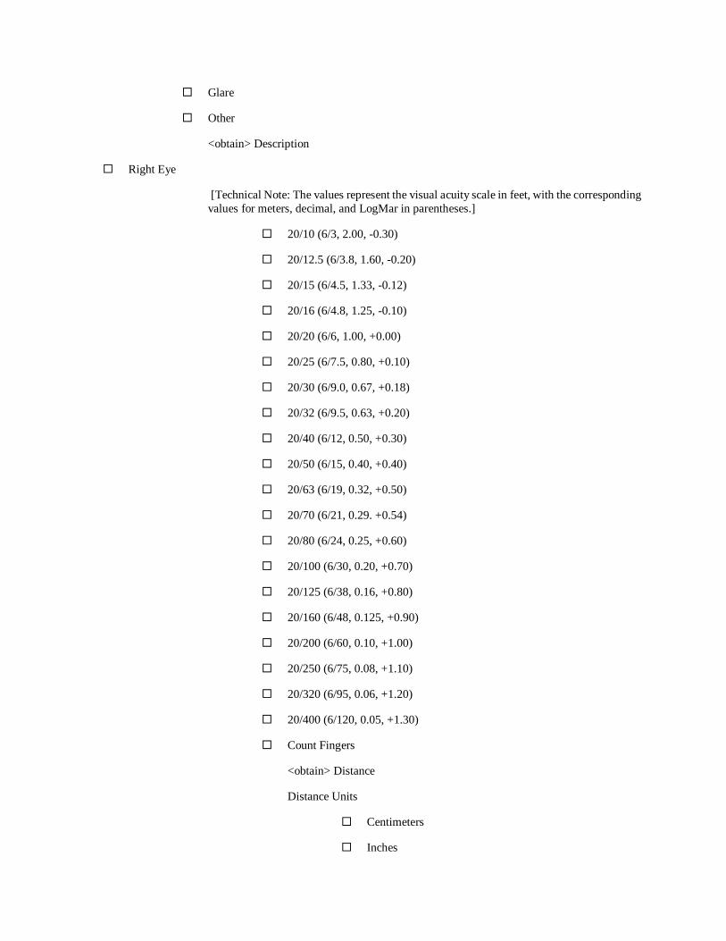
Glare
Other
<obtain> Description
Right Eye
[Technical Note: The values represent the visual acuity scale in feet, with the corresponding
values for meters, decimal, and LogMar in parentheses.]
20/10 (6/3, 2.00, -0.30)
20/12.5 (6/3.8, 1.60, -0.20)
20/15 (6/4.5, 1.33, -0.12)
20/16 (6/4.8, 1.25, -0.10)
20/20 (6/6, 1.00, +0.00)
20/25 (6/7.5, 0.80, +0.10)
20/30 (6/9.0, 0.67, +0.18)
20/32 (6/9.5, 0.63, +0.20)
20/40 (6/12, 0.50, +0.30)
20/50 (6/15, 0.40, +0.40)
20/63 (6/19, 0.32, +0.50)
20/70 (6/21, 0.29. +0.54)
20/80 (6/24, 0.25, +0.60)
20/100 (6/30, 0.20, +0.70)
20/125 (6/38, 0.16, +0.80)
20/160 (6/48, 0.125, +0.90)
20/200 (6/60, 0.10, +1.00)
20/250 (6/75, 0.08, +1.10)
20/320 (6/95, 0.06, +1.20)
20/400 (6/120, 0.05, +1.30)
Count Fingers
<obtain> Distance
Distance Units
Centimeters
Inches
Feet
Meters
Hand Motion
<obtain> Distance
Distance Units
Centimeters
Inches
Feet
Meters
Light Perception without Projection
Light Perception with Projection
No Light Perception
Number of Missed Letters
[Technical Note: Do not display Number of Missed Letters and Number of Additional
Letters Seen questions if Count Fingers, Hand Motion, Light Perception without Projection,
Light Perception with Projection, or No Light Perception is selected for Visual Acuity
Measurement.
-1
-2
-3
Number of Additional Letters Seen
+1
+2
+3
Left Eye
[Technical Note: The values represent the visual acuity scale in feet, with the corresponding
values for meters, decimal, and LogMar in parentheses.]
20/10 (6/3, 2.00, -0.30)
20/12.5 (6/3.8, 1.60, -0.20)
20/15 (6/4.5, 1.33, -0.12)
20/16 (6/4.8, 1.25, -0.10)
20/20 (6/6, 1.00, +0.00)

20/25 (6/7.5, 0.80, +0.10)
20/30 (6/9.0, 0.67, +0.18)
20/32 (6/9.5, 0.63, +0.20)
20/40 (6/12, 0.50, +0.30)
20/50 (6/15, 0.40, +0.40)
20/63 (6/19, 0.32, +0.50)
20/70 (6/21, 0.29. +0.54)
20/80 (6/24, 0.25, +0.60)
20/100 (6/30, 0.20, +0.70)
20/125 (6/38, 0.16, +0.80)
20/160 (6/48, 0.125, +0.90)
20/200 (6/60, 0.10, +1.00)
20/250 (6/75, 0.08, +1.10)
20/320 (6/95, 0.06, +1.20)
20/400 (6/120, 0.05, +1.30)
Count Fingers
<obtain> Distance
Distance Units
Centimeters
Inches
Feet
Meters
Hand Motion
<obtain> Distance
Distance Units
Centimeters
Inches
Feet
Meters
Light Perception without Projection
Light Perception with Projection
No Light Perception
Number of Missed Letters
[Technical Note: Do not display Number of Missed Letters and Number of Additional
Letters Seen questions if Count Fingers, Hand Motion, Light Perception without Projection,
Light Perception with Projection, or No Light Perception is selected for Visual Acuity
Measurement.]
-1
-2
-3
Number of Additional Letters Seen
+1
+2
+3
Both Eyes
[Technical Note: The values represent the visual acuity scale in feet, with the corresponding
values for meters, decimal, and LogMar in parentheses.]
20/10 (6/3, 2.00, -0.30)
20/12.5 (6/3.8, 1.60, -0.20)
20/15 (6/4.5, 1.33, -0.12)
20/16 (6/4.8, 1.25, -0.10)
20/20 (6/6, 1.00, +0.00)
20/25 (6/7.5, 0.80, +0.10)
20/30 (6/9.0, 0.67, +0.18)
20/32 (6/9.5, 0.63, +0.20)
20/40 (6/12, 0.50, +0.30)
20/50 (6/15, 0.40, +0.40)
20/63 (6/19, 0.32, +0.50)
20/70 (6/21, 0.29. +0.54)
20/80 (6/24, 0.25, +0.60)
20/100 (6/30, 0.20, +0.70)
20/125 (6/38, 0.16, +0.80)

20/160 (6/48, 0.125, +0.90)
20/200 (6/60, 0.10, +1.00)
20/250 (6/75, 0.08, +1.10)
20/320 (6/95, 0.06, +1.20)
20/400 (6/120, 0.05, +1.30)
Count Fingers
<obtain> Distance
Distance Units
Centimeters
Inches
Feet
Meters
Hand Motion
<obtain> Distance
Distance Units
Centimeters
Inches
Feet
Meters
Light Perception without Projection
Light Perception with Projection
No Light Perception
Number of Missed Letters
[Technical Note: Do not display Number of Missed Letters and Number of Additional
Letters Seen questions if Count Fingers, Hand Motion, Light Perception without Projection,
Light Perception with Projection, or No Light Perception is selected for Visual Acuity
Measurement.]
-1
-2
-3
Number of Additional Letters Seen
+1

+2
+3
[End Distance Vision.]
Section 2.11.1.2 - Near Vision
[Begin Near Vision.]
[Section Prompt: Near Vision]
[Technical Note: Snellen is default for the Visual Acuity Chart. The default chart distance is 16 inches. Both should
be editable.]
[Section Prompt: Snellen is default for the Visual Acuity Chart.]
<Obtain> Visual Acuity Chart
[Section Prompt: Indicate below if you wish to select a different visual acuity chart.]
Jaeger
LogMAR
M Notation
Other
<obtain> Chart Name
[Technical Note: Auto populate Chart Distance as 16 and preselect “Inches” as Distance Units. Allow user to edit.]
<obtain> Chart Distance
[Section Prompt: Distance Units]
Centimeters
Inches
[Section Prompt: Allow user to repeat Near Vision section from here down as many times needed by selecting
Yes for New Near Acuity Exam below.]
Testing Conditions
Uncorrected
Glasses
<obtain> Description
Contact Lenses
<obtain> Description
Other
<obtain> Description
Right Eye

[Technical Note: The values represent the visual acuity scale in feet, with the corresponding
values in inches, centimeters, M notation, decimal and approximate Jaeger in parentheses.)
20/10 (16/8, 40/20, 0.20, 2.00, -)
20/12.5 (16/10, 40,25, 0.25, 1.60, -)
20/16 (16/12.8, 40/32, 0.32, 1.25, -)
20/20 (16/16, 40/40, 0.40, 1.00, 1)
20/25 (16/20, 40/50, 0.50, 0.80, 1)
20/30 (16/23.8, 40/60, 0.60, 0.67, 2)
20/32 (16/25.4, 40/63, 0.64, 0.63, 2)
20/40 (16/32, 40/80, 0.80, 0.50, 5)
20/50 (16/40, 40/100, 1.0, 0.40, 7)
20/63 (16/50, 40/125, 1.3, 0.32, 4-7)
20/80 (16/64, 40/160, 1.6, 0.25, 5-9)
20/100 (16/80, 40/200, 2.0, 0.20, 8-12)
20/125 (16/100, 40/250, 2.5, 0.16, 9-12)
20/160 (16/128, 40/400, 4.0, 0.10, 15-18)
Number of Missed Letters
-1
-2
-3
Number of Additional Letters Seen
+1
+2
+3
Left Eye
[Technical Note: The values represent the visual acuity scale in feet, with the corresponding
values in inches, centimeters, M notation, decimal and approximate Jaeger in parentheses.]
20/10 (16/8, 40/20, 0.20, 2.00, -)
20/12.5 (16/10, 40,25, 0.25, 1.60, -)
20/16 (16/12.8, 40/32, 0.32, 1.25, -)
20/20 (16/16, 40/40, 0.40, 1.00, 1)

20/25 (16/20, 40/50, 0.50, 0.80, 1)
20/30 (16/23.8, 40/60, 0.60, 0.67, 2)
20/32 (16/25.4, 40/63, 0.64, 0.63, 2)
20/40 (16/32, 40/80, 0.80, 0.50, 5)
20/50 (16/40, 40/100, 1.0, 0.40, 7)
20/63 (16/50, 40/125, 1.3, 0.32, 4-7)
20/80 (16/64, 40/160, 1.6, 0.25, 5-9)
20/100 (16/80, 40/200, 2.0, 0.20, 8-12)
20/125 (16/100, 40/250, 2.5, 0.16, 9-12)
20/160 (16/128, 40/400, 4.0, 0.10, 15-18)
Number of Missed Letters
-1
-2
-3
Number of Additional Letters Seen
+1
+2
+3
Both Eyes
[Technical Note: The values represent the visual acuity scale in feet, with the corresponding
values in inches, centimeters, M notation, decimal and approximate Jaeger in parentheses.]
20/10 (16/8, 40/20, 0.20, 2.00, -)
20/12.5 (16/10, 40,25, 0.25, 1.60, -)
20/16 (16/12.8, 40/32, 0.32, 1.25, -)
20/20 (16/16, 40/40, 0.40, 1.00, 1)
20/25 (16/20, 40/50, 0.50, 0.80, 1)
20/30 (16/23.8, 40/60, 0.60, 0.67, 2)
20/32 (16/25.4, 40/63, 0.64, 0.63, 2)
20/40 (16/32, 40/80, 0.80, 0.50, 5)
20/50 (16/40, 40/100, 1.0, 0.40, 7)
20/63 (16/50, 40/125, 1.3, 0.32, 4-7)

20/80 (16/64, 40/160, 1.6, 0.25, 5-9)
20/100 (16/80, 40/200, 2.0, 0.20, 8-12)
20/125 (16/100, 40/250, 2.5, 0.16, 9-12)
20/160 (16/128, 40/400, 4.0, 0.10, 15-18)
Number of Missed Letters
-1
-2
-3
Number of Additional Letters Seen
+1
+2
+3
[Section Prompt: New Near Acuity Exam.]
Yes
<obtain> Visual Acuity Notes
[End Near Vision.]
[End Visual Acuity]
Section 2.11.2. - Lensometry
[Begin Lensometry.]
[Technical Note: Provide link to most recent lensometry findings and allow importation to the electronic health
record by user.]
[Link: Link to Import Most Recent Lensometry Findings.]
[Technical Note: Allow user to repeat Lensometry section from here down as many times as needed by selecting yes
for New Lensometry Exam below.]
[Section Prompt: Lensometry Method.]
Manual
Automated
<obtain> Additional Details
[Section Prompt: Optical Correction Type.]
Current Glasses
Other
<obtain> Description

[Section Prompt: Right Eye.]
<obtain> Sphere (Diopters)
<obtain> Cylinder (Diopters)
<obtain> Axis (Degrees)
<obtain> Add Power #1 (Diopters)
<obtain> Add Power #2 (Diopters)
<obtain> Prism Power (Diopters)
[Section Prompt: Prism Base.]
Up
Down
In
Out
[Section Prompt: Left Eye.]
<obtain> Sphere (Diopters)
<obtain> Cylinder (Diopters)
<obtain> Axis (Degrees)
<obtain> Add Power #1 (Diopters)
<obtain> Add Power #2 (Diopters)
<obtain> Prism Power (Diopters)
[Section Prompt: Prism Base.]
Up
Down
In
Out
[Section Prompt: Interpupillary Distance
<obtain> Far Interpupillary Distance both eyes
<obtain> Near Interpupillary Distance both eyes
<obtain> Right eye Interpupillary Distance
<obtain> Left eye interpupillary Distance
[Section Prompt: New Lensometry Exam?]

Yes
No
<obtain> Lensometry Notes
[End Lensometry.]
Section 2.11.3. - Refraction
[Begin Refraction.]
[Section Prompt: Refraction]
[Technical Note: Provide link to Refraction Exam History for returning patients.]
[Technical Note: Link to Refraction Exam History.]
[Technical Note: Allow user to repeat Refraction section from here down as many times as needed by selecting
“yes” for New Refraction below.]
[Section Prompt: Refraction Testing Method and Conditions.]
Auto-refraction
Subjective refraction
Retinoscopy
Cycloplegic/Mydriatic
Cyclopentolate
Homatropine
Phenylephrine
Tropicamide
Other
<obtain> Cycloplegic Name
Over Refraction with Glasses
Over Refraction with Contacts
Trial Lens Refraction
Other
<obtain> Description
[Section Prompt: Right Eye]
<obtain> Sphere (Diopters)
<obtain> Cylinder (Diopters)
<obtain> Axis (Degrees)
<obtain> Add Power Near (Diopters)

<obtain> Add Power Intermediate (Diopters)
<obtain> Prism Power (Diopters)
[Section Prompt: Prism Base.]
Up
Down
In
Out
[Section Prompt: Left Eye]
<obtain> Sphere (Diopters)
<obtain> Cylinder (Diopters)
<obtain> Axis (Degrees)
<obtain> Add Power Near (Diopters)
<obtain> Add Power Intermediate (Diopters)
<obtain> Prism Power (Diopters)
[Section Prompt: Prism Base.]
Up
Down
In
Out
[End Refraction.]
Section 2.11.3.1 - Distance Visual Acuity with Refraction
[Begin Distance Visual Acuity with Refraction.]
[Section Prompt: Distance Visual Acuity with Refraction]
[Technical Note: Snellen is default for the Visual Acuity Chart. The default chart distance is 10 feet. Both should be
editable.]
[Section Prompt: Snellen is default for the Visual Acuity Chart.]
<Obtain> Visual Acuity Chart
[Section Prompt: Select below if you wish to select a different visual acuity chart.]
Early Treatment Diabetic Retinopathy Study (ETDRS)
Other
<obtain> Chart Name
<obtain> Chart Distance

[Section Prompt: Distance Units]
[Technical Note: Preselect “Feet” as Distance Units. Allow user to edit per preference.]
Centimeters
Inches
Feet
Meters
[Section Prompt: Right Eye.]
[Technical Note: The values represent the visual acuity scale in feet, with the corresponding values for meters, in
decimal, and in LogMAR in parentheses.]
20/10 (6/3, 2.00, -0.30)
20/12.5 (6/3.8, 1.60, -0.20)
20/15 (6/4.5, 1.33, -0.12)
20/16 (6/4.8, 1.25, -0.10)
20/20 (6/6, 1.00, +0.00)
20/25 (6/7.5, 0.80, +0.10)
20/30 (6/9, 0.67, +0.18)
20/32 (6/9.5, 0.63, +0.20)
20/40 (6/12, 0.50, +0.30)
20/50 (6/15, 0.40, +0.40)
20/63 (6/19, 0.32, +0.50)
20/70 (6/21, 0.29, +0.54)
20/80 (6/24, 0.25, +0.60)
20/100 (6/30, 0.20, +0.70)
20/125 (6/38, 0.16, +0.80)
20/160 (6/48, 0.125, +0.90)
20/200 (6/60, 0.10, +1.00)
20/250 (6/75, 0.08, +1.10)
20/320 (6/95, 0.06, +1.20)
20/400 (6/120, 0.05, +1.30)
Number of Missed Letters
-1

-2
-3
Number of Additional Letters Seen
+1
+2
+3
[Section Prompt: Left Eye.]
[Technical Note: The values represent the visual acuity scale in feet, with the corresponding values for meters, in
decimal, and in LogMAR in parentheses.]
20/10 (6/3, 2.00, -0.30)
20/12.5 (6/3.8, 1.60, -0.20)
20/15 (6/4.5, 1.33, -0.12)
20/16 (6/4.8, 1.25, -0.10)
20/20 (6/6, 1.00, +0.00)
20/25 (6/7.5, 0.80, +0.10)
20/30 (6/9, 0.67, +0.18)
20/32 (6/9.5, 0.63, +0.20)
20/40 (6/12, 0.50, +0.30)
20/50 (6/15, 0.40, +0.40)
20/63 (6/19, 0.32, +0.50)
20/70 (6/21, 0.29, +0.54)
20/80 (6/24, 0.25, +0.60)
20/100 (6/30, 0.20, +0.70)
20/125 (6/38, 0.16, +0.80)
20/160 (6/48, 0.125, +0.90)
20/200 (6/60, 0.10, +1.00)
20/250 (6/75, 0.08, +1.10)
20/320 (6/95, 0.06, +1.20)
20/400 (6/120, 0.05, +1.30)
Number of Missed Letters
-1

-2
-3
Number of Additional Letters Seen
+1
+2
+3
[Section Prompt: Both Eyes.]
[Technical Note: The values represent the visual acuity scale in feet, with the corresponding values for meters, in
decimal, and in LogMAR in parentheses.]
20/10 (6/3, 2.00, -0.30)
20/12.5 (6/3.8, 1.60, -0.20)
20/15 (6/4.5, 1.33, -0.12)
20/16 (6/4.8, 1.25, -0.10)
20/20 (6/6, 1.00, +0.00)
20/25 (6/7.5, 0.80, +0.10)
20/30 (6/9, 0.67, +0.18)
20/32 (6/9.5, 0.63, +0.20)
20/40 (6/12, 0.50, +0.30)
20/50 (6/15, 0.40, +0.40)
20/63 (6/19, 0.32, +0.50)
20/70 (6/21, 0.29, +0.54)
20/80 (6/24, 0.25, +0.60)
20/100 (6/30, 0.20, +0.70)
20/125 (6/38, 0.16, +0.80)
20/160 (6/48, 0.125, +0.90)
20/200 (6/60, 0.10, +1.00)
20/250 (6/75, 0.08, +1.10)
20/320 (6/95, 0.06, +1.20)
20/400 (6/120, 0.05, +1.30)
Number of Missed Letters
-1

-2
-3
Number of Additional Letters Seen
+1
+2
+3
[End Distance Visual Acuity with Refraction.]
Section 2.11.3.2. - Near Visual Acuity with Refraction
[Begin Near Vision Acuity Refraction.]
[Section Prompt: Near Visual Acuity with Refraction]
[Technical Note: Snellen is default for the Visual Acuity Chart. The default chart distance is 16 inches. Both should
be editable.]
[Section Prompt: Snellen is default for the Visual Acuity Chart.]
<Obtain> Visual Acuity Chart
[Section Prompt: Indicate below if you wish to select a different visual acuity chart.]
Jaeger
LogMAR
M Notation
Other
<obtain> Chart Name
[Technical Note: Auto populate Chart Distance as 16 and preselect “Inches” as Distance Units. Allow user to edit.]
<obtain> Chart Distance
[Section Prompt: Distance Units]
Centimeters
Inches
[Section Prompt: Right Eye.]
[Technical Note: The values represent the equivalent distance visual acuity scale in feet, with the
corresponding values in inches, centimeters, M notation, decimal, and LogMAR in parentheses.]
20/10 (16/8, 40/20, 0.20, 2.00, -)
20/12.5 (16/10, 40/25, 0.25, 1.60, -)
20/16 (16/12.8, 40/32, 0.32, 1.25, -)
20/20 (16/16, 40/40, 0.40, 1.00, 1)
20/25 (16/20, 40/50, 0.50, 0.80, 1)
20/30 (16/23.8, 40/60, 0.60, 0.67, 2)
20/32 (16/25.4, 40/63, 0.64, 0.63, 2)
20/40 (16/32, 40/80, 0.80, 0.50, 5)
20/50 (16/40, 40/100, 1.0, 0.40, 7)
20/63 (16/50, 40/125, 1.3, 0.32, 4-7)
20/80 (16/64, 40/160, 1.6, 0.25, 5-9)
20/100 (16/80, 40/200, 2.0, 0.20, 8-12)
20/125 (16/100, 40/250, 2.5, 0.16, 9-12)
20/160 (16/128, 40/320, 3.2, 0.125, 10-15)
20/200 (16/160, 40/400, 4.0, 0.10, 15-18)
Number of Missed Letters
-1
-2
-3
Number of Additional Letters Seen
+1
+2
+3
[Section Prompt: Left Eye.]
[Technical Note: The values represent the equivalent distance visual acuity scale in feet, with the
corresponding values in inches, centimeters, M notation, decimal, and LogMAR in parentheses.]
20/10 (16/8, 40/20, 0.20, 2.00, -)
20/12.5 (16/10, 40/25, 0.25, 1.60, -)
20/16 (16/12.8, 40/32, 0.32, 1.25, -)
20/20 (16/16, 40/40, 0.40, 1.00, 1)
20/25 (16/20, 40/50, 0.50, 0.80, 1)
20/30 (16/23.8, 40/60, 0.60, 0.67, 2)
20/32 (16/25.4, 40/63, 0.64, 0.63, 2)
20/40 (16/32, 40/80, 0.80, 0.50, 5)
20/50 (16/40, 40/100, 1.0, 0.40, 7)

20/63 (16/50, 40/125, 1.3, 0.32, 4-7)
20/80 (16/64, 40/160, 1.6, 0.25, 5-9)
20/100 (16/80, 40/200, 2.0, 0.20, 8-12)
20/125 (16/100, 40/250, 2.5, 0.16, 9-12)
20/160 (16/128, 40/320, 3.2, 0.125, 10-15)
20/200 (16/160, 40/400, 4.0, 0.10, 15-18)
Number of Missed Letters
-1
-2
-3
Number of Additional Letters Seen
+1
+2
+3
[Section Prompt: Both Eyes.]
[Technical Note: The values represent the equivalent distance visual acuity scale in feet, with the
corresponding values in inches, centimeters, M notation, decimal, and LogMAR in parentheses.]
20/10 (16/8, 40/20, 0.20, 2.00, -)
20/12.5 (16/10, 40/25, 0.25, 1.60, -)
20/16 (16/12.8, 40/32, 0.32, 1.25, -)
20/20 (16/16, 40/40, 0.40, 1.00, 1)
20/25 (16/20, 40/50, 0.50, 0.80, 1)
20/30 (16/23.8, 40/60, 0.60, 0.67, 2)
20/32 (16/25.4, 40/63, 0.64, 0.63, 2)
20/40 (16/32, 40/80, 0.80, 0.50, 5)
20/50 (16/40, 40/100, 1.0, 0.40, 7)
20/63 (16/50, 40/125, 1.3, 0.32, 4-7)
20/80 (16/64, 40/160, 1.6, 0.25, 5-9)
20/100 (16/80, 40/200, 2.0, 0.20, 8-12)
20/125 (16/100, 40/250, 2.5, 0.16, 9-12)
20/160 (16/128, 40/320, 3.2, 0.125, 10-15)

20/200 (16/160, 40/400, 4.0, 0.10, 15-18)
Number of Missed Letters
-1
-2
-3
Number of Additional Letters Seen
+1
+2
+3
[Section Prompt: New Refraction?]
Yes
No
<obtain> Refraction Testing Notes
[End Near Vision Acuity Refraction.]
Section 2.11.4. - Intraocular Pressure Measurement
[Begin Intraocular Pressure Measurement.]
[Section Prompt: Intraocular Pressure]
[Technical Note: Provide link to Intraocular Pressure History and to display of intraocular pressure over time in
relationship to target intraocular pressure for returning patients.]
[Technical Note: Link to Intraocular Pressure History]
[Technical Note: Allow user to repeat Intraocular Pressure Measurement section from here down as many times as
needed by selecting Yes for Additional Intraocular Pressure Measurement below.]
<obtain> Right Eye (mm Hg)
<obtain> Date and Time Obtained
<obtain> Left Eye (mm Hg)
<obtain> Date and Time Obtained
[Technical Note: Allow user to set default Intraocular Pressure Measurement Method so that it is preselected
whenever the form is used.]
[Section Prompt: Intraocular Pressure Measurement Method]
Goldman Applanation Tonometer
Tono-Pen
Noncontact Tonometry
Icare Tonometer
Other

<obtain> Intraocular Pressure Measurement Method
[Section Prompt: Dilation Before Intraocular Pressure Measurement?]
Yes
No
[Section Prompt: Additional Intraocular Pressure Measurement?]
Yes
No
<obtain> Intraocular Pressure Measurement Notes
[End Intraocular Pressure Measurement.]
Section 2.11.5. - External Eye Exam
[Begin External Eye Exam.]
[Section Prompt: External Eye Exam.]
Unremarkable
[Section Prompt: Findings]
<obtain> Description both eyes
<obtain> Description right eye
<obtain> Description left eye
[Section Prompt: Pupil Exam.]
Normal (Equal, Round, Reactive to Light, No Afferent Pupillary Defect)
<obtain> Description
Relative Afferent Pupillary Defect (RAPD)
Right Eye
<obtain> Description
Left Eye
<obtain> Description
Other Pupil Abnormality
<obtain> Description
[Section Prompt: Motility]
Normal Extraocular Movements Both Eyes
[Section Prompt: Findings]
[Technical Note: Please do not display anything but “both eyes”, “right eye”, and “left eye” until one of these
3 is selected.]

☐Both Eyes:
Esophoria
<obtain> details
Exophoria
<obtain> details
Esotropia
<obtain> details
Exotropia
<obtain> details
Hyperphoria
<obtain> details
Hypertropia
<obtain> details
Hypophoria
<obtain> details
Hypotropia
<obtain> details
Intermittent Esotropia
<obtain> details
Intermittent Esotropia, Mainly Latent
<obtain> details
Intermittent Esotropia, Mainly Manifest
<obtain> details
Intermittent Exotropia
<obtain> details
Intermittent Exotropia, Mainly Latent
<obtain> details
Intermittent Exotropia, Mainly Manifest
<obtain> details

☐Right Eye
Esophoria
<obtain> details
Exophoria
<obtain> details
Esotropia
<obtain> details
Exotropia
<obtain> details
Hyperphoria
<obtain> details
Hypertropia
<obtain> details
Hypophoria
<obtain> details
Hypotropia
<obtain> details
Intermittent Esotropia
<obtain> details
Intermittent Esotropia, Mainly Latent
<obtain> details
Intermittent Esotropia, Mainly Manifest
<obtain> details
Intermittent Exotropia
<obtain> details
Intermittent Exotropia, Mainly Latent
<obtain> details
Intermittent Exotropia, Mainly Manifest
<obtain> details
☐Left Eye

Esophoria
<obtain> details
Exophoria
<obtain> details
Esotropia
<obtain> details
Exotropia
<obtain> details
Hyperphoria
<obtain> details
Hypertropia
<obtain> details
Hypophoria
<obtain> details
Hypotropia
<obtain> details
Intermittent Esotropia
<obtain> details
Intermittent Esotropia, Mainly Latent
<obtain> details
Intermittent Esotropia, Mainly Manifest
<obtain> details
Intermittent Exotropia
<obtain> details
Intermittent Exotropia, Mainly Latent
<obtain> details
Intermittent Exotropia, Mainly Manifest
<obtain> details
<obtain> Additional Details
[Technical Note: Link to Motility and Alignment Document Tool-See Appendix A.]
[End External Eye Exam.]

Section 2.11.6. - Slit Lamp Exam
[Begin Slit Lamp.]
[Section Prompt: Slit Lamp Exam]
[Technical Note: Provide link to Silt Lamp Exam History for returning patients.]
[Clinical Comment: Future Implementation goal to provide a list of potential descriptors which can be linked to ICD
codes.]
[Technical Note: Link to Silt Lamp Exam History.]
[Section Prompt: Dilation for Silt Lamp Examination?]
Yes
No
[Section Prompt: Right Eye.]
Lids and Lashes
Unremarkable
Findings
<obtain> Description
Conjunctiva
Unremarkable
Findings
<obtain> Description
Cornea
Unremarkable
Findings
<obtain> Description
Anterior Chamber
Unremarkable
Findings
<obtain> Description
Iris/Ciliary Body/Pupil
Unremarkable
Findings
<obtain> Description
Lens
Unremarkable

Findings
<obtain> Description
Anterior Vitreous
Unremarkable
Findings
<obtain> Description
[Section Prompt: Left Eye.]
Lids and Lashes
Unremarkable
Findings
<obtain> Description
Conjunctiva
Unremarkable
Findings
<obtain> Description
Cornea
Unremarkable
Findings
<obtain> Description
Anterior Chamber
Unremarkable
Findings
<obtain> Description
Iris/Ciliary Body/Pupil
Unremarkable
Findings
<obtain> Description
Lens
Unremarkable
Findings
<obtain> Description
Anterior Vitreous

Unremarkable
Findings
<obtain> Description
<obtain> Additional Details
[Technical Note: If any findings are indicated, provide links to illustrations of external eyes, cornea, lens, anterior
segment, macula, optic nerves, gonioscopy, and retinas for drawing findings.]
[Technical Note: Link to Printable Illustration of External Eyes]
[Technical Note: Link to Printable Illustration of Cornea]
[Technical Note: Link to Printable Illustration of Lens]
[Technical Note: Link to Printable Illustration of Anterior Segment]
[Technical Note: Link to Printable Illustration of Retinas]
[Technical Note: Link to Printable Illustration of Macula]
[Technical Note: Link to Printable Illustration of Optic Nerves]
[Technical Note: Link to Printable Illustration of Gonioscopy]
[End Slit Lamp.]
Section 2.11.7. - Fundoscopic Exam
[Begin Fundoscopic Exam.]
[Technical Note: Provide link to Fundoscopic Exam History for returning patients.]
[Technical Note: Link to Fundoscopic Exam History]
[Section Prompt: Dilation for Fundoscopic Examination?]
Yes
No
[Section Prompt: Right Eye.]
[Section Prompt: Vitreous]
Unremarkable
Findings
<obtain> Description
[Section Prompt: Optic Nerve]
Unremarkable
Findings
<obtain> Description
<obtain> Cup-to-Disc Ratio
[Technical Note: Range 0.0 to 1.0 to 2 decimal points (probably a drop-down list).]
<obtain> Cup/Disc Details
[Section Prompt: Macula]
Unremarkable
Findings
<obtain> Description
[Section Prompt: Vessels]
Unremarkable
Findings
<obtain> Description
[Section Prompt: Periphery]
Unremarkable
Findings
<obtain> Description
[Section Prompt: Left Eye.]
[Section Prompt: Vitreous]
Unremarkable
Findings
<obtain> Description
[Section Prompt: Optic Nerve]
Unremarkable
Findings
<obtain> Description
<obtain> Cup-to-Disc Ratio
[Technical Note: Range 0.0 to 1.0 to 2 decimal points (probably a drop-down list).]
<obtain> Cup/Disc Details
[Section Prompt: Macula]
Unremarkable
Findings
<obtain> Description
[Section Prompt: Vessels]
Unremarkable
Findings

<obtain> Description
[Section Prompt: Periphery]
Unremarkable
Findings
<obtain> Description
<obtain> Additional Details
[Technical Note: If any abnormal findings are indicated, provide link to illustration of retinas, macula, or optic nerve
for drawing findings.]
[Technical Note: Link to Printable Illustration of Retinas]
[Technical Note: Link to Printable Illustration of Macula]
[Technical Note: Link to Printable Illustration of Optic Nerves]
[End Fundoscopic Exam.]
[End Ocular Exam.]
Section 2.12 - Special Testing [Begin Special Testing.]
[Technical Note: Dates should be captured automatically and presented to the user for all tests in the Special Testing
section.]
[Section Prompt: Special Testing.]
[Section Selection Behavior: One or more. Optional]
Color Vision Testing
Stereo Vision Testing
Amsler Grid Testing
A-Scan Ultrasound
Anterior Segment Optical Coherence Tomography
B-Scan Ultrasound
Confrontation Visual Field Testing
Corneal Topography/Tomography
Gonioscopy
Intraocular Lens (IOL) Calculations
Keratometry
Optic Nerve Analysis
Pachymetry
Perimetry
Retinal Imaging
[Technical Note: Activate subsections below when any of the “Special Testing” checkboxes are selected.]

Section 2.12.1. - Color Vision Testing
[Begin Color Vision Testing.]
[Section Prompt: Color Vision Testing]
[Section Prompt: Color Vision Testing Method?]
Ishihara
Hardy-Rand-Rittler
Farnsworth D-15
Other
<obtain> Description
[Section Prompt: Right Eye Color Vision Findings?]
Normal
Abnormal
<obtain> Description
[Section Prompt: Left Eye Color Vision Findings?]
Normal
Abnormal
<obtain> Description
[End Color Vision Testing.]
Section 2.12.2. - Stereo Vision Testing
[Begin Stereo Vision Testing.]
[Section Prompt: Stereo Vision Testing]
[Section Prompt: Stereo Vision Testing Method]
Titmus Fly Stereo Test
Stereo Wings
Yes
No
<obtain> Number of Items Correctly Identified in Animal Test (of 3)
<obtain> Number of Items Correctly Identified in Circles Test (of 9)
Randot Stereo Test
Gross Stereopsis Evaluation
500 Seconds
200 Seconds

Absent
Graded Circle Test
400 Seconds
200 Seconds
140 Seconds
100 Seconds
70 Seconds
50 Seconds
40 Seconds
30 Seconds
25 Seconds
20 Seconds
Absent
Animal Test
400 Seconds
200 Seconds
100 Seconds
Absent
Other
<obtain> Test Name
<obtain> Test Results
<obtain> Stereo Vision Testing Notes
[End Stereo Vision Testing.]
Section 2.12.3. - Amsler Grid Testing
[Begin Amsler Grid Testing.]
[Section Prompt: Amsler Grid Testing]
[Technical Note: Provide link to Amsler Grid History for returning patients.]
[Technical Note: Link to Prior Amsler Grid History]
[Technical Note: If Amsler Grid Testing is selected, provide the ability to draw on an electronic grid and a link to
printable Amsler Grid images for illustrating findings or using as a patient handout for monitoring.]
[Technical Note: Link to Printable Amsler Grids.]
[Technical Note: Link to Electronic Amsler Grid.]
[Section Prompt: Right Eye Findings.]
Normal
Abnormal
<obtain> Description
[Section Prompt: Left Eye Findings]
Normal
Abnormal
<obtain> Description
[End Amsler Grid Testing.]
Section 2.12.4. - A-Scan Ultrasound
[Begin A-Scan Ultrasound.]
[Section Prompt: A-Scan Ultrasound]
[Technical Note: Provide link to A-Scan Ultrasound History for returning patients.]
[Technical Note: Link to A-Scan Ultrasound History]
[Technical Note: Allow importation of A-Scan Ultrasound results.]
[Section Prompt: Right Eye Findings.]
[Section Prompt: Technique?]
Contact
Immersion
<obtain> Axial Length (Millimeters)
<obtain> Anterior Chamber Depth (Millimeters)
<obtain> Lens Thickness (Millimeters)
<obtain> Vitreous Depth (Millimeters)
[Section Prompt: Left Eye Findings.]
[Section Prompt: Technique?]
Contact
Immersion
<obtain> Axial Length (Millimeters)
<obtain> Anterior Chamber Depth (Millimeters)
<obtain> Lens Thickness (Millimeters)
<obtain> Vitreous Depth (Millimeters)
<obtain> A-Scan Ultrasound Notes
[End A-Scan Ultrasound.]
Section 2.12.5. - Optical Biometry
[Begin Optical Biometry.]
[Section Prompt: Optical Biometry]
[Technical Note: Provide link to Optical Biometry History for returning patients.]
[Technical Note: Link to Optical Biometry History.]
[Technical Note: Allow importation of Optical Biometry results.]
[Section Prompt: Right Eye Findings.]
<obtain> Axial Length (Millimeters)
<obtain> Anterior Chamber Depth (Millimeters)
<obtain> Keratometric Value Median (diopters)
<obtain> Keratometric Value Mean +/- SD (diopters)
[Section Prompt: Left Eye Findings.]
<obtain> Axial Length (Millimeters)
<obtain> Anterior Chamber Depth (Millimeters)
<obtain> Keratometric Value Median (diopters)
<obtain> Keratometric Value Mean +/- SD (diopters)
<obtain> Optical Biometry Notes
[End Optical Biometry.]
Section 2.12.6. - Anterior Segment Optical Coherence Tomography (OCT)
[Begin Anterior Segment Optical Coherence Tomography.]
[Section Prompt: Anterior Segment OCT
[Technical Note: Provide link to Anterior Segment Optical Coherence Tomography History for returning patients.]
[Technical Note: Link to Anterior Segment Optical Coherence Tomography History]
[Technical Note: Allow importation of Anterior Segment Optical Coherence Tomography results.]
[Section Prompt: Right Eye Findings.]
<obtain> Anterior Chamber Depth (Millimeters)
[Section Prompt: Left Eye Findings.]
<obtain> Anterior Chamber Depth (Millimeters)
<obtain> Anterior Segment Optical Coherence Tomography Notes
[End Anterior Segment Optical Coherence Tomography.]
Section 2.12.7. - B-Scan Ultrasound
[Begin B-Scan Ultrasound.]
[Section Prompt: B-Scan Ultrasound]
[Technical Note: Provide link to B-Scan Ultrasound History for returning patients.]
[Technical Note: Link to B-Scan Ultrasound History]
<obtain> Right Eye Findings
<obtain> Left Eye Findings
[Allow importation of B-Scan Ultrasound images.]
<obtain> B-Scan Ultrasound Notes
[End B-Scan Ultrasound.]
Section 2.12.8. - Confrontation Visual Field Testing
[Begin Confrontation Visual Field Testing.]
[Section Prompt: Confrontation Visual Field]
[Technical Note: Provide link to Visual Field Testing History for returning patients.]
[Technical Note: Link to Visual Field Testing History]
[Section Prompt: Right Eye]
Full to Finger Counting
Other
<obtain> Description
[Section Prompt: Left Eye]
Full to Finger Counting
Other
<obtain> Description
[Technical Note: If Other is selected for either side, provide link to illustration for drawing visual field findings;
allow importation of drawings. If possible, provide links to electronic illustrations upon which the user can draw
findings.]
[Technical Note: Link to Electronic Visual Field Illustrations]
[Technical Note: Link to Upload Drawings]
[End Confrontation Visual Field Testing.]
Section 2.12.9. - Corneal Topography/Tomography
[Begin Corneal Topography/Tomography.]
[Technical Note: Provide link to Corneal Topography/Tomography History for returning patients.]
[Technical Note: Link to Corneal Topography/Tomography History]
<obtain> Topography/Tomography Device
[Technical Note: Allow user to set default Topography/Tomography Device so that the field can be prefilled. Allow
editing of the prefilled field.]
<obtain> Right Eye Findings
<obtain> Left Eye Findings
[Technical Note: Allow user to import images and reports from corneal topography/tomography devices.]
[Technical Note: Link to Import Results from Device]

<obtain> Corneal Topography/Tomography Notes
[End Corneal Topography/Tomography.]
Section 2.12.10. - Gonioscopy
[Begin Gonioscopy.]
[Technical Note: Provide link to Gonioscopy History for returning patients.]
[Technical Note: Link to Gonioscopy History]
[Section Prompt: Gonioscopy Lens.]
Zeiss
Goldmann
Sussman
Posner
Other
<obtain> Description
[Technical Note: Allow user to import Gonioscopy drawings.]
[Technical Note: Provide the ability to draw on an electronic illustration of gonioscopy and a link to printable
illustration of Gonioscopy for illustrating findings or using as a patient handout for monitoring.]
[Technical Note: Link to Printable Illustration of Gonioscopy]
[Technical Note: Link to Electronic Illustration of Gonioscopy]
[Technical Note: Link to Import Gonioscopy Drawings.]
[Section Prompt: Right Eye]
[Section Prompt: General Findings.]
Open
<obtain> Description
Occludable
<obtain> Description
Recessed
<obtain> Description
Closed
<obtain> Description
<obtain> Additional Findings
[Section Prompt: Posterior Structure Visible.]
Schwalbe Line
Trabecular Meshwork
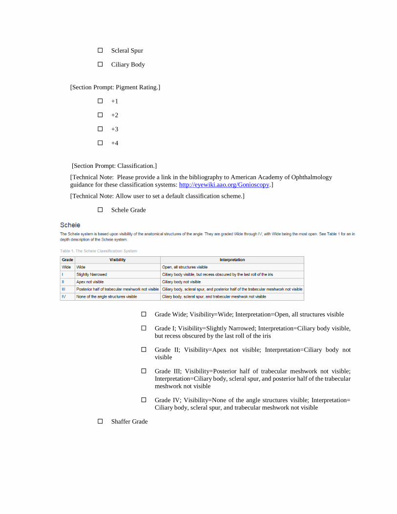
Scleral Spur
Ciliary Body
[Section Prompt: Pigment Rating.]
+1
+2
+3
+4
[Section Prompt: Classification.]
[Technical Note: Please provide a link in the bibliography to American Academy of Ophthalmology
guidance for these classification systems: http://eyewiki.aao.org/Gonioscopy.]
[Technical Note: Allow user to set a default classification scheme.]
Schele Grade
Grade Wide; Visibility=Wide; Interpretation=Open, all structures visible
Grade I; Visibility=Slightly Narrowed; Interpretation=Ciliary body visible,
but recess obscured by the last roll of the iris
Grade II; Visibility=Apex not visible; Interpretation=Ciliary body not
visible
Grade III; Visibility=Posterior half of trabecular meshwork not visible;
Interpretation=Ciliary body, scleral spur, and posterior half of the trabecular
meshwork not visible
Grade IV; Visibility=None of the angle structures visible; Interpretation=
Ciliary body, scleral spur, and trabecular meshwork not visible
Shaffer Grade
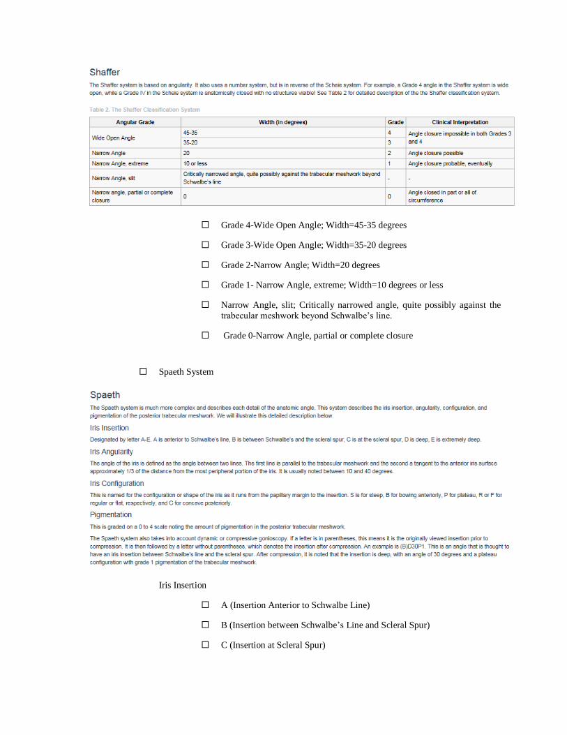
Grade 4-Wide Open Angle; Width=45-35 degrees
Grade 3-Wide Open Angle; Width=35-20 degrees
Grade 2-Narrow Angle; Width=20 degrees
Grade 1- Narrow Angle, extreme; Width=10 degrees or less
Narrow Angle, slit; Critically narrowed angle, quite possibly against the
trabecular meshwork beyond Schwalbe’s line.
Grade 0-Narrow Angle, partial or complete closure
Spaeth System
Iris Insertion
A (Insertion Anterior to Schwalbe Line)
B (Insertion between Schwalbe’s Line and Scleral Spur)
C (Insertion at Scleral Spur)

D (Deep Insertion)
E (Extremely Deep Insertion)
<obtain> Iris Angularity (Degrees)
Iris Angularity (10-40 Degrees)
<obtain> Degrees
Iris Configuration
S (Steep)
B (Anterior Bowing)
P (Plateau)
R (Regular)
F (Flat)
C (Posteriorly Concave)
<obtain> Pigmentation Grade (0 to 4)
Pigmentation
<obtain> Pigmentation Grade
[Technical Note: Scale choices 0, 1+, 2+, 3+, 4+)
Left Eye
[Section Prompt: Right Eye General Findings.]
Open
<obtain> Description
Occludable
<obtain> Description
Recessed
<obtain> Description
Closed
<obtain> Description
<obtain> Additional Findings
[Section Prompt: Posterior Structure.]

Schwalbe Line
Trabecular Meshwork
Scleral Spur
Ciliary Body
[Section Prompt: Pigment Rating.]
+1
+2
+3
+4
[Section Prompt: Classification.]
[Technical Note: Allow user to set a default classification scheme.]
Scheie Grade
Wide (Open Angle; All Structures Visible)
I (Slightly Narrowed Angle; Ciliary Body Visible, Recess Obscured)
II (Angle Apex Not Visible; Ciliary Body Not Visible)
III (Ciliary Body, Scleral Spur, and Posterior Aspect of Trabecular
Meshwork Not Visible)
IV (No Visible Angle Structures)
Shaffer Grade
4 (Wide, Open Angle of 35 to 40 Degrees)
3 (Wide, Open Angle of 20 to 35 Degrees)
2 (Narrowed Angle of 20 Degrees)
1 (Extremely Narrowed Angle of 10 Degrees or Less)
0 (Critically Narrowed Angle with Closure in All or Part of Circumference)
Spaeth System
Iris Insertion
A (Insertion Anterior to Schwalbe Line)
B (Insertion between Schwalbe Line and Scleral Spur)
C (Insertion at Scleral Spur)

D (Deep Insertion)
E (Extremely Deep Insertion)
<obtain> Iris Angularity (Degrees)
Iris Angularity (10-40 Degrees)
<obtain> Degrees
Iris Configuration
S (Steep)
B (Anterior Bowing)
P (Plateau)
F (Flat)
C (Posteriorly Concave)
<obtain> Pigmentation Grade (0 to 4)
Pigmentation
<obtain> Pigmentation Grade
[Technical Note: Scale choices 0, 1+, 2+, 3+, 4+)
[Technical Note: Allow user to repeat Gonioscopy Section by selecting “Yes” for Additional Gonioscopy.]
[Section Prompt: Additional Gonioscopy?]
Yes
No
<obtain> Gonioscopy Notes
[End Gonioscopy.]
Section 2.12.11. - Intraocular Lens (IOL) Calculations
[Begin IOL Calculations.]
[Section Prompt: IOL Calculation method]
Holladay I
Holladay II
Hoffer Q
Haigis

SRK/T
Olsen
Barrett II
Other
<obtain> Description
[Section Prompt: Right Eye Findings.]
[Technical Note; This needs to be presented as a table for each lens model, with a range of lens powers and
associated predicted postoperative refractive error for each lens power.]
[Technical Notes: Users need to be able to define multiple preferred default settings for IOL calculation method and
lens model].
<obtain>Lens Model
<obtain>Lens Power
<obtain> Predicted Postoperative Refractive Error
[Section Prompt: Left Eye Findings.]
<obtain>Lens Model
<obtain>Lens Power
<obtain> Predicted Postoperative Refractive Error
[End IOL Calculations.]
Section 2.12.12. - Keratometry
[Begin Keratometry.]
[Technical Note: Provide link to Keratometry History for returning patients.]
[Technical Note: Link to Keratometry History]
[Technical Note: Allow user to import images and reports from Keratometry devices.]
[Technical Note: Allow user to repeat Keratometry section from here down as many times as needed by selecting
Yes for Additional Keratometry below.]
[Section Prompt: Keratometry]
[Section Prompt: Right Eye]
<obtain> Keratometry 1 (Diopters)
<obtain> Keratometry 1 (Millimeters)
<obtain> Keratometry 2 (Diopters)
<obtain> Keratometry 2 (Millimeters)
<obtain> Axis (Degrees)
[Section Prompt: Left Eye]
<obtain> Keratometry 1 (Diopters)
<obtain> Keratometry 1 (Millimeters)

<obtain> Keratometry 2 (Diopters)
<obtain> Keratometry 2 (Millimeters
[Allow user to repeat Keratometry Section by selecting Yes for Additional Keratometry.]
[Section Prompt: Additional Keratometry?]
Yes
<obtain> Keratometry Notes
[End Keratometry.]
Section 2.12.13. - Optic Nerve Analysis
[Begin Optic Nerve Analysis.]
[Technical Note: Provide link to Optic Nerve Analysis History for returning patients.]
[Technical Note: Link to Optic Nerve Analysis History]
[Technical Note: Allow importation of results from Optic Nerve Analysis.]
[Section Prompt: Type of Optic Nerve Analysis]
Heidelberg Retinal Tomography
GDx Nerve Fiber Analysis
Optical Coherence Tomography
[Section Prompt: Right Eye.]
[Section Prompt: Retinal Nerve Fiber Layer Thickness
<obtain> Inferior (Microns)
<obtain> Superior (Microns)
<obtain> Nasal (Microns)
<obtain> Temporal (Microns)
<obtain> Average Peripapillary Thickness (Microns)
<obtain> Additional Details
[Section Prompt: Left Eye.]
[Section Prompt: Retinal Nerve Fiber Layer Thickness
<obtain> Inferior (Microns)
<obtain> Superior (Microns)
<obtain> Nasal (Microns)
<obtain> Temporal (Microns)
<obtain> Average Peripapillary Thickness (Microns)
<obtain> Additional Details
<obtain> Retinal Nerve Fiber Layer Symmetry
<obtain> Optic Nerve Analysis Notes
[End Optic Nerve Analysis.]

Section 2.12.14. - Pachymetry
[Begin Pachymetry.]
[Technical Note: Provide link to Pachymetry History for returning patients.]
[Technical Note: Link to Pachymetry History]
[Section Prompt: Pachymetry Findings.]
<obtain> Right Corneal Thickness (Micrometers)
<obtain> Left Corneal Thickness (Micrometers)
<obtain> Pachymetry Notes
[End Pachymetry.]
Section 2.12.15. - Perimetry
[Begin Perimetry.]
[Technical Note: Provide link to Perimetry History for returning patients.]
[Technical Note: Link to Perimetry History]
[Section Prompt: Perimetry]
[Section Prompt: Right Eye.]
[Section Prompt: Machine Used.]
Humphrey
Octopus
Goldmann
Other
<obtain> Description
[Section Prompt: Testing Strategy.]
10-2
24-2
30-2
SITA Standard
SITA Fast
Other
<obtain> Description
[Section Prompt: Reliability.]
Good
Fair
Poor

<obtain> Description
<obtain> Pupil Size (Millimeters)
[Section Prompt: Findings.]
Unremarkable
Abnormal Findings
<obtain> Description
[Technical Note: Allow importation of images.]
[Technical Note: Link to Import Images.]
[Section Prompt: Left Eye.]
[Section Prompt: Machine Used.]
Humphrey
Octopus
Goldmann
Other
<obtain> Description
[Section Prompt: Testing Strategy.]
10-2
24-2
30-2
SITA Standard
SITA Fast
Other
<obtain> Description
[Section Prompt: Reliability.]
Good
Fair
Poor
<obtain> Description
<obtain> Pupil Size (Millimeters)
[Section Prompt: Findings.]
Unremarkable
Abnormal Findings

<obtain> Description
[Technical Note: Allow importation of images.]
[Technical Note: Link to Import Images.]
[End Perimetry.]
Section 2.12.16. - Retinal Imaging
[Begin Retinal Imaging.]
[Technical Note: Provide link to Retinal Imaging History for returning patients.]
[Technical Note: If Retinal Imaging selected, allow importation of images and results.]
<obtain> Device Name
[Technical Note: Allow user to set a default device name. Prefill “Device Name” with default name and allow edits.]
[Section Prompt: Type of Imaging.]
Retinal Photography
Fluorescein Angiography
Tomography
Other Retinal Imaging
<obtain> Additional Details
[End Retinal Imaging.]
[End Special Testing.]
Section 2.13 - Procedure Notes [Begin Procedure Notes.]
[Section Prompt: Procedures]
<obtain> Name of Procedure
<obtain> Procedure CPT Code
<obtain> Diagnosis
Right Eye
Left Eye
Both Eyes
[Section Prompt: Document the “Time-Out” process per policy.]
Correct Patient (Two Patient Identifiers)
Correct Procedure
Correct Site
Procedure Confirmed with Patient
Site Marked
<obtain> Pretreatment
[Section Prompt: Injection(s)]
<obtain> Medication
<obtain> Lot Number
<obtain> Inventory Number
<obtain> Dosage
[Section Prompt: Laser(s).]
<obtain> Laser Type
<obtain> Spot Size
<obtain> Power
<obtain> Duration [Technical note: This is duration for Each Spot]
<obtain> Number of Spots
<obtain> Procedure Notes
[End Procedure Notes.]
Section 2.14 - Assessment and Plan [Begin Assessment and Plan.]
Section 2.14.1. - Assessment
[Begin Assessment.]
[Technical Note: For returning patients, propose assessment entries from the most recent Eye Care KNART
Assessment section for selection. For all users, propose items for confirmation based on entries in the Past Ocular
History and findings from the Ocular Exam and Special Testing sections of this documentation template. Allow user
to edit and comment on items. Also allow user to insert as many free text boxes as needed. For patients with
glaucoma, include editable entries for target intraocular pressures as seen in the Past Ocular History.]
[Technical Note: Each entry in the assessment pulls up the plan so that the user can document the part of the plan
that is relevant to that entry.]
[End Assessment.]
Section 2.14.2. - Plan
[Begin Plan.]
[Clinical Comment: Future development of the Eye Care Order Set KNART will result in the order set being
activated from the plan section of the documentation template.]
Section 2.14.2.1. - Vision Correction Prescription Glasses
[Begin Vision Correction Prescription Glasses.]
[Technical Note: Populate fields with results from Refraction section of Ocular Exam above. Allow user to edit
fields.]
[Section Prompt: Prescription for glasses- Right Eye.]
<obtain> Sphere (Diopters)
<obtain> Cylinder (Diopters)

<obtain> Axis (Degrees)
<obtain> Add Power # 1 (Diopters)
<obtain> Add Power # 2 (Diopters)
<obtain> Prism Power (Diopters)
Prism Base
Up
Down
In
Out
[Section Prompt: Prescription for glasses- Left Eye
<obtain> Sphere (Diopters)
<obtain> Cylinder (Diopters)
<obtain> Axis (Degrees)
<obtain> Add Power # 1 (Diopters)
<obtain> Add Power # 2 (Diopters)
<obtain> Prism Power (Diopters)
Prism Base
Up
Down
In
Out
<obtain> Additional Information
[Technical Note: Make Glasses Prescription section exportable.]
[End Vision Correction Prescription Glasses.]
Section 2.14.2.2. - Vision Correction Prescription – Contact Lenses
[Begin Vision Correction Prescription – Contact Lenses.]
[Section Prompt: Contact lenses- Right Eye.]
<obtain> Power (Diopters)
<obtain> Base Curve (Millimeters)
<obtain> Diameter (Millimeters)
<obtain> Brand
<obtain> Color
<obtain> Add Power (Diopters)
<obtain> Cylinder (Diopters)
<obtain> Axis (Degrees)

[Section Prompt: Contact lenses- Left Eye.]
<obtain> Power (Diopters)
<obtain> Base Curve (Millimeters)
<obtain> Diameter (Millimeters)
<obtain> Brand
<obtain> Color
<obtain> Add Power (Diopters)
<obtain> Cylinder (Diopters)
<obtain> Axis (Degrees)
<obtain> Additional Information
[End Vision Correction Prescription – Contact Lenses.]
Section 2.14.2.3. - Eye Prosthetic Devices
[Begin Eye Prosthetic Devices.]
[Section Prompt: Eye Prosthetic Devices]
<obtain> Description
[End Eye Prosthetic Devices.]
Section 2.14.2.4. - Laboratory Tests
[Begin Laboratory Tests.]
Complete Blood Count
Complete Blood Count with Differential
Basic Metabolic Panel
Chem-12
Blood Urea Nitrogen
Creatinine
Fasting Glucose
Hemoglobin A1c
Fluorescent Treponemal Antibody Absorption
Angiotensin Converting Enzyme Level
Antinuclear Antibody
HLA-B27 Typing
Lyme Serologic Testing
Lipid Panel

Erythrocyte Sedimentation Rate
C-Reactive Protein
Vitamin B12
Folate
Thiamine
Other
<obtain> Other Lab Tests
[End Laboratory Tests.]
Section 2.14.2.5. - Imaging Studies
[Begin Imaging Studies.]
X-Ray Chest Posterior-Anterior and Lateral
CT Head with contrast
CT Orbits with Contrast
[Technical Note: Present the following warning if CT Head or Orbits with Contrast is selected for a patient
with a documented contrast allergy: “Patient has a documented contrast allergy.” If available, provide link to
an order set KNART for contrast allergy.]
CT Head without Contrast
CT Orbits without Contrast
MRI Head with Contrast
MRI Orbits with Contrast
[Technical Note: Present the following warning if MRI Head or Orbits with Contrast is selected for a patient
with a documented contrast allergy: “Patient has a documented contrast allergy.” If available, provide link to
an order set KNART for contrast allergy.
MRI Head without Contrast
MRI Orbits without Contrast
Carotid Doppler
Sleep Study
Other
<obtain> Other Imaging Studies
[End Imaging Studies.]
Section 2.14.2.6. - Eye Clinic Ancillary Study and Procedure Orders
[Begin Eye Clinic Ancillary Study and Procedure Orders.]
Amsler Grid

Refraction
Keratometry
Scanning Laser Polarimetry
Electrooculography
Refractive Surgery Measurements and Calculations
Electroretinography
Pachymetry
Intraocular Pressure Measurement
Intraocular Lens Calculations
Optical Coherence Tomography
Angiography
Angle
Anterior Segment
Cornea
Ganglion Cell Analysis
Macular Change Analysis
High Definition
Macular Cube
Macular Thickness Analysis
Optic Nerve and Retinal Nerve Fiber Layer Analysis
Progression Analysis
Single Eye Summary
A Scan
B Scan
Confocal Microscopy
Corneal Topography
External Eye Photography
<obtain> Description
Slit Lamp Photography
<obtain> Description
Fundus Photography
<obtain> Description
Fluorescein Angiography
ICG - Indocyanine Green Angiography
Other
<obtain> Description
[End Eye Clinic Ancillary Study and Procedure Orders.]
Section 2.14.2.7. - Medications
[Begin Medications.]
[Section Prompt: Current Medications.]
<obtain> Current Medication list.]
[Section Prompt: Current Eye Medications]
<obtain> Current Eye Medication list.]
[Technical Note: Dynamically provide a link to the appropriate formulary based on the individual clinical site.]
[Technical Note: The Medications text field should behave as a search field in which suggestions are made based on
the appropriate site-specific formulary.]
[Technical Note: Future Implementation KNART that links directly to pharmacy EHR system for all medications
and then this connection will print selected medications back into the Eye Care plan.]
[Technical Note: The user should have the ability to either print or electronically transmit a prescription, either on-
formulary or off-formulary.]
[Selection Behavior: One required for each medication selected.]
Continue
Discontinue
Increase
Decrease
Add
[Section Prompt: Medication.]
Acetazolamide
<obtain> Detail
Acyclovir
<obtain> Detail
Aflibercept (Restricted to Ophthalmology)
Both Eyes
Right eye

Left Eye
<obtain> Detail
Artificial Tears
1 drop every 2 hours as needed
<obtain> Detail
Atropine Sulfate 1% Ophthalmic Solution
<obtain> Detail
Bacitracin/Polymyxin Ophthalmic Ointment
3.5 gram
<obtain> Detail
Betaxolol Hydrochloride 0.25% Ophthalmic Suspension (Restricted to Eye Clinic)
<text> Detail
Bevacizumab Intravitreal Injection (Restricted to Ophthalmology)
Right eye
Left eye
Both eyes
<obtain> Detail
Brimonidine 0.2% Ophthalmic Solution
<obtain> Detail
Brimonidine 0.2%/Brinzolamide Ophthalmic Solution
<obtain> Detail
Carboxymethylcellulose 1% Ophthalmic Gel
<obtain> Detail
Carboxymethylcellulose 0.5% Ophthalmic Solution
<obtain> Detail
Cromolyn 4% Ophthalmic Solution
<obtain> Detail
Cyclosporine 0.05% Ophthalmic Emulsion (Restricted to Eye Clinic)
<obtain> Detail
Diclofenac Sodium 0.1% Ophthalmic Solution (Restricted to Ophthalmology)

<obtain> Detail
Dorzolamide Hydrochloride 2% Ophthalmic Solution (Restricted to Eye Clinic)
<obtain> Detail
Dorzolamide Hydrochloride 2%/Timolol 0.5% Ophthalmic Solution (Restricted to Eye Clinic)
<obtain> Detail
Doxycycline
<obtain> Detail
Erythromycin 0.5% Ophthalmic Ointment
<obtain> Detail
Fluorometholone 0.1% Ophthalmic Suspension (Restricted to Ophthalmology)
<obtain> Detail
Ketorolac 0.025% Ophthalmic
<obtain> Detail
Ketorolac Tromethamine 0.5% Ophthalmic Solution
<obtain> Detail
Latanoprost 0.005% Ophthalmic Solution
<obtain> Detail
Levobunolol Hydrochloride 0.25% Ophthalmic Solution (Restricted to Eye Clinic)
<obtain> Detail
Lubricating Ophthalmic Ointment
<obtain> Detail
Methazolamide
<obtain> Detail
Mitomycin (Restricted to Ophthalmology)
<obtain> Detail
Multivitamin/Ophthalmologic AREDS2/Lutein/Zeaxanthin
2 times daily
Other
<obtain> Detail
Naphazoline/Pheniramine Ophthalmic Solution

<obtain> Detail
Ofloxacin 0.3% Ophthalmic Suspension (Restricted to Ophthalmology)
<obtain> Other
Pilocarpine Hydrochloride 1% Ophthalmic Solution
<obtain> Detail
Pilocarpine Hydrochloride 2% Ophthalmic Solution
<obtain> Detail
Prednisolone Acetate 1% Ophthalmic Suspension (Restricted to Ophthalmology)
<obtain> Detail
Ranibizumab Injection (Restricted to Ophthalmology)
Both eyes
Right eye
Left eye
<obtain> Detail
Sodium Chloride Ophthalmic Solution
<obtain> Detail
Timolol Maleate 0.25% Ophthalmic Solution
<obtain> Detail
Timolol Maleate 0.5% Ophthalmic Solution
<obtain> Detail
Travoprost Z Ophthalmic Solution (Restricted; Submit National Formulary Request)
<obtain> Detail
Other
<obtain> Additional Details
[End Medications.]
Section 2.14.2.8. - Patient Education and Instructions
[Begin Patient Education and Instructions.]
<obtain> Patient Education and Instructions
[Technical Note: Link to Site-Specific Patient Educational Materials.]
[Technical Note: Include links to printable illustrations that can be provided to the patient. Creating and storing
annotated drawings templates electronically is a future implementation concern.]
[Technical Note: Link to Printable Illustration of External Eyes.]
[Technical Note: Link to Printable Illustration of Cornea.]
[Technical Note: Link to Printable Illustration of Anterior Segment.]
[Technical Note: Link to Printable Illustration of Lens.]
[Technical Note: Link to Printable Illustration of Retinas.]
[Technical Note: Link to Printable Illustration of Optic Nerves.]
[Technical Note: Link to Printable Illustration of Macula.]
[Technical Note: Link to Printable Illustration of Gonioscopy.]
[End Patient Education and Instruction.]
[End Plan.]
Section 2.14.3. - Consultation Requests
[Begin Consultation Requests.]
<obtain> Consultation Requests
[End Consultation Requests.]
Section 2.14.4. - Surgery Requests
[Begin Surgery Requests.]
<obtain> Surgery Requests
[End Surgery Requests.]
Section 2.14.5. - Return for Follow-Up
[Begin Return for Follow-Up.]
[Section Prompt: Follow up location.]
[Technical Note: Allow user to add and remove locations in this list.]
Retina Clinic
Tele-Retina Clinic
Other
<obtain> Description
[Section Prompt: Follow up timing]
<obtain> number of time increments
Days
Weeks
Months
Years
[Section Prompt: Dilation Required Next Visit.]
Yes

No
[End Return for Follow-Up.]
[End Assessment and Plan.]
[End Eye Care Documentation Template.]

Bibliography/Evidence American Academy of Ophthalmology Preferred Practice Pattern Cataract/Anterior Segment Panel. Preferred
Practice Pattern Guidelines. Cataract in the Adult Eye. American Academy of Ophthalmology Preferred Practice
Pattern Guidelines website. https://www.aao.org/preferred-practice-pattern/cataract-in-adult-eye-ppp-2016. Updated
October 2016.
American Academy of Ophthalmology Preferred Practice Pattern Committee. Preferred Practice Pattern Guidelines.
Comprehensive Adult Medical Eye Evaluation. American Academy of Ophthalmology Preferred Practice Pattern
Guidelines website. https://www.aao.org/preferred-practice-pattern/comprehensive-adult-medical-eye-evaluation-
2015. Updated November 2015.
American Academy of Ophthalmology Preferred Practice Pattern Glaucoma Panel. Preferred Practice Pattern
Guidelines. Primary Open-Angle Glaucoma. American Academy of Ophthalmology Preferred Practice Pattern
Guidelines website. https://www.aao.org/preferred-practice-pattern/primary-open-angle-glaucoma-ppp-2015.
Updated November 2015.
American Academy of Ophthalmology Preferred Practice Pattern Glaucoma Panel. Preferred Practice Pattern
Guidelines. Primary Open-Angle Glaucoma Suspect. American Academy of Ophthalmology Preferred Practice
Pattern Guidelines website. https://www.aao.org/preferred-practice-pattern/primary-open-angle-glaucoma-suspect-
ppp-2015. Updated November 2015.
American Academy of Ophthalmology Retina/Vitreous Panel. Preferred Practice Pattern Guidelines. Age-Related
Macular Degeneration. American Academy of Ophthalmology Preferred Practice Pattern Guidelines website.
https://www.aao.org/preferred-practice-pattern/age-related-macular-degeneration-ppp-2015. Updated January 2015.
American Academy of Ophthalmology Retina/Vitreous Panel. Preferred Practice Pattern Guidelines. Diabetic
Retinopathy. American Academy of Ophthalmology Preferred Practice Pattern Guidelines website.
https://www.aao.org/preferred-practice-pattern/diabetic-retinopathy-ppp-updated-2016. Updated February 2016.
Bailey IL. Visual acuity. In: Benjamin WJ, ed. Borish’s Clinical Refraction. 2nd edition. St. Louis, MO:
Butterworth-Heinemann, 2006.
Digital Imaging and Communications in Medicine. DICOM Standards Committee. RR.2 Reference Tables For
Equivalent Visual Acuity Notations. DICOM PS3.17 2017e webpage.
http://dicom.nema.org/medical/dicom/current/output/pdf/part17.pdf. Updated November 2017.
Eleu Pacific Partners. Reusable Data Elements: Research Initiative for Optimal Vision Care Prototype Deliverable
10. Updated March 2015.
Eleu Pacific Partners. Storyboards and Mockups: Research Initiative for Optimal Vision Care Prototype Deliverable
3. Updated July 2015.
Eye Care SMARTForm. Eye Care Wireframes 031915.pdf. March 19, 2015. (Veterans Administration, provided
documentation from Linda Wedemeyer, MD, U.S. Veterans Administration).
Holladay JT. Visual acuity measurements. J Cataract Refract Surg. 2004;30(2):287-290.
Lynch MG, business owner; U.S. Department of Veterans Affairs VA Clinical Capabilities Management Board.
Program name: integrated electronic health record and registry for eye care. January 17, 2017. Business Owner
Presentation Integrated Electronic Health Record Eye Care_FY19-23 IT MYP CCMB.PPTX (U.S. Department of
Veterans Affairs, provided documentation from Mary Lynch, MD, U.S. Department of Veterans Affairs).
Lynch MG, business owner; U.S. Department of Veterans Affairs. VA FORM 10-150058(WS) – class III NSR
initial assessment questionnaire: 20160323 Medflow electronic health record for eye care. 20160323 Medflow EHR
for Eye Care NSR.PDF (U.S. Department of Veterans Affairs, provided documentation from Mary Lynch, MD, U.S.
Department of Veterans Affairs).
Lynch MG, Varga J, Skarda D, Maa A; Atlanta VA Medical Center. Medflow/DSS electronic health record for eye
care in the VA. Poster presented at: Healthcare Information and Management Systems Society (HIMSS) 2012
Annual Meeting; February 20-24, 2012; Las Vegas, NV. HIMSS Poster Medflow Feb 2012.pdf (U.S. Department of
Veterans Affairs, provided documentation from Mary Lynch, MD, U.S. Department of Veterans Affairs).
U.S. Department of Veterans Affairs. Optimal Vision Care Prototype (OVCP) Functional Requirements. Updated
April 2015.

Appendix A - Existing Sample VA Artifacts
OVCP Functional Requirements April 15, 2015
The authorized user shall have the capability to document ocular care using various approved DoD devices /
methods (ID: 178)
Link: https://vaww.infoshare.va.gov/sites/chio/KBS/cdsknart2017/Shared%20Documents/KNART%20Domain-
Eye%20Care/Clinical%20Content%20(VA%20and%20non-VA)/non-
VA%20Clinical%20Content/OVCP/Ocular_Clinical_Workflow_Report_Del1_v8_31Mar2015_Final%20Artifact.pd
f
File Name: Eye Care Wireframes 031915 Artifact
Link: https://vaww.infoshare.va.gov/sites/chio/KBS/cdsknart2017/Shared%20Documents/KNART%20Domain-
Eye%20Care/Clinical%20Content%20(VA%20and%20non-VA)/non-
VA%20Clinical%20Content/OVCP/Eye%20Care%20Wireframes%20031915%20Artifact.pdf
File Name: Ocular_Data_Map_Rpt_Del7_v7_30Apr2015_Final Artifact
Link: https://vaww.infoshare.va.gov/sites/chio/KBS/cdsknart2017/Shared%20Documents/KNART%20Domain-
Eye%20Care/Clinical%20Content%20(VA%20and%20non-VA)/non-
VA%20Clinical%20Content/OVCP/Ocular_Data_Map_Rpt_Del7_v7_30Apr2015_Final%20Artifact.pdf
File Name: Opioid_TSWF COT CPG Paper Backup Jan Apr 2017 Artifact
Link: https://vaww.infoshare.va.gov/sites/chio/KBS/cdsknart2017/Shared%20Documents/KNART%20Domain-
Eye%20Care/Clinical%20Content%20(VA%20and%20non-VA)/non-
VA%20Clinical%20Content/OVCP/Opioid_TSWF%20COT%20CPG%20Paper%20Backup%20Jan%20Apr%2020
17%20Artifact.pdf
Figure 1. Motility and Alignment Document Tool

Appendix B: Basic Laboratory Panel Definition
• Blood urea nitrogen
• Calcium
• Chloride
• CO2 (Carbon dioxide, bicarbonate)
• Creatinine
• Glucose
• Potassium
• Sodium

Appendix C: Acronyms/Abbreviations AMD Age-Related Macular Degeneration
AREDS Age-Related Eye Disease Study
CCWP Clinical Content White Paper
CDS Clinical Decision Support
CO2 Carbon Dioxide
CPT Current Procedural Terminology
CT Computed Tomography
DICOM Digital Imaging and Communications in Medicine
DSAEK Descemet Stripping Automated Endothelial Keratoplasty
DSEK Descemet’s Stripping Endothelial Keratoplasty
EHR Electronic Health Record
ENDS Electronic Nicotine Delivery Systems
ETDRS Early Treatment of Diabetic Retinopathy Study
HIV Human Immunodeficiency Virus
HLA Human Leukocyte Antigens
HL7 Health Level 7
ICD Internal Classification of Diseases
ICG Indocyanine Green
IOL Intraocular Lens
KBS Knowledge Based Systems
KNART Knowledge Artifact
LASIK Laser-Assisted in Situ Keratomileusis
LRI Limbal Relaxing Incisions
MRI Magnetic Resonance Imaging
NAG Narrow-Angle Glaucoma
OAG Open-Angle Glaucoma
OIIG Office of Informatics and Information Governance
OVCP Optimal Vision Care Prototype

PRK Photorefractive Keratectomy
RAPD Relative Afferent Pupillary Defect
RPC Remote Procedure Call
SME Subject Matter Expert
SNOMED CT Systematized Nomenclature of Medicine Clinical Terms
TIA Transient Ischemic Attack
TO Task Order
VA Department of Veteran Affairs
VAMC VA Medical Center
YAG Yttrium Aluminum Garnet


