Characteristics of acute inflammation (cellular events, chemical mediators, systemic effects) Morphologic patterns of acute inflammation according to the exudate Chronic inflammation Granulomatous inflammation Tissue repair and wound healing

Chronic inflammation is a response of prolonged duration (weeks or months) in which inflammation, tissue injury, and attempts at repair coexist, in varying combinations.
Chronic inflammation
Causes of Chronic Inflammation -Persistent infections -Hypersensitivity diseases-autoimmun diseases -Prolonged exposure to potentially toxic agents, either exogenous or endogenous -The role of inflammation in neurodegenerative diseases,metabolic syndrome and the associated type 2 diabetes, and certain cancers -Foreign bodies
Cellular players: macrophages, lymphocytes, plasma cells, eosinophyls,mast cells
Morphologic Features
Infiltration with mononuclear cells
Tissue destruction
Attempts at healing
Non- exudative

1. Macrophages
The dominant cells in most chronic inflammatory reactions
Circulating cells of this lineage are known as monocytes .
To kill ingested organisms, and secrete cytokines that stimulate inflammation.
-Classical macrophage activation-
-Alternative macrophage activation-
Tissue repair: They secrete growth factors that promote angiogenesis, activate fibroblasts, and stimulate collagen synthesis.
2.Lymphocytes
As the mediators of adaptive immunity, provides defense against infectious pathogens These cells are often present in chronic inflammation and when they are activated, the inflammation tends to be persistent and severe. 3.Eosinophils In immune reactions mediated by IgE and in parasitic infections
4.Mast cells
In both acute and chronic infl ammatory reactions
5. Leukocytes „acute on chronic”
Th1 Th2

Fibrosis •Fibroblast activation, collagen production: TGF-ß, PDGF, FGF •Angiogenesis: VEGF (vascular endothelial growth factor), FGF Cicatrisatio-cicatrix formation
Pleuritis chronica adhaesiva Pericarditis chronica adhesiva Cicatrix myocardii Cirrhosis hepatis
Macrophages
Morfological type
Foamy cytoplasm
Haemosiderin-laden
Epitheloid cells
Giant cells

Giemsa
HE


Granulomatous Inflammation
Granulomatous inflammation is a form of chronic inflammation characterized by the presence of granuloma
epithelioid cells (modified macrophages) compulsary giant cells (fusion of epithelioid cells) –often lymphocytes fibroblasts
CLASSIFICATION INFECTIVE (tuberculosis, lues, leprosy cat-scratch disease) 1.Immune-granulomas NON-INFECTIVE (sarcoidosis, rheumatic fever,PBC, Wegener) 2.Foreign-bodies granulomas (keratin, lipogranuloma, surgical suture,)
OR
a/ necrotising (tbc, lues, rheumatic nodule) b/ non necrotising (sarcoidosis, Crohn disease)
giant cells
Langhans Foreign-body type Touton
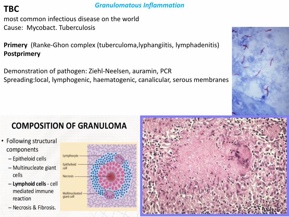
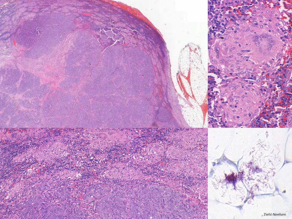
TBC
most common infectious disease on the world Cause: Mycobact. Tuberculosis Primery (Ranke-Ghon complex (tuberculoma,lyphangiitis, lymphadenitis) Postprimery Demonstration of pathogen: Ziehl-Neelsen, auramin, PCR Spreading:local, lymphogenic, haematogenic, canalicular, serous membranes
Granulomatous Inflammation

Syphilis, lues Cause: Treponema pallidum Acquired form Primary: ulcus durum, bubo indolens Secundary: bakteremia, exanthaemas Tertiary: granulomatous – gumma Cardiovasc: aneurysma Neuoro sy: tabes dorsalis, paralysis prgr. Congenital Severe
Pemphigus syphiliticus Hepato-splenomegaly Pneumonia alba Dubois-abscess
Mild form: Hutchinson's triad -blunted upper incisor teeth known as Hutchinson's teeth -inflammation of the cornea known as interstitial keratitis -deafness from auditory nerve disease
Granulomatous Inflammation

Rheumatic fever
+vegetations
Granulomatous Inflammation

Non-caseating, non-infective granulomas
Giant cells with: - Asteroid body - Schaumann ~s body
Granulomatous Inflammation

Foreign body granuloma
Lipogranuloma Keratin-pearl granuloma
Granulomatous Inflammation
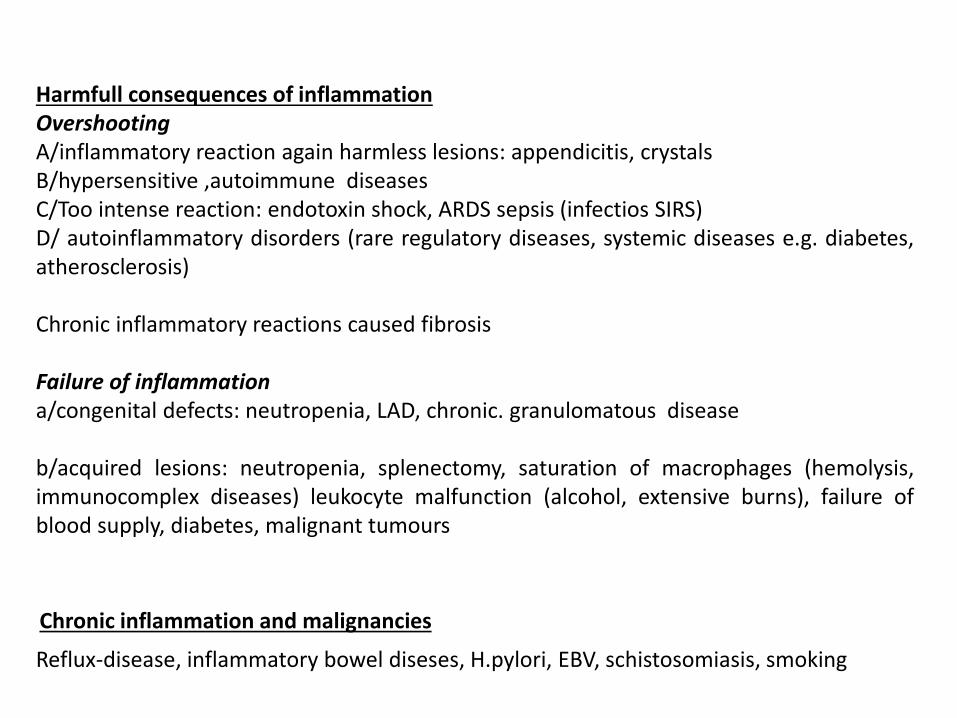
Harmfull consequences of inflammation Overshooting A/inflammatory reaction again harmless lesions: appendicitis, crystals B/hypersensitive ,autoimmune diseases C/Too intense reaction: endotoxin shock, ARDS sepsis (infectios SIRS) D/ autoinflammatory disorders (rare regulatory diseases, systemic diseases e.g. diabetes, atherosclerosis) Chronic inflammatory reactions caused fibrosis Failure of inflammation a/congenital defects: neutropenia, LAD, chronic. granulomatous disease b/acquired lesions: neutropenia, splenectomy, saturation of macrophages (hemolysis, immunocomplex diseases) leukocyte malfunction (alcohol, extensive burns), failure of blood supply, diabetes, malignant tumours
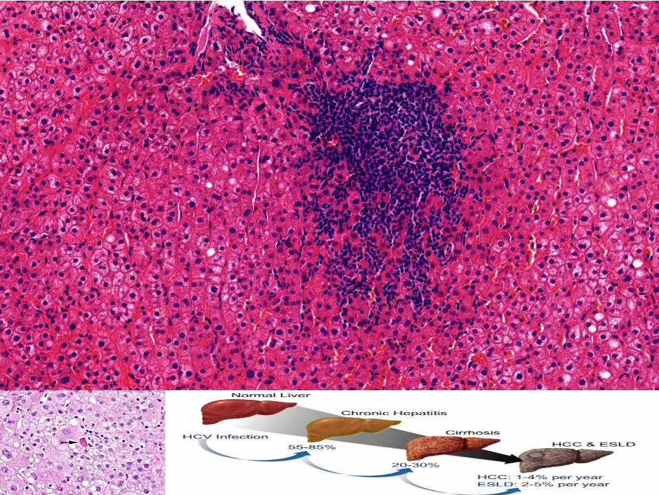
Chronic inflammation and malignancies
Reflux-disease, inflammatory bowel diseses, H.pylori, EBV, schistosomiasis, smoking


TISSUE REPAIR
Repair of damaged tissues occurs
regeneration : proliferation of residual (uninjured)
cells
healing : the deposition of connective tissue to
form a scar

We rank cells according to their ability to regenerate:
LABILE CELLS ("continuous replicators") are constantly replenishing their neighbors that have died or been shed. Examples include the epithelium of skin, mucous membranes, oviducts, ducts; urothelium; endometrium; seminiferous tubules; bone marrow; lymphoid tissue.
STABLE CELLS ("discontinuous replicators") can proliferate rapidly in response to need, especially when required to replace lost neighbors. These include all glandular parenchymal cells, as well as fibroblasts, endothelial cells (cuboidal, and called "angioblasts", when they are healing), and osteoblasts.
PERMANENT CELLS ("non-replicators") have very limited ability to undergo mitosis or be replenished after birth. These cells include glia, neurons, and cardiac (non-failing heart).

Tissue regeneration can occur in parenchymal organs whose cells are capable of proliferation, but with the exception of the liver, this is usually a limited process.
Liver Regeneration
From hepatocyte- after partial hepatectomy, or pvl From stem cells- when the hepatocyte are unable to divide -Oval cells, intermediary hepatobiliary cells
Mechanisms of Tissue Regeneration
In epithelia of the intestinal tract and skin, injured cells are rapidly replaced by proliferation of residual cells
Liver regeneration from stem cells
The size of the lobules in control liver and after regeneration

Control liver After regeneration PV - CV PV - CV
Cyp450 Cyp450
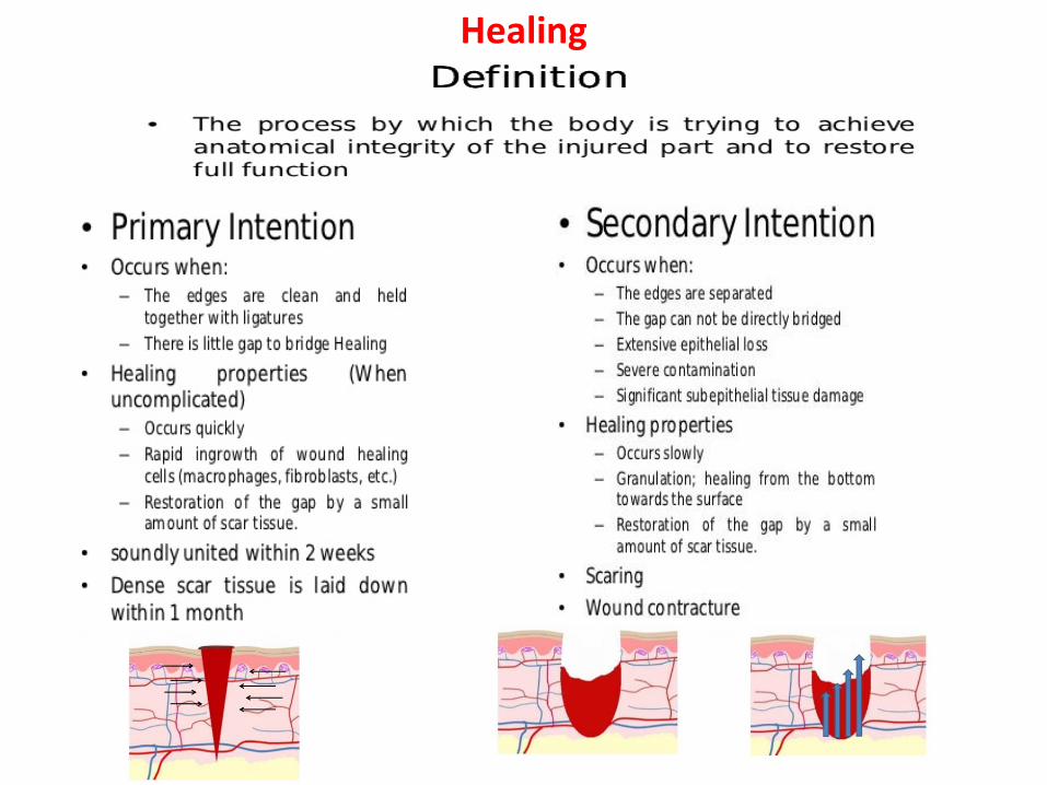
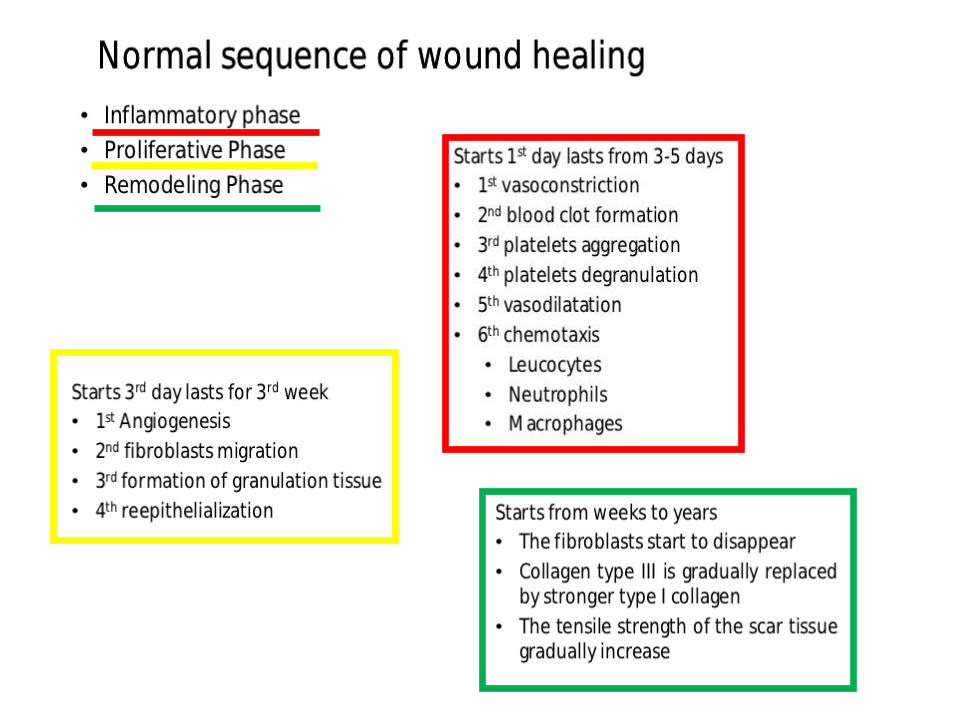
Healing


Steps in Scar Formation

Ten
sil s
tre
ngh
t
Intact skin
Intact bowel
Anastomosis
Wound
days

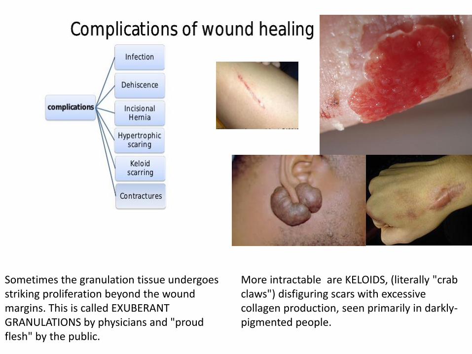
Sometimes the granulation tissue undergoes striking proliferation beyond the wound margins. This is called EXUBERANT GRANULATIONS by physicians and "proud flesh" by the public.
More intractable are KELOIDS, (literally "crab claws") disfiguring scars with excessive collagen production, seen primarily in darkly-pigmented people.


