9/18/2012
1
Chapter 29
Advanced Airway Management
2
Learning Objectives
Describe indications for advanced airway management
List complications associated with advanced airway management
Explain rationale for securing endotracheal tube
State consequence of and need to recognize unintentional esophageal intubation
3
Learning Objectives
State consequence of and need to recognize unintentional esophageal intubation
List equipment required for orotracheal intubation
Describe proper use of curved blade for orotracheal intubation
Describe proper use of straight blade for orotracheal intubation
Copyright © 2013 by Jones & Bartlett Learning, LLC, an Ascend Learning Company

9/18/2012
2
4
Learning Objectives
Describe methods of choosing appropriate-size endotracheal tube in an adult patient
State reasons for proper use of stylet in orotracheal intubation
Explain rationale for use of the stylet
Describe skill of orotracheal intubation in the adult patient
5
Learning Objectives
Describe skill of confirming endotracheal tube placement in adult, infant, child
Differentiate airway anatomy in infant, child, adult
State formula for sizing an infant/child endotracheal tube
Define various alternative methods for sizing infant, child endotracheal tube
6
Learning Objectives
Describe skill of orotracheal intubation in infant, child
Describe skill of securing endotracheal tube in adult, infant, child patient
Describe indications, contraindications, technique for insertion of nasogastric tubes
Copyright © 2013 by Jones & Bartlett Learning, LLC, an Ascend Learning Company
9/18/2012
3
7
Introduction
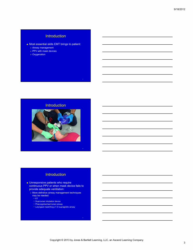
Most essential skills EMT brings to patient: Airway management
PPV with mask devices
Oxygenation
8
Introduction
9
Introduction
Unresponsive patients who require continuous PPV or when mask device fails to provide adequate ventilation: More definitive airway management techniques
may be needed:• ET
• Dual-lumen intubation device
• Pharyngotracheal lumen airway
• Laryngeal mask/King LT-D supraglottic airway
Copyright © 2013 by Jones & Bartlett Learning, LLC, an Ascend Learning Company
9/18/2012
4
10
Introduction
Most systems do not allow EMT to use advanced airway skills Determined by:
• State rules
• System medical director
• Leadership within EMS system
11
Introduction
Factors guiding use of particular intervention: Time, difficulty training providers
Ability to maintain skill proficiency, based on frequency of use
Cost to EMS system
12
Introduction
Advanced and alternative airway devices are only as good as the skill level of the provider who is using them Inappropriate use can be fatal to the patient
Must well trained and well practiced• Preparation
• Practice
• Reinforcement
• Continued evaluation of your competency
Copyright © 2013 by Jones & Bartlett Learning, LLC, an Ascend Learning Company
9/18/2012
5
13
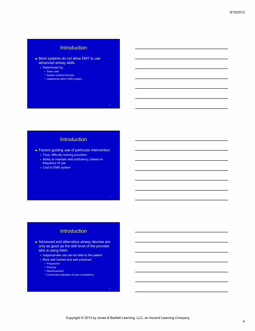
Cricoid Pressure
Developed for use in OR to prevent passive regurgitation of food related to medication and paralysis
Useful for patient without cough/gag reflex to help prevent regurgitation and aspiration during ET intubation
14
Cricoid Pressure
15
Cricoid Pressure
Can be used while patient is being ventilated with bag-mask
Pressure is applied to cricoid cartilage Complete circle of rigid cartilage
Presses esophagus against the spine• Closes esophagus, helps prevent gastric inflation and
regurgitation
Copyright © 2013 by Jones & Bartlett Learning, LLC, an Ascend Learning Company

9/18/2012
6
16
Cricoid Pressure
17
Cricoid Pressure
Cricoid cartilage is inferior to the cricothyroidmembrane To find it, locate thyroid cartilage
Slide finger to the depression just below the cricothyroid membrane
Prominence below cricothyroid membrane is cricothyroid cartilage
18
Cricoid Pressure
During application of cricoid pressure, perform the following measures: Verify correct anatomy to avoid damage to other
structures
Avoid excess pressure in infants and children
Use technique only when sufficient personnel are available
Copyright © 2013 by Jones & Bartlett Learning, LLC, an Ascend Learning Company

9/18/2012
7
19
Endotracheal Intubation
Most effective form of airway management
Properly placed ET tube rests in the trachea and seals against the internal wall with inflated cuff
Seal around the ET tube helps prevent aspiration of liquids and other material into lungs
20
Endotracheal Intubation
Nasotracheal intubation ET tube passed through nose into the trachea
Considered “blind” placement technique• ET tube is placed into the trachea through the nose
without the EMT being able to see where the tube is going
Carries higher risk of complications and injury
21
Endotracheal Intubation
Purpose Orotracheal intubation is the most effective way of
controlling the patient’s airway
EMTs perform orotracheal intubation for patients in respiratory or cardiac arrest
Provides complete control of airway and minimizes risk of aspiration
Copyright © 2013 by Jones & Bartlett Learning, LLC, an Ascend Learning Company

9/18/2012
8
22
Endotracheal Intubation
Permits better O2 delivery, more effective ventilation, and deeper suctioning than other methods
Indications for use: Prolonged PPV required and cannot be achieved
by other methods
Apneic patients
Unresponsive patients who cannot protect their airway
23
Endotracheal Intubation
Advantages of use: Prevents gastric distention
Minimizes aspiration risk
Allows access to lower airway for suctioning
24
Endotracheal Intubation
Complications Esophageal intubation
• Most dangerous is unrecognized intubation of the esophagus
• Tube passes into the esophagus rather than trachea
• Important to confirm proper placement after every attempt at intubation and periodically thereafter
• If tube becomes dislodged and not corrected within minutes: Inadequate ventilation
Severe hypoxia
Gastric inflation
Copyright © 2013 by Jones & Bartlett Learning, LLC, an Ascend Learning Company
9/18/2012
9
25
Endotracheal Intubation
Complications Inadequate ventilation and oxygenation
• Produced by prolonged attempts at intubation without intervening periods of PPV
• Results in hypoxia, hypoxemia
• Attempt no longer than 30 seconds without intervening periods of ventilation with mask device
26
Endotracheal Intubation
Complications Soft tissue trauma
• Lips, teeth, tongue, gums, and airway structure damage can occur if laryngoscope is used forcefully
• Also occurs when top teeth are used for leverage to view the vocal cords
• Laryngoscope must be carefully inserted in the mouth and used gently to lift the jaw and epiglottis, without tilting the blade back over the teeth
27
Endotracheal Intubation
Complications Right main stem bronchus intubation
• If tube is inserted too far, it enters the right main stem bronchus because of its straight angle off the trachea and larger size
• Results in ventilation of only 1 lung
• After intubation, check breath sounds on both sides
• Stop insertion of the tube when the proximal end of the cuffed ET tube passes vocal cords
• Use reference marks on ET tube to assist in estimating location
Copyright © 2013 by Jones & Bartlett Learning, LLC, an Ascend Learning Company
9/18/2012
10
28
Endotracheal Intubation
Complications Vomiting
• Laryngoscope can induce gag reflex in unresponsive patients
• Results in vomiting and possible aspiration of stomach contents into the lungs
• Always have suction ready
29
Endotracheal Intubation
Complications Bradycardia & dysrhythmias
• React to stimulus of intubation because of stimulation of the autonomic nervous system
• Check heart rate periodically
30
Endotracheal Intubation
Complications Tube dislodgement
• Potential hazard during patient movement
• Always reassess and confirm tube position after moving patient
• Never blindly attempt to reposition or reinsert the tube
• If any doubt about tube placement, remove the ET tube and ventilate with a bag-mask device and oral adjunct
• Continuous monitoring using waveform capnography is the standard to confirm proper tube placement
Copyright © 2013 by Jones & Bartlett Learning, LLC, an Ascend Learning Company
9/18/2012
11
31
Endotracheal Intubation
Equipment PPE
• Gloves
• Mask
• Protective eyewear
32
Endotracheal Intubation
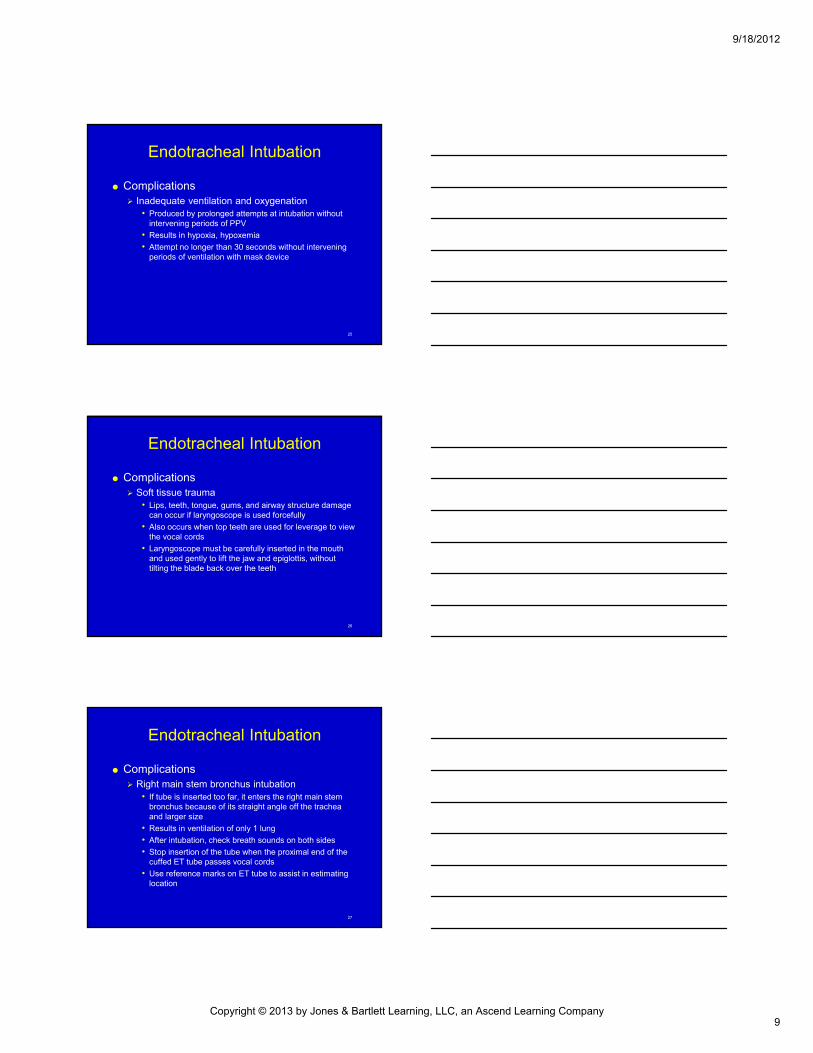
Equipment Laryngoscope
• Handle & blade attached by locking bar at end of handle
• When in use, blade hooks around locking bar and extends to a 90° angle
33
Endotracheal Intubation
Equipment Laryngoscope
• 2 types blades used: straight and curved Blade is used to sweep the bulk of the tongue to the side
and lifts the remainder of the tongue and jaw upward
Also lifts the epiglottis and allows direct visualization of the vocal cords and glottic opening
Copyright © 2013 by Jones & Bartlett Learning, LLC, an Ascend Learning Company

9/18/2012
12
34
Endotracheal Intubation
Equipment Laryngoscope
• Straight blade (Miller blades) Narrow with curved central channel
Available sizes: 0 to 4
Directly lifts epiglottis upward, allows visualization of vocal cords
Tube should be inserted in right side of mouth to maintain visualization of glottis opening until point of insertion
35
Endotracheal Intubation
Equipment Laryngoscope
• Curved blade (MacIntosh blade) Available sizes: 0 to 4
Inserted into vallecula
Indirectly elevates epiglottis away from larynx, allowing visualization of vocal cords and glottis opening
36
Endotracheal Intubation
Equipment Laryngoscope
• Assembled by: Inserting notch on blade onto locking bar or handle
Lift blade up until it locks into place and light comes on
Copyright © 2013 by Jones & Bartlett Learning, LLC, an Ascend Learning Company
9/18/2012
13
37
Endotracheal Intubation
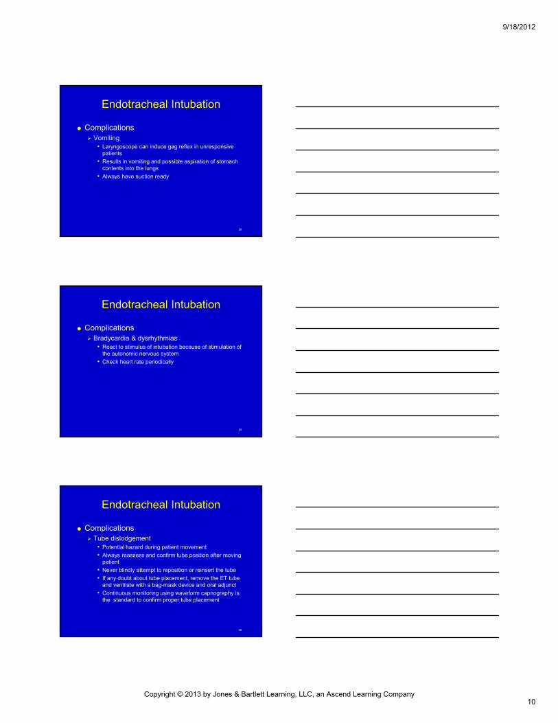
Equipment Endotracheal tubes
• Come in variety of sizes
• Have universal features, important to procedure
• Sizes 2.5 to 10 mm, internal diameter
38
Endotracheal Intubation
Equipment Endotracheal tubes
• Components
15-mm adapter
Larger tubes have a cuff filled with air to seal space between the tube and the tracheal wall
Murphy’s eye
• Adult length is 33 cm
• Markings on the tube assist in proper placement after insertion
39
Endotracheal Intubation
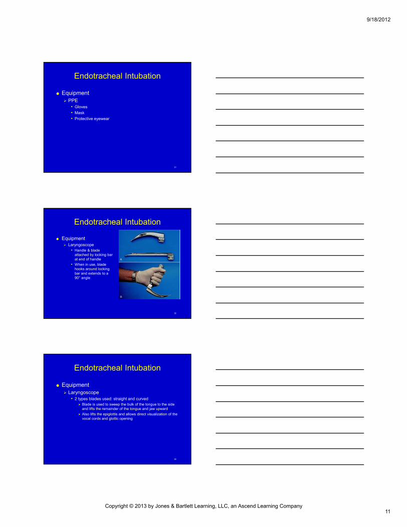
Equipment Stylet
• Malleable metal tube that is inserted into ET tube
• Provides stiffness & shape to help guide tube during intubation
Copyright © 2013 by Jones & Bartlett Learning, LLC, an Ascend Learning Company

9/18/2012
14
40
Endotracheal Intubation
Equipment Water-soluble lubricant
• Eases insertion of the ET tube into the airway
• Place liberally over the cuffed end of the tube
41
Endotracheal Intubation
Equipment Syringe
• 10-mL syringe is used to test cuff for leaks before insertion
• Remains attached and can be used to reinflate the cuff after placement
• After cuff is inflated, remove the syringe and test the pilot balloon near syringe insertion point for fullness
• If it remains attached after inflation, air may bleed back into syringe, reducing the seal of the cuff in the trachea
• If pilot balloon collapses, select a new tube
42
Endotracheal Intubation
Equipment Securing device
• Tape Tape loosens when wet
After securing the ET tube in place, use an oral airway as a bite block
• Commercial devices More likely to secure tube in place
Function as bite block and securing device
Learn and use the one advocated in your system protocols
Copyright © 2013 by Jones & Bartlett Learning, LLC, an Ascend Learning Company
9/18/2012
15
43
Endotracheal Intubation
Equipment Suction
• Should be readily available during intubation procedure
• Rigid, large-bore catheter should be available to evacuate secretions, blood, or vomitus from the upper airway during ET tube placement
• After tube placement, attach a soft, sterile French (fr) catheter to the suction unit for ET suctioning
44
Endotracheal Intubation
Equipment Towels
• Helps place head in sniffing position
• Elevates shoulders in infants/small children
• Elevating the back of the head in adults and shoulders in infants often necessary to achieve visual alignment of structures between the mouth and glottis opening
45
Endotracheal Intubation
Procedure Orotracheal intubation is most frequently used by
advanced EMTs • Secures the airway and ventilates an apneic patient in
respiratory or cardiac arrest
• May be used for anyone unresponsive to painful stimuli or lacks gag reflex to facilitate PPV and prevent aspiration
Copyright © 2013 by Jones & Bartlett Learning, LLC, an Ascend Learning Company
9/18/2012
16
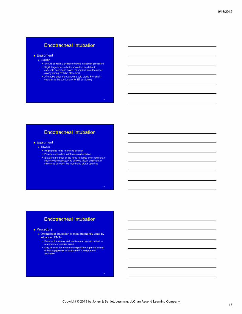
46
Skill 29-1: Inserting an Orotracheal Tube
Take appropriate personal precautions
Provide adequate PPV by bag-mask @ 100% O2
47
Skill 29-1: Inserting an Orotracheal Tube
Assemble, test all equipment
Check cuff for leaks by inflating
Deflate cuff after checking
48
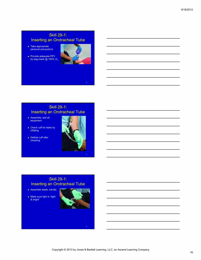
Skill 29-1: Inserting an Orotracheal Tube
Assemble blade, handle
Make sure light is “tight & bright”
Copyright © 2013 by Jones & Bartlett Learning, LLC, an Ascend Learning Company
9/18/2012
17
49
Skill 29-1: Inserting an Orotracheal Tube
Place head in head-tilt/chin position to allow visualization
Holding laryngoscope handle in left hand, insert blade into right corner of mouth
50
Skill 29-1: Inserting an Orotracheal Tube
With right hand, gently insert ET in right side of oral cavity, through vocal cord
Remove laryngoscope blade, extinguish lamp, remove stylet if used
Inflate cuff with 5 to10 mL of air, remove syringe
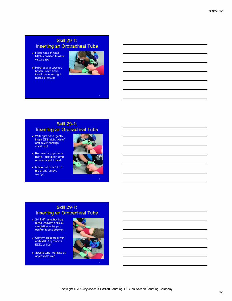
51
Skill 29-1: Inserting an Orotracheal Tube
2nd EMT, attaches bag-mask, delivers artificial ventilation while you confirm tube placement
Confirm placement with end-tidal CO2 monitor, EDD, or both
Secure tube, ventilate at appropriate rate
Copyright © 2013 by Jones & Bartlett Learning, LLC, an Ascend Learning Company
9/18/2012
18
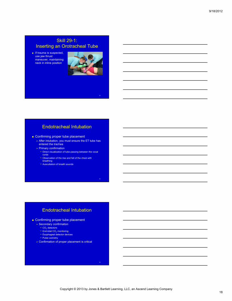
52
Skill 29-1: Inserting an Orotracheal Tube
If trauma is suspected, use jaw thrust maneuver, maintaining neck in inline position
53
Endotracheal Intubation
Confirming proper tube placement After intubation, you must ensure the ET tube has
entered the trachea
Primary confirmation• Direct visualization of tube passing between the vocal
cords
• Observation of the rise and fall of the chest with breathing
• Auscultation of breath sounds
54
Endotracheal Intubation
Confirming proper tube placement Secondary confirmation
• CO2 detectors
• End-tidal CO2 monitoring
• Esophageal detector devices
• Pulse oximetry
Confirmation of proper placement is critical
Copyright © 2013 by Jones & Bartlett Learning, LLC, an Ascend Learning Company
9/18/2012
19
55
Endotracheal Intubation
Confirming proper tube placement If sounds are present only in the epigastrium,
assume an esophageal intubation• Unrecognized esophageal intubation results in profound
hypoxia, possible brain damage, death
• In this situation, deflate cuff and remove tube
• Hyperoxygenate with bag-mask device with 100% O2
• Attempt to reintubate
• Only make 2 attempts at intubation
56
Endotracheal Intubation
Confirming proper tube placement CO2 detectors
• Checking for CO2 is helpful in confirming proper placement of ET tube
• CO2 exists in minimal amounts in ambient air, compared with amount present in exhaled air
• Designed to monitor and identify amount of CO2 present in exhaled air
57
Endotracheal Intubation
Confirming proper tube placement CO2 detectors
• Some devices provide numeric value
• Other devices express quantity with a wave on a monitor
• Colorimetric end-tidal detector, uses color change on paper to express presence or absence of CO2 in exhaled air
• Can also be built into bag-mask device
Copyright © 2013 by Jones & Bartlett Learning, LLC, an Ascend Learning Company

9/18/2012
20
58
Endotracheal Intubation
59
Endotracheal Intubation
Confirming proper tube placement Esophageal detector devices
• Consists of suction mechanism attached to opening of the ET tube
• Extending from the top of the device is a large syringe/bulb
• Generates suction needed to confirm placement of ET tube
• After placement of the ET tube and primary confirmation, the device is attached to the opening of the tube
• Bulb is then squeezed and released, or the syringe is pulled back
60
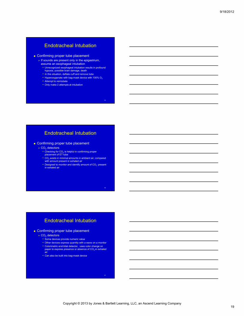
Endotracheal Intubation
Secondary confirmation Pulse oximetry
• Monitors O2 saturation through measurements of light transfer through capillary beds and hemoglobin
• Attach to tip of the patient’s finger over the nail bed or attached to an earlobe
• Reads transmission of red/IR light through capillary bed below
• Uses colorimetric method of red/IR light waves to determine percentage of O2 saturation of hemoglobin
Copyright © 2013 by Jones & Bartlett Learning, LLC, an Ascend Learning Company
9/18/2012
21
61
Endotracheal Intubation
Secondary confirmation Pulse oximetry
• Not useful as only tool for confirmation of the effectiveness of O2 therapy or ET tube placement Data from devices are too slow
Unreliable as source of feedback and decision making
Use as adjunct to assessment
62
Endotracheal Intubation
63
Alternative Airway Devices
Alternative airway devices Esophageal tracheal combitude (ETC)
Pharyngotracheal lumen (PTL)
Laryngeal mase airway (LMA)
King LT-D supraglottic airway
Often, basic airway maneuvers and a bag-mask device are adequate to maintain airway until hospital arrival
Copyright © 2013 by Jones & Bartlett Learning, LLC, an Ascend Learning Company
9/18/2012
22
64
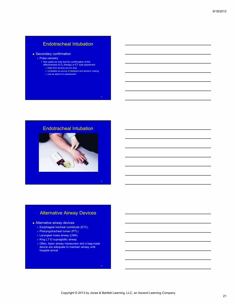
Alternative Airway Devices
ETC & PTL Look similar to ET tube
but have 2 internal lumens
65
Alternative Airway Devices
ETC & PTL After insertion, 2 balloons are inflated
When bag-mask device is attached to the proper port, air is forced into the pharynx and lungs
Esophageal balloon prevents air from entering the esophagus and prevents regurgitation from vomiting
If inadvertently inserted into the trachea, device can be used the same as an ET tube
66
Alternative Airway Devices
ETC & PTL Complications and contraindications
• Most significant: ventilation through the wrong port after attaching the device
• It is critical to check for primary placement and secondary confirmation after insertion
• May cause esophageal wall damage because of its invasive nature
Copyright © 2013 by Jones & Bartlett Learning, LLC, an Ascend Learning Company
9/18/2012
23
67
Alternative Airway Devices
ETC & PTL Complications and contraindications
• May cause esophageal wall damage because of its invasive nature
• Do not use if patient:
Is less than 5 feet tall
Is less than 14 years
Has a history of caustic ingestion
Has esophageal disease
Has inactive gag reflex
68
Alternative Airway Devices
ETC & PTL Equipment needed to insert ETC or PTL:
• PPE: gloves, eyewear, mask
• Stethoscope
• Suction
• End-tidal CO2 monitoring device
• Water-soluble lubricant
• 2 syringes to inflate the pharyngeal and distal cuffs
• Bag-mask device with O2 tubing
• O2
• Securing device
69
Alternative Airway Devices
Esophageal-tracheal combitube (ETC) & (PTL) Procedure
• Insertion is indicated when prolonged PPV is required but cannot be achieved by other methods
• Either device can be used as a backup to ET intubation
• Laryngeal mask airway
• Tube with small, air-filled mask at distal end
• Tip of mask rests above upper end of esophagus and surrounds opening of larynx
• Mask is inflated, creating seal around laryngeal opening
• Bag-mask attached to external port, air is directed into larynx, lungs
Copyright © 2013 by Jones & Bartlett Learning, LLC, an Ascend Learning Company
9/18/2012
24
70
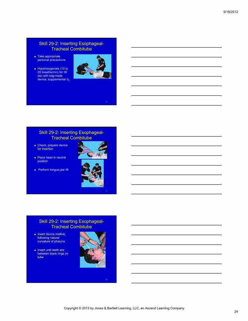
Skill 29-2: Inserting Esophageal-Tracheal Combitube
Take appropriate personal precautions
Hyperoxygenate (10 to 20 breaths/min) for 30 sec with bag-mask device, supplemental 02
71
Skill 29-2: Inserting Esophageal-Tracheal Combitube
Check, prepare device for insertion
Place head in neutral position
Perform tongue-jaw lift
72
Skill 29-2: Inserting Esophageal-Tracheal Combitube
Insert device midline, following natural curvature of pharynx
Insert until teeth are between black rings on tube
Copyright © 2013 by Jones & Bartlett Learning, LLC, an Ascend Learning Company
9/18/2012
25
73
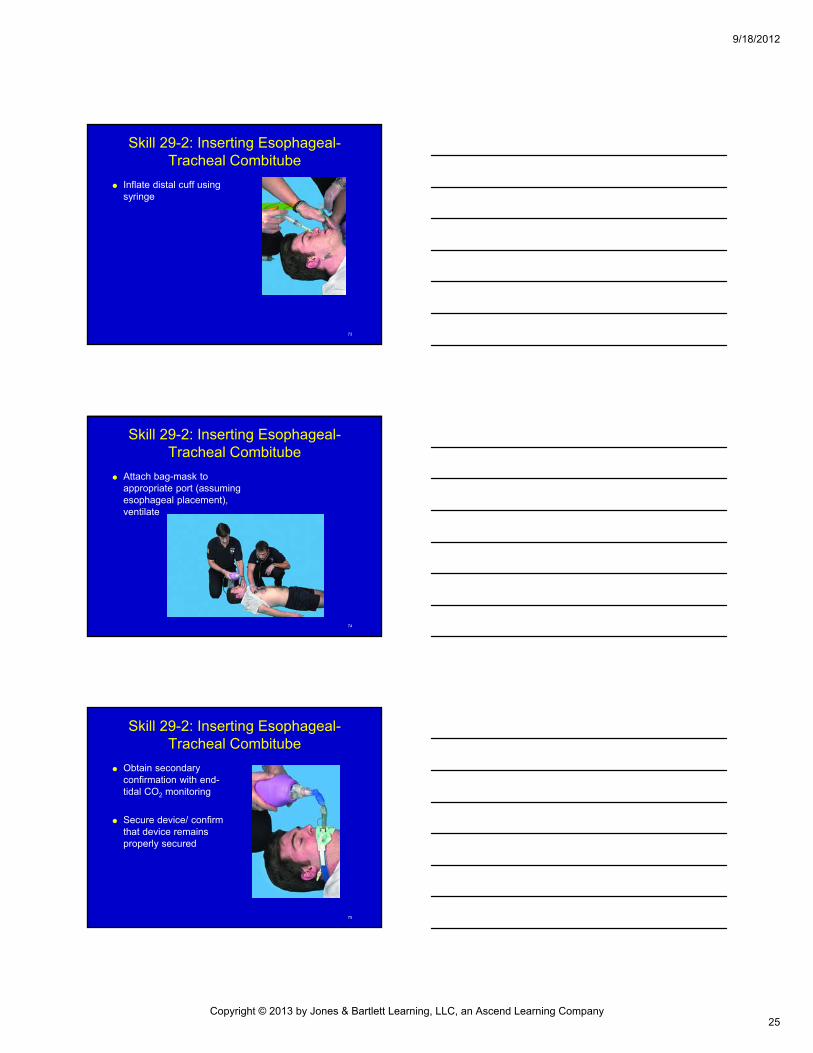
Skill 29-2: Inserting Esophageal-Tracheal Combitube
Inflate distal cuff using syringe
74
Skill 29-2: Inserting Esophageal-Tracheal Combitube
Attach bag-mask to appropriate port (assuming esophageal placement), ventilate
75
Skill 29-2: Inserting Esophageal-Tracheal Combitube
Obtain secondary confirmation with end-tidal CO2 monitoring
Secure device/ confirm that device remains properly secured
Copyright © 2013 by Jones & Bartlett Learning, LLC, an Ascend Learning Company

9/18/2012
26
76
Alternative Airway Devices
Laryngeal mask airway Tube with small, air-filled
mask at distal end
When properly inserted, the tip of the mask rests above the upper end of the esophagus and surrounds the opening of the larynx
77
Alternative Airway Devices
Laryngeal mask airway (LMA) Complications and effectiveness
• Failure to achieve adequate placement
• Not as effective as other devices in preventing gastric inflation and regurgitation
• Mask seal does not provide same degree of protection afforded by a tracheal tube or ETC
• LMA is better than a bag-mask in preventing regurgitation
• Patients should be monitored carefully for patency of the airway with suction immediately available in case of vomiting
78
Alternative Airway Devices
Laryngeal mask airway (LMA) Equipment needed to insert an LMA
• PPE: gloves, eyewear, mask
• Stethoscope
• Suction
• End-tidal CO2 monitoring device
• Water-soluble lubricant
• Syringe to inflate mask
• Bite block/bite stick
• Bag-mask O2 tubing
• O2
• Securing device
Copyright © 2013 by Jones & Bartlett Learning, LLC, an Ascend Learning Company
9/18/2012
27
79
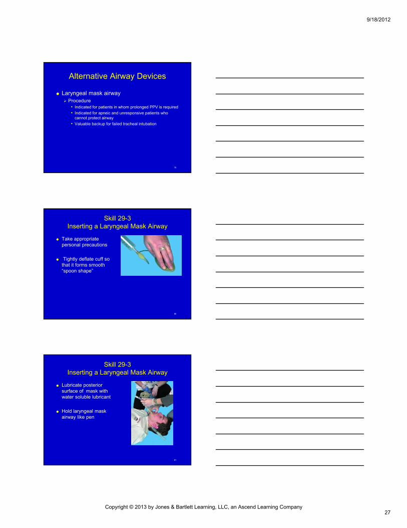
Alternative Airway Devices
Laryngeal mask airway Procedure
• Indicated for patients in whom prolonged PPV is required
• Indicated for apneic and unresponsive patients who cannot protect airway
• Valuable backup for failed tracheal intubation
80
Skill 29-3Inserting a Laryngeal Mask Airway
Take appropriate personal precautions
Tightly deflate cuff so that it forms smooth “spoon shape”
81
Skill 29-3Inserting a Laryngeal Mask Airway
Lubricate posterior surface of mask with water soluble lubricant
Hold laryngeal mask airway like pen
Copyright © 2013 by Jones & Bartlett Learning, LLC, an Ascend Learning Company

9/18/2012
28
82
Skill 29-3Inserting a Laryngeal Mask Airway
With patient’s head extended and neck flexed, carefully flatten LMA tip against hard palate
Use index finger to push cranially
83
Skill 29-3Inserting a Laryngeal Mask Airway
Advance mask until definite resistance felt at base of pharynx
Gently maintain cranial pressure with nondominant hand while removing index finger
84
Skill 29-3Inserting a Laryngeal Mask Airway
Attach bag-mask, ventilate
Copyright © 2013 by Jones & Bartlett Learning, LLC, an Ascend Learning Company

9/18/2012
29
85
Alternative Airway Devices
King LT-D supraglottic airway Disposable supraglottic airway created as an
alternative to tracheal intubation or mask ventilation
Designed for PPV and spontaneously breathing patients
Easy to insert and results in minimal airway trauma
86
Alternative Airway Devices
King LT-D supraglottic airway Complications & contraindications
• Similar to other blind placement, multilumen devices
• Correct-size airway must be used to avoid damage
• Must not be used on patients with esophageal varices or damage to the throat and neck
• Once placed, ensure the airway and lungs are inflating and patient is being ventilated
• Has only 1 ventilation port
• If the tube is improperly placed, it must be removed
87
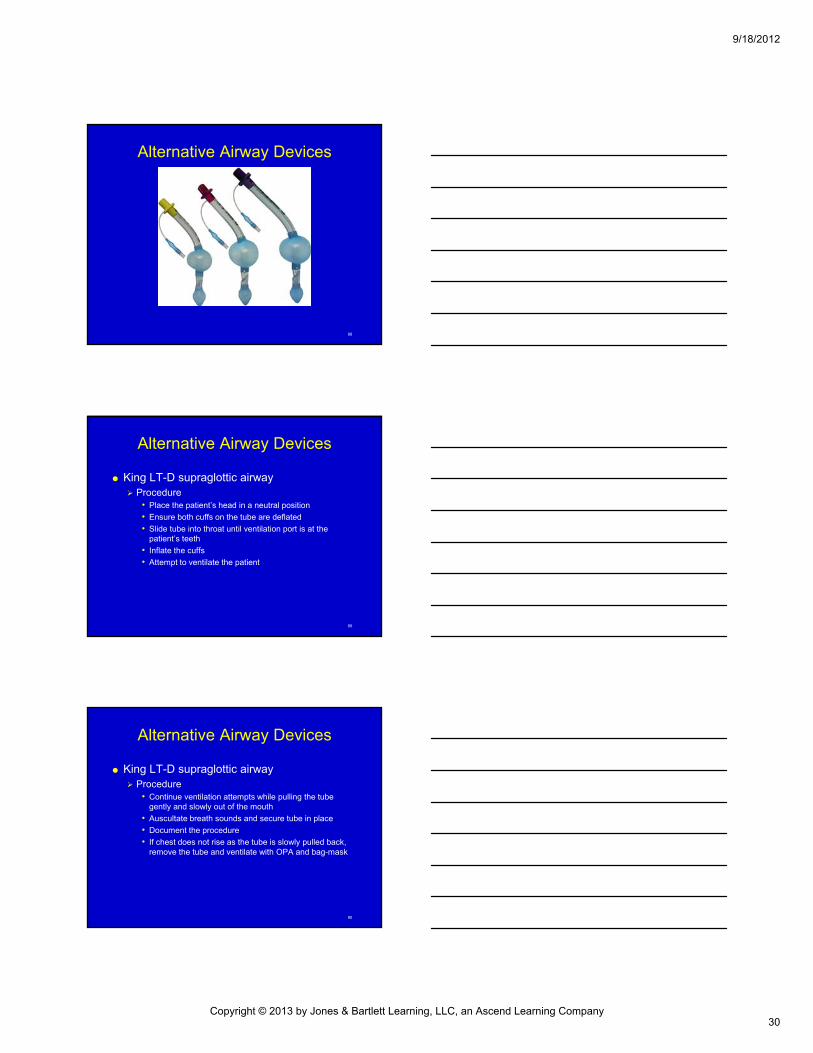
Alternative Airway Devices
King LT-D supraglottic airway Equipment
• Rigid tube with 2 distal cuffs
• Both cuffs are inflated through 1 port
• Side port allows for gastrostomy tube placement
• Designed to slide into the trachea and provide a seal around the trachea, allowing patient to be ventilated
Copyright © 2013 by Jones & Bartlett Learning, LLC, an Ascend Learning Company
9/18/2012
30
88
Alternative Airway Devices
89
Alternative Airway Devices
King LT-D supraglottic airway Procedure
• Place the patient’s head in a neutral position
• Ensure both cuffs on the tube are deflated
• Slide tube into throat until ventilation port is at the patient’s teeth
• Inflate the cuffs
• Attempt to ventilate the patient
90
Alternative Airway Devices
King LT-D supraglottic airway Procedure
• Continue ventilation attempts while pulling the tube gently and slowly out of the mouth
• Auscultate breath sounds and secure tube in place
• Document the procedure
• If chest does not rise as the tube is slowly pulled back, remove the tube and ventilate with OPA and bag-mask
Copyright © 2013 by Jones & Bartlett Learning, LLC, an Ascend Learning Company
9/18/2012
31
91
Suctioning
Used for patients intubated with a tracheal tube
Deep suctioning is indicated when a patient has aspirated material into the lungs or copious amounts of water after a submersion incident
92
Suctioning
Once intubated, insert the catheter through the ET tube Size of the catheter must be small enough to fit
through the tube
Should be soft as to not damage airway
93
Suctioning
Pay particular attention to sterile technique Entering deep into body cavity
Use low to medium suction Hypoxia is a common side effect
Limit attempts to no more than 15 seconds
Copyright © 2013 by Jones & Bartlett Learning, LLC, an Ascend Learning Company

9/18/2012
32
94
Suctioning
Indications for endotracheal suctioning Obvious secretions
Poor compliance when using bag-mask technique
95
Suctioning
Complications of orotracheal suction Abnormal heart rhythms
Hypoxia
Coughing
Mucosal damage
Bronchospasm
Proper technique minimizes potential for complication
96
Skill 29-4Performing Orotracheal Suctioning
Check equipment before proceeding, use sterile technique
Copyright © 2013 by Jones & Bartlett Learning, LLC, an Ascend Learning Company

9/18/2012
33
97
Skill 29-4Performing Orotracheal Suctioning
Insert catheter without suction on
Advance catheter just above carina
Apply suction, withdraw catheter with twisting motion
98
Skill 29-4Performing Orotracheal Suctioning
If necessary, stop hyperoxygenate patient
Repeat suctioning
99
Advanced Airway Management in Infants/Children
Intubation Airway management is particularly important
because respiratory problems are common cause of death
Copyright © 2013 by Jones & Bartlett Learning, LLC, an Ascend Learning Company
9/18/2012
34
100
Advanced Airway Management in Infants/Children
Intubation Anatomic and physiologic considerations
• All structures are smaller and more easily obstructed than adults
• Suctioning is particularly important
• More difficult to create a single, clear visual plane from the mouth through the pharynx to view the glottis opening for orotracheal intubation
• Children have narrower and softer tracheas
• Cricoid ring is the narrowest portion of airway
• Because cartilage is less rigid and developed, a cuff is not inflated
101
Advanced Airway Management in Infants/Children
Intubation Equipment
• Special considerations to help determine proper type and size of bag-mask device, laryngoscope blade and ET tube
• Proper size of bag-mask is necessary
• Markers on the tube assist in placing tube at proper depth in trachea
102
Advanced Airway Management in Infants/Children
Intubation Procedure
• Orotracheal intubation is the most effective means to secure the airway
• In apneic patients, the use of orotracheal intubation allows: Complete control of airway
Protection from aspiration
Better delivery of O2
Deeper suctioning
Copyright © 2013 by Jones & Bartlett Learning, LLC, an Ascend Learning Company

9/18/2012
35
103
Advanced Airway Management in Infants/Children
Intubation Procedure
• Confirmation of tube placement is the same as adults
• For infants and small children, assess for symmetrical rise and fall of chest Best indicator of tube placement because breaths sounds
may be misleading
104
Skill 29-5: Intubating an Infant/Child
Take appropriate personal precautions
Ensure adequate ventilations by bag-mask at age-appropriate rate
Administer 100% O2
105
Skill 29-5: Intubating an Infant/Child
Align patient’s head to ensure ease of visualization
Unless trauma is suspected, tilt head, lift chin, attempt to visualize vocal cords
Copyright © 2013 by Jones & Bartlett Learning, LLC, an Ascend Learning Company

9/18/2012
36
106
Skill 29-5: Intubating an Infant/Child
Use minimal force for intubation (touch is critical)
Holding laryngoscope handle in your left hand, insert laryngoscope blade into right corner of mouth
107
Skill 29-5: Intubating an Infant/Child
Visualize glotticopening, vocal cords
108
Skill 29-5: Intubating an Infant/Child
Do not lose sight of vocal cords
Copyright © 2013 by Jones & Bartlett Learning, LLC, an Ascend Learning Company
9/18/2012
37
109
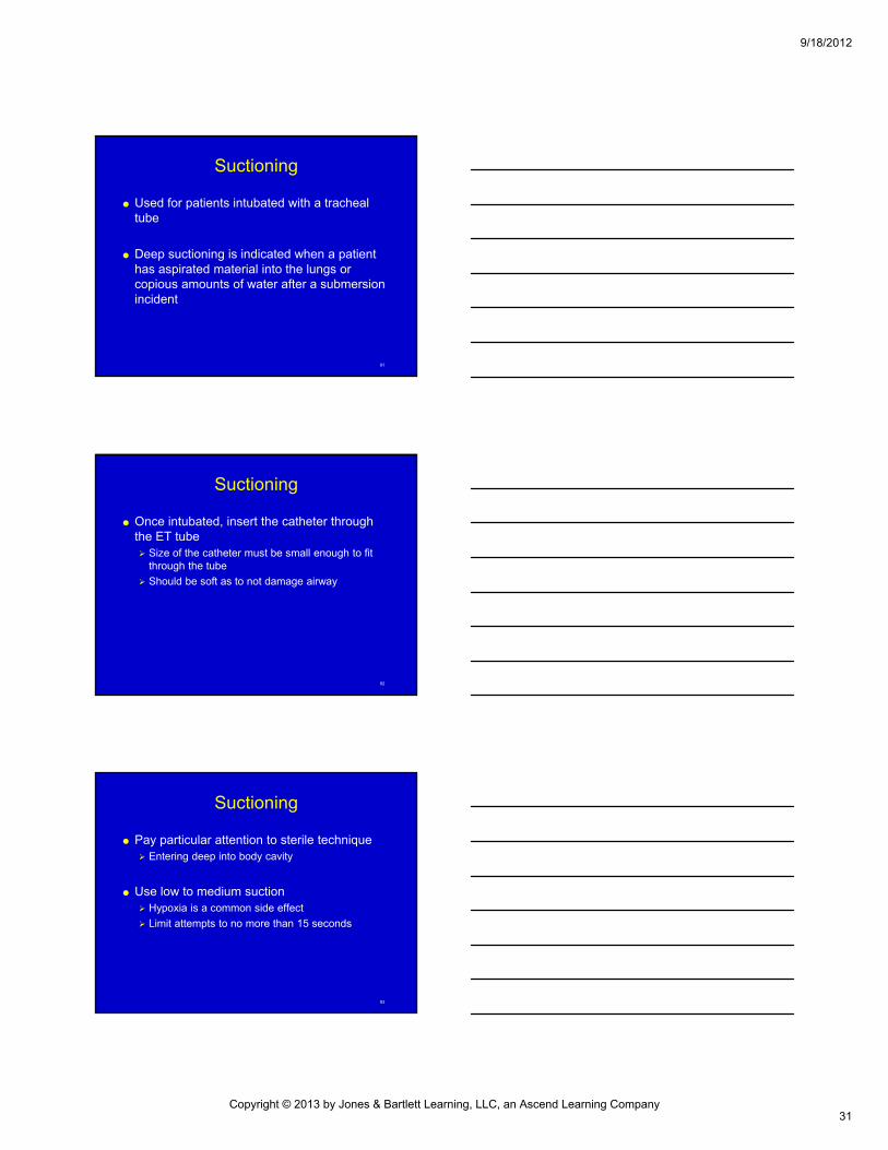
Skill 29-5: Intubating an Infant/Child
With right hand, gently insert ET until glottic marker, if present, is placed at level of vocal cords
110
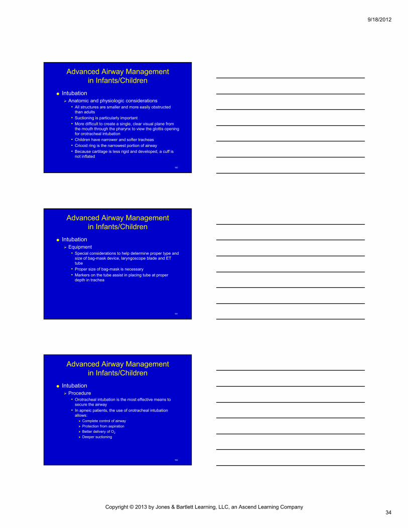
Skill 29-5: Intubating an Infant/Child
If breath sounds are equal bilaterally and no sounds are heard in epigastrium Secure ET in place
111
Advanced Airway Management in Infants/Children
Intubation Complications
• In 1 study in an urban EMS system, ET did not improve survival over bag-mask ventilation
• Continuously monitor heart rate during intubation attempts
• If a slow heart beat is noted, interrupt the intubation attempt and reventilate with a bag-mask
Copyright © 2013 by Jones & Bartlett Learning, LLC, an Ascend Learning Company

9/18/2012
38
112
Advanced Airway Management in Infants/Children
Orotracheal suctioning Use rigid catheter
Do not touch back of the airway
Suctioning time should be less than adults• Can become significantly hypoxic if prolonged
Perform nasal suctioning with:• Bulb suction
• Small French catheter with low-medium suctioning
113
Advanced Airway Management in Infants/Children
Nasogastric tube insertion (NG) Removes air and decompresses the stomach
If unresponsive, used when there is difficulty performing PPV due to gastric inflation
114
Advanced Airway Management in Infants/Children
Nasogastric tube insertion Complications and contraindications
• Tracheal insertion of the tube
• Nasal trauma
• Bleeding
• Induced vomiting
• Passage into the cranium in basilar skull fractures
• Presence of major head, facial/spinal trauma
Copyright © 2013 by Jones & Bartlett Learning, LLC, an Ascend Learning Company
9/18/2012
39
115
Advanced Airway Management in Infants/Children
Nasogastric tube insertion Equipment
• Nasogastic tubes in assorted sizes
• 20-mL syringe
• Water-soluble lubricant
• Emesis basin
• Tape
• Stethoscope
• Suction unit: suction catheter
• Towels to pad the shoulders, as needed
116
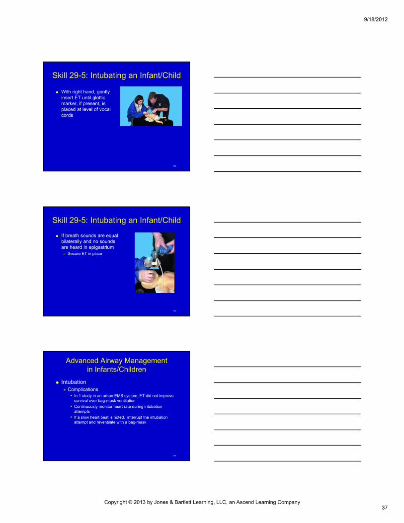
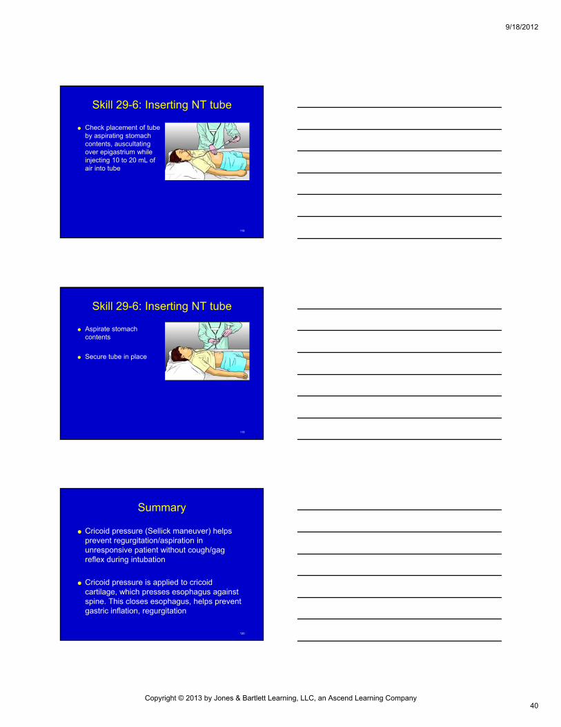
Skill 29-6: Inserting NT tube
Prepare/assemble equipment
Measure tube from tip of nose, around ear, to below xiphoid process
117
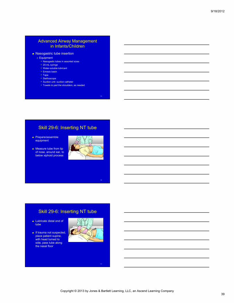
Skill 29-6: Inserting NT tube
Lubricate distal end of tube
If trauma not suspected, place patient supine, with head turned to side, pass tube along the nasal floor
Copyright © 2013 by Jones & Bartlett Learning, LLC, an Ascend Learning Company
9/18/2012
40
118
Skill 29-6: Inserting NT tube
Check placement of tube by aspirating stomach contents, auscultating over epigastrium while injecting 10 to 20 mL of air into tube
119
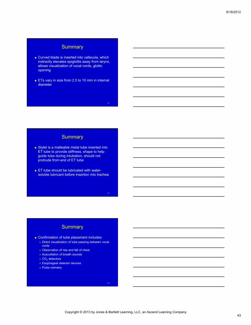
Skill 29-6: Inserting NT tube
Aspirate stomach contents
Secure tube in place
120
Summary
Cricoid pressure (Sellick maneuver) helps prevent regurgitation/aspiration in unresponsive patient without cough/gag reflex during intubation
Cricoid pressure is applied to cricoid cartilage, which presses esophagus against spine. This closes esophagus, helps prevent gastric inflation, regurgitation
Copyright © 2013 by Jones & Bartlett Learning, LLC, an Ascend Learning Company

9/18/2012
41
121
Summary
Most effective form of airway management is endotracheal intubation
ET can be passed (orotracheal)
122
Summary
Indications for ET intubation include situations in which prolonged PPV is required and cannot be effectively achieved by other methods Indicated for apneic patients & unresponsive
patients who cannot protect airway, as evidenced by absence of cough or gag reflex
123
Summary
Advantages of orotracheal intubation include: Preventing gastric distention
Minimizing risk of aspiration
Allowing for suctioning of airway
Copyright © 2013 by Jones & Bartlett Learning, LLC, an Ascend Learning Company

9/18/2012
42
124
Summary
Complications of intubation include : Esophageal intubation
Inadequate ventilation
Oxygenation from prolonged attempts
Soft tissue trauma
Right main stem bronchus intubation
Vomiting
Bradycardia, dysrhythmias
Tube dislodgement
Self-extubation
125
Summary
Esophageal intubation is most dangerous complication of endotracheal intubation because it leads to inadequate ventilation, severe hypoxia, gastric inflation if not corrected within minutes
Intubation attempts should take ≥30 sec without intervening periods of ventilation with mask device
126
Summary
It is important to secure ET tube after intubation to prevent tube dislodgement during patient movement
Straight laryngoscope blade directly lifts epiglottis upward to allow visualization of the vocal cords, often used when intubating infant &, children
Copyright © 2013 by Jones & Bartlett Learning, LLC, an Ascend Learning Company
9/18/2012
43
127
Summary
Curved blade is inserted into vallecula, which indirectly elevates epiglottis away from larynx, allows visualization of vocal cords, glottic opening
ETs vary in size from 2.5 to 10 mm in internal diameter
128
Summary
Stylet is a malleable metal tube inserted into ET tube to provide stiffness, shape to help guide tube during intubation, should not protrude from end of ET tube
ET tube should be lubricated with water-soluble lubricant before insertion into trachea
129
Summary
Confirmation of tube placement includes: Direct visualization of tube passing between vocal
cords
Observation of rise and fall of chest
Auscultation of breath sounds
CO2 detectors
Esophageal detector devices
Pulse oximetry
Copyright © 2013 by Jones & Bartlett Learning, LLC, an Ascend Learning Company
9/18/2012
44
130
Summary
Confirmation of proper tube placement is critical because unrecognized esophageal intubation results in profound hypoxia, possible brain damage/death
Alternative airway devices include: Esophageal-tracheal Combitube (ETC)
Pharyngotracheal lumen (PTL)
Laryngeal mask airway (LMA)
King LT-D supraglottic airway
131
Summary
ETC , PTL are dual-lumen devices that are blindly inserted into esophagus/ trachea Different ports are used to ventilate depending on
where tube is inserted
Most significant complication of ETC and PTL use is ventilating through wrong port after attaching device EDD cannot be used to check placement with
these devices
132
Summary
Primary complication of LMA is failure to achieve adequate placement
Indications for endotracheal suctioning are obvious secretions, poor compliance, which may indicate an obstructed airway
Complications include abnormal heart rhythms, hypoxia, coughing, mucosal damage, bronchospasms
Copyright © 2013 by Jones & Bartlett Learning, LLC, an Ascend Learning Company
9/18/2012
45
133
Summary
Infants/children have smaller anatomic airway structures that are more easily obstructed Tongue is larger, can impede visualization of vocal
cords with intubation
Narrower, softer tracheas, swelling can more easily obstruct their tracheas
Cricoid ring - narrowest portion of airway
Because cartilage is less rigid, developed, uncuffed ET is used
134
Summary
To estimate ET tube size for children, use this formula: 16 plus the patient’s age divided by 4
Other methods include selecting tube the size of child’s little finger/nasal opening
Uncuffed tube should be used in children younger than 8 y/o
135
Summary
In unresponsive infants/children, NG tubes are used when there is difficulty performing PPV because of gastric distention
Contraindications to NG tube insertion include: Presence of major facial, head/spinal trauma
Copyright © 2013 by Jones & Bartlett Learning, LLC, an Ascend Learning Company

9/18/2012
46
136
Summary
Complications of NG tube insertion include: Tracheal insertion of tube
Nasal trauma
Bleeding
Vomiting
Passage into skull
Providers must weigh risks vs. benefits of placing an advanced airway In many cases, basic airway procedures are adequate for
prehospital airway maintenance
137
Questions?
Copyright © 2013 by Jones & Bartlett Learning, LLC, an Ascend Learning Company


