Download - Ch01 mission
Copyright © 2006 Pearson Education, Inc., publishing as Benjamin Cummings
Human Anatomy & PhysiologySEVENTH EDITION
Elaine N. Marieb
Katja Hoehn
PowerPoint® Lecture Slides
prepared by Vince Austin,
Bluegrass Technical
and Community College
C H
A P
T E
R
1The Human Body: An Orientation
P A R T A

Copyright © 2006 Pearson Education, Inc., publishing as Benjamin Cummings
Overview of Anatomy and Physiology
Anatomy – the study of the structure of body parts
and their relationships to one another
Gross or macroscopic
Microscopic
Developmental
Physiology – the study of the function of the
body’s structural machinery
Copyright © 2006 Pearson Education, Inc., publishing as Benjamin Cummings
Gross Anatomy
Regional – all structures in one part of the body
(such as the abdomen or leg)
Systemic – gross anatomy of the body studied by
system
Surface – study of internal structures as they relate
to the overlying skin

Copyright © 2006 Pearson Education, Inc., publishing as Benjamin Cummings
Microscopic Anatomy
Cytology – study of the cell
Histology – study of tissues
Copyright © 2006 Pearson Education, Inc., publishing as Benjamin Cummings
Developmental Anatomy
Traces structural changes throughout life
Embryology – study of developmental changes of
the body before birth

Copyright © 2006 Pearson Education, Inc., publishing as Benjamin Cummings
Specialized Branches of Anatomy
Pathological anatomy – study of structural changes
caused by disease
Radiographic anatomy – study of internal
structures visualized by specialized scanning
procedures such as X-ray, MRI, and CT scans
Molecular biology – study of anatomical structures
at a subcellular level
Copyright © 2006 Pearson Education, Inc., publishing as Benjamin Cummings
Physiology
Considers the operation of specific organ systems
Renal – kidney function
Neurophysiology – workings of the nervous
system
Cardiovascular – operation of the heart and blood
vessels
Focuses on the functions of the body, often at the
cellular or molecular level

Copyright © 2006 Pearson Education, Inc., publishing as Benjamin Cummings
Physiology
Understanding physiology also requires a
knowledge of physics, which explains
electrical currents
blood pressure
the way muscle uses bone for movement
Copyright © 2006 Pearson Education, Inc., publishing as Benjamin Cummings
Principle of Complementarity
Function always reflects structure
What a structure can do depends on its specific
form
Copyright © 2006 Pearson Education, Inc., publishing as Benjamin Cummings
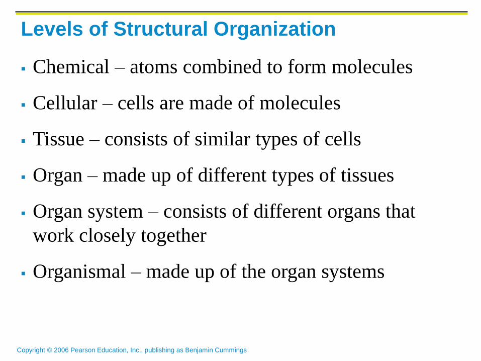
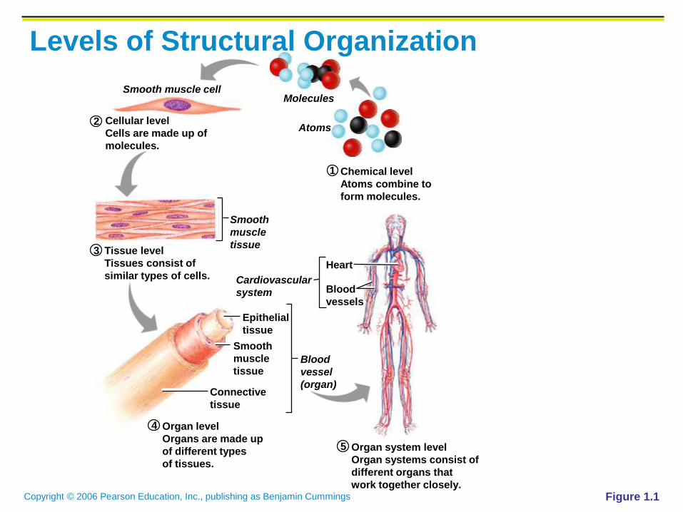
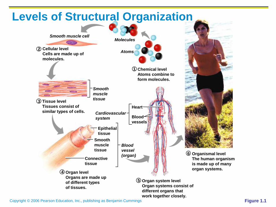
Levels of Structural Organization
Chemical – atoms combined to form molecules
Cellular – cells are made of molecules
Tissue – consists of similar types of cells
Organ – made up of different types of tissues
Organ system – consists of different organs that
work closely together
Organismal – made up of the organ systems

Copyright © 2006 Pearson Education, Inc., publishing as Benjamin Cummings
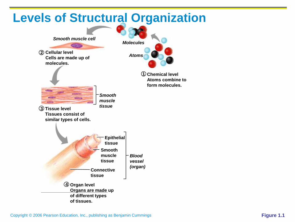
1
2
4
5
6
3
Smooth muscle cellMolecules
Atoms
Smooth
muscle
tissue
Epithelial
tissue
Heart
Blood
vessels
Smooth
muscle
tissue
Connective
tissue
Blood
vessel
(organ)
Cardiovascular
system
Cellular level
Cells are made up of
molecules.
Tissue level
Tissues consist of
similar types of cells.
Organ level
Organs are made up
of different types
of tissues.
Organ system level
Organ systems consist of
different organs that
work together closely.
Organismal level
The human organism
is made up of many
organ systems.
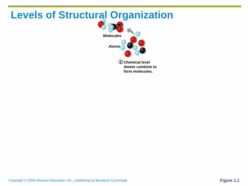
Chemical level
Atoms combine to
form molecules.
Levels of Structural Organization
Figure 1.1
Copyright © 2006 Pearson Education, Inc., publishing as Benjamin Cummings
1
Molecules
Atoms
Chemical level
Atoms combine to
form molecules.
Levels of Structural Organization
Figure 1.1

Copyright © 2006 Pearson Education, Inc., publishing as Benjamin Cummings
1
2
Smooth muscle cellMolecules
AtomsCellular level
Cells are made up of
molecules.
Chemical level
Atoms combine to
form molecules.
Levels of Structural Organization
Figure 1.1

Copyright © 2006 Pearson Education, Inc., publishing as Benjamin Cummings
1
2
3
Smooth muscle cellMolecules
Atoms
Smooth
muscle
tissue
Cellular level
Cells are made up of
molecules.
Tissue level
Tissues consist of
similar types of cells.
Chemical level
Atoms combine to
form molecules.
Levels of Structural Organization
Figure 1.1
Copyright © 2006 Pearson Education, Inc., publishing as Benjamin Cummings
1
2
4
3
Smooth muscle cellMolecules
Atoms
Smooth
muscle
tissue
Epithelial
tissue
Smooth
muscle
tissue
Connective
tissue
Blood
vessel
(organ)
Cellular level
Cells are made up of
molecules.
Tissue level
Tissues consist of
similar types of cells.
Organ level
Organs are made up
of different types
of tissues.
Chemical level
Atoms combine to
form molecules.
Levels of Structural Organization
Figure 1.1
Copyright © 2006 Pearson Education, Inc., publishing as Benjamin Cummings
1
2
4
5
3
Smooth muscle cellMolecules
Atoms
Smooth
muscle
tissue
Epithelial
tissue
Heart
Blood
vessels
Smooth
muscle
tissue
Connective
tissue
Blood
vessel
(organ)
Cardiovascular
system
Cellular level
Cells are made up of
molecules.
Tissue level
Tissues consist of
similar types of cells.
Organ level
Organs are made up
of different types
of tissues.
Organ system level
Organ systems consist of
different organs that
work together closely.
Chemical level
Atoms combine to
form molecules.
Levels of Structural Organization
Figure 1.1
Copyright © 2006 Pearson Education, Inc., publishing as Benjamin Cummings
1
2
4
5
6
3
Smooth muscle cellMolecules
Atoms
Smooth
muscle
tissue
Epithelial
tissue
Heart
Blood
vessels
Smooth
muscle
tissue
Connective
tissue
Blood
vessel
(organ)
Cardiovascular
system
Cellular level
Cells are made up of
molecules.
Tissue level
Tissues consist of
similar types of cells.
Organ level
Organs are made up
of different types
of tissues.
Organ system level
Organ systems consist of
different organs that
work together closely.
Organismal level
The human organism
is made up of many
organ systems.
Chemical level
Atoms combine to
form molecules.
Levels of Structural Organization
Figure 1.1

Copyright © 2006 Pearson Education, Inc., publishing as Benjamin Cummings
Integumentary System
Forms the external body
covering
Composed of the skin, sweat
glands, oil glands, hair, and
nails
Protects deep tissues from
injury and synthesizes vitamin
D
Figure 1.3a

Copyright © 2006 Pearson Education, Inc., publishing as Benjamin Cummings
Skeletal System
Composed of bone, cartilage,
and ligaments
Protects and supports body
organs
Provides the framework for
muscles
Site of blood cell formation
Stores minerals
Figure 1.3b

Copyright © 2006 Pearson Education, Inc., publishing as Benjamin Cummings
Muscular System
Composed of muscles and
tendons
Allows manipulation of the
environment, locomotion, and
facial expression
Maintains posture
Produces heat
Figure 1.3c

Copyright © 2006 Pearson Education, Inc., publishing as Benjamin Cummings
Nervous System
Composed of the brain, spinal
column, and nerves
Is the fast-acting control
system of the body
Responds to stimuli by
activating muscles and glands
Figure 1.3d

Copyright © 2006 Pearson Education, Inc., publishing as Benjamin Cummings
Cardiovascular System
Composed of the heart and
blood vessels
The heart pumps blood
The blood vessels transport
blood throughout the body
Figure 1.3f

Copyright © 2006 Pearson Education, Inc., publishing as Benjamin Cummings
Lymphatic System
Composed of red bone marrow, thymus, spleen, lymph nodes, and lymphatic vessels
Picks up fluid leaked from blood vessels and returns it to blood
Disposes of debris in the lymphatic stream
Houses white blood cells involved with immunity
Figure 1.3g
Copyright © 2006 Pearson Education, Inc., publishing as Benjamin Cummings
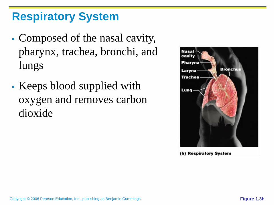
Respiratory System
Composed of the nasal cavity,
pharynx, trachea, bronchi, and
lungs
Keeps blood supplied with
oxygen and removes carbon
dioxide
Figure 1.3h

Copyright © 2006 Pearson Education, Inc., publishing as Benjamin Cummings
Digestive System
Composed of the oral cavity,
esophagus, stomach, small
intestine, large intestine,
rectum, anus, and liver
Breaks down food into
absorbable units that enter the
blood
Eliminates indigestible
foodstuffs as feces
Figure 1.3i

Copyright © 2006 Pearson Education, Inc., publishing as Benjamin Cummings
Urinary System
Composed of kidneys, ureters,
urinary bladder, and urethra
Eliminates nitrogenous wastes
from the body
Regulates water, electrolyte,
and pH balance of the blood
Figure 1.3j

Copyright © 2006 Pearson Education, Inc., publishing as Benjamin Cummings
Male Reproductive System
Composed of prostate gland, penis, testes, scrotum, and ductus deferens
Main function is the production of offspring
Testes produce sperm and male sex hormones
Ducts and glands deliver sperm to the female reproductive tract
Figure 1.3k
Copyright © 2006 Pearson Education, Inc., publishing as Benjamin Cummings
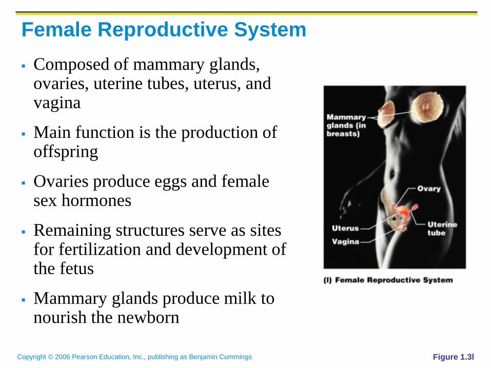
Female Reproductive System
Composed of mammary glands, ovaries, uterine tubes, uterus, and vagina
Main function is the production of offspring
Ovaries produce eggs and female sex hormones
Remaining structures serve as sites for fertilization and development of the fetus
Mammary glands produce milk to nourish the newborn
Figure 1.3l

Copyright © 2006 Pearson Education, Inc., publishing as Benjamin Cummings
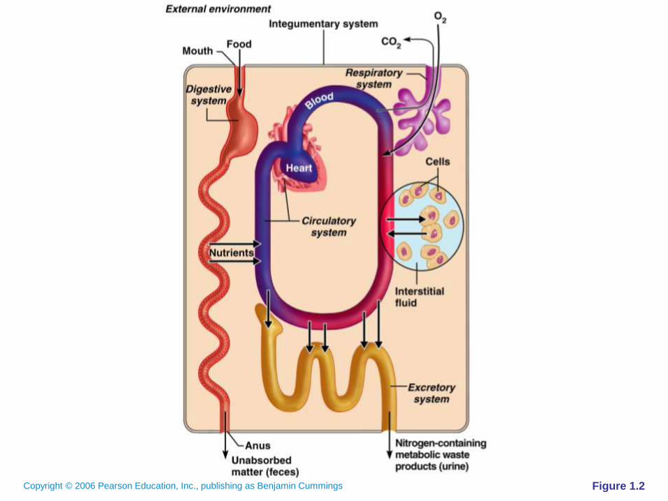
Organ Systems Interrelationships
The integumentary system protects the body from
the external environment
Digestive and respiratory systems, in contact with
the external environment, take in nutrients and
oxygen

Copyright © 2006 Pearson Education, Inc., publishing as Benjamin Cummings
Organ Systems Interrelationships
Nutrients and oxygen are
distributed by the blood
Metabolic wastes are
eliminated by the urinary
and respiratory systems
Figure 1.2
Copyright © 2006 Pearson Education, Inc., publishing as Benjamin Cummings Figure 1.2
Copyright © 2006 Pearson Education, Inc., publishing as Benjamin Cummings
Necessary Life Functions
Maintaining boundaries – the internal environment
remains distinct from the external environment
Cellular level – accomplished by plasma
membranes
Organismal level – accomplished by the skin
Movement – locomotion, propulsion (peristalsis),
and contractility

Copyright © 2006 Pearson Education, Inc., publishing as Benjamin Cummings
Necessary Life Functions
Responsiveness – ability to sense changes in the
environment and respond to them
Digestion – breakdown of ingested foodstuffs
Metabolism – all the chemical reactions that occur
in the body
Excretion – removal of wastes from the body

Copyright © 2006 Pearson Education, Inc., publishing as Benjamin Cummings
Necessary Life Functions
Reproduction – cellular and organismal levels
Cellular – an original cell divides and produces two
identical daughter cells
Organismal – sperm and egg unite to make a whole
new person
Growth – increase in size of a body part or of the
organism

Copyright © 2006 Pearson Education, Inc., publishing as Benjamin Cummings
Survival Needs
Nutrients – needed for energy and cell building
Oxygen – necessary for metabolic reactions
Water – provides the necessary environment for
chemical reactions
Normal body temperature – necessary for chemical
reactions to occur at life-sustaining rates
Atmospheric pressure – required for proper
breathing and gas exchange in the lungs

Copyright © 2006 Pearson Education, Inc., publishing as Benjamin Cummings
Homeostasis
Homeostasis – ability to maintain a relatively
stable internal environment in an ever-changing
outside world
The internal environment of the body is in a
dynamic state of equilibrium
Chemical, thermal, and neural factors interact to
maintain homeostasis

Copyright © 2006 Pearson Education, Inc., publishing as Benjamin Cummings
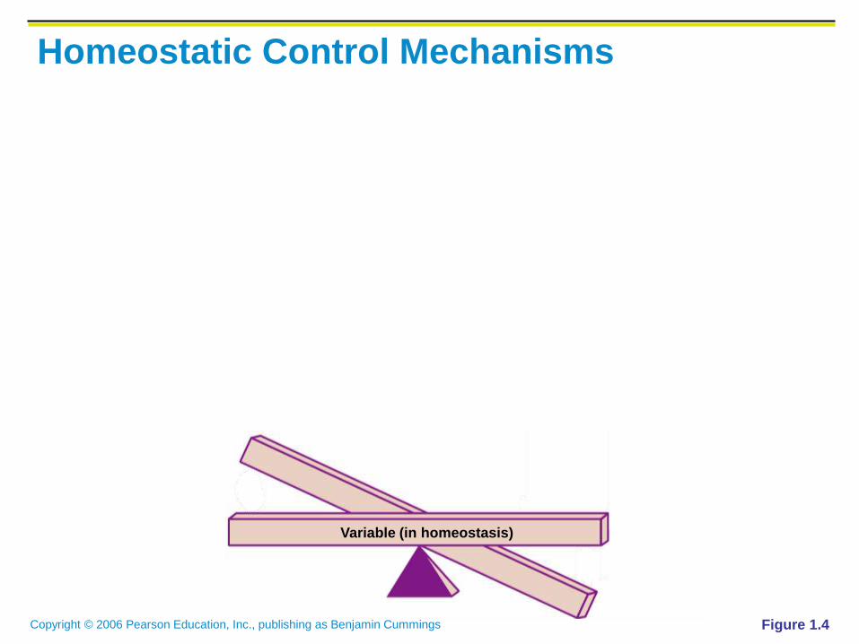
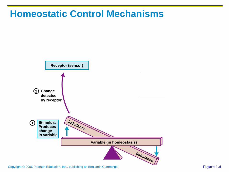
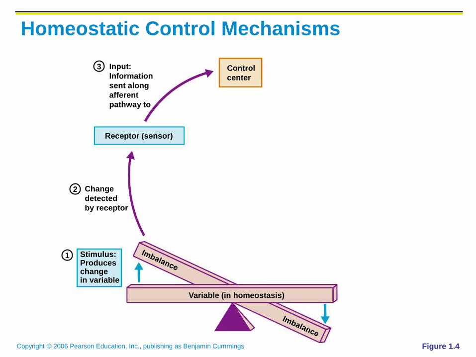
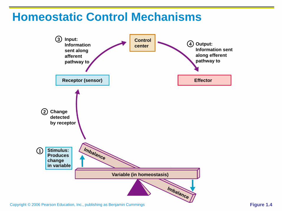
Homeostatic Control Mechanisms
Variables produce a change in the body
The three interdependent components of control
mechanisms:
Receptor – monitors the environments and
responds to changes (stimuli)
Control center – determines the set point at which
the variable is maintained
Effector – provides the means to respond to stimuli

Copyright © 2006 Pearson Education, Inc., publishing as Benjamin Cummings
Homeostatic Control Mechanisms
Figure 1.4
Change
detected
by receptor
Stimulus: Produces changein variable
Input:
Information
sent along
afferent
pathway to
Receptor (sensor) Effector
Control
center
Variable (in homeostasis)
Response of
effector feeds
back to
influence
magnitude of
stimulus and
returns variable
to homeostasis
Output:
Information sent
along efferent
pathway to
2
34
5
1
Copyright © 2006 Pearson Education, Inc., publishing as Benjamin Cummings
Homeostatic Control Mechanisms
Figure 1.4
Variable (in homeostasis)

Copyright © 2006 Pearson Education, Inc., publishing as Benjamin Cummings
Homeostatic Control Mechanisms
Figure 1.4
Stimulus: Produces changein variable
Variable (in homeostasis)
1
Copyright © 2006 Pearson Education, Inc., publishing as Benjamin Cummings
Homeostatic Control Mechanisms
Figure 1.4
Change
detected
by receptor
Stimulus: Produces changein variable
Receptor (sensor)
Variable (in homeostasis)
2
1
Copyright © 2006 Pearson Education, Inc., publishing as Benjamin Cummings
Homeostatic Control Mechanisms
Figure 1.4
Change
detected
by receptor
Stimulus: Produces changein variable
Input:
Information
sent along
afferent
pathway to
Receptor (sensor)
Control
center
Variable (in homeostasis)
2
3
1
Copyright © 2006 Pearson Education, Inc., publishing as Benjamin Cummings
Homeostatic Control Mechanisms
Figure 1.4
Change
detected
by receptor
Stimulus: Produces changein variable
Input:
Information
sent along
afferent
pathway to
Receptor (sensor) Effector
Control
center
Variable (in homeostasis)
Output:
Information sent
along efferent
pathway to
2
34
1

Copyright © 2006 Pearson Education, Inc., publishing as Benjamin Cummings
Homeostatic Control Mechanisms
Figure 1.4
Change
detected
by receptor
Stimulus: Produces changein variable
Input:
Information
sent along
afferent
pathway to
Receptor (sensor) Effector
Control
center
Variable (in homeostasis)
Response of
effector feeds
back to
influence
magnitude of
stimulus and
returns variable
to homeostasis
Output:
Information sent
along efferent
pathway to
2
34
5
1

Copyright © 2006 Pearson Education, Inc., publishing as Benjamin Cummings
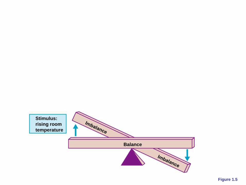
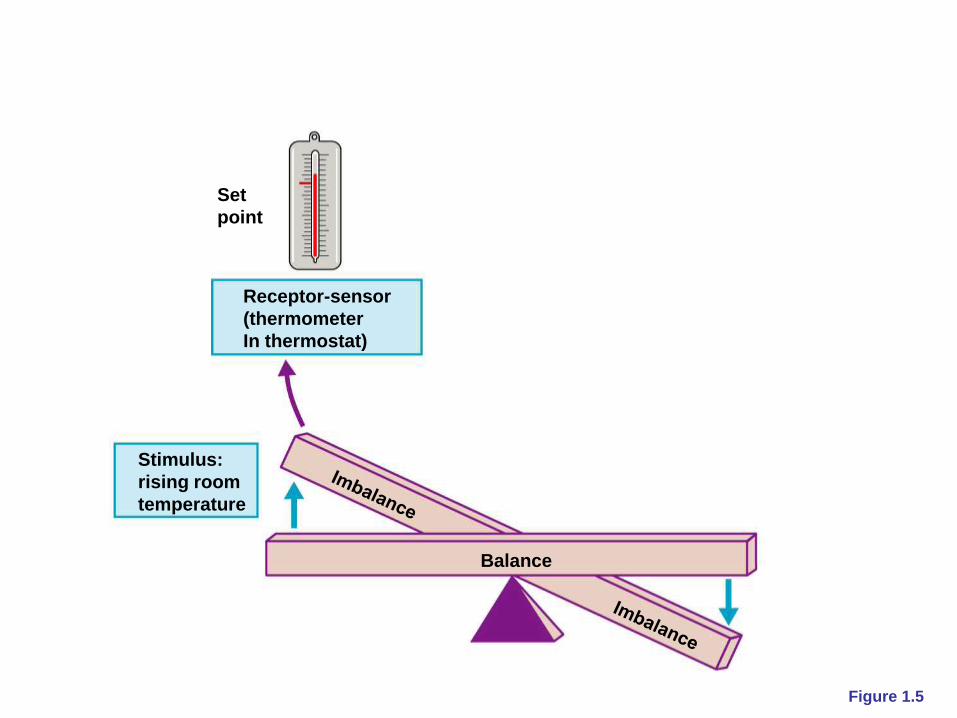
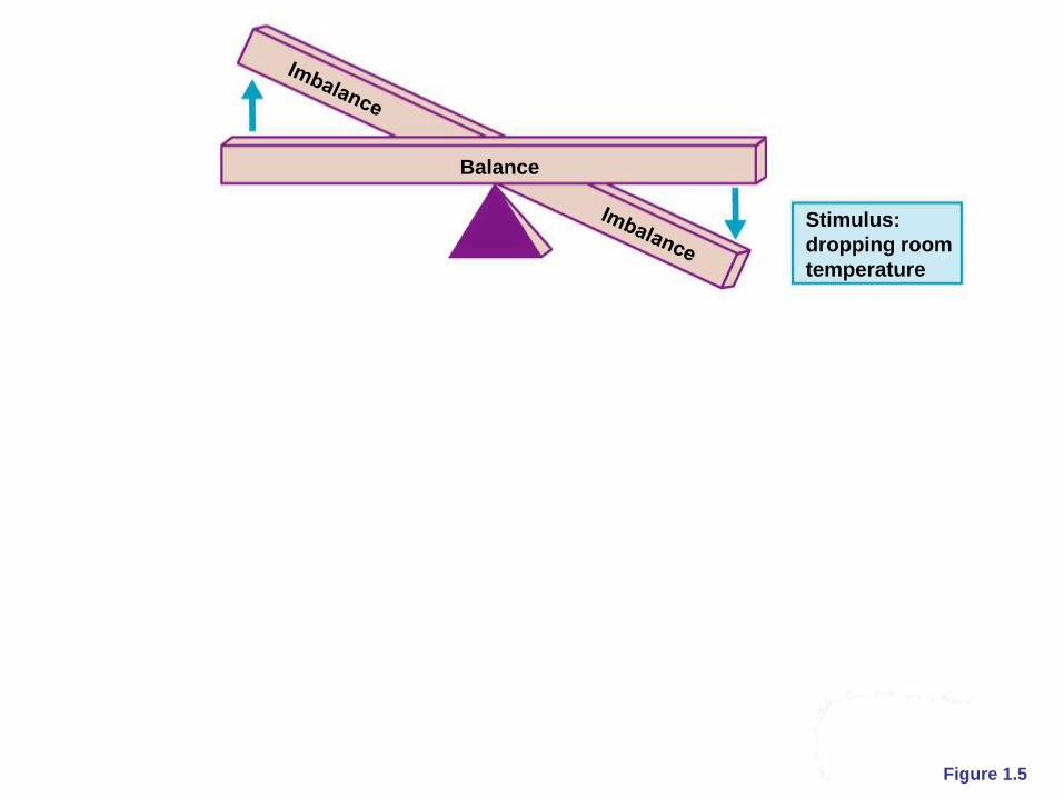
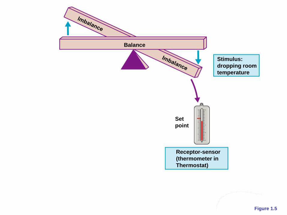
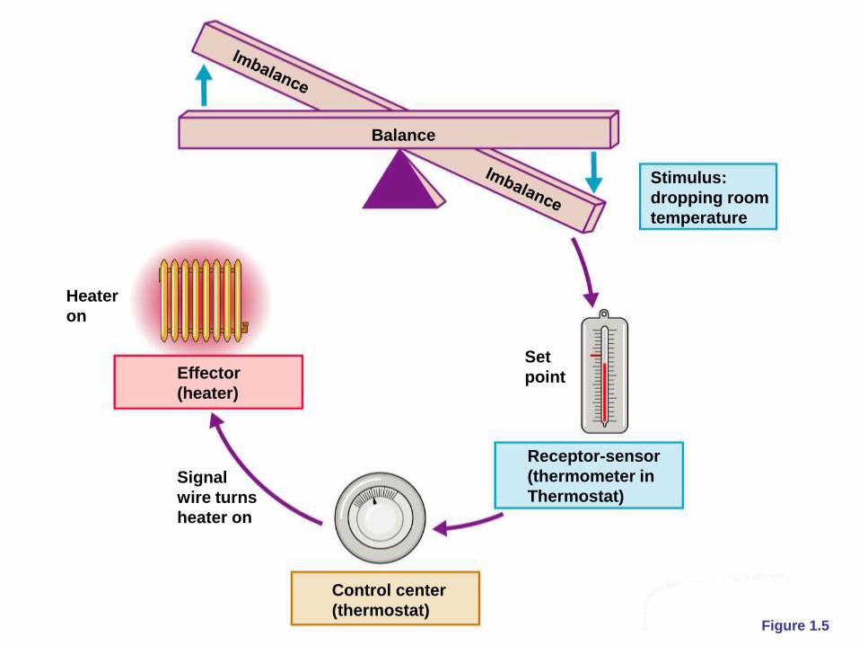
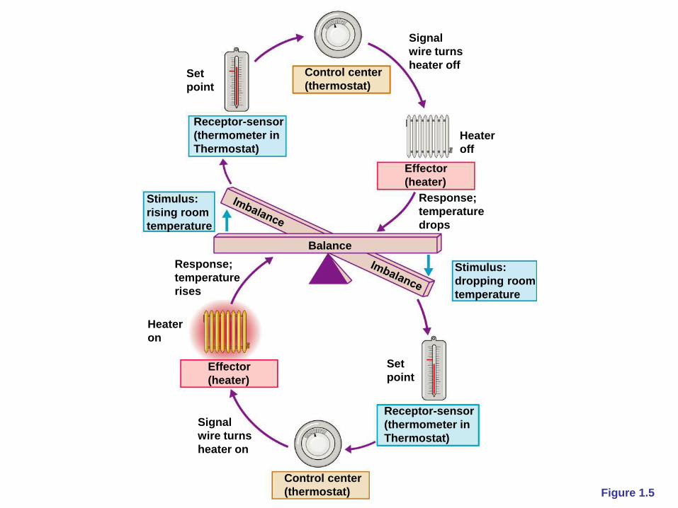
Negative Feedback
In negative feedback systems, the output shuts off
the original stimulus
Example: Regulation of room temperature

Copyright © 2006 Pearson Education, Inc., publishing as Benjamin Cummings Figure 1.5
Signal
wire turns
heater on
Signal
wire turns
heater off
Response;
temperature
rises
Response;
temperature
drops
Stimulus:
rising room
temperature
Stimulus:
dropping room
temperature
Balance
Effector
(heater)
Effector
(heater)
Set
point
Control center
(thermostat)
Heater
off
Set
point
Receptor-sensor
(thermometer in
Thermostat)
Control center
(thermostat)
Heater
on
Receptor-sensor
(thermometer in
Thermostat)
Copyright © 2006 Pearson Education, Inc., publishing as Benjamin Cummings Figure 1.5
Balance
Copyright © 2006 Pearson Education, Inc., publishing as Benjamin Cummings Figure 1.5
Stimulus:
rising room
temperature
Balance
Copyright © 2006 Pearson Education, Inc., publishing as Benjamin Cummings Figure 1.5
Stimulus:
rising room
temperature
Balance
Receptor-sensor
(thermometer
In thermostat)
Set
point

Copyright © 2006 Pearson Education, Inc., publishing as Benjamin Cummings Figure 1.5
Stimulus:
rising room
temperature
Balance
Receptor-sensor
(thermometer
In thermostat)
Set
point
Control center
(thermostat)
Copyright © 2006 Pearson Education, Inc., publishing as Benjamin Cummings Figure 1.5
Signal
wire turns
heater off
Stimulus:
rising room
temperature
Balance
Effector
(heater)
Stimulus:
dropping room
temperature
Receptor-sensor
(thermometer
In thermostat)
Set
point
Control center
(thermostat)
Heater
off

Copyright © 2006 Pearson Education, Inc., publishing as Benjamin Cummings Figure 1.5
Signal
wire turns
heater off
Response;
temperature
drops
Stimulus:
rising room
temperature
Balance
Effector
(heater)
Stimulus:
dropping room
temperature
Receptor-sensor
(thermometer
In thermostat)
Set
point
Control center
(thermostat)
Heater
off
Copyright © 2006 Pearson Education, Inc., publishing as Benjamin Cummings Figure 1.5
Stimulus:
dropping room
temperature
Balance
Copyright © 2006 Pearson Education, Inc., publishing as Benjamin Cummings Figure 1.5
Stimulus:
dropping room
temperature
Balance
Set
point
Receptor-sensor
(thermometer in
Thermostat)

Copyright © 2006 Pearson Education, Inc., publishing as Benjamin Cummings Figure 1.5
Stimulus:
dropping room
temperature
Balance
Set
point
Receptor-sensor
(thermometer in
Thermostat)
Control center
(thermostat)
Copyright © 2006 Pearson Education, Inc., publishing as Benjamin Cummings Figure 1.5
Signal
wire turns
heater on
Stimulus:
dropping room
temperature
Balance
Effector
(heater)
Set
point
Receptor-sensor
(thermometer in
Thermostat)
Control center
(thermostat)
Heater
on

Copyright © 2006 Pearson Education, Inc., publishing as Benjamin Cummings Figure 1.5
Signal
wire turns
heater on
Response;
temperature
rises
Stimulus:
dropping room
temperature
Balance
Effector
(heater)
Set
point
Receptor-sensor
(thermometer in
Thermostat)
Control center
(thermostat)
Heater
on
Copyright © 2006 Pearson Education, Inc., publishing as Benjamin Cummings Figure 1.5
Signal
wire turns
heater on
Signal
wire turns
heater off
Response;
temperature
rises
Response;
temperature
drops
Stimulus:
rising room
temperature
Stimulus:
dropping room
temperature
Balance
Effector
(heater)
Effector
(heater)
Set
point
Control center
(thermostat)
Heater
off
Set
point
Receptor-sensor
(thermometer in
Thermostat)
Control center
(thermostat)
Heater
on
Receptor-sensor
(thermometer in
Thermostat)
Copyright © 2006 Pearson Education, Inc., publishing as Benjamin Cummings
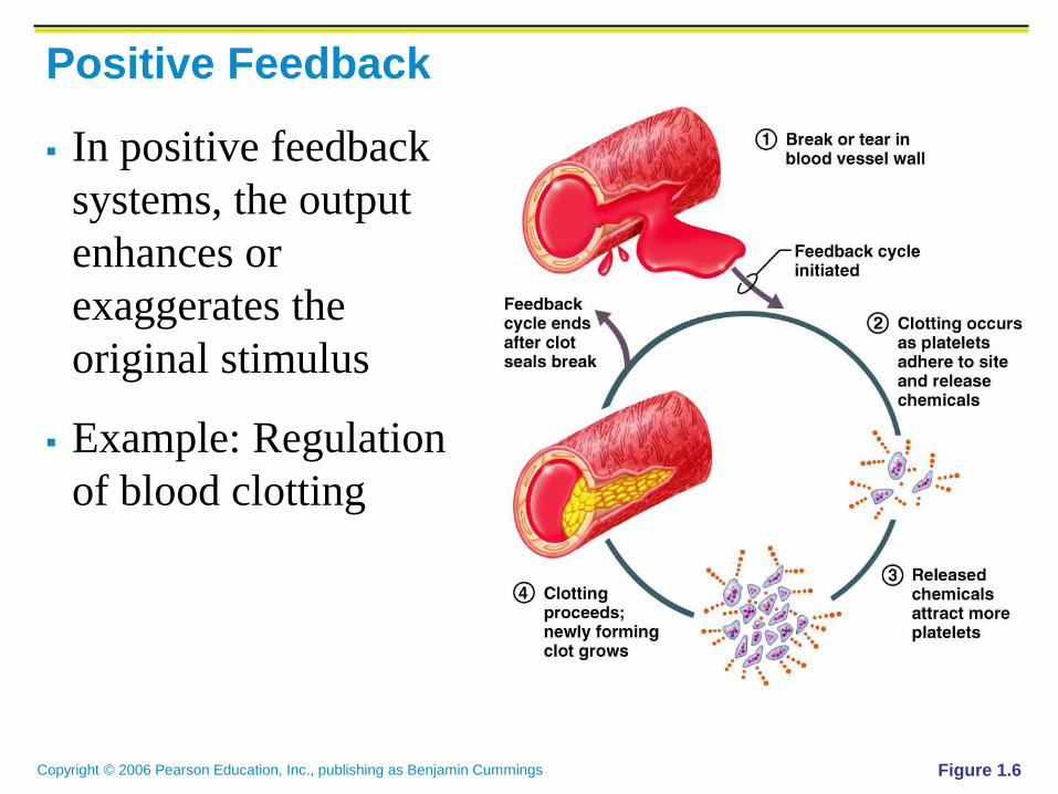
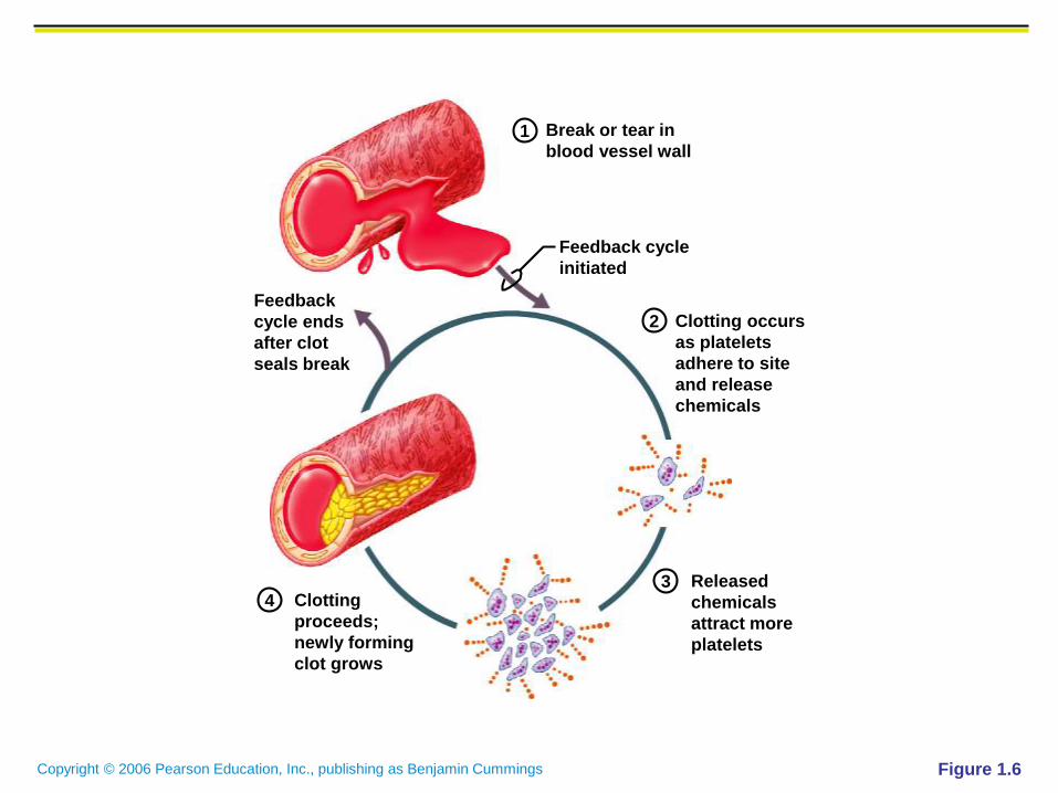
Positive Feedback
In positive feedback
systems, the output
enhances or
exaggerates the
original stimulus
Example: Regulation
of blood clotting
Figure 1.6

Copyright © 2006 Pearson Education, Inc., publishing as Benjamin Cummings Figure 1.6
Released
chemicals
attract more
platelets
Clotting occurs
as platelets
adhere to site
and release
chemicals
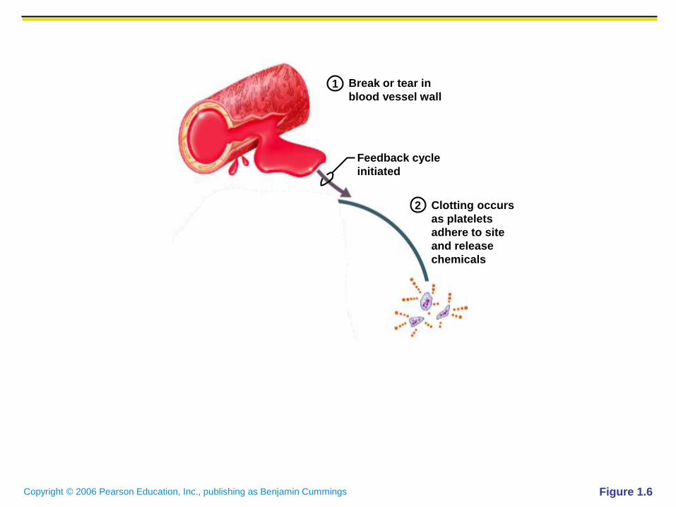
Break or tear in
blood vessel wall
Feedback cycle
initiated
Feedback
cycle ends
after clot
seals break
Clotting
proceeds;
newly forming
clot grows
2
1
34

Copyright © 2006 Pearson Education, Inc., publishing as Benjamin Cummings Figure 1.6
Break or tear in
blood vessel wall
Feedback cycle
initiated
1
Copyright © 2006 Pearson Education, Inc., publishing as Benjamin Cummings Figure 1.6
Clotting occurs
as platelets
adhere to site
and release
chemicals
Break or tear in
blood vessel wall
Feedback cycle
initiated
2
1

Copyright © 2006 Pearson Education, Inc., publishing as Benjamin Cummings Figure 1.6
Released
chemicals
attract more
platelets
Clotting occurs
as platelets
adhere to site
and release
chemicals
Break or tear in
blood vessel wall
Feedback cycle
initiated
2
1
3

Copyright © 2006 Pearson Education, Inc., publishing as Benjamin Cummings Figure 1.6
Released
chemicals
attract more
platelets
Clotting occurs
as platelets
adhere to site
and release
chemicals
Break or tear in
blood vessel wall
Feedback cycle
initiated
Clotting
proceeds;
newly forming
clot grows
2
1
34
Copyright © 2006 Pearson Education, Inc., publishing as Benjamin Cummings Figure 1.6
Released
chemicals
attract more
platelets
Clotting occurs
as platelets
adhere to site
and release
chemicals
Break or tear in
blood vessel wall
Feedback cycle
initiated
Feedback
cycle ends
after clot
seals break
Clotting
proceeds;
newly forming
clot grows
2
1
34

Copyright © 2006 Pearson Education, Inc., publishing as Benjamin Cummings
Homeostatic Imbalance
Disturbance of homeostasis or the body’s normal
equilibrium
Overwhelming the usual negative feedback
mechanisms allows destructive positive feedback
mechanisms to take over


