
Ch. 8 The Cellular Basis of Reproduction and Inheritance
© 2012 Pearson Education, Inc.
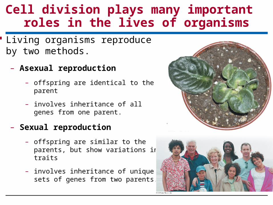
Cell division plays many important roles in the lives of organisms
Living organisms reproduce by two methods.
– Asexual reproduction
– offspring are identical to the parent
– involves inheritance of all genes from one parent.
– Sexual reproduction
– offspring are similar to the parents, but show variations in traits
– involves inheritance of unique sets of genes from two parents.

Cell division plays many important roles in the lives of organisms
Cell division is reproduction at the cellular level,
Requires duplication of chromosomes, sorting new sets of chromosomes into the resulting pair of daughter cells.
© 2012 Pearson Education, Inc.

Cell division is used for:
– Reproduction of single-celled organisms
– Growth of multicellular organisms from a fertilized egg into an adult,
– Repair and replacement of cells
– Sperm and egg production.
Cell division plays many important roles in the lives of organisms
© 2012 Pearson Education, Inc.

Prokaryotes (bacteria and archaea) reproduce by binary fission “dividing in half”
Binary Fission Occurs in Three Steps:
1. duplication of the chromosome and separation of the copies,
2. continued elongation of the cell and movement of the copies
3. division into two daughter cells.
The chromosome of a prokaryote is:
A singular circular DNA molecule & associated proteins
Much smaller than eukaryotic DNA
Prokaryotes reproduce by binary fission
© 2012 Pearson Education, Inc.
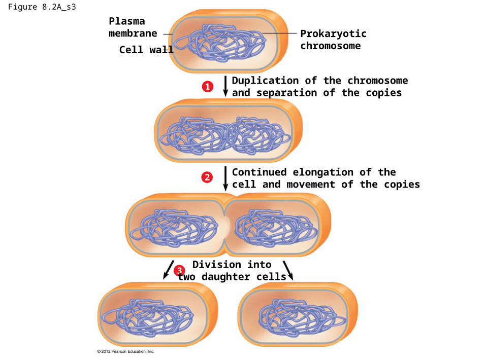
Figure 8.2A_s3
Plasmamembrane
Cell wall
Duplication of the chromosomeand separation of the copies
Continued elongation of thecell and movement of the copies
Prokaryoticchromosome
1
2
3Division into
two daughter cells
THE EUKARYOTIC CELL CYCLE AND MITOSIS
© 2012 Pearson Education, Inc.
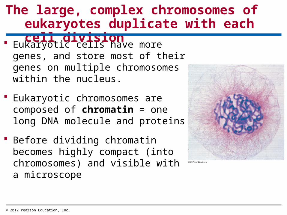
Eukaryotic cells have more genes, and store most of their genes on multiple chromosomes within the nucleus.
Eukaryotic chromosomes are composed of chromatin = one long DNA molecule and proteins
Before dividing chromatin becomes highly compact (into chromosomes) and visible with a microscope
The large, complex chromosomes of eukaryotes duplicate with each cell division
© 2012 Pearson Education, Inc.
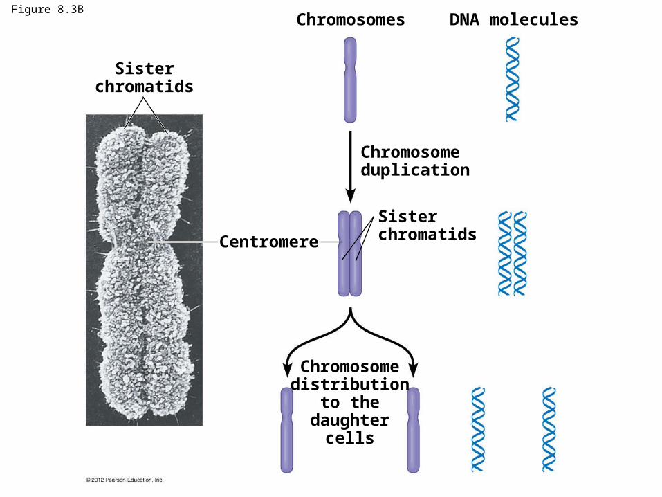
Figure 8.3B
Sisterchromatids
Chromosomes
Centromere
Chromosomeduplication
Sisterchromatids
Chromosomedistribution
to thedaughter
cells
DNA molecules

Before a eukaryotic cell divides, it duplicates all of its chromosomes, resulting in two copies called sister chromatids joined together by a narrowed “waist” called the centromere.
When a cell divides, the sister chromatids separate from each other, (now called chromosomes) and sort into separate daughter cells.
The large, complex chromosomes of eukaryotes duplicate with each cell division
© 2012 Pearson Education, Inc.
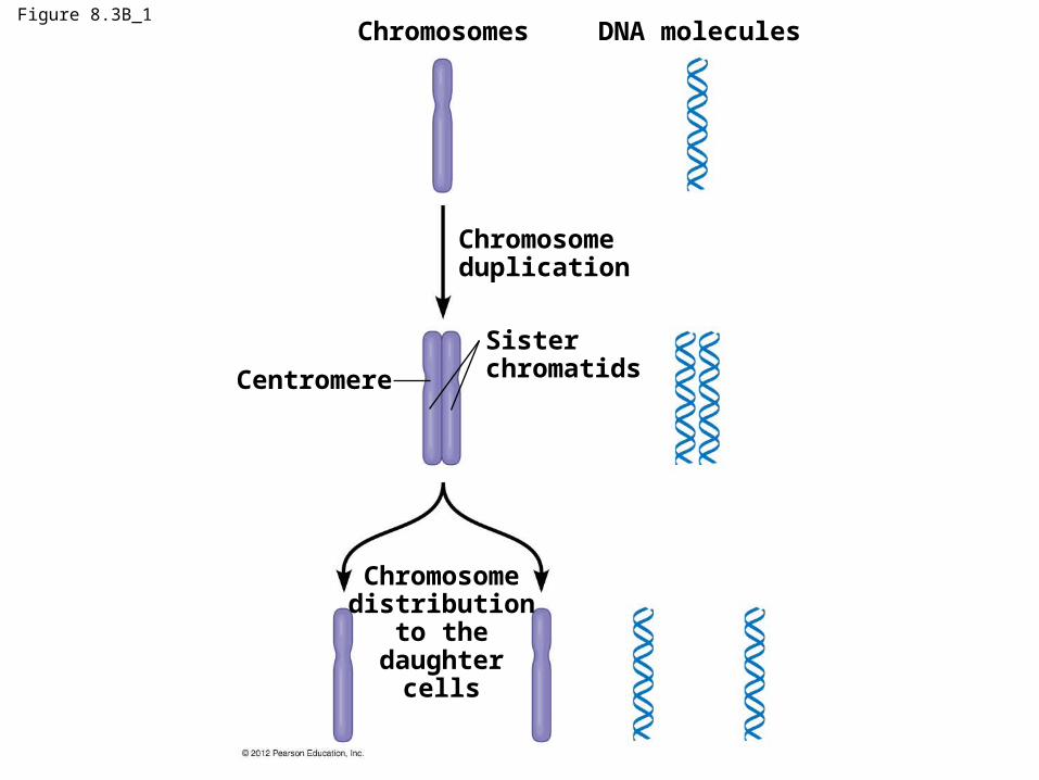
Figure 8.3B_1Chromosomes
Centromere
Chromosomeduplication
Sisterchromatids
Chromosomedistribution
to thedaughter
cells
DNA molecules

The cell cycle extends from the time a cell is first formed until its own division.
The cell cycle multiplies cells
© 2012 Pearson Education, Inc.

The cell cycle consists of two stages, characterized as follows:
1. Interphase:
G1—growth, increase in cytoplasm
– S—duplication of chromosomes
– G2—growth, preparation for division
1. Mitotic phase: nuclear division
– Mitosis—division of the chromosomes in nucleus
– Cytokinesis—division of cytoplasm
The cell cycle multiplies cells
© 2012 Pearson Education, Inc.

G1
(first gap)S
(DNA synthesis)
G2
(second gap)
M
CytokinesisM
itosi
s
I N T E R P H A S E
PHASE
TT
MI
OIC

Mitosis progresses through a series of stages (nuclear division):
– prophase
– prometaphase
– metaphase
– anaphase
– telophase.
Cytokinesis often overlaps telophase.
Cell division is a continuum of dynamic changes
© 2012 Pearson Education, Inc.
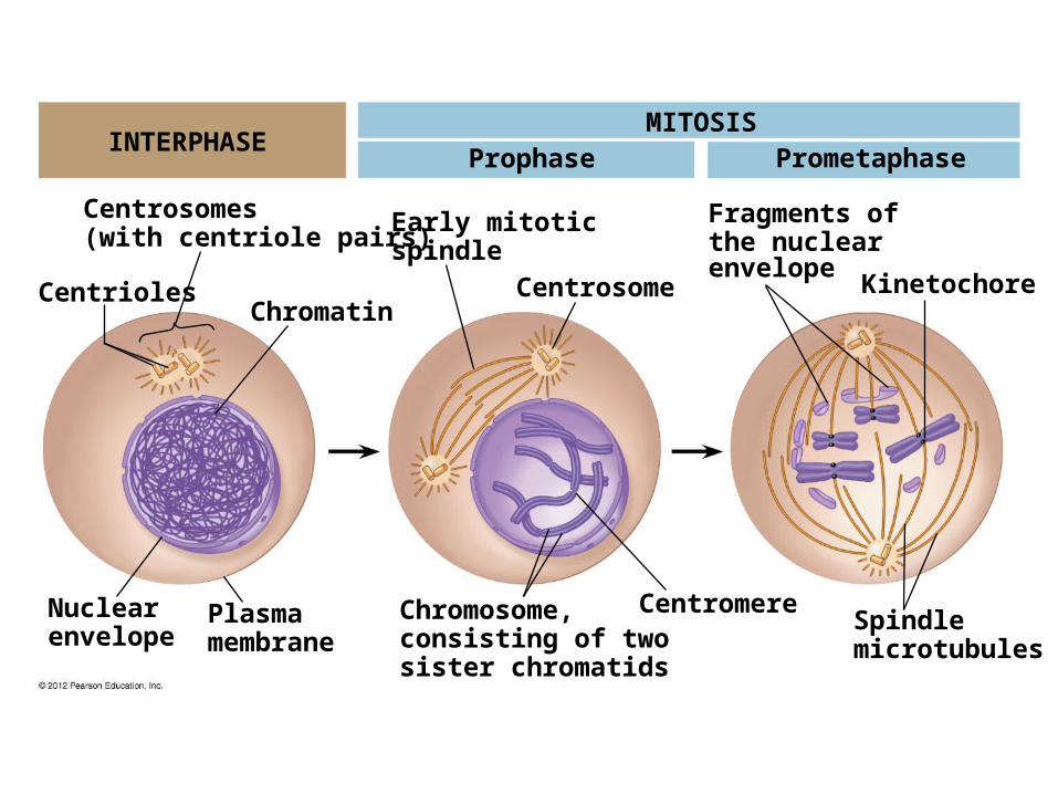
INTERPHASEMITOSIS
Prophase Prometaphase
Centrosome
Early mitoticspindle
Chromatin
Fragments ofthe nuclearenvelope
Kinetochore
Centrosomes(with centriole pairs)
Centrioles
Nuclearenvelope
Plasmamembrane
Chromosome,consisting of twosister chromatids
CentromereSpindlemicrotubules

Prophase
– In the cytoplasm microtubules begin to emerge from centrosomes, forming the mitotic spindle.
– In the nucleus chromosomes coil and become compact and visible with a light microscope
– Nucleoli disappear.
Cell division is a continuum of dynamic changes
© 2012 Pearson Education, Inc.
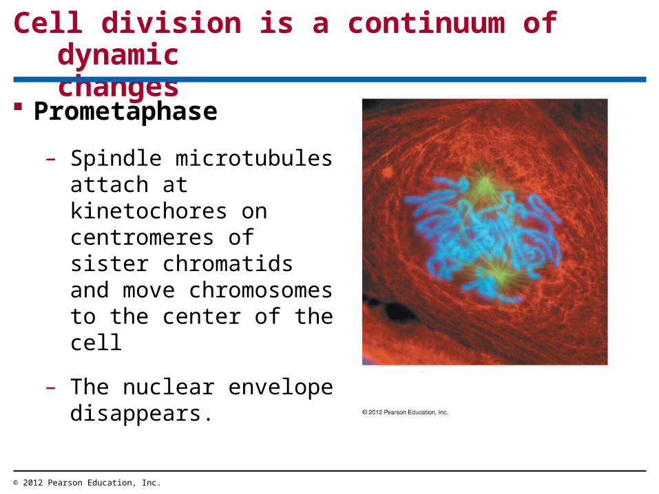
Prometaphase
– Spindle microtubules attach at kinetochores on centromeres of sister chromatids and move chromosomes to the center of the cell
– The nuclear envelope disappears.
Cell division is a continuum of dynamic changes
© 2012 Pearson Education, Inc.

MITOSIS
AnaphaseMetaphase Telophase and Cytokinesis
Metaphaseplate Cleavage
furrow
NuclearenvelopeformingDaughter
chromosomesMitoticspindle

Metaphase
– The mitotic spindle is fully formed and chromosomes align at the cell equator.
– Kinetochores of sister chromatids are facing the opposite poles of the spindle.
Cell division is a continuum of dynamic changes
© 2012 Pearson Education, Inc.

Anaphase – Sister chromatids separate
at centromeres and daughter chromosomes are moved to opposite poles of the cell
– The cell elongates due to lengthening of nonkinetochore microtubules.
Cell division is a continuum of dynamic changes
© 2012 Pearson Education, Inc.

Telophase– The cell continues to elongate
and nuclear envelope forms around chromosomes at each pole, establishing daughter nuclei
– Chromatin uncoils and nucleoli reappear.
– The spindle disappears.
Cell division is a continuum of dynamic changes
© 2012 Pearson Education, Inc.

Cytokinesis differs in animal and plant cells.
In animal cells a cleavage furrow forms from a contracting ring of microfilaments, interacting with myosin; the cleavage furrow deepens to separate the contents into two cells.
Cell division is a continuum of dynamic changes
© 2012 Pearson Education, Inc.

Cytokinesis
Cell wallof theparent cell
Daughternucleus
Cell wall
Plasmamembrane
Vesiclescontainingcell wallmaterial
Cell plateforming
Newcell wall
Cell plate Daughtercells
In plant cells a cell plate forms in the middle, from vesicles containing cell wall material; the cell plate grows outward to reach the edges, dividing the contents into two cells, each
with a plasma membrane and cell wall.

Anchorage, cell density, and chemical growthfactors affect cell division
© 2012 Pearson Education, Inc.
Cell division is controlled by
– presence of nutrients
– density-dependent inhibition
– anchorage dependence
– growth factors
The addition ofgrowthfactor
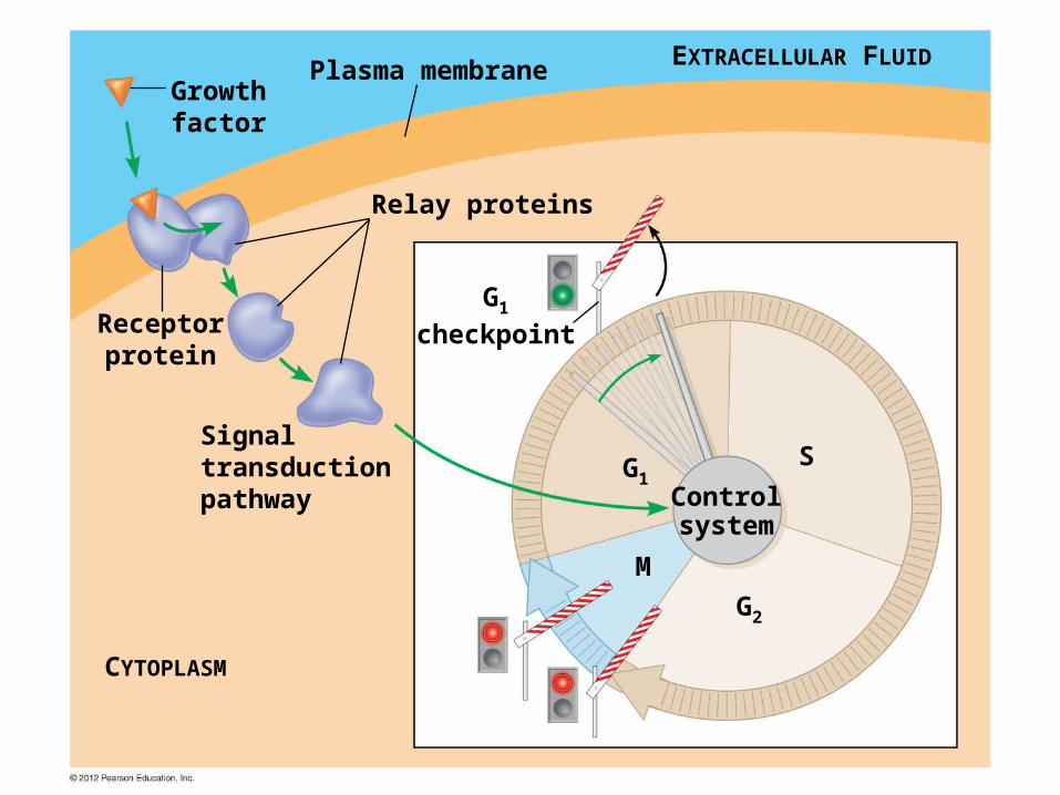
Receptorprotein
Signaltransductionpathway
Growthfactor
Relay proteins
Plasma membrane EXTRACELLULAR FLUID
CYTOPLASM
G1
checkpoint
G1S
M
G2
Controlsystem

Tumor
Glandulartissue
Cancer cells invade neighboring tissue
Metastasis
Tumor cells can spread through lymph
and blood vessels
Cancer cells escape controls on the cell cycle.
Cancer cells divide rapidly, often in the absence of growth factors

Cancer Treatment
Radiaiton
Proton Therapy
Chemotherapy
© 2012 Pearson Education, Inc.
MEIOSIS AND CROSSING OVER
© 2012 Pearson Education, Inc.

Meiosis is a process that converts diploid nuclei to haploid nuclei.
– Diploid cells have two sets of chromosomes.
– Haploid cells have one set of chromosomes.
Meiosis occurs in sex organs, producing gametes: sperm and eggs.
Fertilization is the union of sperm and egg.
The zygote has a diploid chromosome number, one set from each parent.
Gametes have a single set of chromosomes
© 2012 Pearson Education, Inc.

Haploid gametes (n 23)
Egg cell
Sperm cell
Fertilization
n
n
Meiosis
Ovary Testis
Diploidzygote(2n 46)
2n
MitosisKey
Haploid stage (n)Diploid stage (2n)
Multicellular diploidadults (2n 46)
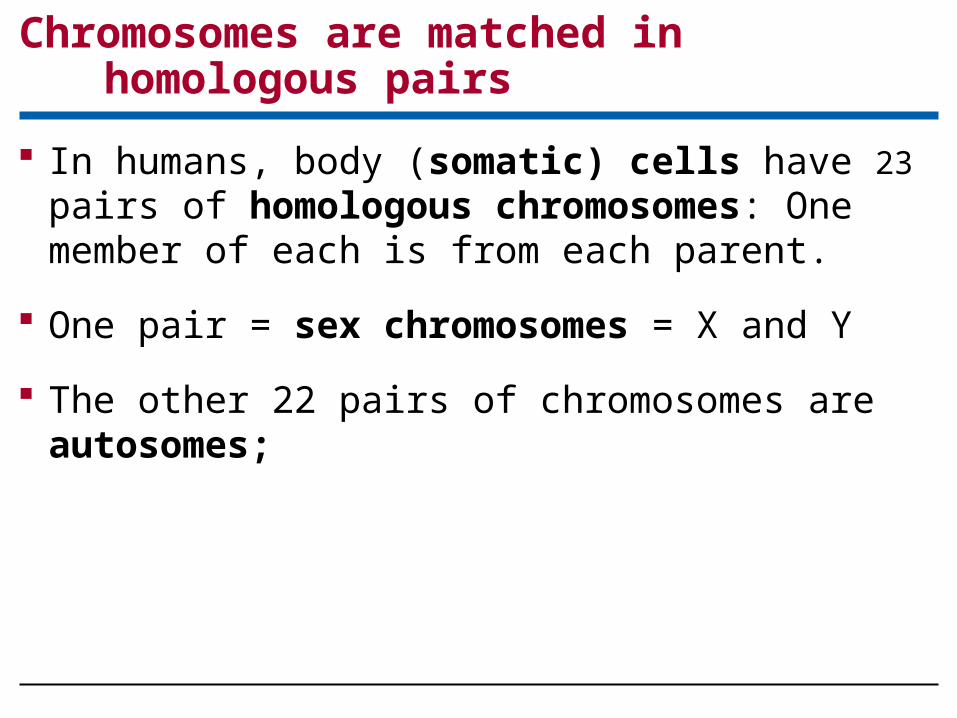
In humans, body (somatic) cells have 23 pairs of homologous chromosomes: One member of each is from each parent.
One pair = sex chromosomes = X and Y
The other 22 pairs of chromosomes are autosomes;
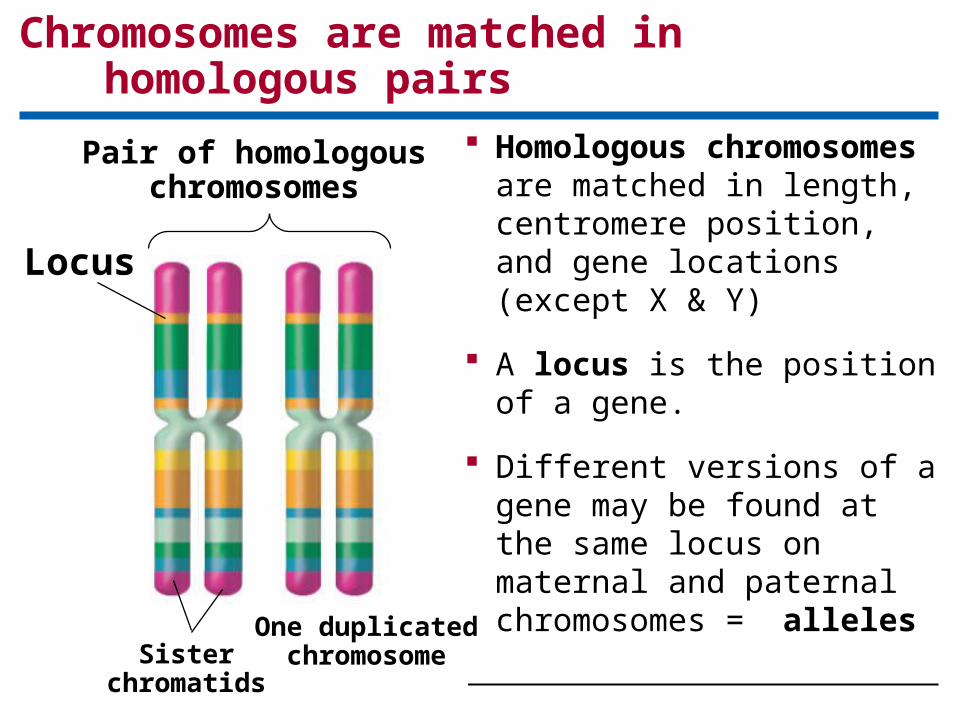
Chromosomes are matched in homologous pairs
Chromosomes are matched in homologous pairs
© 2012 Pearson Education, Inc.
Homologous chromosomes are matched in length, centromere position, and gene locations (except X & Y)
A locus is the position of a gene.
Different versions of a gene may be found at the same locus on maternal and paternal chromosomes = alleles
Locus
Pair of homologouschromosomes
One duplicatedchromosomeSister
chromatids

A pair ofhomologouschromosomesin a diploidparent cell
A pair ofduplicatedhomologouschromosomes
Sisterchromatids
1 2 3
INTERPHASE MEIOSIS I MEIOSIS II

Meiosis I – Prophase I
– Chromatin condensation
– Homologous chromosomes come together
– The homologous pairs, now form a tetrad.
– Nonsister chromatids exchange genetic material by crossing over at the chiasma
Meiosis reduces the chromosome number from diploid to haploid
© 2012 Pearson Education, Inc.

Centrosomes(with centriolepairs) Centrioles
Sites of crossing over
Spindle
Tetrad
Nuclearenvelope
Chromatin Sisterchromatids Fragments
of thenuclearenvelope
Chromosomes duplicate Prophase IINTERPHASE:
MEIOSIS I

Meiosis I – Metaphase I – Tetrads align at the cell equator.
Meiosis I – Anaphase I – Homologous pairs separate and move toward opposite poles of the cell.
Meiosis reduces the chromosome number from diploid to haploid
© 2012 Pearson Education, Inc.
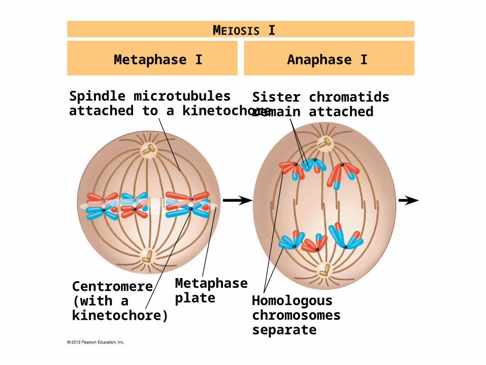
Centromere(with akinetochore)
Spindle microtubulesattached to a kinetochore
Metaphaseplate Homologous
chromosomesseparate
Sister chromatidsremain attached
Metaphase I Anaphase I
MEIOSIS I

Meiosis I – Telophase I –duplicated chromosomes have reached the poles, nuclear envelope re-forms around chromosomes, each nucleus has the haploid number of chromosomes.
Meiosis reduces the chromosome number from diploid to haploid
© 2012 Pearson Education, Inc.

Meiosis II follows meiosis I without chromosome duplication.
Each of the two haploid products enters meiosis II.
Meiosis II – Prophase II
– Chromosomes condense (if uncoiled).
– If re-formed nuclear envelope breaks up again.
Meiosis reduces the chromosome number from diploid to haploid
© 2012 Pearson Education, Inc.

Meiosis II – Metaphase II – Duplicated chromosomes align at equator.
Meiosis II – Anaphase II sister chromatids separate, move towards opposite pole.
Meiosis II – Telophase II - four haploid cells are produced
Meiosis reduces the chromosome number from diploid to haploid
© 2012 Pearson Education, Inc.

Figure 8.13_right
Cleavagefurrow
Telophase I and Cytokinesis Prophase II Metaphase II Anaphase II
MEIOSIS II: Sister chromatids separate
Sister chromatidsseparate
Haploid daughtercells forming
Telophase IIand Cytokinesis

Mitosis and meiosis both
– begin with diploid parent cells
– have chromosomes duplicated during interphase.
However the end products differ.
– Mitosis produces two genetically identical diploid somatic daughter cells.
– Meiosis produces four genetically unique haploid gametes.
Mitosis and meiosis have important similarities and differences
© 2012 Pearson Education, Inc.
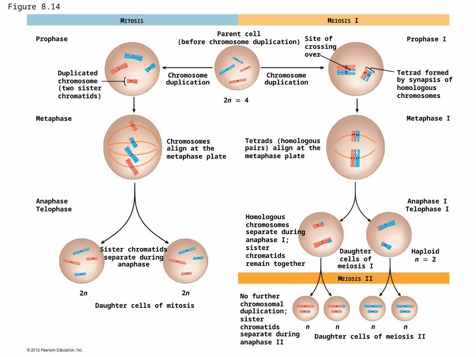
Figure 8.14
Prophase
Metaphase
Duplicatedchromosome(two sisterchromatids)
MITOSIS
Parent cell(before chromosome duplication)
Chromosomeduplication
Chromosomeduplication
Site ofcrossingover
2n 4
Chromosomesalign at themetaphase plate
Tetrads (homologouspairs) align at themetaphase plate
Tetrad formedby synapsis ofhomologouschromosomes
Metaphase I
Prophase I
MEIOSIS I
AnaphaseTelophase
Sister chromatidsseparate during
anaphase
2n 2n
Daughter cells of mitosis
No furtherchromosomalduplication;sisterchromatidsseparate duringanaphase II
n n n n
Daughter cells of meiosis II
Daughtercells of
meiosis I
Haploidn 2
Anaphase ITelophase I
Homologouschromosomesseparate duringanaphase I;sisterchromatidsremain together
MEIOSIS II

Possibility A
Two equally probablearrangements ofchromosomes at
metaphase I
Possibility B
Metaphase II
Gametes
Combination 3 Combination 4Combination 2Combination 1

A karyotype is an ordered display of magnified images of an individual’s chromosomes arranged in pairs.
Karyotypes
– are often produced from dividing cells arrested at metaphase of mitosis and
– allow for the observation of
– homologous chromosome pairs,
– chromosome number, and
– chromosome structure.
A karyotype is a photographic inventory of an individual’s chromosomes
© 2012 Pearson Education, Inc.

Bloodculture
Packed redand whiteblood cells
Centrifuge
Fluid
Hypotonicsolution Fixative
Whitebloodcells
Stain
3
2
1

4
5

Trisomy 21
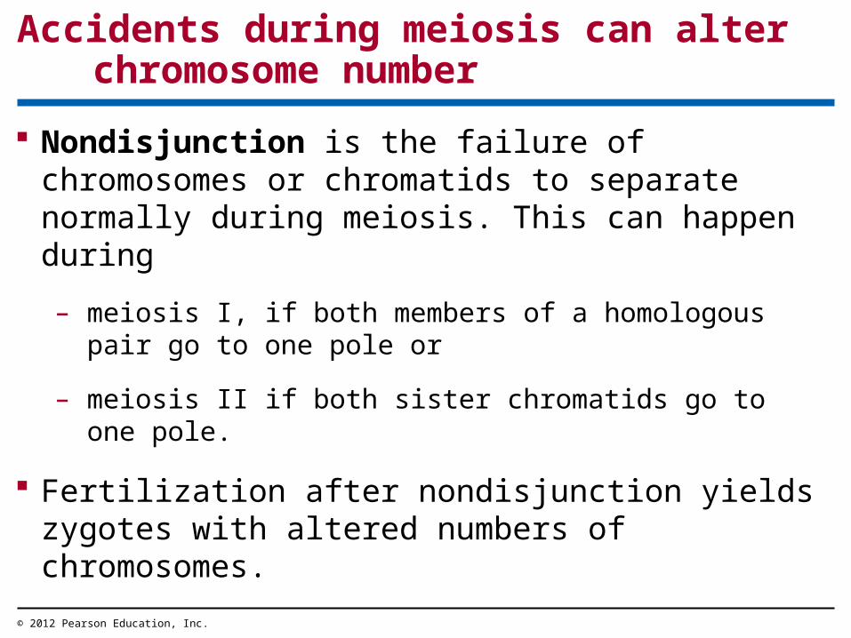
Nondisjunction is the failure of chromosomes or chromatids to separate normally during meiosis. This can happen during
– meiosis I, if both members of a homologous pair go to one pole or
– meiosis II if both sister chromatids go to one pole.
Fertilization after nondisjunction yields zygotes with altered numbers of chromosomes.
Accidents during meiosis can alter chromosome number
© 2012 Pearson Education, Inc.
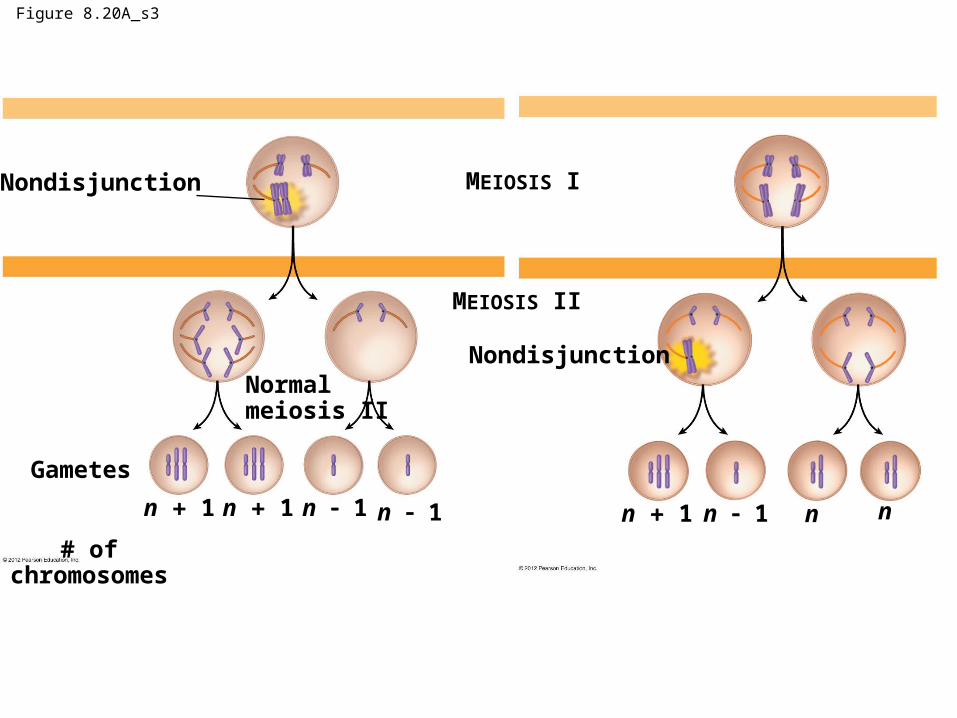
Figure 8.20A_s3
Nondisjunction
Normalmeiosis II
Gametes
# ofchromosomes
n 1 n 1 n 1 n 1
Nondisjunction
MEIOSIS II
MEIOSIS I
n 1 n 1 nn

Chromosome breakage can lead to rearrangements that can produce
– genetic disorders or, if changes occur in somatic cells, cancer.
These rearrangements may include
– a deletion, the loss of a chromosome segment,
– a duplication, the repeat of a chromosome segment,
– an inversion, the reversal of a chromosome segment, or
– a translocation, the attachment of a segment to a nonhomologous chromosome that can be reciprocal
Alterations of chromosome structure can cause birth defects and cancer
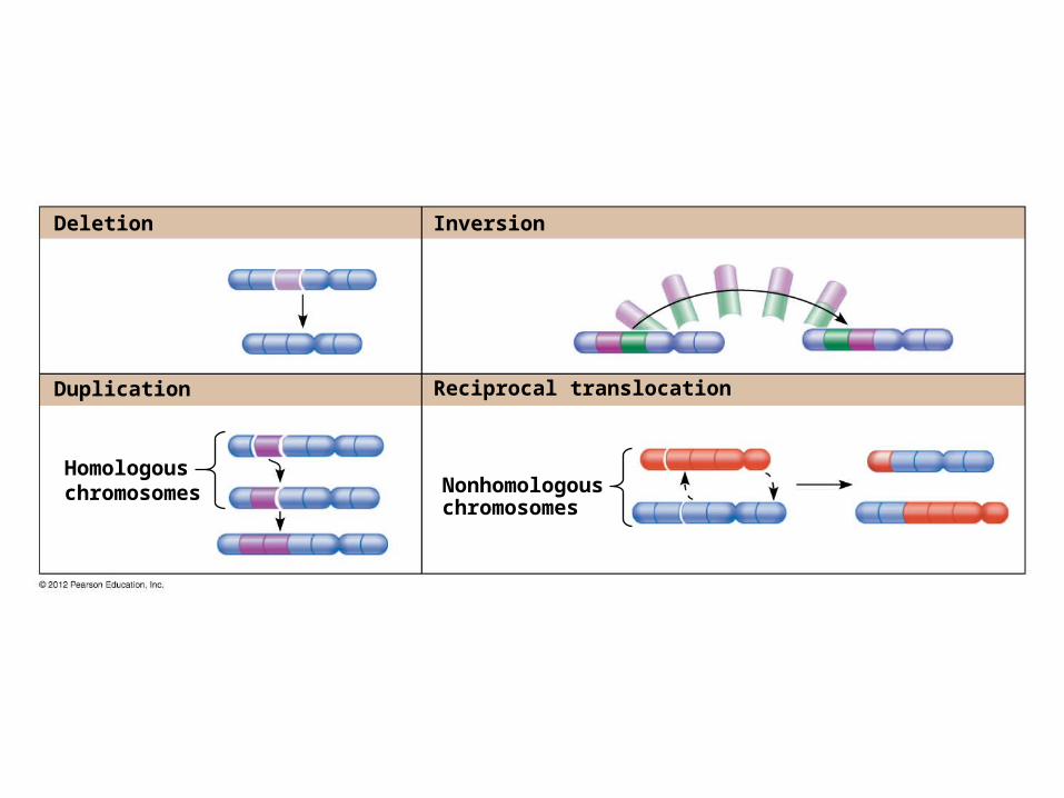
Deletion
Duplication
Inversion
Reciprocal translocation
Homologouschromosomes Nonhomologous
chromosomes
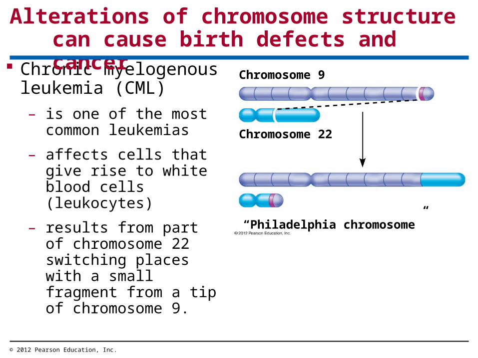
Chronic myelogenous leukemia (CML)
– is one of the most common leukemias
– affects cells that give rise to white blood cells (leukocytes)
– results from part of chromosome 22 switching places with a small fragment from a tip of chromosome 9.
Alterations of chromosome structure can cause birth defects and cancer
© 2012 Pearson Education, Inc.
Chromosome 9
Chromosome 22
“Philadelphia chromosome”
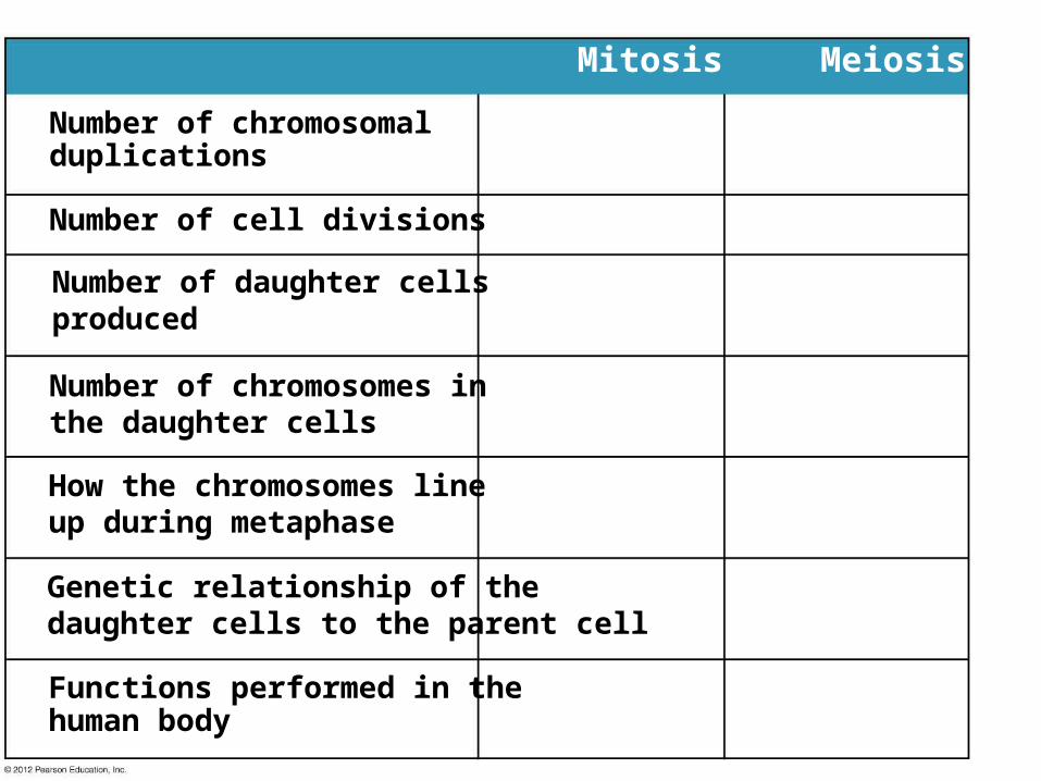
Number of chromosomalduplications
Number of cell divisions
Number of daughter cellsproduced
Number of chromosomes inthe daughter cells
How the chromosomes lineup during metaphase
Genetic relationship of thedaughter cells to the parent cell
Functions performed in thehuman body
Mitosis Meiosis


