
British Liver Transplant Group
Pathology meeting
September 2014
Leeds cases

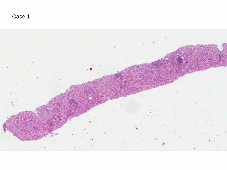
Leeds Case 1
Male 61 years
Liver transplant for HCV cirrhosis with HCC in
January 2014.
Now raised ALT and bilirubin, ? acute rejection.
Biopsy 4 ½ months post transplant.
Case 1

Case 1

Case 1

Case 1

Case 1

Case 1
CK7

Case 1
CK7

Case 1
CK7

Case 1
CK7

Leeds case 1
4 ½ months post Tx hep C. Raised ALT and bili
Preferred diagnosis: fibrosing cholestatic hepatitis
Differential diagnosis: biliary obstruction
acute rejection – biliary pattern ? antibody mediated
CK7 ductular reaction without CK7+ve intermediate
hepatobiliary cells – favours FCH
C4d (not scanned) – non-specific mild positivity.

Leeds case 1
03/06/14 4 ½ months post Tx hep C. Raised ALT and bili
At biopsy: ALT111, bili 23, alk phos 366, HCV PCR 5.2*106.
Diagnosis FCH.
Follow up:
Application for compassionate Hep C treatment
24/06/14: At 3 weeks post biopsy, started Sofosbuvir and
Ledipasvir 12 weeks course.
Viral load 43 at 2 weeks, -ve at 4 and 8 weeks
Most recent liver tests: ALT 18, bili 8, alk phos 261
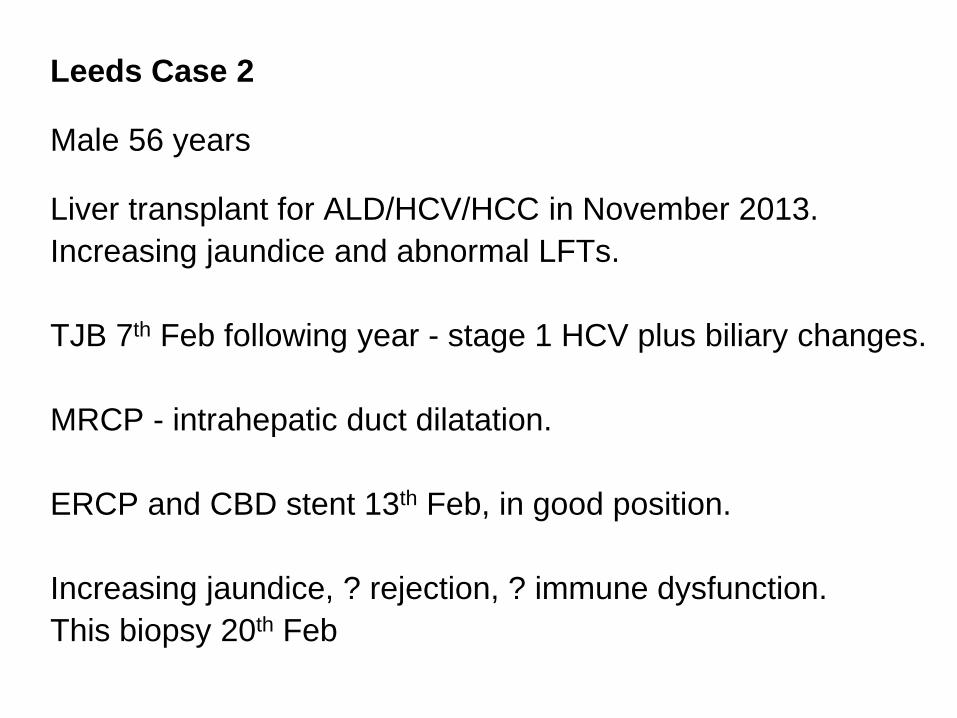
Leeds Case 2 Male 56 years Liver transplant for ALD/HCV/HCC in November 2013.
Increasing jaundice and abnormal LFTs.
TJB 7th Feb following year - stage 1 HCV plus biliary changes.
MRCP - intrahepatic duct dilatation.
ERCP and CBD stent 13th Feb, in good position.
Increasing jaundice, ? rejection, ? immune dysfunction.
This biopsy 20th Feb

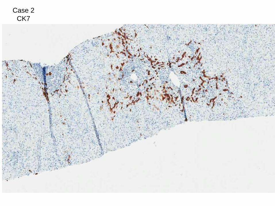
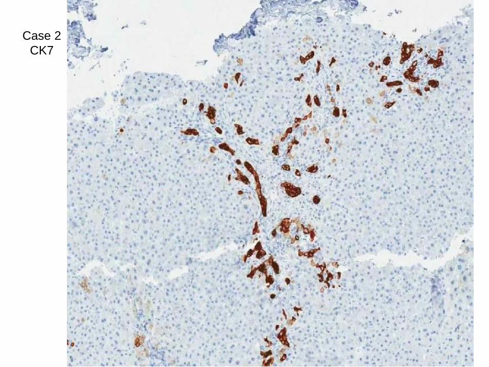
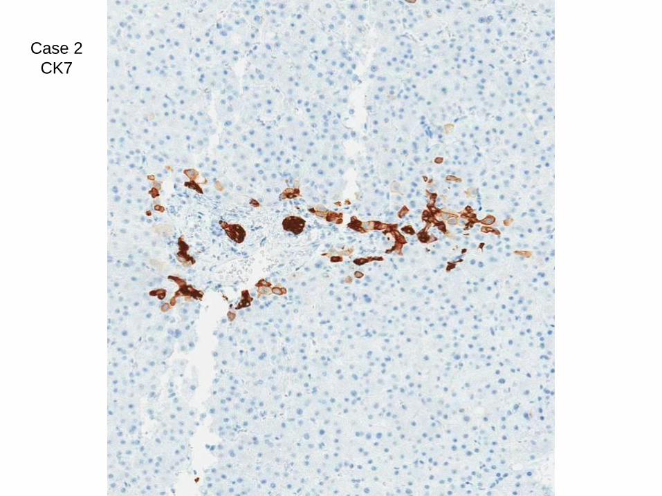
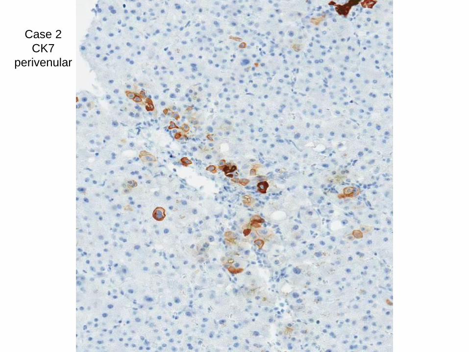
Case 2

Case 2
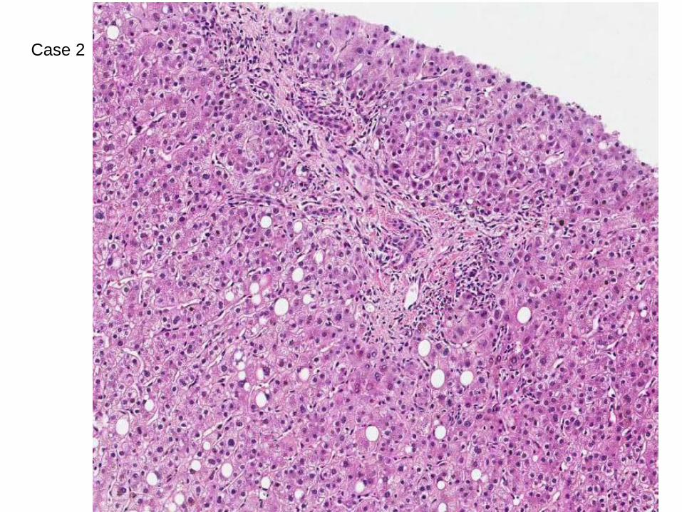
Case 2
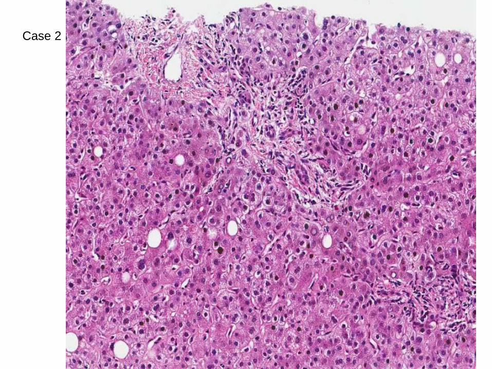
Case 2

Case 2

Case 2
CK7
Case 2
CK7
Case 2
CK7

Case 2
CK7
Case 2
CK7
Case 2
CK7
perivenular

Case 2
Male 56 years Liver transplant for ALD/HCV/HCC in November 2013.
Increasing jaundice and abnormal LFTs.
Diagnosis: cholestasis, features suggestive of large bile duct
obstruction. Minimal changes attributable to hep C. Mild
steatosis. Not FCH. No copper associated protein.
Compared with previous biopsy – LBDO features more
evident now.

Case 2
Male 56 years Liver transplant for ALD/HCV/HCC in November 2013.
Increasing jaundice and abnormal LFTs.
Explant – 3x HCC of which largest 50mm, with macroscopic vascular invasion.
Follow up ERCP July 2014: bile duct narrowing at anastomosis unchanged, good flow.
Jaundice cleared, Alk phos 706, bili 11.
Follow up: Sept 2014 CT surveillance = 3x pulmonary nodules in right middle lobe of lung. Likely mets – waiting AFP and further imaging.

Histopathologic distinction between
fibrosing cholestatic hepatitis C
and biliary obstruction
BO group - HCV-ve with imaging-proven biliary obstruction (BO).
n=38, of which 16 post OLT
FCH-C group – HCV post transplant, cholestasis, no BO or vascular
complication n=13
at least 3 of:
a) marked ductular reaction with expansion of at least some portal tracts
b) marked hepatocyte swelling/ballooning with lobular disarray, involving
majority of hepatic lobules
c) periportal sinusoidal/pericellular fibrosis
d) any degree of canalicular cholestasis with or without intracellular
cholestasis
Salomayo M, Verna EC, Lefkowitch JH, Moreira RK.
Am J Surg Pathol 2013;37;1837-1844
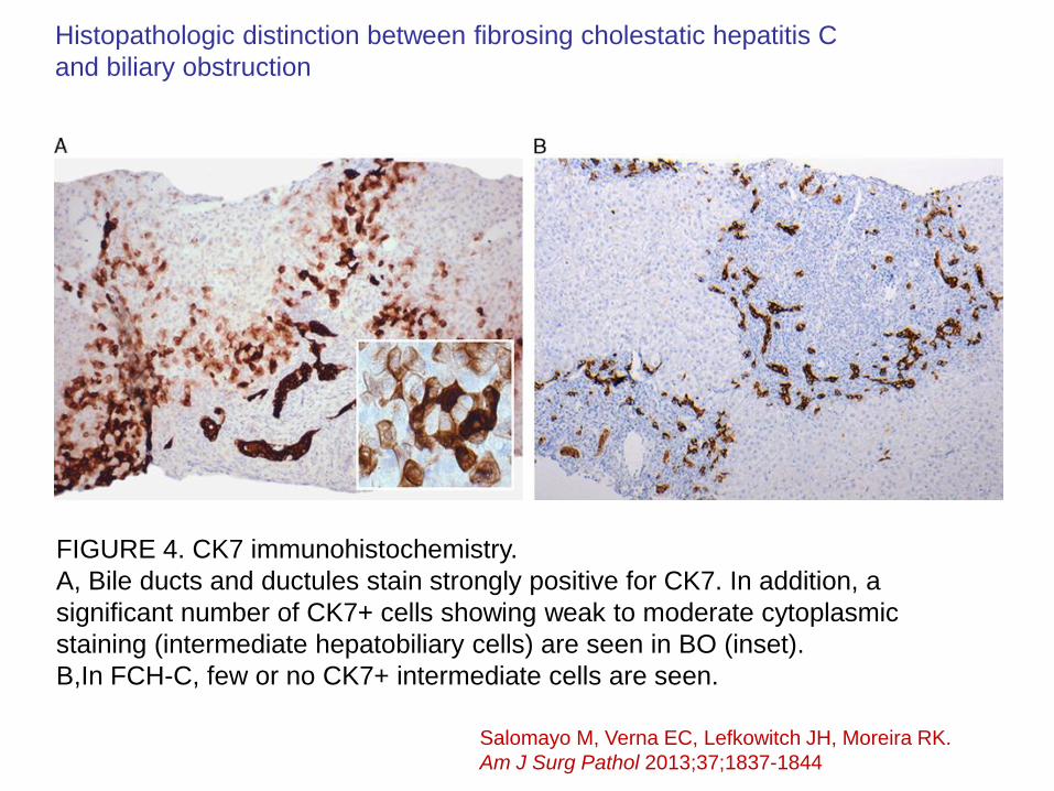
FIGURE 4. CK7 immunohistochemistry.
A, Bile ducts and ductules stain strongly positive for CK7. In addition, a
significant number of CK7+ cells showing weak to moderate cytoplasmic
staining (intermediate hepatobiliary cells) are seen in BO (inset).
B,In FCH-C, few or no CK7+ intermediate cells are seen.
Histopathologic distinction between fibrosing cholestatic hepatitis C
and biliary obstruction
Salomayo M, Verna EC, Lefkowitch JH, Moreira RK.
Am J Surg Pathol 2013;37;1837-1844

FIGURE 5. Distribution of FCH-C (solid) and BO (stripes) cases according to
(A) degree of ductular reaction,
(B) copper accumulation within hepatocytes,
(C) extent of CK7+ intermediate cells in cases with moderate/marked ductular reaction.
Histopathologic distinction between fibrosing cholestatic hepatitis C
and biliary obstruction
FCH, n=13 Biliary obstruction, n=38

Aberrant cytokeratin 7 expression of centrilobular
hepatocytes: a clinicopathological study
Matsukuma S, Takeo H, Kono T, Nagata Y, Sato K.
Department of Pathology, Japan Self Defense Forces Central Hospital,
Tokyo, Japan
113 biopsies, range of diagnoses,
CK7+ve centrilobular hepatocytes in 49.6%
17/34 HCV (50%)
8/21 HBV (38%)
17/27 ASH or NASH (63%)
4/7 PBC (57%)
2/5 AIH (40%)
0/4 DILI (0%)
8/15 other or mixed (53%)
Histopathology 2012, 61, 857–862
Correlates with:
Age
AST (not ALT)
Late stage in viral hepatitis
Centrilob scar in fatty liver
Prominent in BCS
Overall and periportal CK7+
cells, but not always parallel
with periportal CK7+

Figure 1. Cytokeratin 7 (CK7) expression of centrilobular hepatocytes surrounding central vein (CV).
A, CK7-positive centrilobular hepatocytes focally showing a binuclear feature (arrow) (CK7 immunostain).
B, CK7-positive hepatocytes distributed diffusely around CV in a case of Budd–Chiari syndrome (CK7 immunostain).
C, CK7-positive hepatocytes involving centrilobular area (arrows) and periportal area (arrowhead) surrounding portal area (P).
High-power view (C, inset) showing aggregated CK7-positive centrilobular hepatocytes with mono- or bi-nuclear features (CK7
immunostain).
D, CK7-positive centrilobular hepatocytes (arrows) and scattered CK7-positive rounded ⁄ cuboidal cells
(arrowheads), accompanied by centrilobular scar (asterisks) surrounding CV. High-power view of CK7-positive rounded cell
with scanty cytoplasm (D, inset), resembling features of hepatic progenitor cell (CK7 immunostain).

Leeds Case 3 Male 52 years Liver transplant for HCV and HCC in February 2014.
Readmitted with jaundice 16 weeks post Tx,
ALT >1000 ? cause. (biopsy 3.1: Slides 269970 and 269972).
Received pulsed steroids, LFTs improved for some days.
Now again worsening, so repeat biopsy, 18 weeks post Tx
(biopsy 3.2, slides 269973, 269975 and 269976)

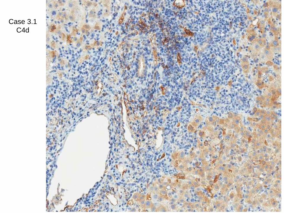
Case 3.1 – first biopsy, 16 weeks

Case 3.1

Case 3.1

Case 3.1

Case 3.1

Case 3.1
C4d

Case 3.1
C4d
Case 3.1
C4d

Case 3.2 second biopsy, 18 weeks

Case 3.2

Case 3.2

Case 3.2

Case 3.2

Case 3.2
CK7

Case 3.2
CK7

Case 3.2
C4d

Case 3.2
C4d

Leeds Case 3 Male 52 years Liver transplant for HCV and HCC in February 2014.
Readmitted with jaundice 16 weeks post transplant,
ALT >1000, bili >300 ? cause. (slides 3.1. 16 weeks)
Diagnosis: marked acute rejection (8/9) also lobular inflammation, ? Rec HCV, ? CMV
Received pulsed steroids, LFTs improved for some days.
Now again worsening, so repeat biopsy (Slides 3.2, 18 weeks)
Diagnosis: ongoing acute rejection, improved since previous biopsy. Cholestasis – but biopsy features overall not suggestive of LBDO or FCH – little ductular reaction on CK7.

Leeds Case 3 Male 52 years Liver transplant for HCV and HCC in February 2014.
Follow up – ALT and histology improving so no further pulse steroids after second biopsy.
Since then – ALT improved steadily down to 120, but this week up again at 630, bili 52, so repeat biopsy today (17th Sept, 33 weeks post Tx), result awaited.
Patient feels very well.
Was this AMR? – DSA were not done (not routine in Leeds).
C4d was being done as protocol for 2 months – first one was not reported at the time of biopsy report.
DSA – only done in a handful of cases last year, if severe acute rejection not responding – clinicians feel this has not altered management
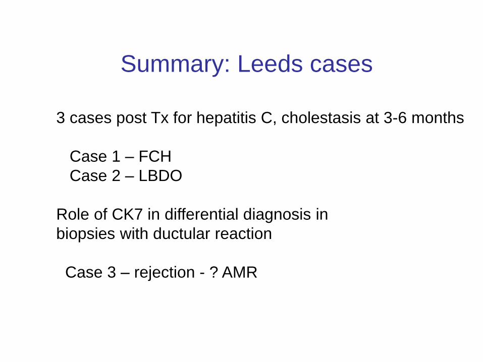
Summary: Leeds cases
3 cases post Tx for hepatitis C, cholestasis at 3-6 months
Case 1 – FCH
Case 2 – LBDO
Role of CK7 in differential diagnosis in
biopsies with ductular reaction
Case 3 – rejection - ? AMR


