BIRTH ASPHYXIA and
The “New” Consensus Statement
Keith Bolton
Rahima Moosa Mother & Child Hospital

THE HERD IS UNDER THREAT

HPCSA CIVIL COURTS CRIMINAL COURTS

Background
• The child with cerebral palsy, intellectual
impairment and epilepsy naturally generates
huge sympathy from society and the courts.
• Apart from ―poor care‖, the capping of the RAF,
the introduction of ―contingency fees‖ and the
empowerment of the people have all resulted in
a massive increase in litigation…especially for
birth asphyxia!
• There is a Tsunami of cases in the courts
especially in Gauteng, KZN & the Eastern
Cape!

The Size of the Problem!
• A world-wide problem.
• In RSA, as with all other
financial matters; Gauteng
(North & South High Courts)
lead the way in attracting
litigation.
• South Gauteng has
approximately 1000 cases
pending against them – 80%
are CP cases.
• The current “quantum” in
these cases is between R10-
30,000,000
• GHD are losing 90% of cases
• This means R10.8 billion in
payouts!
• Private obstetricians and
paediatricians not exempt!

‗It has never been safer to have a baby
and never been more dangerous to be an
obstetrician1‘
(or paediatrician/neonatologist2)
1. MacLennan A, Nelson KB, Hankins G, Speer M. Who will deliver our grandchildren?
Implications of cerebral palsy litigation. JAMA 2005;294(13):1688-1690.
2. Bolton K. Personal opinion

Cerebral Palsy
• Was there someone to blame?

Clark SM et al Antenatal antecedents and the impact of obstetric care in the
etiology of cerebral palsy Clin Obstet Gynecol 2008; 51(4):

N Badawi et al
BMJ 1998;317:1554

The Causes for Cerebral Palsy
( Developed Communities)
Nelson KB. Causative factors in cerebral palsy. Clin Obstets Gynec 2008; 51(4): 749-62

The Causes for Cerebral Palsy
( Developed Communities)
Country Years of Birth % related to
asphyxia
USA 1959-66 12%
Australia 1975-80 17%
Finland 1978-82 24%
Ireland 1981-1983 23%
England 1984-87 17%
Sweden 1987-90 17%
Sweden 1991-94 24%
Volpe JJ. Neurology of the Newborn 5th Edition, 2008; ISBN: 978-1-4160-3995-2
Relationship between intrapartum asphyxia and cerebral palsy: Term infants

The Causes for Cerebral Palsy
( Less Developed Communities)
Van Toorn R et al. Aetiology of cerebral palsy in children presenting at Tygerberg Hospital
SAJ Child Health 2007; 1(2): 74-77

The Causes for Cerebral Palsy
( Less Developed Communities)
Van Toorn R et al. Aetiology of cerebral palsy in children presenting at Tygerberg Hospital
SAJ Child Health 2007; 1(2): 74-77
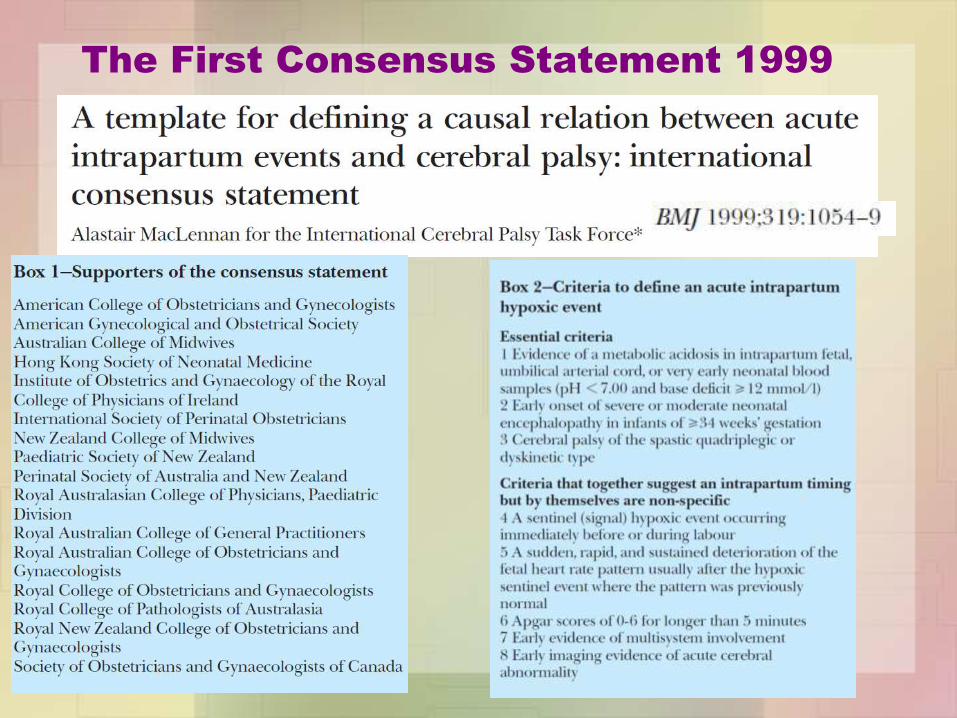
The First Consensus Statement 1999

Neonatal Encephalopathy 1
Neonatal encephalopathy is a clinically defined
syndrome of disturbed neurologic function in the
earliest days of life in an infant born at, or
beyond 35 weeks of gestation, manifested by a
subnormal level of consciousness or seizures,
and often accompanied by difficulty with initiating
and maintaining respiration and depression of
tone and reflexes.
Definition
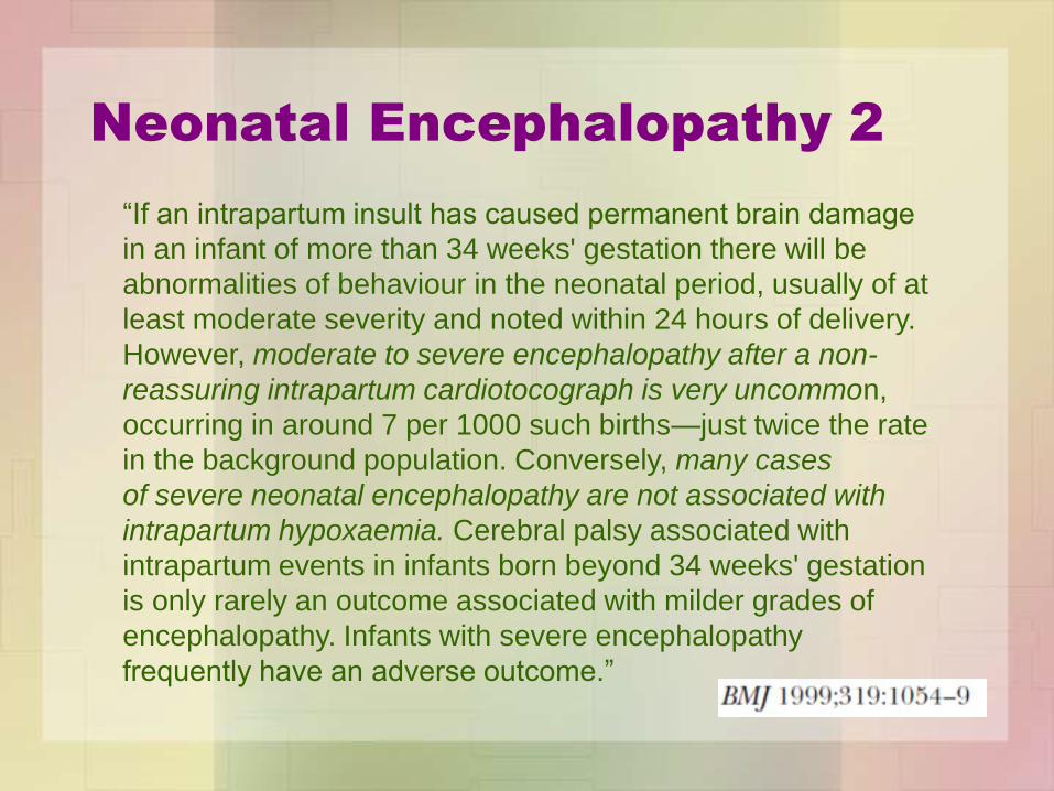
Neonatal Encephalopathy 2
―If an intrapartum insult has caused permanent brain damage
in an infant of more than 34 weeks' gestation there will be
abnormalities of behaviour in the neonatal period, usually of at
least moderate severity and noted within 24 hours of delivery.
However, moderate to severe encephalopathy after a non-
reassuring intrapartum cardiotocograph is very uncommon,
occurring in around 7 per 1000 such births—just twice the rate
in the background population. Conversely, many cases
of severe neonatal encephalopathy are not associated with
intrapartum hypoxaemia. Cerebral palsy associated with
intrapartum events in infants born beyond 34 weeks' gestation
is only rarely an outcome associated with milder grades of
encephalopathy. Infants with severe encephalopathy
frequently have an adverse outcome.‖

Sarnat HB & Sarnat MS. Neonatal encephalopathy following fetal distress.
A clinical and electroencephalographic study. Arch Neurol. 1976 Oct;33(10):696-705
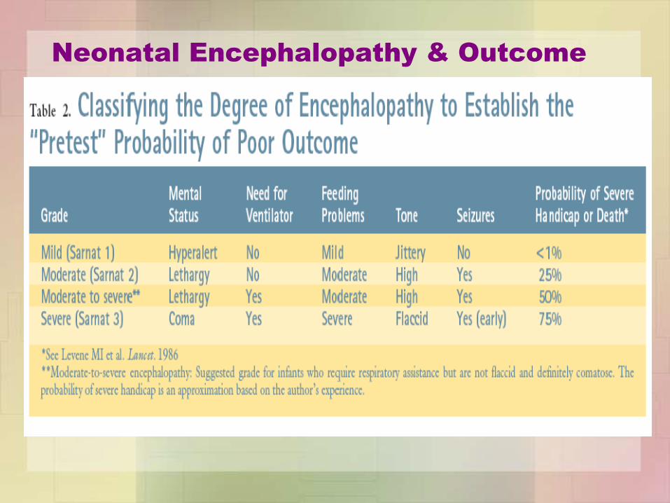
Neonatal Encephalopathy & Outcome

Thompson Score* for Neonatal
Encephalopathy
*Thompson CM et al: The value of a scoring system for hypoxic ischaemic
encephalopathy in predicting neurodevelopmental outcome.
Acta Paediatr 1997, 86(7):757-761.
Sign 0 1 2 3
Tone normal hyper hypo flaccid LOC normal hyperalert, stare lethargic comatouse Fits none < 3 per day > 2 per day Posture normal fisting, cylcing strong distal flexion decerebrate Moro normal partial absent Grasp normal poor absent Suck normal poor absent ± bites Respir normal hyperventilation brief apnea IPPV (apnea) Fontanell normal full, not tense tense

The 2nd
Consensus Statement 2003
Essential Criteria
1. Evidence of metabolic acidosis
2. Early onset moderate or severe NE
3. Spastic quadriplegic/dyskinetic CP
AND:
4. Exclusion of other identifiable
etiologies, such as trauma,
coagulation disorders, infectious
conditions, or genetic disorders.
Criteria suggestive of intrapartum timing
5. Sentinel Event
6. Sudden fetal brady CTG Criteria
7. Apgar Scores 0-3 > 5 mins
8. Multisystem involvement < 72 hours
9. Early Imaging criteria

The 3rd
Consensus Statement 2014
• American Academy of Pediatrics
• American College of Nurse-Midwives
• American Gynecologic and Obstetrical Society
• American Society for Reproductive Medicine
• Association of Women‘s Health, Obstetric and Neonatal Nurses
• Australian Collaborative Cerebral Palsy Research Group
• Child Neurology Society
• Japan Society of Obstetrics and Gynecology
• March of Dimes Foundation
• Royal Australian and New Zealand College of Obstetricians and Gynaecologists
• Royal College of Obstetricians and Gynaecologists
• Society for Maternal-Fetal Medicine
• Society of Obstetricians and Gynaecologists of Canada

The 3rd
Consensus Statement 2014;
Motivation for a “second edition”
• The Task force recognised that a broader perspective
was necessary.
• Based on ―the sober recognition that knowledge gaps
still preclude a definitive test or set of markers that
accurately identifies, with high sensitivity and specificity,
an infant in whom neonatal encephalopathy is
attributable to an acute intrapartum event.‖
• As a comprehensive etiologic evaluation is not possible,
the term hypoxic–ischemic encephalopathy should best
be replaced by neonatal encephalopathy because
neither hypoxia nor ischemia can be assumed to have
been the unique initiating causal mechanism.

The 3rd
Consensus Statement 2014 -
Causal Pathways to CP in Term Infants

The 3rd
Consensus Statement 2014
Clinical Examples of Causal Pathway A
• Causal Pathway A – PRF is a sentinel
event. Examples: Abruptio, Prolapsed cord,
Ruptured uterus etc
• Causal Pathway A – PRF is not a sentinel
event. Examples: Pregnancy induced
hypertension, Antepartum haemorrhage,
Fetal growth retardation etc.
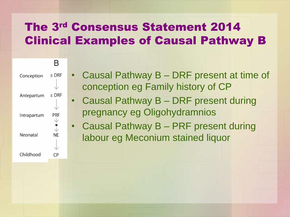
The 3rd
Consensus Statement 2014
Clinical Examples of Causal Pathway B
• Causal Pathway B – DRF present at time of
conception eg Family history of CP
• Causal Pathway B – DRF present during
pregnancy eg Oligohydramnios
• Causal Pathway B – PRF present during
labour eg Meconium stained liquor

The 3rd
Consensus Statement 2014
Clinical Example of Causal Pathway C
• Causal Pathway C DRF present at conception
eg Advanced maternal age
• Causal Pathway C PRF occurs early intrapartum
eg Chorioamnionitis
• Causal Pathway C – Neonatal Encephalopathy
may be absent
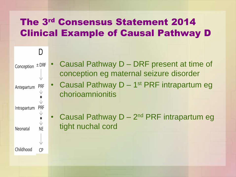
The 3rd
Consensus Statement 2014
Clinical Example of Causal Pathway D
• Causal Pathway D – DRF present at time of
conception eg maternal seizure disorder
• Causal Pathway D – 1st PRF intrapartum eg
chorioamnionitis
• Causal Pathway D – 2nd PRF intrapartum eg
tight nuchal cord
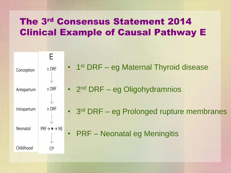
The 3rd
Consensus Statement 2014
Clinical Example of Causal Pathway E
• 1st DRF – eg Maternal Thyroid disease
• 2nd DRF – eg Oligohydramnios
• 3rd DRF – eg Prolonged rupture membranes
• PRF – Neonatal eg Meningitis
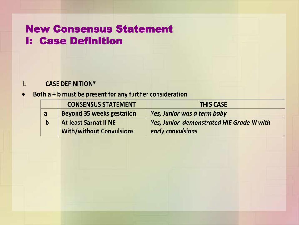
New Consensus Statement
I: Case Definition
I. CASE DEFINITION*
Both a + b must be present for any further consideration
CONSENSUS STATEMENT THIS CASE
a Beyond 35 weeks gestation Yes, Junior was a term baby
b At least Sarnat II NE With/without Convulsions
Yes, Junior demonstrated HIE Grade III with early convulsions
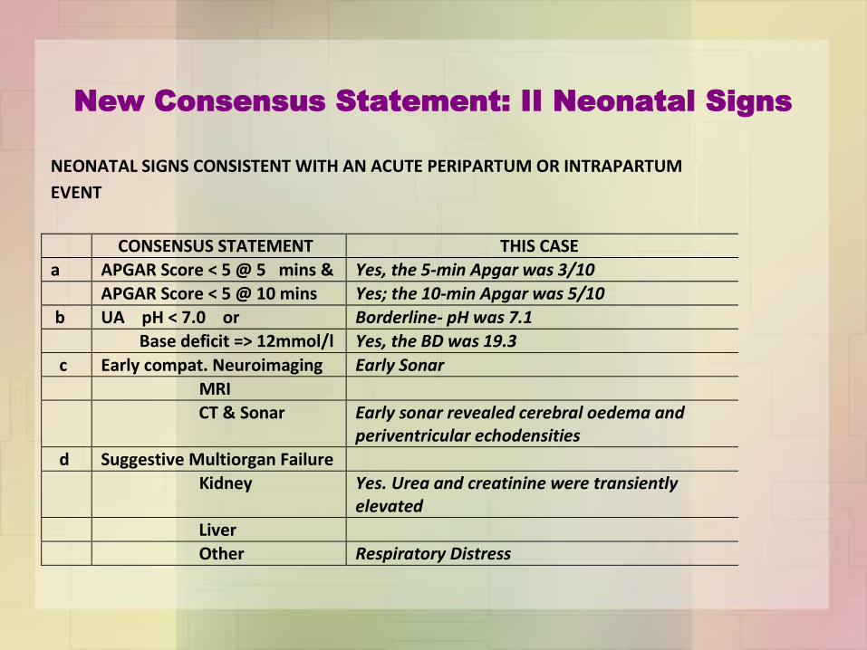
New Consensus Statement: II Neonatal Signs
I. NEONATAL SIGNS CONSISTENT WITH AN ACUTE PERIPARTUM OR INTRAPARTUM
EVENT
CONSENSUS STATEMENT THIS CASE
a APGAR Score < 5 @ 5 mins & Yes, the 5-min Apgar was 3/10
APGAR Score < 5 @ 10 mins Yes; the 10-min Apgar was 5/10
b UA pH < 7.0 or Borderline- pH was 7.1
Base deficit => 12mmol/l Yes, the BD was 19.3
c Early compat. Neuroimaging Early Sonar
MRI
CT & Sonar Early sonar revealed cerebral oedema and periventricular echodensities
d Suggestive Multiorgan Failure
Kidney Yes. Urea and creatinine were transiently elevated
Liver
Other Respiratory Distress

New Consensus Statement: III Time & Timing of
events suggestive of intrapartum/peripartum
role
I. TYPE AND TIMING OF CONTRIBUTORY FACTORS THAT ARE CONSISTENT WITH AN
ACUTE PERIPARTUM OR INTRAPARTUM EVENT
CONSENSUS STATEMENT THIS CASE
a Sentinel H/I Event No details are available to me
Ruptured Uterus
Major Abruptio placentae
Umbilical Cord Prolapse
Amniotic Fluid Embolus
Maternal CVS Collapse
Fetal exsanguination
Other
b Fetal Heart Rate Patterns No details are available to me
Category I or II without asph
Initial vs Labour CTG Abn
Cat II Initially
Cat I > Cat III
Cat I > Other CTG Abn
c Imaging & Timing of insult
Cranial Ultrasonography Early Sonar was compatible with intrapartum asphyxia
Early MRI Not done. An MRI is essential at this stage.
Patterns of Damage HI
Patterns of Damage not HI
d Proximal contributing factor Yes. Evidence of Chorioamnionitis was present at birth
Distal contributing factor/s None apparent

New Consensus Statement: IV
Developmental Outcome Compatible
I. DEVELOPMENTAL OUTCOME IS SPASTIC QUADRIPLEGIA OR DYSKINETIC CEREBRAL
PALSY
CONSENSUS STATEMENT THIS CASE
a Spastic/Dystonic Quad Apparently “mixed” spastic / dystonic quadriplegic CP is present. Neurological examination is required
b Other Subtypes CP
c Other development disorder ? Epilepsy, Intellectual impairment Neurological examination is required
Apgar Scores
• Low Apgar scores at 5 and10 minutes clearly confer
an increased relative risk of CP and the degree of
Apgar abnormality at 5 & 10 minutes correlates with
the risk of CP.
• BUT, most infants with low Apgars will not develop
CP!
• If the Apgar score at 5 minutes is > 6/10, then it is
highly improbably that peri-partum hypoxia-iscaemia
played major role in causing neonatal
encephalopathy.

Apgar Scores –
Policy Statement AAP & ACOG1
• It is inappropriate to use an Apgar score alone to establish the
diagnosis of asphyxia.
• An Apgar score assigned during resuscitation is not equivalent
to a score assigned to a spontaneously breathing infant.
• An Apgar score of 0-3 at five minutes is associated with a
slightly increased risk of CP. Conversely 75% of children with
CP have had normal scores at 5 minutes.
• A five minute score of 7 to 10 is considered normal.
• The risk of poor neurological outcome increases when the
Apgar score is 3 or less at 10, 15 and 20 minutes.
• [Apart from asphyxia] the Apgar score is affected by gestational
age, maternal medications, resuscitation, cardiorespiratory and
neurological conditions.
1. AAP & ACOG. The Apgar Score. Pediatrics 2006; 117(4): 1444-1447

Three-Tier Fetal Heart
Rate Interpretation System
• Category I
Baseline rate 110-160 beats/min
Moderate variability
Absence of any late or variable
decelerations
Early decelerations may or may not be
present
Accelerations may or may not be present
Require routine observations without any specific action required

Three-Tier Fetal Heart
Rate Interpretation System
Category II
• Baseline Rate
Tachycardia or Bradycardia not with absent baseline variability
• Baseline FHR Variability
Minimal, Absent or Marked baseline variability
• Absence of Induced Accelerations (eg scalp stimulation)
• Periodic or Episodic Decelerations
Recurrent variable decels with min or mod baseline variability
Prolonged decels >2min but < 10min
Recurrent late decels with mod baseline variability
Variable decels with other characteristics such as slow return to
baseline, ―overshoots‖ or ―shoulders‖.
Indeterminate tracings; require continued surveillance & re-evaluation

Three-Tier Fetal Heart
Rate Interpretation System
Category III
• Absent baseline FHR variability with any of:
Recurrent late decels
Recurrent variable decels
Bradycardia
• Sinusoidal Pattern
Abnormal tracings predictive of fetal acidemia. Require prompt actions.

CTG
1. A Category I or Category II fetal heart rate tracing when
associated with Apgar scores of 7 or higher at 5 minutes,
normal umbilical cord arterial blood (± 1 standard deviation), or
both is not consistent with an acute hypoxic–ischemic event.
2. There is a great distinction to be made between a patient who
initially presents with an abnormal fetal heart rate pattern and
one who develops an abnormal fetal heart rate pattern during
labour.
a) A category II fetal heart rate pattern lasting 60 minutes or more that was
identified on initial presentation with persistently minimal or absent
variability and lacking accelerations, even in the absence of decelerations,
is suggestive of a previously compromised or injured fetus.
b) The patient who presents with a Category I fetal heart rate pattern that
converts to Category III as defined by the Eunice Kennedy Shriver National
Institute of Child Health and Human Development guidelines is suggestive
of a hypoxic–ischemic event.
c) Additional fetal heart rate patterns that develop after a Category I fetal
heart rate pattern on presentation, which may suggest intrapartum timing of
a hypoxic–ischemic event, include tachycardia with recurrent decelerations
and persistent minimal variability with recurrent decelerations.

Cerebral Palsy Litigation:
Change Course or Abandon Ship
• One of the cardinal drivers of birth injury claims
is electronic fetal monitoring
• The scientific foundation for it‘s use is almost
non-existent
• Its false-positive rate exceeds 99%
• It does not predict cerebral palsy
• In the last 40 years monitoring has harmed more
mothers and babies than it ever helped
• Birth is a dangerous journey and monitoring
doesn‘t help
Sartwelle TP & Johnston JC. Journal of Child Neurology 2015, Vol. 30(7) 828-841
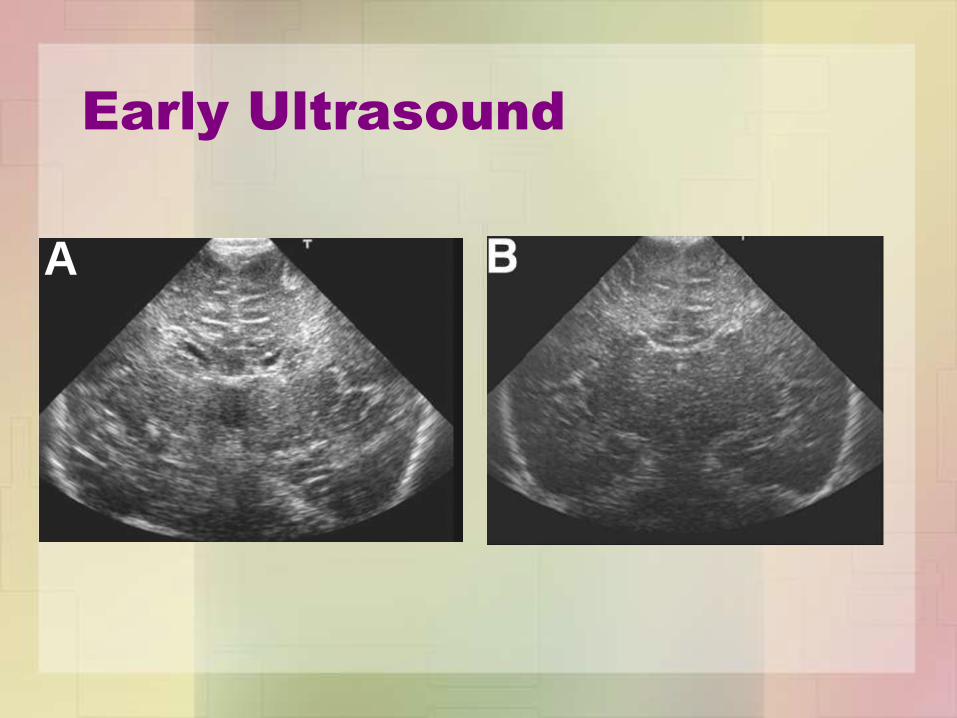
Early Ultrasound
A

Early Ultrasound in Term Asphyxia
BOO NY, CHANDRAN V, ZULFIQAR MA. Early cranial ultrasound changes as predictors of outcome during
first year of life in term infants with perinatal asphyxia. J. Paediatr. Child Health (2000) 36, 363–369
Imaging; the MRI
• An MRI is the best modality for demonstrating the
nature and extent of cerebral injury.
• Cranial ultrasonography and CT lack sensitivity
needed to define the injury.
• The optimal time to do the MRI is 10 days (7-21days)
• If an MRI done anytime after 24 hours shows no
injury, then it is unlikely that peripartum or
intrapartum H-I brain injury was a significant factor in
NE.

Classic MRI Patterns of an
Intrapartum Aetiology
Acute Profound HI Partial Prolonged Asphyxia
A
Predominantly Basal Ganglia Predominant “watershed” White Matter


Summary
• The ―epidemic‖ in litigation for alleged negligence in
obstetric & neonatal care has unsustainable economic
and morale consequences in both the private and
public health sectors
• There are many areas which need to be tackled to
minimise this problem. These involve medical, legal
and societal remedies.
• Understanding the complex nature of the role of birth
asphyxia in aetiology of irreversible brain damage and
the resultant cerebral palsy is important if we are to
justly compensate where negligence occurred but
equally, to vigorously defend healthcare professionals
against unjust prosecution.
―..the salient question being – ‗who, if anyone, will
be performing deliveries in private practice by the
end of the decade?‘ If the answer to the question is
‗nobody‘, the consequences will extend beyond
private healthcare. There are also serious
implications for the state sector that will require
addressing1‖.
1. Howarth GR. Obstetric risk avoidance; will anyone be offering obstetrics in
private practice by the end of the decade? S Afr Med J 2013;103(8):513-514.

Thank you!
• Keith Bolton declares that he has referenced
any work that was not his own.
• The visuals used are either referenced or
common property.
• He has received precious little incentive,
perverse or otherwise, for this talk.
• He declares no existing or potential conflicts
of interest other than giving numerous ―expert
paediatric opinions‖ to assist the Court in
negligence cases.


