
Basic Laboratory Skills: Making Microscopic
Measurements : Lab 2d
MICROSCOPIC MEASUREMENT AND DRAWING
Word of the Day:
Magnification: the process of enlarging something

How big is a cell?
• http://www.cellsalive.com/howbig.htm

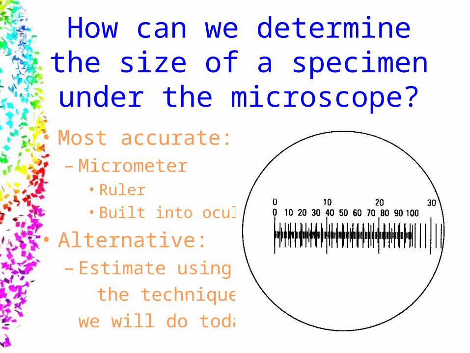
How can we determine the size of a specimen under the
microscope?• Most accurate:
– Micrometer• Ruler• Built into ocular
• Alternative:– Estimate using
the technique
we will do today!
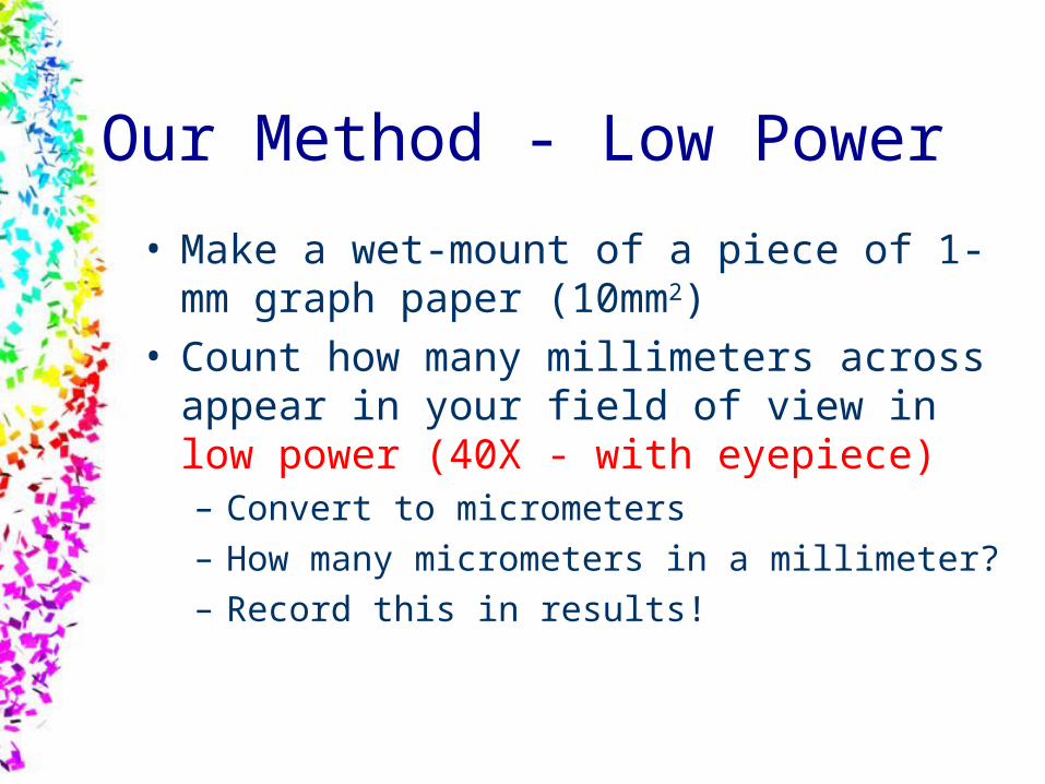
Our Method - Low Power
• Make a wet-mount of a piece of 1-mm graph paper (10mm2)
• Count how many millimeters across appear in your field of view in low power (40X - with eyepiece)– Convert to micrometers– How many micrometers in a millimeter?– Record this in results!

Making Microscope Measurements
1 Box = 2 mm
Total view = 2 boxes =4 mm across=4000 μm
40X

Field of Vision Sizes
• Field of visions for higher powers are based off of the 40X field
• 100X is 2.5 times smaller then 40X
• 400X is 10 times smaller then 40X
40X
100X 400X
2.5 times smaller
10 times smaller

Fields of Vision• 40X = 2 boxes = 4 mm 4000 μm
• 100X = 2 boxes/2.5 = .8 boxes = 1.6 mm 1600 μm
• 400X = 2 boxes/10 = .2 boxes = 0.4 mm 400 μm
*All measurement are for width across the middle*
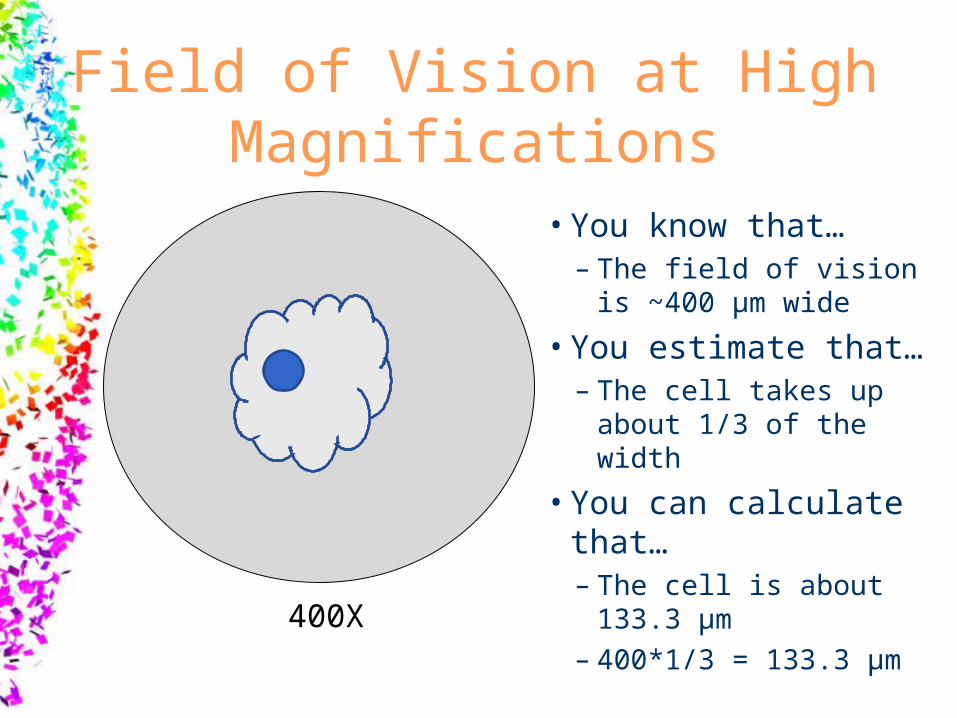
Field of Vision at High Magnifications
• You know that…– The field of vision is
~400 μm wide
• You estimate that…– The cell takes up
about 1/3 of the width
• You can calculate that…– The cell is about
133.3 μm– 400*1/3 = 133.3 μm
400X

Procedure1. Make a wet mount of mm grid graph paper. 2. Count how many 1-mm square go across
the diameter of the low power field.3. Convert the diameter measurement from
mm to µm.1 mm = 1000 µm
4. Create a data table and record the size of the low power field in both mm and µm
mm µm
40x
100x
400x
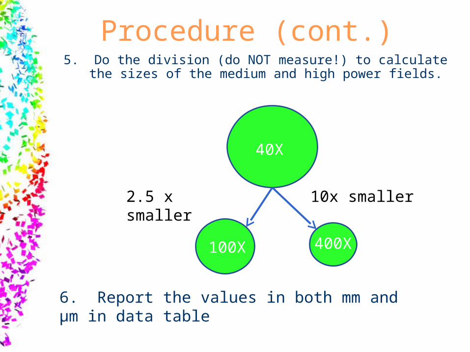
Procedure (cont.)5. Do the division (do NOT measure!) to calculate the
sizes of the medium and high power fields.
40X
100X 400X
10x smaller2.5 x smaller
6. Report the values in both mm and µm in data table

Next!!!!!!!
• Choose a slide and draw your organism– Use details– Use shading– List power drawn under– Make large

Example
• Paramecium– 400X


