Download - Bacteria - Morphology & Classification
Bacteria – Morphology & Classification
II MBBS
Dr Ekta ChourasiaLecturer, Microbiology
07.09.08 Dr Ekta Chourasia
Learning Objectives After completing this section you should be able to perform the
following objectives:
Define a prokaryotic cell and list the differences between prokaryotic and eukaryotic cell
Describe the structure of a bacterial cell and explain the function of its components
Explain why cell wall forms the basis for classification of bacteria
Explain the structural modifications (flagella) of the cell and their functional importance
07.09.08 Dr Ekta Chourasia
Introduction: Microorganisms – several classes of living beings
Based on the organization of their cellular structures, all living cells can be divided into two groups: eukaryotic and prokaryotic
Eukaryotic cell types - Animals, plants, fungi, protozoans, and algae
Prokaryotic cell types - bacteria & blue green algae

07.09.08 Dr Ekta Chourasia
Schematic of typical animal (eukaryotic) cell, showing subcellular components.
Organelles: (1) nucleolus (2) nucleus (3) ribosome (4) vesicle (5) rough endoplasmic reticulum (ER) (6) Golgi apparatus (7) Cytoskeleton (8) smooth ER (9) mitochondria (10) vacuole (11) cytoplasm (12) lysosome (13) centrioles
07.09.08 Dr Ekta Chourasia
Prokaryotic Cells much smaller (microns) and more simple than
eukaryotes
prokaryotes are molecules surrounded by a membrane and cell wall.
they lack a true nucleus and don’t have membrane bound organelles like mitochondria, etc.
large surface-to-volume ratio : nutrients can easily and rapidly reach any part of the cells interior

07.09.08 Dr Ekta Chourasia
Size of Bacteria
Unit of measurement in bacteriology is the micron (micrometre, µm)
Bacteria of medical importance 0.2 – 1.5 µm in diameter 3 – 5 µm in length
07.09.08 Dr Ekta Chourasia
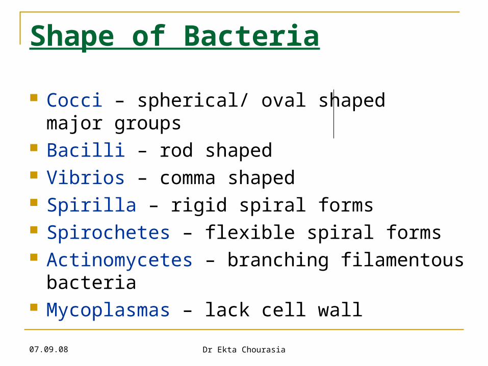
Shape of Bacteria
Cocci – spherical/ oval shaped major groups Bacilli – rod shaped Vibrios – comma shaped Spirilla – rigid spiral forms Spirochetes – flexible spiral forms Actinomycetes – branching filamentous
bacteria Mycoplasmas – lack cell wall
07.09.08 Dr Ekta Chourasia
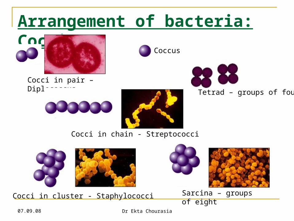
Arrangement of bacteria: Cocci
Cocci in pair – Diplococcus
Sarcina – groups of eight
Tetrad – groups of four
Cocci in chain - Streptococci
Cocci in cluster - Staphylococci
Coccus
07.09.08 Dr Ekta Chourasia
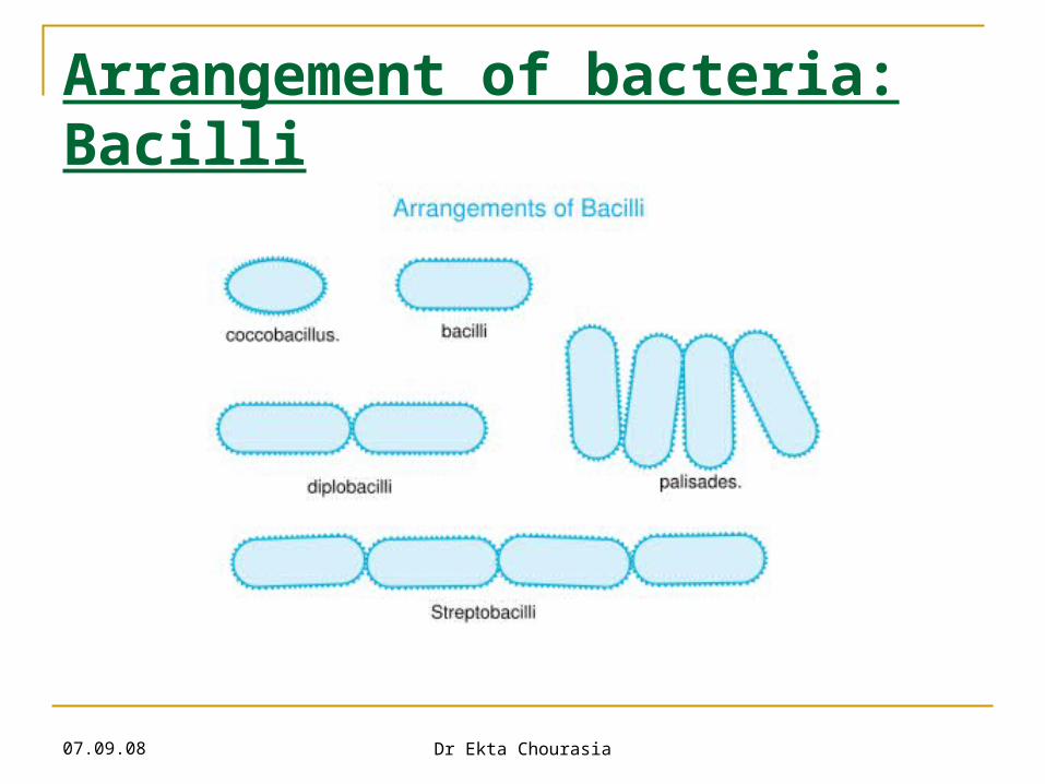
Arrangement of bacteria: Bacilli
07.09.08 Dr Ekta Chourasia
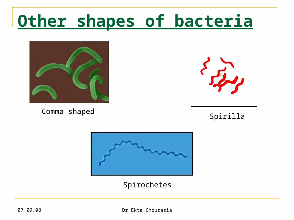
Other shapes of bacteria
Comma shaped
Spirochetes
Spirilla
07.09.08 Dr Ekta Chourasia
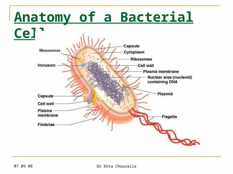
Anatomy of a Bacterial Cell
07.09.08 Dr Ekta Chourasia
Anatomy of A Bacterial Cell Outer layer – two components:
1. Rigid cell wall2. Cytoplasmic (Cell/ Plasma) membrane – present
beneath cell wall
Cytoplasm – cytoplasmic inclusions, ribosomes, mesosomes and nucleus
Additional structures – plasmid, slime layer, capsule, flagella, fimbriae (pili), spores

Structure & Function of Cell Components
07.09.08 Dr Ekta Chourasia
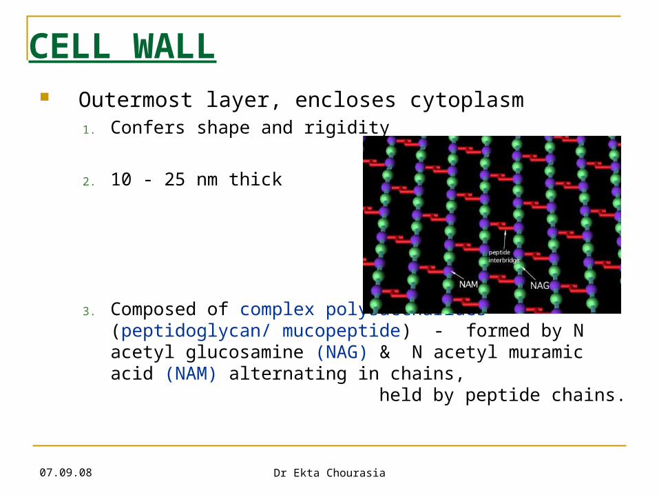
CELL WALL Outermost layer, encloses cytoplasm
1. Confers shape and rigidity
2. 10 - 25 nm thick
3. Composed of complex polysaccharides (peptidoglycan/ mucopeptide) - formed by N acetyl glucosamine (NAG) & N acetyl muramic acid (NAM) alternating in chains, held by peptide chains.
07.09.08 Dr Ekta Chourasia
Cell Wall
Cell wall – 4. Carries bacterial antigens – important in virulence &
immunity
5. Chemical nature of the cell wall helps to divide bacteria into two broad groups – Gram positive & Gram negative
6. Gram +ve bacteria have simpler chemical nature than Gram –ve bacteria.
7. Several antibiotics may interfere with cell wall synthesis e.g. Penicillin, Cephalosporins
07.09.08 Dr Ekta Chourasia
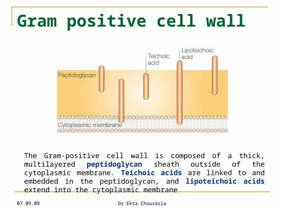
Gram positive cell wall
The Gram-positive cell wall is composed of a thick, multilayered peptidoglycan sheath outside of the cytoplasmic membrane. Teichoic acids are linked to and embedded in the peptidoglycan, and lipoteichoic acids extend into the cytoplasmic membrane
07.09.08 Dr Ekta Chourasia
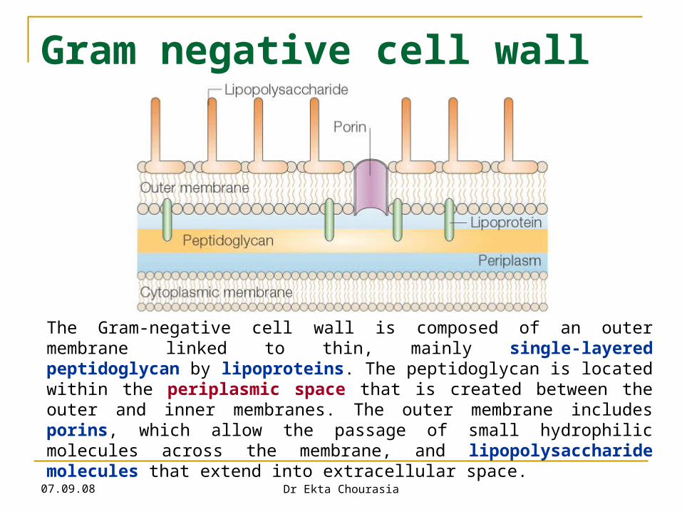
Gram negative cell wall
The Gram-negative cell wall is composed of an outer membrane linked to thin, mainly single-layered peptidoglycan by lipoproteins. The peptidoglycan is located within the periplasmic space that is created between the outer and inner membranes. The outer membrane includes porins, which allow the passage of small hydrophilic molecules across the membrane, and lipopolysaccharide molecules that extend into extracellular space.
07.09.08 Dr Ekta Chourasia
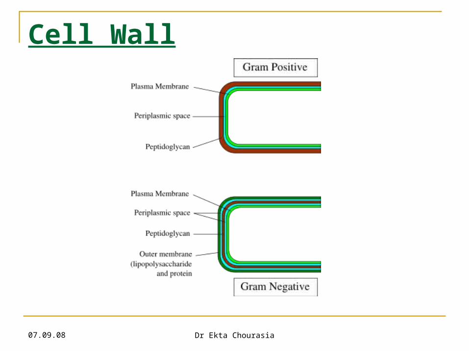
Cell Wall
07.09.08 Dr Ekta Chourasia
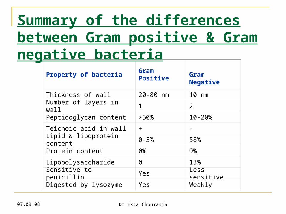
Summary of the differences between Gram positive & Gram negative bacteria
Property of bacteriaGram Positive
Gram Negative
Thickness of wall 20-80 nm 10 nm
Number of layers in wall 1 2
Peptidoglycan content >50% 10-20%
Teichoic acid in wall + -
Lipid & lipoprotein content 0-3% 58%
Protein content 0% 9%
Lipopolysaccharide 0 13%
Sensitive to penicillin Yes Less sensitive
Digested by lysozyme Yes Weakly
07.09.08 Dr Ekta Chourasia
Cytoplasmic (Plasma) membrane Thin layer 5-10 nm, separates cell wall from
cytoplasm
Acts as a semipermeable membrane: controls the inflow and outflow of metabolites
Composed of lipoproteins with small amounts of
carbohydrates
07.09.08 Dr Ekta Chourasia
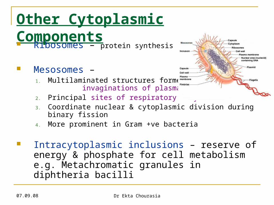
Other Cytoplasmic Components Ribosomes – protein synthesis
Mesosomes – 1. Multilaminated structures formed as
invaginations of plasma membrane2. Principal sites of respiratory enzymes3. Coordinate nuclear & cytoplasmic division during binary
fission4. More prominent in Gram +ve bacteria
Intracytoplasmic inclusions – reserve of energy & phosphate for cell metabolism e.g. Metachromatic granules in diphtheria bacilli
07.09.08 Dr Ekta Chourasia
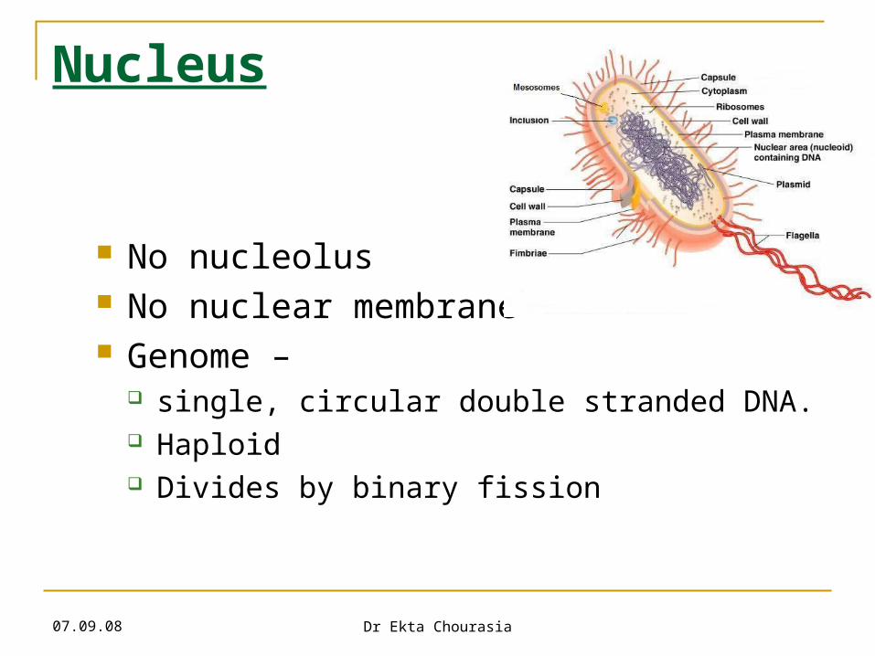
Nucleus
No nucleolus No nuclear membrane Genome –
single, circular double stranded DNA. Haploid Divides by binary fission
07.09.08 Dr Ekta Chourasia
Additional Organelles
1. Plasmid – Extranuclear genetic elements consisting of DNA
Transmitted to daughter cells during binary fission
May be transferred from one bacterium to another
Not essential for life of the cell
Confer certain properties e.g. drug resistance, toxicity
07.09.08 Dr Ekta Chourasia
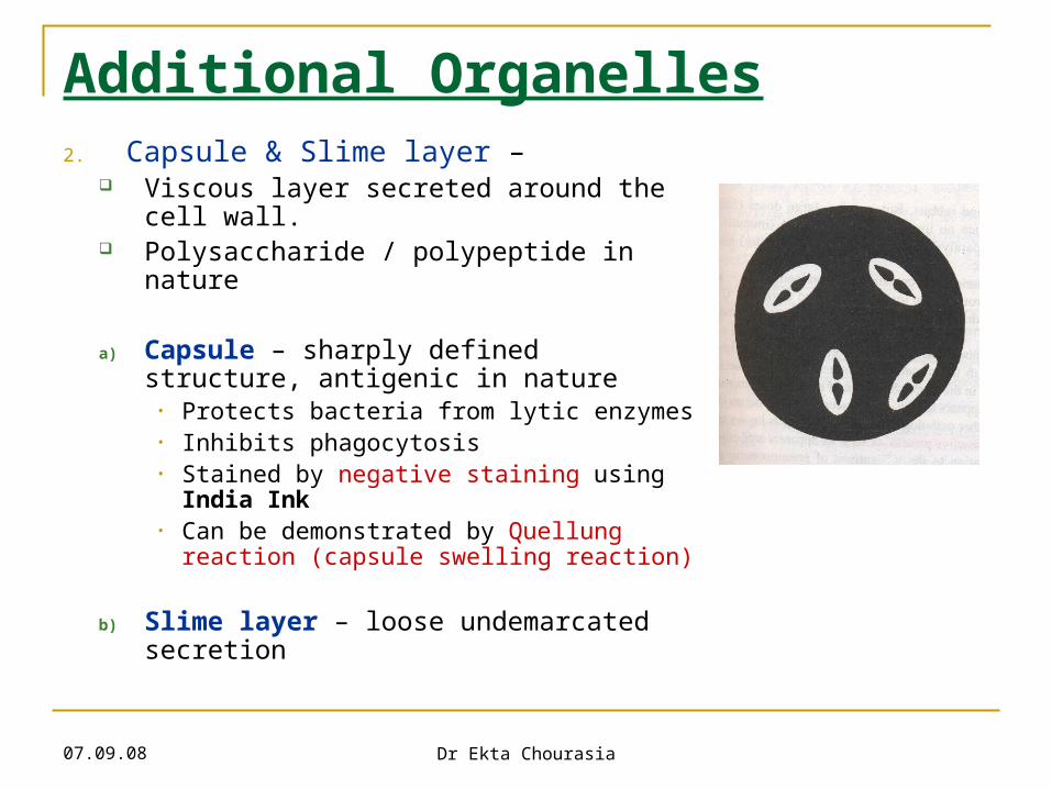
Additional Organelles2. Capsule & Slime layer –
Viscous layer secreted around the cell wall.
Polysaccharide / polypeptide in nature
a) Capsule – sharply defined structure, antigenic in nature
• Protects bacteria from lytic enzymes• Inhibits phagocytosis• Stained by negative staining using India
Ink• Can be demonstrated by Quellung reaction
(capsule swelling reaction)
b) Slime layer – loose undemarcated secretion
07.09.08 Dr Ekta Chourasia
Additional Organelles3. Flagella –
Long (3 to 12 µm), filamentous surface appendages
Organs of locomotion
Chemically, composed of proteins called flagellins
The number and distribution of flagella on the bacterial surface are characteristic for a given species - hence are useful in identifying and classifying bacteria
Flagella may serve as antigenic determinants (e.g. the H antigens of Gram-negative enteric bacteria)
Presence shown by motility e.g. hanging drop preparation

07.09.08 Dr Ekta Chourasia
Types of flagellar arrangementPolar/ Monotrichous – single flagellum at one pole
Lophotrichous – tuft of flagella at one pole
Peritrichous – flagella all over
Amphitrichous – flagella at both poles
Amphilophotrichous – tuft of flagella at both ends
07.09.08 Dr Ekta Chourasia
Additional Organelles4. Fimbriae/ Pili –
Thin, hairlike appendages on the surface of many Gram-negative bacteria
10-20µ long, acts as organs of adhesion (attachment) - allowing bacteria to colonize environmental surfaces or cells and resist flushing
Made up of proteins called pilins.
Pili can be of two types – Common pili – short & abundant Sex pili - small number (one to six), very long pili,
helps in conjugation (process of transfer of DNA)http://student.ccbcmd.edu/courses/bio141/lecguide/unit1/prostruct/yespili.html
http://student.ccbcmd.edu/courses/bio141/lecguide/unit1/prostruct/nopili.html
07.09.08 Dr Ekta Chourasia
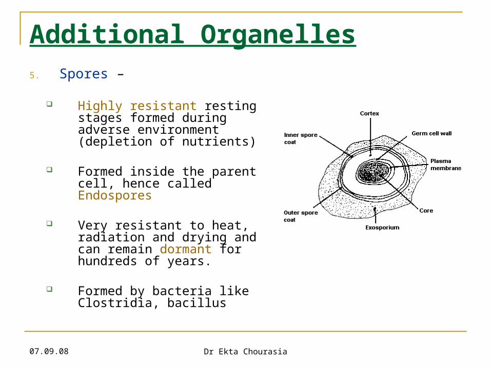
Additional Organelles5. Spores –
Highly resistant resting stages formed during adverse environment (depletion of nutrients)
Formed inside the parent cell, hence called Endospores
Very resistant to heat, radiation and drying and can remain dormant for hundreds of years.
Formed by bacteria like Clostridia, bacillus
07.09.08 Dr Ekta Chourasia
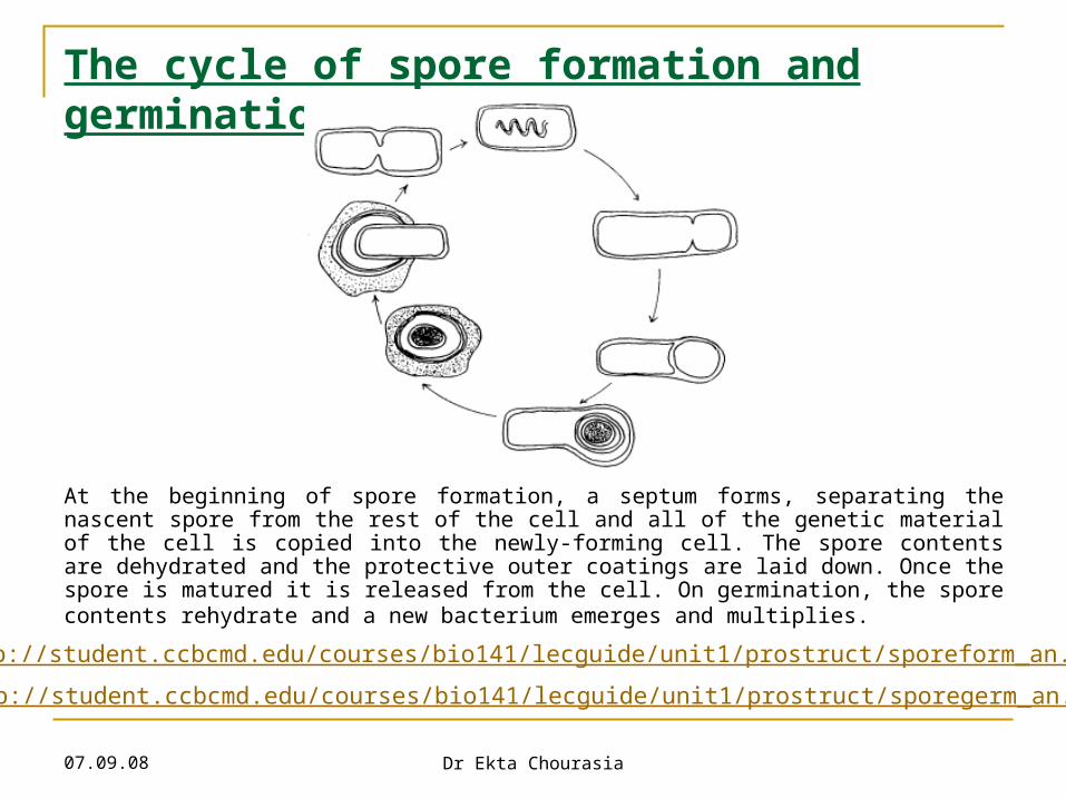
The cycle of spore formation and germination
At the beginning of spore formation, a septum forms, separating the nascent spore from the rest of the cell and all of the genetic material of the cell is copied into the newly-forming cell. The spore contents are dehydrated and the protective outer coatings are laid down. Once the spore is matured it is released from the cell. On germination, the spore contents rehydrate and a new bacterium emerges and multiplies.
http://student.ccbcmd.edu/courses/bio141/lecguide/unit1/prostruct/sporeform_an.html http://student.ccbcmd.edu/courses/bio141/lecguide/unit1/prostruct/sporegerm_an.html
07.09.08 Dr Ekta Chourasia
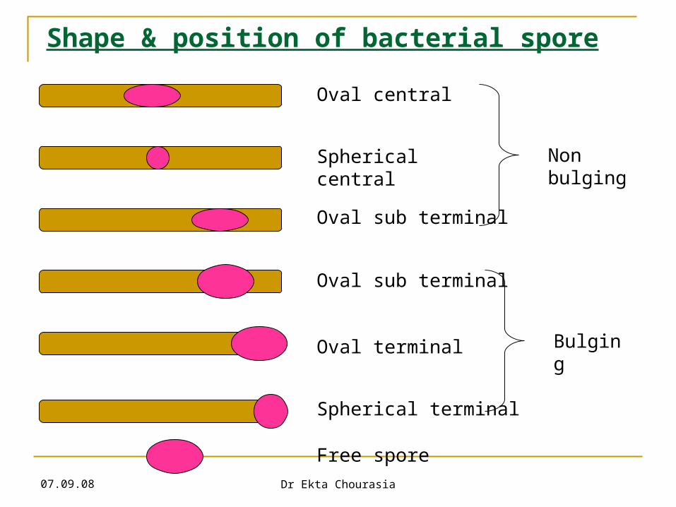
Shape & position of bacterial spore
Oval central
Spherical central
Oval sub terminal
Oval sub terminal
Oval terminal
Spherical terminal
Free spore
Non bulging
Bulging

07.09.08 Dr Ekta Chourasia
Pleomorphism & Involution forms Pleomorphism – great variation in shape & size of
individual cells e.g. Proteus species
Involution forms – swollen & aberrant forms in ageing cultures, especially in the presence of high salt concentration e.g. plague bacillus
Cause – defective cell wall synthesis
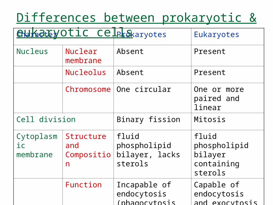
Differences between prokaryotic & eukaryotic cellsCharacter Prokaryotes Eukaryotes
Nucleus Nuclear membrane
Absent Present
Nucleolus Absent Present
Chromosome One circular One or more paired and linear
Cell division Binary fission Mitosis
Cytoplasmic membrane
Structure and Composition
fluid phospholipid bilayer, lacks sterols
fluid phospholipid bilayer containing sterols
Function Incapable of endocytosis (phagocytosis and pinocytosis) and exocytosis
Capable of endocytosis and exocytosis
07.09.08 Dr Ekta Chourasia
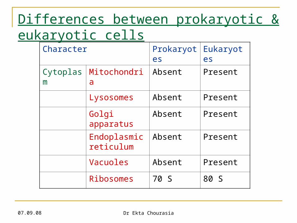
Differences between prokaryotic & eukaryotic cells
Character Prokaryotes Eukaryotes
Cytoplasm Mitochondria Absent Present
Lysosomes Absent Present
Golgi apparatus
Absent Present
Endoplasmic reticulum
Absent Present
Vacuoles Absent Present
Ribosomes 70 S 80 S
07.09.08 Dr Ekta Chourasia
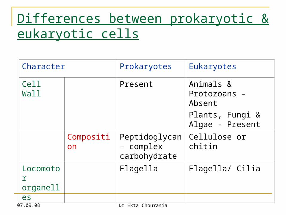
Differences between prokaryotic & eukaryotic cells
Character Prokaryotes Eukaryotes
Cell Wall Present Animals & Protozoans – Absent
Plants, Fungi & Algae - Present
Composition Peptidoglycan – complex carbohydrate
Cellulose or chitin
Locomotor organelles
Flagella Flagella/ Cilia
07.09.08 Dr Ekta Chourasia
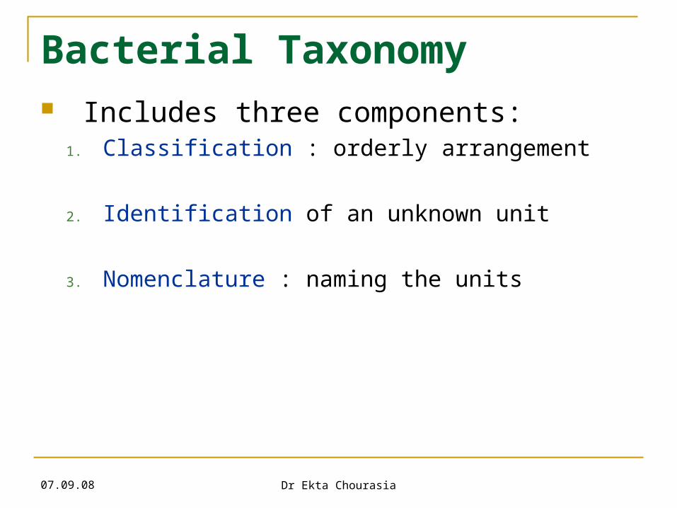
Bacterial Taxonomy Includes three components:
1. Classification : orderly arrangement
2. Identification of an unknown unit
3. Nomenclature : naming the units
07.09.08 Dr Ekta Chourasia
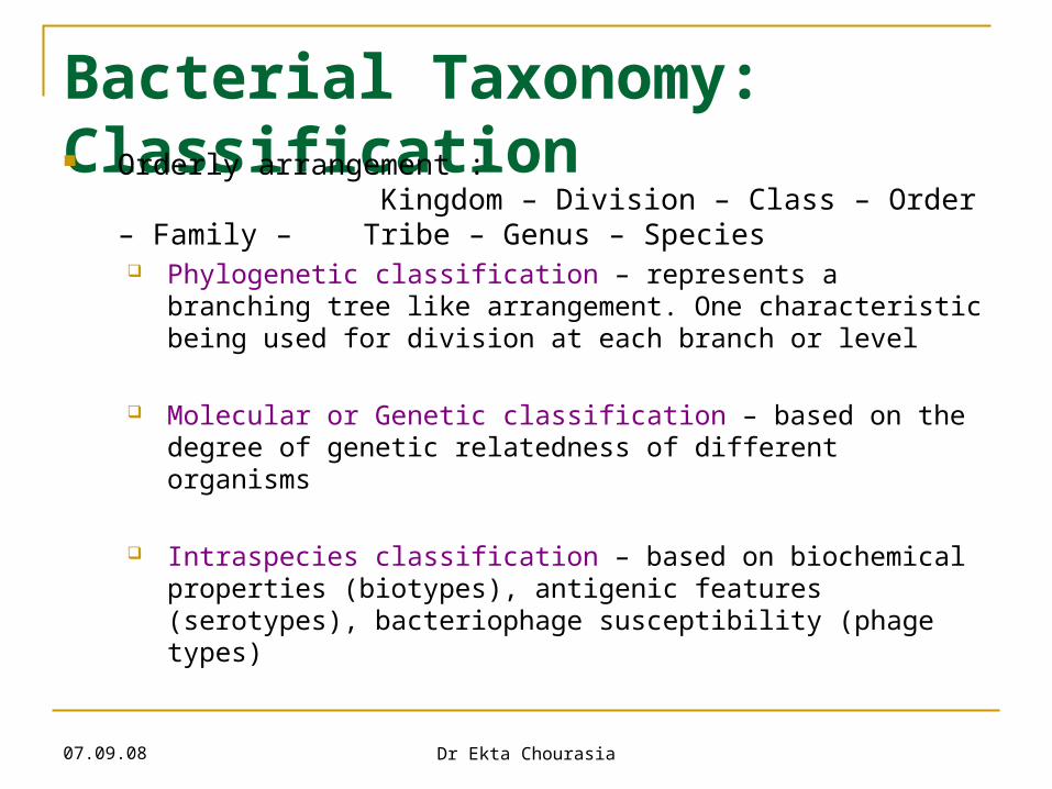
Bacterial Taxonomy: Classification Orderly arrangement :
Kingdom – Division – Class – Order – Family – Tribe – Genus – Species Phylogenetic classification – represents a branching
tree like arrangement. One characteristic being used for division at each branch or level
Molecular or Genetic classification – based on the degree of genetic relatedness of different organisms
Intraspecies classification – based on biochemical properties (biotypes), antigenic features (serotypes), bacteriophage susceptibility (phage types)
07.09.08 Dr Ekta Chourasia
07.09.08 Dr Ekta Chourasia
Bacterial Taxonomy: Nomenclature Two kinds of name are given to bacteria
Casual / common name – for local use, varies from country to country e.g. “typhoid bacillus”
Scientific / International Name – same all over world, consists of two words (in Italics) e.g. Salmonella typhi, Staphylococcus aureus


