
Avoiding Spinal Cord DisastersThe Key is Early Recognition
J. Stephen Huff, MD, FACEPUniversity of Virginia
Departments of Emergency Medicine and NeurologyJune 25 - 27, 2009

Spinal Cord DisastersThe Key is Early Recognition
• Injury patterns
• Differential diagnosis
• Cases
• Pitfalls
• Pearls

Spine or Spinal Cord?

Spinal Cord Syndromesand Injury Patterns
• Complete
• Incomplete– Anterior– Posterior – Central Cord– Brown-Sequard– Cauda equina lesion– Conus medullaris lesion

Spinal Cord Syndromesand Injury Patterns
• Complete– Transverse sensory pattern– Transverse motor pattern
• What’s the level?

Motor levels
• C4 level – quadriplegia
• C5 level + deltoid, biceps
• C6 level + wrist extensors, brachioradialis
• C7 level + triceps
• T1 level + finger abductors

Motor levelsT2 – T12 paraplegic
• L1 intact – Iliopsoas (hip flexion)
• L2 + hip adductors
• L3 + quadriceps
• L4 + tibialis anterior (dorsiflexion)
• L5 + hamstrings
• S1 + gastrocs (plantarflexioin)

Motor
• Weakness
– Sudden or progressive
• Fatigability
• Clumsiness
• Atrophy / fasciculations

Patterns of sensory loss
• Bilateral segmental loss
• Pinprick loss alternating with position & vibration loss
• Sacral sparing
• Sacral loss

Reflexes
• Reflex assessment may be unreliable in acute lesions
• Autonomic reflexes
• “Spinal Shock”
Pitfalls
• Time constraints
• Incomplete history
• Incomplete examination
• Unusual presentations

“Levels”
• Vertebral
• Cord
• Disability
• Function
• => say what you mean…

Imaging
• Plain radiography
• CT
• MRI
• Myelography

Case 1 - multiple trauma
• Unrestrained driver
• Head injury
• Intubated at scene
• Immobilized / IV’s

Case 1 - arrival
• Intubated / unresponsive
• Hypotensive
• Stable chest
• Rigid abdomen

Case 1 - Management
• Airway verified
• Resuscitation continued
• Examination
• Ancillary tests

Case 1 - Pressure problems
• Hypotensive…
• No fractures on early xrays
– CXR
– Pelvis
• Peritoneal lavage negative
• ? Why hypotensive?

Neurogenic Shock
• “Vasogenic shock”– Diagnosis of exclusion– Fluids– Pressors
• Not “spinal shock”
Pitfalls – complete lesions
• Failed recognition
• ABCD
• Attributing hypotension to the spinal cord injury erroneously
• Steroid stumble

Case 2 - football player
• Tackling injury
• Ambulatory after accident
• Immobilized
• Helmet on….

Case 2 - football player
• Awake, alert
• Strength exam normal
• Severe pain upper extremities
• Grip good

Central Cord Syndrome
• Upper extremity symptoms
• Lower extremities intact
• Variable sensory findings
• Variable bladder dysfunction

Central Cord Syndrome
• “Burning Hands” in football players with spinal cord injuries….
• Cord at risk
• Narrow canal – etiology?
• Advanced imaging
• Restriction of play?

Case 3 – chest pain
• 53 year-old man with chest pain and upper back pain
• Left-sided, sharp, + movement
• Hx COPD, sarcoidosis, CHF, pulmonary embolism, diabetes
• On prednisone, metformin, diuretic
• Wheelchair at times, active

Case 3 – chest pain
• Afebrile
• CXR, CT-PA obtained
• WBC 23,000
Case 3 – chest pain
• Leukocytosis attributed to steroids
• Pain medications, discharged
Case 3 – Clinical course
• Returned 48 hours with leg weakness
• Blood cultures + Staph aureus
• MRI- epidural fluid collection

Sensory
• Paresthesias-positive
• Negative symptoms
• Pain – Local pain– Radicular pain– Diffuse burning/aching
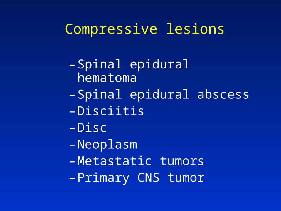
Compressive lesions
– Spinal epidural hematoma– Spinal epidural abscess– Disciitis– Disc– Neoplasm– Metastatic tumors– Primary CNS tumor
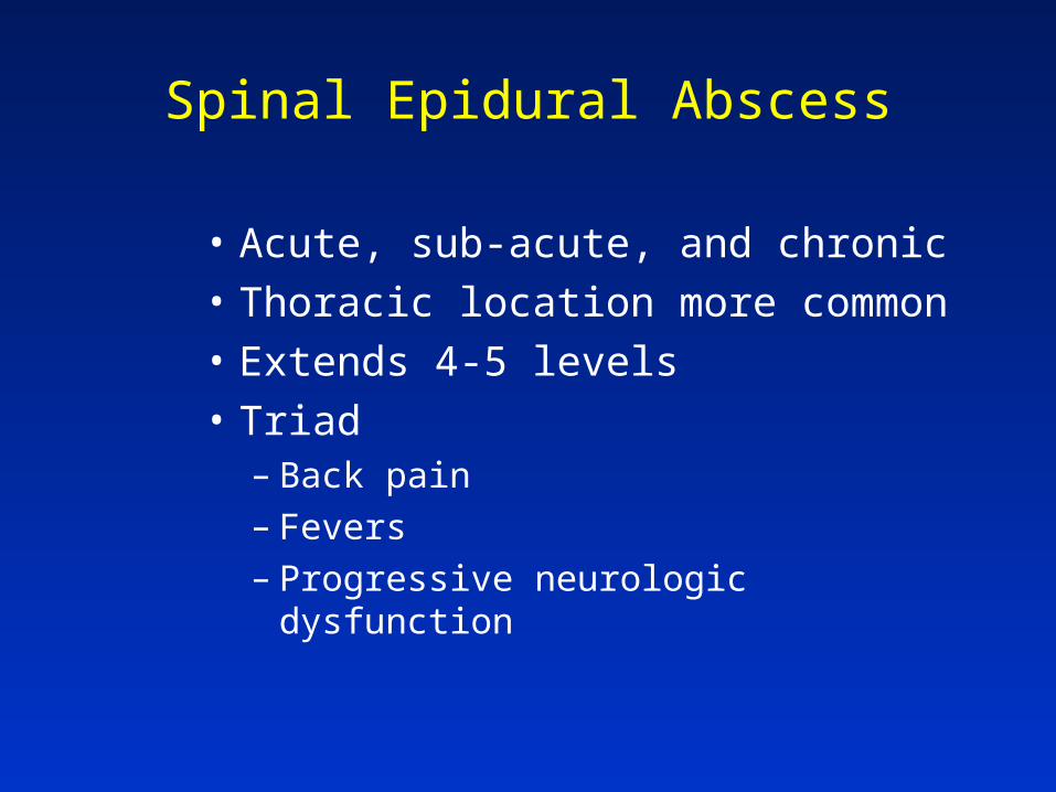
Spinal Epidural Abscess
• Acute, sub-acute, and chronic
• Thoracic location more common
• Extends 4-5 levels
• Triad– Back pain– Fevers– Progressive neurologic dysfunction
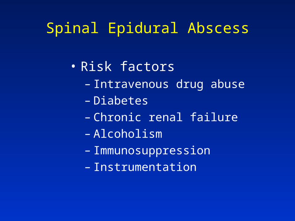
Spinal Epidural Abscess
• Risk factors– Intravenous drug abuse– Diabetes– Chronic renal failure– Alcoholism– Immunosuppression– Instrumentation

Spinal Epidural Abscessdiagnosis
• MRI diagnostic test of choice
• ESR elevated
• LP relatively contraindicated

Spinal Epidural AbscessTherapy
• Surgical decompression
• Antibiotics*– Staph coverage– MRSA
• Prognosis related to pre-op state

Compressive lesions
– Treatment generally similar…– Diagnosis…
• Exclude remedial causes…
– Steroids … – Decompression…– XRT for tumors…
• “the only XRT emergency….”

Case 4 – crack in neck
• Awakened with severe neck pain
• Became weak on way to ED
• Right-sided weakness– No facial droop – No speech difficulty

Case 4 – crack in neck
• At arrival, weak right arm and leg
– 4/5
– Left side normal
• Additional history– Strong family history of stroke– No medical history other than mild
hypertension

Spinal Epidural Hematoma
• Sudden, severe back pain
• Radicular component
• Progressive neurologic deficits

Spinal Epidural Hematoma
• Anticoagulant use
• Thrombocytopenia
• Liver disease / alcoholism
• Instrumentation
• MRI imaging modality of choice

Case 5
• 16 year old
• Abrupt inability to walk

Case 5
• Awake, alert
• Sitting on side of bed
• Lifting legs with arms
• Sensory level at umbilicus

Case 5
• Normal tone
• Normal reflexes
• “Don’t worry about me…”

“Hysterical paraplegia”
• Untenable patterns
– Sensory loss
– Motor loss
• Normal muscle tone
• Normal reflexes
• No bladder dysfunction
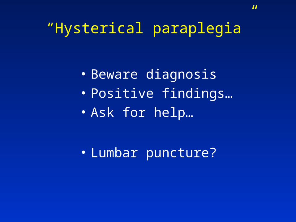
“Hysterical paraplegia”
• Beware diagnosis
• Positive findings…
• Ask for help…
• Lumbar puncture?
“Stable” vs. “unstable”
• Mechanical
• Deficit
• General condition

Low lesions
• Conus medullaris lesion
• Cauda equina lesion– Overlap / coexist– Sphincter involvement– UMN vs. LMN– Bilateral vs. unilateral

Nontraumatic etiologies of spinal cord dysfunction
• Demyelination– Multiple sclerosis / Transverse myelitis– Stroke
• AVM / SAH– Syringomyelia– Traumatic– Tumor
• Idiopathic spastic paraparesis– HIV myelopathy– Other myelopathies
• Compressive lesions

THINK REVERSIBLE

Avoiding Spinal Cord DisastersThe Key is Early Recognition
J. Stephen Huff, MD, FACEPUniversity of [email protected]


