Download - Activity of soluble OX40 ligand is enhanced by oligomerization and cell surface immobilization

Activity of soluble OX40 ligand is enhanced byoligomerization and cell surface immobilizationNicole Muller1,*, Agnes Wyzgol1,*, Sabine Munkel2, Klaus Pfizenmaier2 and Harald Wajant1
1 Department of Molecular Internal Medicine, Medical Clinic and Polyclinic II, University of Wuerzburg, Germany
2 Institute of Cell Biology and Immunology, University of Stuttgart, Germany
The ligands of the tumor necrosis factor (TNF) family
and their corresponding receptors are crucially
involved in a variety of immunoregulatory and devel-
opmental processes [1]. TNF ligands activate receptors
belonging to the TNF receptor superfamily, which can
be subdivided into death receptors and non-death
receptors. The death receptors are characterized by a
conserved protein–protein interaction domain in their
cytoplasmic tail that links these molecules to death
domain-containing adaptor proteins and proapoptotic
proteases, such as caspase-8. The non-death receptors
lack a death domain and interact with adapter proteins
of the TNF receptor-associated factor family, which
are involved in the activation of nuclear factor kappa B
(NF-jB) transcription factors and various mitogen-
activated kinases [1]. TNF receptor-associated factor
binding receptors especially include CD27, 4-1BB and
OX40, which ensure the ongoing cell division of ini-
tially T-cell receptor ⁄CD28 costimulated T-cells and
further control T-cell differentiation [2–4]. For exam-
ple, OX40 has been implicated in the generation and
reactivation of memory T-cells. Accordingly, inhibitors
of the OX40 ligand (OX40L)–OX40 interaction are
under consideration for the treatment of autoimmune
and allergic diseases, whereas OX40 agonists are
exploited as adjuvants to stimulate antitumor immu-
nity [2–4].
With the exception of lymphotoxin-a, all members
of the TNF ligand family are single spanning trans-
membrane proteins with an amino-terminal cytoplas-
mic tail (type II membrane protein). Due to
proteolytic processing or alternative splicing, naturally
Keywords
FAP; NF-jB; oligomerization, OX40L; single
chain
Correspondence
H. Wajant, Department of Molecular
Medicine, Medical Clinic and Polyclinic II,
University of Wuerzburg, Roentgenring 11,
97070 Wuerzburg, Germany
Fax: +49 931 201 71070
Tel: +49 931 201 71010
E-mail: [email protected]
*These authors contributed equally to this
work
(Received 7 December 2007, revised 25
February 2008, accepted 6 March 2008)
doi:10.1111/j.1742-4658.2008.06382.x
OX40 ligand (OX40L) and OX40 are typical members of the tumor
necrosis factor ligand family and the tumor necrosis factor receptor super-
family, respectively, and are involved in the costimulation and differentia-
tion of T cells. Like other tumor necrosis factor ligands, OX40L is a
type II transmembrane protein. Recombinant soluble human OX40L
assembles into trimers and is practically inactive despite binding to OX40.
However, oligomerization of soluble OX40L trimers by cross-linking with
antibodies or by expression as a hexameric fusion protein strongly
increased the activity of the ligand. Moreover, a fusion protein of OX40L
with a single chain fragment recognizing the tumor stroma antigen fibro-
blast activation protein showed a cell surface antigen-dependent increase
in the activity of the ligand domain of the molecule and thus mimicked
the activity of membrane OX40L upon antigen binding. Trimeric single
chain OX40L fusion proteins therefore represent a novel type of OX40L-
derived immunostimulatory molecule with potentially reduced systemic
side effects.
Abbreviations
FACS, fluorescence activated cell sorting; FAP, fibroblast activation protein; IL, interleukin; IjBa, inhibitor of jB-alpha; JNK, c-Jun N-terminal
kinase; NF-jB, nuclear factor kappa B; THD, TNF homology domain; TNC, tenascin-C; TNF-a, tumor necrosis factor-alpha; TRAIL, TNF-related
apoptosis inducing ligand.
2296 FEBS Journal 275 (2008) 2296–2304 ª 2008 The Authors Journal compilation ª 2008 FEBS
occurring soluble variants also exist for most of the
TNF ligands. The structural hallmark defining the
TNF ligand family is the carboxy-terminal TNF
homology domain (THD), which is composed of two
stacked b-pleated sheets adopting a conserved jelly-
roll-like tertiary fold [5,6]. The THD mediates self
association into trimers and is necessary for receptor
binding. In accordance with the carboxy-terminal
localization of the THD, the transmembrane forms of
TNF ligands, as well as soluble ligand variants,
assemble into trimers [5,6]. Despite their similar tri-
meric organization, membrane-bound and soluble
TNF ligands can differ in their activity. With respect
to receptor binding and receptor activation by TNF
ligands, three cases can be distinguished. First, both
the transmembrane as well as the corresponding solu-
ble variant of a particular TNF ligand bind and acti-
vate their cognate TNF receptor. An example for this
case is TNF receptor-1, which becomes properly acti-
vated by soluble as well as membrane TNFa [7]. In
the second case, the transmembrane variant of a TNF
ligand interacts and stimulates the corresponding
receptor, whereas the soluble variant neither binds,
nor activates the receptor. An example of this case is
murine CD95L [8]. Third, a TNF receptor interacts
with both the transmembrane variant of its corre-
sponding ligand as well as with the derived soluble
counterpart, but is not, or only poorly, stimulated by
the later. Such a differential activity of the soluble
and transmembrane ligand form has been demon-
strated for human CD95 and human TNF receptor-2,
[7,9]. The reasons underlying the quite diverse quality
of soluble TNF ligands with respect to receptor bind-
ing and receptor activation are currently poorly
understood.
Notably, inactive soluble TNF ligands that still
bind their receptors can be converted into highly
active molecules by increasing their avidity or by
immobilization on a cell surface or the extracellular
matrix. The most prominent and best investigated
example in this respect is human CD95L. Soluble tri-
meric human CD95L binds its corresponding receptor
CD95 at physiological relevant concentrations, but
fails to stimulate it properly and even acts as an
antagonist for membrane CD95L-induced activation
of CD95 [9]. When soluble CD95L trimers were
secondarily multimerized by antibody mediated cross-
linking or genetic fusion with an oligomerizing pro-
tein domain, the increase in avidity correlated with
an up to thousand-fold enhancement of the specific
activity of CD95L [9–11]. A similar increase in activ-
ity was observed upon binding of the soluble CD95L
to components of the extracellular matrix via a bind-
ing motif located N-terminally to the THD of the
ligand [12]. Likewise, soluble trimeric single chain-
CD95L fusion proteins achieve high activity after
docking to a cell surface antigen [13,14]. Other solu-
ble ligands of the TNF family that only gain high
activity after oligomerization are TNF-a in respect to
TNF receptor-2 and CD40L, 4-1BBL and TNF-
related apoptosis inducing ligand (TRAIL) in respect
to TRAIL receptor-2 [9,15–20]. Notably, both aggre-
gated and non-aggregated TNFa and TRAIL trimers
activate comparably well with TNF receptor-1 and
TRAILR1, respectively. This indicates that the
requirement of a soluble TNF ligand for oligomeriza-
tion to gain high activity is not ligand intrinsic, but
also depends on the receptor. The previously noted
observation that trimeric soluble fusion proteins of
inactive or poorly active TNF ligands acquire high
activity after immobilization opens the possibility of
restricting the activation of their cognate receptors to
cell surface antigen positive cells and tissue
[13,14,18,21–24]. This is of potential relevance for the
therapeutic application of these molecules because it
avoids therapy restricting side effects on cell surface
antigen negative cells and tissue. Noteworthy, for sev-
eral of the TNF ligand family members, including
OX40L, it is not known whether the soluble variant
of the molecule depends on oligomerization to elicit
maximal activity.
In the present study, we report that oligomerization
of soluble OX40L trimers by cross-linking with anti-
bodies or by expression as a hexameric fusion protein
increases the activity of the molecule by more than
200-fold. Accordingly, a fusion protein of OX40L
linked to a single chain fragment recognizing the
tumor stroma antigen fibroblast activation protein
(FAP) shows a significant increase in activity after
binding to its cognate cell surface antigen, thus defin-
ing a novel type of OX40 agonist with potentially
reduced systemic side effects.
Results and Discussion
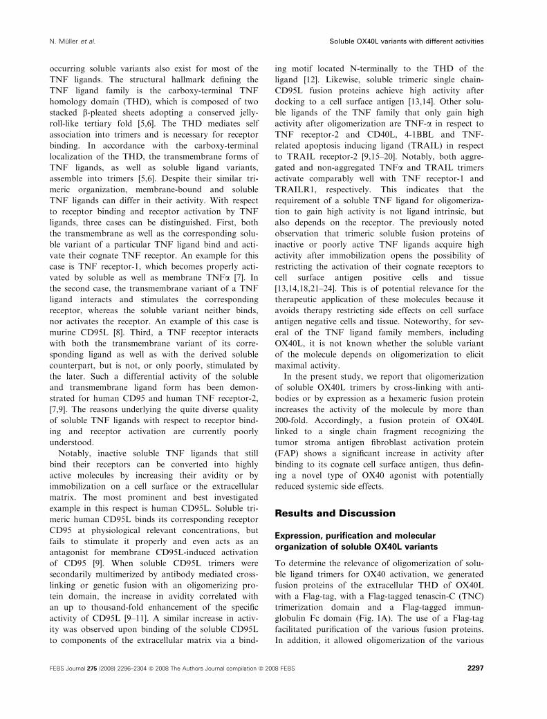
Expression, purification and molecular
organization of soluble OX40L variants
To determine the relevance of oligomerization of solu-
ble ligand trimers for OX40 activation, we generated
fusion proteins of the extracellular THD of OX40L
with a Flag-tag, with a Flag-tagged tenascin-C (TNC)
trimerization domain and a Flag-tagged immun-
globulin Fc domain (Fig. 1A). The use of a Flag-tag
facilitated purification of the various fusion proteins.
In addition, it allowed oligomerization of the various
N. Muller et al. Soluble OX40L variants with different activities
FEBS Journal 275 (2008) 2296–2304 ª 2008 The Authors Journal compilation ª 2008 FEBS 2297

OX40L variants by treatment with Flag-specific
monoclonal antibody. The Flag-TNC-OX40L fusion
protein was included in the study due to our recent
finding that a disulfide-bonded TNC trimerization
domain can enhance receptor binding by some mem-
bers of the TNF ligand family (e.g. murine TRAIL
and murine or human CD95L) without changing the
principle trimeric assembly of these molecules [8]. The
dimerizing Fc domain of human immunoglobulin G1
was used with the intention to drive the formation of
hexameric ligands, as recently shown for a Fc-CD95L
fusion protein [10]. All fusion proteins were produced
in Hek293 cells and were purified by affinity chroma-
tography on monoclonal M2 anti-Flag agarose
(Fig. 1B,C). The migration pattern of Flag-OX40L in
nonreducing SDS ⁄PAGE analysis revealed three
bands with molecular masses of 20, 23 and 27 kDa,
respectively. Flag-TNC-OX40L showed a band of
approximately 100 kDa and a broad fuzzy band of
60–80 kDa. Fc-Flag-OX40L migrated as single band
with a molecular mass of 90 kDa. Under reducing
conditions, Flag-Ox40L ran as a triplet with 23, 27
and 30 kDa, Flag-TNC-OX40L migrated with 27 and
31 kDa and Fc-Flag-OX40L revealed a single band of
60 kDa. The data obtained for Flag-TNC-OX40L are
in accordance with the expected disulfide-bonded
formation of Flag-TNC-OX40L trimers. The values
observed for Fc-Flag-OX40L suggest that this mole-
TNF homology domain
Flag-OX40L (52–183)Flag OX40L aa 137-281Flag OX40L aa 52–183
Flag-TNC-OX40L (52–183)TNCFlag OX40L aa 137-281OX40L aa 52–183
Fc-Flag-OX40L (52–183)Flag OX40L aa 137-281Flag OX40L aa 52–183hIgG1 (hinge + Fc)
A
Flag-OX40L
Fc-Flag-OX40L
Flag-TNC-OX40L
83
6248
33
25
17
C
+ – + – + –dithiothreitol dithiothreitol
Flag-OX40L
Fc-Flag-OX40L
Flag-TNC-OX40L
836248
33
25
17
175
B
+ – + – + –
D
Elution volume
Fc-Flag-OX40LFlag-OX40L TNC-Flag-OX40LMW standards
HMW670
150
4417
Abs
orba
nce
Fig. 1. Characterization of recombinant variants of soluble OX40L. (A) Scheme of the OX40L variants used in the present study. Fc, human
immunoglobulin G1 Fc fragment; F, Flag tag; OX40L, human OX40L (amino acids 52–183); TNC, chicken tenascin-C (amino acids 110–139).
(B) The indicated Flag-tagged variants of soluble OX40L containing the THD were produced in Hek293 cells, purified by M2 affinity chroma-
tography, separated by SDS ⁄ PAGE under reducing and nonreducing conditions and finally visualized by silver staining. (C) Western blot anal-
ysis of the various OX40L variants using the monoclonal M2 anti-Flag serum. (D) The OX40L variants were separated by gel filtration on a
BioSep-Sec-S3000 column. Arrows indicate the elution volume of the molecular weight standards thyroglobulin (670 kDa), IgG (150 kDa),
ovalbumin (44 kDa) and myoglobulin (17 kDa).
Soluble OX40L variants with different activities N. Muller et al.
2298 FEBS Journal 275 (2008) 2296–2304 ª 2008 The Authors Journal compilation ª 2008 FEBS
cule assembles into hexamers composed of three
noncovalently linked, disulfide bonded Fc-Flag-
OX40L dimers. The observed molecular masses signif-
icantly exceed the calculated mass of 17, 21 and
47 kDa for the various OX40L fusion proteins, but
are in good accordance with glycosylation of OX40L
on three asparagine residues in the THD because one
N-linked carbohydrate accounts for 2–5 kDa by
SDS ⁄PAGE. Based on gel filtration using a BioSep-
Sec-S3000 column, molecular masses of 80, 130 and
580 kDa were calculated for native Flag-OX40L,
Flag-TNC-OX40L and Flag-Fc-OX40L (Fig. 1D).
The molecular mass standards used for calibration in
the gel filtration analysis experiments were all globular
shaped proteins, whereas, from X-ray crystallography,
it is known that ligand trimers of the TNF family
form elongated bell-shaped structures. Thus, these val-
ues, despite deviating somewhat from the expected
calculated molecular masses, are in reasonable agree-
ment with the expected trimeric organization of Flag-
OX40L and Flag-TNC-OX40L and a hexameric orga-
nization of Flag-Fc-OX40L. A hexameric organization
has recently also been shown for an immunoglobulin
OX40L fusion protein that was linked with an isoleu-
cine zipper domain [25].
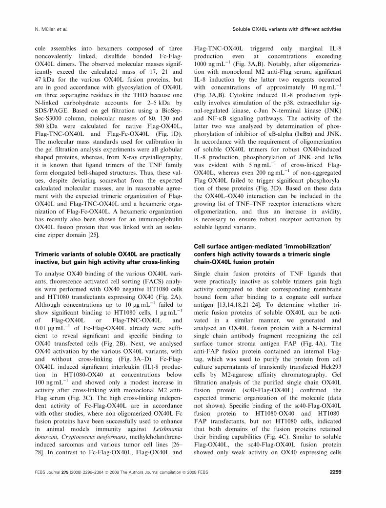
Trimeric variants of soluble OX40L are practically
inactive, but gain high activity after cross-linking
To analyse OX40 binding of the various OX40L vari-
ants, fluorescence activated cell sorting (FACS) analy-
sis were performed with OX40 negative HT1080 cells
and HT1080 transfectants expressing OX40 (Fig. 2A).
Although concentrations up to 10 lgÆmL)1 failed to
show significant binding to HT1080 cells, 1 lgÆmL)1
of Flag-OX40L or Flag-TNC-OX40L and
0.01 lgÆmL)1 of Fc-Flag-OX40L already were suffi-
cient to reveal significant and specific binding to
OX40 transfected cells (Fig. 2B). Next, we analysed
OX40 activation by the various OX40L variants, with
and without cross-linking (Fig. 3A–D). Fc-Flag-
OX40L induced significant interleukin (IL)-8 produc-
tion in HT1080-OX40 at concentrations below
100 ngÆmL)1 and showed only a modest increase in
activity after cross-linking with monoclonal M2 anti-
Flag serum (Fig. 3C). The high cross-linking indepen-
dent activity of Fc-Flag-OX40L are in accordance
with other studies, where non-oligomerized OX40L-Fc
fusion proteins have been successfully used to enhance
in animal models immunity against Leishmania
donovani, Cryptococcus neoformans, methylcholanthrene-
induced sarcomas and various tumor cell lines [26–
28]. In contrast to Fc-Flag-OX40L, Flag-OX40L and
Flag-TNC-OX40L triggered only marginal IL-8
production even at concentrations exceeding
1000 ngÆmL)1 (Fig. 3A,B). Notably, after oligomeriza-
tion with monoclonal M2 anti-Flag serum, significant
IL-8 induction by the latter two reagents occurred
with concentrations of approximately 10 ngÆmL)1
(Fig. 3A,B). Cytokine induced IL-8 production typi-
cally involves stimulation of the p38, extracellular sig-
nal-regulated kinase, c-Jun N-terminal kinase (JNK)
and NF-jB signaling pathways. The activity of the
latter two was analyzed by determination of phos-
phorylation of inhibitor of jB-alpha (IjBa) and JNK.
In accordance with the requirement of oligomerization
of soluble OX40L trimers for robust OX40-induced
IL-8 production, phosphorylation of JNK and IjBawas evident with 5 ngÆmL)1 of cross-linked Flag-
OX40L, whereas even 200 ngÆmL)1 of non-aggregated
Flag-OX40L failed to trigger significant phosphoryla-
tion of these proteins (Fig. 3D). Based on these data
the OX40L–OX40 interaction can be included in the
growing list of TNF–TNF receptor interactions where
oligomerization, and thus an increase in avidity,
is necessary to ensure robust receptor activation by
soluble ligand variants.
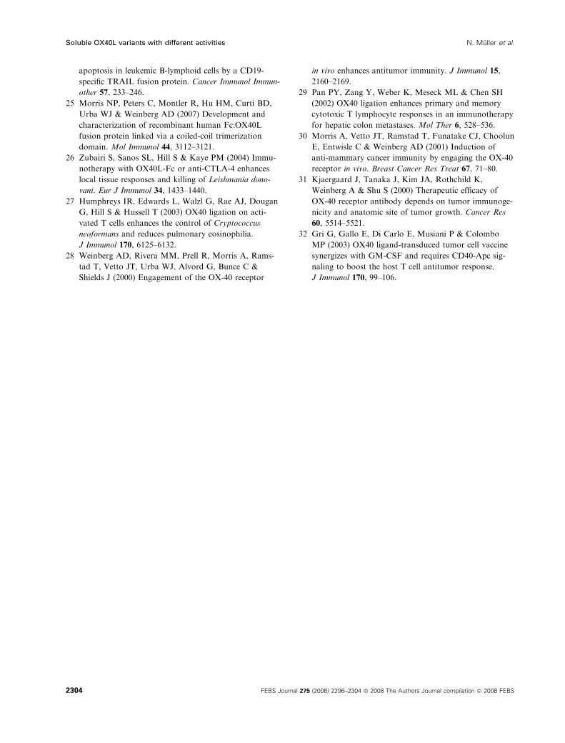
Cell surface antigen-mediated ‘immobilization’
confers high activity towards a trimeric single
chain-OX40L fusion protein
Single chain fusion proteins of TNF ligands that
were practically inactive as soluble trimers gain high
activity compared to their corresponding membrane
bound form after binding to a cognate cell surface
antigen [13,14,18,21–24]. To determine whether tri-
meric fusion proteins of soluble OX40L can be acti-
vated in a similar manner, we generated and
analysed an OX40L fusion protein with a N-terminal
single chain antibody fragment recognizing the cell
surface tumor stroma antigen FAP (Fig. 4A). The
anti-FAP fusion protein contained an internal Flag-
tag, which was used to purify the protein from cell
culture supernatants of transiently transfected Hek293
cells by M2-agarose affinity chromatography. Gel
filtration analysis of the purified single chain OX40L
fusion protein (sc40-Flag-OX40L) confirmed the
expected trimeric organization of the molecule (data
not shown). Specific binding of the sc40-Flag-OX40L
fusion protein to HT1080-OX40 and HT1080-
FAP transfectants, but not HT1080 cells, indicated
that both domains of the fusion proteins retained
their binding capabilities (Fig. 4C). Similar to soluble
Flag-OX40L, the sc40-Flag-OX40L fusion protein
showed only weak activity on OX40 expressing cells
N. Muller et al. Soluble OX40L variants with different activities
FEBS Journal 275 (2008) 2296–2304 ª 2008 The Authors Journal compilation ª 2008 FEBS 2299

at high concentrations (Fig. 4D). After cross-linking
with monoclonal M2 anti-Flag serum, the single
chain OX40 fusion protein was comparably active as
cross-linked Flag-OX40L or Fc-Flag-OX40L (data
not shown). More importantly, in the presence of
FAP expressing cells, sc40-Flag-OX40L showed an
approximately 200-fold higher activity in inducing
IL-8 production in cocultured HT1080-OX40 cells
(Fig. 4D). Using agonistic antibodies or OX40L
immunoglobulin fusion proteins, it has been demon-
strated that OX40 activation, either alone or in com-
bination with other immunstimuli, can enhance an
anti-tumoral immune response in mouse tumor
models with melanoma, colon cancer, glioma and
breast cancer [28–32]. Potential problems that might
arise from clinical applications of OX40 activating
reagents are the induction of autoimmunity and
inflammatory side effects [4]. Trimeric OX40L fusion
proteins that show cell surface antigen-restricted acti-
vation of OX40L ⁄OX40 thus appear to be promising
Rel
ativ
e ce
ll nu
mbe
r
log fluorescence intensity
con.
HT1080-OX40 IgG1
log fluorescence intensity
Pos
itive
cel
ls (
%) con.
Flag-OX40L
Flag-CD27L
0
80
20
60
40
101 10–1 10–2 10 µg·mL–1
CD27L-F
0
20
40
60
80
100
A
Rel
ativ
e ce
ll nu
mbe
rR
elat
ive
cell
num
ber
Rel
ativ
e ce
ll nu
mbe
r
log fluorescence intensity
B
con.
Flag-TNC-OX40L
Flag-CD27L
HT1080-OX40 aOX40
Flag-OX40L (µg·mL–1)
Flag-TNC-OX40L (µg·mL–1)
10–1 10–2 100
Fc-Flag-OX40L (µg·mL–1)
10–3 10–4
Pos
itive
cel
ls (
%)
0
80
20
60
40
log fluorescence intensity
con.
Fc-Flag-OX40L
Fc-Flag-4-1BBL
101 10–1 10–2 100 10 µg·mL–1
CD27L-F
10 µg·mL–1
Fc-F-41BBL
Fig. 2. OX40 binding of OX40L variants.
(A) OX40 cell surface expression of HT1080
and HT1080-OX40 cells was determined by
FACS analysis with PE-labelled anti-OX40.
(B) HT1080 and HT1080-OX40 cells were
incubated with increasing concentrations of
Flag-OX40L, Flag-TNC-OX40L and Flag-
Fc-OX40L on ice and, after repeated
washes, bound proteins were detected
using monoclonal M2 anti-Flag serum and
PE-labelled anti-mouse IgG. Amino-termi-
nally Flag-tagged soluble human CD27L was
used as a control.
Soluble OX40L variants with different activities N. Muller et al.
2300 FEBS Journal 275 (2008) 2296–2304 ª 2008 The Authors Journal compilation ª 2008 FEBS

for making therapy concepts that rely on OX40 acti-
vation safer.
Experimental procedures
Production and purification of recombinant
proteins
The various OX40L fusion proteins were produced in
Hek293 cells. In brief, cells were electroporated
(10–20 · 106 cellsÆmL)1; 4 mm cuvette; 250 V, 1800 lF,maximum resistance) in culture medium with 10% fetal
bovine serum and 40 lg of the corresponding expression
plasmid using an Easyject Plus electroporator (PeqLab,
Erlangen, Germany). Transfected cells were cultured over-
night and, the next day, medium was changed to RPMI
containing 0.5% fetal bovine serum. Supernatants were col-
lected 3 days post transfection and recombinant proteins
were purified by affinity chromatography using anti-Flag
M2 agarose columns. After elution with 100 lgÆmL)1 Flag
peptide (Sigma, Steinheim, Germany) in NaCl ⁄Pi, fractions
containing the OX40L variants were dialyzed against
NaCl ⁄Pi and analyzed by SDS ⁄PAGE.
Gelfiltration chromatography on
BioSep-SEC-S3000
Purified OX40L variants (25 lL) were applied to a BioSep-
SEC-S3000 (300 · 7.8) column (Phenomenex, Aschaffen-
burg, Germany) equilibrated in NaCl ⁄Pi and eluted at a
flow rate of 0.5 mLÆmin)1. The columns were calibrated
with thyroglobulin (670 kDa), IgG (150 kDa), ovalbumin
(44 kDa) and myoglobulin (17 kDa).
Flow cytometry with OX40L variants
Cells were incubated for 2 h at 4 �C with the indicated con-
centration of the OX40L variant of interest and, after three
cycles of washing with NaCl ⁄Pi, 0.2% fetal bovine serum
and 0.02% sodium azide, bound proteins were detected
by monoclonal M2 anti-Flag serum (1 lgÆmL)1; Sigma)
and phycoerythrin (PE)-labelled, mouse IgG-specific rabbit
C
A B
P-I B
I B
P-JNK
+ Flag mAb M2
JNK
1000
405 5000
200– Flag-OX40L (ng·mL–1)1000
405 5000
200–
0
1
2
3
Fc-Flag-OX40L (ng·mL–1)
+M2
4
5
6
0
2
4
6
8
Flag-TNC-OX40L (ng·mL–1)
+ M2
102100 101 1030
D
0
1
2
3
4
IL8
(ng·
mL–
1 )
IL8
(ng·
mL–
1 )
Flag-OX40L (ng·mL–1)102100 101 1030
–+ M2
102100 101 1030
IL8
(ng·
mL–
1 )
–
–
Fig. 3. Activity of OX40L trimers is
enhanced by oligomerization. (A–C) HT1080-
OX40 cells were stimulated in triplicates
with the indicated concentrations of Flag-
OX40L (A), Flag-TNC-OX40L (B) and Fc-Flag-
OX40L (C) with and without anti-Flag
cross-linking. After 6 h, supernatants were
removed and their relative IL-8 content was
determined by ELISA. Cell culture medium
was changed before stimulation to minimize
the contribution of OX40-independent IL-8
production due to constitutive IL-8 expres-
sion. (D) To determine JNK and NF-jB acti-
vation, lysates of HT1080-OX40 cells
stimulated for 10 min with the indicated
concentrations of cross-linked and non-
cross-linked Flag-OX40L were analyzed by
SDS ⁄ PAGE and western blotting with anti-
JNK, anti-phospho-JNK, anti-IjBa, anti-phos-
pho-IjBa and anti-tubulin.
N. Muller et al. Soluble OX40L variants with different activities
FEBS Journal 275 (2008) 2296–2304 ª 2008 The Authors Journal compilation ª 2008 FEBS 2301

antibodies (1 lgÆmL)1; Sigma). Analyses were performed
using FACSCalibur (BD Biosciences, Heidelberg, Germany)
according to standard procedures.
IL-8 ELISA
Cells (2 · 104 well)1) were seeded in 96-well tissue culture
plates and grown overnight. The next day, the medium was
changed to minimize the contribution of basal IL-8 produc-
tion and cells were stimulated in triplicates with varying
concentrations of the indicated OX40L variants. After 6 h,
supernatants were collected and analyzed for IL-8 produc-
tion using a commercially available ELISA kit (BD
Biosciences) according to the manufacturer’s instructions.
Western blot detection of phosphorylated
proteins
For western blot analysis of phosphorylated proteins, cells
were scraped into ice-cold NaCl ⁄Pi with a rubber policeman,
collected by centrifugation and lysed after sonification (ten
pulses) by boiling (5 min, 96 �C) in 4 · Laemmli sample buf-
fer (8% SDS, 0.1 m dithiothreitol, 40% glycerol, 0.2 m Tris,
pH 8.0) supplemented with phosphatase inhibitor cocktails
I and II (Sigma). Proteins were separated by SDS ⁄PAGE
and transferred to nitrocellulose membranes. After blocking
of nonspecific binding sites by incubation in Tris-buffered
saline containing 0.1% Tween 20 and 5% dry milk, immuno-
blotting was performed with primary antibodies recognizing
JNK, phospho-JNK, phospho-IjBa (Cell Signalling, Frank-
furt, Germany) and IjBa (Santa Cruz Biotechnologies Inc.,
Heidelberg, Germany), horseradish peroxidase-conjugated
secondary antibodies (Dako, Hamburg, Germany) and the
ECL western blotting detection reagents and analysis system.
Acknowledgements
This work was supported by Deutsche Krebshilfe
[Grants 106222 (H.W.) and 106235 (K.P.)] and Deut-
sche Forschungsgemeinschaft (SFB 487 project B7).
References
1 Locksley RM, Killeen N & Lenardo MJ (2001) The
TNF and TNF receptor superfamilies: integrating mam-
malian biology. Cell 104, 487–501.
HT1080-FAP
CHT1080
log fluorescence intensity log fluorescence intensity
HT1080
HT1080-OX40
0
1
2
3
4
sc40-Flag-OX40L (ng·mL–1)102100 101 1030
HT1080 + HT1080-OX40HT1080-FAP + HT1080-OX40
D
B175
83
62
48
33
25
+ –
SC40-Flag-OX40L (52–183)Flag OX40Laa137-281Flag OX40L aa 52–183FAP-specific single chain fragment “sc40”A
Rel
ativ
e ce
ll nu
mbe
r
Rel
ativ
e ce
ll nu
mbe
rIL
8 (n
g·m
L–1)
dithiothreitol
Fig. 4. A single chain-OX40L fusion protein
(sc40-Flag-OX40L) recognizing the extracel-
lular domain of FAP displays enhanced activ-
ity after antigen binding. (A) Scheme of
sc40-Flag-OX40L. (B) Purified sc40-Flag-
OX40L was separated by SDS ⁄ PAGE under
reducing and nonreducing conditions and
visualized by silver staining. (C) HT1080,
HT1080-FAP and HT1080-OX40 cells were
incubated with sc40-Flag-OX40L on ice and,
after repeated washes, bound proteins were
detected by FACS using monoclonal M2
anti-Flag serum and PE-labelled anti-mouse
IgG. (D) HT1080-FAP cells were mixed 1 : 1
with HT1080-OX40 or HT1080 cells and the
cell mixtures were seeded in triplicates in
96-well plates (30 · 103 cellsÆwell)1). The
next day, medium was changed and cells
were challenged for 6 h with the indicated
concentrations of sc40-Flag-OX40L. I-L8 pro-
duction was finally determined by ELISA.
Soluble OX40L variants with different activities N. Muller et al.
2302 FEBS Journal 275 (2008) 2296–2304 ª 2008 The Authors Journal compilation ª 2008 FEBS

2 Barr TA, Carlring J & Heath AW (2006) Co-stimula-
tory agonists as immunological adjuvants. Vaccine 24,
3399–3407.
3 Croft M (2003) Costimulation of T cells by OX40,
4-1BB, and CD27. Cytokine Growth Factor Rev 14,
265–273.
4 Sugamura K, Ishii N & Weinberg AD (2004) Therapeu-
tic targeting of the effector T-cell co-stimulatory mole-
cule OX40. Nat Rev Immunol 4, 420–431.
5 Fesik SW (2000) Insights into programmed cell death
through structural biology. Cell 103, 273–282.
6 Bodmer JL, Schneider P & Tschopp J (2002) The
molecular architecture of the TNF superfamily. Trends
Biochem Sci 27, 19–26.
7 Grell M, Douni E, Wajant H, Lohden M, Clauss M,
Maxeiner B, Georgopoulos S, Lesslauer W, Kollias G,
Pfizenmaier K et al. (1995) The transmembrane form of
tumor necrosis factor is the prime activating ligand of
the 80 kDa tumor necrosis factor receptor. Cell 83,
793–802.
8 Berg D, Lehne M, Muller N, Siegmund D, Munkel S,
Sebald W, Pfizenmaier K & Wajant H (2007) Enforced
covalent trimerization increases the activity of the TNF
ligand family members TRAIL and CD95L. Cell Death
Differ 14, 2021–2034.
9 Schneider P, Holler N, Bodmer JL, Hahne M, Frei K,
Fontana A & Tschopp J (1998) Conversion of mem-
brane-bound Fas(CD95) ligand to its soluble form is
associated with downregulation of its proapoptotic
activity and loss of liver toxicity. J Exp Med 187, 1205–
1213.
10 Holler N, Tardivel A, Kovacsovics-Bankowski M,
Hertig S, Gaide O, Martinon F, Tinel A, Deperthes D,
Calderara S, Schulthess T et al. (2003) Two adjacent
trimeric Fas ligands are required for Fas signaling and
formation of a death-inducing signaling complex. Mol
Cell Biol 23, 1428–1440.
11 Greaney P, Nahimana A, Lagopoulos L, Etter AL,
Aubry D, Attinger A, Beltraminelli N, Huni B, Bassi I,
Sordat B et al. (2006) A Fas agonist induces high levels
of apoptosis in haematological malignancies. Leuk Res
30, 415–426.
12 Aoki K, Kurooka M, Chen JJ, Petryniak J, Nabel EG
& Nabel GJ (2001) Extracellular matrix interacts with
soluble CD95L: retention and enhancement of cytotox-
icity. Nat Immunol 2, 333–337.
13 Samel D, Muller D, Gerspach J, Assohou-Luty C, Sass
G, Tiegs G, Pfizenmaier K & Wajant H (2003) Genera-
tion of a FasL-based proapoptotic fusion protein
devoid of systemic toxicity due to cell-surface antigen-
restricted activation. J Biol Chem 278, 32077–32082.
14 Bremer E, Ten Cate B, Samplonius DF, de Leij LF
& Helfrich W (2006) CD7-restricted activation of
Fas-mediated apoptosis: a novel therapeutic approach
for acute T-cell leukaemia. Blood 107, 2863–2870.
15 Haswell LE, Glennie MJ & Al-Shamkhani A (2001)
Analysis of the oligomeric requirement for signaling by
CD40 using soluble multimeric forms of its ligand,
CD154. Eur J Immunol 31, 3094–3100.
16 Stone GW, Barzee S, Snarsky V, Kee K, Spina CA, Yu
XF & Kornbluth RS (2006) Multimeric soluble CD40
ligand and GITR ligand as adjuvants for human immu-
nodeficiency virus DNA vaccines. J Virol 80, 1762–
1772.
17 Muhlenbeck F, Schneider P, Bodmer JL, Schwenzer R,
Hauser A, Schubert G, Scheurich P, Moosmayer D,
Tschopp J & Wajant H (2000) The tumor necrosis fac-
tor-related apoptosis-inducing ligand receptors TRAIL-
R1 and TRAIL-R2 have distinct cross-linking require-
ments for initiation of apoptosis and are non-redundant
in JNK activation. J Biol Chem 275, 32208–32213.
18 Wajant H, Moosmayer D, Wuest T, Bartke T, Gerlach
E, Schonherr U, Peters N, Scheurich P & Pfizenmaier
K (2001) Differential activation of TRAIL-R1 and -2
by soluble and membrane TRAIL allows selective sur-
face antigen-directed activation of TRAIL-R2 by a sol-
uble TRAIL derivative. Oncogene 20, 4101–4106.
19 Kelley RF, Totpal K, Lindstrom SH, Mathieu M, Billeci
K, Deforge L, Pai R, Hymowitz SG & Ashkenazi A
(2005) Receptor-selective mutants of apoptosis-inducing
ligand 2 ⁄ tumor necrosis factor-related apoptosis-induc-
ing ligand reveal a greater contribution of death receptor
(DR) 5 than DR4 to apoptosis signalling. J Biol Chem
280, 2205–2212.
20 Rabu C, Quemener A, Jacques Y, Echasserieau K,
Vusio P & Lang F (2005) Production of recombinant
human trimeric CD137L (4-1BBL). Cross-linking is
essential to its T cell co-stimulation activity. J Biol
Chem 280, 41472–41481.
21 Bremer E, Kuijlen J, Samplonius D, Walczak H, de Leij
L & Helfrich W (2004) Target cell-restricted and
-enhanced apoptosis induction by a scFv:sTRAIL
fusion protein with specificity for the pancarcinoma-
associated antigen EGP2. Int J Cancer 109, 281–290.
22 Bremer E, Samplonius DF, van Genne L, Dijkstra MH,
Kroesen BJ, de Leij LF & Helfrich W (2005) Simulta-
neous inhibition of epidermal growth factor receptor
(EGFR) signaling and enhanced activation of tumor
necrosis factor-related apoptosis-inducing ligand
(TRAIL) receptor-mediated apoptosis induction by an
scFv:sTRAIL fusion protein with specificity for human
EGFR. J Biol Chem 280, 10025–10033.
23 Zhang N, Sadun RE, Arias RS, Flanagan ML, Sachs-
man SM, Nien YC, Khawli LA, Hu P & Epstein AL
(2007) Targeted and untargeted CD137L fusion proteins
for the immunotherapy of experimental solid tumors.
Clin Cancer Res 13, 2758–2767.
24 Stieglmaier J, Bremer E, Kellner C, Liebig TM,
Ten Cate B, Peipp M, Schulze-Koops H, Pfeiffer M,
Buhring HJ, Greil J et al. (2008) Selective induction of
N. Muller et al. Soluble OX40L variants with different activities
FEBS Journal 275 (2008) 2296–2304 ª 2008 The Authors Journal compilation ª 2008 FEBS 2303
apoptosis in leukemic B-lymphoid cells by a CD19-
specific TRAIL fusion protein. Cancer Immunol Immun-
other 57, 233–246.
25 Morris NP, Peters C, Montler R, Hu HM, Curti BD,
Urba WJ & Weinberg AD (2007) Development and
characterization of recombinant human Fc:OX40L
fusion protein linked via a coiled-coil trimerization
domain. Mol Immunol 44, 3112–3121.
26 Zubairi S, Sanos SL, Hill S & Kaye PM (2004) Immu-
notherapy with OX40L-Fc or anti-CTLA-4 enhances
local tissue responses and killing of Leishmania dono-
vani. Eur J Immunol 34, 1433–1440.
27 Humphreys IR, Edwards L, Walzl G, Rae AJ, Dougan
G, Hill S & Hussell T (2003) OX40 ligation on acti-
vated T cells enhances the control of Cryptococcus
neoformans and reduces pulmonary eosinophilia.
J Immunol 170, 6125–6132.
28 Weinberg AD, Rivera MM, Prell R, Morris A, Rams-
tad T, Vetto JT, Urba WJ, Alvord G, Bunce C &
Shields J (2000) Engagement of the OX-40 receptor
in vivo enhances antitumor immunity. J Immunol 15,
2160–2169.
29 Pan PY, Zang Y, Weber K, Meseck ML & Chen SH
(2002) OX40 ligation enhances primary and memory
cytotoxic T lymphocyte responses in an immunotherapy
for hepatic colon metastases. Mol Ther 6, 528–536.
30 Morris A, Vetto JT, Ramstad T, Funatake CJ, Choolun
E, Entwisle C & Weinberg AD (2001) Induction of
anti-mammary cancer immunity by engaging the OX-40
receptor in vivo. Breast Cancer Res Treat 67, 71–80.
31 Kjaergaard J, Tanaka J, Kim JA, Rothchild K,
Weinberg A & Shu S (2000) Therapeutic efficacy of
OX-40 receptor antibody depends on tumor immunoge-
nicity and anatomic site of tumor growth. Cancer Res
60, 5514–5521.
32 Gri G, Gallo E, Di Carlo E, Musiani P & Colombo
MP (2003) OX40 ligand-transduced tumor cell vaccine
synergizes with GM-CSF and requires CD40-Apc sig-
naling to boost the host T cell antitumor response.
J Immunol 170, 99–106.
Soluble OX40L variants with different activities N. Muller et al.
2304 FEBS Journal 275 (2008) 2296–2304 ª 2008 The Authors Journal compilation ª 2008 FEBS


