
Research Collection
Doctoral Thesis
Green fluorescent protein (GFP)a tool to study root interactions in mixed plant stands
Author(s): Faget, Marc
Publication Date: 2009
Permanent Link: https://doi.org/10.3929/ethz-a-006078617
Rights / License: In Copyright - Non-Commercial Use Permitted
This page was generated automatically upon download from the ETH Zurich Research Collection. For moreinformation please consult the Terms of use.
ETH Library

Diss. ETH No. 18805
GREEN FLUORESCENT PROTEIN (GFP) A TOOL TO STUDY ROOT INTERACTIONS IN MIXED PLANT STANDS
A dissertation submitted to
ETH ZURICH
For the degree of
Doctor of Sciences
Presented by
MARC FAGET
M.Sc. University Bordeaux I
Born December 21, 1979
Citizen of France
Accepted on the recommendation of
Prof. Dr. Peter Stamp
Prof. Dr. Emmanuel Frossard
Dr. Juan Herrera
2009
[…] toute l'invention consiste à faire quelque chose de rien. Racine (1670)

Table of content
Table of content
TABLE OF CONTENT I
LIST OF ABBREVIATIONS III
SUMMARY 1
RESUME 3
GENERAL INTRODUCTION 5
MAIZE AS A PLANT MODEL FOR ROOT RESEARCH 5 MAIZE IN THE WORLD 5 MAIZE CROPPED IN LIVING MULCHES 6 ROOT SYSTEMS 7 THE ROOT SYSTEM OF MAIZE 7 THE ROOT SYSTEM OF ITALIAN RYEGRASS 8 ROOT INTERACTIONS 9 METHODS TO STUDY ROOTS 10 GENERAL OVERVIEW 10 METHODS TO STUDY ROOT INTERACTIONS 11 HYPOTHESES AND OBJECTIVES 13
CHAPTER 1: A MINIRHIZOTRON IMAGING SYSTEM TO IDENTIFY ROOTS OF SINGLE SPECIES IN MIXED PLANT STANDS. 15
ABSTRACT 15 INTRODUCTION 16 MATERIALS AND METHODS 17 COMPONENTS OF A MINIRHIZOTRON IMAGING SYSTEM BASED ON A WEBCAM 17 COMPARISON OF WEBCAMS 21 PLANT MATERIAL 21 RESULTS AND DISCUSSION 22 DEVELOPMENT OF A MINIRHIZOTRON IMAGING SYSTEM 22 ADAPTATION OF THE IMAGING SYSTEM TO IDENTIFY FLUORESCENT ROOTS 24 CONCLUSION 26
i

Table of content
CHAPTER 2: THE USE OF GREEN FLUORESCENT PROTEIN (GFP) AS A TOOL TO IDENTIFY ROOTS IN MIXED PLANT STANDS 27
ABSTRACT 27 INTRODUCTION 28 MATERIAL AND METHODS 30 PLANT MATERIAL 30 EXPERIMENTAL CONDITIONS 31 IMAGING EQUIPMENT 32 SCREENING OF ROOTS GROWING IN SOIL 34 RESULTS 35 EXPRESSION OF GFP ALONG ETH-M72GFP ROOTS IN POUCHES 35 ROOT SCREENING OF ETH-M72GFP GROWN ALONE IN THE SOIL 35 DISCRIMINATION OF ROOTS BETWEEN INTERSPECIFIC NEIGHBOURS 39 DISCUSSION 42
CHAPTER 3: ROOT GROWTH OF MAIZE IN A LIVING ITALIAN RYEGRASS MULCH 47
ABSTRACT 47 INTRODUCTION 48 MATERIAL AND METHODS 50 PLANT MATERIAL 50 EXPERIMENTAL CONDITIONS 50 SAMPLING AND SCREENING OF DATA 52 DATA ANALYSIS 53 RESULTS 54 EFFECTS OF ITALIAN RYEGRASS ON THE SHOOT AND ROOT GROWTH AT THE ANTHESIS OF MAIZE 54 EFFECTS OF ITALIAN RYEGRASS ON THE SPATIAL DISTRIBUTION OF MAIZE ROOTS AT ANTHESIS 55 DYNAMICS OF THE ROOT GROWTH OF MAIZE IN BARE SOIL AND IN THE LIVING MULCH 58 DISCUSSION 60 CONCLUSIONS 62
GENERAL CONCLUSION AND OUTLOOK 63
GENERAL CONCLUSION 63 OUTLOOK 65
REFERENCES 68
ACKNOWLEDGEMENTS 75
CURRICULUM VITAE 76
ii

List of abbreviations
iii
List of abbreviations
AOV Analysis of Variance
BS Bare Soil
Ca Calcium
CCD Charge Coupled Devices
CMOS Complementary Metal Oxide Semi-conductor
d.f. Degrees of freedom
GFP Green Fluorescent Protein
ITS Image Time Serie
K Potassium
KCl Potassium Chloride
K2O Potassium Oxide
LED Light Emitting Diode
LM Living Mulch
lm Linear Model
lx Lux
Mg Magnesium
N Nitrogen
NH4 Ammonium
NH4NO3 Ammonium nitrate
P Phosphorus
P2O5 Phosphorus pentoxide
p Probability level
PVC Polyvinyl Chloride
S Sulfur
SE Standard Error
USB Universal Serial Bus
WT Wild Type

Summary
Summary
Maize (Zea mays L.) is, with more than 158 millions hectares, the most cultivated crop in
the world. For conventional cropping the soil is tilled rather intensively and seeds are sown in
rows into the bare soil. Though this system is most efficient in terms of grain yield, it has a
negative environmental impact associated to soil erosion and pollution of water sources. The
cultivation of maize in living mulch systems has been proposed as an environmentally sound
option for suppressing weeds and minimizing soil erosion. Despite the environmental advantage
of living mulch systems, maize grain yield is significantly reduced under such cropping system.
Since the shoot competition between maize and the cover crop is usually controlled, the poor
performance of maize was attributed to below-ground interactions that have not been assessed in
detail.
Roots are fundamental for the anchorage, water and nutrient uptake of plants. Root
systems usually share the soil with intra- or interspecific neighbors. Due to methodological
constraints, only few studies measured root parameters of coexisting plants. This knowledge is
crucial to understand belowground interactions of plants in the soil, to determine the precise
location of roots of different plants in space and time and thus to understand and manage the
coexistence of plants in natural and managed ecosystems.
The overall objectives of this thesis were: i) to develop a method which allows to identify
and localize roots from different plants into the soil, and ii) to apply this method to understand
root interactions between maize and Italian ryegrass (Lolium multiflorum Lam.) in a living mulch
system.
In a first step it was attempted to develop a method that allows identifying roots from
different plants in the soil. For this purpose, ETH-M72GFP maize plants expressing the green
fluorescent protein (GFP) in its roots were chosen. To detect these roots in images obtained from
minirhizotron (i.e. transparent tubes inserted in the soil) it was necessary to develop and construct
a new minirhizotron camera system. This camera was constructed using affordable and easily
obtainable materials and allowed for detecting fluorescent roots from transgenic maize expressing
1
Summary
GFP (ETH-M72GFP). This was the first minirhizotron imaging system that allows detecting
fluorescent roots and thus identifying roots in mixed plant stands.
Quantification of fluorescent roots in minirhizotron images was a powerful approach to
study the relative distribution of roots in mixed plant stands. This approach was validated in
experiments using ETH-M72GFP with its corresponding wild type, Italian ryegrass, and soybean
(Glycine max (L.) Merr.). We demonstrated that a transformed plant expressing a fluorescent
protein combined with the minirhizotron technique allows distinguishing roots from different
intra-specific or inter-specific plants in the soil. It was possible to recognize roots, even fine ones,
and to precisely determine their relative position. This method allows studying the dynamic
interaction of roots from different plants throughout space and time and it provides the urgently
needed method for the simple, inexpensive, non-destructive, and objective assignment of roots in
mixture of plants.
Understanding below-ground interactions is fundamental to identify opportunities to
increase the grain yield of maize in living mulch systems. An experiment was conducted growing
ETH-M72GFP alone or together with Italian ryegrass in a living mulch system. The root growth
and relative distribution were followed using the newly developed method, and the shoot biomass
was harvested at anthesis. Maize biomass was strongly decreased by the presence of Italian
ryegrass. The maize root density and leaf area were 41% and 39% lower in the living mulch
maize than in the mono-cropped maize. Italian ryegrass decreased strongly the root density of
maize, but without affecting the patterns of its spatial distribution. The proliferation of the root
system of maize was delayed by the presence of Italian ryegrass and this effect was especially
evident in the area where the shoot of Italian ryegrass was not suppressed.
In conclusion, the outlined method allows unravelling fundamental belowground
ecological processes and it was used here to obtain unique and novel results about below-ground
interactions of two different species. Because transgenic constructs expressing fluorescent
proteins in different colors are available, a new avenue for studying complex belowground plant
interactions in natural and managed ecosystems is now open.
2
Résumé
Résumé
Couvrant plus de 158 millions d’hectares, le maïs (Zea mays L.) est la plante la plus
cultivée du monde. Traditionnellement le maïs est semé sur un sol nu après labour. Bien que ce
système permette de bonnes récoltes, il engendre de nombreuses contraintes environnementales
associées à l’érosion des sols et à la pollution des eaux. Le semis de maïs dans une culture de
couverture a été proposé comme alternative pour limiter l’érosion, la pollution des sols et le
développement des mauvaises herbes. Malgré l’avantage environnemental de ces semis sous
couverture, ils conduisent souvent à une diminution des rendements en grain. La compétition
pour la lumière étant normalement limitée grâce à un contrôle des parties aériennes de la culture
de couverture, les faibles rendements doivent être attribuées aux interactions ayant lieu dans le
sol et qui n’ont pas encore pu être étudiées en détail.
Les racines assurent des fonctions essentielles pour les plantes : ancrage et absorption de
l’eau et des éléments nutritifs. Le système racinaire d’un plant de maïs partage l’espace avec des
plants voisins appartenant à la même espèce ou à d’autres espèces. A cause de contraintes
méthodologiques, seules quelques études ont permis de mesurer les paramètres racinaires de
plantes co-existantes. Ces connaissances sont pourtant essentielles pour comprendre les
interactions plante/sol, pour déterminer la position précise des racines de différentes plantes dans
l’espace et le temps et pour appréhender et gérer la coexistence des plantes dans les écosystèmes
naturels et cultivés.
Les principaux objectifs de cette étude étaient : i) de développer une méthode permettant
d’identifier et de localiser les racines de différentes plantes dans le sol, et ii) d’appliquer cette
méthode pour comprendre les interactions racinaires entre le maïs et le ray-grass italien (Lolium
multiflorum Lam.) installé comme plante de couverture.
L’objectif de la première partie a été de développer une méthode permettant d’identifier
les racines de différentes plantes dans le sol. Pour cette étape, des plants de maïs ETH-M72GFP
exprimant la protéine fluorescente verte (GFP) au niveau racinaire ont été sélectionnés. Afin de
détecter ces racines dans des images issues de minirhizotron (i.e. tubes transparents insérés dans
le sol), nous avons développé et construit une nouvelle caméra. Cette caméra a été construite en
3
Résumé
4
utilisant des composants abordables et facilement trouvables pour détecter les racines
fluorescentes du maïs transgénique exprimant la GFP. Il s’agit du premier système d’imagerie
permettant de détecter des racines fluorescentes et ainsi d’identifier les racines dans un
peuplement mixte. Cette approche a été validée au travers d’expérimentations utilisant le maïs
ETH-M72GFP et son correspondant « naturel », ray-grass italien et le soja (Glycine max L. Merr.).
Nous avons ainsi démontré qu’une plante transformée exprimant une protéine fluorescente
combinée avec la technique du minirhizotron permettait de distinguer les racines de différentes
plantes intra- ou interspécifiques dans le sol. Il a été possible de reconnaître les racines, même les
fines, et de spécifier leurs positions relatives. Cette méthode permet d’étudier la dynamique des
interactions racinaires de différentes plantes dans l’espace et le temps, et cela répond au besoin
urgent de bénéficier d’une méthode simple, abordable et non destructive pour l’étude des racines
d’un peuplement mixte.
La compréhension des interactions ayant lieu dans le sol est fondamentale afin d’apporter
des solutions pour augmenter les rendements en maïs grain sous culture de couverture. Nous
avons conduit une expérimentation dans laquelle le maïs ETH-M72GFP était associé à une culture
de ray-grass italien. La croissance racinaire et la distribution relative des racines ont été suivies
en utilisant cette nouvelle méthode, et la biomasse aérienne a été récoltée à la floraison. La
biomasse du maïs a été fortement diminuée par la présence de ray-grass italien. La densité
racinaire du maïs et sa surface foliaire étaient respectivement 41% et 39% plus faibles en
association avec le ray-grass italien en comparaison avec la monoculture. Le ray-grass italien
diminue fortement la densité racinaire du maïs sans affecter sa distribution spatiale. La croissance
du système racinaire du maïs a été retardée par la présence du ray-grass italien et cet effet était
plus évident dans les zones où la partie aérienne de la plante de couverture n’avait pas été
supprimée.
En conclusion, cette nouvelle méthode permet d’appréhender des processus écologiques
fondamentaux ayant lieu dans le sol et elle a été utilisée pour obtenir des résultats originaux sur
les interactions racinaires de deux espèces. La disponibilité de constructions transgéniques
exprimant des protéines fluorescentes de différentes couleurs permet d’envisager l’étude
d’interactions racinaires plus complexes au niveau d’une communauté dans des écosystèmes
naturels ou cultivés.

General Introduction
General Introduction
Maize as a plant model for root research
Maize in the world
Maize (Zea mays L.) is the most produced crop in the world; its world production is 791
millions of tons in 2007, (FAOSTAT 2007). It was domesticated between 7 000 - 10 000 years
ago in North America, more probably southwestern or south central Mexico (Goodman, 1988)
from Mexican teosinte (Zea mays subsp. parviglumis). The propagation of maize culture started
first on the southern and northern American continent and has been spread since the XVI century
to Europe, Africa, Asia and Oceania. In Europe, maize was introduced by Columbus after his
arrival to Spain in 1493, the first illustrations of maize in Europe were published in 1534 in
Venice (Sauer, 1960). Maize is an annual cereal with a large morphological diversity among its
varieties. There are more than 300 maize races (Taba, 2003) with maturation times that range
from 60-70 days to 11 months and plant heights from 0.30 m to 10 m. Maize can grow at
different altitudes and climates, and as a consequence of breeding during the 20 century, it can be
cultivated to rather high latitudes such as 55° in both southern and northern hemisphere (Shaw,
1988).
Utilizations of maize vary depending on the economical development of the countries. In
developing countries maize is more often used for direct human consumption, whereas in
industrialized countries the main use of maize is to feed cattle and to produce processed products.
As a consequence, maize is an essential crop for the global food security and an important
industrial input.
Because of its high economic importance, more than 158 millions hectares are used
worldwide to crop maize. For the conventional cropping of maize the soil is tilled rather
intensively and maize is sowed in rows into the bare soil. The conventional cropping is the most
efficient in terms of grain yield, but it has negative environmental impacts.
5

General Introduction
Maize cropped in living mulches
Leaving the soil bare in the conventional cropping of maize is associated to soil erosion
and pollution of water sources by the fertilizers and the agrochemicals (Ploey 1989). In addition,
the soil water content is reduced by higher water evaporation. These negative effects can be
reduced by having the soil covered. Different alternatives exist to cover the soil, such as
intercropping systems. Andrews and Kassman (1976) define an intercropping system where a
crop is grown simultaneously with one or more species. Another alternative is to use cover crops
as living mulches: “A cover crop is any living ground cover that is planted into or after a main
crop and then commonly killed before the next crop is planted. Living mulches are cover crops
planted either before or with a main crop and maintained as a living ground cover throughout the
growing season (Hartwig and Ammon, 2002).“
Cover crops reduce soil erosion and thus have a direct positive effect on the soil
productivity. When maize is planted into living mulches, agrochemicals loss and water runoff can
be reduced by 95% compared with the conventional practice (Hall et al., 1984). The cover crops
add organic matter into the soil which increases the soil productivity (Bullock, 1992). Soil tilth is
also improved due to the formation of biopores (Nakamoto, 2000) where roots can grow (Rasse
and Smucker, 1998) and this improve soil structure (Reicosky and Forcella, 1998) and increase
soil productivity. The pollution of water sources resulting from nitrogen loss is reduced with
cover crops (Danso et al., 1991; Randall et al., 1997; Rasse et al., 2000). Maize also benefits from
the nitrogen release by the cover crop, especially if it is a legume (Ebelhar et al., 1984; Fox and
Piekielek, 1988; Hargrove, 1986). Living mulches also have a role in weed control, which in
terms of efficiency can be comparable to a commercial herbicide (Degregorio and Ashley, 1985).
Despite the environmental advantages of using living mulches, there is a significant
decrease in the grain yield of maize (Garibay et al., 1997; Liedgens et al., 2004b; Martin et al.,
1999). When two plants grow in the same space, there is an interaction between them.
Competition appears at different levels and can empirically be split in an above and belowground
competition. Plant competition takes place when the resources are shared (Grace and Tilman,
1990), and different levels and indices of plant competition can be measured (Weigelt and
6
General Introduction
Jolliffe, 2003). The aboveground competition is essentially for light, and can be easily studied.
However, the shoot competition in living mulches is avoided by mechanical or chemical control.
In contrast, the belowground competition is difficult to study due to its little accessible
environment. Furthermore, roots have to take up with the exception of light and carbon, all the
resources needed for the growth, survival and fitness of plants.
Root Systems
Roots are primordial in plant development but due to the fact of their relatively
inaccessibility they were much less studied than shoot. Besides their major function of anchorage,
absorption, and transport of water and nutrients, roots have, depending on the species, secondary
functions like storage, aerial absorption, and aeration for the pneumatophores (Bell and Bryan,
1991).
“Root systems are composed of the primary root that originates as part of the developing
embryo in the seed, postembryonic, shoot-borne roots, and lateral roots that emerge from all root
types” (Jackson and Daniel, 2005).This study will focus principally on the maize root system as a
plant model for growth in living mulches of Italian ryegrass.
The root system of Maize
Maize (Zea Mays) forms a complex root system (Fig1) comprising embryonic and post-
embryonic roots. The embryonically formed root system is made of the primary root and a
variable number of seminal roots (Hochholdinger et al., 2004). The primary roots appear on
successive phytomeres until the flowering period (Girardin et al., 1986; Hoppe et al., 1986).
However as the development progresses, the post embryonic shoot-borne root system becomes
dominant and responsible together with its lateral roots (i.e. root branches) for the major portion
of water and nutrient uptake. The morphology of the root system of a maize plant in an advanced
developmental stage can be described by the nomenclature of Girardin et al (1986) and the
different types of roots are referred as: primary roots, adventitious roots and lateral roots
(Kozinka, 1992). Although, there are many different ways to refer to root types, one that may
integrate them all is the one that refers to roots as “axile roots” and “lateral roots” (Cahn et al.,
7

General Introduction
1989). Fine roots become especially important for the uptake of nutrients and water (Zobel,
2003). The main function of the root system is to explore the soil, adopting very different spatial
distributions. The growth of maize root decreases strongly with the depth and the distance from
the plant (Barber, 1971a).
The root system of Italian ryegrass
Italian ryegrass has an expansive and dense fibrous root system (Fig1) (Liedgens et al.,
2004a). It can tolerate temporary floods but has a better growth when the soil is well drained
(Miller, 1984). It responds positively to N and P supply. When Italian ryegrass is used as a cover
crop of a living mulch system, it covers efficiently the soil surface, minimizing its erosion and
reduces significantly the N loss by leaching (Liedgens et al., 2004b).
Figure 1: Drawings of excavated root systems (Kutschera, 1960). A: Maize; B: Ryegrass.
8
General Introduction
Root interactions
When two or more plants species grow together, they share the same soil volume and
therefore the same resources. Ricklefs and Miller (1999) defined root competition as reductions
in the availability of soil resources for a root caused by another root. Competition belowground is
more complex than aboveground where competition is mainly for light. Belowground, the
competition is essentially for water, nutrients and space (Casper and Jackson, 1997). Plants have
to arrange and rearrange the roots depending on the conditions to increase their access to the soil
resources (Robinson, 1994). Therefore plant growth depends on the ability of a plant to capture
nutrient and water and this is directly associated to the spatial root distribution.
The response of root systems to the soil environment is conditioned by the presence of
neighbors. Plant species respond in different ways depending if they are grown in intraspecific or
interspecific competition; plants have the capacity to recognize between self and non-self
neighbors and inhibit the root growth in the presence of roots from the same individual (Falik et
al., 2006; Murphy and Dudley, 2009). Architectural adjustments can improve the capture of
resources. Plants increase the efficiency in which the soil is explored by means of root
segregation. This was observed by supplying nutrients (Chassot et al., 2001) and water (Machado
and Oliveira, 2003) in a localized fashion. This segregation of the root system can be temporal or
a vertical or horizontal spatial segregation (Hutchings et al., 2003). Architecture of root systems
can anyway vary depending on species and present a high plasticity (Osmont et al., 2007).
Higher root proliferation increases the capture of soil resources (Fransen et al., 1999). The
relative distribution of roots also affects the capture of soil resources; roots which are at the
surface will be more efficient because a higher access for the nutrient and the water (van Wijk
and Bouten, 1999).
In living mulches, one of the strongest competitions is for the water, maize biomass in
living mulches is most reduced in a dry year (Carreker et al., 1972; Kurtz et al., 1952). Therefore
living mulches set a high level of competition for water with maize, except under wet conditions
(Echtenkamp and Moomaw, 1989). A further major competitive factor is N (Feil et al., 1997).
The lesser the available resources, the higher the competition is (Pugnaire and Luque, 2001). As a
9

General Introduction
consequence, maize cropping is only possible in living mulches by reducing or suppressing
partially this competition. This is often done by mechanical or chemical methods. When living
mulches are suppressed, the level of competition is decreased, but the soil surface is still
protected. However, controlling the growth of the cover crop may not be always possible with
mechanical mowing (Wilson et al., 1982).
Methods to study roots
General overview
Root systems are inherently difficult to study due to their underground environment, the
complexity of the dynamic interactions with the environment, and the diverse type of functions
root systems accomplish (Robinson, 1986). Destructive methods like core sampling of root mass
(Böhm, 1979) are the most common approach that has been used in root studies; e.g. Pierret et al.
(2005). But this method does not allow knowing the precise root position. A further main
limitation of the latter approach is the impossibility to repeat the measurements for the same set
of root systems. Non-destructive methods are more appropriate for the study of root interactions
and especially root segregation since the temporal characteristics of these two processes make
time one of the most important dimensions to take under consideration.
Observing the roots in rhizotrons with transparent windows interfaces, allows to follow
the spatial and temporal distribution of root growth (Taylor et al., 1990), but this method has
limitations because it is strongly disturbing the environment and compressing it almost into two
dimensions. Minirhizotrons are transparent tubes inserted into the soil for the observation of root
that belong to the category of non-destructive methods (Bates, 1937; Liedgens and Richner,
2001). They have the advantage to minimize the interference to root growth compared to the
precedent method (Taylor, 1987).
10
General Introduction
Methods to study root interactions
With more than one species sharing the same soil, like for example in living mulches
(Feil, 2001), interactions between the root systems will take place. Grace and Tilman (1990)
qualify this interaction as a competition for shared and limited resources. Connell (1990) refers to
it as an exploitative competition. However, there is few experimental data about root interactions
due to the fact that roots as compared to the shoot grow in a rather inaccessible environment
(Fitter, 2002) and to the lack of suitable methods to study these interactions (Hutchings et al.,
2003). According to Liedgens et al. (2004b) it is impossible to distinguish the roots from maize
and Italian ryegrass when grown in a living mulch system. This limitation prevents a better
understanding of the cropping system and its further adoption by farmers. Only few studies exist
for plant interactions at the root level between plants of two different species.
Pechakova et al. (1999) excavated the soil and tracked the roots to the plant they belonged
to. Caldwell et al. (1996) combined a microscale root mapping approach and chemical technique
to differentiate roots from a shrub and two species of grasses. They could show that at the micro
scale shrub and grass roots tended to avoid each other (i.e. segregated). According to Dudley and
File (2007) the allocation of biomass to the root system of Cakile edentula increased when groups
of foreign species shared a common pot, but not when the groups of plants were genetically
related. These results demonstrate that plants can discriminate individuals in competitive
interactions and indicate that the root interactions may provide the cue for this recognition.
According to Gregory and Reddy (1982), trench-profile techniques can also be useful in
observing interactions of root systems of different species in terms of spatial distribution.
Radioisotopes can be incorporated into the plant and autoradiography used to study the
distribution of roots in soil or the spatial relationships between the root systems of neighboring
plants. These techniques are not used as widely as they were because of health and safety issues
and because non-uniform distribution of label can make interpretation of results difficult. For
example, when Bouteloua gracilis and Gutierrezia sarothrae were labelled with 14C and 86Rb,
Milchunas et al. (1992) found that 14C activity was concentrated near the soil surface and 86Rb
activity was highly variable and randomly distributed; neither technique produced the same
11

General Introduction
estimate of root distribution as excavation on nail boards. Jumpponnen et al (2002) studied root
distribution by relative uptake of tracers as 15N, 2H and 18O. Lehmann et al. (1998) used the
discrimination of δ13C incorporation between C3 and C4 plant. It is also possible to use
molecular tools based on the DNA and microsatellite analysis of root fragments (Brunner et al.,
2001; Mommer et al., 2008).
In conclusion, up to now there is no reliable method available to determine the precise
location of roots from different plants in space and time. Having such a method would allow
understanding the belowground interaction of different plants in natural and managed
ecosystems.
12

General Introduction
13
Hypotheses and objectives
The main hypothesis of this thesis is that a methodology that allows the study of spatial
and temporal dynamics of coexisting root systems would be an important step forward towards
the understanding of plant interactions in living mulch systems.
In consequence the first objective was to develop a method for the study of root
interactions in mixed plant stands. The method was based on a maize genotype that had been
previously transformed with a molecular marker, the green fluorescent protein (GFP). It was
hypothesized that roots from this genotype would be exclusively visible in an inducing light.
Therefore this fluorescent signal could be reliably traced in minirhizotron images and used to
identify one of the plants in the mixed plant stand.
A further objective was to demonstrate that using maize transformed with the GFP
combined with the adequate minirhizotron camera technique can allow distinguishing roots from
different plants and studying their spatial and temporal distribution in a non destructive way.
The final objective was to study and characterize the spatial and temporal root distribution
of maize and Italian ryegrass in a living mulch as an example of a mixed plant stand.


Chapter 1: A minirhizotron imaging system to identify roots of single species in mixed plant stands.
Chapter 1: A minirhizotron imaging system to identify roots of single species in mixed plant stands.
Abstract
The limited flexibility available in the configuration of commercial minirhizotron imaging
systems makes it difficult to adapt these systems to new applications. It is also too expensive to
introduce modifications, which are often very temporary, to these systems at the end of the
development process.
In order to identify the roots of a single species in mixed plant stands, we developed a
new minirhizotron imaging system that makes it possible to observe roots expressing green
fluorescent protein (GFP). This system is based on affordable and easily obtainable components
such as webcams. Here, we report a protocol to identify suitable webcams for constructing a
minirhizotron imaging system and demonstrate the application of this protocol to build a
minirhizotron imaging system that can identify the roots of a transformed maize plant expressing
(GFP).
Keywords: roots, minirhizotron, webcam, imaging, green fluorescent protein.
15
Chapter 1: A minirhizotron imaging system to identify roots of single species in mixed plant stands.
Introduction
Minirhizotron imaging systems are a combination of light sources and small cameras
constructed to fit into minirhizotrons (i.e., transparent tubes inserted into the soil). Minirhizotrons
make it possible to study a broad array of biological processes such as: i) root development in the
soil profile (Liedgens et al., 2000), ii) root turnover (Pregitzer et al., 2008), iii) root parasitism
(Eizenberg et al., 2005), and iv) proliferation of fungal hyphae (Vargas and Allen, 2008).
Commercial minirhizotron imaging systems are available, but they have limitations such
as: i) a relatively high price (>$10,000), ii) rare updates incorporating recent technological
progress due to low commercial demand, and iii) little flexibility in their configuration to enable
the study of multiple processes with the same imaging system (e.g., root growth of multiple
species in mixed plant stands or interactions with microorganisms).
Today, the widespread adoption of webcams could provide an opportunity to
introduce faster, newer technology into minirhizotron research because they are affordable and
are continuously updated to incorporate the latest developments in digital image-capturing
technology. Many models include high-quality image sensors, and there is literature available on
how to adapt them to different applications (e.g. Bendix et al., 2008; Faro et al., 2008; Gil et al.,
2006; Janesick et al., 2002; Nedev and Ivanova, 2006; Ogren et al., 2004; Richardson et al., 2007;
Sample, 2003).
The objectives of this study were the following: i) to develop a protocol to identify
suitable webcams for constructing minirhizotron imaging devices and ii) to demonstrate the
application of such a protocol to the building of an imaging system for identifying the roots of a
transformed maize plant expressing green fluorescent protein (GFP).
16

Chapter 1: A minirhizotron imaging system to identify roots of single species in mixed plant stands.
Materials and Methods
Components of a minirhizotron imaging system based on a webcam
Fig. 1 shows the principal components of a minirhizotron imaging system. These include:
i) an image sensor, ii) optics, iii) a panel for the optics, iv) light, v) a case for the image sensor,
vi) a handle.
Figure 1: Schematic representation of the components of a minirhizotron imaging system: Long pass filter (A). Band blocking filter (B). Panel for optics (C). Chassis to hold filter (D).Camera case (E). Light (F). Image sensor (G). Handle (H).
17

Chapter 1: A minirhizotron imaging system to identify roots of single species in mixed plant stands.
Image sensor
Modern image sensors included in webcams are nowadays very efficient; in practice
about 50% of the incoming light is captured, as compared to only 5% of the old pioneer systems
(Blanksby and Loinaz, 2000). Two types of image sensors that are common nowadays: charge
coupled devices (CCD) and complementary metal oxide semi-conductor (CMOS). Originally,
CCD sensors had higher resolution and were more expensive. However, CMOS sensors had
improved significantly in the last years and are nowadays at least as good as CCD for the
proposed application in this paper (Bigas et al., 2006; Fischer, 2006; Litwiller, 2002; Litwiller,
2005).
There are two types of sizes to be considered when selecting an image sensor: i) the
physical size of the sensor and ii) the image sensor grid size. In terms of the former, CMOS
sensors have the additional advantage of being smaller for identical image resolution, allowing
for further miniaturization. The physical size of the sensor is important as it has to fit inside the
minirhizotron tube, determining the final size of the camera system. The image sensor grid size
determines the resolution which can be achieved by the system. For the same image size a bigger
grid size will allow a higher resolution. Image grid sizes of commercially available webcams are
typically of 352 x 288 to 640 x 480 pixels. However, larger sensors are becoming available with
grid sizes of 1280 x 1024 pixels. Conventional image sensors used for minirhizotron imaging
have grid sizes of 640 x 480 pixels allowing for a resolution of about 60 μm for images of 18.0 x
13.5 mm (e.g. Bartz, Bartz Technology Co., Santa Barbara, CA, USA).
Optics
It is fundamental that the optics can focus the roots properly. Since the optics of many
webcams are of bad quality (plastic) or missing (pin-holes), webcams that guarantee the quality
of their optics must be chosen. For example, the QuickCam v11.5 by Logitech (Logitech
International S.A., Romanel-sur-Morges, Switzerland) incorporates optics manufactured by Zeiss
(Carl Zeiss AG, Oberkochen, Germany). An alternative may be achieved by ordering specific
optics from specialized providers.
18
Chapter 1: A minirhizotron imaging system to identify roots of single species in mixed plant stands.
Panel for the optics
In order to make the minirhizotron imaging system more versatile and suitable for
changing its configuration to different research objectives, we constructed a panel to hold
different combinations of filters and lenses. Lenses and filters to excite or block light emissions
of specific range can be attached to this panel to provide the imaging system a specific
configuration.
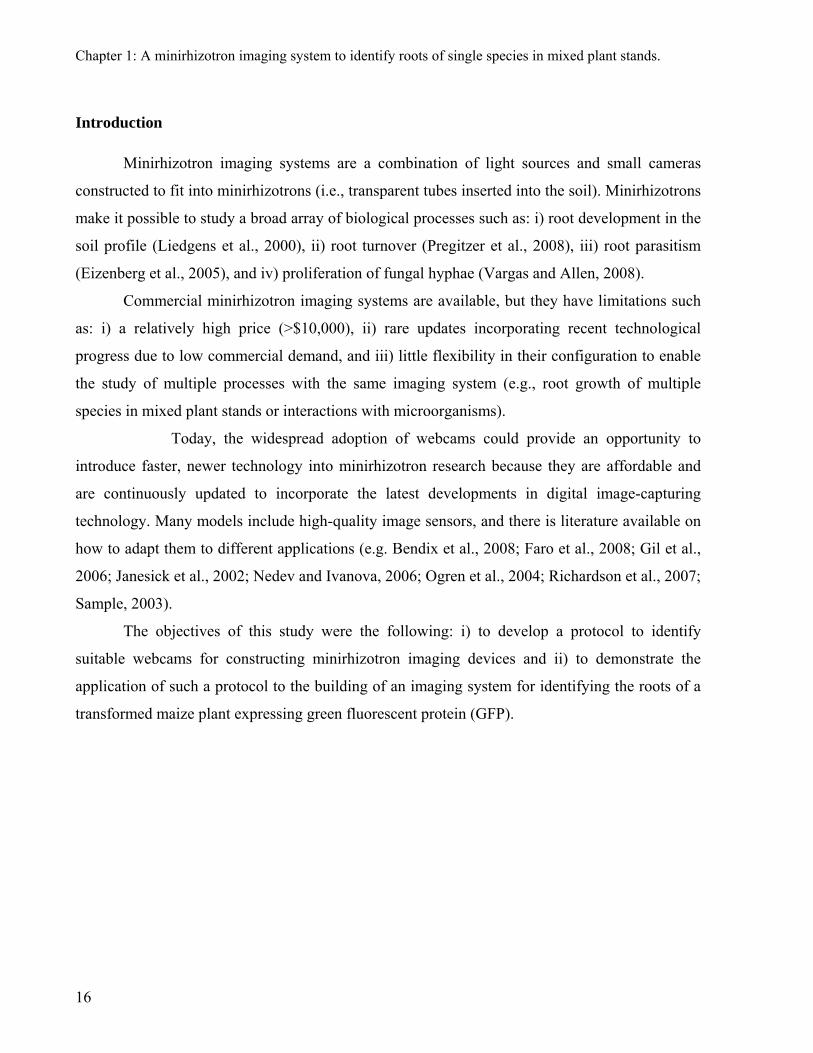
Light
The dark environment in the minirhizotron makes artificial illumination necessary. A
simple and affordable solution is to use LEDs (light emitting diode). Two LEDs (5500 K, Luxeon
III Star, Philips Lumileds Lighting Company, San Jose, CA, USA) arranged around the optic are
enough for obtaining images where roots and soil structures are visible. Strong light intensity has
to be avoided since it causes undesired reflections at the minirhizotron interface. This issue was
controlled by an assemblage of resistors to supply appropriate light intensity.
Case for the image sensor
The sensor’s location relative to the soil-minirhizotron interface can be rotated by 90° or
placed in front of it, depending on the space available for the camera components inside the
minirhizotron. The former allows for minimizing the space required, but it requires mirrors
between the image sensors and the target objects. This makes the camera more complicated,
expensive and difficult to maintain. As a result systems based on mirrors or that prevent the
camera from being introduced in the minirhizotron were discarded a priori but could be
considered for applications that demand very narrow minirhizotrons. A frontal localization has a
simpler implementation as long as the space available for the image sensor and the optics is
enough. For routine and safe operation, all components of the imaging system have to be
assembled in a suitable case. During the development of the imaging system and for testing
purposes we used a prototype made from a PVC tube with a smaller diameter than the
19
Chapter 1: A minirhizotron imaging system to identify roots of single species in mixed plant stands.
minirhizotron. However, for the later routine operation a more robust housing in metal was
developed.
Handle
For the construction of a handle to hold the minirhizotron imaging system and to ensure
proper image registration we followed a protocol reported elsewhere (Johnson and Meyer, 1998).
This is a mechanical handle attached to the case for the image sensor that allows for introducing
the system inside the minirhizotron and registering and collecting images. The handle couples a
ratchet advancing mechanism with another mechanism that locks the handle to the minirhizotron
tube, ensuring proper soil position registration. The handle allows moving easily the imaging
system from one minirhizotron to another. Such a handle is commercialized as a separate
component by Bartz (Bartz Technology Co., Santa Barbara, CA, USA).
Power supply
The camera is supplied with power via the USB port, which is also used to connect the
camera to a computer, where images are digitally stored. The power of the USB port is not high
enough to supply the lightning system. Currently an AC/DC power supply is used (Voltcraft
Stecknetzgerät 3-12 V/1000 MA, Hirschau, Germany).
Software for the capture and analysis of images
Software to trigger the capture and name numerous images is available for free
(http://videocapture.sourceforge.net/) or in commercial packages
(http://www.bartztechnology.com/products.html). Once the digital images are acquired, image
processing software of general characteristics (e.g. imageJ; http://rsbweb.nih.gov/ij/) or
specifically developed to analyze minirhizotron images
(http://www.ces.clemson.edu/~stb/rootfly/) can be used to determine the physical size of the
pixels covered by roots in the images.
20
Chapter 1: A minirhizotron imaging system to identify roots of single species in mixed plant stands.
Comparison of webcams
We took a set of webcams available in the market and tested the quality of their images
with an imaging test chart (Kodak Digital Science Imaging Test Chart TL-5003, 1995 Eastman
Kodak Company). The set of cameras included: Quickcam pro 300 (Logitech International S.A.,
Romanel-sur-Morges, Switzerland), Zicplay TalkCam Tracer CCD (Zicplay S.A., St-Sulpice,
Switzerland), Philips SPC 900 NC (Royal Philips Electronics Inc., Amsterdam, Netherlands),
Creative livecam notebook pro (Creative Technology Limited, Singapore City, Singapore), and
Logitech QuickCam express (Logitech International S.A., Romanel-sur-Morges, Switzerland).
We tested the webcams under two conditions: i) horizontal at a distance of 30 mm from
the imaging test chart positioned flat and, ii) inside of a minirhizotron tube made of Plexiglas at
30 mm from the imaging test chart that was rolled over a minirhizotron. The cameras were
compared on the basis of images of 640 x 480 pixels, with a resulting resolution of 40.6 μm.
Plant material
We tested the imaging system configured to identify roots of single species in mixed plant
stands using a transgenic line of maize (Zea mays L.) expressing the GFP (Aulinger et al.,
(2003). The maize genotype ETH-M72 was genetically transformed to include the gene for the
GFP (ETH-M72GFP). The transformation construct contains the gfp gene flanked by the ubiquitin
promoter (ubi::gfp) and the NOS terminator and was cloned into the pUC19 vector, which
contains the gene for ampicillin resistence (ampR) at the restriction sites SpeI and XbaI. The gfp
gene was cloned into the cassette at the NcoI and SalI sites. The expressed GFP protein is
reported to have a fluorescence peak between 500 and 520 nm when excited by light at 450 to
470 nm.
We grew the transgenic line of maize expressing GFP alone or together with the negative
control (i.e., the non-transgenic maize line) in rectangular containers filled of soil (0.365 m x
0.265 m and 0.255 m) with two horizontal minirhizotrons at a soil depth of 0.10 m. Eight plants
per box were sown. We took pictures using the minirhizotron imaging system with the
conventional configuration and that to identify roots of single species in mixed plant stands.
21
Chapter 1: A minirhizotron imaging system to identify roots of single species in mixed plant stands.
Results and discussion
Development of a minirhizotron imaging system
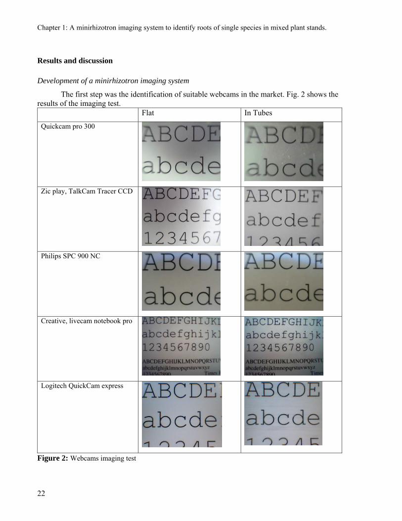
The first step was the identification of suitable webcams in the market. Fig. 2 shows the results of the imaging test. Flat In Tubes
Quickcam pro 300
Zic play, TalkCam Tracer CCD
Philips SPC 900 NC
Creative, livecam notebook pro
Logitech QuickCam express
Figure 2: Webcams imaging test
22

Chapter 1: A minirhizotron imaging system to identify roots of single species in mixed plant stands.
This test measures the imaging characteristics of webcams based on the following quality
parameters: i) aspect ratio, ii) geometric distortion, iii) exposure uniformity, and iv) light
uniformity. By this test it was possible to discard those webcams that produced images with
distortions like Quickcam pro 300 and Zic play (Fig. 2). A set of webcams that produced images
of acceptable quality remained. The imaging test also provided information to decide whether
was better to use the build in optics or those from a specialized provider. Using optics supplied
by an external provider was against the objective of building an affordable camera. Therefore
livecam notebook pro, manufactured by Creative, was the right choice. This was a good
compromise, too, among a camera size small enough to be included in a tube, the area of the
captured images, and the quality of the images. The 640 by 480 pixels images cover an area of
26.0 x 19.5 mm, with a resulting resolution of 40.6 μm. However, any of the other two webcams
tested that had satisfactory quality parameters would have probably been also suitable for
developing a minirhizotron imaging system.
The second step was to identify the settings of the other components of the imaging
system. This must be done in a specific manner according to the webcam that was selected. The
relatively small size of the webcam that we chose allowed for assembling the image captor
directly under the target area of the images. For calibrating the webcam, it was necessary to
determine the physical size of the images captured by placing a graduated millimeter ruler over
the captor.
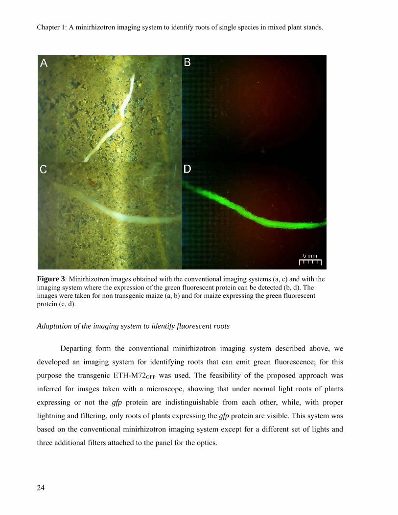
Figures 3a and 3c show images obtained with the conventional minirhizotron imaging
system; roots and soil pores are clearly visible which demonstrates the high resolution of these
images. These images are of similar quality as those from commercial minirhizotron cameras and
fulfill the requirements of software to analyze root images (e.g. rootfly). Therefore, they
demonstrate that constructing a minirhizotron imaging system using webcams is possible.
Furthermore, following the steps described here, the resolution can be increased as well as the
light characteristics to satisfy specific research goals.
23
Chapter 1: A minirhizotron imaging system to identify roots of single species in mixed plant stands.
Figure 3: Minirhizotron images obtained with the conventional imaging systems (a, c) and with the imaging system where the expression of the green fluorescent protein can be detected (b, d). The images were taken for non transgenic maize (a, b) and for maize expressing the green fluorescent protein (c, d).
Adaptation of the imaging system to identify fluorescent roots
Departing form the conventional minirhizotron imaging system described above, we
developed an imaging system for identifying roots that can emit green fluorescence; for this
purpose the transgenic ETH-M72GFP was used. The feasibility of the proposed approach was
inferred for images taken with a microscope, showing that under normal light roots of plants
expressing or not the gfp protein are indistinguishable from each other, while, with proper
lightning and filtering, only roots of plants expressing the gfp protein are visible. This system was
based on the conventional minirhizotron imaging system except for a different set of lights and
three additional filters attached to the panel for the optics.
24

Chapter 1: A minirhizotron imaging system to identify roots of single species in mixed plant stands.
The detection of the roots of ETH-M72GFP depends on the excitation of the protein by
supplying light in the appropriate range. In microscopy Hg light is typically used. This is,
however, no practical with minirhizotrons since the heat that they produce damage the
minirhizotron tubes. Because LEDs that emit light at wave lengths required to excite the protein
exist, we used LEDs that covered the narrow light spectrum of 440 to 460nm (Luxeon V Star,
Philips Lumileds Lighting Company, San Jose, CA, USA).
Upon correct activation of the gfp protein, fluorescence should be visible with an
adequate set of filters. On selecting such filters for an affordable imaging system it was important
to find a compromise between price and quality. Cheap plastic filters did not provide satisfactory
blocking of the incoming light spectrum, therefore more specific glass filters had to be chosen. A
band blocking filter was used to affine the wavelength window of the light (Dichroic 555 IM 25,
Comar Instruments, Cambridge, UK). A longpass color glass filter (LONG 515 nm, Edmund
Optics, Barrington, USA) was installed in front of the image captor to block the wavelengths
outside the fluorescence spectrum of the GFP. This precise adjustment of the light source and the
captured light was important to avoid the detection of auto-fluoresce from roots and elements in
the rhizosphere.
Figures 3b and 3d shows images obtained with the minirhizotron imaging system
modified to detect roots of ETH-M72GFP. In these images the roots of ETH-M72GFP are visible
and it is easier to distinguish roots from the background in these images (Fig. 3b and 3d) than in
conventional images (Fig. 3a and 3c) because of the better contrast. Therefore, the basic
configuration of the minirhizotron imaging system based on a webcam could be easily adapted to
detect the fluorescent roots of ETH-M72GFP and overcome the problem of the low flexibility in
the construction of commercial equipments. This low flexibility makes difficult and costly to
conduct the tests that are necessary to extend the applications of minirhizotrons. The cost of the
imaging systems that we reported here is of $195 for the conventional images and $390 for the
images where fluorescent roots are visible.
25
Chapter 1: A minirhizotron imaging system to identify roots of single species in mixed plant stands.
26
Conclusion
Here we demonstrate the feasibility of developing an affordable minirhizotron imaging
system for capturing conventional images and for images where fluorescent roots are visible.
This was necessary to overcome the low flexibility in the configuration of commercial systems
and thus extend the application of minirhizotrons. The system to detect roots of ETH-M72GFP
constitutes the first report of an imaging system to identify roots of single species in mixed plant
stands.
Furthermore, the steps proposed here can be implemented to develop and configure
imaging systems to extent the applications of minirhizotrons. This could be the case for devices
to screen rhizotrons or to capture images of high resolution where mycelium of ectomycorrhiza is
visible. There are currently no imaging devices to conduct the latter two tasks.

Chapter 2: The use of green fluorescent protein (GFP) as a tool to identify roots in mixed plant stands
Chapter 2: The use of green fluorescent protein (GFP) as a tool to identify roots in mixed plant stands
Published in Functional Plant Biology (2009), volume 36, number 10 & 11, pages 930-937. Faget, M., Herrera, J.M., Stamp, P., Aulinger-Leipner, I., Frossard, E., and Liedgens, M. The use of green fluorescent protein as a tool to identify roots in mixed plant stands.
Abstract
Although roots take up most of the resources required by the plant, a lack of efficient
research tools hinders our understanding of the function and relevance of the root system. This is
even more evident when the research focus is not on a single plant, but on multiple plants that
share the same soil resources. None of the available methods allow for simple, inexpensive, non-
destructive, and objective assignment of observed roots in a mixture of plants to a target plant.
Here, we demonstrate that transgenic plants expressing the green fluorescent protein (GFP)
combined with the well-established minirhizotron technique are a route to overcoming this
limitation. We planted transgenic maize (Zea mays L.) in combination with either its
corresponding wild-type, Italian ryegrass (Lolium multiflorum Lam.) or soybean (Glycine max
(L.) Merr.). The identification of fluorescent roots allows the relative distribution of roots of each
plant type and their interaction and interference with each other to be observed. The selected
plants are suitable for model experiments to unravel fundamental belowground ecological
processes. Because genetic transformation of plants is an established technique that can be
applied to a large set of plant species, this method will be of interest to a broad range of research
areas.
Keywords: root research methodology, root interactions, green fluorescent protein (GFP), mixed
plant stands, minirhizotron, and imaging system.
27
Chapter 2: The use of green fluorescent protein (GFP) as a tool to identify roots in mixed plant stands
Introduction
Roots are essential for plant growth, survival, and fitness. Plant species differ in their
temporal and spatial exploration of the soil and in their adaptation to biotic and abiotic stresses.
Due to the heterogeneous nature of soil environments, root systems must respond to a wide
spectrum of physical, biological, and chemical conditions, including resource availability, all of
which show spatial and temporal variation. Consequently, root systems develop into a complex
array of irregularly distributed roots. These root arrangements ultimately determine the ability of
the plant to access soil resources (Robinson, 1994).
To determine the distribution of roots in the soil, a diverse set of approaches was
employed. Destructive sampling of roots has been the research method of choice for more than a
century. The most common approach is to sample soil cores, which are small compared to the
rooting volume (Pierret et al., 2005). However, with these methods it is impossible to determine
the exact position of the roots. Root position is important because resource uptake is usually
affected to a greater extent by the relative spatial distribution of roots and soil resources than by
the size of the root system (de Kroon, 2007). Also, these methods do not enable multiple
measurements of the same roots. Non-destructive screening by means of transparent observation
interfaces overcomes this limitation and is effective for following the spatial and temporal
dynamics of root growth (Taylor et al., 1990). A disadvantage of this method is that it modifies
the rooting environment. However, the degree of interference is much smaller for minirhizotrons
(i.e., transparent tubes inserted into the soil) compared to classical rhizotrons (Taylor et al.,
1990). Despite the abovementioned approaches, a simple, inexpensive method to assess the
distribution of roots in the soil does not exist.
Root systems usually share the soil with intraspecific or interspecific neighbours. Thus,
the relative arrangement of the roots will influence processes such as resource capture and root
exudation (Li et al., 2007). However, the methodological constraints to assess root distribution
are even more acute when the focus is on the coexistence of multiple plants, i.e., when they share
the same soil volume, rather than on single plants. For such cases, it is also necessary to
28
Chapter 2: The use of green fluorescent protein (GFP) as a tool to identify roots in mixed plant stands
determine which root belongs to which plant. This is very difficult to be done visually on a high
number of samples since roots as opposed to flowers or leaves show few distinctive external
features that would permit identification of species (Fitter, 2002).
The relative distribution of roots of different plant species was studied by the following
methods: (i) the relative uptake of tracers such as 15N, 2H, and 18O (e.g. Jumpponen et al., 2002);
(ii) δ13C discrimination between C3 and C4 plants (Lehmann et al., 1998); (iii) labelling the shoots
of each species with different radioactive tracers in order to distinguish their roots from the
differential signature on autoradiographs (Baldwin and Tinker, 1972); (iv) excavating the soil and
tracing roots back to the plant to which they belong (Pechackova et al., 1999); (v) mapping all the
roots at the cut surface of a core and conducting a chemical extraction to distinguish roots of
different species according to the colour and fluorescence intensity of the resulting eluant
(Caldwell et al., 1996); and (vi) using molecular tools based on the extraction of DNA and
microsatellite analysis of root fragments (Brunner et al., 2001). None of these approaches has
become a standard investigation tool, and, except for the method used by Caldwell et al. (1996),
they do not allow the determination of the relative location of roots belonging to different plants.
However, such information is important for understanding and managing the coexistence of
plants in natural and managed ecosystems (de Kroon, 2007; Schenk, 2006).
Fluorescence is widely applied in the natural sciences as a visual reporter for localisation
purposes. For example, autofluorescence has been used to report the colonisation of roots by
microorganisms (Gamalero et al., 2003), and staining with fluorescent substances has been used
to analyse plant structures (Lux et al., 2004). The use of fluorescent proteins was such an
impressive methodological achievement for research that it was awarded the Nobel Prize for
Chemistry in 2008. By direct or associated tagging, fluorescent proteins allow for a plethora of
scientific applications aimed at the temporal and spatial monitoring of processes in living cells
and organisms (Chalfie and Kain, 2006).
29

Chapter 2: The use of green fluorescent protein (GFP) as a tool to identify roots in mixed plant stands
We genetically transformed the maize genotype ETH-M72 to include the gfp gene for the
green fluorescent protein (GFP) from the jellyfish Aequorea victoria (Aulinger et al., 2003).
Here, we demonstrate that fluorescent roots are a powerful tool for investigating the relative
distribution of roots, especially fine roots, belonging to different plant species, a new
experimental approach in functional root ecology. Furthermore, we show that minirhizotrons
enable continuous and non-destructive observation of the fluorescence expression of GFP maize.
The plant set used for this study includes the genetically transformed maize genotype, its
corresponding wild-type, Italian ryegrass, and soybean.
Material and methods
Plant material
The maize genotype ETH-M72 was genetically transformed to include the gene for GFP
(ETH-M72GFP). The transformation construct contains the gfp gene flanked by the ubiquitin
promoter (ubi::gfp) and the NOS terminator. It was cloned into the pUC19 vector, which contains
the gene for ampicillin resistance (ampR) at the restriction sites SpeI and XbaI. The gfp gene was
cloned into the cassette at the NcoI and SalI sites. The expressed GFP is reported to have a
fluorescence peak between 500 and 520 nm when excited by light at 450 to 470 nm. Validation
of the genetic transformation, the integration pattern of the inserted gene, and comparisons of the
homozygosity of transgenic and non-transgenic controls are reported in Aulinger et al. (2003).
The ETH-M72GFP maize was grown together with either the untransformed wild-type
(ETH-M72WT), Italian ryegrass (Lolium multiflorum Lam., cv. Ellie), or soybean (Glycine max
Merill, cv. Amphor). The arrangement with ETH-M72WT and with non-maize plants represents
monocrop and intercropping systems, respectively.
30

Chapter 2: The use of green fluorescent protein (GFP) as a tool to identify roots in mixed plant stands
Experimental conditions
The data presented herein were collected from two sets of experiments in which plants
were grown either in pouches without soil or in large containers with soil. Maize ETH-M72GFP
was grown alone, with wild-type maize and with the other plant species.
Germinated seeds of ETH-M72GFP (n=30) and ETH-M72WT (n=30) maize were
transferred alone or together to moistened blotting paper (21×29.5 cm; Anchor Paper, St. Paul,
MN, USA) in pouches (Hund et al., in press). The pouches were hung in plastic containers
(27×37×32 cm; Arcawa GmbH, Chatillon, Switzerland) in order to submerge the bottom 20 mm
of the blotting paper in water. To avoid a light impact on root growth, the pouches were covered
with a 0.5-mm thick black polyethylene foil (PE-Teichfolie Typ WA-1200, Walser
Kunststoffwerk AG, Buerglen, Switzerland). In addition, to avoid heating the pouches, the
containers were covered with aluminium foil, except for a narrow opening through which the
seedling grew. The plants were grown for 7 days in a growth chamber (PGW36 Conviron,
Winnipeg, MB, Canada) at a temperature of 24ºC, a relative humidity of 70%, a photosynthetic
active radiation of 400 μmol sec-1 m-2, and a photoperiod of 16 h. The blotting papers with the
plants were removed from the pouches to acquire images of the roots.
For the experiments with soil, ETH-M72GFP maize was grown alone and together with
Italian ryegrass in large containers, i.e., wooden boxes (0.60 m wide x 0.80 m long and 0.80 m
high), allowing for unrestricted root growth. Minirhizotrons (54 mm inner diameter, 60 mm outer
diameter) were installed horizontally in these wooden boxes at a depth of 0.15 m. Minirhizotrons
were in the middle of the boxes, at 0.30 m from the edges. Italian ryegrass was sown at a density
of 6.27 g seed per m2. After 40 days, a 0.30-m wide strip of Italian ryegrass in the mid-section of
the boxes and perpendicular to the longer side was cut and removed from the box. Within that
strip, ETH-M72GFP maize plants were sown at a density of 10.4 plants per m2. The orientation of
the resulting maize row was perpendicular to the minirhizotrons. Thereafter, the remaining grass
plants were cut to 0.15 m at weekly intervals. The mean temperature in the greenhouse was 23°C,
and the photoperiod was 16 hours. The water content of the soil was maintained by irrigation, as
estimated by a time domain reflectometry probe (EC-5 Soil Moisture Sensor, Decagon Devices
31
Chapter 2: The use of green fluorescent protein (GFP) as a tool to identify roots in mixed plant stands
Inc., Pullman, WA, USA). The experiment was conducted until the anthesis of maize, which
occurred approximately at 83 days after sowing.
ETH-M72GFP maize was also grown alone and together with either ETH-M72WT or
soybean in circular PVC containers (0.60 m inner diameter x 0.70 m high) with a single
horizontal minirhizotron at a depth of 0.20 m, in the same greenhouse environment as the
experiment in rectangular wooden containers. Ten ETH-M72GFP maize plants in the containers
with ETH-M72GFP alone, and five plants of each type in the containers with two types of plants
were sown at the same time. The containers with two types of plants were separated into two
equal parts perpendicular to the orientation of the minirhizotrons, to which one of each plant type
was assigned. Plants within a single half of each container were arranged in a regular pattern. The
minimum distance between two plants of different types was 0.12 m.
The substrate in all containers was a mix of loamy field soil and sand and had according
to the method given in parentheses the following characteristics: 30 g kg-1 organic matter (Blake-
Walkley), 60 g kg-1 clay, 110 g kg-1 silt, 810 g kg-1 sand, pH 7.7, 66.3 mg kg-1 assimilable K
(NH4 acetate), 4.1 mg kg-1 P (Olsen), and 12 g kg-1 N (2M KCl). The wooden boxes and the PVC
containers were filled three months before starting the experiments to enable the soil to settle.
Foskal® (P2O5, K2O, Mg, Ca, and S; Agroline, Basel, Switzerland) and ammonium nitrate (NH4
NO3) were applied before sowing to supply 1.4 g P, 8 g K, 0.57 g Mg, 0.20 g Ca, 0.87 g S, and
1.1 g N per m2.
Imaging equipment
Although there is commercial equipment available for imaging the soil-minirhizotron
interface (e.g., Bartz Technology Company, Carpinteria, CA, USA), its configuration does not
allow for the visualisation of fluorescence. However, it is possible to construct imaging systems
to test the suitability of the fluorescence method, as devices for capturing images (such as web
cameras) are widespread and inexpensive. Therefore, we selected a web camera (Live Cam
Notebook Pro®, Creative Technology Limited, Singapore City, Singapore) to digitally capture
minirhizotron images. The unmodified camera, one of the smallest currently available, was
32

Chapter 2: The use of green fluorescent protein (GFP) as a tool to identify roots in mixed plant stands
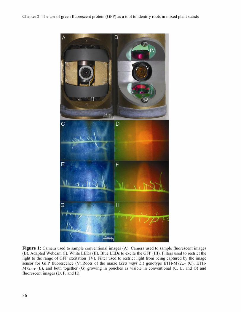
integrated into a very simple assembly (Fig. 1a) with additional light emission devices (LED). Its
basic configuration did not deviate fundamentally from the design proposed by Upchurch and
Ritchie (1983). The 640 x 480 pixel images cover an area of 26.0 x 19.5 mm, with a resulting
resolution of 40.6 μm. The LEDs were connected to an external power supply (Voltcraft
Stecknetzgerät 3-12 V/1000 MA, Hirschau, Germany), and the camera was powered by the USB
computer interface. Based on this basic module, two configurations were constructed (Fig. 1a and
1b). One configuration included white LEDs (5500 K, Luxeon III Star, Philips Lumileds Lighting
Company, San Jose, CA, USA) for the sampling of images to display all structures at the
interface of the minirhizotron. These images will be referred to as “conventional images”. The
second configuration differed from the first one by: (i) LEDs covered a narrower light spectrum
of 440 to 460 nm (Luxeon V Star, Philips Lumileds Lighting Company, San Jose, CA, USA)
close to the GFP excitation range; (ii) a band-blocking filter was used to avoid sources of green
light (Dichroic 555 IM 25, Comar Instruments, Cambridge, UK); and (iii) a long-pass colour
glass filter (LONG 515 nm, Edmund Optics, Barrington, USA) was installed in front of the
webcam to block as many of the long light waves outside the fluorescence spectrum of GFP as
possible. This precise adjustment of the light source and the captured light is important to avoid
mfthe detection of auto-fluorescing substances from the roots and the rhizosphere. Images
sampled by the latter configuration display the fluorescent roots of ETH-M72GFP and are referred
to as “fluorescent images”. The assemblies used to capture the images described above were
mounted on the tip of a handle similar to the one described by Ferguson and Smucker (1989).
This guarantees that the same position at the minirhizotron interface may be imaged repeatedly
and precisely with both configurations. The use of identical handles for both cameras enabled us
to repeatedly sample images of both types at the same position in the minirhizotrons over the
entire duration of the experiment. Conventional minirhizotron images and fluorescent images
were obtained from the blotting paper with roots and from the upper surfaces of the soil-
minirhizotron interface. The minirhizotron images covered a strip of 19.5 x 312 mm (i.e. 12
positions) in both experiments with soil. Images for the experiments in pouches were obtained 7
days after germination, while those for the soil experiments were obtained up to 48 (for ETH-
M72GFP and Italian ryegrass) or 72 (for ETH-M72GFP, ETH-M72WT and, soybean) days after
sowing the maize plants.
33
Chapter 2: The use of green fluorescent protein (GFP) as a tool to identify roots in mixed plant stands
Screening of roots growing in soil
The number of roots (i.e. number of root incidences on the minirhizotron interface) and
root length for each of the sampled images was determined according to Upchurch and Ritchie
(1983) and Atkinson (2000), respectively. Although these estimates can be obtained from single
minirhizotron images, the accuracy of identifying root structures in images increases when
images acquired at the same position are captured across a set of sampling dates (Smit et al.,
2000). Therefore, all images recorded at a single minirhizotron position over the entire growing
season were organised into an image time series (ITS). Since no objective visual criteria exist to
determine whether a root is functional (Smit et al., 2000), all roots identified in an image were
accounted for in each ITS. Thus, the obtained response variables were cumulative no. of roots
and cumulative root length (cm cm-2) observed per image since the sowing date. These were
converted into a surface unit (cm2) and are referred to hereafter as root density (roots cm-2) and
root length density (cm cm-2), respectively.
The image set (n=3200) used to validate the proposed method consisted of 100 ITS, each
including images sampled on 16 dates, randomly selected from plots containing only ETH-
M72GFP. Conventional and fluorescent images were screened for each ITS. Images without roots
were included to account for false positives (i.e., identification of non-root objects as roots).
Statistical analysis of the effect of image type on root parameters
Effects of the type of image, i.e., conventional or fluorescent, on estimates of root density
and root length density of ETH-M72GFP were tested using analysis of variance. Image type, plot,
and the interaction between image type and plot were considered as factors in the statistical
model. Deviations from the 1:1 relationship between conventional and fluorescent images were
studied by taking the values from conventional images as the reference (expected values) and
comparing them those in fluorescent images (observed values). The analysis of variance and the
deviation from the 1:1 relationship for root density and root length density between the values
obtained from fluorescent images and those from conventional images were performed for the
last image of the ITS using the functions aov() and lm() in the statistical software R, respectively.
34

Chapter 2: The use of green fluorescent protein (GFP) as a tool to identify roots in mixed plant stands
Results
Expression of GFP along ETH-M72GFP roots in pouches
Images of roots obtained from pouches are shown in Figure 1 (c to h) as examples of the
type of results obtained with fluorescing roots. Images c, e, and g depict conventional images,
whereas images d, f, and h depict fluorescent images. The latter images show that although roots
from ETH-M72WT could sometimes be noted in fluorescent images (Fig. 1d), the expression of
GFP in the ETH-M72GFP results in roots with green light emited (Fig. 1f) that is not observable
for ETH-M72WT (Fig. 1d). Furthermore, we did not observe ETH-M72GFP roots that lacked this
fluorescent intensity. As a result, roots from ETH-M72WT and ETH-M72GFP are indistinguishable
in conventional images (Fig. 1g), but clearly distinguishable in fluorescent images (Fig. 1h).
Therefore, it is possible to conclude that: (i) roots showing bioluminescence belong to ETH-
M72GFP plants (Fig. 1h), and (ii) the GFP expression and thus fluorescence is observable
throughout the extension of the main root axis and its branches (Fig. 1f and 1h).
Root screening of ETH-M72GFP grown alone in the soil
The image set used to validate the proposed approach consisted of 100 ITS obtained from
plots in which ETH-M72GFP was grown alone. The same positions at the soil-minirhizotron
interface were screened independently using either conventional or fluorescent images and the
results obtained from each type of image were compared. For single ITS’, root density from the
fluorescent images varied between +52% and -28%, compared to the conventional images.
Similarly, for root length density, these differences ranged between +62% and -10%. On average,
root density and root length density for the ITS were recorded as 28% and 16% higher in
fluorescent images compared with conventional minirhizotron images. Figure 2a and 2b show the
relationships between values obtained from fluorescent images and those from conventional
minirhizotron images for root density and root length density, respectively.
35
Chapter 2: The use of green fluorescent protein (GFP) as a tool to identify roots in mixed plant stands
Figure 1: Camera used to sample conventional images (A). Camera used to sample fluorescent images (B). Adapted Webcam (I). White LEDs (II). Blue LEDs to excite the GFP (III). Filters used to restrict the light to the range of GFP excitation (IV). Filter used to restrict light from being captured by the image sensor for GFP fluorescence (V).Roots of the maize (Zea mays L.) genotype ETH-M72WT (C), ETH-M72GFP (E), and both together (G) growing in pouches as visible in conventional (C, E, and G) and fluorescent images (D, F, and H).
36

Chapter 2: The use of green fluorescent protein (GFP) as a tool to identify roots in mixed plant stands
Figure 2: Relationship of the root densities (A) and root length densities (B) measured with fluorescent and conventional images. The line indicates the 1:1 ratio. The box diagrams in each plot show the distribution of the values obtained by subtracting the results of conventional images from those of fluorescent images.
37
Chapter 2: The use of green fluorescent protein (GFP) as a tool to identify roots in mixed plant stands
It was significantly different from the 1:1 relationship for root density (F-value=9.35, p-
value=0.002, d.f.=194) and for root length density (F-value=17.0, p-value<0.001, d.f.=194) and it
was not linear,with systematic deviations from the estimated line identified for larger values.
Also the median differences between the estimates of the root parameters for both image types
(box diagrams) were higher than 0, indicating that the values were skewed towards high values.
The differences in fluorescent compared to conventional images were especially high for root
densities over 3 roots cm-2 and root length densities over 2 cm cm-2. Box diagrams also showed
that, except for some potential outliers, the differences between the estimates for both image
types were approximately normally distributed, and the distribution of the differences among root
length densities were more skewed than the distribution of the differences among root densities.
An analysis of variance revealed marginal effects from the type of image used to measure root
density (F-value = 3.22, p-value = 0.07, d.f. = 197) and insignificant effects for root length
density (F-value = 1.39, p-value = 0.24, d.f. = 197).
Larger values in fluorescent images compared with conventional images often resulted
from the fact that some of the roots were only observed in the fluorescent images (Fig. 3d and 3l)
because of the improved contrast between the roots and the background in those images. In
conventional images, transparent roots may be overlooked and some roots are very difficult to
distinguish from other image structures. This is not a factor for the ETH-M72GFP roots in
fluorescent images. Similarly, in fluorescent images, two or more root segments that are closely
located to other root segments are more easily recognised as separate root segments. Lower
values for the fluorescent images can arise from multiple root segments not being recognised as a
single unit; i.e., the integrity of the root segment is lost due to poor contrast with the background.
In conventional images, these segments are interpreted as separated root segments
according to the rule proposed by Upchurch and Ritchie (1983). Furthermore, lower values for
fluorescent images compared to conventional images also denote the risk in conventional images
due to the uniform background, to erroneously identify image structures as roots when they are
not. In fluorescent images this is not a factor. Nevertheless, the fact that higher values for both
root density and root length density are more common in fluorescent images shows that
38
Chapter 2: The use of green fluorescent protein (GFP) as a tool to identify roots in mixed plant stands
underestimation rather than overestimation is the more serious source of error when screening
roots with conventional minirhizotron images. Similarly to the pouches experiment, we did not
observe any ETH-M72GFP roots that lacked green light emitted at any point during the screened
sequence from the minirhizotrons inserted in the soil. This observation was expected because the
gfp gene is constitutively expressed. These results confirm that all ETH-M72GFP roots can be
identified in fluorescent images obtained from minirhizotrons inserted in the soil.
Discrimination of roots between interspecific neighbours
For each of the two combinations of plants studied in containers with soil, one set of four
pictures is shown (Fig. 3). Images a to d and e to h depict roots of Italian ryegrass and soybean
together with ETH-M72GFP, respectively. The images depict conventional and fluorescent images
sampled at two different dates. All of the conventional images include structures that can be
identified as roots, but other elements, such as soil particles and voids, are also visible.
From the conventional images of roots, it was not possible to assign roots to the plant
type. The fluorescent images proved that only roots of Italian ryegrass, soybean, and ETH-
M72WT exist at the sampled location on the first sampling date (Fig. 3), whereas ETH-M72GFP
roots (white arrows) coexist with roots of the companion plants on the second sampling date. The
fluorescence of the meristematic tissues, such as the tips and branch insertions of ETH-M72GFP,
were particularly bright. Some ETH-M72GFP roots were visible only in the fluorescent images.
Structures in the conventional images are the sole result of light reflection. Roots must
fluoresce within the range defined by the filter (515 nm) installed in front of the imaging sensor
in order to be detected in the fluorescent images (Fig. 1b). The low amount of light detected by
the image sensor originates from reflection because the wavelength range of the LEDs (440 to
460 nm, Fig. 1b) and the fluorescence do not overlap. The identifiable roots in both types of
images are of one of three types: those that are only visible in the conventional images, those that
are visible in the conventional and in the fluorescent images, and those that are only visible in the
fluorescent images. The roots in the conventional images have no characteristics that would
enable them to be assigned to one of the plants in the group. Since the green fluorescence of the
39
Chapter 2: The use of green fluorescent protein (GFP) as a tool to identify roots in mixed plant stands
ETH-M72GFP roots is the consequence of the expression of a protein naturally absent in plants, it
is concluded that roots visible in the fluorescent images belong to ETH-M72GFP. In the images
obtained for containers with ETH-M72GFP plants only, roots observed in conventional images
were also visible in fluorescent images.
In addition, some ETH-M72GFP roots were visible only in the fluorescent images (Fig. 3d
and 3l). These are typically fine roots that, as found for containers with ETH-M72GFP only, are
not identifiable in the corresponding conventional images (Fig. 3c and 3k) because they are either
too fine or are transparent. Roots that lack green light emitted, as shown in the pouches
experiment (Fig. 1c to 1h), belong to companion plants rather than to the ETH-M72GFP plants.
40
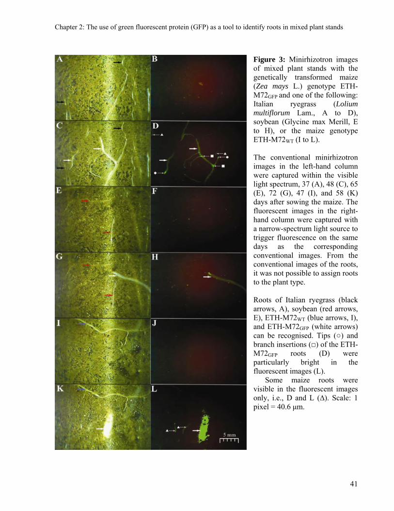
Chapter 2: The use of green fluorescent protein (GFP) as a tool to identify roots in mixed plant stands
Figure 3: Minirhizotron images of mixed plant stands with the genetically transformed maize (Zea mays L.) genotype ETH-M72GFP and one of the following: Italian ryegrass (Lolium multiflorum Lam., A to D), soybean (Glycine max Merill, E to H), or the maize genotype ETH-M72WT (I to L). The conventional minirhizotron images in the left-hand column were captured within the visible light spectrum, 37 (A), 48 (C), 65 (E), 72 (G), 47 (I), and 58 (K) days after sowing the maize. The fluorescent images in the right-hand column were captured with a narrow-spectrum light source to trigger fluorescence on the same days as the corresponding conventional images. From the conventional images of the roots, it was not possible to assign roots to the plant type. Roots of Italian ryegrass (black arrows, A), soybean (red arrows, E), ETH-M72WT (blue arrows, I), and ETH-M72GFP (white arrows) can be recognised. Tips (○) and branch insertions (□) of the ETH-M72GFP roots (D) were particularly bright in the fluorescent images (L). Some maize roots were visible in the fluorescent images only, i.e., D and L (∆). Scale: 1 pixel = 40.6 μm.
41

Chapter 2: The use of green fluorescent protein (GFP) as a tool to identify roots in mixed plant stands
Discussion
This is the first report to demonstrate the use of fluorescence resulting from GFP
expression to study the interactions and interference of roots in mixed plant communities. It
presents a method that allows for the distinction of roots belonging to different plants that are
grown together. This method is based on the use of genetically modified plants in which the
constitutive expression of GFP results in a bioluminescence throughout the root extension (Fig.
1f and 1h). This green emitted light is not observed in non-genetically transformed maize,
soybean, and Italian ryegrass. The fluorescence of the ETH-M72GFP roots can be used as a visual
marker to quantify the root density and root length density of intraspecific and interspecific
neighbours.
Although this method requires at least one transgenic fluorescent plant species, it can
finally replace methods that rely on the separation of rooting volumes by barriers (Li et al., 2007)
or trenches (Ludwig et al., 2004). Such methods drastically disturb the soil environment and, in
extreme situations, can lead to an artificially-induced response in the plant (Connell, 1990).
However, Campbell (1994), for example, has reported to have distinguished wild cherry (Prunus
avium L.) from grass roots by their generally thicker diameter, opacity, more succulent
appearance, and colour. The lack of further reports applying these criteria for distinguishing roots
indicates the difficulty in adapting the procedure to a larger range of plant species combinations.
Furthermore, for universal applications, a new methodology for discriminating between root
systems should allow for sampling of a sufficiently large numbers of roots, as required for a
representative estimation of fine root traits (Pierret et al., 2005). This new method meets these
requirements and will provide the urgently needed experimental evidence of the relative
distribution of root systems in the soil on the condition that at least one of the desired genotypes
or plant partners in the mixture can be genetically transformed for experimental purposes.
This method allows for a very fine-scaled resolution in time and space, which is not the
case for other proposed approaches. Tracers (e.g. Jumpponen et al., 2002) and 13C discrimination
(Lehmann et al., 1998) only allow for coarse temporal and spatial resolution due to the expense
of the tracers and of their chemical analysis. Furthermore, the relative arrangement of fine roots
42

Chapter 2: The use of green fluorescent protein (GFP) as a tool to identify roots in mixed plant stands
cannot be determined. Although these methods enable estimates of the root mass fractions, the
distribution of roots is more suitable for measuring resource capture (Atkinson, 2000). Root
distribution can be precisely generated by this proposed method by delivering data for the root
arrangement and enabling the measurement of other root traits, e.g., diameter, branching,
turnover, and vitality.
In addition to the original goal of discriminating roots, this method also represents a
means of increasing the accuracy and precision of minirhizotron assessments because errors in
estimating root parameters can be reduced by using GFP as a visual marker. Analysis of variance
did not reveal a significant effect of the image type on the measured root parameters. Therefore,
to compare treatments, both types of images will likely result in similar conclusions. However,
root fluorescence can improve the capacity of researchers to precisely estimate root parameters,
information which is critical to generate input data in the context of model simulations. For
example, Robinson (2007) estimated that the imprecise quantification of root biomass is
responsible for 68% of the underestimation of the root carbon pool commonly assumed for global
carbon inventories. We found that root density and root length density were on average +28%
and +16% higher using fluorescent images than using conventional images.
An interesting method based on molecular diagnostics has been presented for precise
distinction of roots in combinations of plants (Brunner et al., 2001; Mommer et al., 2008). The
method based on molecular diagnostics is a powerful tool because if molecular markers for the
species exist, it has the capacity to distinguish the roots of a single plant and to assign all roots in
the sample to a certain species. Due to the necessity for destructive sampling and sample cost,
this molecular method would be more suitable for studies focusing on static rather than dynamic
characteristics of coexisting roots. In contrast, the GFP-based method focuses on the dynamic
analysis of coexisting root systems.
It may be interesting to use the fluorescence method with destructive (auger) sampling as
well. This is theoretically possible, and simply depends on the construction of imaging devices
with lights and filters suitable for detection of fluorescence in the collected samples. However,
the persistence of the fluorescence after destructive sampling may limit the suitability of this
43

Chapter 2: The use of green fluorescent protein (GFP) as a tool to identify roots in mixed plant stands
approach for large numbers of samples. Additional drawbacks that may be relevant when
considering this method are: (i) it has been reported that there is no GFP expression under
anaerobic conditions (Chalfie and Kain, 2006), preventing its use in flooded soils, and (ii) it is
based on genetically modified plants, which may restrict its application for open air experiments
in several countries.
Compared to all of the other methods proposed to study the relative distribution of roots
in plant mixtures, we believe that observation of the root fluorescence of genetically transformed
plants through minirhizotrons is currently the simplest method for the large number of samples
necessary for reliable statistical inference. Transgenic constructs expressing fluorescent proteins
in colours other than green are available, opening up new avenues for studying complex
belowground plant interactions with more than two plant species. Such a study would be
especially interesting for natural or managed ecosystems, provided that transgenic experimental
plant types can be obtained.
44

Chapter 2: The use of green fluorescent protein (GFP) as a tool to identify roots in mixed plant stands
45
Acknowledgments
We thank Dr. Christoph Sautter and Dr. Stefan Otto Peter for transforming the maize
plants for our research. The assistance of Mr. Patrick Flütsch was essential for adaptation of the
imaging system. This project was supported by the Swiss National Science Foundation (Grant
3100A0-112634)


Chapter 3: Root growth of maize in a living Italian ryegrass mulch
Chapter 3: Root growth of maize in a living Italian ryegrass mulch
Abstract
The use of living mulches for the cultivation of maize (Zea mays L.) may reduce soil
erosion and nitrate leaching. The grain yield of maize in such environmentally sound cropping
systems is often low, however. We recently developed an innovative method that allows for the
direct and non-destructive observation of the root growth of maize cropped in a living mulch
system. This method is based on the use of transgenic maize expressing the green fluorescent
protein (GFP) and minirhizotrons. In the present study, transgenic maize expressing GFP was
grown as sole crop on bare soil or together with living Italian ryegrass (Lolium multiflorum Lam.)
in boxes (0.48 m2 surface area x 0.80 m depth) in a greenhouse. Minirhizotrons (60 mm outer
diameter) were installed horizontally and perpendicular to the maize row at three soil depths.
Root growth was screened from sowing to the onset of anthesis of maize. Compared to maize
cultivated on bare soil, the living mulch reduced the shoot dry matter of maize at anthesis by
56%. The reductions in leaf area (39%) and average root density (41%) were almost identical. At
maize anthesis, maize roots constituted 32% (0.05 m soil depth), 25% (0.15 m), and 38% (0.45
m) of the total root density in the living mulch system.
Keywords: maize, Italian ryegrass, living mulch, below-ground competition, roots, root density,
green fluorescent protein (gfp), minirhizotrons.
47
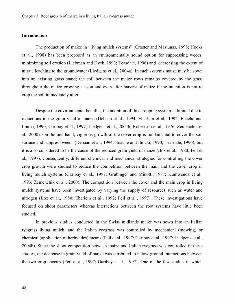
Chapter 3: Root growth of maize in a living Italian ryegrass mulch
Introduction
The production of maize in “living mulch systems” (Croster and Masiunas, 1998; Hooks
et al., 1998) has been proposed as an environmentally sound option for suppressing weeds,
minimizing soil erosion (Liebman and Dyck, 1993; Teasdale, 1996) and decreasing the extent of
nitrate leaching to the groundwater (Liedgens et al., 2004a). In such systems maize may be sown
into an existing grass stand; the soil between the maize rows remains covered by the grass
throughout the maize growing season and even after harvest of maize if the intention is not to
crop the soil immediately after.
Despite the environmental benefits, the adoption of this cropping system is limited due to
reductions in the grain yield of maize (Dehaan et al., 1994; Eberlein et al., 1992; Enache and
Ilnicki, 1990; Garibay et al., 1997; Liedgens et al., 2004b; Robertson et al., 1976; Zemenchik et
al., 2000). On the one hand, vigorous growth of the cover crop is fundamental to cover the soil
surface and suppress weeds (Dehaan et al., 1994; Enache and Ilnicki, 1990; Teasdale, 1996), but
it is also considered to be the cause of the reduced grain yield of maize (Box et al., 1980; Feil et
al., 1997). Consequently, different chemical and mechanical strategies for controlling the cover
crop growth were studied to reduce the competition between the main and the cover crop in
living mulch systems (Garibay et al., 1997; Grubinger and Minotti, 1987; Kumwenda et al.,
1993; Zemenchik et al., 2000). The competition between the cover and the main crop in living
mulch systems have been investigated by varying the supply of resources such as water and
nitrogen (Box et al., 1980; Eberlein et al., 1992; Feil et al., 1997). These investigations have
focused on shoot parameters whereas interactions between the root systems have little been
studied.
In previous studies conducted in the Swiss midlands maize was sown into an Italian
ryegrass living mulch, and the Italian ryegrass was controlled by mechanical (mowing) or
chemical (application of herbicides) means (Feil et al., 1997; Garibay et al., 1997; Liedgens et al.,
2004b). Since the shoot competition between maize and Italian ryegrass was controlled in these
studies, the decrease in grain yield of maize was attributed to below-ground interactions between
the two crop species (Feil et al., 1997; Garibay et al., 1997). One of the few studies in which
48
Chapter 3: Root growth of maize in a living Italian ryegrass mulch
roots in a living mulch system have been investigated showed that the maximum root densities
for monocropped maize and maize as the main crop in a living Italian ryegrass mulch were
attained approximately at the anthesis of maize (Liedgens et al., 2004b). Furthermore, the root
density of monocropped maize was markedly lower than that of the crop mixture (maize plus
cover crop) and monocropped Italian ryegrass. Therefore, it was hypothesized that the reduced
shoot growth of maize in the living mulch is related to increased allocation of carbon to root
growth as a response to the lower availability of soil resources. The lack of a suitable method for
determining the proportion of roots that maize and Italian ryegrass contribute to the total amount
of roots of the living mulch system made it impossible to test this hypothesis.
DNA extraction (Brunner et al., 2001; Mommer et al., 2008) and tracers (Baldwin and
Tinker, 1972; Jumpponen et al., 2002; Lehmann et al., 1998) have been used as methods to
discriminate the roots formed by the various species in mixed plant stands. However, to better
understand the effects of root interactions between maize an Italian ryegrass in a living mulch
system a method is needed that allows to (i) collect a large number of samples at different soil
depths throughout the growing season of the main crop, (ii) directly quantify the roots of each
crop, and (iii) determine the relative arrangement of the fine roots of each crop. We have recently
developed a method (Faget et al., 2009) that fulfills these requirements and, thus, makes it
possible to study the spatial distribution of the roots of maize growing in an established lay of
Italian ryegrass in detail.
The main goals of the present study were to determine (i) the extent to which shoot and
root growth of maize are reduced by the presence of a permanent Italian ryegrass cover crop and
(ii) whether the spatial distribution of the root system of maize is modified by the companion
cover crop.
49

Chapter 3: Root growth of maize in a living Italian ryegrass mulch
Material and methods
In order to observe legal restrictions for the use of genetically modified organisms in
Switzerland, the experiment was conducted in a greenhouse and harvested at the onset of the
anthesis of maize.
Plant material
The maize genotype ETH-M72 was genetically transformed to include the gene for gfp
(ETH-M72GFP). The transformation construct contains the gfp gene flanked by the ubiquitin
promoter (ubi::gfp) and the NOS terminator. It was cloned into the pUC19 vector, which contains
the gene for ampicillin resistance (ampR) at the restriction sites SpeI and XbaI. The gfp gene was
cloned into the cassette at the NcoI and SalI sites. The expressed GFP has been reported to have a
fluorescence peak between 500 and 520 nm when excited by light at 450 to 470 nm. Information
about the validation of the genetic transformation, the integration pattern of the inserted gene, and
comparisons of the homozygosity of transgenic and non-transgenic controls can be found in
Aulinger et al. (2003).
Italian ryegrass (Lolium multiflorum Lam. cv. Ellie) was the cover crop in the living
mulch system, ETH-M72GFP was the main crop.
Experimental conditions
The experiment was conducted twice; the first run took place from 12 December 2007 to
8 April 2008 and the second one from 11 January 2008 to 16 May 2008.
The plants were grown in wooden boxes (0.60 m wide x 0.80 m long and 0.80 m high).
Three minirhizotrons (60 mm outer diameter) were installed horizontally in the boxes at depths of
0.05, 0.15, and 0.45 m. They were horizontally spaced at 0.15 m from each other and from the
edges of the boxes (Fig. 1).
50

Chapter 3: Root growth of maize in a living Italian ryegrass mulch
Figure 1: Schematic representation of the boxes and plant arrangement used in the experiment.
The boxes were filled three months before starting the experiments to enable the soil to
settle, thereby avoiding effects of unsettled soil (Hutchings and John, 2004) and patches with
high concentrations of nutrients (Fransen et al., 1999) on root growth. The soil was a sandy loam
from a nearby field mixed with sand. According to the methods given in parentheses, the
properties of the soil mixture were: 30 g kg-1 organic matter (Blake-Walkley), 60 g kg-1 clay, 110
g kg-1 silt, 810 g kg-1 sand, pH 7.7, 66.3 mg kg-1 assimilable K (NH4 acetate), 4.1 mg kg-1 P
(Olsen), and 12 g kg-1 N (2M KCl). The mean day and night temperatures in the greenhouse were
23°C and 19°C. Plants were grown under white-light photoperiod of 16 h at 3680 lx provided by
high pressure mercury lamps (Philips HPL-N-400W; Royal Philips Electronics Inc., Amsterdam,
Netherlands).
Italian ryegrass was sown at a density of 6.27 g seed m2 (i.e. ~6000 plants m2). After 40
days, one 0.30 m wide strip of Italian ryegrass in the mid-section of the boxes and perpendicular
to the longer side was removed. ETH-M72GFP maize was sown at a density of 10.4 seeds per m2
51

Chapter 3: Root growth of maize in a living Italian ryegrass mulch
into this strip. At the same time, ETH-M72GFP maize was sown in boxes in which the soil was left
bare. The maize rows were orientated perpendicular to the minirhizotrons (Fig. 1). Hereafter,
maize plants from the living mulch and from the bare soil plots will be referred to as LM and BS
maize, respectively.
After sowing the maize, the remaining Italian ryegrass was cut at a height of 0.10 m at
weekly intervals to exclude competition between maize and the cover crop for light. Foskal®
(P2O5, K2O, Mg, Ca, and S; Agroline, Basel, Switzerland) and ammonium nitrate (NH4NO3) were
evenly applied to the soil surface before sowing the maize supplying 1.4 g P, 8 g K, 0.57 g Mg,
0.20 g Ca, 0.87 g S, and 1.1 g N per m2. The water content of the soil was estimated by time
domain reflectometry (EC-5 Soil Moisture Sensor, Decagon Devices Inc., Pullman, WA, USA)
and maintained close to field capacity by irrigation.
Sampling and screening of data
The shoots of maize were cut approximately 83 days after sowing at ground level when
the experiments were terminated. The maize and Italian ryegrass shoots were dried at 60°C until
constant weight. The biomass yield reported for Italian ryegrass represents the sum of the
biomass yields of the weekly cuttings and includes the biomass at the end of the experiments. The
leaf area of maize was measured at the termination of each experimental run with a leaf-area
meter (LI-COR 3100, Lincoln, NE, USA). The leaf area of Italian ryegrass was not determined.
Root images were recorded at the minirhizotron – soil interface using two camera
configurations for distinguishing roots of maize and Italian ryegrass. One camera configuration
allowed for the sampling of images that display all structures at the minirhizotron-soil interface
while the second configuration allowed for identifying precisely the roots of ETH-M72GFP as a
result of the GFP expression (Faget et al., 2009). The use of identical handles for both camera
configurations enabled us to repeatedly sample images of both types at the same position in the
minirhizotrons over the entire duration of the experiment. The images were obtained from the
upper surfaces of the minirhizotrons. At each of the soil depths analyzed, the first image (19.5
mm wide) of the sequence was taken immediately under the maize row. From there, the camera
covered a strip of 312 mm towards the border of the plot; images were taken at 12 positions. The
52

Chapter 3: Root growth of maize in a living Italian ryegrass mulch
first six positions were below the strip on which the Italian ryegrass cover was removed shortly
before maize sowing (maize zone, hereafter). The remaining six images were taken below the
living ryegrass mulch (grass zone, hereafter).
The number of roots (i.e. number of root incidences on the minirhizotron interface) for
each of the sampled images was determined according to Upchurch and Ritchie (1983). Although
this estimate can be obtained from single minirhizotron images, the accuracy of identifying root
structures in images increases when images acquired at the same position are captured across a
set of sampling dates (Smit et al., 2000). Therefore, all images recorded at a single minirhizotron
position over the entire growing season were organized into an image time series (ITS). Images
obtained with each camera configuration were intercalated in each ITS. Since no objective visual
criteria exist to determine whether a root is functional (Smit et al., 2000), all roots identified in an
image were accounted for in each ITS. Thus, the obtained response variable was the cumulative
number of roots observed per image since the sowing date. This was converted into a surface unit
(cm2) and is referred to hereafter as root density (roots cm-2).
Data analysis
Root density was analyzed throughout the growing season of the crops and for the class
distances from the maize row. An estimate of the root density for the soil profile is provided in
Fig. 2; this was obtained as the mean root density for the three studied soil depths. In addition, we
calculated ratios of root density in the grass zone to root density in the maize zone.
For each experimental run (n=2) we used a completely randomized block design with five
replications and two treatments. The treatments were maize grown as a monocrop on bare soil
(BS maize) and maize sown into living ryegrass mulch (LM maize). The analysis of variance of
all the datasets was performed with R (R Development Core Team, 2007). Bars presented in
graphs are standard errors of the mean.
53
Chapter 3: Root growth of maize in a living Italian ryegrass mulch
Results
Effects of Italian ryegrass on the shoot and root growth at the anthesis of maize
Italian ryegrass as living mulch significantly reduced the biomass yield, leaf area, and root
density of maize (Fig. 2).
Bio
ma
ss y
ield
(g
)
01
00
200
30
04
00
Le
af a
rea
(m
2 m2
so
il)
0.0
0.2
0.4
0.6
0.8
1.0
1.2
1.4
Ro
ot d
ensi
ty (
roo
ts c
m2
)
0.0
0.5
1.0
1.5
2.0
BS MaizeLM MaizeItalian ryegrass
Values are mean +SE of 10 measurements
Figure 2: Means of shoot and root parameters of maize and Italian ryegrass measured at harvest in two experimental runs. Root density is the mean of three soil depths (0.05, 0.15, and 0.45 m).
The biomass yield of BS maize was, with 354 g m-2, the higher than that of Italian
ryegrass (156 g m-2) and LM maize (155 g m-2). The leaf area, which was measured only for
maize, was higher for BS maize (0.96 m2 m-2 soil) than for LM maize (0.58 m2 m-2 soil). In
contrast to the biomass yield, the highest root density was found for Italian ryegrass (1.43 roots
cm-2), followed by BS maize (1.13 roots cm-2) and LM maize (0.66 roots cm-2). In interpreting
these data, it must be borne in mind that the final root density of the grass in the living mulch
54

Chapter 3: Root growth of maize in a living Italian ryegrass mulch
system was the result of root growth before and after the sowing of maize. The biomass yield,
leaf area and root density of LM maize were 56%, 39%, and 41%, respectively, lower than that of
BS maize. Therefore, the relative reduction that was found for LM maize as compared to BS
maize was almost identical for the assimilatory organs (i.e. leaf area and root density).
Effects of Italian ryegrass on the spatial distribution of maize roots at anthesis
Figure 3 shows the mean root density of LM maize, BS maize and Italian ryegrass at three
different soil depths, namely 0.05, 0.15, and 0.45 m. Root density of BS and LM maize was
significantly different at all depths; the root density of LM maize was 36%, 46% and 42%,
respectively, lower at 0.05, 0.15 and 0.45 m compared to BS maize. Furthermore, maize roots
accounted for 32% (0.05 m), 25% (0.15 m), and 38% (0.45 m) of the total root density in the
living mulch plots.
The maximum root density of Italian ryegrass and maize was observed at 0.05 m; for
maize this was independent of the cropping method. In all crops, root densities at 0.15 and 0.45 m
depth were similar. Except for 0.45 m depth, values were higher for Italian ryegrass than for BS
maize in the two upper soil layers, but the root density of Italian ryegrass was always
significantly higher than that of LM maize. This suggests a strong negative effect of Italian
ryegrass on the below-ground growth of LM maize.
Immediately below the soil surface, at 0.05 m depth, the root density of maize decreased
with increasing distance from the maize row, irrespective of the maize cropping system (Figure
4).
55
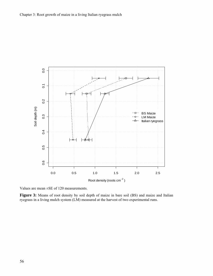
Chapter 3: Root growth of maize in a living Italian ryegrass mulch
0.0 0.5 1.0 1.5 2.0 2.5
0.6
0.5
0.4
0.3
0.2
0.1
0.0
Root density (roots cm2
)
Soil
depth
(m
)
BS MaizeLM MaizeItalian ryegrass
Values are mean ±SE of 120 measurements.
Figure 3: Means of root density by soil depth of maize in bare soil (BS) and maize and Italian ryegrass in a living mulch system (LM) measured at the harvest of two experimental runs.
56
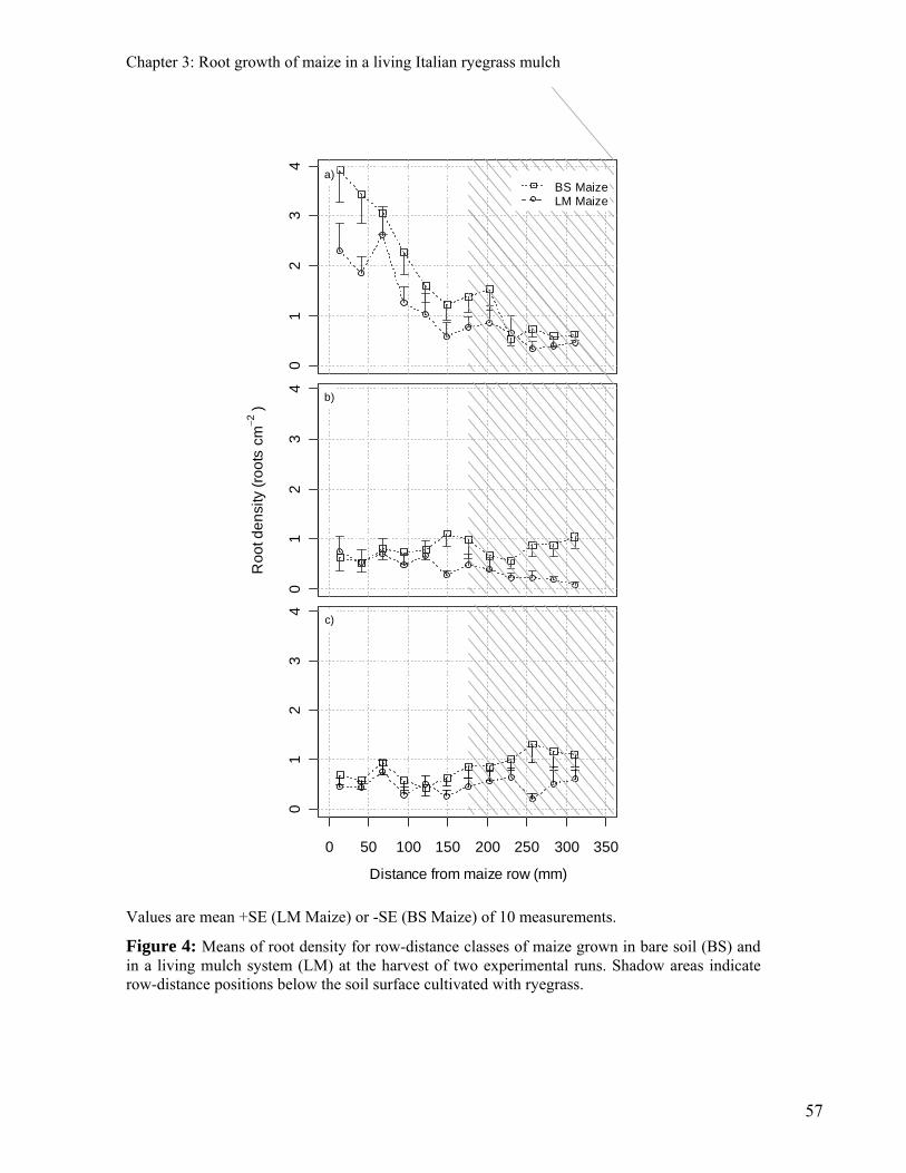
Chapter 3: Root growth of maize in a living Italian ryegrass mulch
01
23
4
BS MaizeLM Maize
a)
01
23
4
Ro
ot d
en
sity
(ro
ots
cm
2 )
b)
0 50 100 150 200 250 300 350
01
23
4
c)
Distance from maize row (mm)
Values are mean +SE (LM Maize) or -SE (BS Maize) of 10 measurements.
Figure 4: Means of root density for row-distance classes of maize grown in bare soil (BS) and in a living mulch system (LM) at the harvest of two experimental runs. Shadow areas indicate row-distance positions below the soil surface cultivated with ryegrass.
57

Chapter 3: Root growth of maize in a living Italian ryegrass mulch
Root densities of LM maize were consistently lower than those of BS maize, but the
differences were only small at distances of more than 200 mm. At soil depths of 0.15 m and 0.45
m, the effect of distance on root density from the row depended on the maize cropping system.
The differences between the maize cropping systems were relatively small at low distances from
the maize row. At distances of more than 150 mm, however, BS maize clearly formed more roots
than LM maize. In contrast to the 0.05 m soil depth, root density at 0.45 m soil depth increased
from about 150 mm distance from the maize row for BS maize.
We calculated the ratios of root density in the grass zone to root density in the maize
zone. BS maize had ratios of 0.57, 1.80, and 1.88 at depths of 0.05, 015, and 0.45 m, respectively.
The respective values for LM maize were 0.40, 0.38, and 1.80. The differences between BS
maize and LM maize were non-significant at p=0.05. These figures indicate that, immediately
below the soil surface, relatively more maize roots occurred in the maize zone, irrespective of
maize cropping system. In contrast, relatively more maize roots were formed in the grass zone in
deep soil layers in both maize production systems. At 0.15 m depth, however, there were marked
differences between the maize cropping systems.
Dynamics of the root growth of maize in bare soil and in the living mulch
The temporal patterns of root density below the maize and grass zones are shown in
Figure 5. The root density of BS maize increased at the three soil depths until anthesis. The root
density of LM maize was generally lower than that of BS maize throughout the growing season.
In the maize zone, the lag in root growth of LM maize compared to BS maize markedly
decreased with increasing soil depth. (Figs. 5a-c). This effect was not observed in the grass zone
(Figs. 5d-f).
58
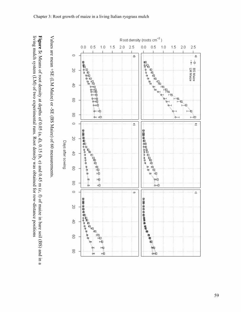
Chapter 3: Root growth of maize in a living Italian ryegrass mulch
Figu
re 5: Means of root density at depths of 0.05 (a, d), 0.15 (b, e) and 0.45 m
(c, f) of maize in bare soil (B
S) and in a
living mulch system
(LM
) of two experim
ental runs. Root density w
as obtained for row-distance positions
Values are m
ean +S
E (L
M M
aize) or -SE
(BS
Maize) of 60 m
easurements.
59

Chapter 3: Root growth of maize in a living Italian ryegrass mulch
Discussion
The reduction in the shoot biomass yield of maize (Fig. 2) agrees well with results found
in other studies in which maize was grown in living grass mulches (Feil et al. 1997; Garibay et al.
1997; Liedgens et al. 2004b). Reduced above-ground biomass of LM maize, compared to BS
maize, was due to a smaller photosynthetic area, which can be inferred from the smaller leaf area
of LM maize (Fig. 2). Similar reductions in leaf area were found by Liedgens et al. (2004b) in a
three-year outdoor lysimeter study in which LM maize showed a faster leaf senescence than BS
maize after anthesis. Unfortunately, we could not investigate the postfloral period due to legal
restrictions associated with the use of transgenic maize.
In this study, it was significant that the living grass mulch had approximately the same
adverse effects on leaf area and total root density (Fig. 2). Root growth of maize in living mulch
systems has been investigated in previous studies, but the maize root density was determined by
calculating the difference between total root density of the living mulch system and monocropped
Italian ryegrass (Liedgens et al. 2004b). This approach represents only a rough estimate of the
contribution that maize makes to the total root density in the living mulch system. By
discriminating between GFP-derived fluorescent and non-fluorescent roots (Faget et al. 2009), we
were able to directly quantify the relative proportion of maize and Italian ryegrass roots in the
living mulch in situ.
The spatial distribution of the roots of crops in mixtures has been analyzed in a few
studies using methods that rely on the separation of rooting volumes by barriers (e.g., Li et al.
2007; Thorsted et al. 2006) or trenches (e.g., Li et al. 2006; Ludwig et al. 2004). Such methods
dramatically disturb the soil environment and can lead to artefacts in plant growth (Connell 1990;
McPhee and Aarssen 2001). Our results were obtained with minirhizotrons, a method that
minimizes the interference to root growth (Taylor et al. 1990). Thus, our study provides reliable
information about the spatial distribution of maize roots in a crop mixture grown on undisturbed
soils, which may further understanding and management of the coexistence of plants in crop
mixtures (Schenk 2006).
60
Chapter 3: Root growth of maize in a living Italian ryegrass mulch
It may be assumed that the roots of LM maize preferentially grow in the soil below a strip
where Italian ryegrass has been removed (we have previously defined this area as the maize
zone). An indicator of a shift of LM maize root growth to the maize zone would be a variation in
the ratio of root density in the grass zone to root density in the maize zone, between LM maize
and BS maize. We found differences in the relative spatial distribution, especially at a soil depth
of 0.15 m, but the differences were not significant. Consequently, further research, ideally in an
outdoor environment with 10 observation depths at minimum, is needed to elucidate the nature of
this effect. In interpreting the results for root distribution, it should be noted that the fertilizers
were evenly distributed on the soil surface in the present study. In farmers’ fields, however,
nitrogen fertilizers could be broadcasted or applied to the maize rows (Malhi et al. 2001). The
latter method is likely to increase the production of roots below the maize rows (Chassot et al.
2001).
Differences in root density between BS maize and LM maize occurred as early as at the
seedling stage (Fig. 5). This effect was likely caused by nitrogen deficiency due to nitrogen
removal by grass from the living mulch system before the maize was planted.
Root density decreases with soil depth, but it may be higher at a depth of 0.15 m than at
0.05 m for monocropped maize (Liedgens and Richner 2001) or for maize and Italian ryegrass in
a maize - Italian ryegrass living mulch system (Liedgens et al. 2004b). Such differences may be
explained by the fact that the soil surface is usually dryer in outdoor experiments that have erratic
rainfall (Liedgens and Richner 2001; Liedgens et al. 2004b) than in greenhouse studies, where
the soil surface is frequently watered. The root densities in our study were slightly lower than
those reported by other experimenters (e.g., Liedgens and Richner 2001; Liedgens et al. 2004b;
Liedgens et al. 2000; Pages and Pellerin 1994; Smit and Groenwold 2005). For example, for the
BS maize, we found 1.7, 0.8, and 0.9 roots cm-2 at soil depths of 0.05 m, 0.15 m, and 0.45 m,
respectively. By contrast, Smit and Groenwold (2005) found densities of 2.3±1.5, 2.3±2.1, and
0.6±0.4 roots cm-2 at the same respective soil depths. Continuously decreasing root densities of
maize in the topsoil and a pronounced response to increasing soil depth in the subsoil were
similarly found in other studies using minirhizotrons (Liedgens and Richner 2001; Liedgens et al.
2004b).
61

Chapter 3: Root growth of maize in a living Italian ryegrass mulch
62
Conclusions
The present study demonstrates that the use of transgenic maize expressing GFP
combined with the minirhizotron technique allows for reliably tracing the dynamics of maize root
growth planted into established Italian ryegrass sods. A potential drawback of the GFP method is
that, in many countries, it can only be applied in confined environments due to legal restrictions.
This study provides unique data on the growth of maize roots into an established Italian
ryegrass sod and it demonstrates that the leaf area and mean root density of maize cropped in a
living mulch system were almost identically reduced by the companion crop.
General Conclusion and Outlook
General Conclusion and Outlook
General Conclusion
Roots are essential for plant growth due to their role of anchorage and absorption of water
and nutrients. This hidden part of plants is less studied than the corresponding shoot due to its
relative inaccessibility in the soil without perturbing the environment. Plant species differ in the
temporal and spatial exploitation of the soil, interacting in crop stands as well as in natural
systems with other plants. However, there is no method to follow in a regular manner the
belowground interactions in between different plants or species in-situ. The main questions were
“How to distinguish roots belonging to different plants?”, “How do roots interact?” .Here we
demonstrated that using transgenic plants expressing a fluorescent protein; combined with the
minirhizotron technique brings a first reliable answer to those questions.
By using plants transformed with the GFP and constructing a specific minirhizotron camera,
albeit is possible from now on to distinguish roots from different plants in the soil. The
prerequisite for the application was the use of the green fluorescent protein that has not been
applied before for agronomic and ecological root studies.
The two cameras developed are of a similar construction but with different goals. The
conventional minirhizotron camera type was already available in the market; however the one
constructed here is easily affordable in terms of price and highly flexible for further potential
modifications. It allows taking digital images of the soil root interface with a white light as used
in the conventional minirhizotron systems. The GFP minirhizotron camera system is a new
powerful tool which allows unequivocally identifying roots from plants expressing the GFP.
Therefore, we demonstrated that it is possible to develop an affordable imaging system for
capturing imaging of conventional and fluorescent roots growing in the soil.
The conventional images were of similar quality as those from commercial equipment and
fulfill the requirements of software to analyze root images. The small size of the captor (640 *
63

General Conclusion and Outlook
480 pixels) was not a limitation for the latter. The roots, soil, and even soil interstices were highly
visible and images were similar than the ones obtained with commercial devices. The quality of
images obtained with the GFP device was the same standard as those obtained with the
conventional device. Roots from transformed plants were exclusively visible in a bright green
color. It was a strong additional advantage that with this light the background is black; thus a
better contrast exists that increases the precision to distinguish the roots from the background
compared to the conventional images. Another advantage concerns the fine roots; because of they
transparency, they are not completely visible in conventional images and parameters such as root
density are often underestimated. In images obtained with the GFP minirhizotron camera, these
fine roots are also visible in a bright green. Therefore, this method represents an improvement for
the minirhizotron studies by increasing its accuracy. Interestingly, it was furthermore possible to
identify precisely the meristematic tissues, i.e. root tips and lateral branch insertion because there
the fluorescence is stronger than in the others parts of the roots.
By growing the genetically transformed maize genotype ETH-M72 GFP with its
corresponding wild type, Italian ryegrass or soybean, we demonstrated that this method allows
indeed an accurate non destructive observation of different root systems. The green emitted light
was only observable in the transformed plants and enabled to investigate the dynamics of the
relative distribution of roots between intraspecific or interspecific neighbors. Using root
fluorescence of genetically transformed plants with minirhizotrons seems to be, compared to
other methods the simplest one to study the relative distribution of roots in mixed plant stands. In
addition to distinguishing roots of different plants, the root density quantified in images using the
GFP system was 28% higher than that obtained with the conventional method. Therefore it can
be envisioned that GFP transformed plants may be even useful in single plant studies when
morphological developments have to be screened with a high descriptive accuracy.
The main study with this new method was conducted for maize cropped in a living mulch
of Italian ryegrass. This is a promising system for the sustainable production of maize. Maize
expressing GFP was grown either alone in bare soil or together with Italian ryegrass in boxes. By
using the GFP minirhizotron method, it was possible to measure the root proportion of each
64

General Conclusion and Outlook
species in space and time until anthesis. As known before, Italian ryegrass decreased strongly the
root density and leaf area of maize by 41% and 39%, respectively. This is unique information that
answers a long open question on how the roots of a staple crop establish in the soil occupied by
living grass; it could have never been answered without using our novel approach that allows
quantitatively discriminating roots and studying their relative distribution. It was another
advantage that the dynamics of this competitive distribution could be followed. These first results
already showed that Italian ryegrass induced a delay in the root growth of the main crop; the
temporal distribution was more affected than the spatial distribution. This paves the way to devise
strategies to increase the sustainability of multi-species cropping systems in the future.
Outlook
It will be important to conduct similar experimentation with different plant species as a
cover crop, for example legumes where the association could be more beneficial for the main
crop by less competition for N and even a possible nutrient transfer.
At the present state, more fluorescent proteins with different wave lengths, i.e. colors exist. Thus
this novel approach to identify and study roots could be extend to a larger plant communities with
more than two species together when many plants with different fluorescent proteins as blue,
cyan, yellow, red can be used with an easy adaptation of minirhizotron camera, tracking all the
different roots system to study the belowground interactions in a whole community.
We noticed in some pictures that the fluorescence of meristematic tissues, i.e. root tips
and lateral branch insertion was stronger than in the others parts, and in opposite older roots were
loosing gradually their fluorescence. This characteristic could be used to adapt this new method
to study the senescence of root systems.
This will depend on finding a relationship between root activity and fluorescence intensity. Most
of the common approaches with minirhizotron studies imply that a huge quantity of images has to
be analyzed. Due to the heterogeneous soil background and the poor visibility of some fine roots,
a manual drawing of root has to be done. With the GFP minirhizotron technique, a more accurate
65

General Conclusion and Outlook
66
identification of roots is possible due to the better contrast between the roots and the background.
The clear contrast between the black background and the fluorescent green roots could allow for
applying a simple image analysis procedure to track the roots automatically. This could bring an
advance in this domain where a lot of time is needed to analyze every image of an experiment.
By the possibility to study root interaction in space and time and extending this method to
new fields of research, this novel approach literally brings light into this underground
environment.


References
References
Andrews, D.J., and Kassam, A.H. (1976) The importance of multiple cropping in increasing world food supplies. American Society of Agronomy, Madison, WI., ASA Special Publication 27.
Atkinson, D. (2000) Root characteristics: Why and what to measure. Root Methods: A Handbook, 1-32.
Aulinger, I.E., Peter, S.O., Schmid, J.E., and Stamp, P. (2003) Rapid attainment of a doubled haploid line from transgenic maize (Zea mays L.) plants by means of anther culture. In Vitro Cellular & Developmental Biology-Plant, 39(2), 165-170.
Baldwin, J.P., and Tinker, P.B. (1972) Method for estimating lengths and spatial patterns of two interpenetrating root systems. Plant and Soil, 37(1), 209-213.
Barber, S.A. (1971a) Effect of Tillage Practice on Corn (Zea-Mays L) Root Distribution and Morphology. Agronomy Journal, 63(5), 724-&.
Barber, S.A. (1971b) Effect of tillage practice on corn (Zea mays L) root distribution and morphology. Agronomy Journal, 63(5), 724-726.
Bates, G. (1937) A device for the observation of root growth in the soil. Nature, 139, 966-967. Bell, A.D., and Bryan, A. (1991) Plant form-an illustrated guide to flowering plant morphology.
Oxford University Press, New York. Bendix, J., Rollenbeck, R., Goettlicher, D., Naubss, T., and Fabian, P. (2008) Seasonality and
diurnal pattern of very low clouds in a deeply incised valley of the eastern tropical Andes (South Ecuador) as observed by a cost-effective WebCam system. Meteorological Applications, 15(2), 281-291.
Bigas, M., Cabruja, E., Forest, J., and Salvi, J. (2006) Review of CMOS image sensors. Microelectronics Journal, 37(5), 433-451.
Blanksby, A.J., and Loinaz, M.J. (2000) Performance analysis of a color CMOS photogate image sensor. Ieee Transactions on Electron Devices, 47(1), 55-64.
Böhm, W. (1979) Methods of studying root systems Springer-verlag,Berlin,Germany. Box, J.E., Wilkinson, S.R., Dawson, R.N., and Kozachyn, J. (1980) Soil-Water Effects on No-
Till Corn Production in Strip and Completely Killed Mulches. Agronomy Journal, 72(5), 797-802.
Brunner, I., Brodbeck, S., Buchler, U., and Sperisen, C. (2001) Molecular identification of fine roots of trees from the Alps: reliable and fast DNA extraction and PCR-RFLP analyses of plastid DNA. Molecular Ecology, 10(8), 2079-2087.
Bullock, D.G. (1992) Crop-Rotation. Critical Reviews in Plant Sciences, 11(4), 309-326. Cahn, M.D., Zobel, R.W., and Bouldin, D.R. (1989) Relationship between Root Elongation Rate
and Diameter and Duration of Growth of Lateral Roots of Maize. Plant and Soil, 119(2), 271-279.
Caldwell, M.M., Manwaring, J.H., and Durham, S.L. (1996) Species interactions at the level of fine roots in the field: Influence of soil nutrient heterogeneity and plant size. Oecologia, 106(4), 440-447.
Campbell, C.D., Mackiedawson, L.A., Reid, E.J., Pratt, S.M., Duff, E.I., and Buckland, S.T. (1994) Manual recording of minirhizotron data and its application to study the effect of
68

References
herbicide and nitrogen-fertilizer on tree and pasture root-growth in a silvopastoral system. Agroforestry Systems, 26(2), 75-87.
Carreker, J.R., Adams, W.E., and Morris, H.D. (1972) Corn Yields in Relation to Rainfall and Cropping Treatments. Journal of Soil and Water Conservation, 27(3), 117-&.
Casper, B.B., and Jackson, R.B. (1997) Plant Competition Underground. Annual Review of Ecology and Systematics, 28, 545-570.
Chalfie, M., and Kain, S. (2006) Green fluorescent protein: properties, applications and protocols. Hoboken, New Jersey, USA: John Wiley & Sons.
Chassot, A., Stamp, P., and Richner, W. (2001) Root distribution and morphology of maize seedlings as affected by tillage and fertilizer placement. Plant and Soil, 231(1), 123-135.
Connell, J.H. (1990) Apparent versus "real" competition in plants. In Perspectives on plant competition (J.B. Grace, and D. Tilman, eds), San Diego, CA, USA: Academic Press, pp 9-26.
Croster, M.P., and Masiunas, J.B. (1998) The effect of weed-free period and nitrogen on eastern black nightshade competition with English pea. Hortscience, 33(1), 88-91.
Danso, S.K.A., Curbelo, S., Labandera, C., and Pastorini, D. (1991) Herbage Yield and Nitrogen-Fixation in a Triple-Species Mixed Sward of White Clover, Lotus and Fescue. Soil Biology & Biochemistry, 23(1), 65-70.
de Kroon, H. (2007) Ecology - How do roots interact? Science, 318(5856), 1562-1563. Degregorio, R., and Ashley, R.A. (1985) No Till Vegetables - Suppression of Clover Weeds by
Plastic Mulch. Hortscience, 20(2), 187-187. Dehaan, R.L., Wyse, D.L., Ehlke, N.J., Maxwell, B.D., and Putnam, D.H. (1994) Simulation of
Spring-Seeded Smother Plants for Weed-Control in Corn (Zea-Mays). Weed Science, 42(1), 35-43.
Dudley, S.A., and File, A.L. (2007) Kin recognition in an annual plant. Biology Letters, 3(4), 435-438.
Ebelhar, S.A., Frye, W.W., and Blevins, R.L. (1984) Nitrogen from Legume Cover Crops for No-Tillage Corn. Agronomy Journal, 76(1), 51-55.
Eberlein, C.V., Sheaffer, C.C., and Oliveira, V.F. (1992) Corn Growth and Yield in an Alfalfa Living Mulch System. Journal of Production Agriculture, 5(3), 332-339.
Echtenkamp, G.W., and Moomaw, R.S. (1989) No-till corn production in a living mulch system. Weed Technol., 3, 261-266.
Eizenberg, H., Shtienberg, D., Silberbush, M., and Ephrath, J.E. (2005) A new method for in-situ monitoring of the underground development of Orobanche cumana in sunflower (Helianthus annuus) with a mini-rhizotron. Annals of Botany, 96(6), 1137-1140.
Enache, A.J., and Ilnicki, R.D. (1990) Weed-Control by Subterranean Clover (Trifolium-Subterraneum) Used as a Living Mulch. Weed Technology, 4(3), 534-538.
Faget, M., Herrera, J.M., Stamp, P., Aulinger-Leipner, I., Frossard, E., and Liedgens, M. (2009) The use of green fluorescent protein as a tool to identify roots in mixed plant stands. Functional Plant Biology, 36, 930-937.
Falik, O., de Kroon, H., and Novoplansky, A. (2006) Physiologically-Mediated Self/Nonself Root Discrimination in Trifolium repens Has Mixed Effects on Plant Performance
Plant Signaling and Behavior, 1(3), 116-121.
69

References
Faro, A., Giordano, D., and Spampinato, C. (2008) Evaluation of the traffic parameters in a metropolitan area by fusing visual perceptions and CNN processing of webcam images. Ieee Transactions on Neural Networks, 19(6), 1108-1129.
Feil, B., Garibay, S.V., Ammon, H.U., and Stamp, P. (1997) Maize production in a grass mulch system - seasonal patterns of indicators of the nitrogen status of maize. European Journal of Agronomy, 7(1-3), 171-179.
Feil, B., Liedgens, M. (2001) Pflanzenproduktion in lebenden Mulchen - eine Übersicht. Pflanzenbauwiss, 5, 15-23.
Ferguson, J.C., and Smucker, A.J.M. (1989) Modifications of the minirhizotron video camera system for measuring spatial and temporal root dynamics. Soil Science Society of America Journal, 53(5), 1601-1605.
Fischer, A.L. (2006) CMOS gets a leg up on CCD. Photonics Spectra, 40(1), 93-+. Fitter, A.H. (2002) Characteristics and functions of root systems. In Plant roots, the hidden half
(Y. Waisel, A. Eshel, and U. Kafkafi, eds), New York, USA: Marcel Dekker, pp 15-32. Fox, R.H., and Piekielek, W.P. (1988) Fertilizer nitrogen equivalence of alfalfa, BFT and red
clover for succeeding corn crops. J. Prod. Agric., 4(313-317). Fransen, B., Blijjenberg, J., and de Kroon, H. (1999) Root morphological and physiological
plasticity of perennial grass species and the exploitation of spatial and temporal heterogeneous nutrient patches. Plant and Soil, 211(2), 179-189.
Gamalero, E., Lingua, G., Berta, G., and Lemanceau, P. (2003) Methods for studying root colonization by introduced beneficial bacteria. Agronomie, 23(5-6), 407-418.
Garibay, S.V., Stamp, P., Ammon, H.U., and Feil, B. (1997) Yield and quality components of silage maize in killed and live cover crop sods. European Journal of Agronomy, 6(3-4), 179-190.
Gil, S., Reisin, H.D., and Rodriguez, E.E. (2006) Using a digital camera as a measuring device. American Journal of Physics, 74(9), 768-775.
Girardin, P., Jordan, M.O., Picard, D., and Trendel, R. (1986) Harmonization of Terms Used for the Morphological Description of Maize. Agronomie, 6(9), 873-875.
Goodman, M.M. (1988) The History and Evolution of Maize. Crc Critical Reviews in Plant Sciences, 7(3), 197-220.
Grace, J.B., and Tilman, D. (1990) Perspectives on Plant Competition. Grace, J. B. And D. Tilman. Perspectives on Plant Competition. Xiv+484p. Academic Press, Inc.: San Diego, California, USA; London, England, Uk. Illus, XIV+484P.
Gregory, P.J., and Reddy, M.S. (1982) Root growth in an intercrop of pearl-millet groundnut. Field Crop. Res., 5(3), 241-252.
Grubinger, V.P., and Minotti, P.L. (1987) Mechanical Suppression of White Clover Living Mulch and Its Effect on the Nitrogen Nutrition of Sweet Corn. Hortscience, 22(5), 1047-1047.
Hall, J.K., Hartwig, N.L., and Hoffman, L.D. (1984) Cyanazine Losses in Runoff from No-Tillage Corn in "Living" and Dead Mulches vs. Unmulched, Conventional Tillage, pp 105-110.
Hargrove, W.L. (1986) Winter Legumes as a Nitrogen-Source for No-Till Grain-Sorghum. Agronomy Journal, 78(1), 70-74.
Hartwig, N.L., and Ammon, H.U. (2002) Cover crops and living mulches Weed Science, 50, 688-699.
70
References
Hochholdinger, F., Woll, K., Sauer, M., and Dembinsky, D. (2004) Genetic dissection of root formation in maize (Zea mays) reveals root-type specific developmental programmes. Annals of Botany, 93(4), 359-368.
Hooks, C.R.R., Valenzuela, H.R., and Defrank, J. (1998) Incidence of pests and arthropod natural enemies in zucchini grown with living mulches. Agriculture Ecosystems & Environment, 69(3), 217-231.
Hoppe, D.C., McCully, M.E., and Wenzel, C.L. (1986) The Nodal Roots of Zea - Their Development in Relation to Structural Features of the Stem. Canadian Journal of Botany-Revue Canadienne De Botanique, 64(11), 2524-2537.
Hund, A., Trachsel, S., and Stamp, P. (in press) Growth of axile and lateral roots of maize: I development of a phenotying platform. Plant and Soil.
Hutchings, M.J., and John, E.A. (2004) The effects of environmental heterogeneity on root growth and root/shoot partitioning. Annals of Botany, 94(1), 1-8.
Hutchings, M.J., John, E.A., and Wijesinghe, D.K. (2003) Toward understanding the consequences of soil heterogeneity for plant populations and communities. Ecology, 84(9), 2322-2334.
Jackson, L.E., and Daniel, H. (2005) ROOT ARCHITECTURE AND GROWTH. In Encyclopedia of Soils in the Environment, Oxford: Elsevier, pp 411-421.
Janesick, J., Gunawan, F., Dosluoglu, T., Tower, J., and McCaffrey, N. (2002) Scientific CMOS pixels. Experimental Astronomy, 14(1), 33-43.
Jumpponen, A., Hogberg, P., Huss-Danell, K., and Mulder, C.P.H. (2002) Interspecific and spatial differences in nitrogen uptake in monocultures and two-species mixtures in north European grasslands. Functional Ecology, 16(4), 454-461.
Kozinka, V. (1992) Uptake and transport of water. In: Kolek J , Kozinka V, eds. Physiology of the plant' s Jstem.Dordrecht,
The Netherlands : Kluwer Academic Publishers, 129-203. Kumwenda, J.D.T., Radcliffe, D.E., Hargrove, W.L., and Bridges, D.C. (1993) Reseeding of
Crimson Clover and Corn Grain-Yield in a Living Mulch System. Soil Science Society of America Journal, 57(2), 517-523.
Kurtz, T., Melsted, S.W., and Bray, R.H. (1952) The Importance of Nitrogen and Water in Reducing Competition between Intercrops and Corn. Agronomy Journal, 44(1), 13-17.
Lehmann, J., Peter, I., Steglich, C., Gebauer, G., Huwe, B., and Zech, W. (1998) Below-ground interactions in dryland agroforestry. Forest Ecology and Management, 111(2-3), 157-169.
Li, L., Li, S.M., Sun, J.H., Zhou, L.L., Bao, X.G., Zhang, H.G., and Zhang, F.S. (2007) Diversity enhances agricultural productivity via rhizosphere phosphorus facilftation on phosphorus-deficient soils. Proceedings of the National Academy of Sciences of the United States of America, 104(27), 11192-11196.
Li, L., Sun, J.H., Zhang, F.S., Guo, T.W., Bao, X.G., Smith, F.A., and Smith, S.E. (2006) Root distribution and interactions between intercropped species. Oecologia, 147(2), 280-290.
Liebman, M., and Dyck, E. (1993) Crop-Rotation and Intercropping Strategies for Weed Management. Ecological Applications, 3(1), 92-122.
Liedgens, M., Frossard, E., and Richner, W. (2004a) Interactions of maize and Italian ryegrass in a living mulch system: (2) Nitrogen and water dynamics. Plant and Soil, 259(1-2), 243-258.
71
References
Liedgens, M., and Richner, W. (2001) Minirhizotron observations of the spatial distribution of the maize root system. Agronomy Journal, 93(5), 1097-1104.
Liedgens, M., Soldati, A., and Stamp, P. (2004b) Interactions of maize and Italian ryegrass in a living mulch system: (1) Shoot growth and rooting patterns. Plant and Soil, 262(1-2), 191-203.
Liedgens, M., Soldati, A., Stamp, P., and Richner, W. (2000) Root development of maize (Zea mays L.) as observed with minirhizotrons in lysimeters. Crop Science, 40(6), 1665-1672.
Litwiller, D. (2002) CCD vs. CMOS: The battle cools off. Photonics Spectra, 36(1), 102-103. Litwiller, D. (2005) CMOs vs. CCD: Maturing technologies, maturing markets. Photonics
Spectra, 39(8), 54-+. Ludwig, F., Dawson, T.E., Prins, H.H.T., Berendse, F., and de Kroon, H. (2004) Below-ground
competition between trees and grasses may overwhelm the facilitative effects of hydraulic lift. Ecology Letters, 7(8), 623-631.
Lux, A., Sottnikova, A., Opatrna, J., and Greger, M. (2004) Differences in structure of adventitious roots in Salix clones with contrasting characteristics of cadmium accumulation and sensitivity. Physiologia Plantarum, 120(4), 537-545.
Machado, R.M.A., and Oliveira, M. (2003) Comparison of tomato root distributions by minirhizotron and destructive sampling. Plant and Soil, 255(1), 375-385.
Malhi, S.S., Grant, C.A., Johnston, A.M., and Gill, K.S. (2001) Nitrogen fertilization management for no-till cereal production in the Canadian Great Plains: a review. Soil & Tillage Research, 60(3-4), 101-122.
Martin, R.C., Greyson, P.R., and Gordon, R. (1999) Competition between corn and living mulch. Can. J. Plant Sci., 79, 579-586.
McPhee, C.S., and Aarssen, L.W. (2001) The separation of above- and below-ground competition in plants - A review and critique of methodology. Plant Ecology, 152(2), 119-136.
Milchunas, D.G., Lee, C.A., Lauenroth, W.K., and Coffin, D.P. (1992) A Comparison of C-14, Rb-86, and Total Excavation for Determination of Root Distributions of Individual Plants. Plant and Soil, 144(1), 125-132.
Miller, H.G. (1984) Deposition - Plant-Soil Interactions. Philosophical Transactions of the Royal Society of London Series B-Biological Sciences, 305(1124), 339-352.
Mommer, L., Wagemaker, C.A.M., De Kroon, H., and Ouborg, N.J. (2008) Unravelling below-ground plant distributions: a real-time polymerase chain reaction method for quantifying species proportions in mixed root samples. Molecular Ecology Resources, 8(5), 947-953.
Murphy, G.P., and Dudley, S.A. (2009) Kin recognition: Competition and cooperation in Impatiens (Balsaminaceae). Am. J. Bot., 96, 1990-1996.
Nakamoto, T. (2000) The distribution of wheat and maize roots as influenced by biopores in a subsoil of the Kanto loam type. Plant Production Science, 3, 140-144.
Nedev, S., and Ivanova, V.C. (2006) Web camera as a measuring tool in the undergraduate physics laboratory. European Journal of Physics, 27(5), 1213-1219.
Ogren, P.J., Deibel, M., Kelly, I., Mulnix, A.B., and Peck, C. (2004) Web camera use in developmental biology, molecular biology & biochemistry laboratories. American Biology Teacher, 66(1), 57-+.
Osmont, K.S., Sibout, R., and Hardtke, C.S. (2007) Hidden branches: Developments in root system architecture. Annual Review of Plant Biology, 58, 93-113.
72
References
Pages, L., and Pellerin, S. (1994) Evaluation of parameters describing the root-system architecture of field-grown maize plants (Zea mays L). 2. Density, length, and branching of first-order lateral roots. Plant and Soil, 164(2), 169-176.
Pechackova, S., During, H.J., Rydlova, V., and Herben, T. (1999) Species-specific spatial pattern of below-ground plant parts in a montane grassland community. Journal of Ecology, 87(4), 569-582.
Pierret, A., Moran, C.J., and Doussan, C. (2005) Conventional detection methodology is limiting our ability to understand the roles and functions of fine roots. New Phytologist, 166(3), 967-980.
Pregitzer, K.S., Burton, A.J., King, J.S., and Zak, D.R. (2008) Soil respiration, root biomass, and root turnover following long-term exposure of northern forests to elevated atmospheric Co-2 and tropospheric O-3. New Phytologist, 180(1), 153-161.
Pugnaire, F.I., and Luque, M.T. (2001) Changes in plant interactions along a gradient of environmental stress. Oikos, 93(1), 42-49.
R Development Core Team. (2007) R: A language and environment for statistical computing, Vienna, Austria: R Foundation for Statistical Computing.
Randall, G.W., Huggins, D.R., Russelle, M.P., Fuchs, D.J., Nelson, W.W., and Anderson, J.L. (1997) Nitrate losses through subsurface tile drainage in Conservation Reserve Program, alfalfa, and row crop systems. Journal of Environmental Quality, 26(5), 1240-1247.
Rasse, D.P., Ritchie, J.T., Peterson, W.R., Wei, J., and Smucker, A.J.M. (2000) Rye cover crop and nitrogen fertilization effects on nitrate leaching in inbred maize fields. Journal of Environmental Quality, 29(1), 298-304.
Rasse, D.P., and Smucker, A.J.M. (1998) Root recolonization of previous root channels in corn and alfalfa rotations. Plant and Soil, 204(2), 203-212.
Reicosky, D.C., and Forcella, F. (1998) Cover crop and soil quality interactions in agroecosystems. Journal of Soil and Water Conservation, 53(3), 224-229.
Richardson, A.D., Jenkins, J.P., Braswell, B.H., Hollinger, D.Y., Ollinger, S.V., and Smith, M.L. (2007) Use of digital webcam images to track spring green-up in a deciduous broadleaf forest. Oecologia, 152(2), 323-334.
Ricklefs, R.E., and Miller, G.L. (1999) 4th edn. W.H. Freeman. Ecology. Robertson, W.K., Lundy, H.W., Prine, G.M., and Currey, W.L. (1976) Planting Corn in Sod and
Small Grain Residues with Minimum Tillage. Agronomy Journal, 68(2), 271-274. Robinson, D. (1994) The Responses of Plants to Nonuniform Supplies of Nutrients. New
Phytologist, 127(4), 635-674. Robinson, D. (2007) Implications of a large global root biomass for carbon sink estimates and for
soil carbon dynamics. Proceedings of the Royal Society B-Biological Sciences, 274, 2753-2759.
Robinson, D., Rorizon, I. H. (1986) Mineral nutrition. Methods in plant ecology, 145-213. Sample, I. (2003) Introducing Dr Webcam. New Scientist, 177(2381), 20-20. Sauer. (1960) Maize into Europe. Acts Int Am. Congr (Vienna), 34. Schenk, H.J. (2006) Root competition: beyond resource depletion. Journal of Ecology, 94(4),
725-739. Shaw, R.H. (1988) Climatic requirement. In G.F. Sprague and J.W. Dudley (ed.) Corn and corn
improvement. 3rd ed. ASA, CSSA, and SSSA, Madison, WI., p. 315-341.
73
References
74
Smit, A.L., George, E., and Groenwold, J. (2000) Root observations and measurements at (transparent) interfaces with soil. Root Methods: A Handbook, 235-271.
Smit, A.L., and Groenwold, J. (2005) Root characteristics of selected field crops: Data from the Wageningen Rhizolab (1990-2002). Plant and Soil, 272(1-2), 365-384.
Taba, S. ( 2003) Preliminary breeder core subsets and prebreeding in Latin American maize germplasm conservation: regeneration, in-situ conservation, core subsets, and prebreeding: proceedings of a workshop held at CIMMYT. Mexico, D.F.:CIMMYT., 9-25.
Taylor, H.M. (1987) Minirhizotron Observation Tubes: Methods and Applications for Measuring Rhizosphere Dynamics. American Society of Agronomy, Madison, WI., ASA Special Publication 50.
Taylor, H.M., Upchurch, D.R., and McMichael, B.L. (1990) Applications and limitations of rhizotrons and minirhizotrons for root studies. Plant and Soil, 129(1), 29-35.
Teasdale, J.R. (1996) Contribution of cover crops to weed management in sustainable agricultural systems. Journal of Production Agriculture, 9(4), 475-479.
Thorsted, M.D., Weiner, J., and Olesen, J.E. (2006) Above- and below-ground competition between intercropped winter wheat Triticum aestivum and white clover Trifolium repens. Journal of Applied Ecology, 43(2), 237-245.
Thorup-Kristensen, K. (1993) The Effect of Nitrogen Catch Crops on the Nitrogen Nutrition of a Succeeding Crop. Acta Agriculturae Scandinavica Section B-Soil and Plant Science, 43(2), 74-81.
Upchurch, D.R., and Ritchie, J.T. (1983) Root observations using a video recording-system in mini-rhizotrons. Agronomy Journal, 75(6), 1009-1015.
van Wijk, M.T., and Bouten, W. (1999) Towards understanding tree root profiles: simulating hydrologically optimal strategies for root distribution. Hydrol. Earth Syst. Sci., 5(4), 629-644.
Vargas, R., and Allen, M.F. (2008) Dynamics of fine root, fungal rhizomorphs, and soil respiration in a mixed temperate forest: Integrating sensors and observations. Vadose Zone Journal, 7(3), 1055-1064.
Weigelt, A., and Jolliffe, P. (2003) Indices of plant competition. Journal of Ecology, 91(5), 707-720.
Wilson, G.F., Lal, R., and Okigbo, B.N. (1982) Effects of cover crops on soil structure and on yield of subsequent arable crops grown under strip tillage on an eroded alfisol. Soil and Tillage Research, 2(3), 233-250.
Zemenchik, R.A., Albrecht, K.A., Boerboom, C.M., and Lauer, J.G. (2000) Corn production with kura clover as a living mulch. Agronomy Journal, 92(4), 698-705.
Zobel, R. (2003) Fine roots - discarding flawed assumptions. New Phytologist, 160(2), 276-279.

Acknowledgements
Acknowledgements I would like to thank:
- My professors and supervisors for giving me the opportunity to work on this topic, and
for all the support: Pr. Dr. Peter Stamp, Pr. Dr. Emmanuel Frossard, Dr. Markus Liedgens.
- My direct supervisor for the scientific discussions, advices and help through this project:
Dr. Juan Herrera.
- The persons who helped me and contributed to this project:
Peter Adam, Brigitta Pichler-Herzog, Deborah Britschgi, Alexandre Strigens, Marianne
Wettstein-Baettig, Ingrid Aulinger-Leipner, Patrick Fluetsch, Boy Feil.
- All the members of Agronomy and Plant Breeding group for the shared moments:
Magali, Christophe, Samy, Caroline, Samuel, Nathinee, Ruth, Tobias, Roland, André, Kathrin,
Regina, Susanne, Chi, Quanjai, David, Niclas, Dominique, Rainer, Andreas, Jörg, Madeleine,
Inge.
- The persons from Eschikon for the time spent together:
Simone, Federica, Sabine, Cécile, Carolin, Estelle, Fabienne, Caroline, Monika, Chris, Kathrin,
Mounir, Adolphe, Lala, Frank, Knut, Mark, André, Astrid, Jan, Martin, Bartek, Else, Jean-
Claude, Urs.
- University Bordeaux I and INRA Bordeaux.
Pr. Dr. Richard Michalet, Dr. Sylvain Pellerin, Dr. Alain Mollier, Dr. Pascal Denoroy,
Dr. Mark Baker.
- Most of my thoughts go to my parents, my brother and Pia for all the support.
- And Damien, Anne, Estelle, Constant, Dominique, Jürgen, Caroline, Vincent, Franck.
This project was financially supported by the Swiss National Science Foundation (Grant 3100A0-
112634).
75
Curriculum Vitae
Curriculum Vitae
Marc Faget
Date of birth: 21 December 1979
Place of Birth: Bordeaux, France
Nationality: French
2006-2009 PhD Student, Institute of Plant Sciences
ETH Zürich, Switzerland
2006 M.Sc. Functioning and Modelling of terrestrial ecosystems
University Bordeaux I – INRA Bordeaux
2004 Maitrise General Biology and Sciences of Earth and Universe
University Bordeaux I
2003 B.Sc. General Biology and Sciences of Earth and Universe
University Bordeaux I
2002 Diploma of Higher Education Life Sciences, Biology and Earth Sciences
University Bordeaux I
76


