
i
A STUDY ON LOW BACK PAIN AMONG AMBULANCE WORKERS
IN THE STATE OF KELANTAN
BY:
DR ENGKU ARIFF BIN TUAN LONIK
Dissertation Submitted in Partial Fulfillment of the Requirement for the Degree of Master of Medicine
(EMERGENCY MEDICINE)
2015

ii
Acknowledgement
Alhamdulillah, all the praises are for Allah SWT as with His blessing I was
able to complete this dissertation. I would like to express my deep gratitude
to my supervisor, Dr Tuan Hairul Nizam Tuan Kamauzaman for his
continuous guidance, enthusiastic encouragement and useful critiques of this
research work.
I would like to thank Dr. Mohd Nazri Shafei, from the Department of
Community Health, Universiti Sains Malaysia (USM) for his advice and
guidance at the early part of this project. A special thanks to the statisticians
and staffs at the Clinical Research Centre (CRC), Hospital Sultanah Bahiyah,
Alor Star, Kedah, Malaysia (HSB) for their advice and guidance in statistical
analysis.
My grateful thanks are also extended to Dr. Fatahul Laham
Mohamed, Head of Department, Emergency and Trauma Department, HSB,
all emergency physicians and colleagues at the Emergency Department,
HSB, as well as all the lecturers in the Emergency Department, School of
Medical Sciences, USM whom without their cooperation and assistance this
study would not be completed.
Lastly, special thank to my family, especially my wife, Nur
Farahwahida Muhammad Azmi and my beloved children, Engku Akiff
Farhan Engku Ariff and Engku Akiffa Farhani Engku Ariff for their
perseverance, patience and moral support throughout my study.

iii
Table of Contents Acknowledgement………………………………………………………. ii
Table of Contents……………………………………………………….. iii
List of Tables…………………………………………………………….. v
List of Figures……………………………………………………………. vi
List of Abbreviations…………………………………………………….. vii
Abstrak (Bahasa Melayu)………………………………………………. viii
Abstract (English Language)…………………………………………...
x
1. INTRODUCTION
1.1 Overview……………………………………………………… 1
1.2 Objective……………………………………………………… 3
1.2.1 General Objective…………………………………… 3
1.2.2 Specific Objectives………………………………….. 3
2. LITERATURE REVIEW
2.1 Epidemiology………………………………………………… 4
2.2 Definition……………………………………………………… 10
2.2.1 Low Back Pain………………………………………. 10
2.2.2 Ambulance workers………………………………… 13
2.3 Functional Anatomy…………………………………………. 14
2.3.1 Lumbar Vertebral Column………………………….. 14
2.3.2 Spinal Stability………………………………………. 18
2.4 Sources of LBP………………………………………………. 21
2.5 Risk Factors………………………………………………….. 22
2.5.1 Individual Risk Factors……………………………... 22
2.5.2 Occupational Risk Factors…………………………. 27
2.6 Assessment Tools…………………………………………… 31
2.6.1 The Nordic Musculoskeletal Questionnaire……… 31
2.6.2 The Low Back Pain Risk Factor Questionnaire….. 32
2.6.3 The Depression, Anxiety and Stress Scale………. 33

iv
3. METHODOLOGY
3.1 Study Design…………………………………………………. 36
3.2 Study Setting………………………………………………… 37
3.3 Sample Size Determination………………………………… 38
3.4 Data Collection…………………………………………….… 39
3.5 Flow Chart of the Study…………………………………….. 42
3.6 Statistical Analysis…………………………………………... 43
4. RESULTS
4.1 Demographic Characteristic………………………………... 44
4.2 Prevalence of LBP…………………………………………... 46
4.3 Risk Factors………………………………………………….. 47
4.3.1 Individual Risk Factors……………………………... 47
4.3.2 Occupational Risk Factors…………………………. 50
5. DISCUSSION
5.1 Prevalence of LBP…………………………………………... 54
5.2 Risk Factors………………………………………………….. 58
5.2.1 Individual Risk Factors……………………………... 58
5.2.2 Occupational Risk Factors…………………………. 64
6. LIMITATIONS AND RECOMMENDATIONS…………………….. 67
7. SUMMARY AND CONCLUSSION……………………………….. 69
REFERENCES…………………………………………………………... 70
APPENDICES
Appendix 1: Questionnaire
Appendix 2: Ethical Approval from HREC, USM
Appendix 3: Ethical Approval from MREC, MOH

v
List of Tables Table 2.1 Differential diagnosis of occupational related LBP 21
Table 2.2 Negative affects and corresponding items assessed in
DASS-21
34
Table 2.3 DASS Severity Ratings
35
Table 4.1 Distribution of respondents in relation to age and job description for male, female and the whole population.
44
Table 4.2 The association between age and LBP
47
Table 4.3 The association between gender and LBP
47
Table 4.4 The association between marital status and LBP
48
Table 4.5 The association between BMI and LBP
48
Table 4.6 The association between dominant hand and LBP
49
Table 4.7 The association between smoking status and LBP
49
Table 4.8 The association between hobbies and LBP
50
Table 4.9 The association between job description and LBP
50
Table 4.10 The association between duration of involvement in EMS and LBP
51
Table 4.11 The association between ergonomic hazards at work and LBP
51
Table 4.12 The association between anxiety, depression, stress and LBP
53
Table 5.1 Prevalence of LBP among health care workers in previous studies
56

vi
List of Figures Figure 2.1 Conceptual model of injury risk characteristics.
9
Figure 2.2 A diagram with shaded area used in the Standardised Nordic Questionnaire for analysis of musculoskeletal symptoms.
11
Figure 2.3 Lumbar vertebrae.
16
Figure 2.4 The division of a lumbar vertebra into its three functional components.
16
Figure 2.5 Parts of a lumbar vertebra (superior view).
17
Figure 2.6 A basic structure of a lumbar intervertebral disc.
17
Figure 2.7 The spine stability system.
18
Figure 2.8 A lumbar X-ray showing normal lumbar lordosis.
19
Figure 2.9 Relationship between intensity of activity and risk of injury.
27
Figure 4.1 Distribution of respondents according to the place of work.
45
Figure 5.1 Age distribution of the respondents.
59

vii
List of Abbreviations AMO Assistant Medical Officer
aOR Adjusted Odd Ratio
BMI Body Mass Index
CI 95% Confidence Interval
DASS Depression, Anxiety and Stress Scale
DASS – 21 21-Items Depression, Anxiety and Stress Scale
EMS Emergency Medical Services
EMT Emergency Medical Technicians
HR Hazard Ration
HREC Human Research Ethics Committee
HUSM Hospital University Science of Malaysia (Universiti Sains
Malaysia)
ILP International Labour Office
ISCO-08 The International Standardized Classification of Occupations
(2008)
LBP Low Back Pain
MOH Ministry of Health, Malaysia
MSD Musculoskeletal Disorder
NMQ Nordic Musculoskeletal Questionnaire
OR Odd Ratio
pOR Pooled Odd Ratio
RFQ Low Back Pain Risk Factor Questionnaire
SPSS Statistical Package for Social Sciences
U.S or USA United State of America
UK United Kingdom
USM University Science of Malaysia (Universiti Sains Malaysia)
WHO World Health Organization

viii
Abstrak PENGENALAN: Masalah sakit belakang merupakan salah satu masalah
utama yang dihadapi oleh kakitangan kesihatan di seluruh dunia amnya, dan
petugas-petugas ambulan khususnya disebabkan oleh rutin kerja harian
mereka. Namun begitu, kajian berkaitan permasalahan ini dikalangan
petugas-petugas ambulan adalah kurang dan tidak menyeluruh.
OBJEKTIF: Objektif kajian ini dilakukan adalah untuk mengenalpasti kadar
prevalen masalah sakit belakang di kalangan petugas-petugas ambulan di
Negeri Kelantan. Selain dari itu, ianya juga bertujuan untuk mengenalpasti
faktor-faktor yang berkaitan dengan permasalah tersebut.
TATACARA KAJIAN: Borang soalselidik telah diedarkan kepada semua
petugas-petugas ambulan yang bertugas di satu hospital universiti (Universiti
Sains Malaysia) dan juga sembilan (9) hospital kerajaan di Negeri Kelantan.
Soalan ini telah menggunapakai tiga (3) siri soalselidik yang telah
digunapakai sebelum ini, iaitu Nordic Musculoskeletal Questionnaire (NMQ),
Low Back Pain Risk Factor Questionnaire (RFQ) dan juga 21-item
Depression, Anxiety and Stress Score (DASS-21). Sejumlah 143 set soalan
yang lengkap dan memenuhi kriteria-kriteria yang ditetapkan telah
dimasukkan ke dalam kajian ini. Analisa statistik telah dilakukan dengan
menggunakan Chi-square test, Fisher’s Exact test dan juga Mann-Whitney
test.

ix
KEPUTUSAN: Kadar prevalen sakit belakang dikalangan petugas-petugas
ambulan di Negeri Kelantan adalah 65.0% (CI: 57.1 – 72.9). Antara faktor
yang dikenalpasti berkait-rapat dengan masalah sakit belakang adalah
jantina lelaki (p value: 0.035), merokok (p value: 0.001) dan juga keterlibatan
dengan aktiviti-aktiviti fizikal (‘out-door’) (p value: 0.001). Manakala
perbuatan membawa barang yang berat dengan menggunakan sebelah
tangan juga dikenalpasti sebagai salah satu faktor yang boleh menyebabkan
sakit pingang (p value: 0.024). Kajian ini juga menunjukkan bahawa ketiga-
tiga simptom psikologi yang dikaji, iaitu kemurungan (depression), cemas
(anxiety) dan tertekan (stress) tidak berkait-rapat dengan permasalahan sakit
belakang (p value: > 0.05).
KESIMPULAN: Kajian ini telah menunjukkan bahawa kadar prevalen
masalah sakit belakang di kalangan petugas-petugas ambulan di Negeri
Kelantan adalah tinggi. Kajian ini juga telah berjaya mengenalpasti beberapa
faktor risiko yang berkait-rapat dengan permasalah sakit belakang. Namun
begitu, kajian yang melibatkan lebih ramai peserta perlu dilakukan pada
masa hadapan bagi mengkaji secara lebih mendalam dari segi kesan dan
impak permasalahan sakit belakang ini dikalangan petugas ambulan.

x
Abstract
INTRODUCTION: Low back pain (LBP) is one of the major musculoskeletal
disorders (MSDs) faced by health care workers worldwide, resulting in
serious social and economic impact. Among them, the ambulance workers
are particularly at risk of developing LBP, due to their nature of work.
However, only a few research were conducted in the past to study the
problem of LBP among the ambulance workers.
OBJECTIVES: The purposes of this study are to describe the prevalence and
associated factors of LBP among ambulance workers in the state of
Kelantan.
METHODOLOGY: A self-administered questionnaire regarding LBP was
distributed to all ambulance workers working in a university hospital
(Universiti Sains Malaysia) and nine (9) government hospitals in the state of
Kelantan. The questionnaire adopted and integrated three existing
questionnaires, which are the Nordic Musculoskeletal Questionnaire (NMQ),
Low Back Pain Risk Factor Questionnaire (RFQ) and 21-items Depression,
Anxiety and Stress Score (DASS-21). A total of 143 completed
questionnaires fulfilled the inclusion and exclusion criteria were included in
the study. Statistical analysis was carried out with Chi-square test, Fisher’s
Exact test and Mann-Whitney test.

xi
RESULTS: The lifetime prevalence of LBP among ambulance workers in the
state of Kelantan is 65.0% (Confidence Interval, CI 57.1 – 72.9). LBP was
associated with male gender (p value: 0.035), smoking (p value: 0.001) and
involvement in out-door activities (p value: 0.001). Carrying load with one
hand is the only work-related ergonomic hazard associated with LBP (p
value: 0.024). The negative psychological affects studied; depression,
anxiety and stress are not associated with LBP (p value > 0.05).
CONCLUSIONS: This study confirmed the high prevalence of LBP among
the ambulance workers. It also identified several factors that are associated
with the development of LBP among this particular group of health workers.
However, larger studies need to be carried out to properly understand the
magnitude and impacts of LBP and other MSD among this profession.

1
1. INTRODUCTION
1.1. Overview
Low-back pain (LBP) is a common musculoskeletal disorder in general
and working population worldwide (Widanarko et al., 2011). About 80% of the
world’s population will develop low-back pain at some time in their life
(Freburger et al., 2009). It was estimated that, on any given day, about 10
million people are experiencing LBP worldwide (Loney and Stratford, 1999).
Most low-back pain episodes are mild and rarely disabling. Nevertheless,
relapses are common and individuals with long-standing low-back pain tend
to shows a more persistent course (Hestbaek et al., 2003; Cassidy et al.,
2005; Dunn et al., 2013). This may result in serious social and economic
impacts on individual and communities (Buckle and Jason Devereux, 2002;
Ng et al., 2014).
LBP affected some of the occupational groups more than other
(Punnett and Wegman, 2004; Myers et al., 2007). High-risk occupations
included those people working in medical facilities, air transportation, mining
and manufacturing. Sport, housewife and systemic diseases, in the other
hand, are among the non-occupational related risk factors (Punnett and
Wegman, 2004). Professional drivers were noted to have highest recurrent
rate of LBP (42.1%), whereas among the recurrent cases, nurses had the
highest average number of recurrences (2.03) (Abenhaim et al., 1988).
It remains as one of the main occupational hazards among healthcare
workers. A number of studies conducted in the Western world proved that the
prevalence of LBP is indeed high in this working group (Studnek and
Crawford, 2007; Simon et al., 2008; Roffey et al., 2010; Studnek et al., 2010).

2
The problem was extensively studied among nurses and doctors, however
relatively less attention has been paid to the ambulance workers.
Nevertheless, the problem of LBP among this particular group of has been
addressed more than 30 years ago. In one of the ambulance service in UK, it
was reported that an average of 27 ambulance workers suffered back pain
every year, between 1968 – 72 (Leyshon and Francis, 1975). Subsequent
studies on LBP among ambulance workers managed to demonstrate that
back injuries are common Musculoskeletal Disorders (MSD) among this
working population. (Hogya and Ellis, 1990; Tam and Yeung, 2006; Studnek
and Crawford, 2007; Studnek et al., 2010).
The fact that there is lack of study on this matter among ambulance
workers resulted in this current study. This study attempts to address the
issue of LBP among ambulance workers in the Malaysia, especially in the
state of Kelantan. Hopefully it may help in further understanding of the
magnitude and impact of MSD, particularly LBP among the front-liners of
medical services – the ambulance workers.

3
1.2. Objective
1.2.1. General Objective
To study the prevalence and factors associated with low-back pain
among ambulance workers involved in the Emergency Medical Services
(EMS) in Kelantan.
1.2.2. Specific Objectives
(1) To determine the prevalence of low-back pain among
ambulance workers involved in EMS in Kelantan.
(2) To determine the associated factors for low-back pain among
ambulance workers involved in EMS in Kelantan.

4
2. LITERATURE REVIEW
2.1. Epidemiology
Musculoskeletal disorders (MSDs) are widespread globally,
experienced by majority of people worldwide, especially in the working-age
population. MSDs are recognized as one of the major cause of long-term sick
leaves and early retirement (Pattani et al., 2001). Although not absolutely
caused by work, they are the single largest category of work-related illness in
many countries (Punnett and Wegman, 2004).
Among the MSDs, the prevalence was highest for low-back, neck and
shoulder pain (Widanarko et al., 2011). The Global Burden of Diseases,
Injuries, and Risk Factors Study 2010 (GBD 2010) showed that LBP was the
top 6 cause of disability globally and the leading cause of disability in
Western Europe and Australasia, in 2010. The same study also showed that,
it was ranked at the third place, after Ischemic Heart Disease (IHD) and
Cerebrovascular Disease as the leading causes of disability, in the non-
communicable disease group (Murray et al., 2012).
Various workplace studies had revealed that LBP was also prevalent
in Malaysia. A study of LBP among commercial drivers in peninsular
Malaysia showed that the prevalence of LBP was 60.4% (Tamrin et al.,
2007). In another study, looking at the relation between LBP and whole body
vibration among Malaysia’s military armored vehicle drivers, the 12-months
prevalence of low back problem was 73.6% (Rozali et al., 2009). Recent
study showed that the 1-month prevalence of LBP among 513 railway
workers in Malaysia was 69% (Ganasegeran et al., 2014). A data obtained
from the National Medical Care Statistic for Primary Care in 2012, published

5
by the Ministry of Health, Malaysia (MOH) revealed that LBP contributed to
nearly 25% of the total MSD diagnosed by both public and private primary
clinics in Malaysia (Sivasampu S, 2014).
LBP was associated with major economical and health implication,
with no effective cure (Blyth et al., 2003; van Middelkoop et al., 2011; Hong
et al., 2013). The problem, if not properly managed, will result in poorer
prognosis and may affect the functional health, long after retirement
(Campbell et al., 2013; Sabbath et al., 2013).
Inconsistency in the definition and classification of LBP leads to a
difficulty in describing the epidemiology of LBP. Nevertheless, numerous
studies has shown that LBP was a major health problem worldwide, with a
life-time prevalence as high as 86% (Ozdemir et al., 2013). The number of
individual with LBP is expected to increase significantly over the next
decades, as the world population ages (Hoy et al., 2012). The prevalence
of recurrence episode of LBP was also high; however the presence of
heterogeneity in the measurement tools used hampers comparisons of
figures between those studies (de Vet et al., 2002; Wasiak et al., 2009;
Cifuentes et al., 2011).
LBP was prevalent in industrialized and non-industrialized countries
alike. LBP was identified as one of the commonest reason for physician visit
and hospitalization in the USA, with high medical cost care (Parthan et al.,
2006). Whereas in North Staffordshire, UK, a study involving 935 subjects of
30-59 years old showed that almost 13% were unemployed as a result of
LBP. Among the employed subjects, 22% were reporting sick leave and 11%
were on light-duties due to LBP (Wynne-Jones et al., 2008). In another study,

6
the 6-months prevalence of LBP among 674 adult population in a
Mediterranean country was 39.5% (Korovessis et al., 2012). Among adult
population in the Australia, a point-, 12-month- and lifetime prevalence of
LBP was 25.6% (CI 23.6 – 27.5), 67.6% (CI 65.5 – 69.7) and 79.2% (CI 77.3
– 81.0), respectively (Walker et al., 2004). In Japan, 25.2% of 20 044
respondents involved in a pain-associated cross-sectional epidemiological
survey reported LBP, and 13.5 % of them reported LBP as their primary pain
(Yamada et al., 2013).
Meanwhile, in a study among farmers in South-West Nigeria, the 12-
month prevalence of LBP among 604 subjects was 74.4% and almost 66% of
them were unable to continue some of the previously enjoyed activities (Tella
et al., 2013). In Nepal, another developing county, the 1-month period
prevalent of LBP among 938 textile workers involved in a cross-sectional
study was 35% (Paudyal et al., 2013).
The prevalence of LBP was comparatively high, both in administrative
working groups and blue-collar workers. LBP, together with fatigue and upper
respiratory symptoms were the commonest complaints in both working group
(Schreuder et al., 2008). A telephone interview of 3003 subjects randomly
selected from the New Zealand Electoral Roll in 2010 showed that the 12-
months period prevalence of LBP was 52% and 57% for white- and blue-
collar workers, respectively (Widanarko et al., 2011). Recently, a study
among male employee in a package producing industry revealed similar
finding, whereby the prevalence of LBP was 51.6% in white-collar workers
and 55.9% in blue-collar workers (Yildirim et al., 2014). Another study,
looking at the impact of pain in different region of the body on long-term

7
sickness absence among Danish workers showed that the prevalence of
severe LBP was 25% for administrative group and 33% for labor group
(Andersen et al., 2011).
The LBP and other MSDs among health care workers, particularly in
nurses, has been addressed in many studies. Among nurses, the prevalence
of disability from LBP and neck-pain were high in those working in hospitals,
compared to nursing home and home care (Simon et al., 2008). In a study
involving hospital staff in Tunisia, musculoskeletal symptoms were commonly
encountered, with a prevalence rate of 74.5% for LBP, followed by neck pain,
38.1% and knee pain, 31.1% (Jellad et al., 2013). Similarly, in Yemen, the
12-month prevalence of LBP among female nurses was 59.8% (Ghilan et al.,
2013). The lifetime prevalence of LBP among nurses in Taiwan was 82.0%,
with a point prevalence of 43.78% (Lin et al., 2012). In Malaysia, the
prevalence of LBP among nurses working in government clinics and hospital
in one of the district in the central region of Peninsular Malaysia was 79.4%
(Rahmah et al., 2008).
Beside nurses, other medical professions at risk of developing LBP
are ambulance personnel. Treating and transporting injured and sick
patients, 24 hours a day, 7 days a week exposed the ambulance workers to
various occupational hazards, from a simple muscle sprain to assault and
fatal motor-vehicular crash (Klontz et al., 1991; Boal et al., 2010; Maguire,
2011; Reichard et al., 2011). A data from the U.S. Department of Labor
showed that, between 2003 and 2007, paramedics and EMTs had an injury
rate 3 times higher than national average, in which 43% of them suffered
from back injury. The most common event leading to the injuries were

8
overexertion (56%), falls (10%) and transportation-related (9%). A total of
530 assaulted cases were reported, whereby 45% involved female
paramedics and EMTs (Maguire and Smith, 2013).
As part of their daily job, ambulance workers are required to lift or
carry patients and cannot always use the ideal methods of lifting due to the
circumstances faced at that particular time; over-weight patients, narrow
stairs or slippery surface. They also need to attend to patient or performing
cardio-pulmonary resuscitation while in a limited and constraint patient-
compartment of a moving ambulance (Yusuff et al., 2013). These tasks put
localized strained on the back (Jones and Lee, 2005).
In an analysis of a sub-population enrolled in the Longitudinal EMT
Attributes and Demographics Study (LEADS), more than half (50.5%) of the
emergency medical services personnel experienced back pain (Studnek et
al., 2010). Locally, one (1) ergonomic study reported that 89% of paramedics
had LBP as a result of working in patient-compartment of an ambulance
(Yusuff et al., 2013).
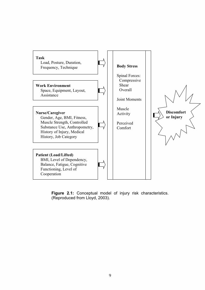
The biodynamic of low back injury among healthcare workers as a
result of manual lifting and recurrent loads was described by a conceptual
model by Lloyd (Llyod, 2003) (Figure 2.1).
9
Figure 2.1: Conceptual model of injury risk characteristics. (Reproduced from Lloyd, 2003).
Task Load, Posture, Duration, Frequency, Technique
Work Environment Space, Equipment, Layout, Assistance
Nurse/Caregiver Gender, Age, BMI, Fitness, Muscle Strength, Controlled Substance Use, Anthropometry, History of Injury, Medical History, Job Category
Patient (Load/Lifted) BMI, Level of Dependency, Balance, Fatigue, Cognitive Functioning, Level of Cooperation
Body Stress Spinal Forces:
Compressive Shear Overall
Joint Moments Muscle Activity Perceived Comfort
Discomfort or Injury

10
2.2. Definition
2.2.1. Low Back Pain
Vrbanic (2011) defined LBP as a pain and discomfort that was
localized below the costal margin and above the inferior gluteal fold, with or
without the presence of leg pain (Vrbanic, 2011). The same definition was
used by Gavira Pavon et al. in a study of LBP-related urinary incontinence
(Gavira Pavon et al., 2013). This definition is in agreement with one of the
earlier definition proposed by Frank et al. in 1996, which defined it as any
pain between the ribs and the top of the leg, from any cause (Frank et al.,
1996).
Kourinka et al. (1987) used similar definition in developing a well-
known Nordic Musculoskeletal Questionnaire (NMQ). However, in the
questionnaire, a diagram with shaded area to define the low back and other
body regions was included to improve the understanding of the definition
(Figure 2.2) (Kuorinka et al., 1987).
Other definition of LBP was a lumbar, sacral or lumbosacral spinal
pain (Malliou et al., 2006). LBP was also considered for a pain that localized
to the paraspinal regions, spreading to the flanks and into the buttocks
(Devereaux, 2009). Variation in the definition of LBP reflected difficulties
faced to make a specific anatomical diagnosis of LBP, owing to the
complexity of the muscular, ligament, bony as well as neural elements of the
back (Hicks et al., 2002).

11
Figure 2.2: A diagram with shaded area used in Standardized Nordic Questionnaire for analysis of musculoskeletal symptoms (Kuorinka et al., 1987).
Many recent studies showed that LBP is a chronic condition with
episodes of recurrence and remission (Itz et al., 2013; Young et al., 2013).
Episode of LBP is defined as a pain lasting more than 24 hours, preceded
and followed by at least 1 month of pain-free (de Vet et al., 2002).
Researchers were divided in defining acute LBP. Previously, some authors
defined an acute LBP if the symptom of LBP lasted for 14 days and less
(Kovacs et al., 2005; Heitz et al., 2009). Contradicted with the other authors,
Scott Kinkade, in his paper ‘Evaluation and Treatment of Acute Low Back
Pain’, had defined an acute LBP as any pain lasted less than six weeks

12
(Kinkade, 2007). His argument was that, existing studies had showed that up
to 90 percent of the cases of LBP recover in six weeks time (Deyo and
Weinstein, 2001; Carragee and Hannibal, 2004). Therefore, in his opinion,
any LBP with no any evidence indicating a serious underlying condition such
as fracture, infection or malignancy should be treated conservatively and was
not indicated for any imaging assessment. Recently any pain that persists
between six to 12 weeks still being considered as an acute event, often non-
specific and self-limited (Casazza, 2012). Chronic LBP was established when
the symptom of pain persist for 3 months or more (Carey et al., 1999; Blyth et
al., 2003; Diamond and Borenstein, 2006; Rozenberg, 2008; Gautschi et al.,
2009; Heitz et al., 2009; Fujii and Matsudaira, 2013).
Researchers also differ in defining the severity of LBP. One study
divided the LBP into low- and high-impact LBP, with duration of 1-week is
taken to distinguish between the groups (Santos-Eggimann et al., 2000). A
recent study classify the severity of LBP based on the radiation of pain, in
which LBP without radiation was considered as mild cases and LBP with
radiation above and below the knee as moderate and severe LBP,
respectively (Murtezani et al., 2011).
The definition of LBP used in the current study was similar to the
definition used by Kourinka et al. (1987), Gavira Pavon et al. (2013) and
Vrbanic (2011). The NMQ was used in the study to help defining the area of
LBP. The definition was chosen since it was accepted by most of the earlier
researchers. Furthermore, the NMQ was used as a part of this study’s
questionnaire.

13
2.2.2. Ambulance Workers
Ambulance workers are personnel that provide emergency health care
to patients who are injured, sick, infirm, or otherwise physically or mentally
impaired prior to and during transport to medical, rehabilitation and other
health care facilities. They were classified under the Technicians and
Associate Professionals group, Health Associate Professionals subgroup, in
The International Standard Classification of Occupations, revised 2008
(ISCO-08) (ILO, 2012). The occupations included in this group, among
others, are emergency paramedics and emergency medical technicians
(EMTs). They received formal training in emergency medical treatment,
patient transport, ambulance principles and practice, or related field (WHO,
2010).
In Malaysia, occupational group included under the ambulance worker
category, as defined by the ISCO-08, are Assistant Medical Officers (AMO)
and Trained Nurses. According to a regulation governed by the MOH, a
minimal requirement of diploma in Assistant Medical Officer or Nursing, is
required for anybody to practice these 2 professions in Malaysia (KKM,
2011a; KKM, 2011b).

14
2.3. Functional Anatomy
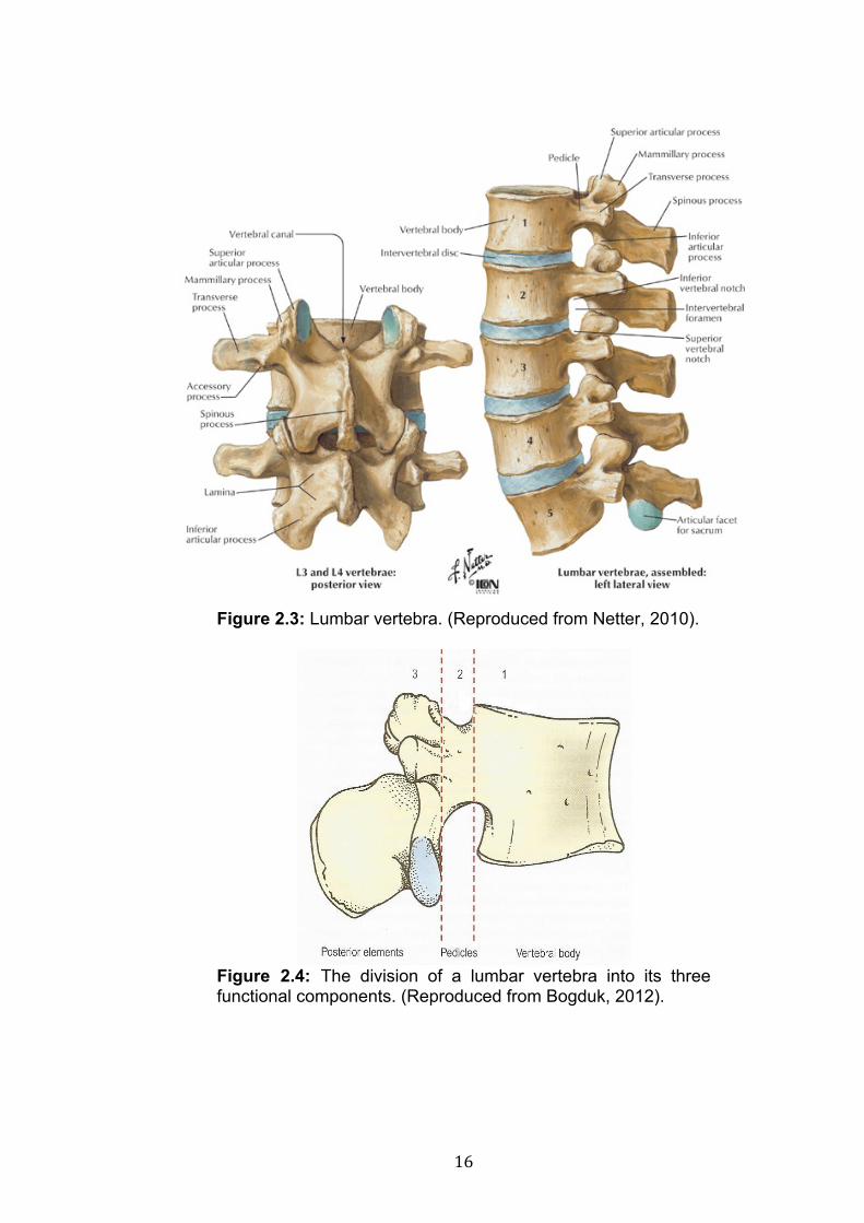
2.3.1. Lumbar Vertebral Column
Human lumbar vertebral column consists of five separate, irregularly
shaped vertebrae (Figure 2.3). Each lumbar vertebral may be divided into
three functional components - vertebral body, the pedicles and the posterior
elements (Figure 2.4). Together, these three elements contributed to the
integrated function of the whole vertebra. Vertebral body is a short, box-
shape bone with a flat superior and inferior surface (Figure 2.4). Each
vertebral body is made up of a cancellous bone surrounded by a shell of
cortical bone. These features give a light but strong structure that subserves
the weight-bearing function of the vertebrae. The posterior elements are
irregular mass of bones, consists of the laminae, the superior articular
processes, the inferior articular processes, the transverse processes, the
accessory processes, the mammillary processes and the spinous process
(Figure 2.5). The inferior articular processes of a superior vertebral body
locked with the superior articular processes of an inferior vertebral body,
forming an apophyseal joint. This synovial joint prevents twisting and forward
sliding of the vertebral bodies. The transverse, accessory, mammillary and
spinous processes act as areas of muscle attachments whereas the longer
transverse and spinous processes act as levers that enhanced the action of
muscles that attached to them. The laminae transmit any forces applied to
the inferior articular processes or the spinous process to the vertebral body,
to execute movement or provide stability. The pedicle connects the vertebral
body to the posterior elements. It transmits both tension and bending forces

15
exerted by muscular actions at the posterior elements to the vertebral body
(Bogduk, 2012).
Adjacent vertebrae bodies are separated by a 5 – 10 mm height
fibrocartilage pad, called intervertebral disc (Adams, 2013) (Figure 2.3). The
intervertebral discs provide small bending, twisting and sliding movements
between the vertebrae. They also dissipate vertical forces evenly on the
vertebral bodies (Adams et al., 1996). The discs comprise of 15 to 25 tough
concentric layers (lamellae) of annulus fibrosus, surrounding a deformable
and soft nucleus pulposus (Figure 2.6). Each lamella consists of 20 to 60
separate bundles of collagen fibers. Type I collagen fibers made up the most
of the annulus, reinforce by some amount of collagen type III and IV. Fibers
of the inner annulus curve around the nucleus pulposus and gradually blend
in with the hyaline cartilage of the endplate. At the outer layer, the fibers are
strongly embedded in the adjacent vertebrae body. The nucleus pulposus
consists mainly of proteoglycan, which is reinforced by fibrous protein. Each
proteoglycan molecules made up of more than 80% tissue water (Frank M.
Phillips, 2010). Another important component of the intervertebral discs is
layers of hyaline cartilage that cover the superior and inferior aspect of the
disc, named endplate (Figure 2.6). Each endplate cover almost the entire
surface of the adjacent vertebral body, binding the intervertebral disc to it
respective vertebral body. A ring apophysis is a narrow rim of bone around
the perimeter of the vertebral body, not covered by the endplate (Adams,
2013).
16
Figure 2.3: Lumbar vertebra. (Reproduced from Netter, 2010).
Figure 2.4: The division of a lumbar vertebra into its three functional components. (Reproduced from Bogduk, 2012).

17
Figure 2.5: Parts of a lumbar vertebra (superior view). (Reproduced from Netter, 2010).
Figure 2.6: A basic structure of a lumbar intervertebral disc. AF, annulus fibrosus; NP, nucleus pulposus; VEP, vertebral endplates. (Reproduced from Bogduk, 2012).

18
2.3.2. Spinal Stability
Spine stability is defined as the ability of the spinal column or its
components to resist buckling when undergoing load. It is maintained by
means of three subsystem namely the central nervous system,
osteoligamentous system and muscle subsystem (Panjabi, 1992) (Figure
2.7).
Figure 2.7: The spine stability system. (Reproduced from Panjabi, 1992).
When viewed from lateral, an upright normal lumbar vertebral curved
posteriorly. Such arrangement resulted in the L1 vertebra to lie vertically
above the sacrum. This posterior concavity of the lumbar vertebrae is called
lumbar lordosis (Figure 2.8). Several factors contribute to this normal
concavity. The first of this is the wedge-shaped of L4/5 intervertebral disc,
whereby its posterior height is 6 – 7 mm shorter than the anterior part.
Control Subsystem
Neural
Passive Subsystem
Spinal Column
Active Subsystem
Spinal Muscles

19
Secondly, the L5 vertebral body is also wedge-shaped; due to its posterior
surface is about 3mm less than the anterior surface. The third factor is slight
backward inclination of each vertebra above L5, in relation to vertebra below.
Figure: 2.8: A lumbar X-ray showing normal lumbar lordosis. (Reproduced from Sullivan, 2003).
The presence of lumbar lordosis allowed a proper articulation between
L5 and the upper part of the sacrum, which is inclined forwards and
downwards. There is a tendency for L4 to slip forwards on L5, and for L5 to
slip forwards on the sacrum due to the anterior tilting of the sacrum. This

20
forward displacement is resisted by a locking mechanism of each apophyseal
joint, especially of L4/L5 and L5/S1, as well as by ligamentous support
provided, particularly, by iliolumbar ligament, anterior longitudinal ligament
and anterior half of annulus fibrosus. (Middleditch and Oliver, 2005).
Muscles surrounding the lumbar spine can be divided into 3 groups –
(i) psoas major muscle, (ii) intertransversarii laterals and quadratus
lumborum, as well as (iii) the lumbar back muscles. Out of these three
groups, the lumbar back muscles played some role in providing stability to
the lumbar spine. This muscle group lies behind and covers the posterior
elements of the lumbar spine. The muscles in this group serve to correct any
possible displacement by gravity or by asymmetrical weight bearing. The
appropriate muscles will be recruited depending on the direction of any
displacement. Morphologically, the lumbar back muscles can be further
divided into three subgroups. The first subgroup is the short intersegmental
muscles, consists of the interspinales and the intertransversarii mediales.
The multifidus and the lumbar components of the longissimus and iliocostalis
represent the second subgroup - the polysegmental muscles, that attach to
the lumbar spines. The third subgroup - the long polysegmental muscles
group, extend from thoracic levels to their attachments on the ilium and
sacrum. This group of muscles, represented by the thoracic component of
the longissimus and iliocostalis lumborum, do not attach to the lumbar
vertebrae (Bogduk, 2012).

21
2.4. Sources of LBP
As LBP is a somatic type of pain, virtually, any structures located at the low
back region and innervated by a nerve supply can be a possible source of
the pain. Nonetheless, reliable evidences to implicate any of the structures
are still lacking, resulting in uncertainty and controversies (Bogduk, 2012).
The possible causes of occupational related LBP are summarized in the
Table 2.1 (Rampal et al., 2007; Bogduk, 2012).
Table 2.1: Differential Diagnosis of Occupational Related LBP
Bone and Joint Vertebral body Fracture Posterior elements Fracture Spondylolysis Kissing spines (Baastrup’s Disease) Lamina impaction Intervertebral Discs Annulus Fibrosus Herniation Torsion injury Nucleus Pulposus Internal Disc Disruption (IDD) End Plates Fracture Avulsion Schmorl’s node formation Ligaments and Muscles Interspinous ligaments sprain Iliac Crest Syndrome Muscle sprain Muscle spasm

22
2.5. Risk Factors
Risk factors for LBP are multidimensional. While several risk factors had
been identified, the evidence for some others is still insufficient or
contradictory. Generally, these factors can be broadly divided into individual
and occupational risk factors (Skovron, 1992; Jellad et al., 2013).
2.5.1. Individual Risk Factors
Individual risk factors frequently implicated for the development of LBP
included older age, female sex, high body mass index (BMI), being married
and unhealthy life-style (Levin et al., 2001; Devereaux, 2009).
2.5.1 (a) Age
LBP, for sometimes, is believed to be a problem of the elderly. A
systemic review of 165 articles on the global prevalence of LBP found out
that the prevalence of LBP was highest among people aged 40 – 80 years
old (Hoy et al., 2012). Another systemic review looking at the pattern of LBP
prevalence with age revealed that the prevalence of benign form of LBP
exhibit curvilinear association with age, whereas the prevalence of the
severe form increases with age (Dionne et al., 2006). A cross-sectional study
of nearly 30,000 subjects aged 16 years old and older in Spain revealed that
LBP was 1.5 times (CI 1.3 – 1.8) higher among subjects in the 31 – 50 years
age group, compared to those in 16 – 30 years group (Fernandez-de-las-
Penas et al., 2011). However, interestingly, children and adolescent were
also equally at risk of developing LBP. A recent meta-analysis study on
prevalence rates of LBP in children and adolescence of 18 years old and

23
younger, revealed a mean lifetime prevalence of 0.399 (CI: 0.342 – 0.459)
(Calvo-Munoz et al., 2013).
2.5.1 (b) Gender
Researchers found contradicting data regarding the gender factor
toward the development of LBP. A recent retrospective study showed that
men between 18 and 34 years old were 1.18 times more risk of getting LBP
(Beaudet et al., 2013). However, few other studies indicated that female were
at higher risk of developing LBP, regardless of the age-group (Widanarko et
al., 2011; Cho et al., 2012; Jimenez-Sanchez et al., 2012; Bener et al., 2013;
Paudyal et al., 2013). A study conducted on undergraduate medical students
in Delhi, India showed that the prevalence among males and females was
45.3% and 50% respectively (Aggarwal et al., 2013). A study on subjects
aged 70 years and older showed that female sex was independently
associated with likelihood of suffering from a short-term restricting back pain
(hazard ratio [HR] 1.30; CI 1.07 – 1.32) and persistent or recurrent back pain
(HR 1.48; CI 1.13 – 1.94) (Makris et al., 2014). A systemic review of 165
articles on prevalence of LBP showed that the problem is more prevalent
among female gender (Hoy et al., 2012).
2.5.1 (c) Body Mass Index
Obesity, defined as excessive accumulation of fat that may affect
health, is classify, among all, based on body mass index (BMI). According to
the World Health Organization (WHO) definition, a BMI of 25 or greater is
considered overweight, whereas a BMI of 30 and more is considered as

24
obese (WHO, 2014). The risk of LBP increases with increasing body mass
index (BMI). Obesity and physical inactivity were the independent risk factors
for LBP (Shiri et al., 2013). In a large epidemiological study involving more
than 800,000 adolescents, LBP was found to be significantly associated with
overweight and obesity, both in male (for overweight, OR 1.097, p value <
0.001; for obesity, OR 1.163, p value < 0.001) and female subjects (for
overweight, OR 1.174, p value < 0.001; for obesity OR1.211, p value <
0.001) (Hershkovich et al., 2013). Recently, a cross-sectional study involving
6,796 US populations showed that 7.7% - 11.6% of obese people (BMI of 31
kg/m2 and more) were at risk of experiencing LBP, as opposed to only 2.9%
among people with normal BMI (20 – 25 kg/m2) (Smuck et al., 2014). Another
recent study involving 145 middle-aged women subjects also demonstrated
that, independent of their recreational activities, obese participants who
involved in predominantly physical activities at work have high level of LBP
compared to the non-obese participants (Urquhart et al., 2014).
A systemic review of studies based on twin subjects also showed that
obesity was associated with LBP (pooled OR, pOR 1.9, CI 1.6 – 2.2)
(Ferreira et al., 2013a). Meanwhile, a study on nearly 13,000 adults in
Taiwan showed that the obesity was associated with LBP, both in poor
people (Hazard Ratio, HR 1.74) and in higher socioeconomic group (HR
1.24) (Hu et al., 2013).
2.5.1 (d) Marital status
A number of studies look into a relation between marital status and
LBP. Apparently being married or staying with a partner was found to have


