
1
A multivalent inhibitor of the DC-SIGN dependent uptake of HIV-1 and Dengue
virus
Norbert Varga,1§ Ieva Sutkeviciute,2,3,4§ Renato Ribeiro-Viana,5§ Angela Berzi6, Rasika Ramdasi,7
Anna Daghetti, 1 Gerolamo Vettoretti, 1 Ali Amara,7 Mario Clerici,8,9 Javier Rojo,*5 Franck Fieschi,*
2,3,4 Anna Bernardi*1,10
1. Universita’ degli Studi di Milano, Dipartimento di Chimica, via Golgi 19, 20133 Milano, Italy; 2.
Univ. Grenoble Alpes, Institut de Biologie Structurale (IBS), F-38027 Grenoble, France ; 3. CEA,
DSV, IBS, F-38027 Grenoble, France; 4. CNRS, IBS, F-38027 Grenoble, France; 5. Instituto de
Investigaciones Químicas (IIQ), CSIC – Universidad de Sevilla, Americo Vespucio 49, 41092
Sevilla, Spain; 6. Università degli Studi di Milano, Dipartimento di Scienze Biomediche e Cliniche
“L. Sacco”, Via GB Grassi 74, 20157 Milano, Italy; 7. INSERM U944, Laboratoire de Pathologie
et Virologie Moléculaire, Hôpital Saint-Louis, 1 Avenue Claude Vellefaux, 75010 Paris, France; 8.
Università degli Studi di Milano, Dipartimento di Fisiopatologia Medico-chirurgica e dei
Trapianti, Via F.lli Cervi 93, 20090 Segrate, Italy; 9. Fondazione Don Gnocchi IRCCS, Via
Capecelatro 66, 20148 Milano, Italy; 10. CNR-ISTM, Institute of Molecular Science and
Technologies, Milan, Italy
§ These authors contributed equally to this paper
Keywords: Glycodendrimers, Glycomimetics, HIV, Dengue, DC-SIGN, Nanotechnology
2
Abstract
DC-SIGN is a C-type lectin receptor on antigen presenting cells (dendritic cells) which has an
important role in some viral infection, notably by HIV and Dengue virus (DV). Multivalent
presentation of carbohydrates on dendrimeric scaffolds has been shown to inhibit DC-SIGN binding
to HIV envelope glycoprotein gp120, thus blocking viral entry. This approach has interesting
potential applications for infection prophylaxis. In an effort to develop high affinity inhibitors of
DC-SIGN mediated viral entry, we have synthesized a group of glycodendrimers of different
valency that bear different carbohydrates or glycomimetic DC-SIGN ligands and have studied their
DC-SIGN binding activity and antiviral properties both in an HIV and a Dengue infection model.
Surface Plasmon Resonance (SPR) competition studies have demonstrated that the materials
obtained bind efficiently to DC-SIGN with IC50s in the M range, which depend on the nature of
the ligand and on the valency of the scaffold. In particular, a hexavalent presentation of the DC-
SIGN selective antagonist 4 displayed high potency, as well as improved accessibility and chemical
stability relative to previously reported dendrimers. At low M concentration the material was
shown to block both DC-SIGN mediated uptake of DV by Raji cells and HIV trans-infection of T-
cells.
3
Introduction
Cells of the innate immune system use pattern recognition receptors (PRRs) to identify
pathogen-associated molecular patterns. The two major families of membrane-bound PRRs found in
sentinel cells, as macrophages and dendritic cells, are Toll-like receptors (TLRs) and C-type lectin
receptors (CLRs). DC-SIGN (Dendritic Cell-Specific Intercellular adhesion molecule-3-Grabbing
Nonintegrin) is a CLR used by immature dendritic cells (DCs) in mucosal tissue to recognize high-
mannose glycans present on the surface of invading microorganisms. For some pathogens,
including viruses like HIV, Ebola or Dengue,[1] this recognition event contributes to infection by
promoting viral transmission, rather than protecting the host. This observation has turned DC-SIGN
into an interesting target for the design of anti-viral agents.[2-10] The task is complicated by the
presence of other C-type lectins of similar selectivity, like Langerin,[11] that has a protective effect
against HIV infection. Thus, selective DC-SIGN ligands that interact only weakly with Langerin are
actively sought after as potentially useful therapeutic tools against HIV and other viruses that use
DC-SIGN as a primary receptor.[12-15]
DC-SIGN is a tetramer and is organized into clustered patches at the cell membrane.[16, 17]
Interactions with pathogens, also expressing multiple copies of clustered glycans, involve a
complex equilibrium that implies multipoint attachments. For this reason the principle of
multivalency has been used with success in the development of antagonists of DC-SIGN and
numerous reports have appeared in the literature concerning mannosylated dendrimers or polymers
that target it.[8, 10, 18-22] Multivalent structures bearing sugar mimics were previously prepared
in our group[10, 18] using Boltorn type dendrimers and dendrons derived from 2,2-
bis(hydroxymethyl) propionic acid. These scaffolds have a polyester backbone of good flexibility
and water solubility, and their outer layers are functionalized with carboxylic groups. Ligand
conjugation occurs via amide bond formation with amine functionalities on the monovalent ligands.
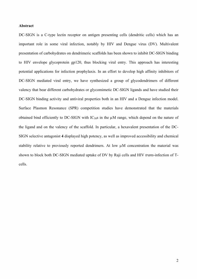
In particular, the tetravalent pseudo-trimannoside 1.2 (Figure 1), bearing four copies of the
trimannoside mimic 2a, was found to interact selectively with DC-SIGN versus Langerin and block
4
HIV-1 infection both in cellular and human cervical explant models.[12, 13] Further analysis of
multivalent constructs bearing up to 30-32 copies of trimannoside mimic 2a or the corresponding
pseudo-disaccharide 3a[18] provided nanomolar inhibitors of an Ebola pseudotyped virus infection,
but showed some drawbacks, such as relatively long synthesis and chemical instability of the
scaffolds. Additionally, these studies revealed that the pseudo-disaccharide 3, once presented on a
polyvalent construct, is only marginally less active as a DC-SIGN antagonist than the more
synthetically complex pseudo-trisaccharide 2. Further campaigns, directed to optimization of the
pseudo-disaccharide structure, led to a new lead, the bis-benzylamide 4, which binds DC-SIGN
with an affinity approaching that measured for 2, while displaying improved selectivity.[15]
In the development of this research, we have sought to exploit antagonist 4 as the active element of
new glycodendrimers. To overcome the problems met with Boltorn-type structures, a set of
polyalkynes were planned as dendrimer cores, in order to take advantage of Cu(I) catalyzed azide-
alkyne cycloaddition (CuAAC, click chemistry) of 4. This approach has the potential to afford
multivalent DC-SIGN antagonists of high potency, that also display improved accessibility and
chemical stability over previously studied materials. Their DC-SIGN binding properties could be
studied in Surface Plasmon Resonance (SPR) and in infection cellular models, using azido-
functionalized mannose 5[23], the pseudo-trisaccharide 2b[24] and pseudo-disaccharide 3b[25] to
synthesize appropriate controls.
2. Materials and methods
2.1 Synthesis
2.1.1 General
Dichloromethane, methanol, N,N-diisopropylethylamine and triethylamine were dried over calcium
hydride; THF was distilled over sodium, N,N-dimethylacetamide (DMA) was dried over activated
molecular sieves (4 Å). Reactions requiring anhydrous conditions were performed under nitrogen.
1H and 13C spectra were recorded at 500 MHz on a Bruker DRX 500, 400 MHz on a Bruker
5
AVANCE-400 and 300 MHz on Bruker DPX-300 instrument. Chemical shifts (δ) for 1H and 13C
spectra are expressed in ppm relative to internal standard (CDCl3: 7.24 for 1H and 77.23 for 13C;
CD3OD: 3.31 for 1H and 49.15 for 13C; D2O: 4.80 for 1H; DMSO-D6: 2.50 for 1H and 39.52 for
13C). Signals were abbreviated as s, singlet; br s, broad singlet; d, doublet; t, triplet; q, quartet; m,
multiplet. Mass spectra were obtained with a ThermoFisher LCQ apparatus (ESI ionization), or
iontrap ESI Esquire 6000 from Bruker, or a Microflex apparatus (MALDI ionization) from Bruker,
or Apex II ICR FTMS (ESI ionization-HRMS). Specific optical rotation values were measured
using a Perkin-Elmer 241, at 589 nm, in a 1 dm cell. Thin layer chromatography (TLC) was carried
out with pre-coated Merck F254 silica gel plates. Flash chromatography (FC) was carried out with
Macherey-Nagel silica gel 60 (230–400 mesh).
Compounds 3, [25] 4, [15] 7, [26] 9, [27] 11, [28] and 14[29] have been previously described. The
tetravalent mannosylated dendrimer 11.5 is a known compound.[28] The synthesis and
characterization of materials derived from monovalent ligand 4 (11.4, 13.4 and 9.15.4) is described
below. The synthesis and characterization of all other glycodendrimers are reported in the
Supplementary Information file.
The numbering used in the NMR characterizations is indicated in the structures reported in the
Supplementary Information file (Figure SI-9). Sugar signals were numbered as customary;
cyclohexane protons are indicated with the letter D followed by numbers. The unusual numbering
of the pseudo-saccharide derivatives in the NMR characterizations was adopted to facilitate
comparison with the native disaccharide
2.1.2 General procedure for the CuAAC reaction (click reactions)
In the optimized procedure of the copper(I) catalyzed 1,3-dipolar cycloaddition, the starting
materials and reagents were added to the reaction mixtures as solutions in water (degased by
bubbling with nitrogen) or THF (freshly distilled). Monovalent ligands (2-4) and dendrons (15.3
and 15.4) with azide groups were added as solids. The reagents were added to the reaction in the
following order: multivalent scaffold (1 eq. in THF), TBTA (1 eq. in THF), copper(II) sulphate
6
(0.1 eq. in H2O), sodium ascorbate (0.4 eq. in H2O) and finally the azide derivative (1.1 eq. per
alkyne). After the addition of all the reagents, the solvent ratio was adjusted to 1:1 by addition of
THF and/or water (c = 0.03 M). The reactions were stirred under nitrogen atmosphere, and
protected from light. The reaction progress was followed by TLC (silica, Hex:EtOAc = 8:2 and
C18, H2O: MeOH = 1:1) or mass spectrometry (MALDI or ESI ionization). Usually, in order to
achieve reaction completion, an additional 0.4 eq. of sodium ascorbate was added (2-4 h after
reaction start). After reaction completion the mixtures were loaded directly on SEPHADEX LH-20
columns (55 cm x 3.5 cm, MeOH as eluent) to purify the products by size exclusion
chromatography. In order to remove copper residues from the products, reverse phase
chromatography was performed (C18, eluent: H2O with gradients of MeOH or MeCN) or to the
solution of product in MeOH a metal scavenger[30] (such as QuadrasilTM MP) was added and
stirred for 5 min. The scavenger was filtered off through a cotton pad and the filtrate was
concentrated to obtain the product.
2.1.3 Synthesis of hexa(2-propynyloxymethyl) bispentaerythritol, 13
To a solution of bispentaerythritol (0.3 g, 1.18 mmol, 1 eq.) in dry DMF (20 mL), sodium hydride
(0.34 g, 14 mmol, 11.8 eq.) was added under argon at -5ºC. The solution was stirred at -5ºC for 1 h
then propargyl bromide (1.15 mL, 14 mmol, 11.8 eq.) was added and the mixture was kept at -5ºC
for additional 20 min. The reaction was let to warm up to room temperature and stirred for 19 h.
The reaction was cooled to 0ºC, quenched by slow addition of water and extracted with diethyl
ether (3 x 30 mL). The combined organic phases were dried over sodium sulphate and concentrated
under reduced pressure. The crude was purified by flash chromatography (silica, hex:Ethyl Acetate
= 9:1 and 8.5:1.5) to afford 130 mg of pure product. Yield: 65%; MS (HRMS) calculated for
[C28H34O7Na]+: 483.2383; found = 483.2379; 1H NMR (300 MHz, CDCl3): δ = 4.12 (d, 12H, H3,
J3-1 = 2.4 Hz), 3.52 (s, 12H, H4), 3.38 (s, 4H, H6), 2.53 (t, 6H, H1, J3-1 = 2.4 Hz); 13C NMR
(75 MHz, CDCl3): δ = δ 80.2 (C1); 74.3 (C2); 69.9 (C3); 69.3 (C4); 58.9 (C6); 45.2 (C5).
7
2.1.4 Synthesis of tetravalent glycodendrimer 11.4
Prepared according to the general procedure starting from 11[28] and 4[15]. Reaction time: 18 h;
Yield: 87%; [α]D 25 = - 4.7 (c = 0.21, MeOH); MS (MALDI, matrix: -cyano-4-hydroxy-
cinnamic acid, solvent: MeOH): calculated for [C145H192N20O48Na]+: 3006.2; found = 3005.4; MS
(ESI-HRMS): calculated for [C145H192N20O48]+: 2981.3198; found = 2981.3244 (after
deconvolution, error: 1.6 ppm); 1H NMR (400 MHz, CD3OD): δ = 7.96 (s, 4H, H16), 7.31 – 7.07
(m, 32H, H12, H13), 4.89 (br s, 4H, H1), 4.54 (s, 16H, H15), 4.51 (t, 8H, H8, J8-7 = 5.4 Hz), 4.44 (s,
8H, H18), 4.27 (s, 8H, H10a), 4.25 (s, 8H, H10b), 3.95 - 3.80 (m, 20H, H2, H6a, D2, H7), 3.72 - 3.60 (m,
12H, H6b, D1, H3), 3.59 - 3.49 (m, 8H, H4, H5), 3.44 (br s, 8H, H19), 2.90 – 2.75 (m, 8H, D4, D5),
1.96 – 1.66 (m, 16H, D3, D6); 13C NMR (100 MHz, CD3OD): δ = 177.1, 176.9 (C9); 146.3 (C17);
141.7 (C14); 139.2 (C11); 128.5, 128.3 (C13, C12); 126.2 (C16); 100.6 (C1); 76.3 (C3); 75.7 (CD1); 72.7
(D2); 72.5 (C2); 72.4 (C5); 70.0 (C19); 69.0 (C4); 68.5 (C7); 65.5 (C18); 65.1 (C15); 63.3 (C6); 52.6
(C10); 51.7 (C8); 46.7 (C20); 43.8 (C10); 41.9, 41.9 (CD4, CD5); 29.9, 29.2 (CD3, CD6).
2.1.5 Synthesis of hexavalent glycodendrimer 13.4
Prepared according to the general procedure starting from 13 and 4[15]. Reaction time: 18 h;
Yield: 70%; [α]D 25 = - 2.8 (c = 0.27, MeOH); MS (MALDI, matrix: 2,5-dihydroxybenzoic acid,
solvent: MeOH); calculated for [C220H292N30O73]+: 4524.8; found = 4524.7 [M]+ and
4549.1[M+Na]+; MS (ESI-HRMS): calculated for [C220H292N30O73]+: 4522.0059; found =
4522.0147 (after deconvolution, error: 2.0 ppm); 1H NMR (400 MHz, CD3OD): δ = 7.95 (s, 6H,
H16), 7.28 – 7.07 (m, 48H, H12, H13), 4.89 ( br s, 6H, H1), 4.54 (s, 24H, H15), 4.51 - 4.45 (m, 12H,
H8), 4.44 (s, 12H, H18), 4.28 - 4.20 (m, 24H, H10), 3.95 - 3.80 (m, 30H, H2, H6a, D2, H7), 3.73 - 3.61
(m, 18H, H6b, D1, H3), 3.60 - 3.47 (m, 12H, H4, H5), 3.38 (br s, 12H, H19), 3.26 (br s, 4H, H21), 2.89
– 2.78 (m, 12H, D4, D5), 1.93 – 1.69 (m, 24H, D3, D6); 13C NMR (100 MHz, CD3OD): δ = 177.1,
176.9 (C9); 146.3 (C17); 141.7 (C14); 139.2 (C11); 128.6, 128.3 (C13, C12); 126.1 (C16); 100.7 (C1);
76.3 (C3); 75.7 (CD1); 72.7 (D2); 72.5 (C2, C5); 71.1 (C21); 70.3 (C19); 69.0 (C4); 68.5 (C7); 65.5
8
(C18); 65.1 (C15); 63.3 (C6); 52.6 (C10); 51.7 (C8); 43.8 (C10); 41.9, 41.9 (CD4, CD5); 29.9, 29.3 (CD3,
CD6).
2.1.6 Synthesis of trivalent glycodendron 15.4
Dendron 14.4 was initially prepared, according to the general procedure and starting from 14[29]
and 4[15]. Reaction time: 3 h; Yield: 81%; MS (MALDI matrix: sinapinic acid, solvent:
MeOH): calculated for [C114H154ClN15O38]+: 2378.0; found = 2378.5; 1H NMR (400 MHz,
CD3OD): δ = 7.98 (s, 3H, H16); 7.28 – 7.16 (m, 24H, H12, H13); 4.89 (br s, 3H, H1); 4.58 – 4.50 (m,
6H, H8); 4.55 (s, 12H, H15); 4.48 (s, 6H, H18); 4.28 (s, 6H, H10); 4.26 (s, 6H, H10); 3.99 - 3.80 (m,
15H, H2, H6a, D2, H7); 3.73 - 3.61 (m, 11H, H6b, D1, H3, H25); 3.61 - 3.44 (m, 12H, H4, H5, H22, H23,
H24), 3.42 (br s, 6H, H19), 3.39 (br s, 2H, H21), 2.90 – 2.76 (m, 6H, D4, D5), 1.96 – 1.68 (m, 12H, D3,
D6); 13C NMR (100 MHz, CD3OD): δ = 177.1, 176.9 (C9), 146.3 (C17), 141.7 (C14), 139.2 (C11),
128.5, 128.3 (C13, C12), 126.2 (C16), 100.5 (C1), 76.3 (C3); 75.7 (CD1); 72.7 (D2); 72.6 (C24); 72.5,
72.4 (C2, C5); 72.2, 71.5 (C22, C23); 70.9 (C21); 70.2 (C19); 69.0 (C4); 68.5 (C7); 65.5 (C18); 65.1
(C15); 63.3 (C6); 51.7 (C8); 46.7 (C20); 44.2 (C25); 43.8 (C10); 41.9, 41.9 (CD4, CD5); 29.9, 29.3 (CD3,
CD6).
To a solution of 14.4 (150 mg, 0.0631 mmol, 1 eq.) in DMF (1 mL) sodium azide (25 mg,
0.378 mmol, 6 eq.) was added. The reaction was stirred at 65ºC for 4 days. The solvent was
removed under reduced pressure and the resulting crude was purified by reverse phase flash
chromatography (C18, water with gradient of MeOH from 0% to 70%) to afford 143 mg of pure
product. Yield: 95%; MS (MALDI matrix: sinapinic acid, solvent: MeOH): calculated for
[C114H154N18O38]+: 2384,5; found = 2385.3; 1H NMR (400 MHz, CD3OD): δ = 7.98 (s, 3H, H16),
7.30 – 7.13 (m, 24H, H12, H13), 4.89 (br s, 3H, H1), 4.60 – 4.50 (m, 6H, H8), 4.55 (s, 12H, H15), 4.48
(s, 6H, H18), 4.28 (s, 6H, H10), 4.26 (s, 6H, H10), 3.97 - 3.80 (m, 15H, H2, H6a, D2, H7), 3.73 - 3.62
(m, 9H, H6b, D1, H3), 3.62 - 3.46 (m, 14H, H4, H5, H22, H23, H24, H25), 3.43 (br s, 6H, H19), 3.40
(br s, 2H, H21), 2.90 – 2.76 (m, 6H, D4, D5), 1.96 – 1.69 (m, 12H, D3, D6).; 13C NMR (100 MHz,
9
CD3OD): δ = 177.1, 176.8 (C9), 146.3 (C17), 141.7 (C14), 139.2 (C11), 128.5, 128.3 (C13, C12), 126.2
(C16), 100.5 (C1), 76.3 (C3); 75.7 (CD1); 72.7 (D2); 72.5, 72.4 (C2, C5); 72.3, 71.5, 71.3 (C22, C23,
C24); 70.9 (C21); 70.2 (C19); 69.0 (C4); 68.5 (C7); 65.5 (C18); 65.1 (C15); 63.2 (C6); 51.9 (C25); 51.7
(C8); 46.7 (C20); 43.8 (C10); 41.9, 41.8 (CD4, CD5); 29.9, 29.2 (CD3, CD6).
2.1.7 Synthesis of nonavalent glycodendrimer 9.15.4
Prepared according to the general procedure starting from 9[27] and 15.4. Reaction time: 18 h;
Yield: 75%; MS (MALDI, matrix: sinapinic acid, solvent: MeOH): calculated for
[C357H474N54O117]+: 7393,9; found = 7394.5; MS (ESI-HRMS): calculated for [C357H474N54O117]
+:
7389.2801; found = 7393.2866 (after deconvolution, error: 0.7 ppm); 1H NMR (400 MHz,
CD3OD): δ = 8.05 (s, 3H, H26), 7.95 (s, 9H, H16), 7.28 – 7.11 (m, 72H, H12, H13), 6.29 (br s, 3H,
H30), 5.03 (br s, 6H, H28), 4.89 (br s, 9H, H1), 4.63 (s, 6H, H25), 4.55 (s, 36H, H15), 4.52 – 4.46 (m,
18H, H8), 4.45 (s, 12H, H18), 4.27 (s, 18H, H10), 4.25 (s, 18H, H10), 3.96 - 3.75 (m, 51H, H2, H6a,
D2, H7, H24), 3.75 - 3.62 (m, 27H, H6b, D1, H3), 3.61 - 3.42 (m, 30H, H4, H5, H22, H23), 3.39 (br s,
18H, H19), 3.36 (br s, 6H, H21), 2.89 – 2.81 (m, 18H, D4, D5), 1.96 – 1.68 (m, 36H, D3, D6).
1H NMR (400 MHz, DMSO-D6): δ = 8.31 (t, 9H, HNH, J10-NH = 5.3 Hz), 8.31 (t, 9H, HNH, J10-NH =
5.3 Hz), 8.17 (s, 3H, H26), 8.01 (s, 9H, H16), 7.24 – 7.07 (m, 72H, H12, H13), 6.33 (br s, 3H, H30),
5.09 (t, 18H, HOH-C15, JH15-OH = 5.6 Hz), 5.05 (br s, 6H, H28), 4.76 (br s, 9H, H1), 4.74 (d, 9H, HOH-
C3, JH3-OH = 4.6 Hz), 4.67 (d, 9H, HOH-C2, JH2-OH = 3.9 Hz), 4.59 (d, 9H, HOH-C4, JH4-OH = 5.5 Hz),
4.54 – 4.46 (m, 27H, H8, HOH-C4), 4.46 - 4.37 (m, 54H, H15, H18), 4.17 (br s, 36H, H10), 3.93 - 3.85
(m, 9H, H7a), 3.83 – 3.72 (m, 18H, D2, H7b), 3.72 – 3.62 (m, 18H, H2, H6a), 3.60 (br s, 9H, D1), 3.52
- 3.20 (m, 84H, H3, H4, H5, H6b, H19, H21, H22, H23, H24, H25), 2.81 – 2.63 (m, 18H, D4, D5), 1.83 –
1.46 (m, 36H, D3, D6).13C NMR (100 MHz, DMSO-D6): δ = 174.1, 174.0 (C9); 144.0 (C29); 140.7
(C14); 138.0 (C11); 126.7, 126.6, 126.3 (C12 ,C13); 124.9 (C26); 124.2 (C16); 98.8 (C1); 94.5 (C30);
74.6 (C5); 74.2 (CD1); 70.9 (C3); 70.5 (C2); 70.4 (D2); 69.1 (C21); 68.7 (C19); 67.1 (C7); 67 - 63 (C22,

10
C23, C24); 62.7 (C15, C18); 61.3 (C28, C6); 49.4 (C8); 44.9 (C25); 41.6 (C10); 39.3 (CD4, CD5); 28.2, 27.9
(CD3, CD6).
2.2 SPR methods.
SPR competition experiments were performed on a Biacore 3000 instrument. Flow cells (Fc) 2 and
1 of sensor chip CM4 were functionalized with mannosylated bovine serum albumine (Man α1-
3[Manα1-6]Man BSA (Man-BSA), Dextra Laboratories) or prepared as control surface,
respectively, as described previously[15]. The final response of immobilized Man-BSA was 5000
RU.
The competition experiment was performed using 25 mM Tris-HCl pH 8, 150 mM NaCl, 4 mM
CaCl2, 0.005% P20 as the running buffer at 5 μL/min flow rate. The binding of soluble tetrameric
DC-SIGN ECD [31]to immobilized Man-BSA was inhibited by the compounds at increasing
concentrations (0.14 μM – 900 μM for multivalent compounds, and 0.69 μM – 4.5 mM for
monovalent ligands). For this reason, 13 L of each DC-SIGN ECD (24 μM)/compound mixture
was injected over the surfaces. The bound lectin was washed off by a 1 min injection of 50 mM
EDTA pH 8. DC-SIGN ECD equilibrium binding responses (Req) for each sample were obtained
from the reference surface corrected sensorgrams 150 s after the start of the injection.
(1)
(2)
where Rhi and Rlo are maximum and minimum asymptotes, A1 is an inflection point and A2 is a slope
of the curve.
The obtained Req values were converted to DC-SIGN residual activity values (y, %) with respect to
Req of DC-SIGN alone, which was assigned a 100% activity value. After plotting residual activity
2
1
1A
lohihi
A
Conc
RRRy
2
1
150 150
A
hi
lohi
R
RRAIC

11
against corresponding compound concentration, the 4-parameter logistic model (eq, 1) was fitted to
the plots, and finally the IC50 values were calculated using equation 2.
2.3 Infection studies methods
2.3.1 HIV infection studies.
For HIV studies, B-THP-1/DC-SIGN cells, that support DC-SIGN mediated HIV-1 transmission
efficiently, were exploited as a model of DCs. Non-transfected B-THP-1 cells were used as a
negative control. B-THP-1/DC-SIGN cells[32] or B-THP-1 cells were pre-incubated for 30 minutes
in the presence or absence of the DC-SIGN inhibitors and afterwards were pulsed with HIV (the R5
tropic laboratory-adapted strain HIV-1 BaL) in the continued presence of inhibitors. After washing,
B-THP-1/DC-SIGN cells were co-cultured with activated CD4+ T lymphocytes from healthy
volunteer donors. To monitor viral infection of CD4+ T lymphocytes, HIV-1 p24 concentration in
the co-culture supernatants was assessed by ELISA. Each point was obtained in triplicate using
CD4+ T lymphocytes derived from three different healthy donors, and each compound was tested at
three different concentrations (1 µM, 10 µM and 100 µM). Details are given in the Supplementary
information.
2.3.2 DV infection studies.
Raji cells over-expressing DC-SIGN were infected with Dengue virus serotype-2 in the presence or
absence of 13.4 at three different concentrations (10 M, 5 M and 1 M). The infection was
scored after 48 hours at 37°C, using DV prM protein specific antibody 2H2. Mannan at 250 μg/mL
was used as positive control for infection inhibition.
2.3.3 Cyotoxicity studies
To assess the potential cytotoxicity of the glycodendrimers tested in cellular models (11.3, 11.4, and
13.4), B-THP-1/DC-SIGN cells or pheripheral blood mononuclear cells (PBMCs) were incubated
for the indicated time intervals with different concentrations of the compounds (1 µM, 10 µM, and
100 µM), followed by staining with 7-aminoactinomicin D (7-AAD), that penetrates cell membrane
of dying or dead cells. Percentage of 7-AAD positive cells (non-viable cells) did not change
12
significantly in the absence of the compounds or in their presence up to 100 µM, the highest
concentration assayed in the cellular infection models (Supplementary Figures SI-4 and SI-5).
The cytotoxicity of 13.4 towards Raji-DC-SIGN cells was also assessed using the MTT test (Figure
SI-6, MTT cytotoxicity testing kit from Sigma). The experiment was performed according to
manufacturer’s instructions. Different concentrations of 13.4 were tested on Raji-DC-SIGN cells
and percentage of live cells was directly proportional to the absorbance detected by
spectrophotometer. The absorbance values were expressed as relative percentage compared to mock
cells. Equal amount of DMSO was used as control. The results indicate that 13.4 is not cytotoxic to
cells at the concentrations used in the infection assays.
3. Results and discussion.
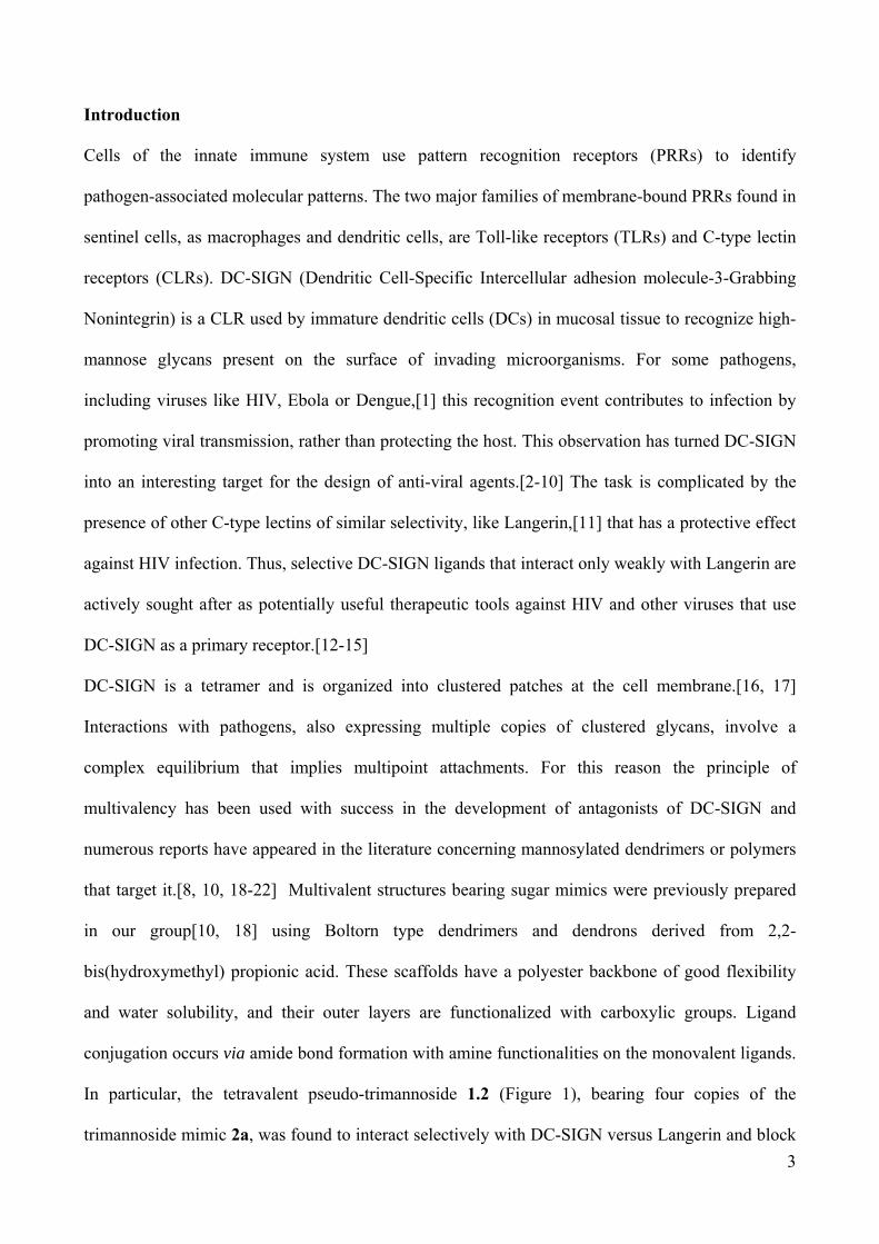
3.1 Synthesis and characterization of the glycodendrimers
The divalent alkyne 7[26] was prepared from ditosylate 6 by reaction with an excess of propargyl
alcohol in the presence of potassium carbonate (Scheme 1). Compounds 9, [27] 11, [27] and 13
were prepared in one step from commercially available starting materials. The general strategy is
based on treating polyalcohols 8, 10 or 12 with an appropriate base in the presence of propargyl
bromide (Scheme 1). The basic structures 7, 9, 11, 13 can lead to di-, tri-, tetra- and hexa-valent
presentations of a monovalent ligand, respectively. Additionally, the known trivalent dendron 14
[29] was prepared from 10 (Scheme 1). Dendron 14 can be functionalized by Cu(I) catalyzed azide-
alkyne cycloaddition (CuAAC) with three copies of a ligand and, after transformation of the
chloride tethered to the focal point into an azide (yielding 15, Figure 2), it can be clicked on other
polyalkynes such as 7, 9, 11, 13, leading to compounds with higher valency and different shapes
(Figure 2).
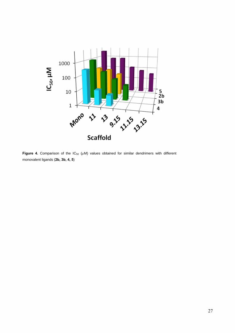
With this approach the 17 polyvalent constructs shown in Figure 2 were synthesized by CuAAC
with ligands 2b, 3b, 4-5. Among them, one tetravalent (11.4), one hexavalent (13.4) and one
nonavalent (9.15.4) dendrimers bear the lead mimic 4 (Figure 2). Controls with valency up to 18
13
were built using mannose 5, the pseudo-dimannoside 3 and the pseudo-trimannoside 2 as the
monovalent ligand (see Supplementary Information). The dendrimers were isolated from the
reaction mixtures by size exclusion chromatography on Sephadex LH20 matrix using MeOH as
eluent. Residual copper was removed either by reverse phase chromatography (C18) or using metal
scavengers (Quadrasil MP)[30]. All materials were found to be stable for months in water solution
and were fully characterized by MALDI-MS analysis (sinapinic acid or DBA) and by 1H and 13C-
NMR spectroscopy.
3.2 Surface Plasmon Resonance studies
All the glycodendrimers were tested by SPR, using a protocol that we have previously
described.[33] The assay allows to compare the relative affinity of ligands on the basis of their
ability to inhibit DC-SIGN binding to mannosylated bovine serum albumin (Man-BSA)
immobilized onto a carboxymethyl dextran- functionalized gold SPR sensor chip (CM4). Inhibition
studies were performed using the extracellular domain (ECD) of DC- SIGN (24 µM) injected alone
or in the presence of increasing concentrations of ligands. The 50% inhibition concentration (IC50)
of the dendrimers was determined and the values were compared to those obtained with the
previously described tetravalent Boltorn-type dendrons 1.
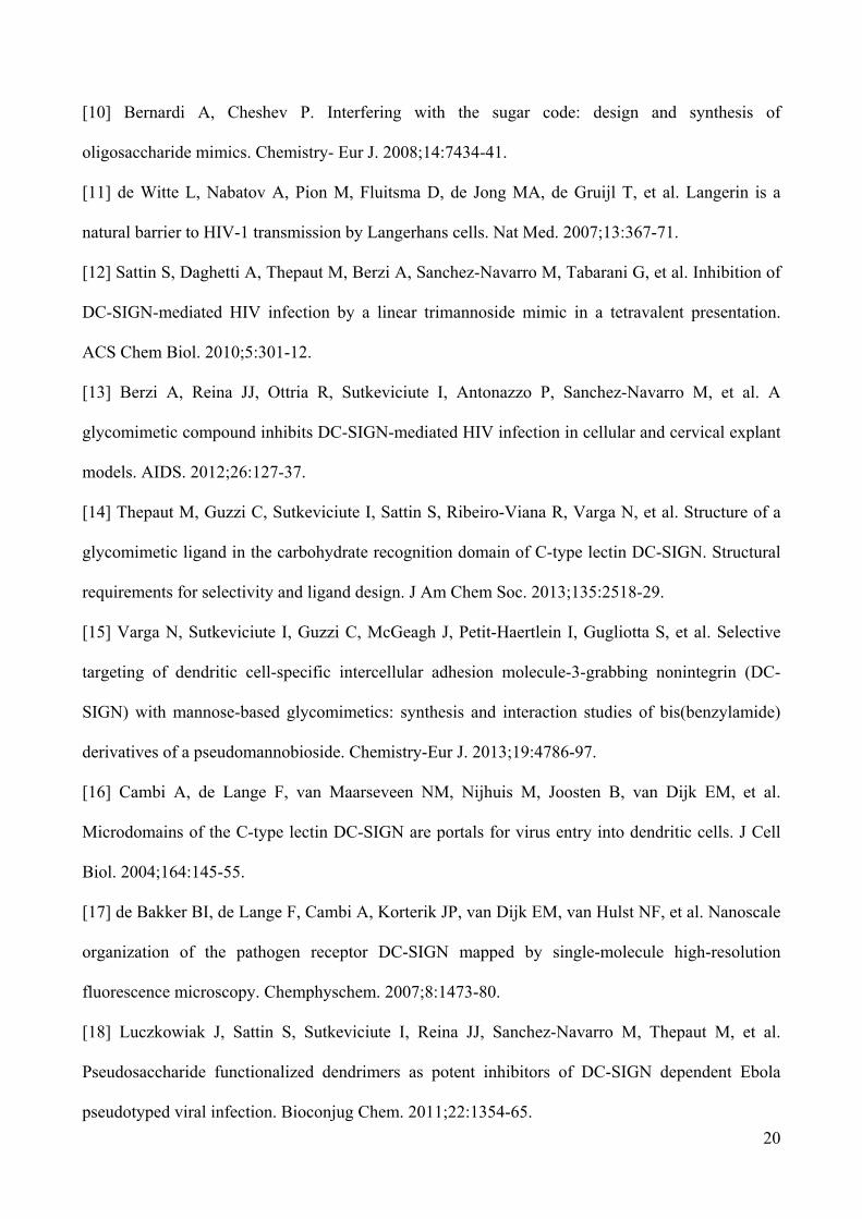
Figure 3 shows the results of a first series of experiments focused on materials derived from the
pseudo-disaccharide 3. This set of experiments revealed that the pentaerythritol based dendrimers
compare well with the previously studied Boltorn-type ones. Indeed, the two tetravalent
presentations 1.3 and 11.3 display basically the same activity and the same relative inhibitory
potency (RIP) per ligand unit (ca. 1.5) compared to the monovalent counterpart 3. The two
hexavalent ligands 13.3 and 7.15.3 showed approximately the same potency with IC50 = 37 µM and
33 µM, respectively. This suggests that, unlike the valency, the shape of the multivalent molecule
may have a minor influence on the activity, at least in this type of assay. On the other hand, the
PEG core of 7.15.3 may just be too flexible to modify the 3D structure of the dendrimer in a

14
significant way. The most remarkable improvement was observed in the case of the nonavalent
system 9.15.3, which, with a 15 µM IC50, displays a RIP of 8.
The SPR competition assay results for all compounds of Figure 2 are shown in Figure 4 and the
corresponding IC50 values are listed in Table 1. A gradual increase of activity was observed as a
function of the scaffold valence increment for all ligands, except those derived from the
pseudotrisaccharide 2, that show RIP < 1 (Table 1, column 3). This unusual behavior of polyvalent
materials derived form 2 was already observed for polyester dendrons[18] and is now confirmed to
occur independently of the nature of the polyvalent support. More structural experiments are being
carried out in our laboratory to analyze the factors that govern this phenomenon. Tethering mannose
on tetravalent and hexavalent scaffolds afforded very minor improvements, but the affinity
gradually increased with higher valence dendrimers, reaching an IC50 = 36 µM for the 18-valent
construct 13.15.5 (RIP 5; Table 1, Entry 7, Man column). Comparing the activities of materials with
different ligands, it is obvious that the selected lead 4 gives the best activity in the group. The
tetravalent and hexavalent constructs 11.4 and 13.4 displayed a RIP of 6 and 9, respectively, leading
to an IC50 = 6 µM for 13.4 (Table 1, Entry 3, column 4). The threshold for water solubility of the
nonavalent construct 9.15.4 was found to be at ca. 2 mg/mL (0.3 µM), which prevented the
determination of activity curves. Nonetheless, the advantage of using a more powerful monovalent
ligand in the preparation of multivalent constructs is obviously shown from these data (Figure 4):
IC50 values in the low micromolar range are rapidly reached using the most powerful ligand 4 even
in tetravalent presentation. Other mannosylated and pseudo-mannosylated materials lag behind,
even when the sugar is presented with higher valency on the dendrimers.
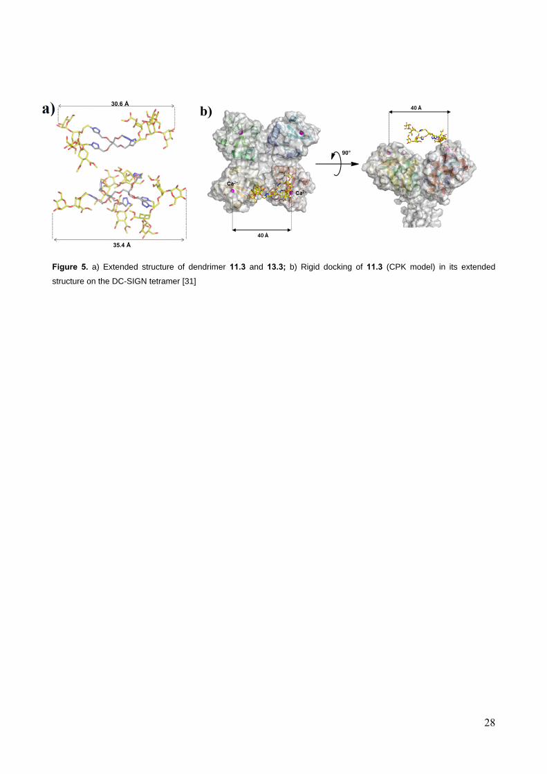
3.3 MD simulations of dendrimers 11 and 13
In order to interpret the multivalency effect observed for the materials under study, it must be kept
in mind that the SPR competition assay used here measures the ability of the dendrimers to inhibit
binding of soluble DC-SIGN tetramers to an immobilized binding partner. Under these conditions,
various effects can be operative. In principle, they may include 1) a high local concentration of the

15
ligand exposed by the dendrimers (or high effective molarity, which favors statistical rebinding;[34]
2) chelation, i.e. simultaneous binding of multiple binding sites by a single dendrimer on a single
DC-SIGN tetramer, or 3) the ability of the dendrimers to cluster soluble DC-SIGN tetramers. [22]
To gauge the average dimension of the most active dendrimers 11.4 and 13.4 and to estimate
whether they can effectively span the distance between two DC-SIGN Ca2+ binding sites which are
separated by approximately 4 nm, molecular dynamics simulations were employed. To speed up the
calculations, the simulations were performed on models 11.3 and 13.3, that ought to have the same
size of the materials derived from 4. Since mannose binds to DC-SIGN Ca2+ ions using O3 and O4,
the distances between these atoms in different Man residues were monitored continuously during
the dynamics, that were run for a total of 60 ns (details for 11.3 are collected in the Supplementary
Information section). Results showed that, even at maximum extension, the distance between the
two farthest Man-O3 is well below 4 nm (30.6 Å for 11.3 and 35.4 Å for 13.3, Figure 5), so that
chelation of two sites on the same DC-SIGN tetramer cannot be achieved. Hence, it is most likely
that the modest multivalency effects measured for 11.4 and 13.4 result from increased effective
molarity of the ligands and/or from an ability to cluster the soluble tetramers in the SPR
experiments. In the real biological settings, where DC-SIGN is exposed on the surface of (dendritic)
cells, this latter effect may be lost, or it may be translated into an ability to promote receptor
clustering at the cell surface. Thus it is of interest to explore the activity of the dendrimers described
here in cellular models of viral infections.
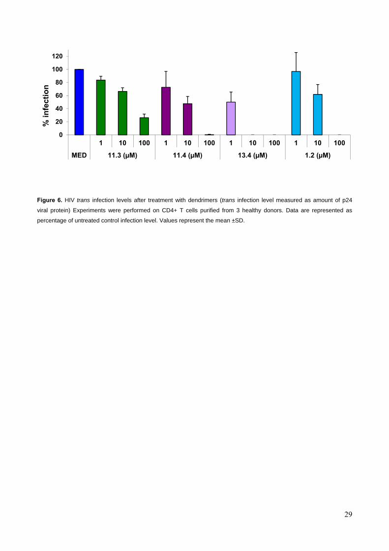
3.4 Infection tests
Tests were performed on both an HIV infection and a Dengue virus (DV) infection models.
DC-SIGN promotes HIV transmission by DCs to CD4+ T cells (infection in trans)[35]. For HIV
studies, B-THP-1/DC-SIGN cells, that support DC-SIGN mediated HIV-1 transmission efficiently,
were exploited as a model of DCs. The cells were pre-incubated for 30 minutes in the presence or
absence of the DC-SIGN inhibitors and afterwards were pulsed with HIV-1 BaL in the continued
presence of inhibitors. After washing, the B-THP-1/DC-SIGN cells were co-cultured with activated
16
CD4+ T lymphocytes from healthy volunteer donors. Viral infection of CD4+ T lymphocytes was
monitored quantifying the concentration of the HIV-1 core protein p24 in the co-culture
supernatants by ELISA. Mannan, a known DC-SIGN inhibitor[35-37] was used as a positive
control. Non-transfected B-THP-1 cells, as expected, did not transmit infection. The HIV trans
infection studies focused on those multivalent structures which were found to be the most active in
the SPR experiment, namely dendrimers 11.4 and 13.4, bearing 4 and 6 copies of the bisamide 4.
The results were compared to those obtained with 11.3 (tetravalent presentation of the pseudo-
disaccharide 3) to examine the effect of the monovalent ligand, and with the previously described
tetravalent Boltorn-type dendron 1.2. [1, 2](Figure 6 and Supplementary Figure SI-3).
Tetravalent 11.3 reduced the infection to 66% and 26% at 10 µM and 100 µM concentrations,
respectively. After treatment with compound 11.4, bearing four copies of 4, 47% of infection was
determined at 10 µM and almost no infection took place at 100 µM concentration. The most
impressive inhibition of HIV trans infection was observed in the case of 13.4 (hexavalent 4). At
1 µM concentration the infection was reduced to 50%, and at 10 µM and 100 µM the infection was
completely suppressed. For comparison, the previously known tetravalent Boltorn-type pseudo-
trisaccharide 1.2 showed similar activity to 11.4 (no infection at 100 µM) and was therefore clearly
outperformed by 13.4, which reduced the infection to 0% at 10-fold lower concentrations.
DC-SIGN also plays an important role during the transmission of Dengue virus and is considered as
a target for therapeutics that block Dengue infection.[38, 39] Dengue infection is primarily
transmitted by mosquitoes and the symptoms include fever, muscle and joint pains and skin rash.
Methods for the control and prevention of Dengue by vaccination have not been established, yet
[40]and previous attempts in our groups to block DC-SIGN mediated DV infections using
mannosylated materials based on Boltorn scaffolds were unsuccessful. In order to examine the
potential activity of the glycodendrimers to inhibit Dengue virus infection, Raji cells over-
expressing DC-SIGN were infected with Dengue virus serotype-2 in the presence or absence of
13.4 at different concentrations, 10 M, 5 M and 1 M (Figure 7). Gratifyingly, dendrimer 13.4
17
showed concentration-dependent antiviral activity. At 10 µM concentration the infection was
inhibited by about 85% and the IC50 was found to be 5.9 µM. The effect of 13.4 lasted even when
the compound was removed 30 minutes after infection (Supplementary Information, Figure SI-7).
3.5 Discussion
A majority of pathogens that infect humans use the mucosal entry pathway. These pathogens
include respiratory viruses and those responsible for sexually transmitted diseases including HIV
and Human Papilloma Virus (HPV). Notably, mucosal infections continue to represent a challenge
for the development of either preventive or therapeutic vaccines. A number of mucosal pathogens
recognize DC-SIGN as their primary target. Therefore, the design of materials capable of
interacting efficiently with DC-SIGN and of blocking the infections by pathogens that use this
lectin to access the target cells is a topic of tremendous interest. Towards this goal, we have
developed a strategy based on a very efficient click chemistry approach that allows to conjugate
different carbohydrate and glycomimetic ligands to a variety of multimeric scaffolds with different
valency. SPR competition studies have demonstrated that the dendrimers obtained bind efficiently
to DC-SIGN with IC50s in the M range.
Among the materials synthesized, those based on the monovalent ligand 4 are the most interesting
ones, since this ligand possesses the best selectivity observed to date for DC-SIGN versus Langerin,
a critical issue for inhibiting HIV infection. These materials were also found to be the most active
ones in the present study. The results showed that multivalent ligands functionalized with 4 can
inhibit both HIV and Dengue virus at low micromolar range. In particular, the hexavalent dendrimer
13.4 exhibits a low M range activity inhibiting trans-infection of T-cells by HIV in cellular studies
and provides 100% inhibition of the infection at 10 M concentration. The activity shown by 13.4
in the Dengue infection model is also very promising and it will be pursued further. Like HIV,
Dengue virus is critically dependent on DC-SIGN for host infection. No clinical treatments are
available for Dengue infection and there is a clear requirement for novel antiviral agents in this

18
field. Carbohydrate-based materials are under study as inhibitors of viral adsorption [40]. In our
studies, the scaffold ligand combination provided by 13.4 is the only one that has proven effective
to block DC-SIGN mediated uptake of DV. All previously tested dendrimers with mannose or 3 as
a ligand were ineffective towards Dengue infection (not shown). Both the multivalent scaffold 13
and monovalent ligand 4 can be prepared in gram scale and only one CuAAC step is required to
obtain the functionalized dendrimer 13.4. Moreover, unlike the previously described multivalent
compounds based on a polyester backbone, the final structure 13.4 is chemically stable. Thus 13.4
represents a clear step forward in the quest for effective antiviral therapeutics.
4. Conclusions
In summary, we present in this work the evolution of glycomimetic ligands of DC-SIGN in
multivalent materials synthesized using very efficient click chemistry reactions. Their relative IC50
were determined with a SPR based test, allowing selection of compounds whose antiviral activity
was tested using cellular models of infection with HIV and DV. The promising results obtained in
these studies establish the bases for the preparation of improved materials with higher antiviral
activities. Indeed, the studies performed here suggest that some improvement may still be obtained
in avidity with scaffolds more apt to favor a chelating effect upon binding to DC-SIGN. It does not
seem likely that such goal is already achieved with this first series of dendrimers that appear to act
mostly through rebinding and clustering effects.
Acknowledgments. The project was supported under the EU ITN Marie-Curie program
(CARMUSYS, Grant number 213592). The CM1102 COST Action Multiglyconano is also
acknowledged. FF thanks the Institut Universitaire de France for financial support. A. Berzi was
supported by a fellowship of University of Milan and Regione Lombardia (Programma “Dote
Ricerca- Rafforzare il capitale umano”, POR-Ob.2 Asse IV-FSE 2007-2013). This work has been
conducted thanks to the access to the SPR and MP3 platforms of the Partnership for Structural

19
Biology and the Institut de Biologie Structurale in Grenoble (PSB/IBS) . HRMS analysis were
obtained at the CIGA center of the University of Milan.
References
[1] van Kooyk Y, Geijtenbeek TBH. DC-sign: Escape mechanism for pathogens. Nat Rev Immunol.
2003;3:697-709.
[2] Anderluh M, Jug G, Svajger U, Obermajer N. DC-SIGN Antagonists, a potential new class of
anti-infectives. Curr Med Chem. 2012;19:992-1007.
[3] Date AA, Destache CJ. A review of nanotechnological approaches for the prophylaxis of
HIV/AIDS. Biomaterials. 2013;34:6202-28.
[4] Borrok MJ, Kiessling LL. Non-carbohydrate inhibitors of the lectin DC-SIGN. J. AmChem Soc.
2007;129:12780-5.
[5] Garber KCA, Wangkanont K, Carlson EE, Kiessling LL. A general glycomimetic strategy
yields non-carbohydrate inhibitors of DC-SIGN. Chem Commun. 2010;46:6747-9.
[6] Mangold SL, Prost LR, Kiessling LL. Quinoxalinone inhibitors of the lectin DC-SIGN. Chem
Sci. 2012;3.
[7] Tran TH, El Baz R, Cuconati A, Arthos J, Jain P, Khan ZK. A novel high-throughput screening
assay to identify inhibitors of HIV-1 gp120 protein interaction with DC-SIGN. J Antivir
Antiretrovir. 2011;3:49-54.
[8] Lasala F, Arce E, Otero JR, Rojo J, Delgado R. Mannosyl glycodendritic structure inhibits DC-
SIGN-mediated Ebola virus infection in cis and in trans. Antimicrob Agents Chemother.
2003;47:3970-2.
[9] Rojo J, Delgado R. Glycodendritic structures: promising new antiviral drugs. J Antimicrob
Chemother. 2004;54:579-81.
20
[10] Bernardi A, Cheshev P. Interfering with the sugar code: design and synthesis of
oligosaccharide mimics. Chemistry- Eur J. 2008;14:7434-41.
[11] de Witte L, Nabatov A, Pion M, Fluitsma D, de Jong MA, de Gruijl T, et al. Langerin is a
natural barrier to HIV-1 transmission by Langerhans cells. Nat Med. 2007;13:367-71.
[12] Sattin S, Daghetti A, Thepaut M, Berzi A, Sanchez-Navarro M, Tabarani G, et al. Inhibition of
DC-SIGN-mediated HIV infection by a linear trimannoside mimic in a tetravalent presentation.
ACS Chem Biol. 2010;5:301-12.
[13] Berzi A, Reina JJ, Ottria R, Sutkeviciute I, Antonazzo P, Sanchez-Navarro M, et al. A
glycomimetic compound inhibits DC-SIGN-mediated HIV infection in cellular and cervical explant
models. AIDS. 2012;26:127-37.
[14] Thepaut M, Guzzi C, Sutkeviciute I, Sattin S, Ribeiro-Viana R, Varga N, et al. Structure of a
glycomimetic ligand in the carbohydrate recognition domain of C-type lectin DC-SIGN. Structural
requirements for selectivity and ligand design. J Am Chem Soc. 2013;135:2518-29.
[15] Varga N, Sutkeviciute I, Guzzi C, McGeagh J, Petit-Haertlein I, Gugliotta S, et al. Selective
targeting of dendritic cell-specific intercellular adhesion molecule-3-grabbing nonintegrin (DC-
SIGN) with mannose-based glycomimetics: synthesis and interaction studies of bis(benzylamide)
derivatives of a pseudomannobioside. Chemistry-Eur J. 2013;19:4786-97.
[16] Cambi A, de Lange F, van Maarseveen NM, Nijhuis M, Joosten B, van Dijk EM, et al.
Microdomains of the C-type lectin DC-SIGN are portals for virus entry into dendritic cells. J Cell
Biol. 2004;164:145-55.
[17] de Bakker BI, de Lange F, Cambi A, Korterik JP, van Dijk EM, van Hulst NF, et al. Nanoscale
organization of the pathogen receptor DC-SIGN mapped by single-molecule high-resolution
fluorescence microscopy. Chemphyschem. 2007;8:1473-80.
[18] Luczkowiak J, Sattin S, Sutkeviciute I, Reina JJ, Sanchez-Navarro M, Thepaut M, et al.
Pseudosaccharide functionalized dendrimers as potent inhibitors of DC-SIGN dependent Ebola
pseudotyped viral infection. Bioconjug Chem. 2011;22:1354-65.

21
[19] Wang SK, Liang PH, Astronomo RD, Hsu TL, Hsieh SL, Burton DR, et al. Targeting the
carbohydrates on HIV-1: interaction of oligomannose dendrons with human monoclonal antibody
2G12 and DC-SIGN. P Natl Acad Sci USA. 2008;105:3690-5.
[20] Becer CR, Gibson MI, Geng J, Ilyas R, Wallis R, Mitchell DA, et al. High-affinity
glycopolymer binding to human DC-SIGN and disruption of DC-SIGN interactions with HIV
envelope glycoprotein. J Am Chem Soc. 2010;132:15130-2.
[21] Martinez-Avila O, Bedoya LM, Marradi M, Clavel C, Alcami J, Penades S. Multivalent
manno-glyconanoparticles inhibit DC-SIGN-mediated HIV-1 trans-infection of human T cells.
Chembiochem. 2009;10:1806-9.
[22] Tabarani G, Reina JJ, Ebel C, Vives C, Lortat-Jacob H, Rojo J, et al. Mannose hyperbranched
dendritic polymers interact with clustered organization of DC-SIGN and inhibit gp120 binding.
FEBS Lett. 2006;580:2402-8.
[23] Arce E, Nieto PM, Diaz V, Castro RG, Bernad A, Rojo J. Glycodendritic structures based on
Boltorn hyperbranched polymers and their interactions with Lens culinaris lectin. Bioconjug Chem.
2003;14:817-23.
[24] Maria S, Sanchez-Medina I, Mereghetti P, Belvisi L, Jimenez-Barbero J, Bernardi A. Synthesis
and conformational analysis of an alpha-D-mannopyranosyl(1 -> 2)-alpha-D-mannopyranosyl-(1 ->
6)-alpha-D-mannopyranose mimic. Carbohydr Res. 2007;342:1859-68.
[25] Reina JJ, Sattin S, Invernizzi D, Mari S, Martinez-Prats L, Tabarani G, et al. 1,2-Mannobioside
mimic: synthesis, DC-SIGN interaction by NMR and docking, and antiviral activity.
ChemMedChem. 2007;2:1030-6.
[26] Canalle LA, van Berkel SS, de Haan LT, van Hest JCM. Copper-free clickable coatings. Adv
Funct Mater. 2009;19:3464-70.
[27] Xie J, Hu L, Shi W, Deng X, Cao Z, Shen Q. Synthesis and nonlinear optical properties of
hyperbranched polytriazole containing second-order nonlinear optical chromophore. J Polym Sci B
Polym Phys. 2008;46:1140-8.
22
[28] Touaibia M, Wellens A, Shiao TC, Wang Q, Sirois S, Bouckaert J, et al. Mannosylated G(0)
dendrimers with nanomolar affinities to Escherichia coli FimH. ChemMedChem. 2007;2:1190-201.
[29] Ortega-Munoz M, Lopez-Jaramillo J, Hernandez-Mateo F, Santoyo-Gonzalez F. Synthesis of
glyco-silicas by Cu(I)-catalyzed "click-chemistry" and their applications in affinity
chromatography. Adv Synth Catal. 2006;348:2410-20.
[30] Lambeth RH, Pederson SJ, Baranoski M, Rawlett AM. Methods for removal of residual
catalyst from polymers prepared by ring opening metathesis polymerization. J Polym Sci A Polym
Chem. 2010;48:5752-7.
[31] Tabarani G, Thepaut M, Stroebel D, Ebel C, Vives C, Vachette P, et al. DC-SIGN neck domain
is a pH-sensor controlling oligomerization. SAXS and hydrodynamic studies of etracellular domain.
J Biol Chem. 2009;284:21229-40.
[32] Huskens D, Vermeire K, Profy AT, Schols D. The candidate sulfonated microbicide, PRO
2000, has potential multiple mechanisms of action against HIV-1. Antiviral Res. 2009;84:38-47.
[33] Andreini M, Doknic D, Sutkeviciute I, Reina JJ, Duan JX, Chabrol E, et al. Second generation
of fucose-based DC-SIGN ligands: affinity improvement and specificity versus Langerin. Org
Biomol Chem. 2011;9:5778-86.
[34] Pieters RJ. Maximising multivalency effects in protein-carbohydrate interactions. Org Biomol
Chem. 2009;7:2013-25.
[35] Geijtenbeek TBH, Torensma R, van Vliet SJ, van Duijnhoven GCF, Adema GJ, van Kooyk Y,
et al. Identification of DC-SIGN, a novel dendritic cell-specific ICAM-3 receptor that supports
primary immune responses. Cell. 2000;100:575-85.
[36] Hong PWP, Flummerfelt KB, de Parseval A, Gurney K, Elder JH, Lee B. Human
immunodeficiency virus envelope (gp120) binding to DC-SIGN and primary dendritic cells is
carbohydrate dependent but does not involve 2G12 or cyanovirin binding sites: Implications for
structural analyses of gp120-DC-SIGN binding. J Virol. 2002;76:12855-65.

23
[37] Lin G, Simmons G, Pohlmann S, Baribaud F, Ni HP, Leslie GJ, et al. Differential N-linked
glycosylation of human immunodeficiency virus and Ebola virus envelope glycoproteins modulates
interactions with DC-SIGN and DC-SIGNR. J Virol. 2003;77:1337-46.
[38] Tassaneetrithep B, Burgess TH, Granelli-Piperno A, Trumpfherer C, Finke J, Sun W, et al.
DC-SIGN (CD209) mediates dengue virus infection of human dendritic cells. J Exp Med.
2003;197:823-9.
[39] Lozach PY, Burleigh L, Staropoli I, Navarro-Sanchez E, Harriague J, Virelizier JL, et al.
Dendritic cell-specific intercellular adhesion molecule 3-grabbing non-integrin (DC-SIGN)-
mediated enhancement of dengue virus infection is independent of DC-SIGN internalization
signals. J Biol Chem. 2005;280:23698-708.
[40] Hidari KI, Abe T, Suzuki T. Carbohydrate-related inhibitors of dengue virus entry. Viruses.
2013;5:605-18.

24
Figure 1. Monovalent glycomimetic ligands of DC-SIGN 2-5 and the tetravalent Boltorn-type dendron 1
O
O
OH
HOHO
OH
MeOOC
O
MeOOC O
O
OH
HOHO
OH
OC
O
COHN
NH
O
O
OH
HOHO
OH
MeOOCMeOOC
OHOHO
OH
O X
O
X
HO4
2
3
OH
O
O
OHO
O OH
O
O
OH
O
O
O
O
O
O
OO
OO O
O
N3= 2, X=NH= 3, X=NH
1
O
OOH
OH
HOHO
5
1.21.3
3a X=NH23b X=N3
HO
N3
N3

25
Figure 2. Dendrimers synthesized in this study.
OO
O
O
NN
N
N N
N
NN
N
NNN
O
O
OO
NN
N
NN
N
NN
N
O
O
ON
NN
NN
N
NNN
OO
OONN
N
NN N
N NN
NN
N
OO
O ONN
N
NN
N
N NN
OO
O
O
NNN
NN N
N NN
OO
O
ONN
N
N NN
NN N
OO
OO
NN N
NNN
NN
N
O
O
O
O
O
O
O
NNN
NN
N
NN
N
O
O
O ON
NN
NN
N
NNN
O
OO
O
O
NNN
NNN
N NN
O
O
O
OO
NN
N
NN
N
NN N
O
11.211.311.411.5
13.213.313.413.5
11.15.59.15.39.15.49.15.5
OO
O ONN
N
NN
N
N N
NO
N3
15.315.415.5
= 3= 4= 5
NNN
OO
O O
NNN
O O
N NN
NNN
OO
3 N NN
O O
OO
N NN
O
NNN
NNN
7.15.3
26
Figure 3. Schematic structures and IC50 values of the monovalent pseudo-disaccharide 3a, glycodendron 1.3 (Boltorn
type) and glycodendrimers 11.3, 13.3, 7.15.3 and 9.15.3 (pentaerythritol based) measured by SPR (competition
experiments with immobilized Man-BSA)
0
300
600
900
1200
3a 1.3 11.3 13.3 7.15.3 9.15.3
904
170 139
37 33 15
IC50
(μ
M)
27
Figure 4. Comparison of the IC50 (M) values obtained for similar dendrimers with different
monovalent ligands (2b, 3b, 4, 5)
28
Figure 5. a) Extended structure of dendrimer 11.3 and 13.3; b) Rigid docking of 11.3 (CPK model) in its extended
structure on the DC-SIGN tetramer [31]
29
Figure 6. HIV trans infection levels after treatment with dendrimers (trans infection level measured as amount of p24
viral protein) Experiments were performed on CD4+ T cells purified from 3 healthy donors. Data are represented as
percentage of untreated control infection level. Values represent the mean ±SD.
0
20
40
60
80
100
120
1 10 100 1 10 100 1 10 100 1 10 100
MED 11.3 (µM) 11.4 (µM) 13.4 (µM) 1.2 (µM)
% i
nfe
ctio
n

30
Figure 7. Dose dependent inhibition of DV infection by 13.4. Raji-DCSIGN cells were infected with DV2 JAM at MOI-1 in
the presence of 13.4 at different concentrations.
61
9.5
30.5
43.4
0
10
20
30
40
50
60
70
Control 10uM 5uM 1uM
% o
f inf
ecte
d ce
lls
13.4

31
Scheme 1. Synthesis of the multivalent alkynes 7, 9, 11, 13, and 14

32
Figure captions
Figure 1. Monovalent glycomimetic ligands of DC-SIGN 2-5 and the tetravalent Boltorn-type dendron 1
Figure 2. Dendrimers synthesized in this study.
Figure 3. Schematic structures and IC50 values of the monovalent pseudo-disaccharide 3a, glycodendron 1.3 (Boltorn
type) and glycodendrimers 11.3, 13.3, 7.15.3 and 9.15.3 (pentaerythritol based) measured by SPR (competition
experiments with immobilized Man-BSA)
Figure 4. Comparison of the IC50 (M) values obtained for similar dendrimers with different
monovalent ligands (2b, 3b, 4, 5)
Figure 5. a) Extended structure of dendrimer 11.3 and 13.3; b) Rigid docking of 11.3 (CPK model) in its extended
structure on the DC-SIGN tetramer [31]
Figure 6. HIV trans infection levels after treatment with dendrimers (trans infection level measured as amount of p24
viral protein) Experiments were performed on CD4+ T cells purified from 3 healthy donors. Data are represented as
percentage of untreated control infection level. Values represent the mean ±SD.
Figure 7. Dose dependent inhibition of DV infection by 13.4. Raji-DCSIGN cells were infected with DV2 JAM at MOI-1 in
the presence of 13.4 at different concentrations.
Scheme 1. Synthesis of the multivalent alkynes 7, 9, 11, 13, and 14

33
Table 1. IC50 + SD values (M) obtained in DC-SIGN inhibition assays (SPR)
Ligand Entry Valency 5
(cmpd numb., RIPa) 2
(cmpd numb., RIPa)
3 (cmpd numb.,
RIPa)
4 (cmpd numb.,
RIPa) 1 1 3292 + 337
(5, 1) 145 + 83
(2, 1) 1018 + 109
(3b, 1) 308 + 40
(4, 1) 2 4 767 + 20
(11.5, 1.1 ) 112
(11.2, 0.3) 136 + 23
(11.3, 1.9 ) 12+ 3
(11.4, 6.4 ) 3 6 800
(13.5, 0.7) 51
(13.2, 0.5) 39
(13.3, 4) 5.7 + 1.6 (13.4, 9)
4 6 - - 32 (7.15.3, 5 )
5 9 128 (9.15.5, 2.8 )
- 14 (9.15.3, 8 )
-b
6 12 67 (11.15.5, 4.1)
- - -
7 18 36 (13.15.5, 5.1)
- - -
a) Relative Inhibitory Potency, calculated as (IC50)mono/IC50*valency ; b) Not measured, due to the low water solubility of 9.15.4


