
1 (13) 2009
A contemporary approach to surgical treatmentof scolioses
ROMAN NOWAK
Katedra i Oddział Kliniczny ŚUMWojewódzki Szpital Specjalistyczny nr 5 im. Św. Barbary, Sosnowiec
Address for correspondence/Adres do korespondencji:Dr n med Roman Nowakul. Rolna 10d/1, 40-555 Katowicetel. +48 668 304 424, e-mail: [email protected]
© J ORTHOP TRAUMA SURG REL RES 1 (13) 2009Review article/Artykuł poglądowy
Współczesne podejście do leczenia operacyjnegoskolioz
Summary
Scoliosis belongs to the most frequent deformations of human axial skeleton. In about 20% of cases,
the pathology constituting the original cause of scoliosis is recognized. As regards the remaining
cases, the diagnosis of scoliosis idiopathica is made by way of elimination. For many years, sur-
gery has been the standard procedure in the treatment of large scolioses, where preservative tre-
atment proves ineffective.
The aim of the work is to present contemporary opinions and controversies concerning the indi-
cations for surgical treatment of scoliosis, the choice of surgical access, modern possibilities and
evolution of surgical methods employed for correction and stabilization of scoliosis.
Therapeutic decisions taken after scoliosis has been diagnosed are based on clinical and radiolo-
gical assessment of a number of factors having a documented influence on the risk of progression
of the curvature. General indications for surgical treatment of scoliosis include progressing sco-
liosis of Cobb’s angle over 40-45° in skeletally immature children, considerable deformations of
Cobb’s angle over 50° in adolescents, irrespectively of their skeletal age, scoliosis-related pain
complaints unresponsive to preservative treatment, thoracic lordosis coexisting with progressive
scoliosis, and cosmetic deformations. In scolioses of known etiology, the decision of surgical tre-
atment is influenced not only by the factors of progression risk, common to idiopathic scolioses
and related to growth potential and extent of the deformation, but also by additional factors, spe-
cific to the original pathology underlying the scoliosis, e.g. the type, number, and location of
congenital defects in the spinal area in congenital (osteopathic) scoliosis, the dynamics and type
of neurological disorders in neuropathic scoliosis. The origin of the modern spinal surgery was
a combination of the concept of spinal fusion and inner stabilization and correction of the scoliosis
with spinal implants. In consideration of the patient’s age, morphology of the scoliosis, as well as
the operator’s experience and preferences, a choice is made between spondylodesis and anterior or
posterior stabilization, or a combination of the two. Among the contemporary surgical methods, two
different approaches can be distinguished with respect to the patient’s age: correction and stabili-
zation of scoliosis with an application of implants and spinal fusion, and stabilization and correc-
tion of scoliosis, utilizing the remaining growth potential for curbing the progression and directing
further spinal growth until final spondylodesis is applied. In spite of enormous progress in spinal
surgery that has been made over the last hundred years, the main objective of surgical treatment
of scoliosis has remained the same: an optimal and permanent correction of the deformation with
the least possible complications.
The evolution of surgical techniques of correction and stabilization of scoliosis from Harrington
instruments to segmental instrumentation with pedicle screws is an indication of the progress from
single-plane to multi-plane correction of scoliotic deformations.
Statistic/Statystyka
Word count/Liczba słów 3500/2782
Tables/Tabele 0
Figures/Ryciny 9
References/Piśmiennictwo 39
Received: 27.11.2008Accepted: 10.01.2009Published: 01.02.2009
A study prepared within the projectof the Ministry of Science and Infor-matization No.2PO5C07430
Praca wykonana w ramach projektuMNiI Nr 2PO5C07430
32 R. NOWAK
THE JOURNAL OF ORTHOPAEDICS TRAUMA SURGERY
AND RELATED RESEARCH
Clinical differentiation of various types of scoliosis as well as numerous controversies concerning
the surgical access, the extent of spondylodesis, the choice of implants and of correction methods
limit the possibility of introducing a universal algorithm of surgical treatment.
Key words: scoliosis, surgical stabilization and correction, spinal fusion, spinal implants.
Streszczenie
Skolioza należy do najczęściej spotykanych zniekształceń szkieletu osiowego człowieka. W przy-
padku około 20% chorych patologia będąca pierwotną przyczyną skoliozy jest znana. W odniesie-
niu do pozostałej grupy, drogą wykluczenia, stawiane jest rozpoznanie scoliosis idiopathica. Lecze-
nie operacyjne od wielu lat jest standardem postępowania w leczeniu dużych skrzywień, których
leczenie zachowawcze jest nieskuteczne.
Celem pracy było przedstawienie obecnych poglądów i kontrowersji dotyczących wskazań do
leczenia operacyjnego bocznych skrzywień kręgosłupa, wyboru dostępu operacyjnego, aktualnych
możliwości i ewolucji operacyjnych metod stosowanych do korekcji i stabilizacji skoliozy.
Decyzje terapeutyczne podejmowane po postawieniu rozpoznania skoliozy oparte są o ocenę kli-
niczną i radiologiczną szeregu czynników o udokumentowanym związku z ryzykiem progresji
skrzywienia. Ogólne wskazania do leczenia operacyjnego skoliozy obejmują narastające skrzywie-
nia o kącie Cobba powyżej 400-450 u dzieci niedojrzałych szkieletowo, znaczne deformacje o kącie
Cobba powyżej 500 u dorastających, bez względu na wiek szkieletowy, związane ze skoliozą
dolegliwości bólowe nieodpowiadające na leczenie zachowawcze, współistniejącą z pogłębiającą
się skoliozą lordozę w odcinku piersiowym kręgosłupa oraz znaczną deformację kosmetyczną.
W skoliozach o znanej etiologii poza wspólnymi ze skrzywieniami idiopatycznymi czynnikami ry-
zyka progresji związanymi z potencjałem wzrostowym i rozmiarami deformacji bardzo istotne zna-
czenie w przy podejmowaniu decyzji o operacji odgrywają dodatkowe czynniki specyficzne dla
zasadniczej patologii będącej przyczyną skrzywienia, np. w skoliozach wrodzonych (kostnopochod-
nych) rodzaj, liczba i lokalizacja wad wrodzonych w obrębie kręgosłupa, w skoliozach neuropo-
chodnych dynamika i rodzaj zaburzeń neurologicznych. Początkiem współczesnej chirurgii kręgo-
słupa było połączenie koncepcji usztywnienia z wewnętrzną stabilizacją i korekcją skrzywienia
opartą na implantach kręgosłupowych. Uwzględniając wiek operowanego, morfologię skrzywienia
oraz doświadczenia i preferencje operatora dokonuje się wyboru pomiędzy spondylodezą i stabi-
lizacją tylną, przednią lub ich połączeniem. W arsenale stosowanych obecnie metod operacyjnych
wyróżnić można dwie odmienne koncepcje podejścia do skoliozy dostosowane do wieku operowa-
nych: korekcję i stabilizację skrzywienia przy użyciu implantów wraz z usztywnieniem kręgosłupa
oraz stabilizację i korekcję skrzywienia z wykorzystaniem pozostałego potencjału wzrostowego do
zahamowania progresji i ukierunkowania dalszego wzrostu kręgosłupa do czasu wykonania osta-
tecznego usztywnienia. Pomimo olbrzymich postępów w chirurgii kręgosłupa jakich dokonano
w ciągu ostatnich stu lat główny cel leczenia chirurgicznego skoliozy pozostał ten sam: optymalna
i trwała korekcja deformacji przy jak najmniejszej liczbie powikłań
Ewolucja technik operacyjnej korekcji i stabilizacji skolioz od instrumentarium Harringtona do
segmentarnej instrumentacji śrubami przeznasadowymi obrazuje postępy na drodze od jedno do
wielopłaszczyznowej korekcji skoliotycznego zniekształcenia.
Zróżnicowanie kliniczne skolioz oraz liczne kontrowersje dotyczące dostępu operacyjnego, rozle-
głości usztywnienia, doboru implantów oraz wyboru metody korekcji ograniczają możliwość
wprowadzenia uniwersalnego algorytmu leczenia operacyjnego.
Słowa kluczowe: skolioza, operacyjna stabilizacja i korekcja, usztywnienie kręgosłupa, implanty
kręgosłupowe
Skolioza czyli boczne wygięcie kręgosłupa należy do
najczęściej spotykanych zniekształceń szkieletu osiowe-
go człowieka. W przypadku około 20% chorych patolo-
gia będąca pierwotną przyczyną skoliozy jest znana.
W odniesieniu do pozostałej grupy, drogą wykluczenia,
stawiane jest rozpoznanie scoliosis idiopathica. (1)
Obszerny temat jakim jest leczenie operacyjne sko-
lioz wymaga odniesienia się do szeregu zagadnień doty-
czących wskazań do interwencji chirurgicznej z uwzględ-
nieniem wieku chorego (usztywnienie versus kontrola
wzrostu kręgosłupa), wyboru dostępu operacyjnego (tyl-
ny, przedni, skojarzony), aktualnych możliwości stabili-
zacji i korekcji skrzywienia oraz klasyfikacji skrzywień
i planowania rozległości spondylodezy.
Scoliosis, or lateral curvature of the spine, belongs to the
most frequent deformations of human axial skeleton. In
about 20% of cases, the pathology constituting the orig-
inal cause of scoliosis is recognized. As regards the
remaining cases, the diagnosis of scoliosis idiopathica is
made by way of elimination. (1)
The vast subject of surgical treatment of scoliosis
requires the consideration of a number of issues related
to the indications for surgical intervention with respect
to the patient’s age (spinal fusion vs. control of the spinal
growth), the choice of surgical access (anterior, posteri-
or, combined), current possibilities of stabilizing and
correcting the curvature, and the classification of curva-
tures and the planned extent of spondylodesis.

33A contemporary approach to surgical treatment of scolioses
1 (13) 2009
LECZENIE OPERACYJNE SKOLIOZ –CEL I WSKAZANIAGłówne dwa cele leczenia chirurgicznego skoliozy to
zahamowanie progresji skrzywienia i uzyskanie jak naj-
większej trójpłaszczyznowej korekcji zniekształcenia
z odtworzeniem skompensowanego tułowia. Istotnym
warunkiem powodzenia jest trwałość uzyskanego wyni-
ku radiologicznego i klinicznego.
Decyzje terapeutyczne podejmowane po postawieniu
rozpoznania skoliozy oparte są o ocenę kliniczną i radio-
logiczną szeregu czynników o udokumentowanym związ-
ku z ryzykiem progresji skrzywienia. W skoliozach idio-
patycznych należą do nich: płeć, wiek w czasie rozpo-
znania (początek choroby), wiek szkieletowy, zaawanso-
wanie dojrzewania płciowego, dotychczasowe tempo
wzrostu (skok wzrostowy), typ morfologiczny skrzywie-
nia i aktualne rozmiary deformacji (kąt Cobba)
(1,2,3,4,5,6,7,8,9,10). W skoliozach o znanej etiologii
poza wspólnymi ze skrzywieniami idiopatycznymi czyn-
nikami ryzyka progresji związanymi z potencjałem wzro-
stowym i rozmiarami deformacji bardzo istotne znacze-
nie rokownicze odgrywają dodatkowe czynniki specyficz-
ne dla zasadniczej patologii będącej przyczyną skrzywie-
nia, np. w skoliozach wrodzonych (kostnopochodnych)
rodzaj, liczba i lokalizacja wad wrodzonych w obrębie
kręgosłupa, w skoliozach neuropochodnych dynamika
i rodzaj zaburzeń neurologicznych, ryc. 1.
Fig. 1. Congenital scoliosis.A female patient, aged 13, withmultiple congenital defects inthe spinal area, syringomyeliaand dimyelia in the thoracolum-bar section. Clinical conditionand radiological image beforeand after surgical treatment.Ryc. 1. Skolioza wrodzona. Cho-ra lat 13 z mnogimi wadamiwrodzonymi w obrębie kręgosłu-pa, syringomyelią i dimyelią wodcinku piersiowo-lędźwiowym.Stan kliniczny i obraz radiolo-giczny przed i po leczeniu ope-racyjnym
SURGICAL TREATMENT OF SCOLIOSIS –AIM AND INDICATIONSThe two main objectives of surgical treatment of scoli-
osis are: curbing the progression of scoliosis and achiev-
ing the largest possible three-dimensional correction of
the deformation with the reconstruction of compensated
trunk. An important evidence of success is durability of
the radiological and clinical result.
Therapeutic decisions taken after scoliosis has been
diagnosed are based on clinical and radiological assess-
ment of a number of factors having a documented influ-
ence on the risk of progression of the curvature. In id-
iopathic scoliosis, they include: sex, age at the moment
of diagnosis (the onset of the disease), skeletal age,
advancement of sexual maturation, the rate of growth up
to the present, morphological type of the curvature, and
its current extent (Cobb’s angle) (1,2,3,4,5,6,7,8,9,10). In
scolioses of known etiology, the prognoses are influenced
not only by the factors of progression risk common to
idiopathic scolioses and related to growth potential and
extent of the deformation but also by additional factors,
specific to the original pathology underlying the scolio-
sis, e.g. the type, number, and location of congenital
defects in the spinal area in congenital (osteopathic)
scoliosis, the dynamics and type of neurological disor-
ders in neuropathic scoliosis. Fig.1.

34 R. NOWAK
THE JOURNAL OF ORTHOPAEDICS TRAUMA SURGERY
AND RELATED RESEARCH
Wiek dziecka, w którym pojawia się zniekształcenie
determinuje przewidywany czas trwania i tempo proce-
su wzrostowego kręgosłupa odgrywających istotną rolę
w ewolucji choroby. W przypadku około 70% skrzywień
idiopatycznych rozpoznanych przed 10 rokiem życia
(skoliozy dziecięce) od 27% do 80% dzieci ze względu
na progresję wymaga leczenia operacyjnego. (7,11,12,13)
W najliczniej reprezentowanej grupie skolioz idiopatycz-
nych dorastających, rozpoznawanych od 10 roku życia,
(około 80%), aktywnego leczenia wymaga około 10%
chorych, a leczenia operacyjnego jedynie 0,1%. (3,8,9)
Towarzyszące ciężkiej deformacji kręgosłupa zniekształ-
cenie klatki piersiowej prowadzi do zmniejszenia jej
rozmiarów i ograniczenia ruchomości żeber co może
skutkować nadciśnieniem płucnym i niewydolnością
prawokomorową.(4,14) Problem, ten głównie dotyczy
chorych o wczesnym początku deformacji. Badania nad
przebiegiem naturalnym skolioz wykazały zwiększoną
śmiertelność z powodu niewydolności krążeniowo-odde-
chowej w tej grupie chorych, nie potwierdziły natomiast
takiego związku w skrzywieniach idiopatycznych dora-
stających. (8,9) Wcześnie narastające zniekształcenie
prawdopodobnie upośledza proces dojrzewania pęcherzy-
ków płucnych, który ulega zakończeniu około 8 roku
życia. (4)
Ogólne wskazania do leczenia operacyjnego skolio-
zy obejmują narastające skrzywienia o kącie Cobba
powyżej 400-450 u dzieci niedojrzałych szkieletowo,
znaczne deformacje o kącie Cobba powyżej 500 u dora-
stających, bez względu na wiek szkieletowy, związane ze
skoliozą dolegliwości bólowe nieodpowiadające na lecze-
nie zachowawcze, współistniejąca z pogłębiającą się
skoliozą lordoza w odcinku piersiowym kręgosłupa oraz
znaczna, zwłaszcza z punktu widzenia osoby chorej
deformacja kosmetyczna. (15) Należy podkreślić, że po-
wyższe wskazania mają charakter ogólny, a ostateczna
decyzja o operacji, jej typie i rozległości podejmowana
jest w przypadku każdego chorego indywidualnie.
W arsenale stosowanych obecnie metod operacyjnych
wyróżnić można dwie odmienne koncepcje podejścia do
skoliozy. Pierwsza z nich, najczęściej stosowana, ma
charakter definitywny i polega na korekcji i stabilizacji
skrzywienia przy użyciu implantów połączonej z mniej
lub bardziej rozległym usztywnieniem kręgosłupa. Dru-
ga koncepcja opiera się na stabilizacji i korekcji skrzy-
wienia z wykorzystaniem pozostałego potencjału wzro-
stowego do zahamowania progresji i ukierunkowania
dalszego wzrostu kręgosłupa do czasu wykonania osta-
tecznego usztywnienia – „nonfusion surgery”. Usztyw-
nienie objętego zniekształceniem odcinka kręgosłupa od
wielu lat stanowi standard w leczeniu operacyjnym scho-
rzeń deformacyjnych kręgosłupa. Uwzględniając wiek
operowanego, morfologię skrzywienia oraz doświadcze-
nia i preferencje operatora dokonuje się wyboru pomię-
dzy spondylodezą i stabilizacją tylną, przednią lub ich
połączeniem ryc.2.
Podjęcie decyzji o leczeniu operacyjnym skoliozy jest
szczególnie trudne w przypadku małych dzieci, z dużym
The child’s age at the occurrence of the deformity
determines the estimated duration and rate of spinal
growth, playing an important role in the development of
the disease. In about 70% of idiopathic curvatures diag-
nosed before the age of 10 (juvenile scoliosis), 27% -
80% of children require surgical treatment due to the
disease’s progression (7,11,12,13). In the largest group of
adolescent idiopathic scolioses, diagnosed after the age
of 10 (about 80%), 10% of patients require active treat-
ment, and only 0.1% - surgical treatment (3,8,9). A chest
deformity, accompanying a severe deformation of the
spine, results in a reduction of the chest’s size and a
limitation of rib mobility, which may cause pulmonary
hypertension and right ventricular failure (4,14). The
problem concerns mostly patients at an early stage of
deformation. Research on natural course of scoliosis
demonstrated increased mortality due to circulatory-res-
piratory failure in this group of patients, whereas similar
relationship among adolescents with idiopathic scoliosis
has not been confirmed (8,9). Probably, an early growth
of a deformation impairs the process of alveoli matura-
tion, which is completed around the age of 8 (4).
General indications for surgical treatment of scolio-
sis include progressing scoliosis of Cobb’s angle over 40-
45° in skeletally immature children, considerable defor-
mations of Cobb’s angle over 50° in adolescents, irrespec-
tively of their skeletal age, scoliosis-related pain com-
plaints unresponsive to preservative treatment, thoracic
lordosis coexisting with progressive scoliosis, and a
considerable, particularly from the patient’s point of view,
cosmetic deformation. (15). It must be emphasized that
the above indications are only general guidelines, and the
final decision on the surgery, its type and extent, is tak-
en individually in each case. Among the contemporary
surgical methods, two different approaches to scoliosis
can be distinguished. The first and most frequent one is
definitive in character and involves correction and stabi-
lization of the curvature by means of implants combined
with more or less extensive spinal fusion. The other
approach is based on stabilization and correction of the
curvature, utilizing the remaining growth potential for
curbing the progression and directing further spinal
growth until final spondylodesis is applied – „nonfusion
surgery”. Spondylodesis of the deformed section of the
spine has been the standard procedure in surgical treat-
ment of spinal deformations for many years. In consid-
eration of the patient’s age, morphology of the scoliosis
as well as the operator’s experience and preferences, a
choice is made between spondylodesis and anterior or
posterior stabilization, or a combination of the two. Fig.2.

35A contemporary approach to surgical treatment of scolioses
1 (13) 2009
Fig. 2. A female patient, aged14, with idiopathic scoliosis ofLenke 1C+ type, Cobb’s angle128°, reduced surgically to 59°.Clinical condition and radiologi-cal image before and after sur-gical treatment. Two-stage cor-rection: anterior release and cor-rection from a posterior accesswith the C-D method and thetechnique of 3 rods.Ryc. 2. Chora lat 14, ze skolio-zą idiopatyczna, typ Lenke 1C+,kąt Cobba 128o, pooperacyjniezmniejszony do 59o. Stan kli-niczny i obraz radiologicznyprzed i po leczeniu operacyj-nym. Korekcja dwuetapowa:uwolnienie przednie i korekcjaz dostępu tylnego metodą C-Dtechniką 3 prętów
potencjałem wzrostowym. Solidny blok spondylodezy
u rosnącego jeszcze dziecka prowadzi do zahamowania
wzrostu liniowego kręgosłupa w jej obszarze. Jest to
w pewnej mierze rekompensowane przez korekcję skrzy-
wienia oraz paradoksalnie poprzez zahamowanie dalszej,
związanej ze wzrostem progresji. Innym istotnym proble-
mem dotykającym niektórych chorych z niedojrzałym
szkieletem, po najczęściej wykonywanym typie operacji
jakim jest korekcja, stabilizacja i tylne usztywnienie
kręgosłupa jest tzw. crankshaft phenomenon. Przyczyną
tego zjawiska objawiającego się pooperacyjną utratą
korekcji skrzywienia i narastaniem zniekształcenia klat-
ki piersiowej jest najprawdopodobniej utrzymujący się
pomimo solidnego bloku tylnej spondylodezy wzrost
przedniej kolumny kręgosłupa. (16) Proponowanym roz-
wiązaniem tego problemu jest uzupełnienie tylnej spon-
dylodezy spondylodezą przednią. Do zmniejszenia wagi
tego problemu może się również przyczynić zastosowa-
nie do instrumentacji tylnej śrub przeznasadowych za-
miast haków. (16,17) Alternatywą dla usztywnienia krę-
gosłupa w przypadku małych pacjentów z dużymi, po-
głębiającymi się skrzywieniami są metody operacyjnej
korekcji i stabilizacji bez usztywnienia – „nonfusion sur-
gery”. Ich główny cel to zahamowanie progresji i kontro-
lowanie skrzywienia do czasu wykonania ostatecznej
spondylodezy. Wśród obecnie stosowanych należy tutaj
wymienić metody instrumentacji w oparciu o tzw. po-
dwójne rosnące pręty (techniki Luque-trolley, Leeds,
The decision of surgical treatment is particularly
difficult in the case of young children, whose growth
potential is very large. A solid block of spondylodesis in
a child who is still growing leads to a linear growth arrest
in this section of the spine. This is to some extent com-
pensated by the correction of the curvature and, paradox-
ically, by curbing further growth-induced progression.
Another serious problem concerning certain patients with
immature skeleton after the most frequent surgery type,
i.e. correction, stabilization and posterior spondylodesis,
is the so-called crankshaft phenomenon. It is manifested
by a post-operative loss of the curvature correction and
an increasing deformity of the chest. The cause of the
phenomenon is most probably the growth of the anterior
spinal column, continuing in spite of the solid block of
posterior spondylodesis (16). A suggested solution to the
problem is complementing the posterior spondylodesis
with an anterior spondylodesis. The problem may also be
diminished by an application of pedicle screws instead
of hooks for posterior instrumentation (16,17). An alter-
native to spondylodesis in the case of very young patients
with large progressing curvatures is offered by the meth-
ods of surgical correction and stabilization without
spondylodesis – „nonfusion surgery”. Their main objec-
tive is to curb the progression and control the curvature
until final spondylodesis is applied. The contemporary
methods include: instrumentation based on the so-called
double growing rods (Luque-trolley, Leeds, McCarthy,
36 R. NOWAK
THE JOURNAL OF ORTHOPAEDICS TRAUMA SURGERY
AND RELATED RESEARCH
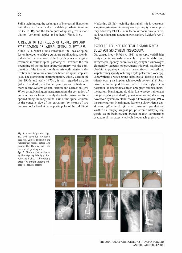
McCarthy, Shilla), technikę dystrakcji międzyżebrowej
z wykorzystaniem pionowej rozciągalnej tytanowej pro-
tezy żebrowej VEPTR, oraz techniki modulowania wzro-
stu kręgosłupa (międzytrzonowe staplery i „lejce”) ryc. 3.
(16)
PRZEGLĄD TECHNIK KOREKCJI I STABILIZACJABOCZNYCH SKRZYWIEŃ KRĘGOSŁUPAOd czasu, kiedy Hibbs w 1911 roku wprowadził ideę
usztywnienia kręgosłupa w celu uzyskania stabilizacji
skrzywienia, spondylodeza stała się jednym z kluczowych
elementów leczenia operacyjnego różnych patologii w
obrębie kręgosłupa. Jednak prawdziwym początkiem
współczesnej spondylochirurgii było połączenie koncepcji
usztywnienia z wewnętrzną stabilizacją i korekcją skrzy-
wienia opartą na implantach kręgosłupowych.(18) Roz-
powszechnione pod koniec lat sześćdziesiątych i na
początku lat siedemdziesiątych ubiegłego stulecia instru-
mentarium Harringtona do dnia dzisiejszego traktowane
jest jako „złoty standard”, punkt odniesienia, dla oceny
nowszych systemów stabilizacyjno-korekcyjnych.(19) W
instrumentarium Harringtona korekcję skrzywienia uzy-
skiwano głównie dzięki sile dystrakcji przyłożonej
wzdłuż osi długiej kręgosłupa, po stronie wklęsłej wy-
gięcia za pośrednictwem dwóch haków laminarnych
osadzonych na przeciwległych biegunach pręta ryc. 4.
Fig. 3. A female patient, aged10, with juvenile idiopathicscoliosis. Clinical condition andradiological image before andduring the therapy with themethod of growing rods.Ryc. 3. Chora lat 10, ze skolio-zą idiopatyczną dziecięcą. Stankliniczny i obraz radiologicznyprzed i w trakcie leczenia me-todą rosnących prętów
Shilla techniques), the technique of intercostal distraction
with the use of a vertical expandable prosthetic titanium
rib (VEPTR), and the techniques of spinal growth mod-
ulation (vertebral staples and tethers). Fig.3. (16).
A REVIEW OF TECHNIQUES OF CORRECTION ANDSTABILIZATION OF LATERAL SPINAL CURVATURESSince 1911, when Hibbs introduced the idea of spinal
fusion in order to achieve curvature stabilization, spondy-
lodesis has become one of the key elements of surgical
treatment in various spinal pathologies. However, the true
beginning of the modern spondylosurgery was the com-
bination of the idea of spondylodesis with interior stabi-
lization and curvature correction based on spinal implants
(18). The Harrington instrumentation, widely used in the
late 1960s and early 1970s , is still regarded as „the
golden standard”, a reference point for an evaluation of
more recent systems of stabilization and correction (19).
When using Harrington instrumentation, the correction of
curvature was achieved mainly due to the distraction force
applied along the longitudinal axis of the spinal column,
at the concave side of the curvature, by means of two
laminar hooks fixed at the opposite poles of the rod. Fig.4.

37A contemporary approach to surgical treatment of scolioses
1 (13) 2009
Zaletą instrumentarium Harringtona była jego prosto-
ta, małe ryzyko powikłań neurologicznych oraz dobra
korekcja zniekształcenia w płaszczyźnie czołowej. Wśród
wad wymienić należy brak wpływu na rotację osiową
kręgosłupa, niejednokrotnie niekorzystny wpływ na pro-
fil strzałkowy w postaci deformacji typu płaskich pleców
oraz małą stabilność zespolenia powodującą konieczność
pooperacyjnego stosowania gorsetu. (18,19). Kolejnym
znaczącym etapem w rozwoju metod stabilizacji i korek-
cji kręgosłupa było wprowadzenie implantów kręgosłu-
powych II generacji do tzw. segmentarnej instrumenta-
cji. Istota tej zaprezentowanej w 1976 roku koncepcji
autorstwa meksykanina Eduardo Luque polega na obję-
ciu instrumentacją każdego segmentu w obszarze plano-
wanej spondylodezy, dzięki czemu możliwe jest rozłoże-
nie sił korygujących deformację na większą liczbę punk-
tów. Zapewnia to większą stabilność zespolenia i umożli-
wia uzyskanie lepszej, niż w instrumentarium Harring-
tona korekcji skrzywienia w płaszczyźnie czołowej
i strzałkowej, a potencjalnie wpływa również na rotację
osiową. (20) W odróżnieniu od metody Harringtona siły
korygujące deformację przyłożone są tu prostopadle do
długiej osi kręgosłupa za pośrednictwem pętli drucianych
przełożonych obustronnie pod łukami kręgowymi, a na-
stępnie dociągniętych w odpowiedniej kolejności do
dwóch prętów w kształcie litery „L”. Konieczność prze-
prowadzania pętli drucianych pod łukami kręgowymi
uznana została za główną wadę metody Luque ze wzglę-
du na wysokie ryzyko powikłań neurologicznych sięga-
jące nawet 17%. (21) Wykorzystanie wiedzy i doświad-
czeń zdobytych dzięki użytkowaniu implantów Harring-
tona i Luque zaowocowało powstaniem szeregu metod
hybrydowych, z których na szczególną uwagę zasługuje
zaprezentowana w 1984 roku przez Drummonda i wsp.
technika Wisconsin. (22) Do korekcji i stabilizacji skrzy-
wienia autorzy wykorzystali dystrakcję prętem Harring-
tona po stronie wklęsłej oraz mechanizm korekcji prętem
Luque w kształce litery C po stronie wypukłej wygięcia.
Przez podstawy wyrostków kolczystych wszystkich in-
Fig. 4. A female patient, aged 16, idiopathic scoliosis, radiologicalimage before and after an application of the Harrington methodRyc. 4. Chora lat 16, skolioza idiopatyczna, obraz radiologiczny przedi po leczeniu metodą Harringtona
Fig. 5. A female patient, aged 15, idiopathic scoliosis, a radiolo-gical image before and after an application of a simplified Wiscon-sin method of segmental instrumentationRyc. 5. Chora lat 15, skolioza idiopatyczna, obraz radiologiczny przed ipo leczeniu uproszczoną metodą segmentarnej instrumentacji Wisconsin
An advantage of the Harrington instrumentation was
its simplicity, low risk of neurological complications, and
good correction of the curvature in the frontal plane. Its
disadvantages included the lack of influence on the axial
rotation of the spine, occasional unfavourable influence
on the sagittal profile, manifested as a deformation of the
„flat back” type, and low stability of the connection,
requiring post-operative use of a jacket (18,19). Another
significant stage in the development of the methods of
spine stabilization and correction was the introduction of
spinal implants of the second generation into the so-called
segmental instrumentation. The concept, elaborated by a
Mexican, Eduardo Luque, and presented in 1976, is based
on instrumenting each segment in the area of the planned
spondylodesis, which makes it possible to distribute the
correcting forces into a larger number of points. This
makes the connection more stable and provides a better
correction in the frontal and sagittal plane than the
Harrington instrumentation, potentially influencing the
axial rotation as well (20). Unlike in the Harrington
method, the forces correcting the deformation are applied
perpendicularly to the longitudinal axis of the spinal
column by means of wire loops placed on both sides
under the vertebral arches, and then pulled in an appro-
priate sequence to the two L-shaped rods. The need to
pass the wire loops under the vertebral arches was regard-
ed as the main disadvantage of Luque’s method, due to
the high risk of neurological complications, reaching even
17% (21). The knowledge and experience gained in the
course of using Harrington’s and Luque’s implants has
produced a number of hybrid methods, including a par-
ticularly interesting approach presented in 1984 by Drum-
mond et al.: the Wisconsin technique (22). In order to
correct and stabilize curvatures, the authors applied dis-
traction with a Harrington rod on the concave side and
correction with a C-shaped Luque’s rod on the convex
side of the curvature. Through the bases of the spinous
processes of all the instrumented segments, wire loops,
passing through nodules, were conducted in the direction
38 R. NOWAK
THE JOURNAL OF ORTHOPAEDICS TRAUMA SURGERY
AND RELATED RESEARCH
strumentowanych segmentów przeprowadzano w kierun-
ku każdego z prętów przewleczone przez guziki pętle
druciane. Pętle dociągano najpierw po stronie wypukłej
do pręta Luque, następnie do pręta Harringtona po stro-
nie wklęsłej wygięcia, ryc. 5.
Wykorzystanie podstaw wyrostków kolczystych jako
miejsca przeprowadzenia pętli drutu pozwoliło zachować
korzyści wynikające z metod Harringtona i Luque,
zmniejszając jednocześnie ryzyko powikłań neurologicz-
nych. Wprowadzenie metody Wisconsin zbiegło się
w czasie z publikacją dwóch francuskich ortopedów Yves
Cotrela i Jean Dubousseta, którzy zaproponowali nowe
podejście do korekcji i stabilizacji skoliozy – metodę
C-D. (23) Metoda C-D zapoczątkowała erę implantów III
generacji ugruntowując spojrzenie na skoliozę, jako na
deformację trójwymiarową, zatem wymagającą odtworze-
nia kształtu kręgosłupa w trzech płaszczyznach: czoło-
wej, strzałkowej i horyzontalnej. W pierwotnej wersji
systemu CD zasadniczą korekcję skrzywienia uzyskiwa-
no poprzez odrotowanie moletowanego pręta osadzone-
go w obrębie odpowiedniej konstrukcji haków zaimplan-
towanych po stronie wklęsłej wygięcia. Pręt wstępnie
doginano do kształtu, jaki pooperacyjnie miał przybrać
kręgosłup w płaszczyźnie strzałkowej. Po odrotowaniu
pręta o 90 stopni, dodatkowe siły korekcyjne w postaci
międzysegmentarnej kompresji lub dystrakcji przykłada-
ne były w odcinkach pomiędzy hakami. Po zablokowa-
niu haków na pręcie instrumentację skrzywienia uzupeł-
niało zamocowanie drugiego pręta w obrębie haków po
stronie wypukłej wygięcia oraz połączenie obydwu prę-
tów dwoma lub trzema ściągaczami poprzecznymi DTT.
Powstała w ten sposób sztywna, ramowa konstrukcja
zapewnia stabilność wystarczającą do wczesnej pioniza-
cji chorego bez zewnętrznego unieruchomienia. Instru-
mentarium C-D pretendowało do miana pierwszego sys-
temu umożliwiającego trójpłaszczyznową korekcję defor-
macji. Okazało się jednak, że wpływ operacji na zmiany
profilu strzałkowego i rotacji osiowej kręgosłupa doty-
czy głównie skrzywień miękkich. Pojawił się również
istotny problem obserwowanej w przypadku niektórych
chorych pooperacyjnej dekompensacji tułowia w płasz-
czyźnie czołowej. (19) W oparciu o fundament utworzo-
ny przez pierwotną wersję instrumentarium C-D, w cią-
gu kolejnych dwóch dziesięcioleci opracowano liczne
systemy stabilizująco-korekcyjne do leczenia operacyjne-
go skolioz z dostępu tylnego. Wprowadzono szereg
nowych rozwiązań konstrukcyjnych polegających między
innymi na zmianie prętów moletowanych na gładkie,
odmiennym sposobie ich osadzenia i zamocowania, dal-
szym zróżnicowaniu typoszeregów haków, a przede
wszystkim wprowadzeniu do instrumentacji jedno i wie-
loosiowych śrub przeznasadowych. Dzięki możliwościom
współczesnej metalurgii i obróbki materiałowej stosowa-
ne do niedawna implanty ze stali austenicznej zostały
wyparte przez wszczepy ze stopów tytanu. Zmiany te
poza obniżeniem profilu implantów i możliwością dosto-
sowania złożonej z nich konstrukcji do indywidualnej
anatomii chorego pozwoliły opracować szereg nowych
of each rod. The loops were first pulled on the convex
side to the Luque’s rod, and then – to the Harrington rod
on the concave side of the curvature. Fig.5
Passing the wire loops at the bases of the spinous
processes allowed to retain the advantages of the Har-
rington and Luque methods, reducing simultaneously the
risk of neurological complications. The introduction of
the Wisconsin method coincided with the publication by
two French orthopaedists, Yves Cotrel and Jean Dubous-
set, who put forward a new approach to correction and
stabilization of scoliosis – the C-D method (23). The C-
D method started the era of the third generation implants,
establishing the attitude to scoliosis as a three-dimension-
al deformation, therefore requiring a reconstruction of the
spinal column’s shape in three planes: frontal, sagittal,
and horizontal. In the original version of the C-D system,
the essential correction of the curvature was achieved
through a derotation of a knurled rod fixed within the area
of hooks of special construction, implanted on the con-
cave side of the curvature. The rod was initially bent to
the shape which the spinal column should acquire in the
sagittal plane after the surgery. After the rod had been
derotated by 90 degrees, additional correcting forces, in
the form of intersegmental compression or distraction,
were applied at the sections between the hooks. The
hooks were blocked on the rod; then the instrumentation
was completed by fixing another rod in the area of the
hooks on the convex side of the curvature and connect-
ing both rods with two or three transverse traction devices
(DTT). In this way a rigid frame construction is created,
providing enough stability to let the patient assume ear-
ly an erect position without external fixation. The C-D
instrumentation appeared to be the first system that al-
lowed three-dimensional correction of deformations.
However, it turned out that the effect of the surgery on
the change of sagittal profile and axial rotation of the
spinal column concerns mainly soft curvatures. In certain
cases, a serious problem of post-operative trunk decom-
pensation in the frontal plane appeared as well (19). On
the foundation of the original C-D instrumentation, within
the next two decades numerous stabilizing-correcting
systems of scoliosis surgery from the posterior access
were elaborated. A number of new solutions were intro-
duced into their construction, including replacing knurled
rods with smooth ones, a different way of fixing them,
further diversification of the series of hook types, and –
first of all – the introduction of monoaxial and polyaxial
pedicle screws. Thanks to the possibilities offered by
modern metallurgy and material processing, the austen-
itic steel implants used until recently were replaced by
implants made of titanium alloys. The changes allowed
not only to lower the implant profiles and to adjust the
implanted constructions to the patient’s individual ana-
tomical traits but also to elaborate a number of new
surgical techniques, opening new possibilities of scolio-
sis correction. The contemporary correction techniques
include, apart from the classical maneuver of rod dero-
39A contemporary approach to surgical treatment of scolioses
1 (13) 2009
technik chirurgicznych dających nowe możliwości korek-
cji skrzywienia. W arsenale stosowanych obecnie tech-
nik korekcyjnych poza klasycznym rękoczynem derota-
cji pręta po stronie wklęsłej wygięcia, ryc.6A, miedzy-
segmentarną dystrakcją i kompresją wymienić należy
translację, technikę dźwigni z doginaniem prętów in situ
(cantilever) oraz coraz częściej ostatnio stosowaną tech-
nikę bezpośredniej derotacji kręgów z instrumentacją
opartą na śrubach przeznasadowych. Technika translacji
polega na dociągnięciu wszczepów osadzonych po stro-
nie wklęsłej wygięcia do wcześniej zamocowanego po tej
samej stronie odpowiednio dogiętego pręta. Pręt jest
wstępnie domodelowany do kształtu kręgosłupa, jaki
pooperacyjnie chce się uzyskać w płaszczyźnie strzałko-
wej, ryc.6B. Instrumentacja strony wypukłej i założenie
ściągaczy poprzecznych uzupełnia korekcję. W porów-
naniu z klasyczną derotacją technika translacji wydaje się
być związana z mniejszym ryzykiem pooperacyjnej de-
kompensacji tułowia. (24) Odmienne podejście do korek-
cji skrzywienia zapewnia technika dźwigni z użyciem
doginaczy in situ (cantilever technique) ryc.6C. Tutaj pręt
domodelowany do kształtu krzywizny wygięcia zostaje
najpierw osadzony w obrębie wszczepów po stronie
wypukłej, a następnie w celu skorygowania wygięcia
bocznego jest odginany specjalnymi doginaczami in situ.
Korekcję uzupełnia założenie drugiego pręta po stronie
wklęsłej, dogięcie obu prętów do pożądanego kształtu
w płaszczyźnie strzałkowej, ich zablokowanie i połącze-
nie ściągaczami poprzecznymi.(25). Zdaniem stosujących
tę technikę, w przypadku wykorzystania instrumentacji
opartej w całości na śrubach przeznasadowych wytwo-
rzone siły korygujące skrzywienie są wystarczające na-
wet do skorygowania dużych, sztywnych skrzywień bez
konieczności uwolnienia kręgosłupa z dostępu przednie-
go. (25).
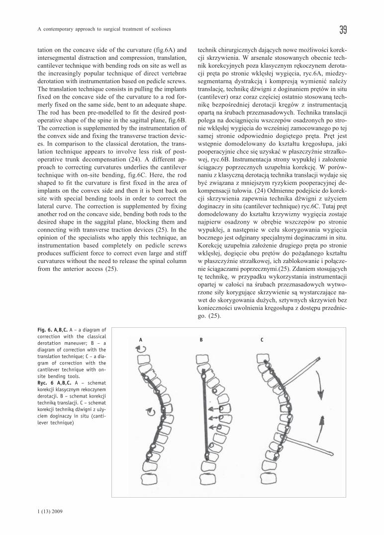
Fig. 6. A,B,C. A – a diagram ofcorrection with the classicalderotation maneuver; B – adiagram of correction with thetranslation technique; C – a dia-gram of correction with thecantilever technique with on-site bending tools.Ryc. 6 A,B,C. A – schematkorekcji klasycznym rekoczynemderotacji. B – schemat korekcjitechniką translacji. C – schematkorekcji techniką dźwigni z uży-ciem doginaczy in situ (canti-lever technique)
A B C
tation on the concave side of the curvature (fig.6A) and
intersegmental distraction and compression, translation,
cantilever technique with bending rods on site as well as
the increasingly popular technique of direct vertebrae
derotation with instrumentation based on pedicle screws.
The translation technique consists in pulling the implants
fixed on the concave side of the curvature to a rod for-
merly fixed on the same side, bent to an adequate shape.
The rod has been pre-modelled to fit the desired post-
operative shape of the spine in the sagittal plane, fig.6B.
The correction is supplemented by the instrumentation of
the convex side and fixing the transverse traction devic-
es. In comparison to the classical derotation, the trans-
lation technique appears to involve less risk of post-
operative trunk decompensation (24). A different ap-
proach to correcting curvatures underlies the cantilever
technique with on-site bending, fig.6C. Here, the rod
shaped to fit the curvature is first fixed in the area of
implants on the convex side and then it is bent back on
site with special bending tools in order to correct the
lateral curve. The correction is supplemented by fixing
another rod on the concave side, bending both rods to the
desired shape in the saggital plane, blocking them and
connecting with transverse traction devices (25). In the
opinion of the specialists who apply this technique, an
instrumentation based completely on pedicle screws
produces sufficient force to correct even large and stiff
curvatures without the need to release the spinal column
from the anterior access (25).

40 R. NOWAK
THE JOURNAL OF ORTHOPAEDICS TRAUMA SURGERY
AND RELATED RESEARCH
Zastosowania śrub przeznasadowych na całej długo-
ści instrumentacji wymaga również zdobywająca coraz
więcej zwolenników korekcja skrzywienia techniką bez-
pośredniej derotacji kręgów (DAVD – Direct Apical
Vertebra Derotation). Koncepcja tej metody oparta jest na
przeniesieniu siły odrotowującej trzony kręgowe za po-
średnictwem wprowadzonych przeznasadowo śrub. Po
wykonaniu manewru odrotowania prętów osadzonych
w kielichach śrub, moment siły derotującej skrzywienie
przykładany jest do śrub po obu stronach wygięcia po-
przez zamocowane do nich derotatory, ryc.7. Dotychcza-
sowe doniesienia wskazują, że dzięki metodzie bezpo-
średniej derotacji kręgów możliwe jest uzyskanie praw-
dziwie trójpłaszczyznowej korekcji skoliozy. (26)
Równolegle z rozwojem technik tylnej korekcji
i stabilizacji skoliozy ewoluowały metody wykorzystu-
jące przedni dostęp do kręgosłupa. Prekursorem w tej
dziedzinie był pod koniec lat sześćdziesiątych XX wie-
Fig. 7. A female patient, aged13, idiopathic scoliosis of ado-lescents, Lenke 1AN type, a cli-nical and radiological image be-fore and after surgical treat-ment with the DAVD techniquewith an application of pediclescrewsRyc. 7. Chora lat 13, skoliozaidiopatyczna dorastających, typLenke 1AN, obraz kliniczny i ra-diologiczny przed i po leczeniuoperacyjnym techniką DAVD zużyciem śrub przeznasadowych
Another technique requiring an application of pedi-
cle screws throughout the length of the instrumentation
is the direct apical vertebra derotation (DAVD), gaining
more and more supporters. The concept is based on trans-
ferring the force derotating vertebral bodies by means of
pedicle screws. After the maneuver of derotating the rods
fixed in the sockets of the screws has been performed,
the moment of the derotating force is applied to the
screws on both sides of the curvature by means of dero-
tators fastened to them, fig.7. Reports have indicated that,
thanks to the method of direct apical vertebra derotation,
truly three-dimensional correction of scoliosis can be
achieved (26).
Simultaneously with the posterior access techniques
of stabilization and correction, the techniques employing
an anterior access to the spinal column evolved as well.
A precursor in this area was in the late 1960s an Aus-
tralian orthopaedist, Dweyer (27). The essence of the

41A contemporary approach to surgical treatment of scolioses
1 (13) 2009
ku australijski ortopeda Dweyer. (27) Zasadniczą ideą
korekcji skrzywienia z dostępu przedniego było zaatako-
wanie szczytu deformacji po stronie wypukłej poprzez
skrócenie i stabilizację przedniej kolumny kręgosłupa.
W metodzie Dwyera korekcję uzyskiwano stosując kom-
presję międzytrzonową wywieraną przez naciąg wielo-
pęczkowego kabla wprowadzonego w obręb śrub zamo-
cowanych w trzonach kręgowych. Dalsza istotna ewolu-
cja instrumentarium do przedniej stabilizacji kręgosłupa
w postaci metody zaproponowanej przez Zielke polega-
ła na zastąpieniu kabla prętem i zmianie kierunku wpro-
wadzania śrub w trzony kręgowe. (28) Wśród zalet tech-
nik z przedniego dostępu do kręgosłupa wymieniano
bardzo dobrą korekcje wygięcia bocznego, większy, niż
w klasycznych technikach tylnych wpływ na rotację
osiową kręgosłupa oraz skrócenie rozległości usztywnie-
nia. (18). Wady polegały na większej liczbie zaburzeń
zrostu, problemów z destabilizacją implantów oraz częst-
szej utracie korekcji. Problemy te w znacznej mierze zni-
welowano w kolejnych generacjach implantów dzięki
konstrukcji złożonej z dwóch prętów mocowanych
w każdym z trzonów dwiema śrubami. Powstały dzięki
temu układ jest biomechanicznie bardziej stabilny i po-
zwala na lepsze odrotowanie wygięcia. Przednie dojście
do skoliozy wykorzystywane jest również w dynamicz-
nie rozwijających się w ciągu ostatnich dwudziestu lat
technikach endoskopowych. Videochirurgia torakoskopo-
wa stanowi małoinwazyjną alternatywę dla wykonywa-
nych metodą klasyczną operacji zarówno przedniego
uwolnienia kręgosłupa, jak i przedniego usztywnienia,
korekcji i stabilizacji skrzywienia. Osiągana korekcja
zniekształcenia kręgosłupa i klatki piersiowej jest po-
równywalna z uzyskiwaną klasycznymi, inwazyjnymi
metodami, natomiast zdecydowanie niższa jest utrata
krwi, lepszy wynik kosmetyczny i krótszy czas hospi-
talizacji. (29)
KLASYFIKACJA CHIRURGICZNA SKOLIOZ IDIOPATYCZ-NYCH. ROZLEGŁOŚĆ USZTYWNIENIA KRĘGOSŁUPACoraz doskonalsze metody korekcji i stabilizacji ofero-
wane przez nowe techniki operacyjne i nowe generacje
implantów wytworzyły zapotrzebowanie na systemy
klasyfikacji skrzywień idiopatycznych łączących typ
skrzywienia z planowanym postępowaniem chirurgicz-
nym, rozumianym głównie jako rozległość instrumenta-
cji i usztywnienia kręgosłupa. W 1983 roku King i wsp.,
analizując wyniki po leczeniu operacyjnym metodą
Harringtona, zaproponowali podział idiopatycznych
skrzywień w odcinku piersiowym kręgosłupa na 5 typów.
W tym podziale zasadniczymi elementami różnicujący-
mi poszczególne typy są: położenie wygięcia lędźwiowe-
go względem centralnej linii krzyżowej CSL (Central
Sacral Line) oraz giętkość wygięcia piersiowego i lę-
dźwiowego, odróżniająca typ 1 od 2. (30) Klasyfikacja
Kinga stworzyła podstawy do racjonalnego planowania
rozległości instrumentacji i spondylodezy, pozwalając
wyłonić skrzywienia w których usztywnienie można
correcting technique with an anterior access was attack-
ing the peak of the deformation on its convex side by
shortening and stabilizing the anterior spinal column. In
the Dweyer’s method, the correction was achieved by
means of compression applied between vertebral bodies
through the tension of a multi-core cable introduced in
the area of the screws fixed in vertebral bodies. A fur-
ther significant modification of the anterior access instru-
mentation was the method suggested by Zielke, where the
cable was replaced with a rod and the direction of intro-
ducing the screws into the vertebral bodies was changed
(28). The advantages of the techniques of anterior ac-
cess included: very good correction of the lateral cur-
vature, stronger effect on the axial rotation of the spine
than in the classical posterior techniques, and shorten-
ing of the extent of spondylodesis (18). Its disadvantages
lay in a higher number of adhesion disorders, problems
with the destabilization of implants, and more frequent
correction loss. The problems were alleviated to a large
extent in the next generations of implants thanks to the
construction consisting of two rods fixed with two
screws in each vertebral body. This system is more
biomechanically stable and facilitates better derotation
of the curvature. The anterior access to scoliosis is used,
as well, in the endoscopic techniques, developing dy-
namically over the last twenty years. Thoracoscopic
video-surgery poses a scarcely invasive alternative to
classical surgery of both anterior release and anterior
spondylodesis, correction, and stabilization of curva-
tures. The achieved correction of spine and chest defor-
mities is comparable to that obtained by means of the
classical, invasive methods, while the blood loss is
definitely lower, the cosmetic result is better, and the
period of hospitalization is shorter (29).
SURGICAL CLASSIFICATION OF IDIOPATHIC SCOLIOSES.THE EXTENT OF SPINAL FUSIONConstantly improved correction and stabilization meth-
ods offered by the new surgical techniques and new
generations of implants create demand for classification
systems of idiopathic scolioses where the type of curva-
ture would be linked with a planned surgical management,
understood mainly as an extent of instrumentation and
spondylodesis. In 1983, King et al., analysing the results
of surgery with the Harrington method, suggested a di-
vision of thoracic idiopathic curvatures into 5 types. The
main differentiating factors of this division are: the lo-
cation of the lumbar curve with relation to the central
sacral line (CSL) and the flexibility of the thoracic and
lumbar curve, being the difference between the type 1 and
2 (30). The King’s classification has created the basis for
rational planning of the extent of instrumentation and
spondylodesis, helping to identify the curvatures where
spondylodesis could be limited to only one curve (selec-
tive spondylodesis). Even though the King’s system is
still frequently applied, in the course of time at least three
of its limitations have become visible: it does not cover

42 R. NOWAK
THE JOURNAL OF ORTHOPAEDICS TRAUMA SURGERY
AND RELATED RESEARCH
ograniczyć jedynie do jednego z wygięć – selektywna
spondylodeza. Pomimo, iż system Kinga jest nadal chęt-
nie stosowany, to z perspektywy czasu widać, że cechu-
ją go co najmniej trzy istotne ograniczenia: nie obejmu-
je wszystkich typów skrzywień idiopatycznych, opiera się
na ocenie kształtu kręgosłupa tylko w jednej płaszczyź-
nie-czołowej i nie jest dostosowany do możliwości ko-
rekcyjnych nowych generacji implantów do trójpłaszczy-
znowej korekcji zniekształcenia kręgosłupa. Równie istot-
ne zarzuty dotyczą niewystarczającej porównywalności
i powtarzalności ocen przy kwalifikowaniu skrzywienia
do określonego typu. (31,32)
Wykorzystując obowiązującą nomenklaturę Scoliosis
Research Society Lenke i wsp. stworzyli klasyfikację
skolioz idiopatycznych uwzględniającą zmiany struktu-
ralne, rozmiary i położenie szczytów wygięć w płaszczyź-
nie czołowej, profil strzałkowy kręgosłupa i położenie
wygięcia lędźwiowego w stosunku do centralnej piono-
wej linii krzyżowej (CSVL- Central Sacral Vertical
Line).(33) Zgodnie z jasno przedstawionymi kryteriami
skrzywienie może zostać zakwalifikowane do jednego
z 6 głównych typów: typ 1 to skrzywienie główne pier-
siowe (MT - Main Thoracic), typ2 skrzywienie podwój-
ne piersiowe (DT - Double Thoracic), typ3 skrzywienie
podwójne większe (DM - Double Major), typ 4 skrzy-
wienie potrójne główne (TM - Triple Major), typ 5 skrzy-
wienie piersiowo-lędźwiowe/lędźwiowe (TL/L - Thora-
columbar/Lumbar) i typ 6 skrzywienie piersiowo-lę-
dźwiowe/lędźwiowe z głównym piersiowym (TL/L–MT
- Thoracolumbar/Lumbar-Main Thoracic), ryc. 8.
Fig. 8. Classification diagramaccording to Lenke (by permis-sion of J Bone Joint Surg, ac-cording to Lenke et al., Biblio-graphy: 33)Ryc. 8. Schemat klasyfikacji wgLenke. (za zgodą J Bone JointSurg wg Lenke G i wsp., pi-śmiennictwo poz. 33)
all the types of idiopathic curvatures, it is based on an
assessment of spinal shape in only one – frontal – plane,
and it does not match the possibilities offered by the new
generations of implants for three-dimensional correction
of spinal deformations. Equally serious objections are
raised due to unsufficient comparability and repeatabil-
ity of the assessment when a curvature is classified to a
certain type (31,32).
Making use of the valid nomenclature of the Scoli-
osis Research Society, Lenke et al. elaborated a classi-
fication of idiopathic scolioses with respect to structural
changes, size and location of curvature peaks in the fron-
tal plane, the saggital profile of the spine, and the loca-
tion of the lumbar curve in relation to the central sacral
vertical line (CSVL) (33). According to clear-cut crite-
ria, a curvature may be qualified into one of the 6 main
types: type 1 – main thoracic (MT), type 2 – double
thoracic (DT), type 3 – double major (DM), type 4 – triple
major (TM), type 5 – thoracolumbar/lumbar (TL/L), type
6 – thoracolumbar/lumbar with main thoracic (TL/L-MT),
fig.8.
43A contemporary approach to surgical treatment of scolioses
1 (13) 2009
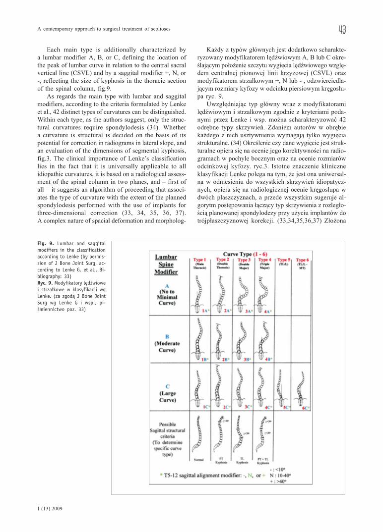
Każdy z typów głównych jest dodatkowo scharakte-
ryzowany modyfikatorem lędźwiowym A, B lub C okre-
ślającym położenie szczytu wygięcia lędźwiowego wzglę-
dem centralnej pionowej linii krzyżowej (CSVL) oraz
modyfikatorem strzałkowym +, N lub - , odzwierciedla-
jącym rozmiary kyfozy w odcinku piersiowym kręgosłu-
pa ryc. 9.
Uwzględniając typ główny wraz z modyfikatorami
lędźwiowym i strzałkowym zgodnie z kryteriami poda-
nymi przez Lenke i wsp. można scharakteryzować 42
odrębne typy skrzywień. Zdaniem autorów w obrębie
każdego z nich usztywnienia wymagają tylko wygięcia
strukturalne. (34) Określenie czy dane wygięcie jest struk-
turalne opiera się na ocenie jego korektywności na radio-
gramach w pochyle bocznym oraz na ocenie rozmiarów
odcinkowej kyfozy. ryc.3. Istotne znaczenie kliniczne
klasyfikacji Lenke polega na tym, że jest ona uniwersal-
na w odniesieniu do wszystkich skrzywień idiopatycz-
nych, opiera się na radiologicznej ocenie kręgosłupa w
dwóch płaszczyznach, a przede wszystkim sugeruje al-
gorytm postępowania łączący typ skrzywienia z rozległo-
ścią planowanej spondylodezy przy użyciu implantów do
trójpłaszczyznowej korekcji. (33,34,35,36,37) Złożona
Fig. 9. Lumbar and saggitalmodifiers in the classificationaccording to Lenke (by permis-sion of J Bone Joint Surg, ac-cording to Lenke G. et al., Bi-bliography: 33)Ryc. 9. Modyfikatory lędźwiowei strzałkowe w klasyfikacji wgLenke. (za zgodą J Bone JointSurg wg Lenke G i wsp., pi-śmiennictwo poz. 33)
Each main type is additionally characterized by
a lumbar modifier A, B, or C, defining the location of
the peak of lumbar curve in relation to the central sacral
vertical line (CSVL) and by a saggital modifier +, N, or
-, reflecting the size of kyphosis in the thoracic section
of the spinal column, fig.9.
As regards the main type with lumbar and saggital
modifiers, according to the criteria formulated by Lenke
et al., 42 distinct types of curvatures can be distinguished.
Within each type, as the authors suggest, only the struc-
tural curvatures require spondylodesis (34). Whether
a curvature is structural is decided on the basis of its
potential for correction in radiograms in lateral slope, and
an evaluation of the dimensions of segmental kyphosis,
fig.3. The clinical importance of Lenke’s classification
lies in the fact that it is universally applicable to all
idiopathic curvatures, it is based on a radiological assess-
ment of the spinal column in two planes, and – first of
all – it suggests an algorithm of proceeding that associ-
ates the type of curvature with the extent of the planned
spondylodesis performed with the use of implants for
three-dimensional correction (33, 34, 35, 36, 37).
A complex nature of spacial deformation and morpholog-

44 R. NOWAK
THE JOURNAL OF ORTHOPAEDICS TRAUMA SURGERY
AND RELATED RESEARCH
natura przestrzennej deformacji i zróżnicowanie morfo-
logiczne skolioz w połączeniu ze złożonością dostępnych
systemów i technik korekcyjnych wpływają na znaczne
zróżnicowanie poglądów dotyczących zarówno wyboru
kręgów granicznych spondylodezy, jaki i optymalnej
instrumentacji. W opublikowanej niedawno pracy Robi-
taille i wsp. analizując propozycje 32 doświadczonych
chirurgów kręgosłupowych dotyczące strategii operacyj-
nej w odniesieniu do 5 przypadków skolioz, stwierdzili
wśród 160 uzyskanych odpowiedzi (32 chirurgów oce-
niających 5 przypadków) 156 odrębnych, niejednokrot-
nie diametralnie różniących się od siebie planów opera-
cyjnych. (38) Należy tutaj podkreślić, że w procesie pla-
nowania operacji korekcji skrzywienia kręgosłupa chirurg
musi brać pod uwagę wiele czynników odnoszących się
do charakterystyki chorego (kształt skrzywienia, kompen-
sacja tułowia, giętkość kręgosłupa, stan neurologiczny,
zniekształcenie klatki piersiowej, dojrzałość szkieletowa,
pozostały potencjał wzrostowy), instrumentacji (typ,
lokalizacja, liczba i rodzaj implantów-haki/śruby, rozle-
głość stabilizacji, średnica, długość i sposób domodelo-
wania prętów kręgosłupowych, kolejność przeprowadza-
nia instrumentacji, stosowane w czasie operacji technika
korekcji skrzywienia). (39) Istotne znaczenie wymagające
uwzględnienia odgrywają również takie czynniki jak:
duża utrata krwi, stan tkanki kostnej chorego, wybór
materiału do przeszczepu kostnego, ryzyko powikłań
neurologicznych oraz sposób postępowania pooperacyj-
nego.
WNIOSKIPomimo olbrzymich postępów w chirurgii kręgosłupa
jakich dokonano w ciągu ostatnich stu lat główny cel
leczenia chirurgicznego skoliozy pozostał ten sam: opty-
malna i trwała korekcja deformacji przy jak najmniejszej
liczbie powikłań.
Ewolucja technik operacyjnej korekcji i stabilizacji
skolioz od instrumentarium Harringtona do segmentarnej
instrumentacji śrubami przeznasadowymi obrazuje postę-
py na drodze od jedno do wielopłaszczyznowej korekcji
skoliotycznego zniekształcenia.
Zróżnicowanie kliniczne skolioz oraz liczne kontro-
wersje dotyczące dostępu operacyjnego, rozległości
usztywnienia, doboru implantów oraz wyboru metody
korekcji ograniczają możliwość wprowadzenia uniwersal-
nego algorytmu leczenia operacyjnego.
ical diversity of scolioses in combination with the com-
plexity of available systems and techniques of correction
result in a great variety of opinions concerning both the
choice of the border vertebra in spondylodesis and its
optimal instrumentation. Robitaille et al., in their recent-
ly published study, analysed suggestions of 32 experi-
enced spinal surgeons with respect to the surgical strat-
egy regarding 5 cases of scoliosis, and found out that
among the 160 answers (32 surgeons evaluating 5 cases)
there were 156 distinct, sometimes diametrically differ-
ent, operation plans (38). It needs emphasizing that, when
planning a spinal correction surgery, the surgeon must
take into account numerous factors concerning the pa-
tient’s characteristics (shape of the curvature, trunk com-
pensation, flexibility of the spine, neurological condition,
chest deformity, skeletal maturity, the remaining growth
potential), instrumentation (type, location, number and
sort of the implants – hooks / screws, the extent of sta-
bilization, the diameter, length, and manner of spinal rod
modelling, the sequence of instrumentation, the technique
of curvature correction applied in the surgery) (39). Other
relevant factors which must be taken into account include:
a significant loss of blood, the condition of the patient’s
bone tissue, the choice of material for bone graft, the risk
of neurological complications, and the kind of post-op-
erative procedure.
CONCLUSIONSIn spite of enormous progress in spinal surgery that has
been made over the last hundred years, the main objec-
tive of surgical treatment of scoliosis has remained the
same: an optimal and permanent correction of the defor-
mation with the least possible complications.
The evolution of surgical techniques of correction and
stabilization of scoliosis from Harrington instruments to
segmental instrumentation with pedicle screws is an
indication of the progress from single-plane to multi-plane
correction of scoliotic deformations.
Clinical differentiation of various types of scoliosis,
as well as numerous controversies concerning the surgi-
cal access, the extent of spondylodesis, the choice of
implants and of correction methods, limit the possibility
of introducing a universal algorithm of surgical treatment.

45A contemporary approach to surgical treatment of scolioses
1 (13) 2009
References/Piśmiennictwo:
1. Mehlman C.T. Idiopathic scoliosis. http://www.emedicine.com/
orthoped/topic504.htm
2. Bunnel W.P. The natural history of idiopathic scoliosis. Cl Or-
thop Rel Res, 1988, 229; 20-25
3. Asher M.A., Burton D.C. Adolescent idiopathic scoliosis: natu-
ral history and long term treatment effects. Scoliosis 2006; 1:2.
http://www.scoliosisjournal.com/content/1/1/2
4. Fernandes P., Weinstein S.L. Natural history of early onset sco-
liosis. J Bone Joint Surg 2007; 89-A: S21-S33
5. Lonstein J.E., Carlson J.M. The prediction of curve progression
in untreated idiopathic scoliosis during growth. J Bone Joint Surg
1984; 66-A: 1061-1071
6. Akbarnia B.A. Management themes in early onset scoliosis. J
Bone Joint Surg 2007; 89-A: S42-S54
7. Robinson C.M., McMaster M.J. Juvenile idiopathic scoliosis.
Curve patterns and prognosis in one hundred and nine patients.
J Bone Joint Surg 1996; 78-A: 1140-1148
8. Weinstein S.L. Natural history. Spine 1999; 24: 2592-2600
9. Weinstein S.L., Dolan L.A., Spratt K.F., Peterson K.K., Spoona-
more M.J., Ponseti I.V. Health and function of patients with untre-
ated idiopathic scoliosis. A 50-year natural history study. JAMA
2003; 289: 559-567
10. Weinstein S.L., Ponseti I.V. Curve progression in idiopathic sco-
liosis. J Bone Joint Surg 1983; 65-A: 447-455.
11. Tolo V.T., Gillespie R. The characteristics of juvenile idiopathic
scoliosis and results of its treatment. J Bone Joint Surg 1978; 60-
B: 181-188
12. Figueiredo U.M., James J.I.P. Juvenile idiopathic scoliosis. J
Bone Joint Surg 1981; 63-B: 61-66
13. Charles Y.P., Daures J., Rosa V., Dimeglio A. Progression risk
of idiopathic juvenile scoliosis during pubertal growth. Spine
2006; 31: 1933-1942
14. Thompson G.H., Lenke L.G., Akbarnia B.A., McCarthy R.E.,
Campbell R.M. Early onset scoliosis: Future directions. J Bone
Joint Surg 2007; 89-A: S163-S166
15. Canale T, Beaty J. Campbell’s Operative Orthopaedics. Mosby
2007, 11 rev. ed. Vol II, part XII
16. Lenke L., Dobbs M.B. Management of juvenile idiopathic sco-
liosis. J Bone Joint Surg 2007; 89-A: S55-S63
17. Akbarnia B.A. Management themes in early onset scoliosis. J
Bone Joint Surg 2007; 89-A: S42-S54
18. Mohan A, Kaushik D. History of surgery for correction of spinal
deformity. Neurosurg Focus. 2003; 14 (1): http://www.aans.org/
education/journal/neurosurgical/jan03/14-1-1.pdf
19. Drummond D. A perspective on recent trends for scoliosis cor-
rection. Clin Orthop Rel Res. 1991; 264: 90-102
20. Wenger D., Carollo J., Wilkerson J. Biomechanics of scoliosis
correction by segmental spinal instrumentation. Spine 1982; 7:
260-264
21. Phillips W., Hensinger R. Wisconsin and other instrumentation
for posterior spinal fusion. Clin Orthop Rel Res 1988; 229: 44-
51
22. Drummond D, Keene J., Breed A. The Wisconsin System: A
technique of interspinous segmental spinal instrumentation. Con-
temporary Orthopaedics 1984; 8: 29-37
23. Cotrel Y, Dubousset J. Novelle technique d’osteosynthese rachi-
dienne segmentaire par voie posterieure. Rev Chir Orthop 1984;
70: 489-494.
24. Muschik M, Schlentzka D, Robinson P, Kupferschmidt C. Dorsal
instrumentation for adolescent thoracic scoliosis: rod rotation
versus translation. Eur Spine J 1999; 8: 93-99
25. Chang K. Cantilever bending technique for treatment of large and
rigid scoliosis. Spine 2003; 28: 2452-2458
26. Lee S, Suk S, Chung E. Direct vertebral rotation: A new tech-
nique of three-dimensional deformity correction with segmental
pedicle screw fixation in adolescent idiopathic scoliosis. Spine
2004; 29: 343-349
27. Dwyer A., Schafer M. Anterior approach to scoliosis. Results of
treatment in fifty-one cases. J Bone Joint Surg 1974; 56B: 218-224
28. Kostiuk J, Ferron S. Anterior Zielke instrumentation for spinal
deformity in adults. J Bone Joint Surg 1989; 71A: 898-912
29. Gatehouse S, Izatt M, Adam J i wsp. Perioperative aspects of
endoscopic anterior scoliosis surgery: the learning curve for a
consecutive series of 100 patients. J Spinal Disord Tech 2007;
20:317-323
30. King H.A., Moe J.H., Bradford D.S., Winter R.B. The selection
of fusion levels in thoracic idiopathic scoliosis. J Bone Joint Surg
1983; 65-A: 1302-1313
31. Lenke L.G., Randal R.B., Bridwell K.H. i wsp. Intraobserver and
interobserver reliability of the classification of thoracic adole-
scent idiopathic scoliosis. J Bone Joint Surg 1998; 80-A: 1097-
1106.
32. Cummings J.R., Loveless E.A., Campbell J., Samelson S., Mazur
J.M. Interobserver reliability and intraobserver reproducibility
of the system of King et al. for the classification of adolescent
idiopathic scoliosis. J Bone Joint Surg 1998; 80-A: 1107-1111
33. Lenke L.G., Betz R.R., Harms J. i wsp. Adolescent idiopathic
scoliosis. A new classification to determine extent of spinal
arthrodesis. J Bone Joint Surg 2001; 83-A; 8: 1169-1181
34. Lenke L.G., Betz R.R., Clements D. i wsp. Curve prevalence of
a new classification of operative adolescent idiopathic scoliosis.
Does classification correlate with treatment? Spine 2002; 27:
604-611.
35. Lenke L.G., Betz R.R., Haher T.R. i wsp. Multisurgeon assessment
of surgical decision-making in adolescent idiopathic scoliosis.
Curve classification, operative approach and fusion levels. Spi-
ne 2001; 21: 2347-2353
36. Lenke L.G., Edwards C.C., Bridwell K.H. The Lenke classifica-
tion of adolescent idiopathic scoliosis: How it organizes curve
patterns as a template to perform selective fusions of the spine.
Spine 2003; 28: S199-S207
37. Puno R.M, An K., Puno R.L., Jacob A., Chung S. Treatment
recomendations for idiopathic scoliosis: an assessment of Lenke
classification. Spine 2003; 28: 2102-2115
38. Robitaille M, Aubin C, Labelle H. Intra and interobserver varia-
bility of preoperative planning for surgical instrumentation in
adolescent idiopathic scoliosis. Eur Spine J 2007; 16: 1604-1614
39. Aubin C, Labelle H, Ciolofan O. Variability of spinal instrumen-
tation configurations in adolescent idiopathic scoliosis. Eur Spine
J 2007; 16: 57-64


