
1031
MD4
Chukwuka Okwu
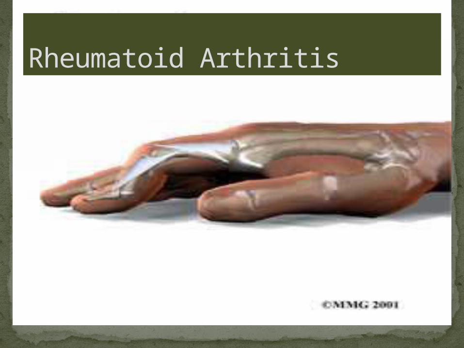
Rheumatoid Arthritis

Jane is a 45 year old woman, who presents to her doctor complaining of morning stiffness of the joints of her fingers for several weeks now with pain. There is also a swelling of her knee joints. She says the pain Improves as she does her daily chores but Tylenol is ineffective in alleviating the pain.
On inspection subcutaneous nodules are found behind the olecranon process with a cyst found on the popliteal fossa.
What are the differentials for this patient?

Presents from early childhood(rare) to late old age.
Most commonly 30-50 years.Worldwide distribution. 0.5-3% of the
populationWomen before menopause are affected more
than men (3:1)
Etiology

HLA DR4 occurs in 50-75% of patientsPresence of pentapeptide QK/RAA in 3rd
allelic hypervariable region of HLA-DRB1 increases susceptibility
Positive rheumatoid factor and anti-citrullinated protein antigen(more specific for RA)
When rheumatoid factor is present with HLA DR4, susceptibility increases 13 times. This rheumatoid factor is typically of IgM type, and is an auto-antibody to the Fc portion of IgG.
Molecular Biology

Inflammation of the synovial lining of joints, tendon sheats & bursae-synovitis
Synovium becomes greatly thickened, with proliferation & infilteration by inflammatory cells(lymphocytes and macrophages)
Hyperplastic synovium(pannus) spreads to cartilage surface & damages the underlying cartilage by blocking its normal routes of nutrition & by effects of cytokines on the chondrocytes.
Fibroblasts from the proliferating synovium also grow along the course of blood vessels between the synovial margins and epiphyseal bone cavity and damage the bone.
Pathogenesis


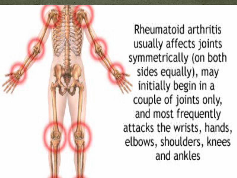
Pain and stiffness of the small joints of the hands(MCP,PIP) and feet(MTP)
Wrist, elbow, shoulders, knees and ankles are also affected
Carpal tunnel syndromePain & stiffness worse in the morning,
improves with activity
Clinical Features

Morning stiffness>1 hourArthritis of three or more joints 6
weeks or moreArthritis of hand joints and wristsSymmetrical arthritisSubcutaneous nodulesPositive serum rheumatoid factorTypical radiological changes(e.g. Erosions,
loss of joint space)Four or more criteria necessary for diagnosis
Diagnosis-American college of rheumatology

Blood count-ESR & CRP are raisedSerology-Rheumatoid factor present in 70%
of cases, anti-citrullinated protein antigen(more specifc, 85% of cases)
Aspiration of the joint-the aspirate looks cloudy owing to white cells
X-ray-Boutonniere deformity, ulnar deviation of fingers
Investigations

Rose weeler test(most specific): ability of IGM rheumatoid factor to agglutinate sheep red cells that have been coated with rabbit anti-sheep antibody.
Latex agglutination test: rheumatoid factor agglutinates latex particles that have been coated with human IgG
Tests

No curative agent for Rheumatoid ArthritisUse NSAIDS & analgesics to control
symptomsIf synovitis recurs, refer to a rheumatologist
to start DMARDs like Methotrexate, sulfasalazine(cytokine inhibition)
Other drugs-Gold(sodium aurothiomalate), corticosteroids, anti-malarials
If no improvement, consider anti TNF-α(infliximab)
Physiotherapy has been found to be very helpful in slowing progression
Treatment

Septic Arthritis-Staphylococcus AureusAmyloidosisCaplan syndrome(coal miners
lung+rheumatoid factor)Interstitial pneumonitis + fibrosing alveolitisAnemia of chronic disorders
complications

Sarah is a 64 year old lady. She was diagnosed with Rheumatoid Arthritis 6 years ago. Her Rheumatoid arthritis was initially treated with methotrexate and sulfalazine which brought the disease under control. Recently Sarah has had several flares and this has resulted in several spells of corticosteroid therapy(among other treatment). Sarah later suffers a prolonged flare which doesn’t respond well to synthetic DMARD treatment. Her rheumatologist suggests commencing treatment with infliximab in addition to methotrexate. What risk associated with biological drug treatment should you be particularly aware?
Case Study

Kumar and clark clinical medicinewww.nhs.co.ukRobbins textbook of pathologywww.umm.edu
References


