Download - 1-s2.0-S0896627311009184-main

Neuron
Review
Neuroplasticity Subserving Motor Skill Learning
Eran Dayan1 and Leonardo G. Cohen1,*1Human Cortical Physiology and Stroke Neurorehabilitation Section, National Institute of Neurological Disorders and Stroke,National Institutes of Health, Bethesda, MD 20892-1428, USA*Correspondence: [email protected] 10.1016/j.neuron.2011.10.008
Recent years have seen significant progress in our understanding of the neural substrates of motor skilllearning. Advances in neuroimaging provide new insight into functional reorganization associated with theacquisition, consolidation, and retention of motor skills. Plastic changes involving structural reorganizationin gray and white matter architecture that occur over shorter time periods than previously thought havebeen documented as well. Data from experimental animals provided crucial information on plausible cellularand molecular substrates contributing to brain reorganization underlying skill acquisition in humans. Here,we review findings demonstrating functional and structural plasticity across different spatial and temporalscales that mediate motor skill learning while identifying converging areas of interest and possible avenuesfor future research.
IntroductionThe acquisition and long-term retention of motor skills play
a fundamental role in our daily lives. Skills such as writing, play-
ing golf, or riding a bicycle are all acquired through repetitive
practice. Motor skill learning refers to the process by which
movements are executed more quickly and accurately with
practice (Willingham, 1998). Our understanding of the neural
substrates underlying the acquisition and retention of motor
skills has been boosted in recent years, owing in a large part to
technological and methodological advances in neuroimaging,
as well as in noninvasive brain stimulation in humans, coupled
with dramatic new insights emerging from animal studies
both in vivo and in vitro, providing additional information about
the recruitment of specific neuronal circuits during the various
stages of motor skill learning. This work has overall demon-
strated a strong link between acquisition of motor skills and
neuronal plasticity at cortical and subcortical levels in the
central nervous system that evolves over time and engages
different spatially distributed interconnected brain regions.
Here, we review novel findings reflecting functional and struc-
tural plasticity associated with the acquisition, consolidation,
and long-term retention of motor skills in humans and experi-
mental animals while identifying points of convergence and
dispute.
A variety of tasks and experimental paradigms have been used
for studying motor skill learning, including juggling, visuomotor
tracking, and isometric force-production tasks, to name a few.
Of particular relevance to the current review are studies of tasks
that require practice of sequential movements: tapping skills like
typing or playing various musical instruments. Here, our main
focus is on learning sequential motor skills that show lasting
improvements beyond baseline performance over lengthy
periods of time. Another model for studying motor learning,
which does not necessarily involve the acquisition of a new skill,
has been adaptation to externally induced perturbations, such
as those induced by a force field (dynamic adaptation) or by
visuomotor rotations (visuomotor adaptation). These perturba-
tions are more commonly introduced while subjects execute
simple motor tasks, for instance, point-to-point ballistic reaching
movements (Krakauer, 2009; Shadmehr et al., 2010; Seidler,
2010; Lalazar and Vaadia, 2008). Yet these paradigms charac-
teristically evaluate the return to baseline levels of performance
following perturbation over relatively short time periods (Kraka-
uer and Mazzoni, 2011). However, it should be noted that repet-
itive practice of adaptation tasks could lead to performance
improvements over time in the form of ‘‘savings,’’ expressed
as faster readaptation to external perturbations relative to the
initial rate of adaptation (e.g., Landi et al., 2011). Moreover, skill
learning tasks, in which lasting improvements are seen over time,
for instance whole-body balancing (Taubert et al., 2010), may
involve an adaptation component.
Motor skills are typically learned slowly over multiple training
sessions until performance reaches nearly asymptotic levels.
Across different experimental paradigms, skill acquisition
develops (Figure 1A) initially relatively fast (i.e., rapid improve-
ments measured over the course of a single training session)
and later more slowly, when further gains develop incrementally
over multiple sessions of practice (Doyon and Benali, 2005;
Doyon and Ungerleider, 2002). Of note, the relative duration of
what can be defined as fast and slow learning is highly task
specific. For example, the fast stage of learning a simple four-
component key-press sequence could last minutes (e.g., Karni
et al., 1995), whereas the fast stage of learning to play a complex
musical piece may last months (Figure 1B). Similarly, nearly
asymptotic levels in end-point measures of skill can be acquired
very rapidly when learning a key-press sequence but muchmore
slowly when learning to play a complex musical piece. Skill
changes can occur during training (online) but also after training
ended (offline; Figure 1C). Offline processes, including skill stabi-
lization and improvement (Fischer et al., 2005; Korman et al.,
2003; Walker et al., 2002), reflect motor memory consolidation
(Doyon and Benali, 2005; Muellbacher et al., 2002; Robertson
et al., 2004a), an intermediate stage between fast and slow
learning (Doyon and Benali, 2005; Doyon et al., 2009a). Online
and offline skill gains can be maintained over time, resulting in
long-term retention (Romano et al., 2010).
Neuron 72, November 3, 2011 ª2011 Elsevier Inc. 443
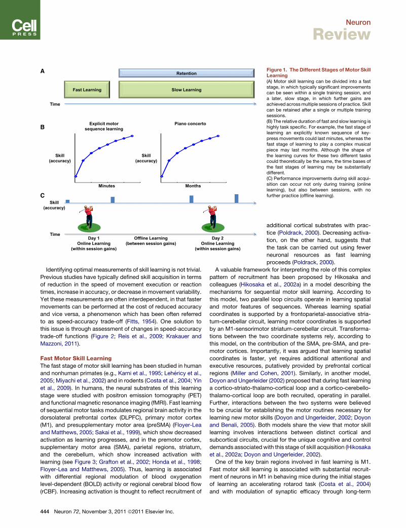
A
B
C
Figure 1. The Different Stages of Motor SkillLearning(A) Motor skill learning can be divided into a faststage, in which typically significant improvementscan be seen within a single training session, anda later, slow stage, in which further gains areachieved acrossmultiple sessions of practice. Skillcan be retained after a single or multiple trainingsessions.(B) The relative duration of fast and slow learning ishighly task specific. For example, the fast stage oflearning an explicitly known sequence of key-press movements could last minutes, whereas thefast stage of learning to play a complex musicalpiece may last months. Although the shape ofthe learning curves for these two different taskscould theoretically be the same, the time bases ofthe fast stages of learning may be substantiallydifferent.(C) Performance improvements during skill acqui-sition can occur not only during training (onlinelearning), but also between sessions, with nofurther practice (offline learning).
Neuron
Review
Identifying optimal measurements of skill learning is not trivial.
Previous studies have typically defined skill acquisition in terms
of reduction in the speed of movement execution or reaction
times, increase in accuracy, or decrease in movement variability.
Yet these measurements are often interdependent, in that faster
movements can be performed at the cost of reduced accuracy
and vice versa, a phenomenon which has been often referred
to as speed-accuracy trade-off (Fitts, 1954). One solution to
this issue is through assessment of changes in speed-accuracy
trade-off functions (Figure 2; Reis et al., 2009; Krakauer and
Mazzoni, 2011).
Fast Motor Skill LearningThe fast stage of motor skill learning has been studied in human
and nonhuman primates (e.g., Karni et al., 1995; Lehericy et al.,
2005; Miyachi et al., 2002) and in rodents (Costa et al., 2004; Yin
et al., 2009). In humans, the neural substrates of this learning
stage were studied with positron emission tomography (PET)
and functional magnetic resonance imaging (fMRI). Fast learning
of sequential motor tasks modulates regional brain activity in the
dorsolateral prefrontal cortex (DLPFC), primary motor cortex
(M1), and presupplementary motor area (preSMA) (Floyer-Lea
and Matthews, 2005; Sakai et al., 1999), which show decreased
activation as learning progresses, and in the premotor cortex,
supplementary motor area (SMA), parietal regions, striatum,
and the cerebellum, which show increased activation with
learning (see Figure 3; Grafton et al., 2002; Honda et al., 1998;
Floyer-Lea and Matthews, 2005). Thus, learning is associated
with differential regional modulation of blood oxygenation
level-dependent (BOLD) activity or regional cerebral blood flow
(rCBF). Increasing activation is thought to reflect recruitment of
444 Neuron 72, November 3, 2011 ª2011 Elsevier Inc.
additional cortical substrates with prac-
tice (Poldrack, 2000). Decreasing activa-
tion, on the other hand, suggests that
the task can be carried out using fewer
neuronal resources as fast learning
proceeds (Poldrack, 2000).
A valuable framework for interpreting the role of this complex
pattern of recruitment has been proposed by Hikosaka and
colleagues (Hikosaka et al., 2002a) in a model describing the
mechanisms for sequential motor skill learning. According to
this model, two parallel loop circuits operate in learning spatial
and motor features of sequences. Whereas learning spatial
coordinates is supported by a frontoparietal-associative stria-
tum-cerebellar circuit, learning motor coordinates is supported
by an M1-sensorimotor striatum-cerebellar circuit. Transforma-
tions between the two coordinate systems rely, according to
this model, on the contribution of the SMA, pre-SMA, and pre-
motor cortices. Importantly, it was argued that learning spatial
coordinates is faster, yet requires additional attentional and
executive resources, putatively provided by prefrontal cortical
regions (Miller and Cohen, 2001). Similarly, in another model,
Doyon and Ungerleider (2002) proposed that during fast learning
a cortico-striato-thalamo-cortical loop and a cortico-cerebello-
thalamo-cortical loop are both recruited, operating in parallel.
Further, interactions between the two systems were believed
to be crucial for establishing the motor routines necessary for
learning new motor skills (Doyon and Ungerleider, 2002; Doyon
and Benali, 2005). Both models share the view that motor skill
learning involves interactions between distinct cortical and
subcortical circuits, crucial for the unique cognitive and control
demands associated with this stage of skill acquisition (Hikosaka
et al., 2002a; Doyon and Ungerleider, 2002).
One of the key brain regions involved in fast learning is M1.
Fast motor skill learning is associated with substantial recruit-
ment of neurons in M1 in behaving mice during the initial stages
of learning an accelerating rotarod task (Costa et al., 2004)
and with modulation of synaptic efficacy through long-term

Figure 2. Shifts in Speed-Accuracy Response Functionsas a Measure of Skill(A) Simulated learning curve, in which performance improvements weredefined in terms of speed. Thus, performance at time point t2 shows clearimprovements relative to performance at time point t1.(B) Inspecting the task’s speed-accuracy response function reveals that theseperformance changes may reflect sampling of two points along the samefunction, thus simply reflecting a switch from movements that are relativelyslow but accurate to movements that are relatively fast but inaccurate.(C) A more reliable measure for skill acquisition may estimate whether learningwas associated with a shift in the speed-accuracy responses, from the blue tothe red function.
Neuron
Review
potentiation (LTP) and long-term depression (LTD) in rodents
(Rioult-Pedotti et al., 1998, 2000). Consistently, by utilizing trans-
cranial magnetic stimulation (TMS), it was shown in humans that
learning a motor task modulates LTP-like plasticity (Ziemann
et al., 2004; Stefan et al., 2006; Rosenkranz et al., 2007). BOLD
activity in M1 progressively decreases as motor skill learning
progresses over a single training session (Karni et al., 1995),
yet it should be noted that the magnitude of engagement of
M1 in fast learning is highly influenced by the specific task and
by attentional demands (Hazeltine et al., 1997; Stefan et al.,
2004). Consistent reorganizational changes in M1 have been
described using TMS. For example, the fast stage of implicit
motor skill learning, as assessed with the serial reaction time
task, is accompanied by increased motor map size of the fingers
engaged in the task. Interestingly, when the sequence becomes
explicitly known, the M1 motor map size returns to baseline
(Pascual-Leone et al., 1994). The cellular mechanisms behind
learning-related plasticity in M1 appear to depend on protein
synthesis within this structure and may specifically involve
brain-derived neurotrophic factor (BDNF; Kleim et al., 2003). In
both humans and animal models, BDNF influences synaptic
plasticity (Akaneya et al., 1997; Lu, 2003). Injection of protein
synthesis inhibitors targeting BDNF into the rat M1 induces
a lasting loss of motor map representation (Kleim et al., 2003).
Moreover, training-dependent increases in motor cortical excit-
ability (Antal et al., 2010; Cheeran et al., 2009) and fMRI signal
(McHughen et al., 2010) are reduced in healthy humans with
a valine-to-methionine substitution at codon 66 (Val66Met) in
the BDNF gene, when compared to subjects without this poly-
morphism (Kleim et al., 2006). These findings led to the hypoth-
esis that the presence of this particular polymorphism could
influence motor skill learning (Fritsch et al., 2010).
Although earlier imaging studies clearly established that the
fast stage of motor skill learning is sustained by activity across
a distributed set of brain regions, conventional univariate fMRI
analysis, in which brain activity is analyzed in a voxel-wise
manner as if each anatomically distinguishable region is inde-
pendent (Marrelec et al., 2006; Tamas Kincses et al., 2008),
does not provide information on interregional interactions that
are required to properly test these models. The most widely
used and straightforward approach for assessing interregional
interactions in neuroimaging data is based on analysis of
functional connectivity (Friston, 1994), which refers to the statis-
tical dependence defined in terms of correlation or covariance
between the activation in spatially remote regions. Using this
approach, it was shown that M1, the premotor cortex, and the
SMA have significantly greater inter- and intrahemispheric
coupling during early, as compared to late, within-session
explicit sequence learning (Sun et al., 2007). Interactions
between M1, SMA, and premotor cortices are likely to reflect
transformations between spatial and motor features of motor
sequences required for fast motor skill learning (Hikosaka
et al., 2002a). Additionally, fast motor skill learning is character-
ized by increased functional connectivity between the DLPFC
and premotor cortex (Sun et al., 2007), relating to the heightened
attentional demands required at this stage of skill acquisition
(Hikosaka et al., 2002a; Petersen et al., 1998).
Additional information on network-level functional reorganiza-
tion mediating fast learning emerged from data-driven model-
free analytical approaches, such as independent component
analysis (ICA), that do not assume prior knowledge of activation
changes (Marrelec et al., 2006). Using this approach, a recent
study characterized two networks involved in fast learning
(Tamas Kincses et al., 2008): (1) an M1-premotor-parietal-cere-
bellar circuit that shows reduction of fMRI activity as learning
progressed, consistent with a developing ability of the network
to economize resources often seen during motor practice (Kelly
and Garavan, 2005; Petersen et al., 1998) and (2) a posterior
parietal-premotor circuit that shows increasing fMRI activity
that correlates with behavioral gains, which may be consistent
with the engagement of spatial processing resources required
for the task (Tamas Kincses et al., 2008; Hikosaka et al.,
2002a). Overall, studies employing functional connectivity anal-
ysis, bothmodel-driven andmodel-free, provided clear evidence
for the reorganization of cortico-cortical and cortico-cerebellar
circuits in fast learning, a pattern of functional plasticity that
is in agreement with previously proposed models (Hikosaka
et al., 2002a; Doyon and Ungerleider, 2002; Doyon and Benali,
2005; see above). On the other hand, functional connectivity
evidence for cortico-striatal interactions as proposed in these
models is currently lacking. Accurate characterization of
Neuron 72, November 3, 2011 ª2011 Elsevier Inc. 445

Figure 3. Neural Substrates of Fast Motor SkillLearningSchematic depiction of the major brain regions recruitedduring the initial stages of motor skill learning, as identifiedusing fMRIandPET:dorsolateral prefrontal cortex (DLPFC),primary motor cortex (M1), premotor cortex (PM), supple-mentary motor area (SMA), presupplementary motor area(preSMA), posterior parietal cortex (PPC), dorsomedialstriatum (DMS), and posterior cerebellum. The arrowsdepict documented increases or decreases in activationassociated with fast skill learning. Inflated cortical andcerebellar surfaces were rendered using CARET (http://brainvis.wustl.edu/wiki/index.php/Main_Page).
Neuron
Review
cortico-striatal interactions during fast learning is likely to benefit
from hypothesis-driven experimental approaches that focus on
these regions (e.g., Di Martino et al., 2008).
Slow Motor Skill LearningBehavioral gains in later stages of motor skill learning are usually
quantitatively smaller than those observed during fast learning
and develop at a slower pace (Doyon and Benali, 2005; Karni
et al., 1995; Ungerleider et al., 2002). The magnitude of changes
and the time course of slow learning are task dependent.
They differ substantially when learning a simple motor sequence
in which performance rapidly reaches near-asymptote levels
and when learning, for example, to play musical pieces on
a violin, in which case performance improvements continue
over many years. It has been proposed that under certain condi-
tions, performance may become automatic, implying lesser
involvement of attentional and executive resources and lesser
susceptibility to interference by a secondary process or task
(Schneider and Shiffrin, 1977; Ashby et al., 2010; Doyon and
Benali, 2005).
Studies that examined the neuronal mechanisms involved
in the slow stage of motor skill learning typically had subjects
learn a motor skill over several weeks and scanned them on
different occasions throughout the training period (Karni et al.,
1995; Floyer-Lea and Matthews, 2005; Coynel et al., 2010;
Lehericy et al., 2005). Slow learning is associated with increased
activation in M1 (Floyer-Lea and Matthews, 2005), primary
somatosensory cortex (Floyer-Lea and Matthews, 2005), SMA
(Lehericy et al., 2005), and putamen (Lehericy et al., 2005;
Floyer-Lea andMatthews, 2005), aswell as decreased activation
in lobule VI of the cerebellum (Figure 4; Lehericy et al., 2005).
446 Neuron 72, November 3, 2011 ª2011 Elsevier Inc.
Thus, progress from early to late stages ofmotor
skill learning is characterized by a shift in fMRI
activation from anterior to more posterior
regions of the brain (Floyer-Lea and Matthews,
2005), a pattern also reported when learning
nonmotor tasks, which is thought to reflect
a progressive decrease in reliance on attentional
resources and executive function (Kelly and
Garavan, 2005). Progressing from fast to
slow motor skill learning is also associated
with a shift in fMRI activation from associative
to sensorimotor striatum (Coynel et al., 2010;
Lehericy et al., 2005), thought to contribute to
slow learning of the motor component of sequences (Hikosaka
et al., 2002a).
Slow learning has been linked with larger-scale functional
reorganization aswell. A recent study tracked functional connec-
tivity using fMRI over a period of 4 weeks of training on an explicit
motor sequence task (Coynel et al., 2010). Early learning was
associated with increased integration, a metric reflecting func-
tional interactions among several brain regions, of a premotor-
associative striatum-cerebellar network. During slow learning,
on the other hand, the authors reported decreased integration
in this premotor-associative striatum-cerebellar network but
stable connectivity within the M1-sensorimotor striatum-
cerebellar network, largely consistent with data emerging from
regional fMRI analysis (Floyer-Lea andMatthews, 2005; Lehericy
et al., 2005).
Engagement of neurons in the sensorimotor striatum during
later stages of learning has been well documented in animal
models (Miyachi et al., 2002; Yin et al., 2009) and has been
proposed as a substrate for the acquisition of habitual and
automatic behavior (Yin et al., 2004, 2009). For example, in vivo
recordings in behaving rodents revealed that the sensorimotor
striatum is engaged later in training, when performance in an
accelerated rotarod task asymptoted (Yin et al., 2009). Consis-
tently, ex vivo recordings from medium spiny neurons in senso-
rimotor striatum following training revealed long-lasting changes
in glutamatergic neurotransmition (Yin et al., 2009). The involve-
ment of the striatum in the stages in which motor skills become
automatic has been confirmed in human neuroimaging studies
(Ashby et al., 2010; Lehericy et al., 2005; Poldrack et al., 2005).
For example, using a dual-task design, in which a sequence of
finger movements was learned while assessing the influence of
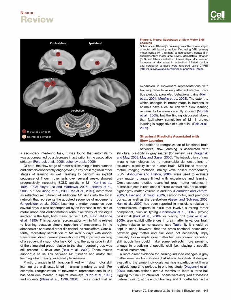
Figure 4. Neural Substrates of Slow Motor SkillLearningSchematics of themajor brain regions active in slow stagesof motor skill learning, as identified using fMRI: primarymotor cortex (M1), primary somatosensory cortex (S1),supplementary motor area (SMA), dorsolateral striatum(DLS), and lateral cerebellum. Arrows depict documentedincreases or decreases in activation. Inflated corticaland cerebellar surfaces were rendered using CARET(http://brainvis.wustl.edu/wiki/index.php/Main_Page).
Neuron
Review
a secondary interfering task, it was found that automaticity
was accompanied by a decrease in activation in the associative
striatum (Poldrack et al., 2005; Lehericy et al., 2005).
Of note, the slow stage of motor skill learning in both humans
and animals consistently engagesM1, a key brain region in other
stages of learning as well. Training to perform an explicit
sequence of finger movements over several weeks showed
progressively increasing BOLD activity in M1 (Karni et al.,
1995, 1998; Floyer-Lea and Matthews, 2005; Lehericy et al.,
2005; but see Xiong et al., 2009; Ma et al., 2010), interpreted
as reflecting recruitment of additional M1 units into the local
network that represents the acquired sequence of movements
(Ungerleider et al., 2002). Learning a motor sequence over
several days is also accompanied by an increase in the size of
motor maps and corticomotoneuronal excitability of the digits
involved in the task, both measured with TMS (Pascual-Leone
et al., 1995). This particular reorganization within M1 is related
to learning because simple repetition of movements in the
absence of a sequential order did not induce such effect. Consis-
tently, facilitatory stimulation of M1 over 5 days with anodal
transcranial direct current stimulation (tDCS) improved learning
of a sequential visuomotor task. Of note, the advantage in skill
of the stimulated group relative to the sham control group was
still present 90 days later (Reis et al., 2009). These results
support a causal link between M1 function and motor skill
learning when training over multiple sessions.
Plastic changes in M1 function linked with slow motor skill
learning are well established in animal models as well. For
example, reorganization of movement representations in M1
has been documented in squirrel monkeys (Nudo et al., 1996)
and rodents (Kleim et al., 1998, 2004). It was found that an
Neuro
expansion in movement representations with
training, detectable only after substantial prac-
tice periods, paralleled behavioral gains (Kleim
et al., 2004; Monfils et al., 2005). The extent to
which changes in motor maps in humans or
animals have a causal link with slow learning
remains to be more carefully studied (Monfils
et al., 2005), but the finding discussed above
that facilitatory stimulation of M1 improves
learning is suggestive of such a link (Reis et al.,
2009).
Structural Plasticity Associated withSlow LearningIn addition to reorganization of functional brain
networks, slow learning is associated with
structural plasticity in gray matter (for review, see Draganski
and May, 2008; May and Gaser, 2006). The introduction of new
imaging technologies led to remarkable demonstrations of
structural plasticity in the human brain. MRI-based morpho-
metric imaging methods, mainly voxel-based morphometry
(VBM; Ashburner and Friston, 2000), were used to evaluate
gray matter changes linked with experience and learning.
Cross-sectional studies quantified gray matter volumes in
human subjects in relation to different levels of skill. For example,
higher gray matter volume in auditory (Bermudez and Zatorre,
2005; Gaser and Schlaug, 2003), sensorimotor, and premotor
cortex, as well as the cerebellum (Gaser and Schlaug, 2003;
Han et al., 2009) has been reported in musicians relative to
nonmusicians. Experts in skills that involve a strong motor
component, such as typing (Cannonieri et al., 2007), playing
basketball (Park et al., 2009), or playing golf (Jancke et al.,
2009), also exhibit differences in gray matter in various brain
regions relative to nonexperts (see Table 1). It should be
kept in mind, however, that the cross-sectional association
between gray matter and skill does not necessarily imply
causality. For example, gray matter features present preceding
skill acquisition could make some subjects more prone to
engage in practicing a specific skill (i.e., playing a specific
musical instrument).
A more direct evidence for learning-induced changes in gray
matter emerges from studies that utilized longitudinal designs,
evaluating the same individuals learning a particular skill over
relatively long time periods. In one key study (Draganski et al.,
2004), subjects trained over 3 months to learn a three-ball
juggling routine. Structural MRI scans were acquired at baseline
(before training), at the end of training, and 3 months later in the
n 72, November 3, 2011 ª2011 Elsevier Inc. 447
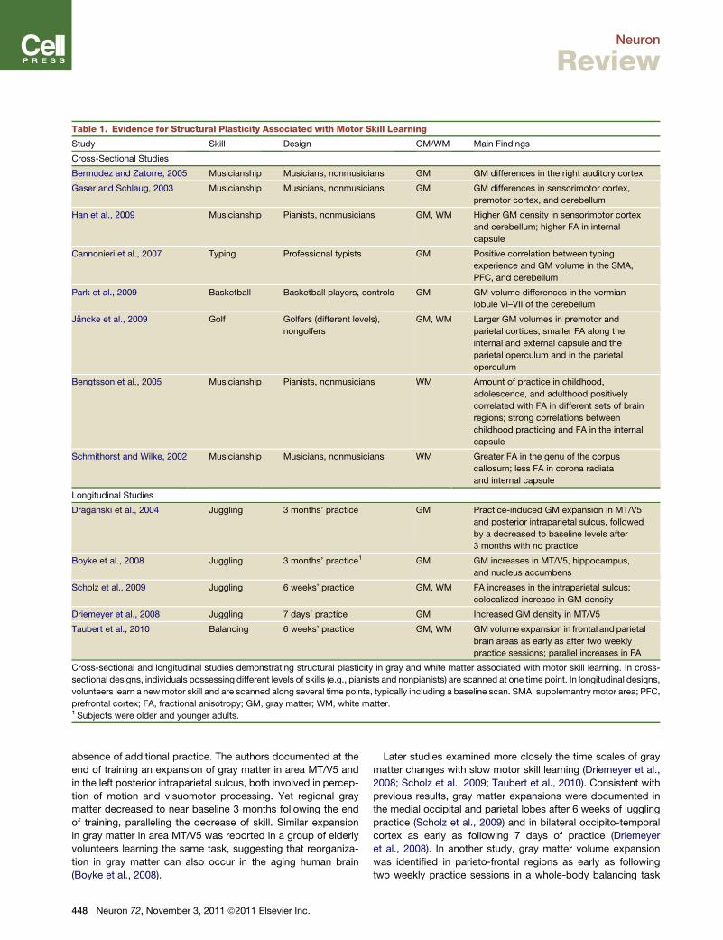
Table 1. Evidence for Structural Plasticity Associated with Motor Skill Learning
Study Skill Design GM/WM Main Findings
Cross-Sectional Studies
Bermudez and Zatorre, 2005 Musicianship Musicians, nonmusicians GM GM differences in the right auditory cortex
Gaser and Schlaug, 2003 Musicianship Musicians, nonmusicians GM GM differences in sensorimotor cortex,
premotor cortex, and cerebellum
Han et al., 2009 Musicianship Pianists, nonmusicians GM, WM Higher GM density in sensorimotor cortex
and cerebellum; higher FA in internal
capsule
Cannonieri et al., 2007 Typing Professional typists GM Positive correlation between typing
experience and GM volume in the SMA,
PFC, and cerebellum
Park et al., 2009 Basketball Basketball players, controls GM GM volume differences in the vermian
lobule VI–VII of the cerebellum
Jancke et al., 2009 Golf Golfers (different levels),
nongolfers
GM, WM Larger GM volumes in premotor and
parietal cortices; smaller FA along the
internal and external capsule and the
parietal operculum and in the parietal
operculum
Bengtsson et al., 2005 Musicianship Pianists, nonmusicians WM Amount of practice in childhood,
adolescence, and adulthood positively
correlated with FA in different sets of brain
regions; strong correlations between
childhood practicing and FA in the internal
capsule
Schmithorst and Wilke, 2002 Musicianship Musicians, nonmusicians WM Greater FA in the genu of the corpus
callosum; less FA in corona radiata
and internal capsule
Longitudinal Studies
Draganski et al., 2004 Juggling 3 months’ practice GM Practice-induced GM expansion in MT/V5
and posterior intraparietal sulcus, followed
by a decreased to baseline levels after
3 months with no practice
Boyke et al., 2008 Juggling 3 months’ practice1 GM GM increases in MT/V5, hippocampus,
and nucleus accumbens
Scholz et al., 2009 Juggling 6 weeks’ practice GM, WM FA increases in the intraparietal sulcus;
colocalized increase in GM density
Driemeyer et al., 2008 Juggling 7 days’ practice GM Increased GM density in MT/V5
Taubert et al., 2010 Balancing 6 weeks’ practice GM, WM GMvolume expansion in frontal and parietal
brain areas as early as after two weekly
practice sessions; parallel increases in FA
Cross-sectional and longitudinal studies demonstrating structural plasticity in gray and white matter associated with motor skill learning. In cross-
sectional designs, individuals possessing different levels of skills (e.g., pianists and nonpianists) are scanned at one time point. In longitudinal designs,
volunteers learn a newmotor skill and are scanned along several time points, typically including a baseline scan. SMA, supplemantry motor area; PFC,
prefrontal cortex; FA, fractional anisotropy; GM, gray matter; WM, white matter.1 Subjects were older and younger adults.
Neuron
Review
absence of additional practice. The authors documented at the
end of training an expansion of gray matter in area MT/V5 and
in the left posterior intraparietal sulcus, both involved in percep-
tion of motion and visuomotor processing. Yet regional gray
matter decreased to near baseline 3 months following the end
of training, paralleling the decrease of skill. Similar expansion
in gray matter in area MT/V5 was reported in a group of elderly
volunteers learning the same task, suggesting that reorganiza-
tion in gray matter can also occur in the aging human brain
(Boyke et al., 2008).
448 Neuron 72, November 3, 2011 ª2011 Elsevier Inc.
Later studies examined more closely the time scales of gray
matter changes with slow motor skill learning (Driemeyer et al.,
2008; Scholz et al., 2009; Taubert et al., 2010). Consistent with
previous results, gray matter expansions were documented in
the medial occipital and parietal lobes after 6 weeks of juggling
practice (Scholz et al., 2009) and in bilateral occipito-temporal
cortex as early as following 7 days of practice (Driemeyer
et al., 2008). In another study, gray matter volume expansion
was identified in parieto-frontal regions as early as following
two weekly practice sessions in a whole-body balancing task

Neuron
Review
(Taubert et al., 2010). These findings are indicative of relatively
rapid structural gray matter plasticity associated with motor skill
learning. What mechanisms could contribute to these striking
findings? Although speculative at this point, it has been
proposed that processes occurring both at the synapse level
and at larger scales, including rapid intracortical remodeling of
dendritic spines and axonal terminals, glial hypertrophy, and
synaptogenesis, might play a contributory role (Draganski and
May, 2008;May andGaser, 2006; Anderson et al., 1994). Consis-
tently, rapid (within an hour) formation of postsynaptic dendritic
spines has been detected in vivo in the pyramidal neurons of the
mouse motor cortex following motor training (Xu et al., 2009),
and the extent of spine remodeling has been shown to correlate
with behavioral improvements after learning, suggesting that
this mechanism of synaptic plasticity may contribute to motor
memory formation (Yang et al., 2009). On the other hand, it
should be noted that other animal studies demonstrated signifi-
cant increases in synapse numbers in the rat M1 only after exten-
sive training (Kleim et al., 1996, 2004).
Slow learning has been linked with structural plasticity in white
matter architecture as well (Table 1). Diffusion MRI-based mea-
sures, such as fractional anisotropy (FA), are believed to reflect
white matter integrity (Fields, 2008), providing a distinctive in-
sight into the microstructural properties of white matter in vivo
(Le Bihan et al., 2001; Mori and Zhang, 2006). Cross-sectional
studies, primarily with highly trained musicians, examined white
matter correlates of skilled behavior (Bengtsson et al., 2005;
Han et al., 2009; Schmithorst andWilke, 2002). Fractional anisot-
ropy in the posterior limb of the internal capsule, which contains
descending corticospinal fibers from the primary sensorimotor
and premotor cortices, correlated with number of practice hours
during childhood in skilled musicians (Bengtsson et al., 2005). It
has been proposed that these results may reflect experience-
inducedplasticity during a critical developmental period (Bengts-
son et al., 2005). A recent pioneering study provided more direct
evidence for experience-induced changes in white matter archi-
tecture, resulting from a relatively short period of practice (Scholz
et al., 2009). In this study, it was shown that 6 weeks of juggling
practice resulted in increased FA in a region of white matter
underlying the intraparietal sulcus. Localized increases in gray
matter were detected in close proximity to these white matter
regions. Yet the magnitude of changes of gray and white matter
showed no correlation and developed over markedly different
time courses. Interestingly, individual differences in white matter
mictrostructure appear to be related to variation in learning
(Johansen-Berg, 2010; Della-Maggiore et al., 2009; Tomassini
et al., 2011). For example, individual differences in learning an
isometric visuomotor tracking task are associated with variability
in FA in the premotor cortex and the cerebellum (Tomassini et al.,
2011). The cellular mechanisms underlying learning-dependent
changes inwhitemattermicrostructure remain to be established,
and so do the links between these changes and measures of
functional plasticity. It has been proposed that changes in white
matter properties, indexed by FA, could affect the velocity and
synchronicity of impulse conduction between distant cortical
regions and thus contribute to the optimization of information
flow required for skill acquisition (see Fields, 2008, 2011),
a hypothesis that requires specific testing.
Altogether, demonstrations of learning-induced gray and
white matter plasticity in humans represent an exciting develop-
ment in systems neuroscience. Yet the contribution of this line of
research to our understanding of motor skill learning is still
limited. The biological mechanisms that underlie these forms of
plasticity remain to be elucidated, and its time scales need to
be more clearly established. Additionally, strict comparative
evaluation of structural and functional plasticity associated
withmotor skill learning is difficult at this point, given the different
experimental paradigms used in the literature. This issue should
be overcome in future investigations by evaluating both forms of
plasticity in longitudinal studies in the same subjects (Thomas
et al., 2009).
Offline Motor Skill LearningProgression from fast to slow motor skill learning is thought to
rely on appropriate consolidation (Doyon and Benali, 2005;
Muellbacher et al., 2002; Robertson et al., 2004a), defined as
the progressive stabilization of a recently acquired memory
(Dudai, 2004). Through consolidation, new memories are trans-
formed from their initial fragile states intomore robust and stabile
forms (Robertson et al., 2004a). In relation to motor skill learning,
the term consolidation has been used in the literature to describe
two different, but not mutually exclusive, phenomena: the offline
behavioral skill improvements that occur after the end of a prac-
tice session (Robertson et al., 2004a) and the reduction in fragility
of a motor memory trace that follows encoding (Robertson,
2009; Robertson et al., 2004a).
In humans, offline skill improvementsmay be affected by sleep
(e.g., Diekelmann and Born, 2010; Fischer et al., 2002; Korman
et al., 2003). Sleep-dependent motor memory consolidation,
which correlates with the amount of stage II nonrapid eye move-
ment sleep (Walker et al., 2002), has been mostly demonstrated
for explicit motor sequence learning (Fischer et al., 2005; Kor-
man et al., 2003; Walker et al., 2002; but see Brawn et al.,
2010; Rickard et al., 2008). Other forms of procedural motor
learning are not necessarily sleep dependent (Debas et al.,
2010; Doyon et al., 2009b; Song et al., 2007). Notably, sleep
does not benefit implicit forms of sequence learning (Robertson
et al., 2004b; Song et al., 2007). In such circumstances, similar
memory gains were reported after sleep and over an equivalent
period of wakefulness (see also Hotermans et al., 2008).
Different brain regions are involved in consolidation of motor
memories. Sleep-dependent improvements in learning a
sequential finger-movement task were linked to reduced BOLD
activity in M1, as measured with fMRI (Fischer et al., 2005).
Furthermore, downregulating excitability of M1 by low-
frequency TMS (virtual lesion) results in reduced motor memory
consolidation (Muellbacher et al., 2002; Robertson et al., 2005),
a time-specific effect because it was not observed when TMS
was applied 6 hr posttraining (Muellbacher et al., 2002). The
finding of differential effects of facilitatory anodal tDCS applied
over M1 on online and offline learning of a sequential motor
task, namely enhancement of offline learning, supports the
existence of relatively different neuronal networks involved in
the two processes (Reis et al., 2009). Another key contributor
to consolidation of sequential motor skills is the striatum (Debas
et al., 2010; Fischer et al., 2005; Albouy et al., 2008; Doyon and
Neuron 72, November 3, 2011 ª2011 Elsevier Inc. 449

Neuron
Review
Ungerleider, 2002). Recent work showed increased striatal
activity in human subjects in whom offline consolidation was
tested following a night of sleep, as compared to those in
whom it was tested after an equivalent period of wakefulness
(Debas et al., 2010; Fischer et al., 2005). Interestingly, BOLD
activity in the ventral striatum and the hippocampus during
the initial stages of oculomotor sequence learning predicted
the magnitude of sleep-dependent behavioral improvements
(Albouy et al., 2008). Additional evidence for the involvement of
these two regions emerged from animal studies demonstrating
that local injections of protein synthesis inhibitors disrupt consol-
idation of motor memories (Buitrago et al., 2004). This effect was
present when injections were applied to M1 (Luft et al., 2004)
and, to a lesser extent, the dorsal striatum (Wachter et al.,
2010) but was absent after injections of control regions (Luft
et al., 2004).
The neural processes leading to successful consolidation
tested posttraining are likely to start operating during practice
and evolve over time after training ended. Typically, evaluation
of changes in BOLD signal induced by task performance
assesses the consequences of these processes as tested
a few hours after or the day after practice was completed.
Thus, the neuronal mechanisms that operate during and early
after practice and during sleep to support motormemory consol-
idation remain to a large extent uncertain. It was recently sug-
gested that a possible way of closing this gap in knowledge is
through measurement of intrinsic resting-state functional
connectivity (Albert et al., 2009; Ma et al., 2011; Taubert et al.,
2011). Spontaneous low-frequency fluctuations in the BOLD
signal, in the absence of any overt input or behavior, have
been widely reported in the past 15 years (for a review, see
Fox and Raichle, 2007; Cole et al., 2010) and can be recorded
at different times before or after training without affecting subse-
quent behavioral testing. Temporally coherent spontaneous
fluctuations at rest have been found between spatially remote
brain regions in areas known to be involved in motor, visual,
and auditory processing, attention, and language (Cole et al.,
2010; Fox and Raichle, 2007). Thus, resting-state functional
connectivity, which may be sampled multiple times during the
period leading to the behavioral measurement of consolidation,
may provide a unique window for examining neural network
activity along the entire course of motor skill acquisition. Avail-
able data are supportive of this contention. Learning a visuomo-
tor tracking task over one session increased resting functional
connectivity in a network that includes the prefrontal, superior,
and inferior parietal cortices, as well as Crus II of the cerebellum
(Albert et al., 2009). Learning a whole-body dynamic balancing
task over multiple sessions showed increased resting-state
connectivity between SMA/preSMA and medial parietal cortex
that correlated with performance improvements (Taubert et al.,
2011). Modulation of resting-state connectivity in parietal circuits
was also observed along 4 weeks of daily training of an explicit
sequence learning task (Ma et al., 2011). Overall, these studies
suggest that functional connectivity in fronto-parietal networks
supports consolidation after fast (Albert et al., 2009) and slow
learning (Taubert et al., 2011; Ma et al., 2011). Comparison
among these studies, however, should be done with caution,
because they involved different motor skill tasks. Notwith-
450 Neuron 72, November 3, 2011 ª2011 Elsevier Inc.
standing, published studies have yet to identify modulation of
connectivity within striatal regions, believed to play a key role
in consolidation of skills (Doyon and Benali, 2005; Doyon and
Ungerleider, 2002), but preliminary findings indeed appear to
support this hypothesis (K. Debas et al., 2011, Human Brain
Mapping, abstract).
It should be kept in mind that previously consolidated memo-
ries are not immune to further modifications. Reactivation of
a consolidated memory renders it once again labile and suscep-
tible to interference (Nader et al., 2000; Walker et al., 2003). For
example, reactivation of fear memories in rodents renders these
memories susceptible to interference achieved through protein
synthesis inhibition (Nader et al., 2000). Thus, reactivation of
consolidated memories initiates a process of reconsolidation,
whereby previously stabilized memories become labile again,
requiring de novo protein synthesis in order to persist (Nader
et al., 2000). In humans, evidence for reconsolidation of motor
memories also exists (Walker et al., 2003; Censor et al., 2010).
Learning a novel sequence of finger movements right after a
previously consolidated procedural memory has been reacti-
vated results in profoundly impaired recollection of the original
procedural memory (Walker et al., 2003). As for the possible
mechanisms underlying reconsolidation of human motor skill
memories, it was recently shown that the application of 1-Hz
repetitive TMS over M1 during the reactivation of an already-
consolidated motor memory, acquired via training in an explicit
sequential finger-tapping task in humans, blocks further memory
modification (Censor et al., 2010). These results suggest that
recurrent interactions of M1 processing with existing memory
traces may be critical for further memory modification through
reconsolidation (Censor et al., 2010; Censor and Cohen, 2011).
Long-Term Retention of Motor SkillsOnce motor skills are acquired and consolidated, they can be
retained over extended periods of time or forgotten. Under
controlled laboratory settings, retention of motor skills has
been demonstrated in humans (Romano et al., 2010; Savion-
Lemieux and Penhune, 2005) over periods of up to a year
(Romano et al., 2010) and in monkeys over similarly extensive
periods (Hikosaka et al., 2002b), yet in real life, retention may
occur over much longer periods. For learning of explicit motor
sequences, even minimal amounts of practice spread over
several days were able to induce long-term retention (Savion-
Lemieux and Penhune, 2005), suggesting that long-term reten-
tion is strongly dependent on successful consolidation.
Various task attributes have a profound influence on long-term
retention of skill learning. For instance, reward during practice
improves long-term retention of a sequential motor skill (Abe
et al., 2011). A reward-related enhancement of long-term
memory has been demonstrated for other forms of memory as
well (Wittmann et al., 2011) and is linked with fMRI activation
in the striatum, ventral tegmental area, and hippocampus (Witt-
mann et al., 2005; Adcock et al., 2006). It has been proposed
that dopaminergic modulation within these circuits, specifically
through dopamine-dependent LTP in the hippocampus, may
contribute to this effect (Calabresi et al., 2007). In the future, it
will be of interest to identify the influence of reward attributes
such as predictability, magnitude, and outcome uncertainty on

Neuron
Review
long-term retention of motor skills. For instance, a recent study
found that reward predictability and to some extent reward
magnitude modulate long-term episodic memory, an effect
that was absent for outcome uncertainty by itself (Wittmann
et al., 2011).
Practice structure influences long-term retention of motor
skills. The contextual interference (CI) effect, demonstrated in
a wide variety of cognitive and motor tasks (Magill and Hall,
1990), refers to the benefits of training under interleaved or
random-order conditions, as opposed to blocked practice
schedules (Shea andMorgan, 1979). Recent studies have shown
that training under different practice schedules implicates
distinct neural substrates (Cross et al., 2007; Kantak et al.,
2010; Tanaka et al., 2010; Wymbs and Grafton, 2009), including
the SMA (Tanaka et al., 2010) andM1 (Kantak et al., 2010). These
findings are consistent with the view that random practice may
lead to more rapid memory stabilization or that motor memory
encoding under random practice is associated with a more rapid
shift from the SMA to other brain regions, such as the striatum or
the parietal cortex (Tanaka et al., 2010). Consistent with this
proposal, it was recently shown that interindividual differences
in the magnitude of benefits of randomized practice schedules
correlate with FA within the corticostriatal tract connecting left
sensorimotor cortex to posterior putamen (Song et al., 2011).
Understanding the influence of practice structure on the consol-
idation and retention of skilled motor behavior has potential
clinical implications, because this knowledge may translate
into improved training-based neurorehabilitative interventions
after brain lesions.
Concluding RemarksTechnological and methodological advances in neuroimaging
and in noninvasive brain stimulation in humans, together with
novel findings stemming from animal-based studies, provide
new insights into the neuroplastic mechanisms that underlie
motor skill learning, suggesting that skill acquisition is subserved
by multiple mechanisms that operate across different temporal
scales. Multivariate and model-free approaches for analyzing
neuroimaging data have emerged andmay turn out to be a useful
tool for examining the larger-scale functional reorganization
associatedwith fast and slowmotor skill learning. Another recent
and intriguing development concerns the analysis of modulation
of resting-state spontaneous fluctuations in BOLD activity as
a possible means for studying the offline consolidation of motor
skills. Noninvasive brain stimulation techniques have been used
to identify a causal role for the activity in various brain regions
in the acquisition of skilled motor behavior, motor memory
consolidation, and long-term retention. Studies in laboratory
animals identified, with fine temporal and spatial resolution, the
involvement of distinct neural substrates in the various stages
of motor skill learning and also helped identify the possible
cellular and molecular underpinnings of learning-induced plas-
ticity. Advances were also made in uncovering the mechanisms
behind structural plasticity associated with the acquisition of
motor skills. Learning-induced structural changes in both gray
and white matter have been documented in humans at increas-
ingly smaller temporal scales. Similar advancesweremade in the
study of learning and experience-induced structural plasticity in
laboratory animals, yet possible links between these findings
and demonstrations of structural plasticity in humans are, to
date, still speculative; however, they show clear translational
value in understanding motor skill learning after brain lesions
(Clarkson et al., 2010, 2011; Li et al., 2010).
ACKNOWLEDGMENTS
We would like to thank Barry Richmond, Sunbin Song, and Nitzan Censor forproviding useful suggestions. This work was supported by the IntramuralResearch Program of the National Institute of Neurological Disorders andStroke, National Institutes of Health.
REFERENCES
Abe, M., Schambra, H.M., Wassermann, E.M., Luckenbaugh, D.,Schweighofer, N., and Cohen, L.G. (2011). Reward improves long-term reten-tion of a motor memory through induction of offline memory gains. Curr. Biol.21, 557–562.
Adcock, R.A., Thangavel, A., Whitfield-Gabrieli, S., Knutson, B., and Gabrieli,J.D. (2006). Reward-motivated learning: mesolimbic activation precedesmemory formation. Neuron 50, 507–517.
Akaneya, Y., Tsumoto, T., Kinoshita, S., and Hatanaka, H. (1997). Brain-derived neurotrophic factor enhances long-term potentiation in rat visualcortex. J. Neurosci. 17, 6707–6716.
Albert, N.B., Robertson, E.M., and Miall, R.C. (2009). The resting human brainand motor learning. Curr. Biol. 19, 1023–1027.
Albouy, G., Sterpenich, V., Balteau, E., Vandewalle, G., Desseilles, M., Dang-Vu, T., Darsaud, A., Ruby, P., Luppi, P.H., Degueldre, C., et al. (2008). Both thehippocampus and striatum are involved in consolidation of motor sequencememory. Neuron 58, 261–272.
Anderson, B.J., Li, X., Alcantara, A.A., Isaacs, K.R., Black, J.E., and Green-ough, W.T. (1994). Glial hypertrophy is associated with synaptogenesisfollowing motor-skill learning, but not with angiogenesis following exercise.Glia 11, 73–80.
Antal, A., Chaieb, L., Moliadze, V., Monte-Silva, K., Poreisz, C., Thirugnana-sambandam, N., Nitsche, M.A., Shoukier, M., Ludwig, H., and Paulus, W.(2010). Brain-derived neurotrophic factor (BDNF) gene polymorphisms shapecortical plasticity in humans. Brain Stimulat. 3, 230–237.
Ashburner, J., and Friston, K.J. (2000). Voxel-based morphometry—themethods. Neuroimage 11, 805–821.
Ashby, F.G., Turner, B.O., and Horvitz, J.C. (2010). Cortical and basal gangliacontributions to habit learning and automaticity. Trends Cogn. Sci. (Regul. Ed.)14, 208–215.
Bengtsson, S.L., Nagy, Z., Skare, S., Forsman, L., Forssberg, H., and Ullen, F.(2005). Extensive piano practicing has regionally specific effects on whitematter development. Nat. Neurosci. 8, 1148–1150.
Bermudez, P., and Zatorre, R.J. (2005). Differences in gray matter betweenmusicians and nonmusicians. Ann. N Y Acad. Sci. 1060, 395–399.
Boyke, J., Driemeyer, J., Gaser, C., Buchel, C., and May, A. (2008). Training-induced brain structure changes in the elderly. J. Neurosci. 28, 7031–7035.
Brawn, T.P., Fenn, K.M., Nusbaum, H.C., and Margoliash, D. (2010).Consolidating the effects of waking and sleep on motor-sequence learning.J. Neurosci. 30, 13977–13982.
Buitrago, M.M., Ringer, T., Schulz, J.B., Dichgans, J., and Luft, A.R. (2004).Characterization of motor skill and instrumental learning time scales in a skilledreaching task in rat. Behav. Brain Res. 155, 249–256.
Calabresi, P., Picconi, B., Tozzi, A., and Di Filippo, M. (2007). Dopamine-mediated regulation of corticostriatal synaptic plasticity. Trends Neurosci.30, 211–219.
Neuron 72, November 3, 2011 ª2011 Elsevier Inc. 451
Neuron
Review
Cannonieri, G.C., Bonilha, L., Fernandes, P.T., Cendes, F., and Li, L.M. (2007).Practice and perfect: length of training and structural brain changes in experi-enced typists. Neuroreport 18, 1063–1066.
Censor, N., and Cohen, L.G. (2011). Using repetitive transcranial magneticstimulation to study the underlying neural mechanisms of human motorlearning and memory. J. Physiol. 589, 21–28.
Censor, N., Dimyan, M.A., and Cohen, L.G. (2010). Modification of existinghuman motor memories is enabled by primary cortical processing duringmemory reactivation. Curr. Biol. 20, 1545–1549.
Cheeran, B.J., Ritter, C., Rothwell, J.C., and Siebner, H.R. (2009). Mappinggenetic influences on the corticospinal motor system in humans. Neurosci-ence 164, 156–163.
Clarkson, A.N., Huang, B.S., Macisaac, S.E., Mody, I., and Carmichael, S.T.(2010). Reducing excessive GABA-mediated tonic inhibition promotes func-tional recovery after stroke. Nature 468, 305–309.
Clarkson, A.N., Overman, J.J., Zhong, S., Mueller, R., Lynch, G., and Carmi-chael, S.T. (2011). AMPA receptor-induced local brain-derived neurotrophicfactor signaling mediates motor recovery after stroke. J. Neurosci. 31, 3766–3775.
Cole, D.M., Smith, S.M., and Beckmann, C.F. (2010). Advances and pitfallsin the analysis and interpretation of resting-state FMRI data. Front SystNeurosci 4, 8.
Costa, R.M., Cohen, D., and Nicolelis, M.A.L. (2004). Differential corticostriatalplasticity during fast and slowmotor skill learning in mice. Curr. Biol. 14, 1124–1134.
Coynel, D., Marrelec, G., Perlbarg, V., Pelegrini-Issac, M., Van deMoortele, P.-F., Ugurbil, K., Doyon, J., Benali, H., and Lehericy, S. (2010). Dynamics ofmotor-related functional integration during motor sequence learning. Neuro-image 49, 759–766.
Cross, E.S., Schmitt, P.J., and Grafton, S.T. (2007). Neural substrates ofcontextual interference during motor learning support a model of active prep-aration. J. Cogn. Neurosci. 19, 1854–1871.
Debas, K., Carrier, J., Orban, P., Barakat, M., Lungu, O., Vandewalle, G., HadjTahar, A., Bellec, P., Karni, A., Ungerleider, L.G., et al. (2010). Brain plasticityrelated to the consolidation of motor sequence learning and motor adaptation.Proc. Natl. Acad. Sci. USA 107, 17839–17844.
Della-Maggiore, V., Scholz, J., Johansen-Berg, H., and Paus, T. (2009). Therate of visuomotor adaptation correlates with cerebellar white-matter micro-structure. Hum. Brain Mapp. 30, 4048–4053.
Di Martino, A., Scheres, A., Margulies, D.S., Kelly, A.M., Uddin, L.Q., Shehzad,Z., Biswal, B., Walters, J.R., Castellanos, F.X., and Milham, M.P. (2008). Func-tional connectivity of human striatum: a resting state FMRI study. Cereb.Cortex 18, 2735–2747.
Diekelmann, S., and Born, J. (2010). The memory function of sleep. Nat. Rev.Neurosci. 11, 114–126.
Doyon, J., and Benali, H. (2005). Reorganization and plasticity in the adult brainduring learning of motor skills. Curr. Opin. Neurobiol. 15, 161–167.
Doyon, J., and Ungerleider, L.G. (2002). Functional anatomy of motor skilllearning. In Neuropsychology of Memory, L.S. Squire and D.L. Schacter,eds. (New York: Guilford Press), pp. 225–238.
Doyon, J., Bellec, P., Amsel, R., Penhune, V., Monchi, O., Carrier, J., Lehericy,S., and Benali, H. (2009a). Contributions of the basal ganglia and functionallyrelated brain structures to motor learning. Behav. Brain Res. 199, 61–75.
Doyon, J., Korman, M., Morin, A., Dostie, V., Hadj Tahar, A., Benali, H., Karni,A., Ungerleider, L.G., and Carrier, J. (2009b). Contribution of night and daysleep vs. simple passage of time to the consolidation of motor sequenceand visuomotor adaptation learning. Exp. Brain Res. 195, 15–26.
Draganski, B., and May, A. (2008). Training-induced structural changes in theadult human brain. Behav. Brain Res. 192, 137–142.
Draganski, B., Gaser, C., Busch, V., Schuierer, G., Bogdahn, U., and May, A.(2004). Neuroplasticity: changes in grey matter induced by training. Nature427, 311–312.
452 Neuron 72, November 3, 2011 ª2011 Elsevier Inc.
Driemeyer, J., Boyke, J., Gaser, C., Buchel, C., and May, A. (2008). Changesin gray matter induced by learning—revisited. PLoS ONE 3, e2669.
Dudai, Y. (2004). The neurobiology of consolidations, or, how stable is theengram? Annu. Rev. Psychol. 55, 51–86.
Fields, R.D. (2008). White matter in learning, cognition and psychiatric disor-ders. Trends Neurosci. 31, 361–370.
Fields, R.D. (2011). Imaging learning: the search for a memory trace. Neurosci-entist 17, 185–196.
Fischer, S., Hallschmid, M., Elsner, A.L., and Born, J. (2002). Sleep formsmemory for finger skills. Proc. Natl. Acad. Sci. USA 99, 11987–11991.
Fischer, S., Nitschke, M.F., Melchert, U.H., Erdmann, C., and Born, J. (2005).Motor memory consolidation in sleep shapes more effective neuronal repre-sentations. J. Neurosci. 25, 11248–11255.
Fitts, P.M. (1954). The information capacity of the human motor system incontrolling the amplitude of movement. J. Exp. Psychol. 47, 381–391.
Floyer-Lea, A., and Matthews, P.M. (2005). Distinguishable brain activationnetworks for short- and long-term motor skill learning. J. Neurophysiol. 94,512–518.
Fox, M.D., and Raichle, M.E. (2007). Spontaneous fluctuations in brain activityobserved with functional magnetic resonance imaging. Nat. Rev. Neurosci. 8,700–711.
Friston, K. (1994). Functional and effective connectivity: a synthesis. Hum.Brain Mapp. 2, 56–78.
Fritsch, B., Reis, J., Martinowich, K., Schambra, H.M., Ji, Y., Cohen, L.G., andLu, B. (2010). Direct current stimulation promotes BDNF-dependent synapticplasticity: potential implications for motor learning. Neuron 66, 198–204.
Gaser, C., and Schlaug, G. (2003). Brain structures differ between musiciansand non-musicians. J. Neurosci. 23, 9240–9245.
Grafton, S.T., Hazeltine, E., and Ivry, R.B. (2002).Motor sequence learningwiththe nondominant left hand. A PET functional imaging study. Exp. Brain Res.146, 369–378.
Han, Y., Yang, H., Lv, Y.-T., Zhu, C.-Z., He, Y., Tang, H.-H., Gong, Q.-Y., Luo,Y.-J., Zang, Y.-F., and Dong, Q. (2009). Gray matter density and white matterintegrity in pianists’ brain: a combined structural and diffusion tensor MRIstudy. Neurosci. Lett. 459, 3–6.
Hazeltine, E., Grafton, S.T., and Ivry, R. (1997). Attention and stimulus charac-teristics determine the locus of motor-sequence encoding. A PET study. Brain120, 123–140.
Hikosaka, O., Nakamura, K., Sakai, K., and Nakahara, H. (2002a). Centralmechanisms of motor skill learning. Curr. Opin. Neurobiol. 12, 217–222.
Hikosaka, O., Rand, M.K., Nakamura, K., Miyachi, S., Kitaguchi, K., Sakai, K.,Lu, X., and Shimo, Y. (2002b). Long-term retention of motor skill in macaquemonkeys and humans. Exp. Brain Res. 147, 494–504.
Honda, M., Deiber, M.P., Ibanez, V., Pascual-Leone, A., Zhuang, P., andHallett, M. (1998). Dynamic cortical involvement in implicit and explicit motorsequence learning. A PET study. Brain 121, 2159–2173.
Hotermans, C., Peigneux, P., de Noordhout, A.M., Moonen, G., and Maquet,P. (2008). Repetitive transcranial magnetic stimulation over the primary motorcortex disrupts early boost but not delayed gains in performance in motorsequence learning. Eur. J. Neurosci. 28, 1216–1221.
Jancke, L., Koeneke, S., Hoppe, A., Rominger, C., and Hanggi, J. (2009). Thearchitecture of the golfer’s brain. PLoS ONE 4, e4785.
Johansen-Berg, H. (2010). Behavioural relevance of variation in white mattermicrostructure. Curr. Opin. Neurol. 23, 351–358.
Kantak, S.S., Sullivan, K.J., Fisher, B.E., Knowlton, B.J., and Winstein, C.J.(2010). Neural substrates of motor memory consolidation depend on practicestructure. Nat. Neurosci. 13, 923–925.
Karni, A., Meyer, G., Jezzard, P., Adams, M.M., Turner, R., and Ungerleider,L.G. (1995). Functional MRI evidence for adult motor cortex plasticity duringmotor skill learning. Nature 377, 155–158.
Neuron
Review
Karni, A., Meyer, G., Rey-Hipolito, C., Jezzard, P., Adams, M.M., Turner, R.,and Ungerleider, L.G. (1998). The acquisition of skilled motor performance:fast and slow experience-driven changes in primary motor cortex. Proc.Natl. Acad. Sci. USA 95, 861–868.
Kelly, A.M.C., and Garavan, H. (2005). Human functional neuroimaging ofbrain changes associated with practice. Cereb. Cortex 15, 1089–1102.
Kleim, J.A., Lussnig, E., Schwarz, E.R., Comery, T.A., and Greenough, W.T.(1996). Synaptogenesis and Fos expression in the motor cortex of the adultrat after motor skill learning. J. Neurosci. 16, 4529–4535.
Kleim, J.A., Barbay, S., and Nudo, R.J. (1998). Functional reorganization ofthe rat motor cortex following motor skill learning. J. Neurophysiol. 80,3321–3325.
Kleim, J.A., Bruneau, R., Calder, K., Pocock, D., VandenBerg, P.M., MacDon-ald, E., Monfils, M.H., Sutherland, R.J., and Nader, K. (2003). Functional orga-nization of adult motor cortex is dependent upon continued protein synthesis.Neuron 40, 167–176.
Kleim, J.A., Hogg, T.M., VandenBerg, P.M., Cooper, N.R., Bruneau, R., andRemple, M. (2004). Cortical synaptogenesis and motor map reorganizationoccur during late, but not early, phase of motor skill learning. J. Neurosci.24, 628–633.
Kleim, J.A., Chan, S., Pringle, E., Schallert, K., Procaccio, V., Jimenez, R., andCramer, S.C. (2006). BDNF val66met polymorphism is associated with modi-fied experience-dependent plasticity in human motor cortex. Nat. Neurosci. 9,735–737.
Korman, M., Raz, N., Flash, T., and Karni, A. (2003). Multiple shifts in the repre-sentation of a motor sequence during the acquisition of skilled performance.Proc. Natl. Acad. Sci. USA 100, 12492–12497.
Krakauer, J.W. (2009). Motor learning and consolidation: the case of visuo-motor rotation. In Motor Control, D. Sternad, ed. (New York: Springer),pp. 405–421.
Krakauer, J.W., and Mazzoni, P. (2011). Human sensorimotor learning: adap-tation, skill, and beyond. Curr. Opin. Neurobiol. 21, 636–644.
Lalazar, H., and Vaadia, E. (2008). Neural basis of sensorimotor learning: modi-fying internal models. Curr. Opin. Neurobiol. 18, 573–581.
Landi, S.M., Baguear, F., and Della-Maggiore, V. (2011). One week of motoradaptation induces structural changes in primary motor cortex that predictlong-term memory one year later. J. Neurosci. 31, 11808–11813.
Le Bihan, D., Mangin, J.F., Poupon, C., Clark, C.A., Pappata, S., Molko, N., andChabriat, H. (2001). Diffusion tensor imaging: concepts and applications. J.Magn. Reson. Imaging 13, 534–546.
Lehericy, S., Benali, H., Van de Moortele, P.-F., Pelegrini-Issac, M., Waechter,T., Ugurbil, K., and Doyon, J. (2005). Distinct basal ganglia territories areengaged in early and advanced motor sequence learning. Proc. Natl. Acad.Sci. USA 102, 12566–12571.
Li, S., Overman, J.J., Katsman, D., Kozlov, S.V., Donnelly, C.J., Twiss, J.L.,Giger, R.J., Coppola, G., Geschwind, D.H., and Carmichael, S.T. (2010). Anage-related sprouting transcriptome provides molecular control of axonalsprouting after stroke. Nat. Neurosci. 13, 1496–1504.
Lu, B. (2003). Pro-region of neurotrophins: role in synaptic modulation. Neuron39, 735–738.
Luft, A.R., Buitrago, M.M., Ringer, T., Dichgans, J., and Schulz, J.B. (2004).Motor skill learning depends on protein synthesis in motor cortex after training.J. Neurosci. 24, 6515–6520.
Ma, L., Wang, B., Narayana, S., Hazeltine, E., Chen, X., Robin, D.A., Fox, P.T.,and Xiong, J. (2010). Changes in regional activity are accompanied withchanges in inter-regional connectivity during 4 weeks motor learning. BrainRes. 1318, 64–76.
Ma, L., Narayana, S., Robin, D.A., Fox, P.T., and Xiong, J. (2011). Changesoccur in resting state network of motor system during 4 weeks of motor skilllearning. Neuroimage 58, 226–233.
Magill, R.A., andHall, K.G. (1990). A review of the contextual interference effectin motor skill acquisition. Hum. Mov. Sci. 9, 241–289.
Marrelec, G., Bellec, P., and Benali, H. (2006). Exploring large-scale brainnetworks in functional MRI. J. Physiol. Paris 100, 171–181.
May, A., and Gaser, C. (2006). Magnetic resonance-based morphometry:a window into structural plasticity of the brain. Curr. Opin. Neurol. 19, 407–411.
McHughen, S.A., Rodriguez, P.F., Kleim, J.A., Kleim, E.D., Marchal Crespo, L.,Procaccio, V., and Cramer, S.C. (2010). BDNF val66met polymorphisminfluences motor system function in the human brain. Cereb. Cortex 20,1254–1262.
Miller, E.K., and Cohen, J.D. (2001). An integrative theory of prefrontal cortexfunction. Annu. Rev. Neurosci. 24, 167–202.
Miyachi, S., Hikosaka, O., and Lu, X. (2002). Differential activation of monkeystriatal neurons in the early and late stages of procedural learning. Exp. BrainRes. 146, 122–126.
Monfils, M.-H., Plautz, E.J., and Kleim, J.A. (2005). In search of the motorengram: motor map plasticity as a mechanism for encodingmotor experience.Neuroscientist 11, 471–483.
Mori, S., and Zhang, J. (2006). Principles of diffusion tensor imaging and itsapplications to basic neuroscience research. Neuron 51, 527–539.
Muellbacher, W., Ziemann, U., Wissel, J., Dang, N., Kofler, M., Facchini, S.,Boroojerdi, B., Poewe, W., and Hallett, M. (2002). Early consolidation in humanprimary motor cortex. Nature 415, 640–644.
Nader, K., Schafe, G.E., and Le Doux, J.E. (2000). Fear memories requireprotein synthesis in the amygdala for reconsolidation after retrieval. Nature406, 722–726.
Nudo, R.J., Milliken, G.W., Jenkins, W.M., and Merzenich, M.M. (1996). Use-dependent alterations of movement representations in primary motor cortexof adult squirrel monkeys. J. Neurosci. 16, 785–807.
Park, I.S., Lee, K.J., Han, J.W., Lee, N.J., Lee, W.T., Park, K.A., and Rhyu, I.J.(2009). Experience-dependent plasticity of cerebellar vermis in basketballplayers. Cerebellum 8, 334–339.
Pascual-Leone, A., Grafman, J., and Hallett, M. (1994). Modulation of corticalmotor output maps during development of implicit and explicit knowledge.Science 263, 1287–1289.
Pascual-Leone, A., Nguyet, D., Cohen, L.G., Brasil-Neto, J.P., Cammarota, A.,and Hallett, M. (1995). Modulation of muscle responses evoked by transcranialmagnetic stimulation during the acquisition of new fine motor skills. J. Neuro-physiol. 74, 1037–1045.
Petersen, S.E., van Mier, H., Fiez, J.A., and Raichle, M.E. (1998). The effects ofpractice on the functional anatomy of task performance. Proc. Natl. Acad. Sci.USA 95, 853–860.
Poldrack, R.A. (2000). Imaging brain plasticity: conceptual andmethodologicalissues—a theoretical review. Neuroimage 12, 1–13.
Poldrack, R.A., Sabb, F.W., Foerde, K., Tom, S.M., Asarnow, R.F., Book-heimer, S.Y., and Knowlton, B.J. (2005). The neural correlates of motor skillautomaticity. J. Neurosci. 25, 5356–5364.
Reis, J., Schambra, H.M., Cohen, L.G., Buch, E.R., Fritsch, B., Zarahn, E., Cel-nik, P.A., and Krakauer, J.W. (2009). Noninvasive cortical stimulation enhancesmotor skill acquisition over multiple days through an effect on consolidation.Proc. Natl. Acad. Sci. USA 106, 1590–1595.
Rickard, T.C., Cai, D.J., Rieth, C.A., Jones, J., and Ard, M.C. (2008). Sleepdoes not enhance motor sequence learning. J. Exp. Psychol. Learn. Mem.Cogn. 34, 834–842.
Rioult-Pedotti, M.S., Friedman, D., Hess, G., and Donoghue, J.P. (1998).Strengthening of horizontal cortical connections following skill learning. Nat.Neurosci. 1, 230–234.
Rioult-Pedotti, M.-S., Friedman, D., and Donoghue, J.P. (2000). Learning-induced LTP in neocortex. Science 290, 533–536.
Robertson, E.M. (2009). From creation to consolidation: a novel framework formemory processing. PLoS Biol. 7, e19.
Robertson, E.M., Pascual-Leone, A., andMiall, R.C. (2004a). Current conceptsin procedural consolidation. Nat. Rev. Neurosci. 5, 576–582.
Neuron 72, November 3, 2011 ª2011 Elsevier Inc. 453

Neuron
Review
Robertson, E.M., Pascual-Leone, A., and Press, D.Z. (2004b). Awarenessmodifies the skill-learning benefits of sleep. Curr. Biol. 14, 208–212.
Robertson, E.M., Press, D.Z., and Pascual-Leone, A. (2005). Off-line learningand the primary motor cortex. J. Neurosci. 25, 6372–6378.
Romano, J.C., Howard, J.H., Jr., and Howard, D.V. (2010). One-year retentionof general and sequence-specific skills in a probabilistic, serial reaction timetask. Memory 18, 427–441.
Rosenkranz, K., Kacar, A., and Rothwell, J.C. (2007). Differential modulation ofmotor cortical plasticity and excitability in early and late phases of humanmotor learning. J. Neurosci. 27, 12058–12066.
Sakai, K., Hikosaka, O., Miyauchi, S., Sasaki, Y., Fujimaki, N., and Putz, B.(1999). Presupplementary motor area activation during sequence learningreflects visuo-motor association. J. Neurosci. 19, RC1.
Savion-Lemieux, T., and Penhune, V.B. (2005). The effects of practice anddelay on motor skill learning and retention. Exp. Brain Res. 161, 423–431.
Schmithorst, V.J., and Wilke, M. (2002). Differences in white matter architec-ture between musicians and non-musicians: a diffusion tensor imaging study.Neurosci. Lett. 321, 57–60.
Schneider, W., and Shiffrin, R.M. (1977). Controlled and automatic humaninformation processing: I. Detection, search and attention. Psychol. Rev. 84,1–66.
Scholz, J., Klein, M.C., Behrens, T.E., and Johansen-Berg, H. (2009). Traininginduces changes in white-matter architecture. Nat. Neurosci. 12, 1370–1371.
Seidler, R.D. (2010). Neural correlates of motor learning, transfer of learning,and learning to learn. Exerc. Sport Sci. Rev. 38, 3–9.
Shadmehr, R., Smith, M.A., and Krakauer, J.W. (2010). Error correction,sensory prediction, and adaptation in motor control. Annu. Rev. Neurosci.33, 89–108.
Shea, J.B., and Morgan, R.L. (1979). Contextual interference effects on theacquisition, retention, and transfer of a motor skill. J. Exp. Psychol. Hum.Learn. Mem. 5, 179–187.
Song, S., Howard, J.H., Jr., and Howard, D.V. (2007). Sleep does not benefitprobabilistic motor sequence learning. J. Neurosci. 27, 12475–12483.
Song, S., Sharma, N., Buch, E.R., and Cohen, L.G. (2011). White mattermicrostructural correlates of superior long-term skill gained implicitly underrandomized practice. Cereb. Cortex., in press. Published online September12, 2011. 10.1093/cercor/bhr247.
Stefan, K., Wycislo, M., and Classen, J. (2004). Modulation of associativehuman motor cortical plasticity by attention. J. Neurophysiol. 92, 66–72.
Stefan, K., Wycislo, M., Gentner, R., Schramm, A., Naumann, M., Reiners, K.,and Classen, J. (2006). Temporary occlusion of associative motor corticalplasticity by prior dynamic motor training. Cereb. Cortex 16, 376–385.
Sun, F.T., Miller, L.M., Rao, A.A., and D’Esposito, M. (2007). Functionalconnectivity of cortical networks involved in bimanual motor sequencelearning. Cereb. Cortex 17, 1227–1234.
Tamas Kincses, Z., Johansen-Berg, H., Tomassini, V., Bosnell, R., Matthews,P.M., and Beckmann, C.F. (2008). Model-free characterization of brain func-tional networks for motor sequence learning using fMRI. Neuroimage 39,1950–1958.
Tanaka, S., Honda, M., Hanakawa, T., and Cohen, L.G. (2010). Differentialcontribution of the supplementary motor area to stabilization of a proceduralmotor skill acquired through different practice schedules. Cereb. Cortex 20,2114–2121.
Taubert, M., Draganski, B., Anwander, A., Muller, K., Horstmann, A., Villringer,A., and Ragert, P. (2010). Dynamic properties of human brain structure:
454 Neuron 72, November 3, 2011 ª2011 Elsevier Inc.
learning-related changes in cortical areas and associated fiber connections.J. Neurosci. 30, 11670–11677.
Taubert, M., Lohmann, G., Margulies, D.S., Villringer, A., and Ragert, P. (2011).Long-term effects of motor training on resting-state networks and underlyingbrain structure. Neuroimage 57, 1492–1498.
Thomas, A.G., Marrett, S., Saad, Z.S., Ruff, D.A., Martin, A., and Bandettini,P.A. (2009). Functional but not structural changes associated with learning:an exploration of longitudinal voxel-based morphometry (VBM). Neuroimage48, 117–125.
Tomassini, V., Jbabdi, S., Kincses, Z.T., Bosnell, R., Douaud, G., Pozzilli, C.,Matthews, P.M., and Johansen-Berg, H. (2011). Structural and functionalbases for individual differences in motor learning. Hum Brain Mapp 32,494–508.
Ungerleider, L.G., Doyon, J., and Karni, A. (2002). Imaging brain plasticityduring motor skill learning. Neurobiol. Learn. Mem. 78, 553–564.
Wachter, T., Rohrich, S., Frank, A., Molina-Luna, K., Pekanovic, A., Hertler, B.,Schubring-Giese, M., and Luft, A.R. (2010). Motor skill learning depends onprotein synthesis in the dorsal striatum after training. Exp. Brain Res. 200,319–323.
Walker, M.P., Brakefield, T., Morgan, A., Hobson, J.A., and Stickgold, R.(2002). Practice with sleep makes perfect: sleep-dependent motor skilllearning. Neuron 35, 205–211.
Walker, M.P., Brakefield, T., Hobson, J.A., and Stickgold, R. (2003). Disso-ciable stages of human memory consolidation and reconsolidation. Nature425, 616–620.
Willingham, D.B. (1998). A neuropsychological theory of motor skill learning.Psychol. Rev. 105, 558–584.
Wittmann, B.C., Schott, B.H., Guderian, S., Frey, J.U., Heinze, H.J., and Duzel,E. (2005). Reward-related FMRI activation of dopaminergic midbrain is asso-ciated with enhanced hippocampus-dependent long-term memory formation.Neuron 45, 459–467.
Wittmann, B.C., Dolan, R.J., and Duzel, E. (2011). Behavioral specifications ofreward-associated long-term memory enhancement in humans. Learn. Mem.18, 296–300.
Wymbs, N.F., and Grafton, S.T. (2009). Neural substrates of practice structurethat support future off-line learning. J. Neurophysiol. 102, 2462–2476.
Xiong, J., Ma, L., Wang, B., Narayana, S., Duff, E.P., Egan, G.F., and Fox, P.T.(2009). Long-term motor training induced changes in regional cerebral bloodflow in both task and resting states. Neuroimage 45, 75–82.
Xu, T., Yu, X., Perlik, A.J., Tobin, W.F., Zweig, J.A., Tennant, K., Jones, T., andZuo, Y. (2009). Rapid formation and selective stabilization of synapses forenduring motor memories. Nature 462, 915–919.
Yang, G., Pan, F., and Gan, W.-B. (2009). Stably maintained dendritic spinesare associated with lifelong memories. Nature 462, 920–924.
Yin, H.H., Knowlton, B.J., and Balleine, B.W. (2004). Lesions of dorsolateralstriatum preserve outcome expectancy but disrupt habit formation in instru-mental learning. Eur. J. Neurosci. 19, 181–189.
Yin, H.H., Mulcare, S.P., Hilario, M.R., Clouse, E., Holloway, T., Davis, M.I.,Hansson, A.C., Lovinger, D.M., and Costa, R.M. (2009). Dynamic reorganiza-tion of striatal circuits during the acquisition and consolidation of a skill. Nat.Neurosci. 12, 333–341.
Ziemann, U., Ili�c, T.V., Pauli, C., Meintzschel, F., and Ruge, D. (2004). Learningmodifies subsequent induction of long-term potentiation-like and long-termdepression-like plasticity in human motor cortex. J. Neurosci. 24, 1666–1672.


