
Biosynthesis of magnetic nanostructuresin a foreign organism by transfer of
bacterial magnetosome gene clusters.
Item Type Article
Authors Kolinko, Isabel; Lohße, Anna; Borg, Sarah; Raschdorf, Oliver;Jogler, Christian; Tu, Qiang; Pósfai, Mihály; Tompa, Eva; Plitzko,Jürgen M; Brachmann, Andreas; Wanner, Gerhard; Müller, Rolf;Zhang, Youming; Schüler, Dirk
Citation Biosynthesis of magnetic nanostructures in a foreign organismby transfer of bacterial magnetosome gene clusters. 2014, 9(3):193-7 Nat Nanotechnol
DOI 10.1038/nnano.2014.13
Journal Nature nanotechnology
Download date 29/05/2018 23:31:32
Link to Item http://hdl.handle.net/10033/334911

- 1 -

- 2 -
Fig. S1: Construction scheme of insertion cassettes for modular expression of
the mam and mms operons. (a) Recombineering of a BAC containing the mamAB
operon (blue) and a vector backbone (Km-p15A-Tps-oriT-Km, orange) harboring a
MycoMar transposase gene (tps), inverted repeats (IR), origin of transfer (oriT), p15A
origin of replication (p15A) and a kanamycinR cassette (kmR, orange). (b) Insertion of
a spectinomycinR cassette (specR, pink) and the mamGFDC operon (green) into
pTps_AB by triple recombination. (c & d) Stitching of pTps_ABG by insertion of the
mms6 operon and a chloramphenicolR cassette. (e) pTps_XYZ consisting of a Tps
vector backbone (orange), mamXYZ (pale blue) and a gentamicinR gene (gmR,
purple) was constructed. (f) Plasmids were transferred by conjugation into R. rubrum.
Transposition of the DNA-fragments within the IR sequences occurred at random
positions at TA dinucleotide insertion sites by a “cut and paste” mechanism1. (g)
Chromosomal insertion sites of the transposed constructs in R. rubrum_ABG6X are
shown with adjacent genes (red) as revealed by whole genome sequencing
performed with a MiSeq sequencer (Illumina) (accession number of R. rubrum
ATCC 11170: NC_007643). pTps_ABG6 inserted within a gene encoding a putative
aldehyd dehydrogenase (YP_426002), and pTps_XYZ inserted within rru_A2927,
encoding a putative acriflavin resistance protein (protein accession number
YP_428011). Sequences of inserted magnetosome operons matched those of the
donor (M. gryphiswaldense) with no detectable mutations, except for a deletion (aa
169-247) within the hypervariable non-essential CAR domain of mamJ, which was
shown to be irrelevant for protein function2.

- 3 -
Fig. S2: Transmission electron micrographs of MSR mutants expressing
various insertional transposon constructs. The plasmids pTps_AB, pTps_ABG
and pTps_ABG6 were transferred into the non-magnetic M. gryphiswaldense
mutants ΔmamAB3 and MSR-1B, the latter lacking most of the magnetosome genes
except of the mamXY operon3,4. After transfer of pTps_AB, a wt-like phenotype was
restored in ΔmamAB_AB as revealed by Cmag (1.2 ± 0.2) and measured crystal sizes
(37 ± 10 nm) in comparison with M. gryphiswaldense wt (36 ± 9 nm, Cmag=1.4 ± 0.2)
(see also Table S1). Mutant MSR-1B was only partly complemented after insertion of
pTps_AB and pTps_ABG, that is, Cmag and crystal sizes were still lower than in the wt
(Table S1). Transfer of pTps_ABG6 restored nearly wt-like magnetosome formation
in MSR-1B (35 ± 8 nm, Cmag=0.9± 0.1). ±= s.d. Scale bar: 1 μm, insets: 0.2 µm.

- 4 -
Fig. S3: HRTEM image of a poorly crystalline iron oxide particle from
R. rubrum_ABG6 with the corresponding Fourier transform (i) that shows
diffuse, faint intensity maxima consistent with the structure of hematite.

- 5 -
Fig. S4: Growth, magnetic response and ICM/Bchl a production of
R. rubrum_AGB6X. (a & b) Cells were grown in ATCC 112 (chemotrophic, 20% O2),
Sistrom A (phototrophic, anoxygenic) and M2SF (chemotrophic, 1%O2) medium for 3
(30 °C), 4 (23 °C) or 10 (18 °C) days. Optical density at 660 nm (minimal Bchl a
absorption, black diamonds), 880 nm (maximal Bchl a absorption) and magnetic
response (grey diamonds) were measured. The ratio OD880/OD660 (white diamonds)
correlates with the amount of chromatophores produced in the cells5 (median values
n=3, error bars indicate s.d.). No Cmag was detectable under aerobic and
microaerobic conditions at 30 °C. (c & d) Absorption spectra of extracted
bacteriochlorophylls from R. rubrum wt (c) and R. rubrum_ABG6X (d) (phototrophic
growth, 30 °C).
- 6 -
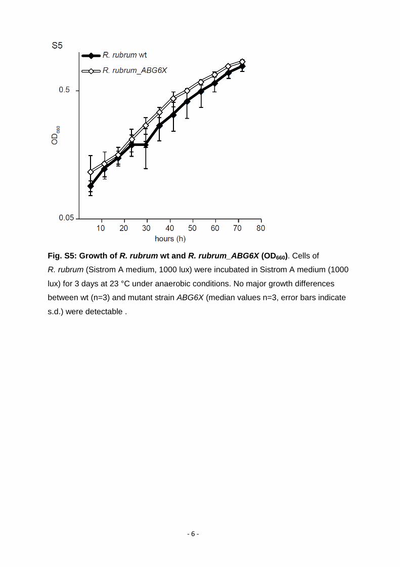
Fig. S5: Growth of R. rubrum wt and R. rubrum_ABG6X (OD660). Cells of
R. rubrum (Sistrom A medium, 1000 lux) were incubated in Sistrom A medium (1000
lux) for 3 days at 23 °C under anaerobic conditions. No major growth differences
between wt (n=3) and mutant strain ABG6X (median values n=3, error bars indicate
s.d.) were detectable .

- 7 -
Fig. S6: Proteomic analysis of magnetosomes from R. rubrum_ABG6X. (a) 1D
SDS-PAGE of Coomassie blue stained proteins solubilized from isolated
magnetosome particles of M. gryphiswaldense and R. rubrum_ABG6X. Bands of the
same size are indicated (arrowheads). (b) Immunodetection of MamC (12.4 kDa) in
blotted fractions of M. gryphiswaldense and R. rubrum_ABG6X using an anti-MamC
antibody6. A signal for MamC was detectable in the magnetic membrane fraction of
R. rubrum_ABG6X (6), which was absent from the soluble fraction, but faintly present
also in the non-magnetic membrane fraction (5), possibly originating from empty
membrane vesicles or incomplete magnetic separation during isolation. Protein
extracts from M. gryphiswaldense: 1. soluble fraction, 2. non-magnetic membrane
fraction, 3. magnetosome membrane. Protein extracts from R. rubrum_ABG6X: 4.
soluble fraction, 5. non-magnetic membrane fraction, 6. magnetic (“magnetosome”)
membrane fraction. M: Marker.

- 8 -
Fig. S7: Fluorescence microscopy of R. rubrum wt and R. rubrum_ABG6X cells
expressing different EGFP-tagged magnetosome proteins. For localization
studies of fluorescently labeled magnetosome proteins, strains were cultivated in
ATCC medium overnight at 30 °C with appropriate antibiotics (Table S3). (a & b)
MamGFDC with a C-terminal MamC-EGFP fusion expressed in R. rubrum wt (n=151)
(a), and R. rubrum_ABG6X (n=112) (b). In the transformed strain, a filamentous
structure is visible for 79% of the cells (n=89). c & d, MamJ-EGFP expressed in
R. rubrum wt (n=109) (c), and in R. rubrum_ABG6X (n=89) displaying a chain-like
fluorescence signal in 63% of the cells (n=56) (d). Scale bar: 2 µm.
- 9 -
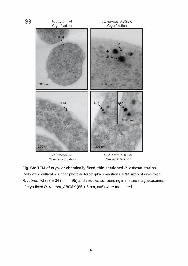
Fig. S8: TEM of cryo- or chemically fixed, thin sectioned R. rubrum strains.
Cells were cultivated under photo-heterotrophic conditions. ICM sizes of cryo-fixed
R. rubrum wt (93 ± 34 nm, n=95) and vesicles surrounding immature magnetosomes
of cryo-fixed R. rubrum_ABG6X (66 ± 6 nm, n=6) were measured.
- 10 -
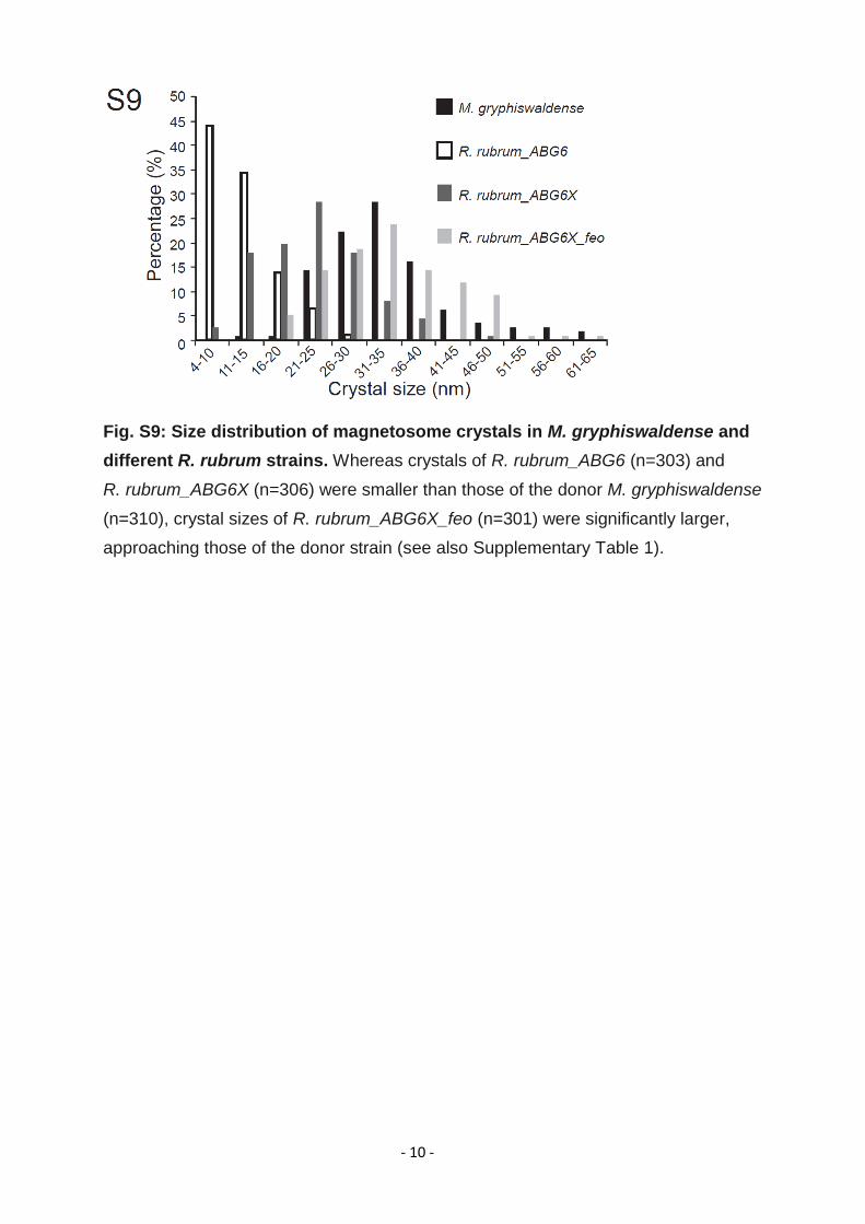
Fig. S9: Size distribution of magnetosome crystals in M. gryphiswaldense and
different R. rubrum strains. Whereas crystals of R. rubrum_ABG6 (n=303) and
R. rubrum_ABG6X (n=306) were smaller than those of the donor M. gryphiswaldense
(n=310), crystal sizes of R. rubrum_ABG6X_feo (n=301) were significantly larger,
approaching those of the donor strain (see also Supplementary Table 1).

- 11 -
Figure S10: Transmission electron micrographs of whole cells of different
R. rubrum strains expressing magnetosome gene clusters. Scale bar: 1 µm,
inset: 0.2 µm.

- 12 -
1 Rubin, E. J. et al. In vivo transposition of mariner-based elements in enteric bacteria and mycobacteria. Proc. Natl. Acad. Sci. U S A 96, 1645-1650 (1999).
2 Scheffel, A. & Schüler, D. The acidic repetitive domain of the Magnetospirillum gryphiswaldense MamJ protein displays hypervariability but is not required for magnetosome chain assembly. J. Bacteriol. 189, 6437-6446 (2007).
3 Lohsse, A. et al. Functional analysis of the magnetosome island in Magnetospirillum gryphiswaldense: the mamAB operon is sufficient for magnetite biomineralization. PLoS One 6, e25561 (2011).
4 Schübbe, S. et al. Characterization of a spontaneous nonmagnetic mutant of Magnetospirillum gryphiswaldense reveals a large deletion comprising a putative magnetosome island. J. Bacteriol. 185, 5779-5790 (2003).
5 Ghosh, R., Hardmeyer, A., Thoenen, I. & Bachofen, R. Optimization of the Sistrom Culture Medium for Large-Scale Batch Cultivation of Rhodospirillum rubrum under Semiaerobic Conditions with Maximal Yield of Photosynthetic Membranes. Appl. Environ. Microbiol. 60, 1698-1700 (1994).
6 Lang, C. & Schüler, D. Expression of green fluorescent protein fused to magnetosome proteins in microaerophilic magnetotactic bacteria. Appl. Environ. Microbiol. 74, 4944-4953 (2008).


