download the lipid spin (spring 2013) - national lipid association
TRANSCRIPT
Volume 11 Issue 2 Spring 2013visit www.lipid.org
Official Publication of the National Lipid Association
LipidSpin
Also in this issue: “HDL-P vs. ApoA1 vs. HDL-C” in Context of the HDL-Hypothesis ControversyThe Role of Remnant Lipoproteins in Atherogenesis
This issue sponsored by the Pacific Lipid Association
Clinical Feature:The Evolution of Lipid, Lipoprotein, and Apolipoprotein Markers of CVD Risk and Therapeutic Targets—Is it Time to Abandon the Cholesterol Content of Atherogenic Lipoproteins?


1
2 From the NLA President Our Pursuit Continues—Peter P. Toth, MD, PhD, FNLA*
3 From the PLA PresidentApolipoproteins in Clinical Practice —J. Antonio G. López, MD, FNLA
4 Letter from the Lipid Spin EditorsThe Times They Are A-changin’—Robert A. Wild, MD, PhD, FNLA*
5 Clinical FeatureIs it Time to Abandon the Cholesterol Content of Atherogenic Lipoproteins?—Paul D. Rosenblit, MD, PhD, FNLA*—Edward A. Gill, MD, FNLA*—Robert G. Thompson, MD*
12 Guest EditorialThe Role of Remnant Lipoproteins in Atherogenesis—John R. Nelson, MD, FNLA*—Paul N. Hopkins, MD, MSPH*
16 EBM Tools for Practice“HDL-P vs. ApoA1 vs. HDL-C” in Context of the HDL-Hypothesis Controversy—Michael D. Shapiro, DO*—Eliot A. Brinton, MD, FNLA*
18 Lipid LuminationsLipoprotein(a)—Clinical Significance, Evaluation, and Management —P. Barton Duell, MD
22 Specialty CornerNephrology Corner—Limitations of Statin Use and Adjuvant Therapies in Stage IV CKD and Dialysis —Michael J. Bloch, MD* —Ali Olyaei, PharmD
27 Practical PearlsCardiac Auscultation for the Lipidologist: A Systolic Murmur You Do Not Want to Miss!—J. Antonio G. López, MD, FNLA*—John R. Nelson, MD, FNLA*
30 Case StudyDigging Deeper—A Case for Apolipoproteins and Lifestyle in Office Practice —Rob Greenfield, MD, FNLA*—Susan Given, cFNP, MN, BSN
32 Chapter Update —B. Alan Bottenberg, DO, FNLA*
34 Member Spotlight: Daniel Steinberg, MD, PhD
35 Education, News and Notes
36 Events Calendar
37 Foundation Update
38 References
41 Patient Tear Sheet
Look for the NLA Community logo to discuss articles online at www.lipid.org
In This Issue: Spring 2013 (Volume 11, Issue 2)
EditorsJAMES A. UNDERBERG, MD, MS, FACPM, FACP, FNLA* Preventive CV Medicine, Lipidology and Hypertension Clinical Assistant Professor of Medicine NYU Medical School and Center for CV Prevention New York, NY
ROBERT A. WILD, MD, PhD, MPH, FNLA* Clinical Epidemiology and Biostatistics andClinical Lipidology ProfessorOklahoma University Health Sciences CenterOklahoma City, OK
Managing EditorMEGAN L. SEERY National Lipid Association
Executive DirectorCHRISTOPHER R. SEYMOUR, MBA National Lipid Association
Contributing EditorKEVIN C. MAKI, PhD, CLS, FNLA
Associate Editor for Patient EducationVANESSA L. MILNE, MS, NP, CLSCardiac Vascular Nurse and Family Nurse PractitionerBellevue Hospital Lipid ClinicNew York, NY
Lipid Spin is published quarterly by the National Lipid Association 6816 Southpoint Parkway, Suite 1000 Jacksonville, FL 32216 Phone: 904-998-0854 | Fax: 904-998-0855
Copyright ©2013 by the NLA. All rights reserved.
Visit us on the web at www.lipid.org.
The National Lipid Association makes every effort to provide accurate information in the Lipid Spin at the time of publication; however, circumstances may alter certain details, such as dates or locations of events. Any changes will be denoted as soon as possible. The NLA invites members and guest authors to provide scientific and medical opinion, which do not necessarily reflect the policy of the Association.
*indicates ABCL Diplomate status

2 LipidSpin
From the NLA President: Our Pursuit Continues
The NLA has consistently held a determined focus on promoting the highest levels of education and professional development. We are defined by the quality of our outstanding leadership, members, partners and staff. As a result, we are continuously challenged to assess and refine our work in an attempt to produce the best and most innovative programming in Clinical Lipidology, leading to a direct and measurable impact in our medical community and across the globe.
With our core mission in mind, a group of NLA leaders met in Miami from February 8-10 for biannual strategic planning, where we were faced with important topics about resources and positioning the association far into the future. I am gratified to report that we emerged with clear priorities in the areas of Professional Education, Policy Statements, and Practice Management
and Research. Additionally, we discussed ongoing and new endeavors in the areas of Communications and Membership.
We have made targeted recommendations regarding Leadership Development and Member Responsibilities. In particular, chapter leaders and members at the regional levels will be given greater responsibility for projects, and chapter leaders will develop annual plans. Chapter bylaws will be made uniform so they are consistent throughout the association. In addition, some NLA committees will review and refine their charges and initiatives.
The strategic planning recommendations will be brought before the NLA Board for consideration during our Annual Scientific Sessions in Las Vegas this coming May. I look forward to sharing our strategic plan with you once adopted and approved.
Additionally, the NLA hosted yet another successful regional meeting, the Spring Clinical Lipid Update, in New Orleans this past February. The conference was co-hosted by the Midwest Lipid Association and Southwest Lipid Association, who provided key expertise in the planning
and execution of the Spring CLU. After the conference concluded, Foundation of the NLA President Anne Goldberg, MD, hosted some of our partner organizations during our inaugural FH Roundtable in New Orleans. At the Roundtable, these groups discussed forming a collective identity known as the FH Consortium, with the goal of further moving FH awareness to the national stage and assisting patients with finding treatment.
Moving ahead, I look forward to seeing you at our Annual Scientific Sessions in Las Vegas from May 30-June 2. I always enjoy the NLA meetings because they are characterized by people who are passionate about ideas and who share common values and intellectual energy. Let’s gather once again to share with and learn from one another in our pursuit of new knowledge as well as personal and career achievement. n
Discuss this article at www.lipid.org/lipidspin
PETER P. ToTh, MD, PhD, FNLAPresident, National Lipid AssociationDirector of Preventative CardiologyCGH Medical CenterSterling, ILProfessor of Clinical Family and Community MedicineUniversity of Illinois School of MedicinePeoria, ILDiplomate, American Board of Clinical Lipidology

Official Publication of the National Lipid Association 3
From the PLA President: Apolipoproteins in Clinical Practice
I am pleased to present to the National Lipid Association the Spring 2013 issue of the Lipid Spin. The Pacific Lipid Association serves the states of Washington, Oregon, California, Idaho, Montana, Nevada, Utah, Alaska, and Hawaii. The theme of this Spring issue is “Roles of Common Apolipoproteins in Cardiovascular Disease (CVD) and How They Affect Our Clinical Practice.” The authors discuss apolipoproteins, their relevance, a review of the literature and the implications for clinical practice. I am proud of the contributions of our PLA members.
I am honored that pioneering lipid researcher, Daniel Steinberg, MD, PhD, professor emeritus of medicine at the University of California-San Diego, is featured in this issue as the member spotlight. Dr. Steinberg is one of the original proponents of the cholesterol hypothesis of atherosclerosis. San Diego, California is my hometown.
At this midterm juncture of my presidency, I am thankful for the support of B. Alan Bottenberg, Do, President-elect; Paul D. Rosenblit, MD, PhD, Treasurer; Wayne S. True, MD, Secretary; and Immediate
Past-President John R. Nelson, MD. For this issue of the Lipid Spin, I am also thankful for the work of the editors, Jamie Underberg, MD, and Robert Wild, MD, PhD. This issue would not have been completed without the assistance of the NLA staff and, in particular, Megan Seery.
An important mission of the PLA and NLA is to promote responsible outreach to all regions of the country to make available the benefits of the NLA in every community. I encourage all of our members to continue our collective effort in achieving this goal.
We invite you to attend the 2013 NLA Annual Scientific Sessions in our own Las Vegas, Nevada, from May 30-June 2. The PLA will serve as the regional host for the sessions at the Red Rock Hotel in Las Vegas. You will not want to miss out on the latest in lipid research and applications. n
J. ANToNIo G. LÓPEZ, MD, FACC, FAhA, FACP, FCCP, FNLAPacific Lipid Association President
Director, Preventive Cardiology and Cardiovascular RehabilitationDirector, Lipid Clinic and LDL Apheresis Program
Chair, Department of CardiologySaint Alphonsus Regional Medical Center
Boise, ID
Diplomate, American Board of Clinical Lipidology
Discuss this article at www.lipid.org/lipidspin
4 LipidSpin
Letter From the Lipid Spin Editors: The Times They Are A-changin’
…the times they are a-changin’ ~Bob Dylan
These are exciting times for Lipidologists! We can always count on change as the one sure thing in life.
Our field is changing. As health care evolves, the NLA is doing its part to position our field to take advantage of the changes that our system demands. Our leaders are providing new ways to navigate systems to the benefit of our patients. As a multidisciplinary organization, we now have even more ways for our many disciplines to contribute to accomplishing our goal: improving the practice of Clinical Lipidology. In addition to mainstream primary care, we also have taken efforts to educate within Pediatrics, Ob/Gyn and Geriatrics.
We all know that reimbursement affects the care of our patients. I am excited to see that the Centers for Medicare and Medicaid
Services are now rewarding efforts to deliver lifestyle interventions in a practical and meaningful way. I am also pleased to see that as ATP IV progresses, recommendations are being developed based on a thorough and systematic examination of existing evidence. Happily, we are living in times that are more influenced by evidence rather than opinion. The NLA has developed task forces to deal with each aspect of ATP IV, recognizing that this is a wonderful way to get our message out based on the best evidence available for risk assessments and therapy.
As you will learn in this issue, many of us participated in a strategic planning session in Miami in February. We came away from the meeting energized. The NLA is consolidating; you will note more evenness in the governance of each region. There are new efforts to recognize regional leaders for their hard work. While we recognize that each region has unique challenges, we also know that uniform rules of governance are more efficient.
Our mission is to educate all professionals who practice Clinical Lipidology so that in turn our patients’ lives will be improved. Steps are under way to bring you the best evidence available to integrate clinical
judgment and patient preferences with systems management. We are making strides to streamline the process, and targeting our educational offerings to move towards mature learning principles. We are looking at newer and better ways to communicate our message.
At the strategic planning meeting we discussed newer and better ways to make the Lipid Spin even more meaningful. We have the fortunate problem of having to turn down submissions! However, this has created a challenge. During the editorial process, we receive submissions that do not fit within an issue’s regional theme or scope. Many of the submissions are of good quality, yet do not fit into the theme of an upcoming issue. We are working hard to develop a solution and look forward to sharing it with you in the coming months. In the meantime, we hope you continue to look to the Lipid Spin as a practical resource to help your practice.
…the times they are a-changin’…and the times are exciting! Prevention is finally earning its spot in the limelight. At the NLA we recognize that an ounce of prevention is worth a pound of cure. Let’s embrace the concept and work even harder to spread our message. n
Discuss this article at www.lipid.org/lipidspin
RoBERT A. WILD, MD, PhD, MPh, FNLAClinical Epidemiology and Biostatistics andClinical Lipidology ProfessorOklahoma University Health Sciences CenterOklahoma City, OKDiplomate, American Board of Clinical Lipidology
Official Publication of the National Lipid Association 5
Clinical Feature: Is it Time to Abandon the Cholesterol Content
of Atherogenic Lipoproteins?
The year 2013 marks the “Silver Anniversary” of three key announcements that identified relationships among various lipid fractions, lipoproteins, apolipoproteins, non-lipid disorders and cardiovascular disease (CVD) risk. In1988, NCEP ATP I1 recognized evidence that high levels of low density lipoprotein cholesterol (LDL-C) contributed to increased CVD risk and subsequently this has become the primary lipoprotein lipid target to reduce CVD.2-4 They recognized that high density lipoprotein cholesterol (HDL-C) was associated with reduced CVD risk.5-7 The 1988 NCEP ‘expert laboratory panels’ also provided guidelines for measuring LDL-
C9, HDL-C10, and triglycerides (TG).11 Both HDL-C and calculated LDL-C have remained cornerstones of lipoprotein lipid measurements for guiding lipid-lowering therapy for over 25 years.
In the second key 1988 announcement, Gerald Reaven, MD, described the etiology of known clustered metabolic CVD risks as, primarily, a consequence of resistance to insulin-stimulated glucose uptake and resistance to insulin-stimulated suppression of adipose tissue lipolysis. Insulin resistance, secondarily, leads to compensatory hyperinsulinemia, impaired glucose tolerance (IGT), elevated circulating free fatty acids and TG and
decreased circulating HDL-C. He suggested that the insulin resistance ‘syndrome X’ played a central role in the pathogenesis and clinical course of type 2 diabetes mellitus (T2DM), hypertension, and CAD, and “likely explained most ofthe CVD risk in the general population” including many obese, overweight, or physically inactive individuals, as well as, those
Discuss this article at www.lipid.org/lipidspin
PAUL D. RoSENBLIT, MD, PhD, FACE, FNLADirector, Diabetes/Lipid Management & Research Center, Huntington Beach, CA
Clinical Professor, Dept. of Medicine, Division Endocrinology/Diabetes/Metabolism,University of California, Irvine (UCI) School of Medicine, Irvine, CA
Co-Director, Diabetes Out-Patient Clinic, UCI Medical Center, Orange, CADiplomate, American Board of Clinical Lipidology
EDWARD A. GILL, MD, FAhA, FASE, FACP, FACC, FNLAProfessor of Medicine, Department of Medicine, Division of Cardiology
Adjunct Professor of Radiology, Director of EchocardiographyHarborview Medical Center, University of Washington
Clinical Professor of Diagnostic UltrasoundSeattle University
Seattle, WA Diplomate, American Board of Clinical Lipidology
RoBERT G. ThoMPSoN, MD, FACCSwedish Hospital Medical Center
Seattle, WADiplomate, American Board of Clinical Lipidology

6 LipidSpin
individuals with T2DM.12 The third 1988 key announcement shed doubt on the simplicity and predictability of LDL-C per se. Researchers from Lawrence Berkeley Laboratory introduced the Atherogenic Lipoprotein Phenotype (ALP) concept, with the identification of two distinct lipoprotein phenotypes; (Pattern A) characterized by a predominance of large, buoyant LDL particles and (Pattern B)characterized by more circulating small, dense LDL particles. They found that, compared with the Pattern A, Pattern B phenotype was associated with greater risk of myocardial infarction.13-15 Pattern B dyslipidemia was also associated with increased apolipoprotein B (ApoB), VLDL, hypertriglyceridemia, and decreased levels of HDL-C and apolipoprotein A-I (ApoA1) levels.13
A link was established between Syndrome X, T2DM, and ALP.16,17 Having plasma TG concentration >130 mg/dL, a TG/HDL >3.0 and insulin concentrations (>109 pmol/L) aided in the identification of overweight individuals who were sufficiently insulin resistant to be at increased risk for CVD outcomes.18 An increased TG/HDL ratio, a surrogate of insulin resistance, was highly predictive of a first coronary event, regardless of BMI value.19
The Copenhagen Prospective Cardiovascular (Male) Study, estimated that approximately 35% of the population-attributable CVD risk, associated with high TG and low HDL-C levels, was independent of LDL-C level, hypertension, smoking history, or level of physical activity.20 The Helsinki Heart Study (HHS) reported a link between the high TG and low HDL-C and greater risk for CHD. They found that while there was an overall 34% relative risk reduction among gemfibrozil users for the entire HHS cohort (mean TG 176 mg/dL); almost the entire benefit of gemfibrozil (a 56-71% RRR) was noted in
the group defined by either low HDL-C or high TG or both. The investigators suggested a personalized or individualized targeting or “tailoring of drug therapy” with fibrates, for this high risk group.21 The early monotherapy fibrate studies may have influenced the NCEP ATP III panel’s recommendations for fibrate use in these subgroups.
Although hypothesis-generating observations (in need of a dedicated clinical trial to test this hypothesis in this population as the primary cohort) similar results were obtained in four subsequent fibrate studies. There was remarkable consistency noted in all of the post-hoc subgroups analyses from each primary study (usually analyzing <20% of the entire primary cohorts). Three independent meta-analyses, combining ‘moderate dyslipidemia’ subgroups, in all five trials (HHS, VA-HIT, BIP, FIELD, ACCORD-Lipid), demonstrated the consistent highly significant fibrate benefit.22-24
Metabolically circulating LDL-C must first undergo some modification and this affects the structure of its apolipoprotein B (ApoB) moiety. This is necessary for it to become a ligand for the scavenger
receptors of monocyte macrophages. As a gradient-driven diffusion process, the more LDL-ApoB particles present in the circulation, whether by overproduction or by reduced clearance, the more LDL-ApoB particles infiltrate arterial walls. This sets in motion the cascade of events that leads to atherosclerosis.25 The intimal retention of LDL-ApoB particles is thought to reflect an imbalance between the entry and the efflux of lipoproteins via the media and its adventitia.26-28
Recognizing the importance of all atherogenic lipoprotein particle concentrations, the NCEP ATP III (2001) identified non-HDL-C as a secondary target for therapy, after LDL-C goals have been met, in patients who have elevated triglyceride levels >200 mg/dL.Non-HDL-C is a surrogate for all of apolipoprotein-B-containing particles, carrying cholesterol into the arterial wall [LDL-C, VLDL-C, IDL-C, chylomicrons, chylomicron remnants, and Lp(a)]. Another secondary target identified by ATP III is having the ‘Metabolic Syndrome.’ ATP III also suggested that advanced cardiovascular panels could include, testing for ‘emerging risks’ such as ApoB and lipoprotein (a), ApoA1.29
Since LDL particles vary in both their cholesterol and triglyceride contents, LDL-C, per se, does not always provide a precise and/or accurate measure of the circulating concentration of heterogeneous LDL particles. This is particularly true in the hypertriglyceridemic environment, when LDL particles are particularly cholesterol-depleted, small in size and large in number. Nuclear magnetic resonance (NMR) spectroscopy, which measures lipoprotein particle concentrations directly has been utilized to study the significance of elevated low density lipoprotein particle concentrations (LDL-P). NMR analysis of the Framingham Offspring Study demonstrated a significant
Once the standardization of these biomarkers
is no longer debatable, particle concentrations are likely to become
mainstream measurements.
discordance between LDL-C and LDL-P in patients with low levels of HDL-C. This implied that the excess CAD risk likely results from an excess of cholesterol-depleted LDL particles and suggested that many patients with normal levels of LDL-C, but low-levels of HDL-C, would benefit from LDL-lowering therapy.30
The Framingham Heart Study data also found that LDL-P>LDL-C discordance is strongly linked to all five metabolic syndrome markers. Thus the enhanced risk of patients with metabolic syndrome may come from underappreciated or unrecognized LDL-P elevations. Of interest, in contrast to a graded association of increased small LDL-P with presence of more components of the metabolic syndrome, LDL-C concentrations per se, did not show a stepwise increase.31
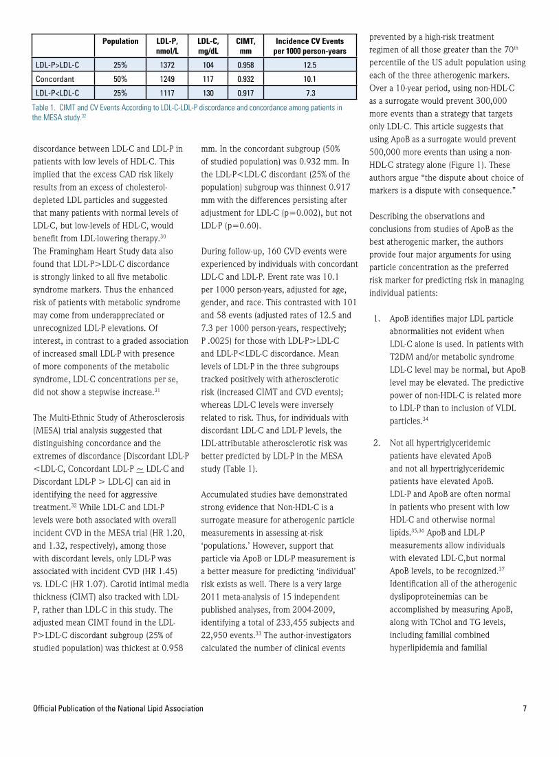
The Multi-Ethnic Study of Atherosclerosis (MESA) trial analysis suggested that distinguishing concordance and the extremes of discordance [Discordant LDL-P <LDL-C, Concordant LDL-P ~ LDL-C and Discordant LDL-P > LDL-C] can aid in identifying the need for aggressive treatment.32 While LDL-C and LDL-P levels were both associated with overall incident CVD in the MESA trial (HR 1.20, and 1.32, respectively), among those with discordant levels, only LDL-P was associated with incident CVD (HR 1.45) vs. LDL-C (HR 1.07). Carotid intimal media thickness (CIMT) also tracked with LDL-P, rather than LDL-C in this study. The adjusted mean CIMT found in the LDL-P>LDL-C discordant subgroup (25% of studied population) was thickest at 0.958
mm. In the concordant subgroup (50% of studied population) was 0.932 mm. In the LDL-P<LDL-C discordant (25% of the population) subgroup was thinnest 0.917 mm with the differences persisting after adjustment for LDL-C (p=0.002), but not LDL-P (p=0.60).
During follow-up, 160 CVD events were experienced by individuals with concordant LDL-C and LDL-P. Event rate was 10.1 per 1000 person-years, adjusted for age, gender, and race. This contrasted with 101 and 58 events (adjusted rates of 12.5 and 7.3 per 1000 person-years, respectively; P .0025) for those with LDL-P>LDL-C and LDL-P<LDL-C discordance. Mean levels of LDL-P in the three subgroups tracked positively with atherosclerotic risk (increased CIMT and CVD events); whereas LDL-C levels were inversely related to risk. Thus, for individuals with discordant LDL-C and LDL-P levels, the LDL-attributable atherosclerotic risk was better predicted by LDL-P in the MESA study (Table 1).
Accumulated studies have demonstrated strong evidence that Non-HDL-C is a surrogate measure for atherogenic particle measurements in assessing at-risk ‘populations.’ However, support that particle via ApoB or LDL-P measurement is a better measure for predicting ‘individual’ risk exists as well. There is a very large 2011 meta-analysis of 15 independent published analyses, from 2004-2009, identifying a total of 233,455 subjects and 22,950 events.33 The author-investigators calculated the number of clinical events
prevented by a high-risk treatment regimen of all those greater than the 70th percentile of the US adult population using each of the three atherogenic markers. Over a 10-year period, using non-HDL-C as a surrogate would prevent 300,000 more events than a strategy that targets only LDL-C. This article suggests that using ApoB as a surrogate would prevent 500,000 more events than using a non-HDL-C strategy alone (Figure 1). These authors argue “the dispute about choice of markers is a dispute with consequence.”
Describing the observations and conclusions from studies of ApoB as the best atherogenic marker, the authors provide four major arguments for using particle concentration as the preferred risk marker for predicting risk in managing individual patients:
1. ApoB identifies major LDL particle abnormalities not evident when LDL-C alone is used. In patients with T2DM and/or metabolic syndrome LDL-C level may be normal, but ApoB level may be elevated. The predictive power of non-HDL-C is related more to LDL-P than to inclusion of VLDL particles.34
2. Not all hypertriglyceridemic patients have elevated ApoB and not all hypertriglyceridemic patients have elevated ApoB. LDL-P and ApoB are often normal in patients who present with low HDL-C and otherwise normal lipids.35,36 ApoB and LDL-P measurements allow individuals with elevated LDL-C,but normal ApoB levels, to be recognized.37 Identification all of the atherogenic dyslipoproteinemias can be accomplished by measuring ApoB, along with TChol and TG levels, including familial combined hyperlipidemia and familial
Population LDL-P, nmol/L
LDL-C, mg/dL
CIMT, mm
Incidence CV Eventsper 1000 person-years
LDL-P>LDL-C 25% 1372 104 0.958 12.5
Concordant 50% 1249 117 0.932 10.1
LDL-P<LDL-C 25% 1117 130 0.917 7.3
Table 1. CIMT and CV Events According to LDL-C-LDL-P discordance and concordance among patients in the MESA study.32
Official Publication of the National Lipid Association 7

dysbetalipoproteinemia.38
3. Recognized errors in the measurement of HDL-C, a component of the Friedwald equation, may in turn affect the accuracy of non-HDL-C measurement.39 Clinical assays for ApoB, on the other hand, have become reliable, robust, and can be measured on non-fasting samples at low cost.40 Accordingly, ApoB is superior to LDL-C and non-HDL-C as a laboratory analysis and reducing laboratory error will in turn reduce clinical errors in individual patient care.
4. While in large statin trial populations, non-HDL-C and ApoB are generally equivalent risk markers, ApoB is superior for identification of the individual that will benefit from an increased dose of statin. In statin-treated ‘populations,’ ApoB level identifies more individuals at increased risk, compared with LDL-C measurements.41
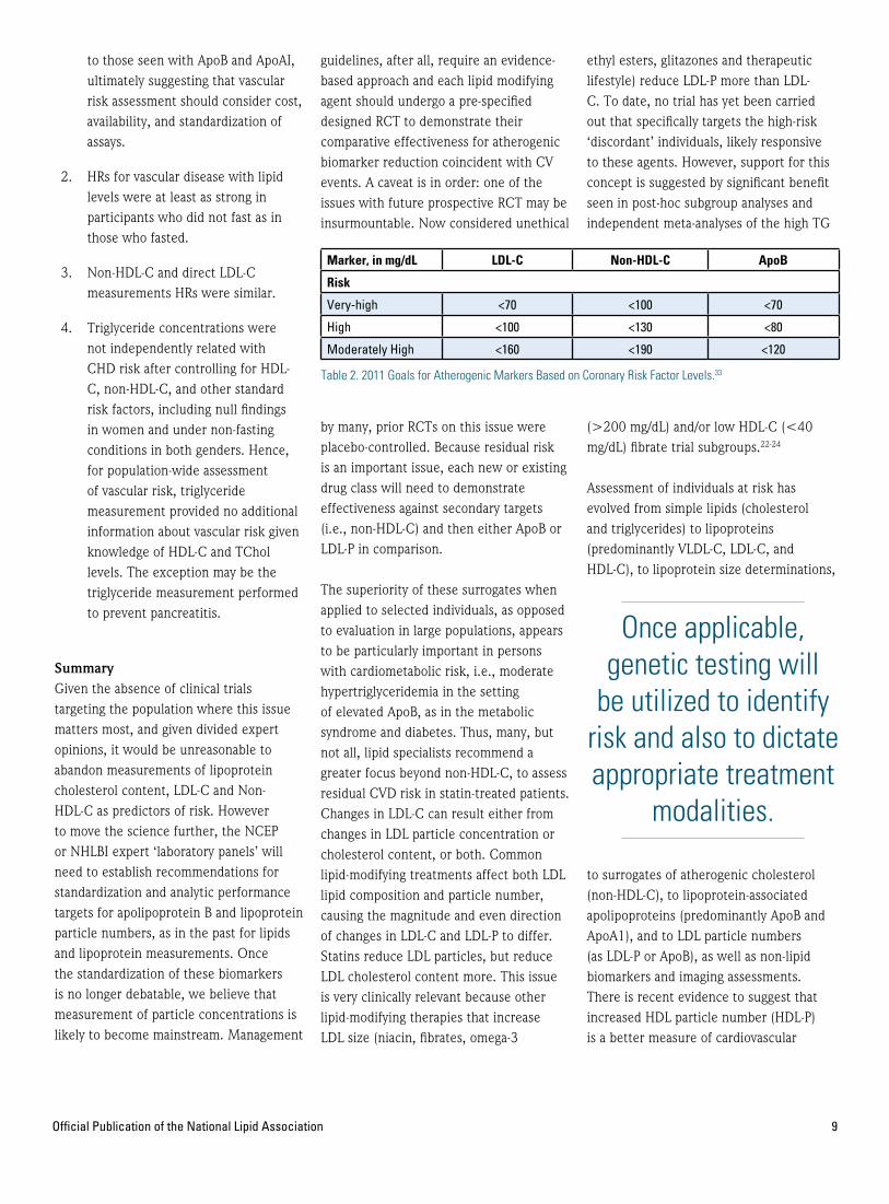
Based on the most recent statin clinical trials, Sniderman, Williams, Contois, et al.32, in 2011, suggested that in patients at ‘very high risk,’ the ApoB target should be <70 mg/dL, with no lower limits. They suggested for those patients at ‘high risk’ an appropriate ApoB target should be <80 mg/dL and for the ‘moderately high risk’ patients, the ApoB target would be <120 mg/dL. For non-HDL-C the targets are <100mg/dL, <130 mg/dL and <190 mg/dL, respectively; and for LDL-C <70mg/dL, <100 mg/dL and <160 mg/dL, respectively (Table 2).
A key concept is the inverse relationship that exists between HDL-C and ApoB or LDL-P, such that lower levels of HDL-C tend to be associated with higher levels of ApoB.42 Both HDL and LDL can
participate in cholesteryl ester transfer protein (CETP)-mediated lipid exchange where the VLDL-triglyceride moves to the HDL and LDL particles in exchange for cholesterol ester moving to the VLDL fraction. Thus, a higher HDL-C points to less core lipid exchange and greater concordance between LDL-C and ApoB. Conversely, a lower HDL-C points to more core lipid exchange and, therefore, greater discordance between LDL-C and ApoB. When ApoB and LDL-C are concordant, they predict risk equally, whereas when they are discordant, ApoB will be superior. Therefore, compositional changes related to CETP mediated lipid exchange explain much of the variance in predictive power between LDL-C and ApoB.
Considerable controversy continues to exist with regard to the need for additional markers beyond LDL-C and non-HDL-C. Not all studies show this superiority of ApoB over non-HDL-C. Among statin-treated patients (n=38,153), on-treatment levels of LDL-C, non-HDL-C, and ApoB
were each associated with risk of future major cardiovascular events, but the strength of association, relative to LDL-C (HR 1.13) was greater for non-HDL-C (HR 1.16, p = 0.002) than for ApoB (HR 1.14, p=0.02).43
In a very large analysis (n=302,430) of people, without initial vascular disease, from 68 long-term prospective studies, mostly in Europe and North America, involving 2.79 million person-years of follow-up, there were 8,857 nonfatal myocardial infarctions, 3,928 coronary heart disease [CHD] deaths, 2,534 ischemic strokes, 513 hemorrhagic strokes, and 2,536 unclassified strokes. The analysis44 demonstrated that lipid risk assessment can be simplified by measurement of either cholesterol levels or apolipoproteins, without the need to fast, and without regard to triglyceride. This conclusion derives from several findings including:
1. Hazard ratios (HRs) with non-HDL-C and HDL-C that were nearly identical
8 LipidSpin
Figure 1.
to those seen with ApoB and ApoAI, ultimately suggesting that vascular risk assessment should consider cost, availability, and standardization of assays.
2. HRs for vascular disease with lipid levels were at least as strong in participants who did not fast as in those who fasted.
3. Non-HDL-C and direct LDL-C measurements HRs were similar.
4. Triglyceride concentrations were not independently related with CHD risk after controlling for HDL-C, non-HDL-C, and other standard risk factors, including null findings in women and under non-fasting conditions in both genders. Hence, for population-wide assessment of vascular risk, triglyceride measurement provided no additional information about vascular risk given knowledge of HDL-C and TChol levels. The exception may be the triglyceride measurement performed to prevent pancreatitis.
SummaryGiven the absence of clinical trials targeting the population where this issue matters most, and given divided expert opinions, it would be unreasonable to abandon measurements of lipoprotein cholesterol content, LDL-C and Non-HDL-C as predictors of risk. However to move the science further, the NCEP or NHLBI expert ‘laboratory panels’ will need to establish recommendations for standardization and analytic performance targets for apolipoprotein B and lipoprotein particle numbers, as in the past for lipids and lipoprotein measurements. Once the standardization of these biomarkers is no longer debatable, we believe that measurement of particle concentrations is likely to become mainstream. Management
guidelines, after all, require an evidence-based approach and each lipid modifying agent should undergo a pre-specified designed RCT to demonstrate their comparative effectiveness for atherogenic biomarker reduction coincident with CV events. A caveat is in order: one of the issues with future prospective RCT may be insurmountable. Now considered unethical
by many, prior RCTs on this issue were placebo-controlled. Because residual risk is an important issue, each new or existing drug class will need to demonstrate effectiveness against secondary targets (i.e., non-HDL-C) and then either ApoB or LDL-P in comparison.
The superiority of these surrogates when applied to selected individuals, as opposed to evaluation in large populations, appears to be particularly important in persons with cardiometabolic risk, i.e., moderate hypertriglyceridemia in the setting of elevated ApoB, as in the metabolic syndrome and diabetes. Thus, many, but not all, lipid specialists recommend a greater focus beyond non-HDL-C, to assess residual CVD risk in statin-treated patients. Changes in LDL-C can result either from changes in LDL particle concentration or cholesterol content, or both. Common lipid-modifying treatments affect both LDL lipid composition and particle number, causing the magnitude and even direction of changes in LDL-C and LDL-P to differ. Statins reduce LDL particles, but reduce LDL cholesterol content more. This issue is very clinically relevant because other lipid-modifying therapies that increase LDL size (niacin, fibrates, omega-3
ethyl esters, glitazones and therapeutic lifestyle) reduce LDL-P more than LDL-C. To date, no trial has yet been carried out that specifically targets the high-risk ‘discordant’ individuals, likely responsive to these agents. However, support for this concept is suggested by significant benefit seen in post-hoc subgroup analyses and independent meta-analyses of the high TG
(>200 mg/dL) and/or low HDL-C (<40 mg/dL) fibrate trial subgroups.22-24
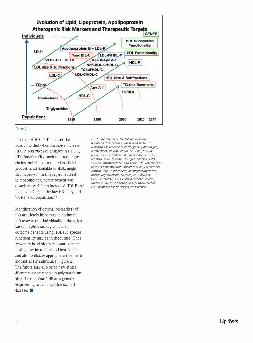
Assessment of individuals at risk has evolved from simple lipids (cholesterol and triglycerides) to lipoproteins (predominantly VLDL-C, LDL-C, and HDL-C), to lipoprotein size determinations,
to surrogates of atherogenic cholesterol (non-HDL-C), to lipoprotein-associated apolipoproteins (predominantly ApoB and ApoA1), and to LDL particle numbers (as LDL-P or ApoB), as well as non-lipid biomarkers and imaging assessments. There is recent evidence to suggest that increased HDL particle number (HDL-P) is a better measure of cardiovascular
Official Publication of the National Lipid Association 9
Marker, in mg/dL LDL-C Non-HDL-C ApoB
Risk
Very-high <70 <100 <70
High <100 <130 <80
Moderately High <160 <190 <120
Table 2. 2011 Goals for Atherogenic Markers Based on Coronary Risk Factor Levels.33
Once applicable, genetic testing will
be utilized to identify risk and also to dictate appropriate treatment
modalities.
10 LipidSpin
risk than HDL-C.47 This raises the possibility that when therapies increase HDL-P, regardless of changes in HDL-C, HDL functionality, such as macrophage cholesterol efflux, or other beneficial properties attributable to HDL, might also improve.42 In this regard, at least as monotherapy, fibrate benefit was associated with both increased HDL-P and reduced LDL-P, in the low-HDL targeted VA-HIT trial population.48
Identification of optimal biomarkers of risk are clearly important to optimum risk assessment. Individualized therapies based on pharmacologic-induced outcome benefits using HDL sub-species functionality may be in the future. Once proven to be clinically relevant, genetic testing may be utilized to identify risk and also to dictate appropriate treatment modalities for individuals (Figure 2). The future may also bring new ethical dilemmas associated with polymorphism identification that facilitates genetic engineering to avoid (cardiovascular) disease. n
Disclosure statement: Dr. Gill has received honoraria from Lantheus Medical Imaging. Dr. Rosenblit has received research grants from Amgen, AstraZeneca, Daiichi Sankyo Inc., Eisai, Eli Lilly & Co., GlaxoSmithKline, Mannkind, Merck & Co, Novartis, Novo Nordisk, Orexigen, Sanofi-Aventis, Takeda Pharmaceuticals, and Tolerx. Dr. Rosenblit has received honoraria from Abbott (Abbvie) Laboratories, Amarin Corp., AstraZeneca, Boeringher Ingelheim, Bristol-Meyers Squibb, Dexcom, Eli Lilly & Co., GlaxoSmithKline, Kowa Pharmaceuticals America, Merck & Co., NovoNordisk, Sanofi, and Santarus. Dr. Thompson has no disclosures to report.
Figure 2.
May 30–June 2, 2013 � National Lipid Association Scientific Sessions
Program Highlights Include:
� Emerging Therapies Special Session: Focus on PCSK9 � Evidence-based presentations direct from world-renowned thought leaders� Interactive breakouts with real-world applications for lipid practice �
Challenging case-based plenary sessions � CME and CE credit forphysicians, nurses, pharmacists and registered dietitians � Poster Sessionand Young Investigator Award
The National Lipid Association is a multidisciplinary healthcare community focused on the preventionof dyslipidemias and their associated cardiometabolic disorders.
www.lipid.org/sessions
The Evidence: Risk, Treatment and Outcomes
Managing an Array of Patients in Your PracticeOnly From the National Lipid Association
NLA.PreventativeMedicine.1.13:Layout 1 2/4/13 3:55 PM Page 1

JohN R. NELSoN, MD, FACC, FASNC, FNLAPacific Lipid Association Immediate Past PresidentDirector, California Cardiovascular InstituteAssistant Clinical Professor, UCSF School of MedicineFresno Medicine Residency Program-VolunteerFresno, CADiplomate, American Board of Clinical Lipidology
PAUL N. hoPkINS, MD, MSPhProfessor of Internal Medicine University of UtahCo-Director, Cardiovascular Disease Risk Reduction ClinicSalt Lake City, UTDiplomate, American Board of Clinical Lipidology
Guest Editorial: The Role of Remnant Lipoproteins in Atherogenesis
Remnant Lipoproteins Promote Foam Cell FormationAtherosclerosis is characterized by accumulation of inflammatory foam cells whose formation is promoted by the subendothelial retention of ApoB-containing lipoproteins. Plaques develop in predisposed areas of the arterial tree where blood flow is either slow or has a back and forth pattern (thus coronary arteries are particularly prone).1 In these predisposed areas endothelium displays increased susceptibility to inflammation as well as greater permeability to lipoproteins with subendothelial retention in these locations. Resident subendothelial dendritic cells may be the first cells to take up
retained lipoproteins to become foam cells. Some dendritic cell subtypes suppress while others promote inflammation.2 Hyperlipidemia initiates greater endothelial expression of inflammatory adhesion molecules (by multiple mechanisms) followed by macrophage and neutrophil transmigration into the subendothelial space. Eventually, macrophages as well as activated smooth muscle cells begin to accumulate and are converted to foam cells. Surprisingly, early acquisition of cholesterol by macrophages actually suppresses inflammatory responses, leading to a reparative macrophage phenotype.3 However, continued cholesterol accumulation, particularly with excessive intracellular unesterified cholesterol combined with stimulation of innate immune receptors (such as toll-like receptors), results in predominantly inflammatory macrophages. Further accumulation of macrophages (and other inflammatory cell types) ensues, followed
eventually by wholesale apoptosis and necrosis with formation of the necrotic core and an unstable plaque.4 These vulnerable plaques have a high cholesterol content, many macrophages at the shoulders, thinned fibrous caps, and are prone to rupture, leading to acute coronary events.5 The physical expansion caused by sudden cholesterol crystallization in such plaques may be a major driving force for their rupture.6
Excess cholesterol accumulation can lead to initiation, promotion, and progression of atherosclerotic lesions and may even precipitate plaque rupture and acute coronary events, but where does the cholesterol come from? In classical in vitro studies, incubation of macrophages with native LDL (low density lipoprotein) did not result in foam cell formation due to downregulation of the LDL receptor.7-9 However, after LDL were oxidized or acetylated they were avidly taken up by
Discuss this article at www.lipid.org/lipidspin
12 LipidSpin

macrophages with conversion to foam cells. Importantly, in these same studies, triglyceride-rich remnant lipoproteins (TGRL) from cholesterol-fed rabbits or dogs (referred to as β-VLDL) needed no modification to promote foam cell formation. Note that β-VLDL are TGRL with abnormal composition and are not equivalent to IDL (intermediate density lipoproteins). They are composed of both intestinal (with ApoB48) and hepatic (with ApoB100) TGRL remnants. β-VLDL have density less than 1.006 (the density of plasma) and float upon ultracentrifugation whereas IDL do not float. Unlike normal VLDL which have pre-β mobility, β-VLDL have β mobility upon electrophoresis, that is, they move like LDL. Finally, β-VLDL are abnormally enriched in cholesterol (mostly esterified) due to prolonged transit time and exchange of cholesteryl ester for triglycerides through the action of cholesterol ester transfer protein (CETP).10
The contribution of various forms of oxidized LDL (including minimally modified LDL) to foam cell formation in vivo continues to be debated.11 In the meantime, a number of additional LDL modifications that promote foam cell formation may even be more quantitatively important than oxidation. These include proteoglycan binding and aggregation, especially after exposure to various phospholipases (including LpPLA2) or sphingomyelinase, which result in so-called electronegative LDL.12 Besides β-VLDL, several other types of TGRL have also been shown to promote foam cell formation, including human VLDL from hypertriglyceridemic subjects13, human chylomicron remnants14, and remnant-like particles (RLP) isolated by incubation with immunoaffinity gels directed against a specific epitope on ApoB and ApoA1 with the intention to remove nascent TGRL and HDL.15,16 TGRL have been directly isolated from human aortic intima.17,18 In one study, 36% of the cholesterol isolated from aortic plaque in patients undergoing aortic
reconstruction was from very low density lipoprotein (VLDL) and intermediate dense lipoprotein (IDL).18
Chylomicron remnants (CR) are cholesterol-rich TGRL remnants produced from the hydrolysis of chylomicrons. These ApoB48-containing particles vary greatly in size and composition, becoming denser and less negatively charged as they lose triglycerides and their associated ApoC lipoproteins while increasing their concentration of cholesteryl ester. Human CR are in the range of 50 to 150 nm in diameter.19 Small VLDL and IDL are TGRL remnants produced from the hydrolysis of triglyceride-rich VLDL. Gradient density ultracentrifugation reveals small VLDL and IDL in the Sf (Svedberg flotation rate) 20 to 60 and 12 to 20 ranges respectively.10 The diameter of LDL, small VLDL, and IDL particles are, respectively, 20 to 25 nm, 30 to 80 nm, and 25 to 35 nm. The density of IDL is greater than 1.006, but less than 1.019 g/mL with a diameter of 27.5 to 30 nm in individuals without dyslipidemia. Approximately 15%
to 20% of the total cholesterol is carried in IDL and a normal plasma concentration of IDL is 5 to 15 mg/dL and a total mass of 10 to 30 mg/dL.10 Lipoproteins greater than 75 nm in diameter are thought to not enter the arterial wall.20 These considerations suggest that small CR and other TGRL remnants can enter the arterial wall and contribute to atherogenesis.
These findings may support the possibility that postprandial CR contribute to atherogenesis.21,22 Recently, much more ApoB48 was reported to be present in human carotid plaque than ApoB100.23 It appears to be the cholesteryl ester component of these remnant TGRL that is atherogenic as demonstrated by ACAT2 deficiency, which almost entirely abrogated atherosclerosis in ApoE null mice. In these knockout mice, there were normal or slightly increased numbers of both ApoB48 and B100 particles having markedly reduced cholesteryl ester and increased triglyceride content.24
Official Publication of the National Lipid Association 13
Figure 1. Risk of premature CAD (MI, CABG, or PTCA by age 60 in men or 70 in women) associated with type III hyperlipidemia among 1170 premature CAD cases and 1759 population-based controls. Type III was defined as measured VLDL-C / total triglycerides ≥ 0.30 with total triglycerides > 150 mg/dL. Risk associated with meeting this criteria (versus not) is given as “all.” Those with type III were further broken down as mild, moderate, and severe, defined as estimated β-VLDL cholesterol <50, 50-79, and 80 mg/dL or more, respectively. Risks were calculated by logistic regression adjusting for age, gender, measured LDL-C, HDL-C, fasting triglyceride category (excluding type III – see Figure 2), hypertension, diabetes, and history of cigarette smoking.
50
45
40
35
30
25
Odd
s Ra
tio
Type III all
0.68%
2.5-13.6
5.8
0.40%
0.3-4.8
1.2
0.17%
1.4-25
5.8
0.11%
7.7-273
46
mild moderate severe
Odds Ratio
95% CI
Prevalence
20
15
10
5
0
TGRL Remnants Can Initiate Endothelial InflammationUpon incubation with TGRL, endothelial cells upregulate their expression of MCP-1, ICAM-1, and VCAM-1.25,26 MCP-1 is a chemokine that stimulates monocyte integrin activation, allowing firm adherence to ICAM-1 and VCAM-1 while also promoting transendothelial migration. Incubation of monocytes with RLP also promotes their adherence to endothelial cells.27 RLP adversely affect endothelial function by directly and indirectly inhibiting endothelial nitric oxide synthase.28 Furthermore, elevated
RLP has been shown to be an independent risk factor for impaired flow-mediated, endothelial-dependant dilatation in patients with coronary artery disease.29 Elevated RLP levels have been associated with impaired coronary vasomotor response and acetylcholine-induced spasm.30,31 Elevated TGRLs were further found to be cytotoxic and induce apoptosis of endothelial cells.32
hydrolysis of TGRL May Also Activate Endothelial CellsHydrolysis of TGRL has been shown to
induce endothelial inflammation with production of TNFα, ICAM-1, and increased reactive oxygen species.33 In this study, it was the free fatty acids derived from the hydrolysis of TGRL, not the cholesteryl ester, triglycerides, free cholesterol or phospholipids that were associated with these effects.
Free fatty acids released during hydrolysis of TGRL can also adversely affect endothelial barrier function and increase subendothelial transfer of lipoproteins. In a study with cultured endothelial cells, exposure to oleic acid resulted in an increased transfer of LDL
across the endothelium.34 TGRL hydrolysis products were reported to increase endothelial permeability by promoting disruption of the zonula occludens-1 complex which is essential for tight junction formation. Increased caspase 3 activation was also seen, which can be associated with apoptosis.35 In another study, RLP were shown to induce a strong inflammatory response with vigorous NADPH oxidase activation and superoxide formation followed by apoptosis in endothelial cells through activation of the LOX1 receptor.36
Further observations on Foam Cell FormationIn the subendothelial space, monocytes differentiate into macrophages where they ingest ApoB-containing lipoproteins. The inaugural event is the subendothelial retention of ApoB lipoproteins.37 In a study of patients undergoing elective carotid endarterectomy, although the influx of LDL cholesterol was 19 times greater than that of TGRL cholesterol, the intimal clearance and fractional loss were similar.38 In a study of heritable hyperlipidemic rabbits, lipoprotein arterial influx was linearly related to plasma concentration; however, efflux was inversely related to lipoprotein diameter,39 suggesting the potential for greater retention of TGRL remnants. The main ApoB proteoglycan binding site is between the positively charged basic amino acids on ApoB (residues 3359 to 3369) and the negatively charged sulfate groups on the glycosaminoglycan chains of proteoglycans.40 Small VLDL and IDL have less affinity for proteoglycans; however, like LDL, sphingomyelinase causes VLDL and IDL to aggregate, fuse, and enhance their binding to proteoglycans.41 It has been shown that sphingomyelinase-induced aggregation of TGRL leads to foam cell formation.42
Although there is much greater penetration of the endothelial barrier by LDL particles, TGRLs carry significantly greater cholesteryl ester molecules per particle. It has been estimated that CR-TGRL of approximately 100 nm in diameter carry 40 times more cholesteryl ester than LDL particles.43 In a study evaluating TGRL and LDL fractions removed by density gradient ultracentrifugation from thoracic and abdominal aorta tissue at autopsy, it was found that when these fractions were incubated with mouse peritoneal macrophages, TGRL increased incorporation of radioactive oleate into cholesteryl esters by 10-to-20 fold as compared to three-to-four fold for LDL.17 Similar increases
14 LipidSpin
Odd
s Ra
tio
8
7
6
5
4
3
2
1
0
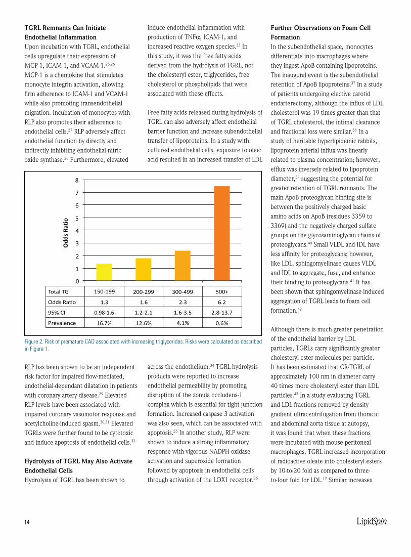
Total TG 150-199
16.7%
0.98-1.6
1.3
12.6%
1.2-2.1
1.6
4.1%
1.6-3.5
2.3
0.6%
2.8-13.7
6.2
200-299 300-499 500+
Odds Ratio
95% CI
Prevalence
Figure 2. Risk of premature CAD associated with increasing triglycerides. Risks were calculated as described in Figure 1.

in cholesteryl ester synthesis by were seen in studies with dogs over 30 years ago.7 In patients with type III or type IV hyperlipidemia, oxidized β-VLDL or VLDL remnants were found to cause greater macrophage cholesteryl ester formation than oxidized LDL.44,45
Coronary Risk Associated with Type III hyperlipidemiaType III hyperlipidemia is characterized by increased accumulation of β-VLDL in plasma. This phenotype is commonly thought to be rare, being the result of an apo E 2-2 genotype (about 1 in 100 persons) together with a genetic predisposition to excess VLDL production, such as APOA5 variants,46 or acquired overproduction of VLDL as with obesity or hypothyroidism. The prevalence of type III is frequently cited as approximately 1 in 10,000.47 However, in the Lipid Research Clinics (LRC) Prevalence Study, type III hyperlipidemia was found in 0.4% of men in the general population.48 This study represents one of the only studies to apply classic criteria to all participants to define type III, namely, the presence of a β-VLDL band upon electrophoresis of the density <1.006 fraction isolated after ultracentrifugation of plasma.
While markedly increased risk of atherosclerotic disease has long been appreciated for patients with type III, a population-based estimate of risk was not available until our recent publication (PNH).49,50 Additional, previously unpublished analyses utilizing data from the more recent of these studies50 are presented in Figures 1-3. The study groups consisted of 1759 population-based controls and 1170 cases with onset of clinical CAD by age 60 in men and 70 in women, all with ultracentrifugation performed on plasma samples. Type III hyperlipidemia was defined as present if the ratio of measured VLDL cholesterol/total triglycerides was ≥ 0.30 with total triglycerides > 150 mg/dL.51
The prevalence of type III (0.68%) we identified in the control population was very similar to the LRC Prevalence Study estimate, especially in consideration of the increased obesity expected in the population. The prevalence among our cases was 2.7%, almost identical to that reported by Goldstein, et al.52 In Figure 1, risk associated with the presence of type III is given with adjustment for LDL-C, HDL-C, triglyceride categories (which excluded type III subjects), hypertension, diabetes, and cigarette smoking. In addition to the traditional yes/no definition of type III, we show the markedly increasing risk associated with more severe type III as defined by an algebraic estimate of plasma β-VLDL cholesterol levels. CAD risk was increased over 40-fold in the most severe category. These severe cases represent only about 1/1000 control subjects yet most did not have any xanthomas. Perhaps those with tuberous xanthomas and/or palmar striae would be found as infrequently as 1/10,000. Elevations in triglycerides without type III were associated with increased CAD risk, but to a much lesser extent as shown in Figure 2. Interestingly, many cases of type III hyperlipidemia would have been
missed if ultracentrifugation had only been performed in those with triglycerides over 300-400 as shown in figure 3. It should be noted that estimates of risk associated with remnant accumulation can vary substantially, depending on the method or parameter used.10,53-55
In summary, despite significantly lower plasma concentrations than LDL, TGRL and TGRL remnants contribute to atherosclerosis plaque formation. With increasing obesity rates, these TGRL-derived particles may play a greater role in the development of atherosclerotic burden. Non-HDL cholesterol goals therefore may become even more important in the management of the dyslipidemic patient. n
Disclosure statement: Dr. Hopkins has received honoraria from Merck & Co. Dr. Hopkins has received research grants from Regeneron and Takeda Pharmaceuticals. Dr. Nelson has received honoraria from Abbott Laboraties, Amarin Corp., AstraZeneca, Atherotech, Bristol-Myers Squibb, Daiichi Sankyo Inc., GlaxoSmithKline, Gilead Pharmaceuticals, Kowa Pharmaceuticals America, Merck & Co., Pfizer Inc., and Novartis Pharmaceuticals.
Official Publication of the National Lipid Association 15
Perc
ent w
ithin
type
III c
ateg
ory
70
mild (n=14) moderate (n=15) severe (n=15)
Type III patients
150-199
200-299
300-499
500+
60
64
36
66
20
40
33
27
0
7 7
0 0
50
40
30
20
10
0
Figure 3. Distribution of type III hyperlipidemia cases (including cases and controls together) according to fasting triglycerides. Note the relatively large number of type III cases with only modest triglyceride elevations.

16 LipidSpin
Recent high-profile interventional studies and a large genetic association analysis have failed to show a benefit of raising high density lipoprotein cholesterol (HDL-C) levels on cardiovascular disease (CVD) outcomes, calling into question the validity of the HDL hypothesis. Among several plausible explanations for these findings, one is that assaying the cholesterol content of HDL (HDL-C) may fail to adequately measure its protective effects. Two potentially better ways to assess the protective effects of HDL are to measure levels of the major HDL apolipoprotein (apo), ApoA1, and to estimate HDL-particle number (HDL-P) by nuclear magnetic resonance (NMR).
The “Atherothrombosis Intervention in
Metabolic Syndrome with Low HDL/High Triglycerides: Impact on Global Health” (AIM-HIGH) study failed to show CVD benefit from HDL-C raising with niacin.1 The lack of benefit of niacin in this trial was surprising given the many pre-AIM-HIGH studies demonstrating that niacin reduces CVD events.2 In patients with low HDL-C and stable coronary artery disease, extended release nicotinic acid (ERNA) was added to statin therapy and subsequent CVD events were assessed. To better understand the impact of the HDL-C raising effect of ERNA, low density lipoprotein-cholesterol (LDL-C) was targeted to 40-80 mg/dL in both groups, leading to higher statin doses and more frequent ezetimibe use in the control group. Low-dose immediate-release nicotinic acid (IRNA) was given to the control group to cause flushing and maintain the study blind. Possible explanations for the surprising lack of CVD benefit included (1) near equalization of LDL-C levels (weighing against the HDL-hypothesis), (2) smaller-than-expected HDL-C difference of only 15% due to IRNA in the control arm, and (3) the short study
duration of only 2 ½ years3 (neither (2) nor (3) weighing against the HDL-hypothesis). Further, in a post hoc subgroup analysis in subjects having both high triglycerides and low HDL-C at baseline, there was a statistically significant 37% decrease in CVD events with high-dose ERNA vs. control. This finding clearly supports the traditional HDL hypothesis.4 An alternative explanation for the surprising results of AIM-HIGH is that the lack of CVD benefit with ERNA was expected since, despite a robust increase in HDL-C and ApoA1 with ERNA, HDL-P may not increase with ERNA treatment.
Another study with results appearing to weigh against the HDL hypothesis is the “Randomized, Double-blind, Placebo-controlled Study Assessing the Effect of RO4607381 on Cardiovascular Mortality and Morbidity in Clinically Stable Patients With a Recent Acute Coronary Syndrome” (dal-OUTCOMES) trial.5 In this study, dalcetrapib, a cholesteryl ester transfer protein inhibitor (CETP-I), failed to lower CVD events despite increasing HDL-C by 31%, (and previously
EBM Tools for Practice: “HDL-P vs. ApoA1 vs. HDL-C” in Context of the HDL-Hypothesis Controversy
Discuss this article at www.lipid.org/lipidspin
MIChAEL D. ShAPIRo, Do, FACC, FSCCTAssistant Professor of Medicine and RadiologyKnight Cardiovascular InstituteOregon Health & Science UniversityDirector, Preventive Cardiology and Atherosclerosis ImagingDiplomate, American Board of Clinical Lipidology
ELIoT A. BRINToN, MD, FNLADirector, Atherometabolic ResearchUtah Foundation for Biomedical ResearchSalt Lake City, UTDiplomate, American Board of Clinical Lipidology
being reported to raise ApoA1 by 13%, and HDL-P by 9%).6 The apparent contradiction of the HDL hypothesis in dal-OUTCOMES (by 3 HDL metrics) might be explained, however by consideration of two study findings: (1) a modest inverse trend between CVD risk and the degree of HDL-C increase with dalcetrapib (suggesting that the increase in HDL-C remained somewhat protective), and (2) a statistically significant increase in blood pressure with dalcetrapib (suggesting that the lack of overall CVD benefit was due to modest adverse adrenal effects, analogous to much greater ones seen with another CETP-I, torcetrapib). Ongoing laboratory and statistical analyses may better explain the apparently paradoxical results of dal-OUTCOMES.
A third very recent clinical trial result also seems to weigh against the HDL hypothesis. According to a preliminary report of The Heart Protection Study-2 (HPS-2), ERNA (with a flush-blocker, laropiprant) added to a statin failed to reduce CVD vs. statin alone.7 Certain problems with the AIM-HIGH clinical trial design were avoided. No IRNA was given to control subjects in HPS-2, since the lack of flushing in the treatment arm did not require flushing in the control arm to maintain the study blind. Also HPS-2 was much larger and longer than AIM-HIGH. Unfortunately, however, baseline HDL-C and triglyceride levels in HPS-2 were even closer to normal than they were in AIM-HIGH. Analyses of HPS-2 subjects with low HDL-C and high triglycerides might show decreased CVD risk similar to the subgroup analysis in AIM-HIGH, which would provide further support for the HDL hypothesis in those important patients.
Beyond these randomized pharmaco-therapeutic trials, a recent Mendelian randomization study also examined the relationship between HDL-C levels and CVD risk.8 A single nucleotide polymorphism in the endothelial lipase gene was associated with HDL-C levels 5.5 mg/dL (roughly 12%) higher than in non-carriers. Surprisingly, this
was not associated with a lower myocardial infarction (MI) rate. Importantly, however, the higher HDL-C was not accompanied by a lower triglyceride level (in contrast to the inverse relationship seen in the general population). Further, polymorphisms in 14 other genes with isolated HDL-C increases (no triglyceride change) also failed to reduce MI. Unfortunately, neither ApoA1 nor HDL-P levels were reported in that study.
As noted above, some of the evidence weighing against the HDL hypothesis might be explained by using different measures of HDL plasma concentration. ApoA1 seems to play many important roles in atheroprevention, and its level is inversely related to CVD, as strongly, or more strongly than HDL-C in many epidemiological studies.9,10 Similarly, HDL-P, a measure of HDL particle concentration independent of both HDL-C and ApoA1, may inversely predict atherosclerosis and CVD as well or better than does HDL-C.11,12 An interesting example of this independent prognostic ability comes from a recent analysis from the prospective observational Multi-Ethnic Study of Atherosclerosis (MESA).13 HDL-P and HDL-C were both strongly inversely associated with carotid intima-media thickness (CIMT) and incident coronary heart disease (CHD), but the relationship with HDL-C was greatly weakened after adjusting for HDL-P and LDL-P (an estimation of LDL particle concentration from NMR). In contrast, adjustment for HDL-C and LDL-P did not affect the relationship of HDL-P with CIMT and CHD. The independence of HDL-P from other lipid/lipoprotein measures is further demonstrated by the fact that it appears to be the only HDL parameter consistently neither increased by niacin treatment nor decreased by high plasma triglyceride levels.
HDL-P was also independent from other HDL parameters in a Mendelian randomization analysis of genetic polymorphisms in the phospholipid transfer protein (PLTP) gene. In this study PLTP-related HDL increases
were associated with decreased CVD rates.14 HDL-C was only modestly and non-significantly increased, whereas HDL-P (especially small HDL-P) was significantly increased and inversely related to CVD.
Although several measures of HDL levels can inversely predict CVD, a dynamic measure of HDL function, such as reverse cholesterol transport (RCT) intuitively might provide even better predictive ability. A recent study by Khera, et al. demonstrated that assaying one aspect of HDL function (cholesterol efflux from cultured cells, related to the first step in RCT) was somewhat more predictive of CIMT and angiographic coronary artery disease than was HDL-C.15
This is a challenging time in the evolution of our understanding of the roles of HDL in atherogenesis and CVD risk. Recent studies suggest reconsideration not only of the HDL hypothesis, but also of the optimal methods to measure potential HDL-mediated beneficial effects on atherosclerosis and CVD events. HDL-C measurements are still clinically useful, but adding independent measures of HDL levels such as ApoA1, HDL-P and possibly assays of HDL function, may provide even better prediction of CVD risk. The HDL hypothesis remains “alive and (presumably) well” for now, even though much additional research is needed to validate old and new diagnostic and therapeutic tools to better assess and enhance the many apparently favorable effects of HDL on atherosclerosis and CVD. n
Disclosure statement: Dr. Brinton has received honoraria from Abbott Laboratories, Aegerion, Amarin Corp., Atherotech, Boehringer Ingelheim, Bristol-Myers Squibb, Daiichi Sankyo Inc., Essentialis, Health Diagnostic Laboratory, Kowa Pharmaceuticals America, Merck & Co., Roche/Genentech, Sanofi, and Takeda Pharmaceuticals. Dr. Brinton has received grants from Amarin Corp., Health Diagnostic Laboratory, Merck & Co., and Roche/Genentech. Dr. Shapiro has received honoraria from LipoScience Inc. and Abbott Laboratories. Dr. Shapiro has received grants from Amgen, Sanofi, and Novartis Pharmaceuticals.
Official Publication of the National Lipid Association 17
18 LipidSpin
IntroductionLipoprotein(a), also referred to as Lp(a), is an unusual plasma lipoprotein that was first described by Berg in 1963.1 The lipoprotein(a) particle consists of a low density lipoprotein (LDL) particle to which a single molecule of apoprotein(a) is covalently bound via a disulfide linkage to apoprotein B-100. The size of the apoprotein(a) moiety varies substantially between individuals because of differences in the number of kringle-4 repeats, as discussed below. Lipoprotein(a) is formed in plasma, possibly on the surface of hepatocytes, primarily from circulating LDL and hepatically secreted apoprotein(a). The distribution of plasma concentrations of lipoprotein(a) in the general population is highly skewed toward zero, with the range varying more than 1000-fold. The median concentration in Caucasians, Asians, and Hispanics is 10 to 20 mg/dL, with levels being 2-3 fold higher among blacks.2
The normal function of lipoprotein(a) is uncertain, since there is no clear deficiency state, most animal species do not produce lipoprotein(a) (it is found only in humans, apes, old world monkeys, and European hedgehogs), and most humans have low concentrations of plasma lipoprotein(a). It is has been proposed that lipoprotein(a) may function to deliver cholesterol to sites of injury and repair in various tissues, but there are other mechanisms for accomplishing this task in the absence of lipoprotein(a). Anticarcinogenic properties have been proposed for lipoprotein(a), and the results of one recent study showed a significant association between prospective cancer risk and low concentrations of lipoprotein(a) in 10,413 participants followed for a median of 12.5 years3, but most studies have shown no association. Lipoprotein(a) is of interest to lipidologists and other health care providers because it is a risk factor for and mediator of
thrombosis and accelerated atherogenesis.
Assays for Lipoprotein(a)Measurements of lipoprotein(a) cannot be interpreted without an understanding of the diverse variations in laboratory methodology. Measurements of plasma lipoprotein(a) concentrations are performed by several different methods, which has been a significant source of ambiguity and confusion in interpreting published data and diagnostic results provided by various laboratories.4 There has been some success in standardizing the quantitative assays used for measuring lipoprotein(a) concentrations, but variability between laboratories can still produce disparate results.4 In addition, various laboratories provide results in units that are not directly interchangeable. The three most common assay units utilized are nmol/L of lipoprotein(a) particles, mg/dL of lipoprotein(a) protein (usually a
Lipid Luminations: Lipoprotein(a)—Clinical Significance, Evaluation, and Management
Discuss this article at www.lipid.org/lipidspin
P. BARToN DUELL, MDDirector, Lipid Disorders Clinic and Lipid-Atherosclerosis LaboratoryOregon Health and Science UniversityPortland, OR
Official Publication of the National Lipid Association 19
measurement of apoprotein(a) by ELISA), and mg/dL of lipoprotein(a) cholesterol. The latter two differ about 3-fold, but the results are easily confused because both are expressed in units of mg/dL, often without designation of measurement of protein or cholesterol. One mg/dL of apoprotein(a) protein is comparable to about 2.4 nmol/L of lipoprotein(a), but the proportion varies from 1.8 for large apoprotein(a) size to 2.9 for small apoprotein(a). Other methods of assessing lipoprotein(a) include determination of the apoprotein(a) genotype and quantification of the number of kringle-4 repeats in the apoprotein(a) molecule. It is estimated that the apoprotein(a) genotype alone accounts for 90% of heterogeneity in plasma concentrations of lipoprotein(a), and the
results of family studies provide a similar estimate of heritability of lipoprotein(a) levels. Other causes of elevated concentrations of lipoprotein(a) are shown in Table1. The molar plasma concentration of lipoprotein(a) is inversely proportional to the number of kringle-4 repeats in the apoprotein(a) molecule, which means that the largest apoprotein(a) molecules are
associated with the lowest concentrations of lipoprotein(a) in plasma. Practitioners need to familiarize themselves with the assay used by their laboratory, including the accuracy and reproducibility of the results, so they can correctly interpret the lipoprotein(a) results from their patients. Reference ranges for lipoprotein(a) are shown in Table 2.
Lipoprotein(a) and Cardiovascular RiskLipoprotein(a) plays a causative role in atherogenesis and cardiovascular disease (CVD) through several mechanisms related to increased thrombogenesis and lipid deposition in the artery wall.5-8 The risks of coronary artery disease, cerebrovascular disease, and peripheral vascular disease are all increased in the setting of high levels of lipoprotein(a). Up to 20% of individuals with early onset CVD have high levels of lipoprotein(a) > the 95th percentile, which demonstrates that elevated lipoprotein(a) is fairly common in this patient population.
The apoprotein(a) molecule is a homologue of the fibrinolytic proenzyme, plasminogen, the precursor of plasmin. The presence of high levels of apoprotein(a) can interfere with plasminogen activation and thereby contribute to thrombosis by decreasing fibrinolysis and enhancing clot stabilization. Lipoprotein(a) may also interfere with the function of tissue factor pathway inhibitor, which increases thrombogenesis. Accordingly, very high concentrations of lipoprotein(a)
can be associated with spontaneous arterial thromboses, and possibly venous thromboses, but a recent Mendelian randomization study of lipoprotein(a) genotype and plasma concentrations in 41,231 individuals did not demonstrate a relationship between lipoprotein(a) and venous thrombosis except when lipoprotein(a) levels were greater than the 95th percentile.9
Lipoprotein(a) also plays an important role in atherogenesis, particularly in the presence of elevated concentrations of LDL or remnant lipoproteins.10,11 The lipoprotein(a) particle appears to be more readily retained in the artery wall and it accumulates at sites of arterial injury or inflammation. In addition to its atherogenic cargo of cholesterol, lipoprotein(a) is also a carrier of pro-atherogenic oxidized phospholipids and lipoprotein-associated phospholipase A2 (Lp-PLA2; also known as PAF acetylhydrolase). Several lines of evidence suggest that the risk of CVD appears to be related to a synergistic relationship between lipoprotein(a) and LDL, as reflected by the attenuation of risk in individuals with high lipoprotein(a) but low LDL-C10,11, the enhancement of risk in subjects with heterozygous familial hypercholesterolemia and high lipoprotein(a)12, and the suppression of risk of CVD events by aggressive LDL-C lowering in patients with pre-existing CVD and high lipoprotein(a) concentrations.13
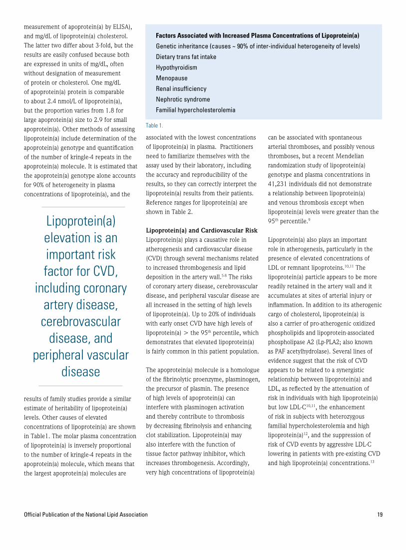
Table 1.
Factors Associated with Increased Plasma Concentrations of Lipoprotein(a)
Genetic inheritance (causes ~ 90% of inter-individual heterogeneity of levels)
Dietary trans fat intake
Hypothyroidism
Menopause
Renal insufficiency
Nephrotic syndrome
Familial hypercholesterolemia
Lipoprotein(a) elevation is an important risk factor for CVD,
including coronary artery disease, cerebrovascular
disease, and peripheral vascular
disease

20 LipidSpin
Lipoprotein(a), Aortic Valve Calcification and Aortic StenosisThe very interesting results of a recent study have suggested that lipoprotein(a) also contributes to aortic valve calcification and incidence of aortic stenosis. Genome-wide associations with the presence of aortic valve calcification were assessed in 6942 subjects in 3 cohorts, which led to the identification of a single nucleotide polymorphism in the lipoprotein(a) (LPA) locus (rs10455872) associated with an odds ratio of 2.05 (P=9.0 x 10⁻¹⁰) for aortic valve calcification.19 Lipoprotein(a) levels predicted by the LPA genotype also were associated with aortic valve calcification. In a prospective analysis, the LPA genotype also was associated with the incidence of aortic stenosis with a hazard ratio of 1.54 (95% CI 1.05 to 2.27).
Screening for Lipoprotein(a) ElevationScreening for lipoprotein(a) elevation is indicated in patients with moderate to high CVD risk because it is helpful for CVD risk stratification and helps guide the aggressiveness of treatment of dyslipidemia. Identification of an individual with high lipoprotein(a) also is a marker of genetically mediated CVD risk, which provides the opportunity for detection of first degree relatives who unknowingly may also have increased CVD risk. Screening of seemingly low-risk patients also needs to be considered because the advent of the statin era 25 years ago has substantially reduced the sensitivity of the family history for detection of familial CVD risk. Since an entire generation of patients have markedly reduced their CVD risk as a consequence of effective LDL-lowering by statins, the offspring of these patients (and their health care providers) can no longer assume that a negative family history of CVD implies low CVD risk. The implication of this is that patients who report having no family history of CVD may actually have increased CVD risk related to lipoprotein(a) elevation
or other genetically mediated CVD risk factors. The European Atherosclerosis Society Consensus Panel recently advocated screening all individuals with the following conditions: (I) premature CVD, (II) familial hypercholesterolemia, (III) a family history of premature CVD and/or elevated Lp(a), (IV) recurrent CVD despite statin treatment, (V) ≥3% 10-year risk of fatal CVD according to the European guidelines, and (VI) ≥10% 10-year risk of fatal and/or non-fatal CHD according to the US guidelines.8 A family history of hypercholesterolemia could also be considered as an alternative criterion for item (III) because of the reasons described above. The National Lipid Association also convened a panel of clinical experts who issued recommendations regarding the clinical use of various biomarkers in 2011, which included recommendations for lipoprotein(a) that mirrored the recommendations from the European
Atherosclerosis Society Consensus Panel.14
TreatmentThere is no direct proof that lowering lipoprotein(a) reduces CVD risk because the studies have not been done. In the meantime, it is reasonable to try to reduce high levels of lipoprotein(a) in selected patients. Niacin is the primary pharmacologic treatment for elevated lipoprotein(a) because it has the greatest lipoprotein(a)-lowering efficacy and it has been shown to reduce CVD events in several patient populations.15 Unfortunately, the efficacy of this intervention is limited to a 20-40% dose-dependent reduction, which is insufficient
to achieve acceptable levels in patients with very high levels. There are reports that statins minimally lower plasma lipoprotein(a) concentrations, but statins are generally ineffective for lipoprotein(a) lowering except in patients with familial hypercholesterolemia, who may achieve modest lipoprotein(a) lowering for unclear reasons (lipoprotein(a) is not cleared by the LDL receptor). A possible mechanism is decreased production of lipoprotein(a) due to a reduced pool of LDL in plasma.
LDL apheresis can acutely lower lipoprotein(a) levels by 50-80% during a 2-3 hour procedure, but the invasiveness of the procedure, high cost, and fairly limited availability are limiting factors. Individuals with very high lipoprotein(a) concentrations and progressive CVD despite aggressive medical therapy may be candidates for initiation of treatment with LDL apheresis. We are currently treating
two individuals with LDL apheresis for this indication, one of whom had severely elevated lipoprotein(a) concentrations and developed rapidly progressive internal carotid artery atherosclerotic occlusions necessitating bilateral revascularizations in her early 50s, despite aggressive combination treatment with a statin plus niacin. In an uncontrolled observational study of 120 patients with CVD and lipoprotein(a) levels greater than the 95th percentile, treatment with LDL apheresis was associated with a reduction in CVD events (MACE rate per patient 1.056 vs. 0.144; P<0.0001).16 The results of a more recent randomized trial of LDL apheresis in 32 patients with lipoprotein(a) > 50
Table 2.
Reference Ranges for Plasma Lipoprotein(a) MeasurementsMeasurement Reference RangeLp(a) molar concentration < 75 nmol/LLp(a) cholesterol concentration < 10 mg/dLLp(a) protein concentration < 30 mg/dL

Official Publication of the National Lipid Association 21
mg/dL and LDL cholesterol < 2.5 mmol/L demonstrated increased regression or stabilization of angiographic coronary atherosclerosis (70% vs 43%, p=0.02) compared to usual care.17
Treatment with estrogen replacement or estrogen analogues in postmenopausal women is associated with a modest reduction in the plasma concentration of lipoprotein(a), but the predictive association between lipoprotein(a) and cardiovascular risk is attenuated in women taking hormone replacement therapy.18 Among postmenopausal women with the highest quintile of lipoprotein(a), however, those women taking hormone replacement appeared to have a lower risk of cardiovascular events compared to those not taking estrogen, particularly among women with high LDL cholesterol concentrations above the median.18 The relationship between estrogen replacement and CVD risk in the general population continues to be controversial, however.
Treatment of hypothyroidism, if detected, and correction of renal insufficiency and proteinuria, if possible, may also have beneficial effects on lipoprotein(a) levels. Anabolic steroids such as stanozolol and danazol may lower lipoprotein(a) levels up to 50% in women, but these agents are not recommended for general use because of adverse side-effects. It is possible that aspirin, L-carnitine, ascorbic acid combined with L-lysine, calcium antagonists, angiotensin converting enzyme inhibitors, and androgens may lower lipoprotein(a) by < 10%8, but these agents are not indicated as primary treatment for elevated lipoprotein(a). Experimental medications are under development that may lower lipoprotein(a) concentrations by more than 20-25%, such as lomitapide (microsomal transfer protein inhibitor), mipomersen (apoprotein B antisense oligonucleotide), anti-PCSK9 agents, and thyroid hormone analogues. Lomitapide and mipomersen
were recently FDA approved for restricted treatment of homozygous familial hypercholesterolemia, but they are still considered experimental for lipoprotein(a) lowering and treatment of other patient populations.
Since it is typically difficult to normalize plasma levels of lipoprotein(a), an
alternative strategy is to aggressively lower levels of LDL and remnant lipoproteins in patients with high lipoprotein(a). The efficacy of this strategy is not proven, but it is supported by the findings from prospective observational and intervention studies that suggest that the risk of CVD events attributable to lipoprotein(a) may be abrogated when the LDL cholesterol concentration is < 70-80 mg/dL.10,11,13
Summary and ConclusionsLipoprotein(a) elevation is an important risk factor for CVD, including coronary artery disease, cerebrovascular disease, and peripheral vascular disease, particularly among individuals with the highest levels of lipoprotein(a) in combination
with elevated LDL cholesterol or particle numbers. Levels of plasma lipoprotein(a) are rarely quantified outside of lipid disorder clinics, so the majority of patients with high levels are undiagnosed. About 90% of the inter-individual heterogeneity in levels of lipoprotein(a) is genetically mediated, so the disorder is highly heritable, necessitating screening of first degree relatives of affected individuals. The methods for quantifying lipoprotein(a) are not well standardized, so practitioners need to understand how the testing is performed and what is actually measured in their laboratory. All moderate- and high-risk patients should have a lipoprotein(a) determination, and some seemingly low risk individuals may warrant evaluation. Niacin is the most efficacious treatment for lowering lipoprotein(a), aside from LDL apheresis. Aggressive LDL lowering is an alternative strategy for managing patients with elevated lipoprotein(a), since the atherogenicity of lipoprotein(a) appears to be attenuated when the LDL-C concentration is low. Clinical trials are needed to demonstrate the best approach to managing patients with high lipoprotein(a), but in the meantime we need to utilize the available strategies in the context of our current understanding of lipoprotein(a) and CVD risk. n
Disclosure statement: Dr. Duell has received honoraria from Aegerion, Genzyme, Merck & Co., and Vivus. Dr. Duell has received grants from Bristol-Myers Squibb, Cerenis, Pfizer Inc., and Genzyme.
Table 3.
Options for Management of High Lipoprotein(a)Niacin – first choicePossible estrogen replacement in postmenopausal womenLDL apheresisMore aggressive LDL loweringUpcoming experimental therapies?Renal transplantation in patients with renal failure
Niacin is the most efficacious treatment
for lowering lipoprotein(a), aside from LDL apheresis.
22 LipidSpin
Coronary heart disease (CHD) remains the leading cause of death in Western civilizations. The role of abnormal lipid metabolism as a modifiable risk factor for CHD is well documented. The presence of chronic kidney disease (CKD) is also associated with an increased risk of CHD, but the pathogenesis of this relationship is not completely understood. Specifically, it remains unclear whether and to what degree the management of dyslipidemia can affect the risk of CHD in patients with advanced CKD, especially given the higher risk of adverse effects with pharmacological therapy in this patient population.
EpidemiologyDecreased kidney function is associated with an increase in the risk of cardiovascular (CV) death, particularly among those with stage IV and V CKD (Figure 1).1 Relative to the general population, advanced CKD does appear to be associated with an increase in triglycerides and VLDL-C and a decrease in HDL-C, but LDL-C appears to be relatively unchanged or perhaps a bit decreased. The epidemiological data has led many to suggest that advanced CKD should be considered a CHD risk equivalent. Indeed, the National Kidney Foundation has published guidelines for management of lipids in CKD that set aggressive goals of therapy, similar to those defined by ATP III
for other CHD risk equivalents (Table 1).2
Pharmocokinetics, Pharmacodynamics and Potential for Drug InteractionsStatins and other lipid-lowering agents have been used clinically for the management of hyperlipidemia and prevention of cardiovascular events in patients with CKD for the past 25 years. Statins with a shorter duration of action should be dosed at night or bedtime since cholesterol biosynthesis undergoes a circadian cycle, with most cholesterol formation occurring while an individual is asleep. The ideal antihyperlipidemic agent should improve patients’ lipid profile without increasing the risk of toxicity. The comparison of pharmacokinetics
Specialty Corner: Nephrology Corner—Limitations of Statin Use and Adjuvant Therapies in Stage IV CKD and Dialysis
MIChAEL J. BLoCh, MDDepartment of MedicineUniversity of Nevada School of MedicineReno, NVDiplomate, American Board of Clinical Lipidology
ALI oLYAEI, PharmDDivision of Nephrology, Hypertension and TransplantationOregon State University and Oregon Health and Science UniversityPortland, OR
Discuss this article at www.lipid.org/lipidspin
Dyslipidemia Goal
LDL-C >100 mg/dL LDL-C <100 mg/dLTriglycerides >500 mg/dL Triglycerides <500 mg/dLTriglycerides >200 mg/dL Non-HDL-C <130 mg/dL
Table 1. National Kidney Foundation Guidelines for Managing Dyslipidemia in Adults with CKD.2
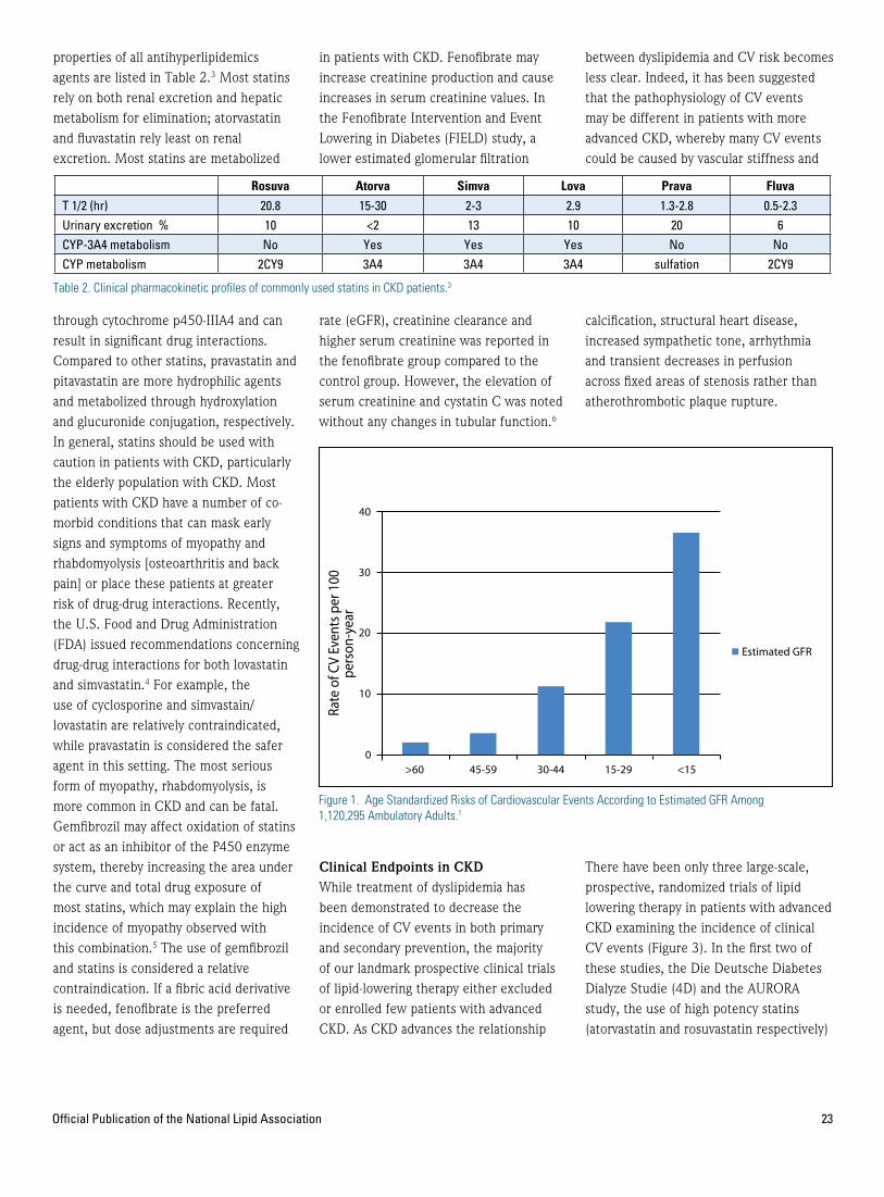
properties of all antihyperlipidemics agents are listed in Table 2.3 Most statins rely on both renal excretion and hepatic metabolism for elimination; atorvastatin and fluvastatin rely least on renal excretion. Most statins are metabolized
through cytochrome p450-IIIA4 and can result in significant drug interactions. Compared to other statins, pravastatin and pitavastatin are more hydrophilic agents and metabolized through hydroxylation and glucuronide conjugation, respectively. In general, statins should be used with caution in patients with CKD, particularly the elderly population with CKD. Most patients with CKD have a number of co-morbid conditions that can mask early signs and symptoms of myopathy and rhabdomyolysis [osteoarthritis and back pain] or place these patients at greater risk of drug-drug interactions. Recently, the U.S. Food and Drug Administration (FDA) issued recommendations concerning drug-drug interactions for both lovastatin and simvastatin.4 For example, the use of cyclosporine and simvastain/lovastatin are relatively contraindicated, while pravastatin is considered the safer agent in this setting. The most serious form of myopathy, rhabdomyolysis, is more common in CKD and can be fatal. Gemfibrozil may affect oxidation of statins or act as an inhibitor of the P450 enzyme system, thereby increasing the area under the curve and total drug exposure of most statins, which may explain the high incidence of myopathy observed with this combination.5 The use of gemfibrozil and statins is considered a relative contraindication. If a fibric acid derivative is needed, fenofibrate is the preferred agent, but dose adjustments are required
in patients with CKD. Fenofibrate may increase creatinine production and cause increases in serum creatinine values. In the Fenofibrate Intervention and Event Lowering in Diabetes (FIELD) study, a lower estimated glomerular filtration
rate (eGFR), creatinine clearance and higher serum creatinine was reported in the fenofibrate group compared to the control group. However, the elevation of serum creatinine and cystatin C was noted without any changes in tubular function.6
Clinical Endpoints in CkD While treatment of dyslipidemia has been demonstrated to decrease the incidence of CV events in both primary and secondary prevention, the majority of our landmark prospective clinical trials of lipid-lowering therapy either excluded or enrolled few patients with advanced CKD. As CKD advances the relationship
between dyslipidemia and CV risk becomes less clear. Indeed, it has been suggested that the pathophysiology of CV events may be different in patients with more advanced CKD, whereby many CV events could be caused by vascular stiffness and
calcification, structural heart disease, increased sympathetic tone, arrhythmia and transient decreases in perfusion across fixed areas of stenosis rather than atherothrombotic plaque rupture.
There have been only three large-scale, prospective, randomized trials of lipid lowering therapy in patients with advanced CKD examining the incidence of clinical CV events (Figure 3). In the first two of these studies, the Die Deutsche Diabetes Dialyze Studie (4D) and the AURORA study, the use of high potency statins (atorvastatin and rosuvastatin respectively)
Official Publication of the National Lipid Association 23
Rosuva Atorva Simva Lova Prava Fluva
T 1/2 (hr) 20.8 15-30 2-3 2.9 1.3-2.8 0.5-2.3Urinary excretion % 10 <2 13 10 20 6CYP-3A4 metabolism No Yes Yes Yes No NoCYP metabolism 2CY9 3A4 3A4 3A4 sulfation 2CY9
Table 2. Clinical pharmacokinetic profiles of commonly used statins in CKD patients.3
Figure 1. Age Standardized Risks of Cardiovascular Events According to Estimated GFR Among 1,120,295 Ambulatory Adults.1
Estimated GFR
<15>60
Rate
of C
V Ev
ents
per
100
pers
on-y
ear
0
10
20
30
40
45-59 30-44 15-29

24 LipidSpin
in dialysis patients did not lead to a significant reduction in cardiovascular events despite robust changes in lipid parameters.7,8
In the SHARP study, the use of simvastatin-ezetimibe in subjects with stage 3-5 CKD did lead to a statistically significant 17% reduction in CVD events.9 In subgroup analysis of SHARP, subjects with stage 3 CKD had a statistically significant 25% reduction in CV events; those with stage 4 CKD had a statistically significant 22% reduction in CV events; while those on hemodialysis at baseline did not have a significant reduction in events. Whether the favorable results seen in SHARP were due to the specific combination of lipid-lowering agents used in that study or the inclusion of patients with less advanced CKD (as seems more likely) is unknown. Importantly, although other combinations of lipid-lowering agents, including statin + nicotinic agent or statin + fibrate, are frequently employed in patients with advanced CKD, endpoint data with these combinations is essentially non-existent in this patient population.
A recent meta-analysis of all trials of lipid-lowering therapy that included patients with CKD suggested that lipid-lowering therapy led to a modest decrease in the risk of cardiac mortality (pooled risk ratio
[RR] from 6 trials 0.82), CV events (pooled RR from 9 trials 0.78), and myocardial infarction (pooled RR from 9 trials 0.74); however, the majority of subjects in the included studies were stage 3 CKD patients, representing subgroups of larger studies.6,10 A second meta-analysis demonstrated that statin therapy reduced all cause and CV mortality, major CV events, myocardial infarction and stroke in person with CKD not receiving dialysis by about 20-25%, but that there was little or no beneficial effect on any mortality or CV endpoints with statin therapy in those on dialysis at baseline.11 Unfortunately, in both meta-analyses, most patients with CKD not on dialysis were stage 3 or lower, and the data was not reported for stage 4 CKD individually.
There are very limited clinical data on the safety and efficacy of combination of statin and nicotinic acid or statin and fibrates therapies in patients with CKD. Due to possible drug interactions and risk of myopathy and recent findings from the Action to Control Cardiovascular Risk in Diabetes (ACCORD) Study and AIM-HIGH (Atherothrombosis Intervention in Metabolic Syndrome With Low HDL/High Triglycerides: Impact on Global Health Outcomes, that combination therapy did not result in a significant reduction of cardiovascular events compared with statin
monotherapy, care should be taken when combination therapy with these agents is utilized with statins in patients with chronic kidney disease.12, 13
While lipid-lowering therapy appears effective in reducing CV events in stage 3 CKD, there is little to no evidence that lipid-lowering therapy reduces CV events in patients already on dialysis. Data specifically in stage 4 patients is quite limited, but based on the SHARP study results, these patients probably do receive a modest benefit.
Safety of Lipid-Lowering Agents in Advanced CkD In general, cumulative data from both primary and secondary prevention studies of statins indicate that the HMG CoA reductase inhibitors (statins) have an excellent safety record and a favorable risk-benefit profile, with a low risk of significant adverse events (<1% incidence).14 However, epidemiological studies indicate the discontinuation rates for lovastatin and simvastatin were 3% and 6% respectively, and much higher incidence of myopathy at high doses (80 mg daily).15
Myopathy—The clinical spectrum of statin-induced myopathy consists of myalgia, myositis, rhabdomyolysis and asymptomatic increases in plasma creatine kinase (CK) levels. Muscle-related adverse events can be difficult to describe because the terminology used can be inconsistent. Generally, ‘myalgia’ refers to the tolerability issue of muscle pain or weakness without elevation of creatine kinase (CK) levels, while ‘myositis’ is defined as the safety issue of muscle pain with considerable elevation of CK levels.16 Rhabdomyolysis refers to muscle symptoms with markedly raised CK, usually >10x upper limit of normal, and is generally considered a serious medical condition that can lead to hospitalization, renal failure, and even death.16 For all statins,
Study Year N Population InterventionResults of Primary Endpoint
4D7 2004 1255Type 2 diabetes mellitus on dialysis
Atorvastatin or placebo
8% non-significant reduction in CV events
AURORA8 2009 2776 Dialysis patients Rosuvastatin or placebo
4% non-significant reduction in CV events
SHARP9 2010 9438
SrCr at least 1.7 md/dL in men or 1.5 mg/dL in women or on dialysis
Simvastatin + Ezetimibe or placebo
17% reduction in major CV events (statistically significant
Table 3. Summary of Outcome Studies of Lipid-Lowering Therapy in Patients with CDK.7-9

Official Publication of the National Lipid Association 25
Table 4. Dosing adjustment for lipid-lowering agents in chronic kidney disease.
the overall risk of rhabdomyolysis is less than 0.5% in the general population.17 This risk may be higher in patients with CKD, elderly, and in patients taking other drugs or food which inhibit CYP3A4, specifically grapefruit, cyclosporine, azole antifungals, macrolide antibiotics, and fibrates.18 Although the exact pathogenesis of myopathy has not been determined, several mechanisms have been postulated. Myopathy may be due to mitochondrial
dysfunction and muscle protein degradation. The genetic marker SLCO1B1 is among the strongest predictors of myopathy risk.19
Hepatotoxicity—Previously, hepatocellular necrosis and hepatotoxicity induced by statins were considered a myth.20 A recent study has concluded that idiosyncratic hepatotoxicity may be associated with the use of statins.21 Asymptomatic hepatic
transaminase elevation (greater than three times the upper limit of normal) may occur in 1-2% of patients on an HMG-CoA reductase inhibitor and in general is dose related. In most patients, elevation of transaminase enzymes are resolved spontaneously with continued therapy, although discontinuation may be required in some patients. Based on current recommendations from the FDA, routine monitoring of transaminases is no longer
Drug Class Medications Dosing in Renal Impairment
HMG-CoA Reductase Inhibitor
Atorvastatin No adjustment is necessary.
Fluvastatin Mild-to-moderate renal impairment: No dosage adjustment necessary.Severe renal impairment: Use with caution (particularly at doses >40 mg/day; has not been studied).
Lovastatin When ClCr <30, Use IR >20 mg daily with caution.Use initial ER 20 mg QHS; (Doses >20 mg daily with caution).
Pitavastatin ClCr 15-60 (not receiving HD): Initial 1 mg QD; max 2 mg QDESRD: Initial 1 mg QD; max 2 mg QD.
Pravastatin Significant impairment: initial 10 mg/day.
Rosuvastatin Mild-to-moderate impairment: No dosage adjustment required.ClCr <30: Initial 5 mg/day; NTE 10 mg QD.
Simvastatin
Manufacturer’s recommendations:Mild-to-moderate renal impairment: No dosage adjustment necessary.Severe renal impairment: ClCr <30: Initial 5 mg/day with close monitoring.Alternative recommendation: No dosage adjustment necessary for any degree of renal impairment.
Bile Acid Sequestrants
Colesevelam No dosage adjustment necessary; not absorbed from the GI tract.
Cholestyramine No dosage adjustment provided in manufacturer’s labeling; however, use with caution in renal impairment; may cause hyperchloremic acidosis.
Colestipol No dosage adjustment necessary; not absorbed from the gastrointestinal tract.
Nicotinic Acid Niacin, etc. No dosage adjustment recommended; use with caution.
Fibric Acid Derivatives
Gemfibrozil
Mild-to-moderate impairment: Use caution; deterioration of renal function has been reported in patients with baseline SCr >2.
Severe impairment: contraindicated.
HD: Not removed by HD; supplemental dose is not necessary.
Fenofibrate ClCr ≥50: No dosage adjustment necessary.
ClCr <50: Initiate at 45 mg/day.
Contraindicated in severe impairment.
Cholesterol Absorption Inhibitor Ezetimibe AUC increased with severe impairment (ClCr <30); no dosing adjustment necessary.
Omega-3 Fatty Acid No dosage adjustment provided in manufacturer’s labeling (has not been studied).
26 LipidSpin
necessary unless the patient exhibits transaminase abnormalities at baseline, has other risk factors for hepatotoxicity, or clinically abnormalities are suspected.
Dosing—With the increasing incidence of chronic renal disease, regular renal function monitoring and dosage adjustment of lipid-lowering agents according to eGFR and pharmacokinetic data are of major importance.3,22 Because large studies of the safety of these agents in patients with CKD is lacking, drug-drug interactions and dosage adjustment recommendations need to be regularly updated following the results of epidemiological and observational studies. Patients with significant renal disease should be started on low doses of statins and other lipid-lowering agents, with doses titrated up only when clinically
indicated. Due to the risk of significant drug-drug interactions, for the renal transplant population fluvastatin has been demonstrated to be safe in a large clinical endpoint trial where tacrolimus was the most common immunosuppressant used, but pravastatin may be a more suitable agent when cyclosporine is used (Table 4).23
SummaryWhile far from conclusive, the available clinical trail evidence supports the hypothesis that lipid-lowering therapy may provide diminishing benefit as CKD advances. In stage 4 CKD and dialysis, the potential for benefit from statin therapy needs to be weighed carefully against the increased risk for adverse effects seen in this patient population. With the exception
of statin-ezetimibe, combination lipid-lowering therapy has not been well studied in this patient population and should be used only with careful monitoring for adverse events. Future studies are needed to help clinicians appropriately balance the benefits and risks of lipid-lowering therapy in CKD, particularly for the increasing number of patients with Stage IV CKD and concomitant dyslipidemia. n
Disclosure statement: Dr. Bloch has received honoraria from Aegerion, AstraZeneca, Chelsea Therapeutics, Daiichi Sankyo Inc., LipoScience Inc., PriMed CME, and Takeda Pharmaceuticals. Dr. Olyaei has no disclosures to report.
IntroducIng the Lipid Luminations serIes on reachMd.
reachMd.com/LipidLuminations
onlineReachMD.comSatellite Radio
XM Channel 167
Mobile Phone Applications
on-the-goon-air
© 2013 ReachMD 500 Off ice Center Dr ive, Sui te 325 • Fort Washington, PA 19034 • 866.423.7849
Access to Medical Information and Education
anYtimE, anYpLaCE
giving you the skinny on fatty acids.

Official Publication of the National Lipid Association 27
It is typically silent, but sometimes you can hear it, a soft systolic ejection murmur heard over the aortic area during cardiac auscultation on physical examination. As cardiovascular risk reduction specialists, we do not to want to miss this cardiac murmur. It is not innocent or benign; this is the murmur of aortic valve sclerosis (ASc). In addition, there is a normal split of the second heart sound, a normal carotid upstroke, and a peak transaortic systolic gradient noted on Doppler transthoracic echocardiography (TTE) typically less than 16 mmHg. The characteristic M-mode tracing on echocardiography is seen in Figure 1. ASc is characterized by calcification and thickening involving the aortic valve cusps with no hemodynamically significant transaortic systolic gradient noted on TTE. ASc shares
many of the same clinical risk factors for coronary heart disease (CHD) including age, hypertension, and cigarette smoking.1
ASc is prevalent among the elderly. Of 5,176 subjects ≥65 years of age enrolled in the prospective Cardiovascular Heart Study undergoing adequate echocardiographic study, 26% were found to have ASc.1 Aortic valve stenosis was identified in 2% of the subjects. ASc is even more common among patients with known coronary heart disease. The prevalence is approximately 40%.2,3 In a study of 425 patients presenting to the emergency room with chest pain, the prevalence of Asc was even higher at 49%.4 In patients undergoing coronary angiography for chest pain, the prevalence was related to the degree of obstructive coronary artery disease
present. In patients with no obstructive CHD, single vessel CHD, double or triple vessel CHD prevalence respectively was 14%, 28%, and 58%.2 Interestingly, ASc was found to be a more powerful predictor of obstructive CHD in patients <60 years of age than in those >60 years.2
In a study of 338 consecutive patients undergoing myocardial perfusion single photon emission computed tomography (SPECT) and TTE, ASc was significantly associated with an abnormal SPECT.5
Practical Pearls: Cardiac Auscultation for the Lipidologist: A Systolic Murmur You
Do Not Want to Miss!
Discuss this article at www.lipid.org/lipidspin
J. ANToNIo G. LÓPEZ, MD, FACC, FAhA, FACP, FCCP, FNLAPacific Lipid Association President
Director, Preventive Cardiology and Cardiovascular RehabilitationDirector, Lipid Clinic and LDL Apheresis Program
Chair, Department of CardiologySaint Alphonsus Regional Medical Center
Boise, IDDiplomate, American Board of Clinical Lipidology
JohN R. NELSoN, MD, FACC, FASNC, FNLAPacific Lipid Association Immediate Past President
Director, California Cardiovascular InstituteAssistant Clinical Professor, UCSF School of Medicine
Fresno Medicine Residency Program-VolunteerFresno, CA
Diplomate, American Board of Clinical Lipidology

28 LipidSpin
Among mitral and aortic valve sclerosis calcification sites, the odds for an abnormal SPECT in younger patients (≤55 years) for 3 sites was nearly 4 times higher than for patients without calcium. In older patients (>55 years of age), the odds were only 1.94 times as high. After univariate analysis, CRP has been found to be associated with ASc (p<0.05) in 425 patients presenting to the emergency room with chest pain.4 After one year, in patients with established ASc, there was a marked decrease in event-free survival for those at the highest tertile of CRP (>1.18 mg/dL) compared to the patients at the lowest tertile (<0.32 mg/dL). The odds for an abnormal SPECT in patients with multiple calcific deposits, diabetes mellitus or multiple cardiac risk factors was striking for women when they had these factors. The OR was 20.00 in younger women (≤55 years of age) vs. 10.00 for older women (>55 years of age) compared to women with no multiple calcium deposits without diabetes mellitus or multiple cardiac risk factors. ASc has important clinical ramifications. Not only can ASc progress to aortic valve stenosis; it is independently associated with adverse cardiovascular events.
In a study of 2,131 patients with ASc over a mean follow-up of 7.4 years, 10.5% developed mild, 2.9% developed moderate and 2.5% developed severe aortic valve stenosis.6 In the Cardiovascular Health Study, 9% of 1,091 subjects followed for a mean 5-year period developed aortic valve stenosis.7 ASc was independently associated with new coronary events (risk ratio 1.8) in a prospective study of 1980 subjects.8 In the large prospective Cardiovascular Health Study of over 5000 men and women ≥65 years of age, followed for 5 years, ASc was independently associated with increased risk of cardiovascular death and myocardial infarction (MI).9 ASc has been shown
to be an independent predictor of MI in patients with known CHD also. In a prospective study of 814 patients with CHD, 40% having prevalent ASc had a 2.4 fold increased risk of MI during 4-years of follow-up.3
ASc is associated with lipid accumulation inflammation in addition to calcium deposition in the valve. Low density lipoprotein (LDL) and lipoprotein (a) [Lp(a)] were found in lesions of aortic stenosis.10 Elevated circulating Lp (a), and low high density lipoprotein (HDL) cholesterol have been shown to be independently associated with ASc.11,12 LDL-C is associated with aortic valve stenosis13-16 in patients with Familial Hypercholesterolemia, however the association in the general population has been inconsistent. In the Cardiovascular Health Study17, increased LDL-C was associated with ASc but not in the SPARC (Stroke Prevention: Assessment of Risk in Community)18 or in the Helsinki Aging Study.19 ASc is associated with systemic endothelial dysfunction as demonstrated by significantly lower flow mediated dilatation (FMD).20 Recently, it has been shown that ASc is associated with platelet resistance to NO.21 Over a 4-year follow-up period, progression of ASc was also independently associated with tissue resistance to NO.22
ASc lesions have histologic similarities with coronary atherosclerotic plaque including inflammatory cells: specifically macrophages and T-lymphocytes, in addition to lipid accumulation and disruption of the basement membrane.23,24 After univariate analysis, CRP has been found to be associated with ASc (p<0.05) in 425 patients presenting to the emergency room with chest pain.4 After one year, in patients with established ASc, there was a marked decrease in event-free survival for those at the highest tertile
of CRP (>1.18 mg/dL) compared to the patients at the lowest tertile (<0.32 mg/dL). In patients without CHD or ASc and a CRP in the upper two tertiles, the cardiac death and MI incidence at 1 year was 0 versus 41% in patients with CHD and ASc. In this study, ASc was not an independent predictor. The presence of CHD and elevated CRP was associated with the adverse cardiovascular events however. Specifically, the increasing tertile of CRP was an independent predictor of cardiac death and nonfatal MI (HR 2.2). However, in a lower risk, prospective population study, CRP was not found to be associated with the development of ASc.7
In addition to inflammation and lipid accumulation, aortic ASc is associated with calcification. Calcification involving the aortic valve is an active osteogenic process which has been characterized as an osteoblast-like phenotype.25
Compared to normal human aortic valves, explanted calcified aortic valves at the time of transplantation were shown to have increased levels of the proteins: Osteocalcin, Osteopontin, bone sialoprotein and the transcription factor Cbfa-1 all characteristics of osteoblast activity.26 Furthermore, in patients with aortic valve stenosis undergoing surgery or routine echocardiography, increased plasma levels of Osteopontin were associated with the presence of aortic valve calcification and stenosis.27 In addition to Osteocalcin and Osteopontin, low-density lipoprotein receptor-related protein 5 (Lrp5) was found to be increased in explanted calcified aortic valves by protein and gene expression at the time of surgical valve replacement.27 Lrp5 is an important receptor in the activation of skeletal bone formation. In a study of hypercholesterolemic rabbits with aortic valve calcification, atorvastatin decreased Lrp5 and the aortic valve calcification.28 Just recently, for the first time, a

Official Publication of the National Lipid Association 29
circulating osteogenic precursor cell in human blood has been identified to be associated with calcification of the aortic valve.29
Patients with ASc are at higher risk for myocardial infarction and CHD and should be assessed for underlying CHD. Multiple retrospective studies30-34 have shown statins to be beneficial in reducing progression of aortic valve stenosis; unfortunately, prospective, randomized35-37
and non-randomized38 clinical trials have not confirmed these observations. Furthermore, there has never been reported a randomized placebo controlled trial in patients with ASc with a TTE Doppler gradient of less than 16 mmHg. The average peak transaortic systolic gradient was at least 36 mmHg in all of these prospective trials. It still remains unknown whether statin therapy will reduce the progression of ASc to aortic stenosis in patients with milder forms
of ASc. Of interest is a recent study in hypercholesterolemic mice with early aortic valve disease demonstrating that reducing plasma lipid levels by genetic inactivation normalizes oxidative stress, reduces pro-osteogenic signaling, and halts the progression of aortic valve disease.39
We believe that in patients with a cardiac murmur, when there is reasonable suspicion of valvular or structural heart disease, it is appropriate to obtain an echocardiogram.39 It is also helpful to add on the TTE request: “Please evaluate for Aortic Valve Sclerosis/Stenosis.” It is also appropriate to obtain an echocardiogram for routine surveillance (≥3 years) of mild valvular stenosis or (≥1 year) for moderate or severe valvular stenosis without a change in clinical status or cardiac exam.40 If a patient is identified with ASc then intensive risk factor modification should be instituted including a regular exercise program. In a LDL receptor deficient
mouse model, regular exercise training prevented ASc through several aspects including reduction and inflammation, oxidative stress and also an inhibition of the osteogenic pathway.41 This may be relevant to humans. Patients with CHD, or CHD risk equivalents including diabetes mellitus, will all need to be on statins regardless of their LDL-C levels or the presence of ASc. We are looking forward to the future as studies address molecular targets for valvular calcification. ASc is an important murmur that we do not want to miss! n
Disclosure statement: Dr. López has received honoraria from Abbott Laboratories, Aegerion, Amarin Corp., AstraZeneca, Bristol-Myers Squibb, Boehringer Ingelheim, Daiichi Sankyo Inc., Forest Pharmaceuticals, Gilead Pharmaceuticals, GlaxoSmithKline, Kowa Pharmaceuticals America, and ZonaHealth. Dr. Nelson has received honoraria from Abbott Laboratories, Amarin Corp., AstraZeneca, Atherotech, Bristol-Myers Squibb, Daiichi Sankyo Inc., GlaxoSmithKline, Gilead Pharmaceuticals, Kowa Pharmaceuticals America, Merck & Co., Pfizer Inc., and Novartis Pharmaceuticals.
R.E. is a 58-year-old Asian male who finally quit smoking one month ago, and is now being treated for hypertension to goal with the combination amlodipine/benazepril. He has type 2 diabetes and takes metformin 500 mg bid. His initial lipid panel revealed a TC=230 mg/dL, LDL=140 mg/dL, HDL=35 mg/dL and TG=265mg/dL and his Non HDL- was 195. His BMI was 29, FBS=109mg/dL and his HbA1C=6.8. In general he has led a sedentary lifestyle. He was placed on Simvastatin, 40 mg q HS and three months later his lipid profile was: TC=145mg/dL, LDL=89mg/dL. HDL= 31mg/dL, and TG=166mg/dL, and non-HDL-C=114. Because he has multiple risk factors including diabetes, using ATP III guidelines
he has an LDL goal of <100mg/dL and a non-HDL-C goal of <130.
Additional data from an NMR lab obtained on the same blood sample: ApoB=97mg/dL, (optimal <60)*LDL-P=1531 nmol/L, (high risk >1000)* sdLDL 32 mg/dL, (optimal <21)* ApoA1=111 mg/dL (optimal >150)*, ApoB/ApoA1 Ratio=0.87 (high risk >0.81)*. He is relatively sedentary and followed no particular diet consistently. Is this additional information useful in clinical practice?
LDL-C concentration, commonly calculated, is simply the amount of cholesterol in the LDL fraction of plasma, and has become the major target for preventing vascular disease associated with dyslipidemia. ApoB is a
measure of the total number of atherogenic particles and in some studies it is a more precise marker for risk of vascular disease.
The cholesterol content of the LDL particle can be quite variable. In patients with type 2 diabetes and obesity associated with high triglyceides, this discrepancy seems to amplify. Cholesterol ester transfer protein, (CETP) transfers triglycerides from triglyceride-rich VLDL to cholesterol containing LDL and HDL lipoproteins in exchange for cholesterol. LDL-C may be deceptively lower as the cholesterol content is decreased but the particle number has not changed. When triglyceride-rich LDL particles are then exposed to hepatic lipase, triglycerides are cleaved from the LDL particle creating small dense LDL. Small LDL particles may be more subject to
Case Study: Digging Deeper—A Case for Apolipoproteins and Lifestyle in Office Practice
RoB GREENFIELD, MD, FACC, FAhA, FNLACalifornia Heart AssociatesClinical Assistant Professor, UC-Irvine Medical CenterOrange County, CADiplomate, American Board of Clinical Lipidology
SUSAN GIVEN, cFNP, MN, BSNPreventive Cardiology CenterSanta Monica, CA
Discuss this article at www.lipid.org/lipidspin
30 LipidSpin

Case Study: Digging Deeper—A Case for Apolipoproteins and Lifestyle in Office Practice
oxidation and entry into blood vessel walls potentially accelerating the atherosclerotic process.
Are apolipoproteins better CVD risk predictors than lipids? The INTERHEART study suggested that the ApoB/ApoA1 ratio was more strongly associated with risk for acute myocardial infarction in all ethnic groups, in both sexes, and at all ages.Additionally, ApoB, and the ApoB/ApoA1 ratio were strongly associated with more fatal myocardial infarction in men and women. ApoA1 was noted to be protective in the AMORIS trial.Analysis from this trial suggested that these values should also be measured to evaluate cardiac risk. Some data, but not all, suggests that this ratio is superior to non-HDL-C calculation {Total Chol – HDL-C= non-HDL-C}. The area remains controversial because the collaborative meta-analysis utilizing individual patient data could not find that ApoB was superior to predicting MI. Although non HDL-C and ApoB are highly correlated in large groups, they may be only moderately concordant for some individuals.
In another study looking at the association between ApoB, ApoA1, the ApoB/ApoA1 ratio, and ApoA1 in the prediction of myocardial infarction in middle-aged men and women, only ApoB and the ApoB/ApoA1 ratio were strong predictors of coronary events. ApoA1 did not add significantly to the estimation of coronary risk.
Patients taking statins need to intensify CVD risk reduction lifestyle measures through proper diet, exercise, and avoidance of tobacco. In a study published by Kokkinos et al., statin treatment and increased fitness were independently associated with lower mortality among dyslipidemic patients. The combination of statins and increased fitness resulted in lowest quartile of mortality risk than either alone.
In our case, we felt there was justification for intensifying drug therapy and life-style intervention measures. The initial lipid panel results in contrast would not have lead us to demand a more vigorous approach for him.
Our primary goal was to further reduce LDL particle number and ApoB levels and improve his ApoB/ApoA1 ratio. Rather than increase simvastatin to 80mg (not recommended by the FDA), we chose to change the statin to rosuvastatin 20 mg, and intensify lifestyle changes. We expect that with cigarette cessation, our patient’s HDL will increase as valuable enzymes in the HDL molecule may have been inhibited by smoking. Regarding lifestyle, we asked him to incorporate exercise into his busy work schedule by purchasing an inexpensive pedometer and use parking spaces further away, and stairs whenever possible. We asked him to find time during his lunch break to walk for at least 15 min, and when possible to devote this amount of time either before or after work. On weekends he was able to walk for an hour each day with his wife. He was instructed to avoid “anything white” in his diet such as breads, rice, and potatoes, as well as sweets. At our request, he purchased a recommended book to learn more about low glycemic index foods. Four months later, he lost ten pounds and repeat labs demonstrated that his FBS was 94mg/dL, HbA1C=6.1, Total Chol=137mg/dL, LDL-C=75mg/dL, TG=120mg/dL, HDL-C=38mg/dL, non-HDL-C=99. Pt’s BMI dropped to 27 and his
lipoprotein data now revealed an ApoB=80, LDL-P=950nmol/L, ApoB/ApoA1=0.70.
In today’s world of medicine, properly assessing patients and treating to achieve optimal risk reduction is a moving target. Which risk-assessment formulae do we use? Is 10-year or lifetime risk assessment best? Patient adherence and compliance are made more difficult by the endless plethora of readily available information and misinformation to which our patients are exposed on the internet and elsewhere.
As clinicians, we read studies that tell us that we are “under-treating” and we are “soft” on lifestyle changes. We encounter barriers to aggressive and multi-drug combinations due to patient reluctance to take “more meds,” the high cost of pharmaceuticals, and formulary roadblocks. Despite this, discovery and innovation continue.
What are “normal” lipids? What will be the “new normal?” What will not change, however, is that as clinicians, we will always be required to use our sound judgment in interpreting and applying the best available evidence with honesty and compassion for our patients. n
Disclosure statement: Ms. Given has no disclosures to report. Dr. Greenfield has received honoraria from Abbott Laboratories and Merck & Co.
Official Publication of the National Lipid Association 31
Figure 1. Not shown here: HDL is also subjected to hepatic lipase and becomes sdHDL which is rapidly degraded and excreted renally.
CETPCETPLDL-C
HDL-C
Triglyceride-Rich LDL
Triglycerides
Small dense LDL
Hepatic lipase
“What makes us normal is knowing that we are not normal.”
~ Haruki Murakami

32 LipidSpin
As President-elect of the Pacific Lipid Association, it gives me a great pleasure to present our chapter update. Our members continue to be very active in their communities as well as throughout the NLA. Our own Matthew Ito, PharmD, will become the NLA President this May.
March of last year, our immediate past president, John Nelson, MD, held his annual CME conference in Fresno, California. The conference, entitled “Treatment of the High Risk Patient with Low HDL Cholesterol,” was attended by 234 people. Several PLA members presented at the meeting.
The PLA, along with Dr. Nelson’s leadership, has recently spearheaded a drive to develop an Advocacy Committee in the NLA. This committee would address specific issues faced by individual members of the NLA. Dr. Nelson is currently working with the State of
California on SB866, a two-page electronic prior authorization form. This would allow practitioners to use a single form for all insurance prior authorizations. Insurance companies would be given two business days to respond to the completed document. This would save physicians a great deal of time as well as improve the timeliness of patient care.
PLA Board member, Julie Bolick, RD, is working with the Patient Adherence Subcommittee to create a patient toolkit for practitioners. She has also convened a meeting of NLA dietitians.
In September, our current chapter president, J. Antonio G. López, MD, hosted the live Lipid Insights program entitled “CETP Inhibition—An Important Potential Strategy in Reducing Cardiovascular Events.” Dr. López, along with Eliot Brinton, MD, and Benjamin Ansell, MD, presented a lively debate focused on the relationship between CETP and atherosclerosis.
Dr. López continues as Chair of the NLA Honor’s and Awards committee. Wayne True, MD, is Chair of the NLA Membership Committee.
Drs. Eliot Brinton and Matt Ito were among the four faculty members to participate in the successful USAGE manuscript and statin adherence public relations campaign (www.StatinUSAGE.com), which included a paper published in the Journal of Clinical Lipidology this past June.
This past October, Paul Rosenblit, MD, PhD, along with Brian Chesnie, MD, Rob Greenfield, MD, and Nathan Wong, PhD, held the 4th Annual Orange County Symposium in Newport Beach, California. This conference, jointly sponsored by the NLA and the American Society for Preventive Cardiology, focused on expanding knowledge with regard to the role of Clinical Lipidology in the primary and secondary prevention of cardiovascular disease.
The 10th Annual World Congress on Insulin Resistance, Diabetes, and Coronary Artery Disease Symposium was held in Los Angeles this past November. Yehuda handelsman, MD, and colleagues assembled a unique multidisciplinary program dedicated to the management of cardiovascular risk factors and disease. This year’s program introduced new aspects
Chapter Update: Pacific Lipid Association
B. ALAN BoTTENBERG, Do, FACoI, FNLAPresident-elect, Pacific Lipid AssociationNorthern Nevada LipidologyCarson City, NV
Diplomate, American Board of Clinical Lipidology
Discuss this article at www.lipid.org/lipidspin

of bone, fat, leptin and adeponectin interactions, as well as mitochondria and associated proteins, to metabolic impairment in human disease.
Our PLA is currently working with the NLA to host the 2013 Annual Scientific Sessions in Las Vegas, Nevada from May 29-June 2. Our NLA President, Peter Toth, MD, PhD, and Drs. Ito, López and I will serve as the program’s co-chairs. The preliminary schedule and faculty look outstanding.
We are also working to invite lipid experts from the Pacific Rim nations to participate in the Spring 2014 Clinical Lipid Update on Maui. More information will be forthcoming.
The PLA remains an active and dynamic group. I have thoroughly enjoyed my interactions with both the PLA and NLA and recommend all those interested to become more involved. n
Official Publication of the National Lipid Association 33
SAVE THE DATEDancing in the Desert
a poolside event in support of the Foundation of the NLA
during the Scienti�c Sessions in Las Vegas
June 1 | 7:00 PM
2013 AnnualScienti�c Sessions
May 30–June 2
Red Rock Hotel • Las Vegas, Nevada
34 LipidSpin
Like many young clinicians starting out, Daniel Steinberg, MD, PhD, did not intend to build a career in Clinical Lipidology. But the Harvard-trained biochemist had motivation to pick a field quickly: it was 1950, the Korean War had just broken out, and he had served less than two years in World War II. His thesis supervisor, Christian B. Anfinsen, MD, had just accepted a position at what was the newly created National Heart Institute—if Dr. Steinberg accepted a job there, he would not need to enlist in the war.
The NHI’s first director, James Shannon, MD, took interest in the research on lipoproteins and heart disease conducted by John Gofman, MD, PhD. His curiosity trickled down to Dr. Anfinsen, who put together a group of clinically oriented researchers, including Dr. Steinberg, to work on cholesterol and lipoproteins in relation to coronary heart disease. The work of that group and others around
the world strongly suggested a causal relationship but the final proof awaited a randomized, double-blind clinical trial. In 1974, the Coronary Primary Prevention Trial (CPPT) launched with 3,600 male subjects who had high cholesterol. Half were given cholestyramine and half were given a placebo; subjects who had taken the bile acid sequestrant had a statistically significant decrease in heart attack risk. The success of the CPPT—the first large-scale, double-blind trial to show that lowering cholesterol decreased heart attack risk—helped position the National Institutes of Health to make the lowering of blood cholesterol levels a major public health goal.
When Dr. Steinberg went to the University of California-San Diego in 1968 he wanted to find out just how LDL led to atherosclerotic heart disease at the molecular level. In the 1980’s, Michael Brown, MD, and Joseph Goldstein, MD, published a paper showing that the macrophages would not take up low-density lipoproteins (LDL) very quickly, which was surprising since macrophages tend to become loaded with cholesterol in atherosclerosis. Drs. Brown and Goldstein postulated that LDL molecules must
first undergo some chemical or physical modification before they could be rapidly taken up by the macrophage. Dr. Steinberg and his colleagues showed that oxidation of LDL converts it to a form recognized by specific receptors, receptors that don’t recognize native, unmodified LDL. Dr. Steinberg now serves as a professor emeritus for the University of California-San Diego, where he worked for the past four decades. Many people would be surprised to learn that, at age 90, he still goes into the office on weekdays and writes avidly—in 2007 he published The Cholesterol Wars, and he just completed a book on “missed Nobel prizes.”
Recently, Dr. Steinberg wrote to the ATP IV Panel, urging its members to be more aggressive in recommending treatment for hypercholesterolemia at a much earlier age. Looking ahead, he hopes research will shed light on why high-density lipoprotein appears to be a negative risk factor. The mystery of HDL is important, Dr. Steinberg said, “because the epidemiology is so clear cut that a breakthrough in this area could yield some of the most promising developments.” n
Member Spotlight: Daniel Steinberg, MD, PhD
Discuss this article at www.lipid.org/lipidspin
DANIEL STEINBERG, MD, PhDUniversity of California-San DiegoLa Jolla, CA
Diplomate, American Board of Clinical Lipidology
Education and Meeting News and Notes
Official Publication of the National Lipid Association 35
Strategic Planning UpdateNLA leaders met in February to discuss and develop new strategic initiatives for the NLA. A key focus of the discussions was improving ways for members to actively participate in committees and Boards and encouraging more member-led projects. There were also recommendations related to developing processes to evaluating the benefit of new ideas, improving relationships with other organizations, membership development, communications, and several other areas. The NLA will report on these initiatives in the coming months.
Lifetime MembershipFor the first time, the NLA is offering a Lifetime Membership program with rates based on the duration of your involvement with the NLA. All Lifetime Memberships include a $1,000 donation to the Foundation of the NLA, which will be set aside to establish training programs and fellowships in Clinical Lipidology. Please visit www.lipid.org/lifemember for more information.
Survey of Physicians’ Responses to Plaque Imaging on CTAPlease provide insight into the use of plaque information obtained from coronary CTA in managing your patients by participating in “Potential Clinical Applications of Plaque Imaging by CTA: A Survey,” conducted by Harvey Hecht, MD, and colleagues from Mount Sinai Medical Center and The University of Erlangen. To participate, please download the survey from the
“NLA Updates” section on the NLA homepage at www.lipid.org, mark your responses on the answer sheet, and then e-mail your answer sheet to [email protected].
Early-bird Rate: Register for Annual Scientific Sessions Now and Save!This 4-day comprehensive learning experience features a wide variety of scientific sessions, symposia, case presentations and poster sessions that provide practical solutions for applying the latest research in your clinical practice. All sessions and educational events are evidence-based and clinically relevant to the practicing lipidologist. Register at www.lipid.org/sessions by March 29 to take advantage of the special Early-bird rate of $525.
New Chapter Bylaws ProposedAt the NLA strategic planning meeting that was held February 9-10, the leaders of the NLA and its Chapter Boards agreed to adopt a uniform set of Bylaws for all five of the NLA’s regional Chapters. The proposed bylaws, available at https://www.lipid.org/about/newbylaws, will need to be adopted by each Chapter’s Board of Directors and are scheduled to take effect on June 2, 2013, after the NLA 2013 Annual Scientific Sessions end. The NLA plans to hold the vote of the Chapter Boards between April 1-5, 2013. If you have comments on the proposed bylaws change, please contact your Chapter President or Lindsey (howard) Mitcham at [email protected].
hDL CME/CE-certified Newsletter Series All three installments of the HDL-themed CME/CE-certified interactive newsletter series have launched and are now open for member participation. This series aims to help clinicians successfully manage and treat residual risk in cardiovascular disease (CVD) due to abnormally low or dysfunctional HDL. This interactive newsletter will evaluate HDL-C as a tool to assess an
individual’s cardiovascular risk and examine the various atheroprotective functions of HDL and their role in determining CHD risk. Please visit www.cmecorner.com and choose “Dyslipidemia” among the featured programs to get started.
ABCL Maintenance of Certification The American Board of Clinical Lipidology (ABCL) recently voted on maintenance of certification requirements in order for diplomates to maintain their certification after 10 years. Applicants must accumulate 500 points in the following areas in order to recertify: Evidence of Professional Standing, Lifelong Learning and Self-Assessment, Cognitive Expertise, Practice Performance Assessment. Stay tuned for more information.
Featured NLA Podcasts on ReachMD Catch broadcasts of the NLA’s latest ReachMD shows featuring Lipid Luminations host Alan Brown, MD, on XM Satellite Radio Channel 167. Below are the first air dates for our latest programs. For the broadcast replay schedule and to access the entire catalogue of NLA podcasts, visit the Lipid Luminations website at www.reachmd.com/lipidluminations.
• March 25: Robert Wild, MD, PhD: “Managing CVD in Women During Childbearing Years”
• April 4: kevin Maki, PhD: “Obesity, Diabetes and the Metabolic Syndrome”
• April 8: Rebecca Reeves, DrPh, RD: “Changing Nutritional Needs Throughout the Lifetime”
Lipid Spin ReviewThanks to Wayne Warren, MD, for reviewing articles for this issue.

36 LipidSpin
Events Calendar
2013 Scientific Meetings 2013 National Lipid Association Scientific Sessions
Hosted by the Pacific Lipid Association May 30–June 2, 2013Red Rock Hotel Las Vegas, Nevadawww.lipid.org/sessions
2013 National Lipid Association Clinical Lipid Update—Fall
Hosted by the Southeast Lipid Association and the Northeast Lipid Association
September 20–22, 2013Hyatt Regency Baltimore HotelBaltimore, Marylandwww.lipid.org/clu
2014 Scientific Meetings2014 National Lipid AssociationClinical Lipid Update—SpringHosted by the Pacific Lipid Association and the Southwest Lipid AssociationMarch 14–16, 2014Grand Wailea HotelMaui, Hawaii
2014 National Lipid Association Scientific SessionsHosted by the Southeast Lipid AssociationMay 1–4, 2014Hyatt Regency Grand Cypress Hotel Orlando, Florida
2014 National Lipid Association Clinical Lipid Update—FallHosted by the Midwest Lipid Association and the Northeast Lipid AssociationAugust 22–24, 2014JW Marriott HotelIndianapolis, Indiana
other 2013 Meetings SCAN Annual MeetingApril 26–28, 2013Westin Michigan AvenueChicago, Illinoiswww.scandpg.org PCNA Annual MeetingMay 2–4, 2013Paris Hotel Las Vegas, Nevadawww.pcna.net

Official Publication of the National Lipid Association 37
Foundation Update
Following the Foundation of the NLA’s inaugural Familial Hypercholesterolemia Roundtable in New Orleans this past February, I am pleased to report meaningful progress on our important FH initiative. We convened with an outstanding delegation of representatives from the American College of Osteopathic Physicians, American Society for Preventive Cardiology, FH Foundation, International FH Foundation, National Lipid Association, National Society of Genetic Counselors, and the Preventive Cardiovascular Nurses Association, as well as several additional representatives from the Foundation of the NLA. The group discussed our shared commitment to the development and achievement of FH awareness. There are plans for meetings throughout the year to share information and ideas to further awareness of FH. February also proved to be an important month for planning the Foundation’s future. At the NLA’s biannual strategic planning session, held February 9-10 in Miami, we reaffirmed the Foundation’s desire to focus on FH awareness and educational efforts. We also discussed the need to establish broader financial
support for the Foundation. Currently, the best ways to donate are to make individual contributions, to apply for Lifetime Membership through the NLA at www.lipid.org/lifemember, or to attend a Foundation event. Supporting the Foundation allows us to provide seed funding for educational and research grants. We are developing a fund to support fellowship programs in Clinical Lipidology, and $1,000 from each Lifetime Membership fee will be set aside for this purpose. Looking ahead, please join us for an evening poolside to go “Dancing in the Desert” on Saturday, June 1, during the Annual Scientific Sessions in Las Vegas. The Red Rock Hotel’s luxurious pools will provide the perfect setting to enjoy a warm summer evening complete with music, a photo booth and premium cocktails. Register at www.lipid.org/sessions—it will be a great time for a great cause! As always, thanks for your support of our Foundation. n
ANNE C. GoLDBERG, MD, FNLAPresident, Foundation of the National Lipid Association
Associate Professor of MedicineWashington University School of Medicine
St. Louis, MO
Diplomate, American Board of Clinical Lipidology
Discuss this article at www.lipid.org/lipidspin
38 LipidSpin
ReferencesClinical Feature References1. National Cholesterol Education Program Expert Panel. Report of the
National Cholesterol Education Program Expert Panel on detection, evaluation, and treatment of high blood cholesterol in adults. Arch Intern Med 1988;148:36-69.
2. Ross R. The pathogenesis of atherosclerosis: An update. NEJM, 1986; 314:488-510.
3. Lipid Research Clinics Program: The Lipid Research Clinics Coronary Primary Prevention Trial: I. Reduction in incidence of coronary heart disease. JAMA, 1984;251:351-364.
4. Kannel W, Castelli W, Gordon T. Serum cholesterol, lipoproteins and the risk of coronary heart disease. Ann Intern Med,1971;74:1-12.
5. Miller G, Miller NE: Plasma high density lipoprotein concentration and development of ischemic heart disease. Lancet, 1975;1:16-19.
6. Pearson TA, Bulkley BH, Achuff SC, Kwiterovich PO, Gordis L. The association of low levels of HDL cholesterol and arteriographically defined coronary artery disease. Am J Epidemiol, 1979;109:285-295.
7. Castelli WP, Garrison RJ, Wilson PF, Abbott RD, Kalousdian S, Kannel WB. Incidence of coronary heart disease and lipoprotein cholesterol levels: The Framingham Study. JAMA,
1986;256:2835-2838.8. National Cholesterol Education Program Laboratory Standardization
Panel. Current status of blood cholesterol measurement in clinical laboratories in the United States. Clin Chem, 1988;34:193–201.
9. National Cholesterol Education Program Laboratory Standardization Panel. Recommendations for improving cholesterol measurement. NIH Publication No 90-2964. Washington: US Department of Health and Human Services, 1990. Bachorik PS, Ross JW, for the National Cholesterol Education Program Working Group on Lipo-protein Measurement. National Cholesterol Education Program recommendations for measurement of low-density lipoprotein cho-lesterol: executive summary. Clin Chem 1995;41:1414-20.
10. Warnick GR, Wood PD, for the National Cholesterol Education Program Working Group on Lipoprotein Measurement. National Cholesterol Education Program recommendations for measurement of high-density lipoprotein cholesterol: executive summary. Clin Chem, 1995;41:1427-1433.
11. Stein EA, Myers GL, for the National Cholesterol Education Program Working Group on Lipoprotein Measurement. National Cholesterol Education Program recommendations for triglyceride measurement: executive summary. Clin Chem, 1995;41:1421-1426.
12. Reaven GM. The role of insulin resistance in human disease. Diabe-tes, 1988;37(12):1595-1607.
13. Austin MA, Breslow JL, Hennekens CH, Buring JE, Willett WC, Krauss RM. Low density lipoprotein subclass patterns and risk of myocardial infarction. JAMA, 1988;260:1917-1921
14. Fisher WR, Heterogeneity of plasma low density lipoprotein mani-festations of the physiologic phenomenon in man. Metabolism, 1983;32:283-291.
15. Crouse JR, Parks JS, Schey HM: Studies of low density lipoprotein molecular weight in human beings with coronary artery disease. J Lipid Res, 1985;26:566-574.
16. Feingold KR, Grunfeld C, Pang M, Doerrler W, and Krauss RM LDL subclass phenotypes and triglyceride metabolism in non-insulin-dependent diabetes. Arterioscler Thromb Vasc Biol., 1992;12:1496-1502.
17. Reaven GM, et al. Insulin Resistance and Hyperinsulinemia in Indi-viduals with Small, Dense, Low Density Lipoprotein Particles. J Clin Invest., 1993;92:141-146.
18. McLaughlin T, Abbasi F, Cheal K, et al. Use of Metabolic Markers to Identify Overweight Individuals Who Are Insulin Resistant. Ann Intern Med., 2003;139:802-809.
19. Cordero A, Andrés E, Ordoñez B, et al. Usefulness of Triglycerides-to–High-Density Lipoprotein Cholesterol Ratio for Predicting the First Coronary Event in Men. Am J Cardiol 2009;104:1393-1397.
20. Jeppesen J, Hein HO, Suadicani P, Gynterberg F. Low triglycerides, high-density lipoprotein cholesterol and risk of ischemic heart dis-ease. Arch Int Med. 2001;161:361-366.
21. Manninen V, Tenkanen L, Koskinen P, et al. Joint effects of serum triglyceride and LDL cholesterol and HDL cholesterol concentra-tions on coronary heart disease risk in the Helsinki Heart Study. Implications for treatment. Circulation,1992;85;37-45
22. Sacks FM, Carey VJ, Fruchart JC. Combination lipid therapy in type 2 diabetes. NEJM. 2010; 363:692-694.
23. Bruckert E, Labreuche J, Deplanque D, et al. Fibrates effect on car-diovascular risk is greater in patients with high triglyceride levels or atherogenic dyslipidemia profile. A systematic review and metanaly-sis. J Cardiovasc Pharmacol. 2011; 57:267-272.
24. Lee M, Savera JL, Towfighic A, et al., Efficacy of fibrates for cardio-vascular risk reduction in persons with atherogenic dyslipidemia: A meta-analysis. Atherosclerosis. 2011;217:492-298
25. Witztum JL, Steinberg D. Role of oxidized low density lipoprotein in atherogenesis. J Clin Invest., 1991;88:1785-1792.
26. Nielsen LB Transfer of low density lipoprotein into the arterial wall and risk of atherosclerosis. Atherosclerosis, 1996;123:1-15.
27. Bjornheden T, Bondjers G, Wiklund O. Direct assessment of lipo-protein outflow from in vivo-labelled arterial tissue as determined in an in vitro perfusion system. Arterioscler Thromb Vasc Biol., 1998;18:1927-1933.
28. Schwenke DC, Carew TE. Initiation of atherosclerotic lesions in cholesterol- fed rabbits, II: selective retention of LDL vs selective increases in LDL permeability in susceptible sites of arteries Arte-riosclerosis, 1989; 9:908-918.
29. National Cholesterol Education Panel. Third report of the Na-tional Cholesterol Education Program (NCEP) Expert Panel on Detection, Evaluation, and Treatment of High Blood Cholesterol in Adults (Adult Treatment Panel III): final report. Circulation, 2002;106:3143-3421.
30. Otvos JD, Jeyarajah EJ, Cromwell WC Amer. J. Cardiol, 2002;90(suppl 8A):22i-29i.
31. Kathiresan S, Otvos JD, Sullivan LM, et al. Increased Small Low-Density Lipoprotein Particle Number: A Prominent Feature of the Metabolic Syndrome in the Framingham Heart Study. Circulation, 2006;113;20-29.
32. Otvos JD, Mora S, Shalaurova I, et al. Clinical implications of dis-cordance between low-density lipoprotein cholesterol and particle number. Journal of Clinical Lipidology 2011;5(2):105–113.
33. Sniderman AD, Williams K, Contois JH. A Meta-Analysis of Low-Density Lipoprotein Cholesterol, Non-High-Density Lipoprotein Cholesterol, and Apolipoprotein B as Markers of Cardiovascular Risk. Circ Cardiovasc Qual Outcomes, 2011;4:337-345
34. Sniderman AD, McQueen MJ, Contois JH, Williams K, Couture P, Furberg CD. Why is non-HDL-cholesterol a better marker of cardio-vascular risk than LDL-cholesterol? J Clin Lipidol. 2010;4:152-155.
35. Genest J Jr, Bard JM, Fruchart JC, Ordovas JM, Schaefer EJ. Familial hypoalphalipoproteinemia in premature coronary artery disease. Arterioscler Thromb., 1993;13:1728-1737.
36. Sniderman AD, Dagenais GR, Cantin B, Despres JP, Lamarche B. High apolipoprotein B with low high-density lipoprotein cholesterol and normal plasma triglycerides and cholesterol. Am J Cardiol., 2001;87:792-793.
37. St-Pierre AC, Cantin B, Dagenais GR, Despres JP, Lamarche B. Apolipoprotein-B, low-density lipoprotein cholesterol, and the long-term risk of coronary heart disease in men. Am J Cardiol. 2006;97:997-1001.
38. Sniderman A, Couture P, de Graaf J. Diagnosis and treatment of apolipoprotein B dyslipoproteinemias. Nat Rev Endocrinol. 2010;6:335-346.
39. Contois JH, Warnick GR, Sniderman AD. Reliability of low-density lipoprotein cholesterol, non-high density lipoprotein cholesterol, and apolipoprotein B measurement. J Clin Lipidol. 2011;5:264-272
40. Contois JH, McConnell JP, Sethi AA, et. al. Apolipoprotein B and cardiovascular disease risk: position statement from AACC Lipo-proteins and Vascular Diseases Division Working Group on Best Practices. Clin. Chem. 2009;55:407-419
41. Sniderman A. Targets for LDL-lowering therapy. Curr Opin Lipidol. 2009;20:282-287.
42. El Harchaoui K, Arsenault BJ, Franssen R, et al. High-density lipoprotein particle size and concentration and coronary risk. Ann Intern Med. 2009;150:84-93.
43. Boekholdt SM, Arsenault BJ, Mora M et al. Association of LDL Cho-lesterol, Non–HDL Cholesterol, and Apolipoprotein B Levels With Risk of Cardiovascular Events Among Patients Treated With Statins: A Meta-analysis. JAMA. 2012;307(12):1302-1309.
44. Di Angelantonio E, Sarwar N, Perry P, et al (for the writing committee--The Emerging Risk Factors Collaboration. Major Lipids, Apolipoproteins, and Risk of Vascular Disease. JAMA. 2009;302(18):1993-2000.
45. Rosenson RS, Davidson MH, Pourfarzi R, et al. Underappreciated opportunities for low-density lipoprotein management in patients
with cardiometabolic residual risk. Atherosclerosis. 213;2010:1-7.46. Barter PJ, Ballantyne CM, Carmena R, Castro Cabezas MC, Chap-
man MJ, Couture P, et al. Apo B versus cholesterol to estimate car-diovascular risk and to guide therapy: report of the Thirty Person/Ten Country Panel. J Intern Med, 2006;259:247–58.
47. deGoma EM, Rader DJ. High-Density Lipoprotein Particle Number A Better Measure to Quantify High-Density Lipoprotein? JACC. 2012;60(6):517-519).
48. Otvos JD, Collins D, Freedman DS et al. Low-Density Lipoprotein and High-Density Lipoprotein Particle Subclasses Predict Coronary Events and Are Favorably Changed by Gemfibrozil Therapy in the Veterans Affairs High-Density Lipoprotein Intervention Trial. Circu-lation. 2006;113:1556-1563.
Guest Editorial References1. Chiu JJ, Chien S. Effects of disturbed flow on vascular endothelium:
pathophysiological basis and clinical perspectives. Physiol Rev. 2011;91:327-87.
2. Manthey HD, Zernecke A. Dendritic cells in atherosclerosis: functions in immune regulation and beyond. Thromb Haemost. 2011;106:772-8.
3. Spann NJ, Garmire LX, McDonald JG, et al. Subramaniam S, Quehen-berger O, Russell DW, Glass CK. Regulated accumulation of des-mosterol integrates macrophage lipid metabolism and inflammatory responses. Cell. 2012;151:138-52.
4. Tabas I. Macrophage death and defective inflammation resolution in atherosclerosis. Nat Rev Immunol. 2010;10:36-46.
5. Felton CV, Crook D, Davies MJ, Oliver MF. Relation of plaque lipid composition and morphology to the stability of human aortic plaques. Arterioscler Thromb Vasc Biol. 1997;17:1337-45.
6. Vedre A, Pathak DR, Crimp M, Lum C, Koochesfahani M, Abela GS. Physical factors that trigger cholesterol crystallization leading to plaque rupture. Atherosclerosis 2009;203:89-96.
7. Goldstein JL, Ho YK, Brown MS, Innerarity TL, Mahley RW. Choles-teryl ester accumulation in macrophages resulting from receptor-mediated uptake and degradation of hypercholesterolemic canine beta-very low density lipoproteins. J Biol Chem. 1980;255:1839-48.
8. Brown MS, Goldstein JL. Lipoprotein metabolism in the macrophage: implications for cholesterol deposition in atherosclerosis. Annu Rev Biochem. 1983;52:223-61.
9. Batt KV, Avella M, Moore EH, Jackson B, Suckling KE, Botham KM. Differential effects of low-density lipoprotein and chylomicron remnants on lipid accumulation in human macrophages. Exp Biol Med. 2004;229:528-37.
10. Cohn JS, Marcoux C, Davignon J. Detection, quantification, and characterization of potentially atherogenic triglyceride-rich remnant lipoproteins. Arterioscler Thromb Vasc Biol 1999;19:2474-86.
11. Steinberg D. The LDL modification hypothesis of atherogenesis: an update. J Lipid Res. 2009;50:S376-81.
12. Tabas I. Nonoxidative modifications of lipoproteins in atherogen-esis. Annu Rev Nutr. 1999;19:123-39.
13. Gianturco SH, Bradley WA, Gotto AM, Jr., Morrisett JD, Peavy DL. Hypertriglyceridemic very low density lipoproteins induce triglycer-ide synthesis and accumulation in mouse peritoneal macrophages. J Clin Invest. 1982;70:168-78.
14. Van Lenten BJ, Fogelman AM, Jackson RL, Shapiro S, Haberland ME, Edwards PA. Receptor-mediated uptake of remnant lipopro-teins by cholesterol-loaded human monocyte-macrophages. J Biol Chem, 1985;260:8783-8.
15. Tomono S, Kawazu S, Kato N, Ono T, Ishii C, Ito Y, Shimizu M, Shimoyama M, Nakano T, Nakajima K. Uptake of remnant like particles (RLP) in diabetic patients from mouse peritoneal macro-phages. J Atheroscler Thromb. 1994;1:98-102.
16. Kawakami A, Tani M, Chiba T, Yui K, Shinozaki S, Nakajima K, Tanaka A, Shimokado K, Yoshida M. Pitavastatin inhibits remnant lipoprotein-induced macrophage foam cell formation through ApoB48 receptor-dependent mechanism. Arterioscler Thromb Vasc Biol. 2005;25:424-9.
17. Yla-Herttuala S, Jaakkola O, Ehnholm C, Tikkanen MJ, Solakivi T, Sarkioja T, Nikkari T. Characterization of two lipoproteins contain-ing apolipoproteins B and E from lesion-free human aortic intima. J Lipid Res. 1988;29:563-72.
18. Rapp JH, Lespine A, Hamilton RL, Colyvas N, Chaumeton AH, Tweedie-Hardman J, Kotite L, Kunitake ST, Havel RJ, Kane JP. Triglyceride-rich lipoproteins isolated by selected-affinity anti-apo-lipoprotein B immunosorption from human atherosclerotic plaque.

Official Publication of the National Lipid Association 39
Arterioscler Thromb, 1994;14:1767-74.19. Shen BW, Scanu AM, Kezdy FJ. Structure of human serum lipopro-
teins inferred from compositional analysis. Proc Natl Acad Sci U S A 1977;74:837-41.
20. Nordestgaard BG, Zilversmit DB. Large lipoproteins are excluded from the arterial wall in diabetic cholesterol-fed rabbits. J Lipid Res 1988;29:1491-500.
21. Zilversmit DB. Atherogenesis: a postprandial phenomenon. Circula-tion. 1979;60:473-85.
22. Zilversmit DB. A proposal linking atherogenesis to the interaction of endothelial lipoprotein lipase with triglyceride-rich lipoproteins. Annu Rev Nutr. 1981;1:95-121.
23. Vazquez-Figueroa JG, Rinehart S, McCree A, et al. Abstract 16886: First quantification of intestinal-apoB48 and hepatic-apoB100 par-ticles in human atherosclerotic plaque by dual immunofluorescence staining. Circulation. 2010;122:A16886.
24. Willner EL, Tow B, Buhman KK, Wilson M, Sanan DA, Rudel LL, Farese RV, Jr. Deficiency of acyl CoA:cholesterol acyltransferase 2 prevents atherosclerosis in apolipoprotein E-deficient mice. Proc Natl Acad Sci U S A. 2003;100:1262-7.
25. Doi H, Kugiyama K, Oka H, Sugiyama S, Ogata N, Koide SI, Naka-mura SI, Yasue H. Remnant lipoproteins induce proatherothrom-bogenic molecules in endothelial cells through a redox-sensitive mechanism. Circulation. 2000;102:670-6.
26. Park SY, Lee JH, Kim YK, Kim CD, Rhim BY, Lee WS, Hong KW. Cilostazol prevents remnant lipoprotein particle-induced monocyte adhesion to endothelial cells by suppression of adhesion molecules and monocyte chemoattractant protein-1 expression via lectin-like receptor for oxidized low-density lipoprotein receptor activation. J Pharmacol Exp Ther 2005;312:1241-8.
27. Kawakami A, Tanaka A, Nakajima K, Shimokado K, Yoshida M. Atorvastatin attenuates remnant lipoprotein-induced monocyte adhesion to vascular endothelium under flow conditions. Circ Res. 2002;91:263-71.
28. Zheng XY, Liu L. Remnant-like lipoprotein particles impair endothe-lial function: direct and indirect effects on nitric oxide synthase. J Lipid Res. 2007;48:1673-80.
29. Nakamura T, Takano H, Umetani K, Kawabata K, Obata JE, Kitta Y, Kodama Y, Mende A, Ichigi Y, Fujioka D, Saito Y, Kugiyama K. Remnant lipoproteinemia is a risk factor for endothelial vasomotor dysfunction and coronary artery disease in metabolic syndrome. Atherosclerosis. 2005;181:321-7.
30. Inoue T, Saniabadi AR, Matsunaga R, Hoshi K, Yaguchi I, Morooka S. Impaired endothelium-dependent acetylcholine-induced coronary artery relaxation in patients with high serum remnant lipoprotein particles. Atherosclerosis. 1998;139:363-7.
31. Kugiyama K, Doi H, Motoyama T, et al. Association of remnant lipoprotein levels with impairment of endothelium-dependent vasomotor function in human coronary arteries. Circulation. 1998;97:2519-26.
32. Nakajima K, Nakano T, Tanaka A. The oxidative modification hy-pothesis of atherosclerosis: the comparison of atherogenic effects on oxidized LDL and remnant lipoproteins in plasma. Clin Chim Acta. 2006;367:36-47.
33. Wang L, Gill R, Pedersen TL, Higgins LJ, Newman JW, Rutledge JC. Triglyceride-rich lipoprotein lipolysis releases neutral and oxi-dized FFAs that induce endothelial cell inflammation. J Lipid Res. 2009;50:204-13.
34. Hennig B, Shasby DM, Spector AA. Exposure to fatty acid increases human low density lipoprotein transfer across cultured endothelial monolayers. Circ Res. 1985;57:776-80.
35. Eiselein L, Wilson DW, Lame MW, Rutledge JC. Lipolysis products from triglyceride-rich lipoproteins increase endothelial permeability, perturb zonula occludens-1 and F-actin, and induce apoptosis. Am J Physiol Heart Circ Physiol. 2007;292:H2745-53.
36. Shin HK, Kim YK, Kim KY, Lee JH, Hong KW. Remnant lipoprotein particles induce apoptosis in endothelial cells by NAD(P)H oxidase-mediated production of superoxide and cytokines via lectin-like oxidized low-density lipoprotein receptor-1 activation: prevention by cilostazol. Circulation. 2004;109:1022-8.
37. Tabas I, Williams KJ, Boren J. Subendothelial lipoprotein retention as the initiating process in atherosclerosis: update and therapeutic implications. Circulation. 2007;116:1832-44.
38. Shaikh M, Wootton R, Nordestgaard BG, Baskerville P, Lumley JS, La Ville AE, Quiney J, Lewis B. Quantitative studies of transfer in vivo of low density, Sf 12-60, and Sf 60-400 lipoproteins be-tween plasma and arterial intima in humans. Arterioscler Thromb. 1991;11:569-77.
39. Nordestgaard BG, Wootton R, Lewis B. Selective retention of VLDL, IDL, and LDL in the arterial intima of genetically hyperlipidemic rabbits in vivo. Molecular size as a determinant of fractional loss from the intima-inner media. Arterioscler Thromb Vasc Biol 1995;15:534-42.
40. Boren J, Olin K, Lee I, Chait A, Wight TN, Innerarity TL. Iden-tification of the principal proteoglycan-binding site in LDL. A single-point mutation in apo-B100 severely affects proteoglycan interaction without affecting LDL receptor binding. J Clin Invest. 1998;101:2658-64.
41. Oorni K, Posio P, Ala-Korpela M, Jauhiainen M, Kovanen PT.
Sphingomyelinase induces aggregation and fusion of small very low-density lipoprotein and intermediate-density lipoprotein particles and increases their retention to human arterial proteoglycans. Arte-rioscler Thromb Vasc Biol, 2005;25:1678-83.
42. Marathe S, Choi Y, Leventhal AR, Tabas I. Sphingomyelinase con-verts lipoproteins from apolipoprotein E knockout mice into potent inducers of macrophage foam cell formation. Arterioscler Thromb Vasc Biol, 2000;20:2607-13.
43. Proctor SD, Vine DF, Mamo JC. Arterial retention of apolipoprotein B(48)- and B(100)-containing lipoproteins in atherogenesis. Curr Opin Lipidol, 2002;13:461-70.
44. Whitman SC, Miller DB, Wolfe BM, Hegele RA, Huff MW. Uptake of type III hypertriglyceridemic VLDL by macrophages is enhanced by oxidation, especially after remnant formation. Arterioscler Thromb Vasc Biol, 1997;17:1707-15.
45. Whitman SC, Sawyez CG, Miller DB, Wolfe BM, Huff MW. Oxi-dized type IV hypertriglyceridemic VLDL-remnants cause greater macrophage cholesteryl ester accumulation than oxidized LDL. J Lipid Res 1998;39:1008-20.
46. Johansen CT, Kathiresan S, Hegele RA. Genetic determinants of plasma triglycerides. J Lipid Res. 2011;52:189-206.
47. Mahley RW, Rall SC. Type III hyperlipoproteinemia (dysbetalipo-proteinemia): the role of apolipoprotein E in normal and abnormal lipoprotein metabolism. In: Scriver CR, Beaudet AL, Sly WS, Valle D, eds. The metabolic and molecular bases of inherited disease. 8th ed. New York: McGraw Hill, Inc; 2001:2835-62.
48. LaRosa JC, Chambless LE, Criqui MH, Frantz ID, Glueck CJ, Heiss G, Morrison JA. Patterns of dyslipoproteinemia in selected North American populations. The Lipid Research Clinics Program Preva-lence Study. Circulation. 1986;73:I12-29.
49. Hopkins PN, Wu LL, Hunt SC, Brinton EA. Plasma triglycerides and type III hyperlipidemia are independently associated with premature familial coronary artery disease. J Am Coll Cardiol. 2005;45:1003-12.
50. Hopkins PN, Nanjee MN, Wu LL, McGinty MG, Brinton EA, Hunt SC, Anderson JL. Altered composition of triglyceride-rich lipopro-teins and coronary artery disease in a large case-control study. Atherosclerosis. 2009;207:559-66.
51. Fredrickson DS, Morganroth J, Levy RI. Type III hyperlipoprotein-emia: an analysis of two contemporary definitions. Ann Intern Med. 1975;82:150-7.
52. Hazzard W, Goldstein J, Schrott H. Hyperlipidemia in coronary heart disease. III. Evaluation of lipoprotein phenotypes of 156 genetically defined survivors of myocardial infarction. J Clin Invest. 1973;52:1569-77.
53. Schaefer EJ, Audelin MC, McNamara JR, et al. Comparison of fast-ing and postprandial plasma lipoproteins in subjects with and with-out coronary heart disease. Am J Cardiol. 2001;88:1129-33.
54. Bjorkegren J, Silveira A, Boquist S, et al. Postprandial enrichment of remnant lipoproteins with apoC-I in healthy normolipidemic men with early asymptomatic atherosclerosis. Arterioscler Thromb Vasc Biol. 2002;22:1470-4.
55. Onat A, Hergenc G, Sansoy V, Fobker M, et al. Apolipoprotein C-III, a strong discriminant of coronary risk in men and a determi-nant of the metabolic syndrome in both genders. Atherosclerosis. 2003;168:81-9.
EBM Tools for Practice References1. Boden WE, Probstfield JL, Anderson T, Chaitman BR, et al; for AIM-
HIGH Investigators. Niacin in patients with low HDL cholesterol levels receiving intensive statin therapy. NEJM. 2011;365:2255-2267.
2. Bruckert E, Labreuche J, Amarenco P. Meta-analysis of the effect of nicotinic acid alone or in combination on cardiovascular events and atherosclerosis. Atherosclerosis. 2010;210:353-361.
3. Brinton EA. Search and rescue for hypotheses surviving AIM-HIGH, the niacin therapy earthquake: still problematic after the primary publication. J Clin Lipidol. 2012;6:312-317.
4. Guyton JR, et al. Oral Presentation, American Heart Association Scientific Sessions, Los Angeles, CA, November, 2012.
5. Schwartz GG, Olsson AG, Abt M, Ballantyne CM, Barter PJ, Brumm J, et al; for dal-OUTCOMES Investigators. Effects of dalcetrapib in patients with a recent acute coronary syndrome. NEJM. 2012;367:2089-2099.
6. Ballantyne CM, Miller M, Niesor EM, et al. Effect of dalcetrapib plus pravastatin on lipoprotein metabolism and high-density lipoprotein composition and function in dyslipidemic patients: Results of a phase IIb dose-ranging study. Am Heart J, 2012;163:515-521
7. Merck press release. Released on December 24, 2012. 8. Voight BF, Peloso GM, Orho-Melander M, et al.
Plasma HDL cholesterol and risk of myocardial infarction: a mendelian randomisation study. Lancet. 2012;380:572-580.
9. McQueen MJ, Hawken S, Wang X, Ounpuu S, Sniderman A, Probstfield J, et al; INTERHEART study investigators. Lipids, lipoproteins, and apolipoproteins as risk markers of myocardial infarction in 52 countries (the INTERHEART study): a case-control study. Lancet. 2008;372:224-233.
10. Sniderman AD, Holme I, Aastveit A, Furberg C, Walldius G, Jungner
I. Relation of age, the apolipoprotein B/apolipoprotein A-I ratio, and the risk of fatal myocardial infarction and implications for the primary prevention of cardiovascular disease. Am J Cardiol. 2007;100:217-221.
11. Parish S, Offer A, Clarke R, Hopewell JC, Hill MR, Otvos JD, et al; for Heart Protection Study Collaborative Group. Lipids and lipoproteins and risk of different vascular events in the MRC/BHF Heart Protection Study. Circulation. 2012;125:2469-2478.
12. Otvos JD, Collins D, Freedman DS, et al. Low-density lipoprotein and high-density lipoprotein particle subclasses predict coronary events and are favorably changed by gemfibrozil therapy in the Veterans Affairs High-Density Lipoprotein Intervention Trial. Circulation. 2006;113:1556-1563.
13. Mackey RH, Greenland P, Goff DC Jr, Lloyd-Jones D, Sibley CT, Mora S. High-density lipoprotein cholesterol and particle concentrations, carotid atherosclerosis, and coronary events: MESA (multi-ethnic study of atherosclerosis). J Am Coll Cardiol. 2012;60:508-516.
14. Vergeer M, Boekholdt SM, Sandhu MS, Ricketts SL, Wareham NJ, Brown MJ. Genetic variation at the phospholipid transfer protein locus affects its activity and high-density lipoprotein size and is a novel marker of cardiovascular disease susceptibility. Circulation. 2010;122:470-477.
15. Khera AV, Cuchel M, de la Llera-Moya M, Rodrigues A, Burke MF, Jafri K, et al. Cholesterol efflux capacity, high-density lipoprotein function, and atherosclerosis. NEJM. 2011;364:127-135.
Lipid Luminations References1. Berg K. “A new serum type system in man-the Lp system.” Acta
Pathol Microbiol Scand 1963;59 (3): 369–82.2. Matthews KA, Sowers MF, Derby CA, et al. Ethnic differences in
cardiovascular risk factor burden among middle-aged women: Study of Women’s Health Across the Nation (SWAN). Am Heart J. 2005;149:1066–1073.
3. Sawabe M, Tanaka N, Mieno MN, et al. JMS Cohort Study Group. Low lipoprotein(a) concentration is associated with cancer and all-cause deaths: a population-based cohort study (the JMS cohort study). PLOS One. 2012;7(4):e31954.
4. Marcovina SM, Albers JJ, Scanu AM, Kennedy H, Giaculli F, Berg K, Couderc R, Dati F, Rifai N, Sakurabayashi I, Tate JR, Steinmetz A. Use of a reference material proposed by the International Federa-tion of Clinical Chemistry and Laboratory Medicine to evaluate analytical methods for the determination of plasma lipoprotein(a). Clin. Chem. 2000;46(12):1956–67.
5. Erqou S, Kaptoge S, Perry PL, Di AE, Thompson A, White IR, Marcovina SM, Collins R, Thompson SG, Danesh J. Lipoprotein(a) concentration and the risk of coronary heart disease, stroke, and nonvascular mortality. JAMA. 2009;302:412–423.
6. Clarke R, Peden JF, Hopewell JC, et al. Genetic variants associ-ated with Lp(a) lipoprotein level and coronary disease. NEJM. 2009;361:2518–2528.
7. Kamstrup PR, Tybjaerg-Hansen A, Steffensen R, Nordestgaard BG. Genetically elevated lipoprotein(a) and increased risk of myocardial infarction. JAMA. 2009;301:2331–2339.
8. Nordestgaard BG, Chapman JM, Ray K, et al., for the European Atherosclerosis Society Consensus Panel. Lipoprotein(a) as a cardio-vascular risk factor: current status. Eur Heart J. 2010;31(23):2844-2853.
9. Kamstrup PR, Tybjærg-Hansen A, Nordestgaard BG. Genetic evi-dence that lipoprotein(a) associates with atherosclerotic stenosis rather than venous thrombosis. Arterioscler Thromb Vasc Biol. 2012;32:1732–1741.
10. Rifai N, Ma J, Sacks FM, Ridker PM, Hernandez WJ, Stampfer MJ, Marcovina SM. Apolipoprotein(a) Size and Lipoprotein(a) Concen-tration and Future Risk of Angina Pectoris with Evidence of Severe Coronary Atherosclerosis in Men: The Physicians’ Health Study. Clin Chem. 2004;50:1364–1371.
11. Suk DJ, Rifai N, Buring JE, Ridker PM. Lipoprotein(a), Measured With an Assay Independent of Apolipoprotein(a) Isoform Size, and Risk of Future Cardiovascular Events Among Initially Healthy Women. JAMA. 2006;296:1363-1370.
12. Seed M, Hoppichler F, Reaveley D, et al. Relation of serum lipoprotein(a) concentration and apolipoprotein(a) phenotype to coronary heart disease in patients with familial hypercholesterol-emia NEJM. 1990; 322:1494–9.
13. Maher VMG; Brown BG; Marcovina SM; Hillger LA; Zhao XQ; Al-bers JJ. Effects of lowering elevated LDL cholesterol on the cardio-vascular risk of lipoprotein(a). JAMA. 1995;274(22):1771-1774.
14. Davidson MH, Ballantyne CM, Jacobson TA, et al. Clinical utility of inflammatory markers and advanced lipoprotein testing: Advice from an expert panel of lipid specialists. Journal of Clinical Lipidol-ogy. 2011;5:338-367.
15. Bruckert E, Labreuche J, Amarenco P. Meta-analysis of the effect of nicotinic acid alone or in combination on cardiovascular events and atherosclerosis. Atherosclerosis. 2010;210:353–361.
16. Jaeger BR, Richter Y, Nagel D, et al. Longitudinal cohort study on the effectiveness of lipid apheresis treatment to reduce high lipoprotein(a) levels and prevent major adverse coronary events. Nat Clin Pract Cardiovasc Med. 2009;6:229–39.

40 LipidSpin
17. European Atherosclerosis Congress Milan. Abstract 865. Safarova M, Ezhov M, Afanasieva O et al. Extracorporeal lipoprotein(a) removing as a new therapeutic option for coronary atherosclerosis regression. EAS Abstract Book, #865. May 2012.
18. Suk Danik J, Rifai N, Buring JE, Ridker PM. Lipoprotein(a), hormone replacement therapy, and risk of future cardiovascular events. J. Am. Coll. Cardiol. 2008;52(2):124–31.
19. Thanassoulis G, Campbell CY, Owens DS, Smith JG, Smith AV, et al. Genetic associations with valvular calcification and aortic steno-sis. NEJM. 2013;368:503-12.
Specialty Corner References 1. Go AS, Chertow GM, Fan D, McCulloch CE, Hsu CY. Chronic
kidney disease and the risks of death, cardiovascular events, and hospitalization. NEJM. 2004;351(13):1296-1305.
2. K/DOQI clinical practice guidelines for management of dyslipid-emias in patients with kidney disease. Am J Kidney Dis 2003;41(4 Suppl 3):I-91.
3. Harper CR, Jacobson TA. Managing dyslipidemia in chronic kidney disease. J Am Coll Cardiol. 2008;51(25):2375-2384.
4. FDA. http://www.fda.gov/Drugs/DrugSafety/ucm293101.htm.5. Erdur FM, Soyoral YU, Emre H, Begenik H, Canbaz ET, Erkoc R.
Fenofibrate-induced rhabdomyolysis in a patient with chronic renal failure due to nephrotic syndrome: a rare case report. Clin Biochem. 2012;45(1-2):162-164.
6. Forsblom C, Hiukka A, Leinonen ES, Sundvall J, Groop PH, Taski-nen MR. Effects of long-term fenofibrate treatment on markers of renal function in type 2 diabetes: the FIELD Helsinki substudy. Diabetes Care. 2010;33(2):215-220.
7. Wanner C, Krane V, Marz W et al. Atorvastatin in patients with type 2 diabetes mellitus undergoing hemodialysis. NEJM. 2005;353(3):238-248.
8. Fellstrom BC, Jardine AG, Schmieder RE et al. Rosuvastatin and cardiovascular events in patients undergoing hemodialysis. NEJM. 2009;360(14):1395-1407.
9. Baigent C, Landray MJ, Reith C et al. The effects of lowering LDL cholesterol with simvastatin plus ezetimibe in patients with chronic kidney disease (Study of Heart and Renal Protection): a randomised placebo-controlled trial. Lancet. 2011;377(9784):2181-2192.
10. Upadhyay A, Earley A, Lamont JL, Haynes S, Wanner C, Balk EM. Lipid-lowering therapy in persons with chronic kidney disease: a systematic review and meta-analysis. Ann Intern Med. 2012;157(4):251-262.
11. Palmer SC, Craig JC, Navaneethan SD, Tonelli M, Pellegrini F, Strippoli GF. Benefits and harms of statin therapy for persons with chronic kidney disease: a systematic review and meta-analysis. Ann Intern Med. 2012;157(4):263-275.
12. Ginsberg HN, Elam MB, Lovato LC et al. Effects of com-bination lipid therapy in type 2 diabetes mellitus. NEJM. 2010;362(17):1563-1574.
13. Michos ED, Sibley CT, Baer JT, Blaha MJ, Blumenthal RS. Niacin and statin combination therapy for atherosclerosis regression and prevention of cardiovascular disease events: reconciling the AIM-HIGH (Atherothrombosis Intervention in Metabolic Syndrome With Low HDL/High Triglycerides: Impact on Global Health Outcomes) trial with previous surrogate endpoint trials. J Am Coll Cardiol. 2012;59(23):2058-2064.
14. Brown WV. Safety of statins. Curr Opin Lipidol. 2008;19(6):558-562.
15. Bruckert E, Hayem G, Dejager S, Yau C, Begaud B. Mild to moderate muscular symptoms with high-dosage statin therapy in hyperlipidemic patients--the PRIMO study. Cardiovasc Drugs Ther. 2005;19(6):403-414.
16. Sathasivam S, Lecky B. Statin Induced Myopathy: Clinical Review. BMJ. 2008;337:1160-1162.
17. Graham DJ, Staffa JA, Shatin D, et al. Incidence of Hospitalized Rhabdomyolysis in Patients Treated with Lipid Lowering Drugs. JAMA. 2004;292(21):2585-2590.
18. Franssen R, Vergeer M, Stroes ES, Kastelein JJ. Combination statin-fibrate therapy: safety aspects. Diabetes Obes Metab. 2009;11(2):89-94.
19. Feng Q, Wilke RA, Baye TM. Individualized risk for statin-induced myopathy: current knowledge, emerging challenges and potential solutions. Pharmacogenomics. 2012;13(5):579-594.
20. Bader T. The myth of statin-induced hepatotoxicity. Am J Gastroen-terol. 2010;105(5):978-980.
21. Bjornsson E, Jacobsen EI, Kalaitzakis E. Hepatotoxicity associated with statins: reports of idiosyncratic liver injury post-marketing. J Hepatol. 2012;56(2):374-380.
22. Kassimatis TI, Konstantinopoulos PA. Statins in patients with chronic kidney disease: a double-edged sword? J Am Coll Cardiol. 2008;52(20):1679.
23. Jardine AG, Holdaas H, Fellstrom B et al. fluvastatin prevents car-diac death and myocardial infarction in renal transplant recipients: post-hoc subgroup analyses of the ALERT Study. Am J Transplant. 2004;4(6):988-995.
Practical Pearls References
1. Stewart BF, Siscovick D, Lind BK, et al. Clinical factors associated with calcific aortic valve disease. Cardiovascular Health Study. J Am Coll Cardiol. 997; 29:630-634.
2. Conte L, Rossi A, Cicoira M, Bonapace S, Amado, Eleas AA, Golia G, Zardini P, Vassanelli C. Aortic Valve Sclerosis: A Marker of Sig-nificant Obstructive Coronary Artery Disease in Patients with Chest Pain. J Am Soc Echocardiogr. 2007;20:703-708.
3. Shah SJ, Ristow B, Ali S, Bee YN, Schiller NB, Whooley MA. Acute Myocardial Infarction in Patients With Versus Without Aortic Valve Sclerosis and Effect of Statin Therapy (from the Heart and Soul Study). Am J Cardiol. 2007, 99:1128-1133.
4. Chandra HR, Goldstein JA, Choudhary N, O’Neill CS, George PB, Gangasani SR, Cronin L, Marcovitz PA, Hauser AM, O’Neill WW. Adverse outcome in aortic sclerosis is associated with coronary artery disease and inflammation. J Am Coll Cardiol, 2004; 43:169-175.
5. Jeon DS, Atar S, Brasch AV, Luo H, Mirocha J, Naqui TZ, Kraus R, Berman DS, Siegel RJ. Association of mitral annulus calcification, aortic valve sclerosis and aortic root calcification with abnormal myocardial perfusion single photon emission tomography in subjects age < 65 years old. J Am Coll Cardiol, 2001; 38:1988-1993.
6. Cosmi JE, Kort S, Tunick PA, Rosenzweig BP, Freedberg RS, Katz ES, Applebaum RM, Kronzon I. The risk of the development of aor-tic stenosis in patients with “benign” aortic valve thickening. Arch Intern Med. 2002; 162:2345-2347.
7. Novaro GM, Katz R, Aviles RJ, Gottdiener JS, Cushman M, Psaty BM, Otto CM, Griffin BP. Clinical factors, but not C-reactive pro-tein, predict progression of calcific aortic-valve disease: the Cardio-vascular Health Study. J Am Coll Cardiol, 2007; 50:1992-1998.
8. Aronow WS, Ahn C, Shirani J, Kronzon I. Comparison of frequency of new coronary events in older subjects with and without valvular aortic sclerosis. Am J Cardiol 1999; 83:599-600.
9. Otto CM, Lind BK, Kitzman DW, Gersh BJ, Siscovick DS. Associa-tion of aortic-valve sclerosis with cardiovascular mortality and mor-bidity in the elderly. NEJM. 1999; 341:142–147.
10. O’Brien KD, Reichenbach DD, Marcovina SM, Kuusisto J, Alpers CE, Otto CM. Apolipoproteins B, (a), and E accumulate in the mor-phologically early lesion of ‘degenerative’ valvular aortic stenosis. Arterioscler Thromb Vasc Biol. 1996; 16:523-32.
11. Gotoh T., Kuroda T., Yamasawa M. Nishinga M, Mitsuhashi T, Seinio Y, Nagoh N, Kayaba K, Yamada S, Matsuo H, Hosue M, Itoh Y, Kawai T, Igarashi M, Shimada K. Correlation between lipoprotein(a) and aortic valve sclerosis assessed by echocardiog-raphy (the JMS Cardiac Echo and Cohort Study). Am J Cardiol. 1995;76:928-32.
12. Aronow WS, Schwartz KS, Koenigsberg M. Correlation of serum lipids, calcium and phosphorus, diabetes mellitus, and history of systemic hypertension with presence or absence of calcified or thickened aortic cusps or root in elderly patients. Am J Cardiol. 1987; 59:998-9.
13. Allen JM, Thompson GR, Myant NB, Steiner R, Oakley CM. Car-diovascular complications of homozygous familial hypercholesterol-emia. Br Heart J 1980; 44: 361-368.
14. Sprecher DL, Schaefer EJ, Kent KM, Gregg RE, Zech LA, Hoeg JM, McManus B, Roberts WC, Brewer HB Jr. Cardiovascular features of homozygous familial hypercholesterolemia: analysis of 16 patients. Am J Cardiol 1984; 54:20-30.
15. Rallidis L, Naoumova RP, Thompson GR, Nihoyannopoulos P. Extent and severity of atherosclerotic involvement of the aortic valve and root in familial hypercholesterolemia. Heart, 1998; 80:583-90.
16. Kawaguchi A, Miyatake K, Yutani C, Beppu S, Tsushima M, Yama-mura T, Yamamoto A. Characteristic cardiovascular manifestation in homozygous and heterozygous familial hypercholesterolemia. Am Heart J, 1999; 137:410-418.
17. Stewart BF, Siscovick D, Lind BK, Gardin JM, Gottdiener JS, Smith VE, Kitzman DW, Otto CM.: Clinical factors associated with calcific aortic valve disease. Cardiovascular Health Study. J Am Coll Cardiol, 1997; 29:630-4.
18. Agmon Y, Khandheria BK, Meissner I, et al. Aortic valve sclerosis and aortic atherosclerosis: different manifestations of the same disease? Insights from a population-based study. J Am Coll Cardiol. 38:827-834, 2001.
19. Lindroos M, Kupari M, Valvanne J, Standberg T, Heikkila J, Telvis R. Factors associated with calcific aortic valve degeneration in the elderly. Eur Heart J. 1994; 15:865-870.
20. Poggianti E, Venneri L, Chubuchny V, Jambrik Z, Baroncini LA, Picano E. Aortic Valve Sclerosis Is Associated With Systemic Endo-thelial Dysfunction. J. Am Coll Cardiol. 2003; 41:136-41.
21. Ngo DT, Sverdlov AL, Willoughby SR, Nightingale AK, Chirkov YY, McNeil JJ, Horowitz JD. Determinants of occurrence of aortic sclerosis in an aging population. JACC. Cardiovasc Imaging. 2009; 2:919–927.
22. Sverdlov AL, Ng DTM, Chan WPA, ChirkoSverdlov AL, Chirko YY, Gersh BJ, McNeil JJ, Horowitz JD. Determinants of aortic sclerosis progression: implications regarding impairment of nitric oxide signaling and potential therapeutics. European Heart Journal 2012; 33:2419-2425.
23. Otto CM, Kuusisto J, Reichenbach DD, Gown AM, O’Brien KD.
Characterization of the early lesion of “degenerative” valvular aortic stenosis. Histological and Immunohistochemical studies. Circula-tion. 1994, 90:844-53.
24. Rajamannan NM, Edwards WD, Spelsberg TC. Hypercholesterolemic Aortic-Valve Disease. NEJM. 2003; 349; 717-718.
25. Rajamannan NM, Subramaniam M, Rickard D, Stock SR, Donovan J, Springett M. Orszulak T, Fullerton DA, Tajik AJ, Bonow RO, Spelsberg T. Human aortic valve calcification is associated with an osteoblast phenotype, Circulation. 2003; 107: 2181-2184.
26. Yu PJ, Skolnick A, Ferrari G, et al. Correlation between plasma os-teopontin levels and aortic valve calcification; potential insights into the pathogenesis of aortic valve calcification and stenosis. J. Thorac. Cardiovasc. Surg. 2009; 138; 196-199.
27. Ciara FC, Stock SR, Gleason TG, et al. Human degenerative valve disease is associated with up-regulation of low-density lipoprotein receptor-related protein 5 receptor-mediated bone formation. J Am Coll Cardiol. 2006; 47; 1707-1712.
28. Rajamannan NM, Subramaniam M, Caira F, Stock SR, Spelsberg TC. Atorvastatin inhibits hypercholesterolemia-induced calcification in the aortic valves via the Lrp5 receptor pathway. Circulation, 2005; 112 Suppl 9; I229-I334.
29. Egan KP, Kim JH, Mohler ER III, Pignolo RJ. Role for Circulating Osteogenic Precursor Cells in Aortic Valvular Disease. Arterioscler. Thromb. Vasc. Biol. 2011; 31; 2965-2971.
30. Shavelle DM, Takasu J, Budoff MJ, Mao SS, Zao XQ, O’Brien K. HMG CoA reductase inhibitor (statin) and aortic valve calcium. Lancet 2002; 359:1125-1126.
31. Aronow WS, Ahn C, Kronzon I, Goldman ME. Association of coro-nary risk factors and use of statins with progression of mild valvular aortic stenosis in older persons. Am J Cardiol. 2001; 88:693-695.
32. Bellamy MF, Pellikka PA, Klarich KW, Tajik AJ, Enriquez-Sarano M. Association of cholesterol levels, hydroxymethylglutaryl coenzyme-A reductase inhibitor treatment, and progression of aortic stenosis in the community. J Am Coll Cardiol, 2002; 40: 1723-1730.
33. Navaro GM, Tiong IY, Pearce GL, Lauer MS, Sprecher DL, Griffen BP. Effect of hydroxymethylglutaryl coenzyme A reductase inhibi-tors on the progression of calcific aortic stenosis, Circulation, 2001;104: 2205-2209.
34. Rosenhek R, Rader F, Loho N, et al. Statins but not angiotensin-converting enzyme inhibitors delay progression of aortic stenosis. Circulation, 2004; 110: 1291-5.
35. Cowell SJ, Newby DE, Prescott RJ, Bloomfield P, Reid J, Northridge DB, Boon NA, for the Scottish Aortic Stenosis and Lipid Lowering Trial, Impact on Regression (SALTIRE) Investigators. A randomized trial of intensive lipid-lowering therapy in calcific aortic stenosis. NEJM, 2005; 352: 2389-2397.
36. Rossebo AB, Pedersen TR, Boman K, et al. SEAS Investigators. Inten-sive lipid lowering with simvastatin and Ezetimibe in aortic stenosis. NEJM. 2008; 359: 1342-1356.
37. Dichtl W, Franz H, Feuchtner GM, et al. Prognosis and risk factors in patients with asymptomatic aortic stenosis and their modulation by atorvastatin (20 mg). Am J Cardiol, 2008; 102; 742-748.
38. Moura LM, Ramos SF, Somorano JL, et al. Rosuvastatin affecting aortic valve endothelium to slow the progression of aortic stenosis. J Am Coll Cardiol. 2007; 49:554-561.
39. Miller JD, Weiss RM, Serrano KM, Brooks RM II, Berry CJ, Zimmer-man K, Young SG, Heistad DD. Lowering plasma cholesterol levels halts progression of aortic valve disease in mice. Circulation. 2009; 119: 2693-2701.
40. ACCF/ASE/AHA/ASNC/HFSA/HRS/SCAI/SCCM/SCCT/SCMR 2011 Appropriate Use Criteria for Echocardiography A Report of the American College of Cardiology Foundation Appropriate Use Cri-teria Task Force, American Society of Echocardiography, American Heart Association, American Society of Nuclear Cardiology, Heart Failure Society of America, Heart Rhythm Society, Society for Car-diovascular Angiography and Interventions, Society of Critical Care Medicine, Society of Cardiovascular Computed Tomography, and Society for Cardiovascular Magnetic Resonance. J Am Coll Cardiol. 2011; 57:1126-1166.
41. Matsumoto Y, Adams V, Jacob S, Mangner N, Schuler G, Linke A. Regular exercise training prevents aortic valve disease in low-density lipoprotein-receptor-deficient mice. Circulation. 2010; 121: 759-767.
Tear Sheet References 1. Appel LJ, Sacks F, Carey V, et al. Effect of protein, monounsaturated
fat and carbohydrate intake on blood pressure and serum lipids: Re-sults of the Omniheart randomized trial. JAMA. 2005; 294:2455-2464.
2. Furtado JD, Campus H, Appel L, et al. Effect of protein, unsaturated fat, and carbohydrate intakes on plasma apolipoprotein B and VLDL and LDL containing apolipoprotein C-III: results from the OmniHeart Trial. Am J Clin Nutr. 2008;87(6): 1623-1630.
3. Sola R, Fito M, Estruch R, et al. Effect of a traditional Mediterranean diet on apolipoproteins, B, A,-I, and their ratio: a randomized con-trolled trial. Atherosclerosis. 2011; 218(1):174-180.

Diet focus Tips for getting it doneGo vegetarian one night a week. Include a serving of legumes and whole and high protein grains.
Try beans with corn or whole wheat tortillas; minestrone, split pea or lentil soup with whole grain crackers; vegetarian chili with whole grain bread or top salad with beans and serve with a whole-wheat roll. Try vegetarian meal substitutes such as veggie burgers on a whole grain bun or tofu with brown rice. Serve bulgur or millet as a side dish instead of rice.
Change the way you think about meat.
Decrease intake of animal protein to 4 oz. daily. Consume lean meat, skinless white meat or poultry once daily or less.
Eat fish. Include tuna, herring, salmon, sardines rich in omega-3 fatty acids and shellfish including mussels, oysters, and clams. Eat fish two times weekly, 4 oz. per serving.
Enjoy fat free or low fat dairy products.
Add a glass of fat-free milk to cereal at breakfast, low fat cottage cheese at lunch and low fat yogurt or low fat string cheese for snacks.
Enjoy one serving of fruit at every meal with an extra serving at breakfast.
Add dried fruits (no added sugar) such as dried raisins, apricots, plums or figs to cold or cooked cereals at breakfast, along with a banana or a serving of berries. You can also think of fruit as dessert.
Eat lots of vegetables. Enjoy 2-3 servings at lunch and dinner.
Choose large salads with a variety of raw vegetables at lunch and dinner. Include lots of raw vegetables and pickles with sandwiches. Include both cooked and raw vegetables with lots of color: brussels sprouts, broccoli, carrots, peppers (green, orange, yellow and red), spinach, tomatoes, etc.
Use good fats. Extra virgin olive oil, nuts, peanuts, sunflower seeds, olives, avocados and unsalted peanut butter.
Use olive oil in cooking, choose nuts and seeds for snacks and add olives and avocado to salads and sandwiches. Choose unsalted, natural peanut butter on sandwiches or toast.
Increase plant sterols and stanols.
Try orange juice, yogurts or margarines fortified with plant sterols or stanols. Choose low sugar options.
Increase soluble fiber. Aim for 10-25 grams daily. One-half cup of cooked oatmeal has 2 grams, one-half cup of lima beans has 3.5 grams, or three tbsp. of psyllium fiber supplement has 6 grams.
Always eat breakfast. Eat breakfast to fuel your day from the start.
A diet lower in carbohydrates and higher in protein and monounsaturated fat may decrease ApoB and reduce risk for coronary heart disease.
Have you been told your ApoB or LDL Particle Number is high?Here are some dietary changes that may help lower these numbers.
—Julie Bolick, RD—Tisha Fonce, RD
Health Care Providers—access this tear sheet atwww.learnyourlipids.com
FOR YOUR PATIENTS
Name:_______________________________ Date:___________ Health Care Provider:________________________________
LDL Goals:__________________________ Weight Loss Goals:______________________________
Activity/Exercise Goals:______________________________________________________________
Medications Recommended:_________________________________________________________Provided by the National Lipid Association6816 Southpoint Pkwy., Ste. 1000 • Jacksonville, FL 32216 • www.learnyourlipids.com

6816 Southpoint PkwySuite 1000Jacksonville, Florida 32216
May 30–June 2, 2013National Lipid Association Scientific Sessions
Program Highlights Include:� Emerging Therapies Special Session: Focus on PCSK9 � Evidence-basedpresentations direct from world-renowned thought leaders � Interactive breakoutswith real-world applications for lipid practice � Challenging case-based plenarysessions � CME and CE credit for physicians, nurses, pharmacists and registereddietitians � Poster Session and Young Investigator Award
www.lipid.org/sessions
The Evidence: Risk, Treatment and OutcomesManaging an Array of Patients in Your PracticeOnly From the National Lipid Association
JournalClinicalLipidologyA.1.13:Layout 1 1/10/13 11:52 AM Page 1