dorsal epidural migration of lumbar sequestered disc fragment: … · 2014-07-12 · posterior...
TRANSCRIPT

34 Copyrights © 2014 The Korean Society of Radiology
INTRODUCTION
A dorsal epidural migration of sequestered lumbar disc frag-ment, known as complete isolation of the disc fragment from the parent disc may occur in approximately one-third of symp-tomatic disc herniations (1). Low back pain and radiculopathy due to nerve root compression as a result of dorsal or dorsal-lat-eral disc rupture are the most common symptoms of the disease (2). Clinically, the differential diagnosis of a dorsal epidural mi-gration of sequestered disc fragment is important, because it can influence both, the treatment and prognosis of the patient.
A dorsal epidural migration of a sequestered lumbar disc fragment is very rare and there are few case reports on the mi-gration of lumbar disc fragment to the dorsal aspect of the epi-dural space only (2-6). Herein we present 3 pathologically con-firmed cases of dorsal epidural migration of sequestered lumbar disc fragment and review the literature on the dorsal epidural migration of the sequestrated disc fragment. Also we discuss the
occurrence tendency of such a migrated disc fragment in the more affected age group.
CASE REPORT
We retrospectively reviewed the imaging features of dorsal epidural migration of sequestered lumbar disc fragment in three male patients. Magnetic resonance (MR) imaging was per-formed in all three patients. All patients underwent surgical ex-cision and a pathological diagnosis was achieved.
The first patient was a 95-year-old man who presented with motor weakness of both lower extremities for 2 weeks. The mo-tor strength of the right hip flexion was 3/5 and the left hip flex-ion, both knee extension, both ankle dorsiflexion, both great toe dorsiflexion and both ankle plantarflexion were 0/5 on motor examination. However, the sensory examination was intact.
Sagittal (Fig. 1A-C) and axial (Fig. 1D) MR images of the pa-tient showed an abnormal mass-like lesion located in the dorsal
Case ReportpISSN 1738-2637 / eISSN 2288-2928J Korean Soc Radiol 2014;71(1):34-38http://dx.doi.org/10.3348/jksr.2014.71.1.34
Received January 24, 2014; Accepted May 13, 2014Corresponding author: Seon-Kwan Juhng, MDDepartment of Radiology, Wonkwang University Hospital, School of Medicine, Wonkwang University, 895 Muwang-ro, Iksan 570-711, Korea.Tel. 82-63-859-1920 Fax. 82-63-851-4749E-mail: [email protected]
This is an Open Access article distributed under the terms of the Creative Commons Attribution Non-Commercial License (http://creativecommons.org/licenses/by-nc/3.0) which permits unrestricted non-commercial use, distri-bution, and reproduction in any medium, provided the original work is properly cited.
This paper was supported by Wonkwang University in 2013.
Herein we report three cases of dorsal epidural migration of sequestered lumbar disc fragment in male patients older than 60 years. One of the patients underwent a microscopic decompression and discectomy for a herniated lumbar intervertebral disc disease five months ago. The magnetic resonance imaging showed characteris-tic features of dorsal epidural migration of a sequestered lumbar disc fragment. We suggest that both, older age and previous intervertebral decompression surgery may be the predisposing factors for the dorsal epidural migration of the seques-tered lumbar disc fragment.
Index termsMRIIntervertebral DiscLongitudinal LigamentDiskectomyRadiculopathy
Dorsal Epidural Migration of Lumbar Sequestered Disc Fragment: Report of Three Cases1
후경막외로 전위된 요추부 추간판 탈출증: 3예 보고1
Seonghyun Wee, MD1, Seon-Kwan Juhng, MD1, Dae Moo Shim, MD2 Departments of 1Radiology, 2Orthopaedic Surgery, Wonkwang University Hospital, School of Medicine, Wonkwang University, Iksan, Korea
Seonghyun Wee, et al
35jksronline.org J Korean Soc Radiol 2014;71(1):34-38
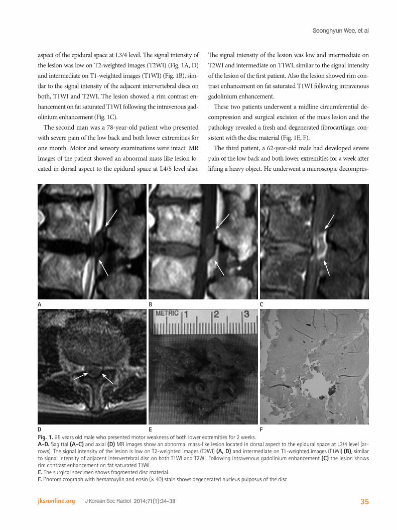
The signal intensity of the lesion was low and intermediate on T2WI and intermediate on T1WI, similar to the signal intensity of the lesion of the first patient. Also the lesion showed rim con-trast enhancement on fat saturated T1WI following intravenous gadolinium enhancement.
These two patients underwent a midline circumferential de-compression and surgical excision of the mass lesion and the pathology revealed a fresh and degenerated fibrocartilage, con-sistent with the disc material (Fig. 1E, F).
The third patient, a 62-year-old male had developed severe pain of the low back and both lower extremities for a week after lifting a heavy object. He underwent a microscopic decompres-
aspect of the epidural space at L3/4 level. The signal intensity of the lesion was low on T2-weighted images (T2WI) (Fig. 1A, D) and intermediate on T1-weighted images (T1WI) (Fig. 1B), sim-ilar to the signal intensity of the adjacent intervertebral discs on both, T1WI and T2WI. The lesion showed a rim contrast en-hancement on fat saturated T1WI following the intravenous gad-olinium enhancement (Fig. 1C).
The second man was a 78-year-old patient who presented with severe pain of the low back and both lower extremities for one month. Motor and sensory examinations were intact. MR images of the patient showed an abnormal mass-like lesion lo-cated in dorsal aspect to the epidural space at L4/5 level also.
Fig. 1. 95 years old male who presented motor weakness of both lower extremities for 2 weeks.A-D. Sagittal (A-C) and axial (D) MR images show an abnormal mass-like lesion located in dorsal aspect to the epidural space at L3/4 level (ar-rows). The signal intensity of the lesion is low on T2-weighted images (T2WI) (A, D) and intermediate on T1-weighted images (T1WI) (B), similar to signal intensity of adjacent intervertebral disc on both T1WI and T2WI. Following intravenous gadolinium enhancement (C) the lesion shows rim contrast enhancement on fat saturated T1WI.E. The surgical specimen shows fragmented disc material.F. Photomicrograph with hematoxylin and eosin (× 40) stain shows degenerated nucleus pulposus of the disc.
E
B
D
A
F
C
Posterior Thecal Sac Migration of Sequestered Disc Fragment
36 jksronline.orgJ Korean Soc Radiol 2014;71(1):34-38
Those fragments often appear heterogeneous, whereas tu-mors usually appear homogeneous on T2WI (1, 4). A peripher-al enhancement around the sequestrated disc fragment is com-monly found on contrast-enhanced MR imaging (7).
Yamashita et al. (8) speculated based of pathologic and con-trast-enhanced MR imaging findings, that the mechanism of peripheral enhancement around the sequestered disc materials is associated with the accumulation of contrast material within the vascularized granulation tissue surrounding the avascular sequestered disc material. In our cases, the fragments appeared as hypo- to isointense mass-like lesion on T1WI and hypo- to mild hyperintense lesion on T2WI. All cases showed a peripher-al enhancement around the disc fragments on gadolinium-en-hanced MR images.
The differential diagnosis of a dorsal epidural mass-like lesion is important, because it may influence both, the treatment and prognosis of the patient. Before operation, those lesions may be diagnosed as an epidural abscess, epidural spinal tumor or epi-dural hematoma. An epidural abscess is typically iso- to hypoin-tense on T1WI and hyperintense on T2WI (9). The enhance-ment can be diffusely homogeneous, heterogeneous or rim-enhanced (3). Epidural tumors show variable signal intensity and enhancement features according to the tumor types (3). They are often hypointense on T1WI and hyper- or hypointense on T2WI (3). Generally, an epidural hematoma appears iso- or hyperintense on T1WI in the acute and sub-acute phase (2).
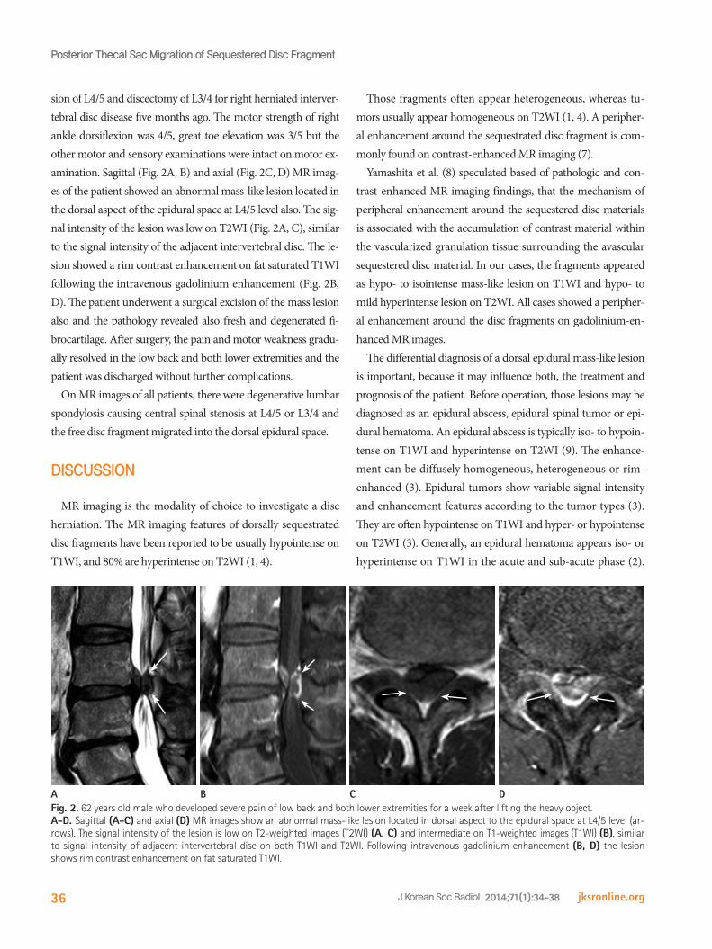
sion of L4/5 and discectomy of L3/4 for right herniated interver-tebral disc disease five months ago. The motor strength of right ankle dorsiflexion was 4/5, great toe elevation was 3/5 but the other motor and sensory examinations were intact on motor ex-amination. Sagittal (Fig. 2A, B) and axial (Fig. 2C, D) MR imag-es of the patient showed an abnormal mass-like lesion located in the dorsal aspect of the epidural space at L4/5 level also. The sig-nal intensity of the lesion was low on T2WI (Fig. 2A, C), similar to the signal intensity of the adjacent intervertebral disc. The le-sion showed a rim contrast enhancement on fat saturated T1WI following the intravenous gadolinium enhancement (Fig. 2B, D). The patient underwent a surgical excision of the mass lesion also and the pathology revealed also fresh and degenerated fi-brocartilage. After surgery, the pain and motor weakness gradu-ally resolved in the low back and both lower extremities and the patient was discharged without further complications.
On MR images of all patients, there were degenerative lumbar spondylosis causing central spinal stenosis at L4/5 or L3/4 and the free disc fragment migrated into the dorsal epidural space.
DISCUSSION
MR imaging is the modality of choice to investigate a disc herniation. The MR imaging features of dorsally sequestrated disc fragments have been reported to be usually hypointense on T1WI, and 80% are hyperintense on T2WI (1, 4).
Fig. 2. 62 years old male who developed severe pain of low back and both lower extremities for a week after lifting the heavy object.A-D. Sagittal (A-C) and axial (D) MR images show an abnormal mass-like lesion located in dorsal aspect to the epidural space at L4/5 level (ar-rows). The signal intensity of the lesion is low on T2-weighted images (T2WI) (A, C) and intermediate on T1-weighted images (T1WI) (B), similar to signal intensity of adjacent intervertebral disc on both T1WI and T2WI. Following intravenous gadolinium enhancement (B, D) the lesion shows rim contrast enhancement on fat saturated T1WI.
A B C D

Seonghyun Wee, et al
37jksronline.org J Korean Soc Radiol 2014;71(1):34-38
In conclusion, our cases suggest that the MR imaging shows characteristic features for the dorsal epidural migration of a se-questered lumbar disc fragment. And both, older age and previ-ous intervertebral decompression surgery may be predisposing factors of dorsal epidural migration of a sequestered lumbar disc fragment.
REFERENCES
1.MasarykTJ,RossJS,ModicMT,BoumphreyF,BohlmanH,
WilberG.High-resolutionMR imagingof sequestered
lumbarintervertebraldisks.AJRAmJRoentgenol1988;
150:1155-1162
2.BonarotiEA,WelchWC.Posteriorepiduralmigrationofan
extrudedlumbardiscfragmentcausingcaudaequinasyn-
drome.Clinicalandmagneticresonanceimagingevalua-
tion.Spine(PhilaPa1976)1998;23:378-381
3.HuangTY,LeeKS,TsaiTH,SuYF,HwangSL.Posteriorepi-
duralmigrationofsequestrated lumbardiscfragments
intothebilateralfacet joints:casereport.Neurosurgery
2011;69:E1148-E1151
4.DösogluM,IsM,GezenF,ZiyalMI.Posteriorepiduralmi-
grationofalumbardiscfragmentcausingcaudaequina
syndrome:casereportandreviewoftherelevantlitera-
ture.EurSpineJ2001;10:348-351
5.RobeP,MartinD,LenelleJ,StevenaertA.Posteriorepidu-
ralmigrationofsequesteredlumbardiscfragments.Re-
portoftwocases.JNeurosurg1999;90(2Suppl):264-266
6.KuzeyliK,CakirE,UsulH,BaykalS,YazarU,KaraarslanG,
etal.Posteriorepiduralmigrationof lumbardiscfrag-
ments:reportofthreecases.Spine(PhilaPa1976)2003;
28:E64-E67
7.RossJS,ModicMT,MasarykTJ,CarterJ,MarcusRE,Bohl-
manH.Assessmentofextraduraldegenerativedisease
withGd-DTPA-enhancedMRimaging:correlationwith
surgicalandpathologicfindings.AJNRAmJNeuroradiol
1989;10:1243-1249
8.YamashitaK,HiroshimaK,KurataA.Gadolinium-DTPA--
enhancedmagneticresonanceimagingofasequestered
lumbarintervertebraldiscanditscorrelationwithpatho-
logicfindings.Spine(PhilaPa1976)1994;19:479-482
9.SandhuFS,DillonWP.Spinalepiduralabscess:evaluation
The differential diagnosis should include synovial cysts that typ-ically communicate with the synovia of a degenerative adjacent articulation, based on the location of the lesion (3). A synovial cyst originating from the facet joint is centered on the facet and has a variable imaging signal, depending on the contents of the cyst. They are usually rim-enhanced and sometimes associated with calcification (5).
To the best of our knowledge, there are few reported cases with lumbar disc migration to the dorsal epidural space yet. At the level of the disc, the posterior longitudinal ligament (PLL) is firmly adherent to the dorsal annulus and is attached to the lat-eral membrane, also called peridural membrane (10) extending medially from the lateral edge of the PLL to the lateral wall of the spinal canal. This limits the movement of the extruded disc fragment beyond the postero-lateral corner of the epidural space and makes it difficult for a disc to migrate dorsal to the epidural space (1, 2).
The mechanism of dorsal migration still remains unclear. Per-haps, a stretching of PLL and other ligaments may push or dis-place the free disc fragment into the spinal canal (4). Possible explanations of dorsal migration of a free disc fragment include an intraspinal pressure gradient during the traction of the spine (4) or a position of the nerve root relative to the intervertebral disc space and the epidural space determining the migration path (6). The hypothesis has been verified that a side-to-side migration of an extruded lumbar disc herniation can be blocked or restrained by the presence of the ventral meningovertebral ligaments (11). In addition, Kuzeyli et al. (6) suggested that conditions of hyper-mobility associated with degenerative disc disease or degenera-tive spondylolisthesis may also be predispositions for a dorsal migration of disc fragments. There were degenerative lumbar spondylosis causing central spinal stenosis at L4/5 and L3/4 and the free disc fragment migrated into the dorsal epidural space in our three presented patients.
We suggest that older age and previous intervertebral decom-pression surgery may be one of the predisposing factors of dor-sal epidural migration of a sequestered lumbar disc fragment. These two factors may be together with conditions of hypermo-bility associated with degenerative disc disease or degenerative spondylolisthesis. The first and the second patient belonged to the older age group and the third patient showed a history of in-tervertebral decompression surgery 5 months ago.

Posterior Thecal Sac Migration of Sequestered Disc Fragment
38 jksronline.orgJ Korean Soc Radiol 2014;71(1):34-38
thedeeplayeroftheposteriorlongitudinalligament.An
anatomical,radiologic,andclinicalstudy.Spine(PhilaPa
1976)1993;18:1030-1043
11.ScapinelliR.Themeningovertebralligamentsasabarrier
totheside-to-sidemigrationofextruded lumbardisc
herniations.ActaOrthopBelg1992;58:436-441
withcontrast-enhancedMRimaging.AJNRAmJNeuro-
radiol1991;12:1087-1093
10.WiltseLL,FonsecaAS,AmsterJ,DimartinoP,Ravessoud
FA.Relationshipofthedura,Hofmann’s ligaments,Bat-
son’splexus,andafibrovascularmembranelyingonthe
posteriorsurfaceofthevertebralbodiesandattachingto
후경막외로 전위된 요추부 추간판 탈출증: 3예 보고1
위성현1 · 정선관1 · 심대무2
저자들은 후경막외로 전위된 요추부 추간판 탈출증 3예를 경험하여 문헌 고찰과 함께 보고하고자 한다. 3명의 환자는 모
두 60세 이상의 남자였고, 3명 중 1명의 환자는 내원 5개월 전에 요추부 추간판 탈출증 진단하에 관혈적 현미경적 감압술
과 추간판 제거술을 받은 과거력을 가지고 있었다. 자기공명영상에서는 세 환자 모두 후경막외로 전위된 요추부 추간판
탈출증의 특징적인 소견을 보였다. 저자들은 이러한 세 증례에서 경험하였듯이, 고령과 이전에 추간판 감압술을 시행받은
과거력이 후경막외로 전위된 요추부 추간판 탈출증의 원인이 될 수 있다고 제언하는 바이다.
원광대학교 의과대학 원광대학교병원 1영상의학과, 2정형외과