doi doc url //eprints.lib.hokudai.ac.jp/dspace/bitstream/2115/...the origin of these bodies is...
TRANSCRIPT

Instructions for use
Title FINE STRUCTURE OF POST-CAPILLARY VENULES IN MOUSE LYMPH NODES
Author(s) SUGIMURA, Makoto; FURUHATA, Kitao; KUDO, Norio; TAKAHATA, Kurahiko; MIFUNE, Yoshikatsu
Citation Japanese Journal of Veterinary Research, 12(4): 83-90
Issue Date 1964-12
DOI 10.14943/jjvr.12.4.83
Doc URL http://hdl.handle.net/2115/1799
Type bulletin
File Information KJ00002369113.pdf
Hokkaido University Collection of Scholarly and Academic Papers : HUSCAP

FINE STRUCTURE OF POST .. CAPILLARY VENULES
IN MOUSE LYMPH NODES
Makoto SUGIMURA, Kitao FURUHATA, Norio KUDO
and Kurahiko TAKAHATA
Department of Veterinary flnatomy, Faculty of Veterinay Medicine,
Hokkaido Uni'versity, Sapporo, Japan
Y oshikatsu MlFUNE
Electron A1icroscope Laboratory, Faculty of Veterinary ,A,ledicine,
Hokkaido University, Sapporo, Japan
(Received for publication, Sept. 21, 1964)
INTRODUCTION
The post-capillary venules in lymphatic tissue differ in some specific features
from other blood vessels. They have a distinctive high endothelium through which
lymphocytes migrate (DAWSON and MASUR, '29; HUMMEL, '35; PIRRO, '54/,55 and
SUGIMURA, '62) and the endothelial cells are metachromatic and react strongly with
the techniques for nonspecific esterase and succinic dehydrogenase (SMITH and
HENON, '59). With the exception of SUGIMURA, the above authors considered that the
lymphocytes moved into the venules from the surrounding lymphatic tissues, but
in a recent study of the recirculation of lymphocytes, GOWANS and KNIGHT ('64)
clearly showed that lymphocytes migrate from the blood to the lymphatic tissues.
At present, only a few electron microscope studies of the venules have been
reported (CLARK; '62, '63 and MARCHESI and GOWANS; '64). These investigations,
however, are primarily concerned with migrating lymphocytes and reticulum, and
there are no detailed descriptions of the fine structure of the endothelial cells.
There is a question whether lymphocytes migrate by penetrating an endothelial
cell or between the cells. MARCHESI and GOWANS stated that lymphocytes migrate
by penetrating an endothelial cell but not pass through the intercellular junction.
In this paper, the authors furnish contrasting evidence that lymphocytes migrate
between the endothelial cells of the venule, in addition to descriptions of the fine
structures of the endothelial cells and the pericytes of the venules.
MATERIALS AND METHODS
The mandibular, superficial axillary, subiliac and cranial mesenteric lymph nodes of 11 del
strain mice were used. The names and locations of the lymph nodes were taken from
JAP. ]. VET. RES., VOL. 12, No.4, 1964

84 SUGIMURA, M. et al.
descriptions by KAWASHIMA, et al. ('64). The mice· were of both sexes, ranging in age from
60 to 215 days and in weight from 21.5 to 37.5 g. Lymph node specimens were fixed for
1-l.5 hours and then dehydrated with graded ethanols. Fixation and dehydration were
carried out at ~OC. The specimens were embedded in methacrylate or Epon 812 (LUFT).
Sections were cut on a HITACHI UM-3 microtome using glass knives. After mounting on
copper grids, the sections were stained with uranyl acetate (WATSON) or lead tartrate
(MILLONIG) and examined with a JEM CHD~·4 electron microscope at initial magnifications
varying from 1,500 to 10, 000. Some thicker sections were stained with toluidine blue for
identification of the area to be sectioned for electron microscopy.
In addition, paraffin sections of lymph nodes from different locations were fixed in
CARNOY's solution and then stained with hematoxylin-eosin, toluidine blue, pyronine-methyl
green and PAS. Some paraffin sections fixed in ice-cold acetone were attempted to demonstrate alkaline and acid phosphatase.
OBSERVATIONS
With the optical microscope, post-capillary venules were observed in the dense, cortical
lymphatic tissue (Figs. 1 and 2). The thick endothelium gradually became thinner as the veins
traversed the medullary cords. The cytoplasm of the endothelial cells was pyroninophilic
(Fig. 3) and stained metachromatically with toluidine blue, but was negative to the other
procedures, such as PAS, acid phosphatase etc. Many lymphocytes were observed at several
levels in the walls of venules, but with this magnification, it was impossible to determine
whether the lymphocytes migrated into the endothelial cells or between them (Fig. 3). There ~ were no smooth muscular cells in the wall of the venules.
With the electron microscope, the post-capillary venules were 0 bserved to consist of cuboid endothelial cells and slender pericytes.
1. Endothelial Cells
The venous lumen is covered by a single continuous layer of endothelial cells. These
cells are usually cuboid, but they frequently are forced into irregular contours by the migrating
lymphocytes (Figs. 4 and 5). The lateral cell surface has terminal bar-like junctions and
interdigitations of the cell membrane (Fig. 14). The basal portions of the cells have irregular
projections which differ from those usually observed in blood vessels, and which are surrounded
by a basement membrane (Fig. 4).
The nuclei of the endothelial cells are forced into various shapes by the migrating
lymphocytes. The nuclear membrane is triple-layered, at any point of which the nuclear
membrane pores are visible (Fig. 9). The nucleoplasm consists of fine dense structures in
a less dense matrix. These structures clustered around the periphery, forming a rim along
the innermost nuclear membrane. Close to this nuclear membrane, there are one or two
large nucleoli which are composed of nucleolonema. Another unknown body consisting of
lower density particles is frequently found in the nuclei (Figs. 4 and 9).
On the free surfaces of the cell, there are a few microvilli and pinocytotic vesicles (Fig. 5).
In comparison with other blood vessels. there are more organelles in the cytoplasm of the
endothelial cells of the venules. There are many. usually oval or elongated, mitochondria
f'

Fine Structure of Post~capillar.y Venules in fl,1ouse Lymph Nodes 85
with regular, plate-like cristae. Well developed Golgi complex, abundant, rough-surfaced
endoplasmic reticula, and three types of inclusions seem to be characteristic of the endothelial
cells. The Golgi complex occupies a large area of the cells and consists of smooth-surfaced
vacuoles and many vesicles. Near the Golgi complex, there is the centrosome containing two
centrioles (Fig. 10). There are many rough-surfaced endoplasmic reticula, which are usually
cystic and which are closely related to the adjacent mitochondria, and also many free ribo
somes in the cytoplasm of the endothelial cells (Figs. 14 and 15).
The inclusion bodies may be separated into three forms. The first type is a multivesicular
body which is groups of small vesicles surrounded by a membrane (Figs. 5 and 9). This
body appears to have been formed from the Golgi vesicles (Fig. 11). Sometimes the matrix
of these bodies is of high electron density, and they closely resemble the second type (Figs.
12 and 13). Inclusions of the second type are round bodies of varying sizes, usually
surrounded by a single thin membrane, which generally contains a homogeneous dense matrix,
but which occasionally has a less dense matrix in which there are denser areas or myelin-like
structures (Figs. 8, 9 and 15). Some of these bodies have no membrane (Figs. 9 and 21).
Bodies of this second type are usually refered to as lysosomes, microbodies or segresomes.
The origin of these bodies is unknown, but it may be the smooth-surfaced endoplasmic
reticulum or the multivesicular body. Inclusion bodies of the third type contain a crystal
and seem to be characteristic of the endothelial cells of the post-capillary venules. The bodies
are spherical, resembling those of the second type, but they do not always have a distinct
membrane. The matrices are of intermediate electron density and contain a cuboid crystal.
Close to the sides of this crystal, there are either two or four highly dense structures (Figs.
4, 15 and Hi). The origin of these third type of bodies seems to be an inclusion body of the
second type.
No mitosis was observed III the endothelial cells.
2. Pericytes
The pericytes surround the endothelium of the post-capillary venule. They are separated
from the endothelial cells by the basement membrane and an intercellular space. Usually
there is at least one layer of these slender pericytes, and sometimes two or more (Fig. 15).
There are only a few organelles in the cytoplasm, some scattered mitochondria, several
rough-surfaced endoplasmic reticula, free ribosomes and vesicles (Figs. 6, 15 and 18). The
nucleus of the pericyte is usually a long ellipsoid, the nucleoplasm is similar in appearance
to that of the endothelial cells, but there are no prominent nucleoli. It was noted that there
was a filamentous structure in the cytoplasm of these pericytes and a basement membrane
surrounding them (Figs. 15, 18 and 21).
3. Reticular Cells
Reticular cells usually separate the pericytes from the lymphocytes of the cortex. There
IS an intercellular space between the pericytes and reticular cells. In some of the sections,
there are gaps between the reticular cells in which there are small lymphocytes (Fig. 20). The
reticular cells are large with irregular contours and cytoplasmic expansions. Some of the
reticular cells have rich, rough·surfaced, tubular endoplasmic reticula but no inclusion bodies

86 SUGIMURA, M. et al.
(Fig. 17). Other cells have many inclusion bodies and vesicles and none of the rough-surfaced
endoplasmic reticula (Figs. 15 and 16). The nuclei of the reticular cells are ovoid and the
nucleoplasm is of low density, similar to that of the pericytes, but they do not have basement
membranes as the pericytes do (Fig. 17).
All of the previously described cells are separated each other by intercellular spaces
containing a low density, amorphous substance and several filamentous structures, but not
typical collagenous fibrils (Fig. 17).
4. Lymphocytes Migrating through the Venules
All of the lymphocytes migrating through the post-capillary venules are small lymphocytes
with a thin cytoplasm containing fairly numerous free ribosomes, one or two tubular rough
surfaced endoplasmic reticula (Fig. 20) and scattered or grouped mitochondria (Fig. 11).
Occasionally, poorly defined Golgi complex and centrioles were observed in the cytoplasm
(Figs. 4, 6 and 8). The nuclei of the lymphocytes are usually rounded and rarely, there is
a deep indentation in the nuclear membrane (Fig. 7). The nucleoplasm is dense with a small
compact nucleolus. Frequently, one or more unknown bodies, resembling those previously
described in the endothelial cells, are observed in the nucleoplasm (Figs. 6, 8 and 20). There
were no other leucocytes in the walls of the venules.
The lymphocytes are located between the endothelial cells, and between the pericytes and
the endothelial cells. Sometimes the lymphocytes seem to be in the endothelial cells but not
between them (Figs. 4 and 6), but in many sections, the lymphocytes are located between the
endothelial cells forcing and misshaping the adjacent endothelial cells. The cell membranes
of both types of cells are always intact (Figs. 19 and 22). The lymphocytes seem to migrate
between the junctions of the endothelial cells, although the direction of the lymphocytic
migration cannot be determined from these observations.
One degenerated cell (lymphocyte ?) was observed between the endothelial cells (Fig. 23).
DISCUSSION
Electron microscope studies of the blood vessels in the lymphatic tissue have
been reported by many investigators (CLARK, '62 and '63; HOSOKAWA, '60; NAITO
and lSOKA W A, '62; TORO, '63). The ultrastructure of the blood vessels in the
lymphatic tissue is apparently similar to that of blood vessels in other tissues, with
the exception of the sinusoids in the liver (HAMfYfON, '64; SCHMIDT, '60), spleen
(WEISS, '57, '61, '62; ROBERT and LATTA, '64) and bone marrow (WEISS, '61; ZAMBONI
and PEASE, '61). On the other hand, only a few authors have described the ultra
structure of the post-capillary venules in the lymph nodes. CLARK (,62) stated that,
in mice, the endothelial cells of the venule contained well developed ergastoplasm
and some dense inclusions, as well as cytoplasmic vesicles and irregular micro
villi along the luminal border, but he did not describe the pericytes. MARCHESI and
Gow ANS (,64) reported briefly that the endothelial cells in rat venules have organelles
which are similar to these in other blood vessels. Their interest seemed to have

Fine Structure of Post-capillary Venules in Mouse Lymph Nodes 87
been focused on lymphocytic migration through the venules.
In the persent paper, the authors have shown that the endothelial cells of the post-capillary venules have many mitochondria, well developed rough-surfaced
endoplasmic reticula and numerous free ribosomes. In addition, the cytoplasm of the
endothelial cells includes a prominent Golgi complex and three types of inclusions;
a multivesicular body, a dense inclusion body without a crystal and an inclusion
body with a cuboid crystal. The latter type of inclusion has never been observed
in cells of other types of mouse lymph nodes. These features are characteristic
of the endothelial cells of the venule which are different from those of the other
blood vessels.
The endothelium is surrounded by pericytes. An intercellular space separates
the basement membrane of the endothelial cell from these pericytes. The pericytes
are also surrounded by a basement membrane. In the cytoplasm of the pericytes,
there is a filamentous structure and poorly developed organelles. The pericytes of
the post-capillary venule are much better developed than those of the arterial
capillaries in the mouse lymph nodes, although HOSOKA WA ('61) reported that the
arterial capillaries of the human lymph node are specific in having discontinuous,
smooth muscular cells which correspond to the pericytes. In addition, NAITO and
ISOKAWA ('62) described corresponding pericytes in the arterial capillaries of the
human lymph node and called then "ROUGET's cells" which are usually found only
in arterial capillaries. From this point of view, it is possible that the post-capillary
venules of the mouse lymph nodes may belong to the capillaries rather than to
the venules. The pericytes may be active in contraction of specific venules.
Many earlier investigators considered the direction of movement of the lym
phocytes through the walls of the venules to be to the venule from the surrounding
lymphatic tissues (DAWSON and MASUR, '29; HUMMEL, '35; PIRRO, '54/'55; SMITH
and HENON, '59), but GOWANS and KNIGHT ('64) clearly demonstrated that the
direction was from the blood to the lymphatic tissue. Furthermore, MARCHESI and
GOWANS ('64) did electron microscope studies of the migration of lymphocytes
through the endothelium of the venule in rat lymph nodes. They stated that all of
cells in the wall of the venule are small lymphocytes, and that no other leucocytes
are normally present. These small lymphocytes migrate across the vessel walls by
entering the endothelial cells and traversing their cytoplasm, but not pass through
the intercellular junction. In inflamed nodes, however, other leucocytes emigrate
through the venules by penetrating between the endothelial cells.
CLARK (,62) stated that small lymphocytes could be observed at several levels
in the walls of venules in mice lymph nodes. They lay between the endothelial
cells in what appeared to be an intercellular space, beneath the endothelium in
a space surrounded by an apparently split or reduplicated endothelial basement
88 SUGIMURA, M. et al.
membrane, and beneath the basement membrane among the reticular fibers.
The present authors found small lymphocytes between the endothelial cells in
the majority of the sections, although, in some of the sections, the lymphocytes
seemed to be in the cytoplasm of the endothelial cells. The lymphocytes produced
irregularities in the outlines of adjacent endothelial cells. Small lymphocytes seemed
to migrate by penetrating the junction between the endothelial cells but not by
penetrating their cytoplasm. These findings agree with CLARK's description ('62).
The direction of the movement of the lymphocytes through the walls of the
venules was not determined in this study. However, GESNER and GOWANS ('62)
showed data indicating that lymphocytes recirculate from the blood to the thoracic
duct, obtained in a study of the output of lymphocytes from the thoracic duct of
unanaesthetized mice.
Certainly, small lymphocytes have a specific affinity for the endothelial cells of
the post-capillary venules, but the present authors do not known the reason for
this. Recently, lymphocytes have attracted attention as "immunological competent
cells" in homograft immunity and other immunological reactions (BURNET, '59,
PRENDERGAST, '64). The results of the present studies indicate that the endothelial
cells, in the cytoplasm of which there are a prominent Golgi complex, well developed
rough-surfaced endoplasmic reticula and specific inclusions, but no mitosis, seem to
produce any substance which would activate recirculating lymphocytes or be useful
in their immunological function. But the actual functional relation between the
endothelial cells and the small lymphocyte requires further investigation.
SUMMARY
The post-capillary venules of mouse lymph nodes were examined with optical
and electron microscopes.
The endothelial cells of these venules contain many mitochondria, well
developed, rough-surfaced endoplasmic reticula, numerous free ribosomes, a promi
nent Golgi complex and three types of inclusions; multivesicular bodies and dense
inclusion bodies with and without a cuboid crystal. The prominent Golgi complex
and the inclusion with a cuboid crystal are not present in the endothelial cells of
other blood vessels in the lymph nodes of mice.
The endothelium of the venules is surrounded by pericytes which correspond
to ROUGET'S cells which are usually only observed in arterial capillaries.
All of cells migrating through the post-capillary venules are small lymphocytes
and no other leucocytes are normally present. The small lymphocytes migrate by
penetrating the junction between the endothelial cells but not by penetratil1g their
cytoplasm.

Fine Structure of Post-capillar." Venules in .~follse Lymph Nodes 89
One degenearted cell (lymphocytes ?) was observed between the endothelial
cells of the venules.
REFERENCES
1) BURNET, M. (1959): The clonal selection theory of aquired immunity, Cambridge
2) CLARK, S. L. JR. (1962): .. 4mer. J. Anat., 110, 217
3) CLARK, S. L. JR. (1963): Ibid., 112, 1
4 ) DAWSON, A. B. & MASUR J. (1929): Anat. Rec., 44, 143
5) GESNER, B. M. & GOWANS, J. L. (1962): Brit. J. e:xp. Path., 43, 424
(») GOWANS,]. L. & KNIGHT, E. J. (1964): Proc. ro,V. Soc. SCI'. B, 159, 257
7) HAMPTON, J. C. (1964): Liver, Electron microscopic anatomy, Edit. by KURTZ,
S. M., Academic Press, New York and London
8) HOSOKAWA, K. (1961): Jii. Z. (Tokyo Jikeikai It1cdical Journal), 76, 1274 (in Japanese)
9) HUMMEL, K. P. (1935): Amer. J. Anat. 57, 351
10) KAWASHIMA, Y, SUGIMURA, M., HWANG, Y. and KUDO, N. (1964): Jap. J. 'l'ct.
Res., 12, 69
11) MARCHESI, V. T. & GOWANS, J. L. (1964): 1'1'0('. roy. Soc. Scr. n, 159, 283
12) NAITO, T. & ISOKAWA, F. (1962): Nichi-So-Roku (11rch. histol. jajJ.) , 22 397 (in
Japanese with French summary)
13) PIRRO, A. F. (1954/55): Quad .• 1nat. practica, 10, 8f) (Cited by SMITH & HENON)
14) PRENDERGAST, R. A. (1964): J. e:xiJ .• r...led., 119, 377
15) ROBERT, D. K. & LATTA, J. S. (1964): Anat. Rcc" 148, 81
16) SCHMIDT, F. C. (1960); Anat. AriZ., 108, 376
17) SMITH, C. & HENON, B. K. (1959): Anat. Rec., 135, 207
18) SUGIMURA, M. (1962): Jap . .J. 'pct Res., 10, 155
19) TaRO, 1. (1962): Verh. anat. Ges., Anat. Anz., Erg., 112, 111
20) WEISS, L. (1957): J. Biophys. Biochem. C:vtol., 3, 599
21) WEISS, L. (1961): Johns HojJk. Hosp. Bull., 108, 171
22) WEISS, L. (19Gl): Anat. Rec., 139, 286
23) WEISS, L. (19t)2): Amer. J ... 4 nat. , 111, 131
24) ZAMBONI, L. & PEASE, D. C. (1961): .1. Ultmstl'llcture Rrs., 5, 65

90 SUGIMURA, M. et al.
ABBREVIATIONS TO PLATES
B Unknown body in nucleoplasm
BA Basement membrane
C Centrosome
D Degenerated cell
E Endothelial cell
F Filamentous structure
G Golgi complex
IC Inclusion body with a crystal
ID Inclusion body without a crystal
J Terminal bar-like junction
L Lymphocyte
M Mitochondria
MD Multivesicular body with dense matrix
MU Multivesicular body
N Nucleus
NP Nuclear membrane pore
NU Nucleolus
P Pericyte
PV Pinocytotic vesicle
R Reticular cell
RE Rough-surfaced endoplasmic reticulum
SE Smooth-surfaced endoplasmic reticulum
EXPLANATION OF FIGURES
Plate I Fig. 1. Iliac node: Post-capillary venules in the cortex. The endothelial cells
gradually flatten as the veins traverse the medullary cord H-E X 60
Fig. 2. Lumbar node: Post-capillary venule connected to a capillary. Many
lymphocytes in the endothelium of the venule H-E X 1,000
Fig. 3. Iliac node: The cytoplasm of the endothelial cells is pyroninophilic.
Lymphocytes at the level of the endothelial cells, and between the endothelial
cell and perivascular sheath Pyronine-Methyl green X 1,000

SUGIMURA, M. et al. PLATE I
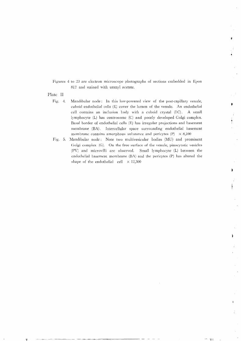
Figures 4 to 23 are electron microscope photographs of sections embedded III Epon
812 and stained with uranyl acetate.
Plate II
Fig. 4. Mandibular node: In this low-powered VIew of the post-capillary venule,
cuboid endothelial cells (E) cover the lumen of the venule. An endothelial
cell contains an inclusion body with a cuboid crystal (I C). A small
lymphocyte (L) has centrosome (C) and poorly developed Golgi complex.
Basal border of endothelial cells (E) has irregular projections and Casement
membrane (EA). Intercellular space surrounding endothelial basement
membrane contains amorphous substance and pericytes (P) X 8,100
Fig. 5. Mandibular node: Note two multivesicular bodies (MU) and prominent
Golgi complex (G). On the free surface of the venule, pinocytotic vesicles
(PV) and microvilli are observed. Small lymphocyte (L) between the
endothelial basement membrane (BA) and the pericytes (P) has altered the
shape of the endothelial cell X 12,300
.. ---~ _ .... _---- ~-------------- -~ - ~ - -,T

SUGIMURA, M. et al. PLATE II

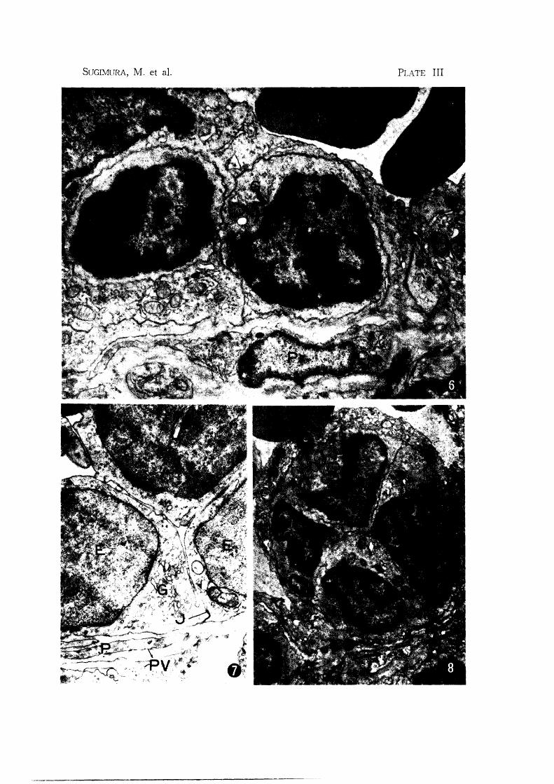
Plate III
Fig. 6. Mandibular node: Two small lymphocytes appear to locate in cytoplasm
of endothelial cell (E). The cell membranes of Loth types of cells are
intact. A lymphocyte has a poorly developed Golgi complex (G) X 13,000
Fig. 7. Subiliac node: Part of the venule in the medullary cord. Venule is inter
mediate form tetween post-capillary venule and usual venule. Note flattened
endothelial cells (E), poorly developed Golgi complex (G) and mitochondria.
Perivascular space surrounding the basement membrane contains two layers
of pericytes (P) X 12,000
Fig. 8. Mandibular node: Four small lymphocytes (L) appear to locate in endo
thelial cell X 8,600
T
SUGIMURA, M. et al. PLATE III

Plate IV
Fig. 9. Mandibular node: Apical portion of the endothelium of the post-capillary
venule. The nuclei of the endothelial cells (E) have one or two nucleoli
(NU), unknown cody (B) and nuclear membrane pore (NP). Terminal
Lar-like junction (J) cetween the endothelial cells, Golgi complex (G), multi
vesicular body (MU) and inclusion bodies (ID) are found X 19,200
Fig. 10. Mandibular node: Four lymphocytes (L) between endothelial cells. Note
projections of endothelial cell between the lymphocytes (arrow). Prominent
Golgi complex (G) and centrioles (C) are oJserved in the cytopJasm of an
endothelial cell X 11,500

SUGIMURA, M. et al. PLATE IV

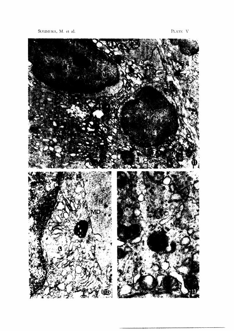
Plate V
Fig. 11. Subiliac node: Basal portion of the post-capillary venule. Note multi
vesicular codies (MU) and prominent Golgi complex (G). In this figure,
the multivesicular bodies seem to be formed from the Golgi vacuole and
vesicles. Small lymphocyte (L) appears to lay between two or three
endothelial cells X 14,000
Fig. 12. Mandibular node: Note multivesicular body with dense matrix (MD) near
Goigi complex (G). Formation of this body seems to be related to the
Golgi vesicles X 26,000
Fig. 13. Subiliac node: Multivesicular lody with dense matrix (MD) X 26,000
I. I .
SUGIMURA, M. et al. PLATE V

Plate VI
Fig. 14. Subiliac node: Basal portion of the venule. Note terminal bar-like junction
(J) and interdigitations of the cell membranes (arrows). In the cytoplasm
of endothelial cells, rich mitochondria (M), well developed endoplasmic
reticula (RE), abundant free ribosomes and prominent Golgi complex (G)
are observed X 16,000
Fig. 15. Adjacent to Figure 14. In this figure, basal portion of endothelial cells,
thin pericyte cytoplasm and reticular cells are observed. In the cytoplasm
of an endothelial cell, there are three inclusion bodies with a clear membrane
(ID) and an inclusion body with a cuboid crystal (IC). Note irregular basal
border of the endothelium and basement membrane (BA). Pericyte (P) has
filamentous structures (F) and basement membrane (BA). Intercellular space
around the pericyte contains low density amorphous substance. Reticular
cell (R) has numerous inclusions X 15,500
, i
~ ..I
, .
\', .
f. ')
"

SUGlMURA, M. et a1. PLATE VI

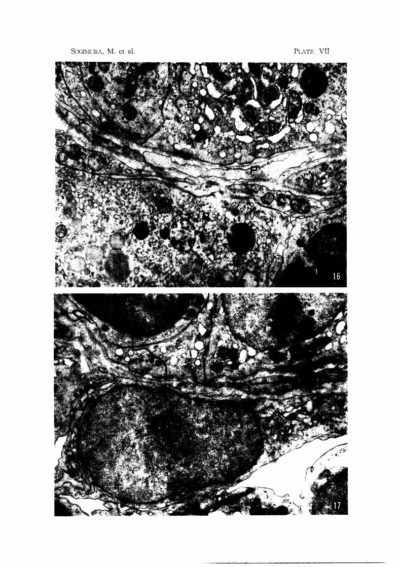
Plate VII Fig. 1(1. Subiliac node: Adjacent to Figure 15. Note basal portion of endothelial
cell (E), pericytes (P) and reticular cell (R). Reticular cell has variety of
inclusions, but none containing crystals X 15,500
Fig. 17. Subiliac node: Basal portion of post-capillary venule. Reticular cell (R)
separates the lymphatic tissue of the cortex from the intercellular space
around the endothelium. Note well developed endoplasmic reticulum (RE)
and prominent Golgi complex (G) in the cytoplasm of the reticular cell
X 14,800
SUGIMCRA, M. et al. PLATE vtt

T
Plate VIn
Fig. 18. Subiliac node: Basal portion of venule. Note nucleus of pericyte with
indentations of the nuclear membrane. The pericyte has poorly developed
organelles, filamentous structure (F) and Casement membrane (EA) X Hi,500
Fig. 19. Subiliac node: Note small lymphocyte (L) between two endothelial cells,
and terminal bar-like junction (.1). The cell membranes of both types of
cells are intact X 12,000
Fig. 20. Subiliac node: Lymphocyte (L) apparently located between three endothelial
cells. Note projection of the lymphocyte through the basement membrane
of the endothelium (arrow). Note small lymphocytes occluding gap in the
perivascular sheath of the reticular cells (I~) in the lower left and pinocytotic
vesicles (PV) X 12,000
. ..

SUGLMURA, M. et al. PLATE VIII

Plate IX
Fig. 21. Subiliac node: Small lymphocyte (L), located between endothelial cells (E)
and a pericytes (P), seems to have amoeboid motion. Note filamentous
structures (F) in the cytoplasm of pericyte X 13,100
Fig. 22. Subiliac node: Lymphocyte (L) located between two endothelial cells (E)
indents the cell l::odies. Note terminal tar-like junction (JJ between the
cell membranes of the endothelial cells X 15,000
Fig. 23. Subiliac node: Note degenerated cell (D) between endothelial cells and a
pericyte (P) X 10,500

PLATE IX