does the use of additional x-ray beam filtration during cine...
TRANSCRIPT
This is a repository copy of Does the use of additional X-ray beam filtration during cine acquisition reduce clinical image quality and effective dose in cardiac interventional imaging?.
White Rose Research Online URL for this paper:http://eprints.whiterose.ac.uk/84804/
Version: Accepted Version
Article:
Davies, AG, Gislason-Lee, AJ, Cowen, AR et al. (4 more authors) (2014) Does the use of additional X-ray beam filtration during cine acquisition reduce clinical image quality and effective dose in cardiac interventional imaging? Radiation Protection Dosimetry, 162 (4). 597 - 604. ISSN 0144-8420
https://doi.org/10.1093/rpd/ncu020
[email protected]://eprints.whiterose.ac.uk/
Reuse
Unless indicated otherwise, fulltext items are protected by copyright with all rights reserved. The copyright exception in section 29 of the Copyright, Designs and Patents Act 1988 allows the making of a single copy solely for the purpose of non-commercial research or private study within the limits of fair dealing. The publisher or other rights-holder may allow further reproduction and re-use of this version - refer to the White Rose Research Online record for this item. Where records identify the publisher as the copyright holder, users can verify any specific terms of use on the publisher’s website.
Takedown
If you consider content in White Rose Research Online to be in breach of UK law, please notify us by emailing [email protected] including the URL of the record and the reason for the withdrawal request.

Does the use of additional X-ray beam filtration during cine acquisition reduce
clinical image quality and effective dose in cardiac interventional imaging?
Andrew G Davies1, Amber J Gislason-Lee1, Arnold R Cowen1, Stephen M
Kengyelics1, Michael Lupton2, Janet Moore2, Mohan Sivananthan2,
1Division of Medical Physics, Room 8.001 Worsley Building, Clarendon Way,
University of Leeds, Leeds, UK, LS2 9JT
2Leeds General Infirmary, Great George Street, Leeds, UK, LS1 3EX
Corresponding Author: Amber J Gislason-Lee, phone 044 113 343 8317 email
Running Title: X-ray beam filtration in cardiac imaging
2
Does the use of additional X-ray beam filtration during cine acquisition reduce
clinical image quality and effective dose in cardiac interventional imaging?
Andrew G Davies1, Amber J Gislason-Lee1, Arnold R Cowen1, Stephen M
Kengyelics1, Michael Lupton2, Janet Moore2, Mohan Sivananthan2,
ABSTRACT
The impact of spectral filtration in digital (“cine”) acquisition was investigated using
a flat panel cardiac interventional X-ray imaging system. A 0.1 mm Cu and 1.0 mm
Al filter added to the standard acquisition mode created the filtered mode for
comparison. Image sequences of 35 patients were acquired; a double blind subjective
image quality assessment was completed and dose area product (DAP) rates were
calculated. Entrance surface dose (ESD) and effective dose (E) rates were determined
for 20 and 30 cm phantoms. Phantom ESD fell by 28% and 41% and E by 1% and
0.7 %, for the 20 and 30 cm phantoms respectively when using the filtration. Patient
DAP rates fell by 43% with no statistically significant difference in clinical image
quality. Adding 0.1 mm Cu and 1.0 mm Al filtration in acquisition substantially
reduces patient ESD and DAP, with no significant change in E or clinical image
quality.
INTRODUCTION
Coronary angiography and percutaneous interventional (PCI) procedures are
becoming more frequent [1] in the cardiac catheterisation laboratory. Moreover, with
technological medical advances there is a tendency to undertake more complex
interventions, increasing the duration of imaging in these cases. There are several

3
reports in the literature of transient and permanent skin damage caused by cardiac
catheterisation procedures [2-9], particularly with patients who require repeated
coronary angiography procedures [10]. There is a need to reduce patient peak skin
dose to a minimum level required for a given procedure in order to avoid these
deterministic effects of radiation. In addition, stochastic effects on human tissue such
as radiation-induced cancer must be avoided, in adherence with the ALARA (As Low
As Reasonably Achievable) principle [11].
Cardiac X-ray systems operate in two imaging modes - fluoroscopy and digital
acquisition; the latter is formerly known as ‘cine’ but in the context of modern digital
systems the term acquisition is more appropriate and therefore used in this paper.
Fluoroscopy is predominantly used to visualise interventional devices as they are
manipulated inside the patient, employing a relatively low radiation dose rate.
Acquisition uses higher dose rates, and commensurately provides higher fidelity
imaging used for diagnosis and assessment of treatment. Although fluoroscopy
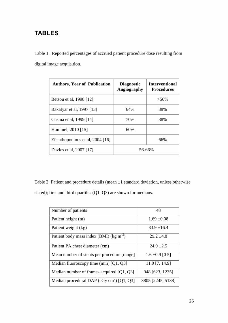
dominates in terms of time, acquisition can account for over 50% of the total accrued
patient procedure dose; percentages reported in the literature vary, as shown in Table
1.
Authors, Year of Publication Diagnostic Angiography
Interventional Procedures
Betsou et al, 1998 [12] >50%
Bakalyar et al, 1997 [13] 64% 38%
Cusma et al, 1999 [14] 70% 38%
Hummel, 2010 [15] 60%
Efstathopoulous et al, 2004 [16] 66%
Davies et al, 2007 [17] 56-66%
Table 1. Reported percentages of accrued patient procedure dose resulting from digital image acquisition.

4
In fluoroscopy, supplementary metal X-ray beam filters commonly made of
copper (Cu) are used to reduce patient skin dose [18 - 26] and have also been shown
to reduce staff dose [23]. Modern X-ray systems can be programmed to employ X-
ray beam filters in acquisition as well as in fluoroscopy, and this has become common
practice, reducing patient dose in cardiac interventions [15, 27] and neurological
interventions [25]. However, the literature demonstrates concern that Cu filtration
degrades angiogram image quality [21, 26]. Although the use of Cu filtration in
acquisition has been investigated, its impact on clinical image quality has not been
assessed [15, 24-26, 28]. It is important to assess the impact on clinical image quality
of any dose saving technique. To the authors’ knowledge, there are no published
studies which assess clinical (i.e. patient) image quality using Cu filtration in
acquisition mode for cardiac X-ray imaging.
In addition to skin dose, the stochastic, longer term effects of radiation damage
should be considered in assessing the impact of X-ray beam filtration. Copper filters
have been shown to increase Dose Area Product (DAP) to effective dose conversion
coefficients [18, 29], leading to the preconception that using Cu filtration may lead to
an undesirable increase in effective dose to the patient. Therefore the effect of Cu
filtration on patient effective dose requires further investigation.
In this prospective study we investigated the effect of a Cu beam filter on
patient dose - both skin dose and effective dose - in a phantom study as well as a
clinical assessment of cardiac patient dose and image quality using dynamic patient
image sequences.
5
MATERIALS AND METHODS
We assessed the effects on patient dose and image quality of introducing Cu
filtration in acquisition on a modern flat panel detector X-ray system in the cardiac
catheterisation laboratory at Leeds General Infirmary, UK. Modifications to an
Allura XPer FD10 system (Philips Healthcare, Best, The Netherlands) were made to
add an additional acquisition operating mode which was identical to the default
acquisition operating mode, except that 0.1 mm Cu and 1 mm aluminium (Al) X-ray
beam spectral filtration were used. The Al was used in conjunction with Cu to absorb
secondary radiation generated in the primary filter [33], as is the norm for the imaging
system. The default operating mode had no added spectral filtration; the total filtration
of the X-ray tube not including any additional pre-filtration was 2.7 mm aluminum
(Al).
Data were obtained using the default “standard” (no added filter) and the
modified “filtered” (0.1 mm Cu + 1.0 mm Al filters added) acquisition modes for two
separate study elements: a phantom dose study using the national standard
measurement techniques [30], and a clinical study of patient dose rates and image
quality using a double blinded subjective assessment. In both these study elements,
results from the two different acquisition modes were compared in order to determine
the effect of the added filtration on clinical image quality and patient dose.
Phantom Study
Phantom Dose
6
Phantom entrance surface dose (ESD) (i.e. skin dose) rates were measured
using protocol outlined by the Institute of Physics and Engineering in Medicine
(IPEM) working group, Martin et al [30]. A polymethylmethacrylate (PMMA)
phantom was used to simulate a “standard” and “large” patient in the posterior-
anterior (PA) projection, using 20 cm and 30 cm high stacks of PMMA blocks
respectively, with the C-arm rotated to place the X-ray tube near the floor underneath
the phantom. The PMMA blocks were placed in the X-ray beam with the table 90 cm
above the floor; the authors had previously reviewed two months’ worth of cardiac
imaging metadata to determine this as a representative working height for PCI
procedures. The phantom was raised from the surface of the patient couch by 5 cm
thick wood spacers, allowing the ionisation chamber (chamber #2 shown in Figure 1)
to be placed on the entrance surface of the phantom, to include backscattered
radiation.
Input air kerma was also measured, in order to calculate the effective dose to
the phantom. The air kerma was measured 32.5 cm in front of (below) the phantom
(chamber #1 in Figure 1), and corrected for the attenuation effects of the patient table.
Radcal 20X6-60 and 20X6-6 ionisation chambers with 2026C dose meters (Radcal
Corp, Monrovia CA, USA), calibrated to national standards, were used to measure the
phantom ESD and input air kerma respectively. Image sequences were acquired in
both the standard and filtered operating modes using the 20 cm nominal (14.1 x 14.1
cm) field of view. Phantom ESD and input air kerma values were recorded once the
system’s automatic dose rate control (ADRC) and dose meter outputs had stabilized.

7
Figure 1. Experimental setup with 20 cm phantom
Imaging geometry, phantom thickness and inverse square law corrected air
kerma were used to calculate effective dose rate using PCXMC software (v2.0,
STUK, Finland), a computer software program which calculates effective dose rate
using Monte Carlo methods and International Commission on Radiological Protection
(ICRP) 103 weighting factors [34]. One million photons (the maximum amount
allowed by the software) were used in the Monte Carlo simulation; this minimised the
error reported by PCXMC, which was always less than 1%. Patient positioning
simulated in PCXMC was the same as in the experimental setup (PA projection), so
the heart, lungs, and skeleton were in the X-ray field of view. All body organ doses
contributed to total effective dose. For the 20 cm phantom, the PCXMC standard
height and mass were used for calculation; the patient model had a BMI of 23 kg m-2.

8
For the 30 cm phantom, the PCXMC standard height was used and the mass was
increased to 110 kg for a BMI of 34 kg m-2; this adjustment, compared with the 20 cm
phantom, changed the proportions of anatomy within the X-ray field of view and the
distribution of radiation through the model patient.
Patient Study
Patient Dose
A group of 48 patients from those allocated to the catheterisation lab with the
modified X-ray system participated in this experiment. Ethical approval was obtained
from the local Research Ethics Committee, and all patients gave informed written
consent to participate. The mean patient body mass index (BMI) was 29.2 ± 4.8 kg
m-2; this and other patient and patient procedure characteristics are shown in Table 2.
Patient procedures began as usual, and the filtered acquisition mode was utilised for
the remaining image sequences acquired during the patient procedure, once the
clinician had established an image quality reference; standard mode image sequences
acquired at the start of the procedure provided this image quality reference to ensure
that image quality provided by the filtered mode was adequate and did not
compromise patient care. DAP values were internally calculated and reported by the
imaging system. The DAP values accrued during acquisition (i.e. excluding
fluoroscopy) and corresponding numbers of image frames were recorded; using the
frame rate (12.5 frames/sec), the average DAP rate per patient per acquisition
operating mode was calculated.
9
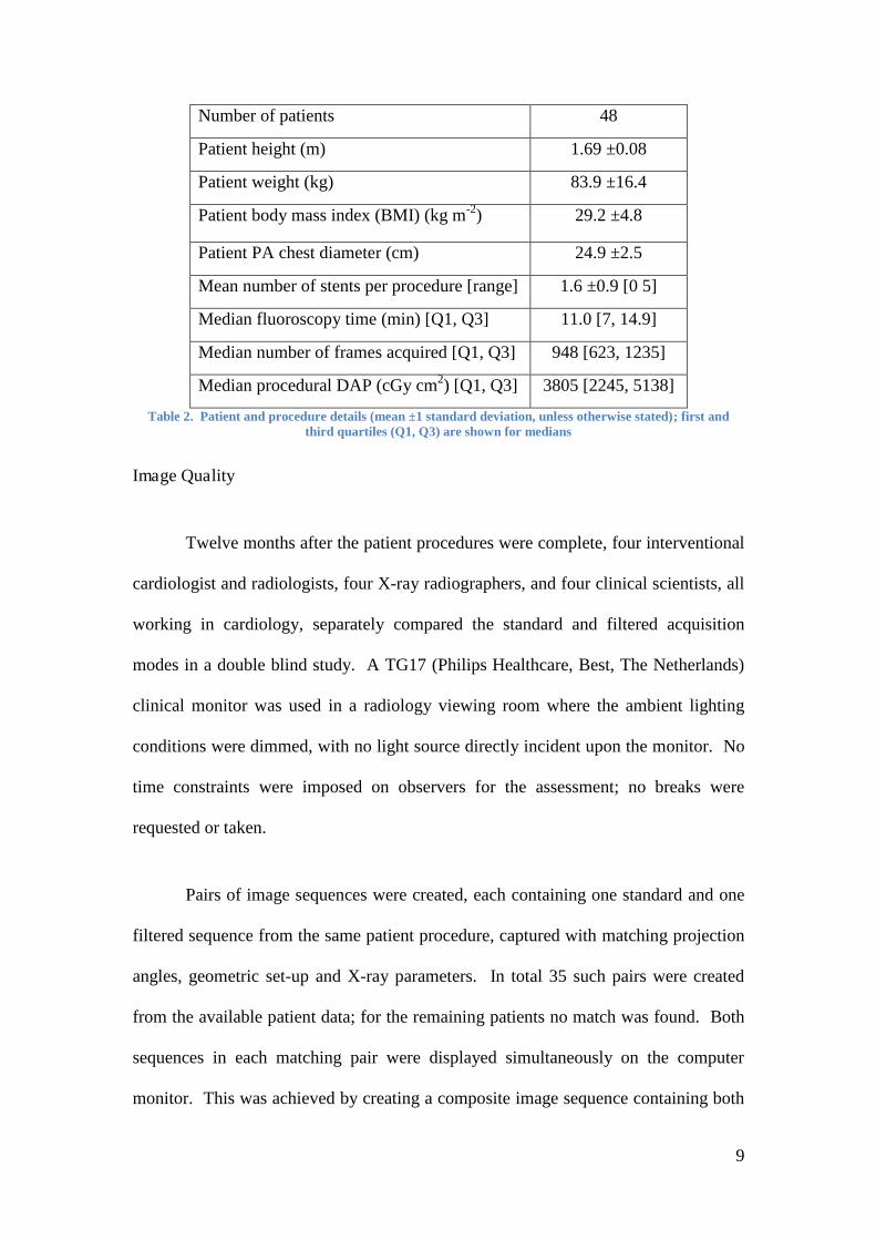
Number of patients 48
Patient height (m) 1.69 ±0.08
Patient weight (kg) 83.9 ±16.4
Patient body mass index (BMI) (kg m-2) 29.2 ±4.8
Patient PA chest diameter (cm) 24.9 ±2.5
Mean number of stents per procedure [range] 1.6 ±0.9 [0 5]
Median fluoroscopy time (min) [Q1, Q3] 11.0 [7, 14.9]
Median number of frames acquired [Q1, Q3] 948 [623, 1235]
Median procedural DAP (cGy cm2) [Q1, Q3] 3805 [2245, 5138]
Table 2. Patient and procedure details (mean ±1 standard deviation, unless otherwise stated); first and third quartiles (Q1, Q3) are shown for medians
Image Quality
Twelve months after the patient procedures were complete, four interventional
cardiologist and radiologists, four X-ray radiographers, and four clinical scientists, all
working in cardiology, separately compared the standard and filtered acquisition
modes in a double blind study. A TG17 (Philips Healthcare, Best, The Netherlands)
clinical monitor was used in a radiology viewing room where the ambient lighting
conditions were dimmed, with no light source directly incident upon the monitor. No
time constraints were imposed on observers for the assessment; no breaks were
requested or taken.
Pairs of image sequences were created, each containing one standard and one
filtered sequence from the same patient procedure, captured with matching projection
angles, geometric set-up and X-ray parameters. In total 35 such pairs were created
from the available patient data; for the remaining patients no match was found. Both
sequences in each matching pair were displayed simultaneously on the computer
monitor. This was achieved by creating a composite image sequence containing both

10
the standard and filtered sequences displayed side-by-side with software written in
Matlab 2008a (The Mathworks, Inc, Natick, USA). This software truncated the
longer of the original sequences to the length of its matching neighbour, and randomly
selected which (standard or filtered) sequence was drawn on the left and which was
on the right hand side of the pair, recording its selection in a key file on the host
computer. This file, and thus knowledge of which sequence was the standard or
filtered sequence, was not available to the observers at any stage of the experiment,
nor was it available to the investigators until all viewing sessions had been completed.
Images sequences were assessed by the observers choosing which of the two
sequences (i.e. the left hand or right hand sequence) in the pair was preferred in terms
of providing superior diagnostic image quality. Observers were also asked to state
whether both images sequences in the pair had a clinically acceptable level of image
quality. An example of a patient image sequence pair used in the double blind study
is shown in Figure 2.
Figure 2. Standard (left) and filtered (right) mode images from the same patient for comparison
Preferred (left hand or right hand) sequences were converted to a score of -1 or
+1, representing observer preference for the standard or filtered acquisition mode,

11
respectively, using the key file from when the sequences were created. Preferences
from all the observers and all image sequence pairs were pooled and a sign test was
performed to test the null hypothesis – that no difference would be found between the
two different types of image sequences, standard and filtered. The sign test was
single (left) tailed, and performed at the 5% significance level. The alternative
hypothesis was that overall observer preference would favour the standard mode. The
one-tailed test provided more power to detect an effect in one direction by not testing
the effect in the other direction.
A binomial logistic regression analysis was completed in order to determine
the influence of the individual observers, clinical roles, and the combination of
observers and their clinical roles on subjective observations. The model was created
and analysed using SPSS v16.0 (SPSS, Chicago, USA). The binomial dependent
variable was the preferred (standard or filtered) image sequence. The categorical
independent variables for the model were the individual observer, clinical role
(interventionalist, radiographer, or clinical scientist), and combination of observer and
clinical role. Pseudo R square values using the log likelihood were calculated
indicating the proportion of variance in the dependent variable associated with the
independent variables. The Wald statistic, which is the ratio of the logistic regression
coefficient and standard error squared, was used to determine the strength of the
independent variables as predictors for the dependent variable.
RESULTS
Phantom Study
12
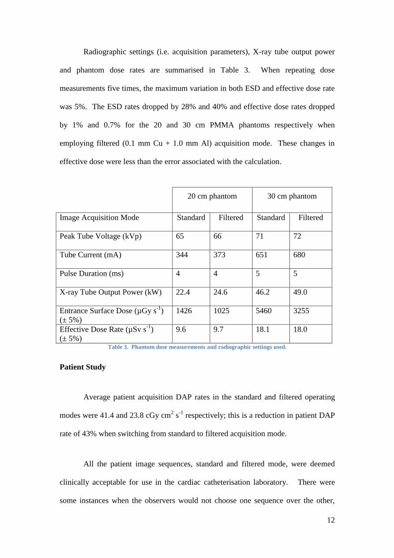
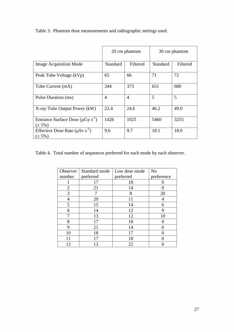
Radiographic settings (i.e. acquisition parameters), X-ray tube output power
and phantom dose rates are summarised in Table 3. When repeating dose
measurements five times, the maximum variation in both ESD and effective dose rate
was 5%. The ESD rates dropped by 28% and 40% and effective dose rates dropped
by 1% and 0.7% for the 20 and 30 cm PMMA phantoms respectively when
employing filtered (0.1 mm Cu + 1.0 mm Al) acquisition mode. These changes in
effective dose were less than the error associated with the calculation.
20 cm phantom 30 cm phantom
Image Acquisition Mode Standard Filtered Standard Filtered
Peak Tube Voltage (kVp) 65 66 71 72
Tube Current (mA) 344 373 651 680
Pulse Duration (ms) 4 4 5 5
X-ray Tube Output Power (kW) 22.4 24.6 46.2 49.0
Entrance Surface Dose (µGy s-1) (± 5%)
1426 1025 5460 3255
Effective Dose Rate (µSv s-1) (± 5%)
9.6 9.7 18.1 18.0
Table 3. Phantom dose measurements and radiographic settings used.
Patient Study
Average patient acquisition DAP rates in the standard and filtered operating
modes were 41.4 and 23.8 cGy cm2 s-1 respectively; this is a reduction in patient DAP
rate of 43% when switching from standard to filtered acquisition mode.
All the patient image sequences, standard and filtered mode, were deemed
clinically acceptable for use in the cardiac catheterisation laboratory. There were
some instances when the observers would not choose one sequence over the other,

13
claiming that the image quality was the same (no preference); the score was then zero.
The result of the sign test on pooled scores from the twelve observers whom took part
in the blind subjective image quality assessment was p = 0.2. The null hypothesis was
therefore accepted at the 5% significance level, indicating that the observers had no
preference for either the standard or filtered image sequences. Each observer’s
preference sums are shown in Table 4. The pseudo R square value correlating model
prediction with the data was 0.021; this and the Wald statistics indicate that no
independent variable in the model was significantly predictive of preference score.
Observer number
Standard mode preferred
Low dose mode preferred
No preference
1 17 18 0 2 21 14 0 3 7 8 20 4 20 11 4 5 15 14 6 6 14 12 9 7 13 12 10 8 17 18 0 9 21 14 0 10 18 17 0 11 17 18 0 12 13 22 0
Table 4. Total number of sequences preferred for each mode by each observer
DISCUSSION
Results verify past findings that a substantial reduction in patient DAP from
acquisition sequences can be achieved when 0.1 mm Cu and 1.0 mm Al spectral beam
filtration is employed in digital acquisition. A novel finding from this study is that
with this reduction in DAP there is no significant change in subjectively assessed
clinical image quality.

14
The hypothesis for the observer study of clinical image quality was that there
would be no perceived difference between the two acquisition modes being compared,
and this was accepted by the sign test. The rationale behind this hypothesis was that
the change in image quality from adding the filtration would be so small that it would
not be generally discernable in clinical image sequences. The authors had previously
found a reduction in contrast to noise ratio (CNR) of 12% and 8% for the 20 and 30
cm PMMA phantoms respectively, using raw image data. Similarly, Fetterly [28]
found a 9% decrease in CNR for water phantoms 15-40 cm thick. According to
Altman’s nomogram [31], the number of observations made in this study was high
enough to detect a difference of 10% in preference between the two imaging modes
with 80% statistical power, using a 0.05 cut-off for statistical significance, assuming
each observation was independent. It is therefore highly unlikely that the result was
due to chance.
An interventional X-ray system’s ADRC will respond differently to different
projection angles within a single patient procedure by changing the radiographic
factors used. Higher patient doses generally result from steeper patient projections
than shallow projections. Therefore one might be concerned that the experimental
phantom setup used in this study, with only the PA projection, might not accurately
represent the range of potential patient doses resulting from a cardiac catheterisation
procedure. However, the reduction in DAP rate found in the patient study (using a
clinically relevant range of projection angles) was found to be very similar to the
reduction in ESD rate measured in the phantom study (which used one projection
angle), indicating that the substantial dose savings measured in the phantom study
would be realised in clinical practice.
15
Another novel finding is that whilst there is a reduction in ESD and/or DAP in
acquisition using added filtration, the effective dose values may not change; those
calculated in the phantom study using 0.1 mm Cu and 1.0 mm Al indicate no
clinically significant change in effective dose due to the additional filtration. The
ICRP [11] makes adjustments to tissue weighting factors used to calculate effective
dose regularly, yet weighting factors for the organs of highest interest in this study are
stable, with no changes made for several decades [32]; therefore there is no concern
for uncertainty in these weighting factors. However, minor changes in PCXMC will
change the results found in this study. For example, if a slightly thinner X-ray tube
filtration was used to specify the input X-ray spectra in PCXMC, the effective dose
would decrease more for the 20 cm phantom and increase slightly for the 30 cm
phantom (still less than 5% differences). If the largest field of view available on the
imaging system was used to calculate effective dose, with the liver and stomach in the
periphery, then the effective dose would rise by about 1% for both phantom sizes. The
BMI of the patient population studied was between the two BMI’s used in PCXMC,
so actual patient size was well represented, however effective dose strongly depends
on patient size [33, 36] as well as sex. Moreover, radiographic factors selected by the
ADRC impact the effective dose because they will change not only the input dose but
also the penetrative characteristics of the X-ray beam. Should published conversion
factors rather than PCXMC be used to calculate effective dose, these influencing
factors may be reduced but results will still vary depending on the imaging system’s
ADRC, projection angle used, amount and type of spectral filtration, and other
factors. Physicists should perform effective dose calculations using the different
modes of a cardiac interventional system under various clinical scenarios in order to

16
assure the impact of spectral filtration on effective dose is understood for that
particular system.
Patient characteristics and case complexity, duration of fluoroscopy time and
number and duration of acquisition sequences varied considerably between patient
procedures, resulting in a large variation in total procedural DAP between cases. This
means that the reported reduction in total DAP due to the added filtration could be
obscured by these confounding factors. However, the assessment of patient dose
savings by using DAP rate, rather than total procedural DAP overcomes these
problems by controlling for the number and length of acquisition sequences per
procedure. Moreover DAP due to fluoroscopy was specifically excluded from this
study.
The amount of dose reduction per patient, although expressed as DAP rate,
was dependent on the thickness of the patient and also on the patient projection angles
used (which may depend on the vessel of interest). However, the observers assessed
intra-patient image sequence pairs, so the variation of BMI within the study will have
had no impact on image quality comparisons.
No alterations to the X-ray system’s ADRC programming were made, other
than the introduction of the added spectral filtration, and no issues were reported
relating to increased tube loading due to the filtration in the clinical cases. For the
phantom study, X-ray tube output power was increased by 10% and 6% for the 20 and
30 cm phantoms respectively when the spectral filtration was added. The ADRC
responded to the filtration with a modest increase of X-ray tube potential difference
(by 1 kVp) for both phantom sizes.
17
Limitations of Study
Due to ethical considerations, use of the two acquisition modes could not be
entirely randomized during patient PCI procedures. The X-ray system operator
utilised the standard acquisition mode first, to ensure quality of patient care, and
changed to the filtered mode once the clinician was comfortable with making the
switch. The patient procedure always began with standard acquisition and ended with
filtered acquisition, however the stage in the procedure when this switch occurred
varied largely from patient to patient. Therefore even the investigators, upon
retrospective image sequence viewing, could not estimate at which point in a patient
procedure the system operator switched modes. There was no preconceived
knowledge of which image sequences were captured using the standard mode and
which were captured using the filtered mode, despite this limitation. This study
design was advantageous in that it allowed for image sequences from the same patient
to be paired for mode comparison.
The X-ray system ADRC was used during this study, as is required for safe
and convenient system operation during patient procedures; radiographic factors were
automatically selected by this particular system’s characteristic ADRC programming.
A different selection of radiographic factors would not only change the patient dose,
but also impact image quality (eg. lower X-ray tube voltage increases contrast, higher
tube current decreases noise). Different manufacturers, countries, and even hospitals
utilize different ADRC programming techniques [34, 35], therefore results on other
interventional systems may differ. Image processing settings which vary between
interventional systems impact clinical image quality as well. It may be possible to
further improve the performance of the imaging system, achieving a better balance

18
between patient dose (ESD or effective dose or both) and image quality than used in
this study, for a given patient size [33, 36]. Use of sophisticated computer based
image enhancement techniques or further “tuning” of the X-ray system’s ADRC
programming could help achieve better balance; however this study demonstrates that
even without these alterations the added filtration in acquisition results in significantly
lower ESD and DAP with no significant change in clinical (patient) image quality and
no significant change in effective dose.
Comparison with Previous Studies
The level of dose reduction (aside from effective dose) found in this study was
in broad agreement with previous studies [15, 24, 28] where a similar filter thickness
was used. The current investigation focussed on the PA projection angle whereas
Dragusin et al considered two completely different projection angles and a different
thickness of Cu filtration [24]. Dragusin et al found an increase in effective dose with
additional Cu and in the current study it remained unchanged; In addition to the
projection angles being different, the ADRC in the current study increased X-ray tube
voltage and current when Cu was added whereas Dragusin et al controlled X-ray
settings independently, and all these factors influence effective dose. Dragusin et al
used an anthropomorphic phantom for image quality assessment [24], therefore it was
difficult to accurately compare dose or image quality results. However, Dragusin et al
found no statistically significant impact on image quality from adding Cu filtration in
acquisition mode, which is in agreement with the current study.
CONCLUSION
19
The impact on clinical image quality from using 0.1 mm thick copper and 1.0
mm thick aluminium spectral X-ray beam filtration in digital (cine) acquisition mode
on a modern cardiac flat panel detector interventional X-ray system was investigated
in a subjective assessment of dynamic patient image sequences. Observers perceived
no significant change in clinical image quality with the added filtration. The same
filtration provided a 43% reduction in patient acquisition DAP rate. A phantom study
using PCXMC to calculate effective dose showed no clinically significant changes
with the added filtration; changes were less than the error in estimation of effective
dose.
The increasingly common practice of using copper X-ray beam filtration in
digital acquisition has been justified in terms of patient dose (ESD and DAP)
reduction, and this study has introduced its justification in terms of clinical patient
image quality. This study also demonstrates that a reduction in effective dose should
not be expected when using copper filtration in digital acquisition; effective dose may
increase or decrease with filtration. Changes in effective dose will vary with
automatic dose rate control (ADRC) programming of interventional cardiac imaging
systems, as well other factors. The results from this study should not be understood
as applicable to other imaging systems; physicists should conduct effective dose
surveys in their interventional imaging suites.
Spectral X-ray beam filters are currently used as standard practice in
fluoroscopy; where they are not yet in use for acquisition they can be programmed via
manufacturer service support. This may require manufacturer assistance or an
existing option may be built in for user programming, depending on the imaging
system.
20
FUNDING
This work was supported in part by a research grant from Philips Healthcare,
The Netherlands.
ACKNOWLEDGEMENTS
This study was made possible by the cooperation of radiology staff at Leeds
General Infirmary’s cardiac catheterization suite and by the technical support
provided by Philips Healthcare, The Netherlands. Thanks to Dr Maurice Pye, Janet
Waines, Lynsey Rickarson, Gershan K. Davis, Tom Bruijns, and Dr Klaus Witte for
viewing the image sequence pairs.

21
REFERENCES
1. Faulkner, K. and Werduch, A. An estimate of the collective dose to the
European population from cardiac X-ray procedures. Br J Radiol 81, 955-962
(2008).
2. Lichtenstein, D.A., Klapholz, L., Vardy, D.A., Leichter, I., Mosseri, M.,
Klaus, S.N. and Gilead, L.T. Chronic Radiodermatitis Following Cardiac
Catheterization. Arch Dermatol 132, 663-667 (1996).
3. Dehen, L., Vilmer, C., Humiliere, C., Corcos, T., Pentousis, D., Ollivaud, L.,
Chatelain, D., Dubertret, L. Chronic radiodermatitis following cardiac
catheterisation: a report of two cases and a brief review of the literature. Heart
81, 308-312 (1999).
4. Koenig, T.R., Mettler, F.A. and Wagner, L.K. Skin Injuries from
Fluoroscopyically Guided Procedures: Part 2, Review of 73 Cases and
Recommendations for Minimizing Dose Delivered to Patient. Am J
Roentgenol 177, 13-20 (2000).
5. Lee, J., Hoss, D. and Phillips, T.J. Fluoroscopy-Induced Skin Necrosis. Arch
Dermatol 139, 140-142 (2003).
6. Vlietstra, R.E., Wagner, L.K., Koenig, T. and Mettler, F. Radiation burns as a
severe complication of fluoroscopy guided cardiological interventions. J
Intervent Cardiol 17, 131-142 (2004).
7. Frazier, T.H., Richardson, J.B., Fabre, V.C. and Callen, J.P. Fluoroscopy-
Induced Chronic Radiation Skin Injury A Disease Perhaps Often Overlooked.
Arch Dermatol 143, 637-640 (2007).

22
8. Henry, M.F., Maender, J.L., Shen, Y., Tschen, J.A., Subrt, P., Schmidt J.D.
and Hsu, S. Fluoroscopy-induced chronic radiation dermatitis: A report of
three cases. Dermatol Online J 15(1):3 (2009).
9. Balter, S., Hopewell, J.W., Miller, D.L., Wagner, L.K. and Zelefsky, M.J.
Fluoroscopically Guided Interventional Procedures: A Review of Radiation
Effects on Patients’ Skin and Hair. Radiology 254, 326-341 (2010).
10. Vano, E., Goicolea, J., Galvan, C., Gonzalez, L., Meiggs, L., Ten, J.I. and
Macava, C. Skin radiation injuries in patients following repeated coronary
angioplasty procedures. Br J Radiol. 74, 1023-1031 (2001).
11. ICRP. Recommendations of the International Commission on Radiological
Protection. ICRP Publication 103. Ann ICRP 2007.
12. Betsou, S., Efstathopoulos, E.P., Datritsis, K., Faulkner, K. and Panayiotakis,
G. Patient radiation doses during cardiac catheterization procedures. Br J
Radiol 71, 634-639 (1998).
13. Bakalyar, D.M., Catellani, M.D. and Safian, R.D. Radiation Exposure to
Patients Undergoing Diagnostic and Interventional Cardiac Catheterization
Procedures. Cathet Cardiovasc Diagn 42, 121-125 (1995).
14. Cusma, J.T., Bell, M.R., Wondrow, M.A., Taubel, J.P. and Holmes, Jr D.R.
Real-time measurement of radiation exposure to patients during diagnostic
coronary angiography and percutaneous interventional procedures. J Am Coll
Cardiol 33, 427-435 (1999).
15. Hummel, W. Comparison of KAP values for CAG examinations: the
influence of parameter settings. Rad Prot Dosim 139, 363-366 (2010).

23
16. Efstathopoulos, E., Karvousi, S., Kottou, S., Tzanalaridou, E., Korovesis, S.,
Giazitzoglou, E. and Katritsis, D.G. Patient dosimetry during coronary
interventions: a comprehensive analysis. Am Heart J 147, 468-475 (2004).
17. Davies, A.G., Cowen, A.R., Kengyelics, S.M., Moore, J. and Sivananthan,
M.U. Do flat detector cardiac X-ray systems convey advantages over image-
intensifier-based systems? Study comparing X-ray dose and image quality.
Eur Radiol 17, 1787-1794 (2007).
18. Bogaert, E., Bacher, K. and Thierens, H. Interventional cardiovascular
procedures in Belgium: effective dose and conversion factors Rad Prot Dosim
129, 77-82 (2008).
19. Nicholson, R., Tuffee, F. and Uthappa, C.M. Skin sparing in interventional
radiology: the effect of copper filtration. Br J Radiol 73, 36-42 (2000).
20. Davies, A.G., Cowen, A., Kengyelics, S.M., Moore, J., Pepper, C., Cowan, C.
and Sivananthan, M.U. X-ray Dose Reduction in Fluoroscopically Guided
Electrophysiology Procedures. Pacing Clin Electrophysiol 29, 262-271 (2006).
21. ICRP. Avoidance of radiation injuries from medical interventional
procedures. ICRP Publication 85. Ann ICRP 30(2), (2000).
22. Livingstone, R.S., Chandy, S., Peace, T.B.S., George, P.V., John, B. and Pati,
P. Audit of radiation dose to patients during coronary angiography. Indian J
Med Sci 61, 83-90 (2007).
23. den Boer, A., de Feyter, P.J., Hummel, W.A., Keane, D. and Roelandt, J.R.
Reduction of radiation exposure while maintaining high-quality fluoroscopic
images during interventional cardiology using novel X-ray tube technology
with extra beam filtering. Circulation 89, 2710-2714 (1994).
24
24. Dragusin, O., Bosmans, H., Pappas, C. and Desmet, W. An investigation of
flat panel equipment variables on image quality with a dedicated cardiac
phantom. Phys Med Biol 53, 4927-4940 (2008).
25. Norbash, A.M., Busick, D. and Marks, M.P. Techniques for reducing
interventional neuroradiologic Skin dose: tube position rotation and
supplemental beam filtration. Am J Neuroradiol 17, 41-49 (1996).
26. Chida, K., Saito, H, Zuguchi, M., Shirotori, K., Kumagai, S., Nakayama, H.,
Matsubara, K. and Kohzuki, M. Does Digital Acquisition Reduce Patients’
Skin Dose in Cardiac Interventional Procedures? Am J Roentgen 183, 1111-
1114 (2004).
27. Bogaert, E., Bacher, K. and Thierens, H. A large-scale multicenter study in
Belgium of dose area product values and effective doses in interventional
cardiology using contemporary x-ray equipment. Rad Prot Dos 128, 312-323
(2008).
28. Fetterly, K.A. Investigation of the practical aspects of an additional 0.1 mm
copper x-ray spectral filter for cine acquisition mode imaging in a clinical care
setting. Health Phys 99, 624-630 (2010).
29. Smans, K., Struelens, L., Hoornaert, M.T., Bleeser, F., Buls, N., Berus, D.,
Clerinx, P., Malchair, F., Vanhavere, F. and Bosmans, H. A study of the
correlation between dose area product and effective dose in vascular
radiology. Rad Prot Dosim 130, 300-308 (2008).
30. Martin, C.J., Sutton, D.G., Workman, A., Shaw, A.J. and Temperton, D.
Protocol for measurement of patient entrance surface dose rates for
fluoroscopic X-ray equipment. Br J Radiol 71, 1283-1287 (1998).
25
31. Altman, D.G., Practical statistics for medical research. London: Chapman &
Hall (1991) ISBN 0412276305.
32. ICRP, “Publication 103: Recommendations of the International Commission
on Radiological Protection,” Ann. ICRP (2007).
33. Gislason, A.J., Davies, A.G. and Cowen, A.R. Dose optimization in pediatric
cardiac x-ray imaging. Med Phys 37, 5258-5269 (2010).
34. Lin, P.J.P., Rauch, P., Balter, S., Fukuda, A., Goode, A., Hartwell, T.,
LaFrance, T., Nickoloff, E., Shepard, J. and Strauss, K. Functionality and
Operation of Fluoroscopic Automatic Brightness Control/Automatic Dose
Rate Control Logic in Modern Cardiovascular and Interventional Angiography
Systems: A Report of AAPM Task Group 125 Radiography/Fluoroscopy
Subcommittee, Imaging Physics Committee, Science Council. AAPM Report
No. 125, AAPM 2012.
35. Gislason, A.J., Hoornaert, B, Davies, A.G. and Cowen, A.R. Allura Xper
Cardiac System Implementation of Automatic Dose Rate Control. Philips
Healthcare, The Netherlands, 2011.
36. Gislason-Lee, A.J., McMillan, C., Cowen, A.R. and Davies, A.G. Dose
optimization in cardiac x-ray imaging. Med Phys 40 (9), (2013).
26
TABLES
Table 1. Reported percentages of accrued patient procedure dose resulting from
digital image acquisition.
Authors, Year of Publication Diagnostic Angiography
Interventional Procedures
Betsou et al, 1998 [12] >50%
Bakalyar et al, 1997 [13] 64% 38%
Cusma et al, 1999 [14] 70% 38%
Hummel, 2010 [15] 60%
Efstathopoulous et al, 2004 [16] 66%
Davies et al, 2007 [17] 56-66%
Table 2: Patient and procedure details (mean ±1 standard deviation, unless otherwise
stated); first and third quartiles (Q1, Q3) are shown for medians.
Number of patients 48
Patient height (m) 1.69 ±0.08
Patient weight (kg) 83.9 ±16.4
Patient body mass index (BMI) (kg m-2) 29.2 ±4.8
Patient PA chest diameter (cm) 24.9 ±2.5
Mean number of stents per procedure [range] 1.6 ±0.9 [0 5]
Median fluoroscopy time (min) [Q1, Q3] 11.0 [7, 14.9]
Median number of frames acquired [Q1, Q3] 948 [623, 1235]
Median procedural DAP (cGy cm2) [Q1, Q3] 3805 [2245, 5138]
27
Table 3. Phantom dose measurements and radiographic settings used.
20 cm phantom 30 cm phantom
Image Acquisition Mode Standard Filtered Standard Filtered
Peak Tube Voltage (kVp) 65 66 71 72
Tube Current (mA) 344 373 651 680
Pulse Duration (ms) 4 4 5 5
X-ray Tube Output Power (kW) 22.4 24.6 46.2 49.0
Entrance Surface Dose (µGy s-1) (± 5%)
1426 1025 5460 3255
Effective Dose Rate (µSv s-1) (± 5%)
9.6 9.7 18.1 18.0
Table 4. Total number of sequences preferred for each mode by each observer.
Observer number
Standard mode preferred
Low dose mode preferred
No preference
1 17 18 0 2 21 14 0 3 7 8 20 4 20 11 4 5 15 14 6 6 14 12 9 7 13 12 10 8 17 18 0 9 21 14 0 10 18 17 0 11 17 18 0 12 13 22 0

28
FIGURE LEGENDS
Figure 1. Experimental setup with 20 cm phantom
Figure 2. Standard (a) and filtered (b) mode images from the same patient for
comparison