does changing weightlifting shoe heel height affect the kinematics of front and olympic back squat...
TRANSCRIPT

!
DOES CHANGING WEIGHTLIFTING SHOE HEEL HEIGHT AFFECT THE
KINEMATICS OF FRONT AND OLYMPIC BACK SQUAT EXERCISES?
!!
BY !!
ANDREW J BRIND-SURCH
A Project submitted in fulfilment of the requirements for Module PSP400 for the Degree of Master of Science in
Sports Biomechanics.
School of Sport, Exercise and Health Sciences
!Loughborough University
!August 2014

Contents
!
Figures
1 Abstract 32 Appendix B (extended literature review)
2 Introduction 49 Appendix C (extended method)
12 Method 55 Appendix D (participation information)
16 Results Appendix A is on the CD attached
29 References
3 Figures 1 & 2 (Squat positioning) 21
Figure 7 (Graph of angle between T1 T2 T3 in front squat)
16 Figure 3 (Graph of trunk angle in back squat) 22
Figure 8 (Graph of angle between T3 T4 T5 in front squat)
17Figure 4 (Graph of angle between L1 L3 L5 in back squat)
35 Figures B1 & B2 (Squat positioning)
18Figure 5 (Graph of angle between L2 L3 L5 and PSIS in back squat) 43
Figure B3 (example of an elite weightlifter using wedges)
20 Figure 6 (Graph of trunk angle in front squat) 51 Figure C1
(ABS wedges used in testing protocol)
I
Abstract
!Evaluating whether changing the heel height of weighting shoes improves
technique, by allowing for a more vertical trunk and less flexion with the
lumbar and thoracic regions of the spine during the front and back squat
exercises. Quantified by calculating the angles of each segment. Eight
athletes who have experience in weightlifting (7 M, 2 F, 24.6 ± 5.7 years,
back squat 143.25 ± 20.57 kg, front squat 124.38 ± 11.16 kg) were split in
three groups based on femur length to dorsiflexion flexibility ratio (<0.20,
0.2-0.3, >0.3). Each performed six front and six back squats at 75% of
1RM for each lift at randomly ordered heel heights (19.05 mm baseline,
22.83 mm, 26.61 mm, 30.39 mm, 34.17 mm, and 37.95 mm). The angle
between the trunk and vertical for both lifts, then between lumbar verte-
brae L1 L3 L5 and L3 L5 and the posterior superior iliac spine for the back
squat and between the thoracic vertebrate T1 T2 T3 and T3 T4 T5. A re-
peated measures ANOVA was carried out, comparing the joint angles us-
ing a flexibility ratio (dorsiflexion at the ankle and thigh length) as the
grouping factor. In all cases, there was a none significant result (F≤ 0.128,
p ≥ 0.247). Meaning that additional heel height past 19.05mm has no addi-
tion benefit in improving the kinematics of the front or back squat. Although
this wasn't what was expected, but it does point towards the other features
�1
of weightlifting shoes (solid heel, strapping and flat sole) having more of
an effect than expected. The other area identified for further research is
the use of additional wedges in other squatting movements, such as over-
head or single leg (pistol) and within weightlifting movements (clean and
jerk, snatch).
!!!!!!!!!!!!!!!
�2
Introduction
!Intensive strength and conditioning programs are used within a wide range
of elite sports; therefore a wide range of athletes are practising advance
weightlifting movements with the aim of maximising their performance.
Within competitive sport specialised shoes are common which are de-
signed to optimise the athletes’ performance and reduce the chance of in-
jury. Examples include lightweight spiked shoes for sprinting and cleated
stiff soled shoes for cycling. More specifically within strength and condi-
tioning, weightlifting shoes are often used which are designed to improve
performance and reduce the chance of injury when performing weightlift-
ing movements and squatting exercises (Clark 2010).
!Squatting Technique
!High compressive forces are found within the lumbar region in both the
front and back squat these are in excess of ten times body weight (Cap-
pozzo 1985) (Russell 1989). The path of the load and changes in joint an-
gle and segment position should be similar with the main difference relat-
ing to the position of the bar relation to the athlete. In a front squat the load
�3

is positioned as it would be during the catch phase of the squat clean. In
�4
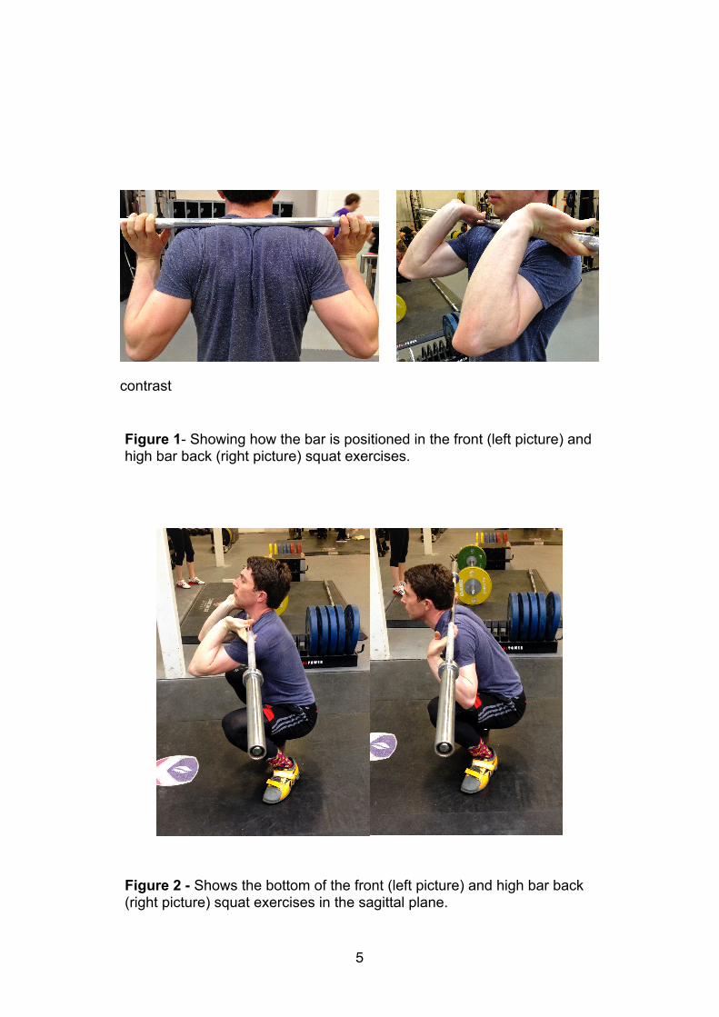
contrast
�5
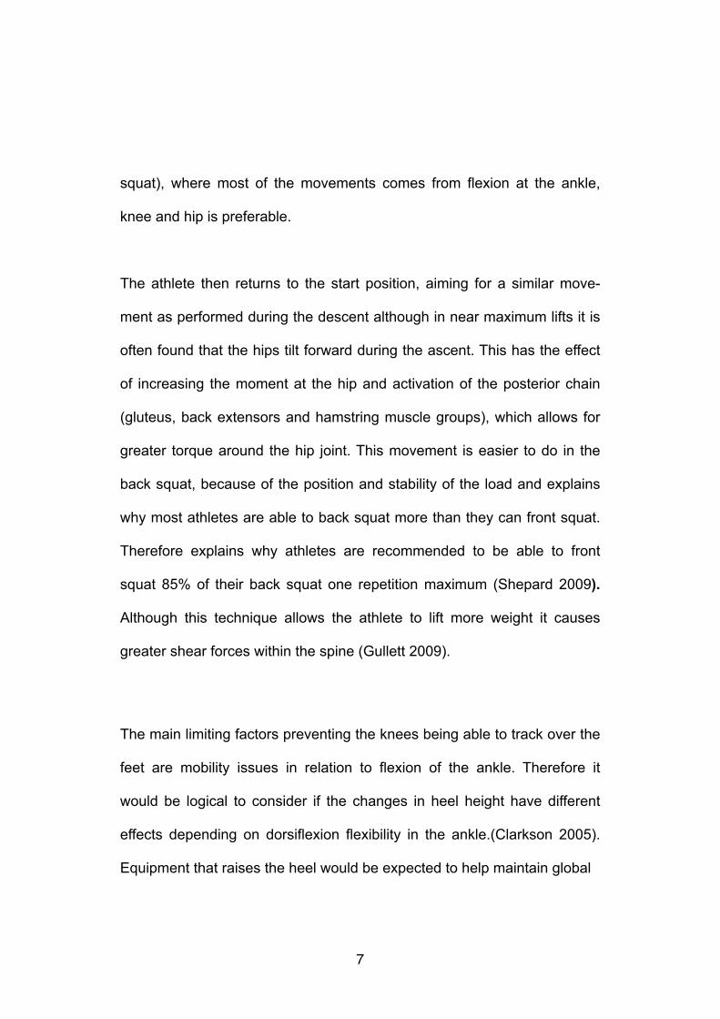
Figure 1- Showing how the bar is positioned in the front (left picture) and high bar back (right picture) squat exercises.
Figure 2 - Shows the bottom of the front (left picture) and high bar back (right picture) squat exercises in the sagittal plane.
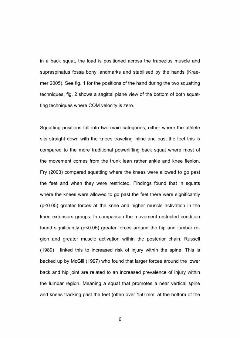
in a back squat, the load is positioned across the trapezius muscle and
supraspinatus fossa bony landmarks and stabilised by the hands (Krae-
mer 2005). See fig. 1 for the positions of the hand during the two squatting
techniques, fig. 2 shows a sagittal plane view of the bottom of both squat-
ting techniques where COM velocity is zero.
!Squatting positions fall into two main categories, either where the athlete
sits straight down with the knees traveling inline and past the feet this is
compared to the more traditional powerlifting back squat where most of
the movement comes from the trunk lean rather ankle and knee flexion.
Fry (2003) compared squatting where the knees were allowed to go past
the feet and when they were restricted. Findings found that in squats
where the knees were allowed to go past the feet there were significantly
(p<0.05) greater forces at the knee and higher muscle activation in the
knee extensors groups. In comparison the movement restricted condition
found significantly (p<0.05) greater forces around the hip and lumbar re-
gion and greater muscle activation within the posterior chain. Russell
(1989) linked this to increased risk of injury within the spine. This is
backed up by McGill (1997) who found that larger forces around the lower
back and hip joint are related to an increased prevalence of injury within
the lumbar region. Meaning a squat that promotes a near vertical spine
and knees tracking past the feet (often over 150 mm, at the bottom of the
�6
squat), where most of the movements comes from flexion at the ankle,
knee and hip is preferable.
!The athlete then returns to the start position, aiming for a similar move-
ment as performed during the descent although in near maximum lifts it is
often found that the hips tilt forward during the ascent. This has the effect
of increasing the moment at the hip and activation of the posterior chain
(gluteus, back extensors and hamstring muscle groups), which allows for
greater torque around the hip joint. This movement is easier to do in the
back squat, because of the position and stability of the load and explains
why most athletes are able to back squat more than they can front squat.
Therefore explains why athletes are recommended to be able to front
squat 85% of their back squat one repetition maximum (Shepard 2009).
Although this technique allows the athlete to lift more weight it causes
greater shear forces within the spine (Gullett 2009).
!The main limiting factors preventing the knees being able to track over the
feet are mobility issues in relation to flexion of the ankle. Therefore it
would be logical to consider if the changes in heel height have different
effects depending on dorsiflexion flexibility in the ankle.(Clarkson 2005).
Equipment that raises the heel would be expected to help maintain global
�7
shank angle because less flexion at the ankle joint is needed to achieve
the same position.
!Lumbar Region of the Spine
!Much of the previous research surrounding squatting technique focuses
on the lower back in relation to technique and injury. The spine’s main
functions are to support the upper body, muscle and ligament attachment
and protect the spinal cord. The spine is divided into five distinct sections
based on the vertebrae structure and position within the back. From top to
bottom they are; the cervical, thoracic, lumbar, sacral and coccygeal. To-
gether they form an S shaped curve that is designed to maximise the safe
axial loading capacity. This comes from lumbar extension and thoracic
flexion when in the neutral position. In this position it can support well in
excess of 15 times body weight, but is weak against shear forces which
force it out of its neutral S shape (Lehto 2012). The lumbar region contains
the five largest non-fused vertebrae and is located at the bottom of the
spine and it allows a large ROM and supports much of the body’s weight.
The thoracic region is made of twelve vertebrate and the attachment point
for the ribs located at the top of the spine. There is expected to be more
�8
flexion and extension within the thoracic region of the spine during the
front squat when compared to the back squat (Gray 1973).
!Pain within the lumbar region has many possible causes, from a damaged
disc causing a nerve compression often requiring surgery whilst soft tissue
strains that normally heal with rest. Soft tissue strains are the most com-
mon cause of back pain within sport. This sort of injury will often resolve
within a few days or weeks of reduced load although this isn't chronic even
a few days of missed training or competition can be serious for an elite
athlete. This sort of injury normally occurs because the shear force acting
on the spine is far greater than the muscles can resist at that time mean-
ing it is forced to flex well outside its neutral posture during movement.
This can easily occur, when athletes are often handling in excess of 100
kg in a fatigued state which has shown to be linked to reduced technique
and muscle strength (Merton 1954) (Sparto 1997). This is why training to
strengthen the muscles within the back and a consistent technique that
promotes a neutral spine is so important. This has the effect of reducing
the amount done by the muscle within the back therefore reducing the
chance of injury. Pointing towards any equipment that is able to promote
better technique therefore reducing the forces causing the back to flex
would be beneficial in aiding in reducing injury and maximising perfor-
�9
mance (Herkowitz 2004). Previous research looking at squatting technique
has only measured the global trunk angle, rather than specific flexion or
extension changes within each region.
!Weightlifting Shoes
!According to section 4.2.1 of the International Weightlifting Federation
(IWF) technical and competition rules the purpose of weightlifting shoes is
to protect the lifters' feet and provide a stable and firm stance on the
weightlifting platform which allows for a deep and safe squat (IWF 2009).
!Research from Sato (2012) who compared back squatting in either run-
ning or weightlifting shoes support this description. His findings show that
the weightlifting shoe group, had significantly (p<0.05) less trunk lean and
greater foot angle in relation to the floor. These changes in technique are
linked to reduced shear forces within the lumbar region of the spine which
are linked to a reduced chance of injury (McGill 1997). There was also
greater knee extensor group activity in the weightlifting shoe group during
the squat. This crosses over to the front squat, where wearing weightlifting
shoes may improve strength and stability in the catch phase of the squat
�10
clean. The improvements in technique are related to the characteristics of
weightlifting shoes these fall into two main areas; this is the construction of
the upper and design of the sole.
!The upper is made of a stiff material, with straps across the fore foot and
mid foot this is designed to maximise the force that is applied to the sole
by not allowing the foot to move inside the shoe. This research is going to
focus on the second area, the design of the sole, because it is though to
have a much greater effect on technique. The outsole is flat which allows a
stable platform to lift from. The midsole has a solid raised heel made of
non-compressible material and is thought to be the most important feature
in weighting lifting shoe design. It has the effect of increasing the angle
between the floor and the shank throughout the movement, meaning less
dorsiflexion is needed at the ankle. This corroborates research that found
this to be the main limiting factor in allowing the knees to track over the
feet and therefore perform the correct squatting technique. This is backed
by Sato (2012) who found significant (p<0.05) improvements in squatting
technique in those wearing weightlifting shoes compared to running shoes.
However this is not completely relevant to elite sport because most high
level athletes will already use weightlifting shoes, therefore it would be
more useful to compare differences within weightlifting shoes design (Cot-
�11
ter 2012). Manipulating the heel height to study how it affects an athlete’s
technique and therefore muscle activation during both the front and back
squat will form the basis of the research. The standard weightlifting heel
height is 19.05 mm across most sizes and brands even though there are
no regulations that stipulate this or the use of additional wedges which
have been used by elite lifters (IWF 2009). With athletes using additional
wedges that increase the heel height past 19.05 mm, it could be logically
thought that this may aid their performance and technique although there
has been no research to back this up. This is because the angle between
the floor and shank will be greater as the heel height increases leading to
less flexion at the ankle being required to achieve the same depth, which
should allow for a straighter front and back squat.
!Summary and Hypotheses
!The literature reviewed around squatting technique and the use of
weightlifting shoes, has led to the hypothesis that changing a weightlifting
shoe’s heel height will affect the technique and muscle activation within
the front and back squat exercises. This will be quantified by confirmation
or rejection of the following statements:
�12

The angle between the trunk and the floor will get closer to vertical, and
there will be less flexion within the lumbar (within back squat) and thoracic
(within front squat) regions of the spine as the weightlifting shoe heel
height increases.
!The flexible groups (0.2-0.3, >0.3) will show less flexion within the back as
the weightlifting shoe heel height increases compared to those in the non-
flexible group (<0.20).
!!!!!!!!
�13
Method
!Participants and Equipment
!The participant group was eight weightlifters who have experience and
sound technique when performing the weightlifting movements and squat-
ting (snatch and clean & jerk) (six male, two female) (24.6 ± 5.7 years,
back squat 143.25 ± 20.57 kg, front squat 124.38 ± 11.16 kg).They were
split into three groups based on the ratio between their thigh length and
ankle dorsiflexion flexibility, because this has shown to be one of the main
contributors in squatting technique (<0.20, 0.2-0.3, >0.3).
!To perform the squat an Olympic weightlifting bar (20 kg, 28 mm diameter)
and incremental weighted discs were used. Co-ordinate data was record-
ed using a 10 camera Vicon T-series system which track passive reflective
markers attached to the body recording at 250 fps. A 19 marker set was
used on the limbs, shoulders, and bar with an additional 7 smaller markers
positioned on the back. The main set was placed on bony landmarks, the
medial and lateral positions of each joint of the limbs. These were posi-
tioned on the legs, hips, shoulders and on each end of the load. With the
�14
additional seven markers placed on spinous processes of the back, three
on alternate lumbar vertebrae (L1, L3, L5), the posterior superior iliac
spine, and four on thoracic vertebrae (T1, T2, T3, T4).
!The athletes wore their own weightlifting shoes to ensure correct fit and to
enable the athlete to feel comfortable during the protocol. This resulted in
the technique being similar to that used during training and competition.
The heel height was incrementally increased in five randomly ordered
stages according to the testing protocol. Standard weight lifting shoes
have a baseline drop between heel and forefoot of 19.05 mm, this was in-
creased by attaching non-compressible thermoplastic wedges to the ath-
letes’ shoes. In five 3.78 mm increments up to a maximum heel height
37.95 mm including the original heel (19.05 mm baseline, 22.83 mm,
26.61 mm, 30.39 mm, 34.17 mm, and 37.95 mm).
!Protocol
The 26 marker set described above was applied to the athlete, who then
performed a standardised squatting warm up approved by a strength and
conditioning coach (decreasing repetitions and increasing weight up 75%
�15
of their one repetition maximum over four sets). A basic dorsiflexion flexi-
bility test was then performed once the athlete had completed the warm
up, that involved the athlete flexing their ankle as far a possible past their
knee. The distance measured was horizontally from the end of the shoe to
the centre of the patella. This ROM should be close to the expected ROM
at the bottom the squat, and was be used to group the athletes.
!Each athlete then completed the testing protocol which involved perform-
ing six front squats followed by six back squats at 75% of their self-report-
ed 1RM for both the front and back squat exercises. The different height
wedges were attached to the shoe (19.05 mm baseline, 22.83 mm, 26.61
mm, 30.39 mm, 34.17 mm, 37.95 mm) in a randomised order. Sandler
(2010) found that minimum of three minutes is needed for full recovered
from low repetition weightlifting. This means that each athletes will have,
three minutes so they are able to sustain a consistent technique through-
out the protocol.
!!
�16
Post Processing
!The marker co-ordinates data was used to calculate internal angles during
the movement with a focus on the point where the angle either reached a
maximum or minimum value depending on the joint.The angles were the
internal angles of the knee and the shank in relation to the floor, and the
abduction and adduction of the knee. The overall trunk and pelvis angles
were calculated relative to the vertical and horizontal respectively. The
markers on the lumbar and thoracic region of the spine were used to cal-
culate flexion or extension of the spine compared to neutral (measured
pre-test with the subject standing in the anatomical position). This was
done by using arctangent to calculate the 2D angles between segments
that only moved within one plane (e.g. pelvis). Angles in 3D between seg-
ments that moved in multiple planes were calculated using the dot (scalar)
product method (e.g. knees) (explained in Robertson 2006, pp.256).
!The statistical test used was a repeated measures ANOVA. This calculat-
ed the group means and standard deviations of the internal/external an-
gles. It was also used to identify whether and where the significant differ-
ences and interactions occurred within each variable (p≤0.05) between
each heel height and group. The statistical outputs and processing outputs
�17

were compared to previous literature and the guidelines put forward in the
hypotheses in order to identify which heel heights would allow the closest
to optimal technique and minimal expected lumbar loading within the dif-
ferent flexibility groups.
!!!!!!!!!!!
�18
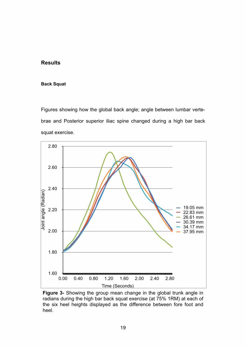
Results
Back Squat
!Figures showing how the global back angle; angle between lumbar verte-
brae and Posterior superior iliac spine changed during a high bar back
squat exercise.
�19
Join
t ang
le (R
adia
n)
1.60
1.80
2.00
2.20
2.40
2.60
2.80
Time (Seconds)0.00 0.40 0.80 1.20 1.60 2.00 2.40 2.80
19.05 mm22.83 mm26.61 mm30.39 mm34.17 mm37.95 mm
Figure 3- Showing the group mean change in the global trunk angle in radians during the high bar back squat exercise (at 75% 1RM) at each of the six heel heights displayed as the difference between fore foot and heel.
!!
�20
Join
t ang
le (R
adia
ns)
-2.40
-2.33
-2.25
-2.18
-2.10
-2.03
-1.95
Time (Seconds)0 0.40 0.80 1.20 1.60 2.00 2.40 2.80
19.05 mm22.83 mm26.61 mm30.39 mm34.17 mm37.95 mm
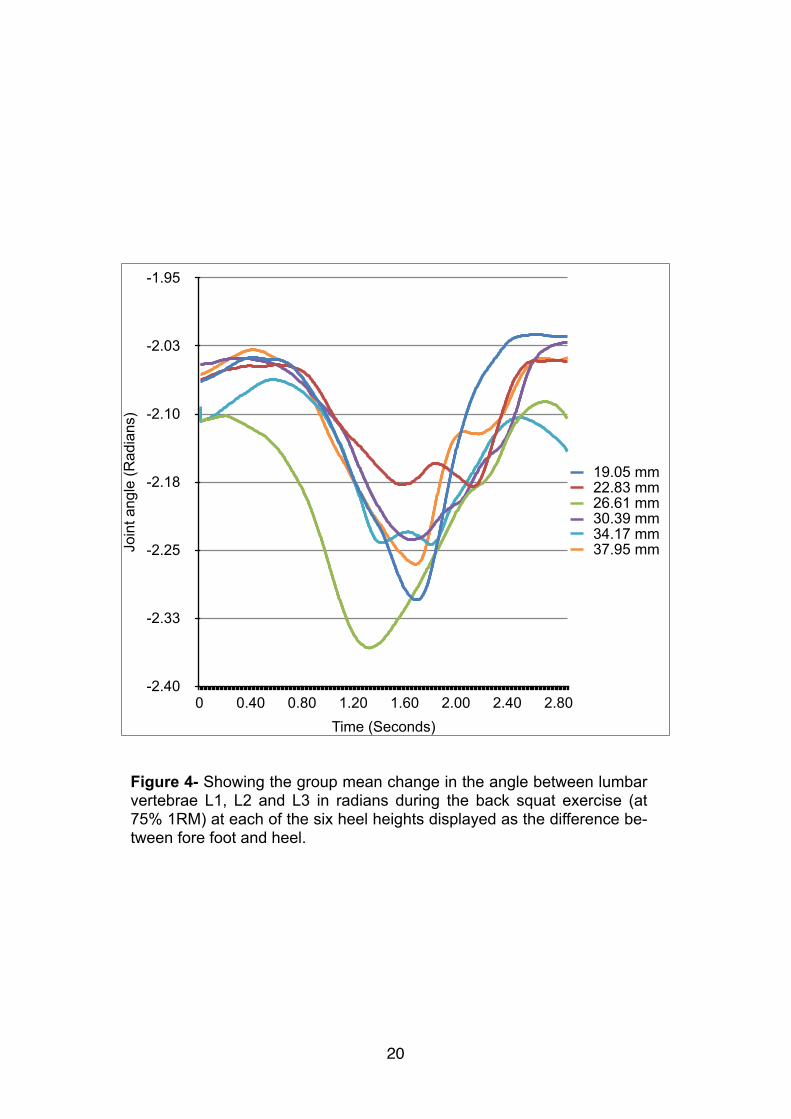
Figure 4- Showing the group mean change in the angle between lumbar vertebrae L1, L2 and L3 in radians during the back squat exercise (at 75% 1RM) at each of the six heel heights displayed as the difference be-tween fore foot and heel.
!
!!
�21
Join
t Ang
le (R
adia
ns)
2.40
2.48
2.57
2.65
2.73
2.82
2.90
Time (Seconds)0.00 0.40 0.80 1.20 1.60 2.00 2.40 2.80
19.05 mm22.83 mm26.61 mm30.39 mm34.17 mm37.95 mm
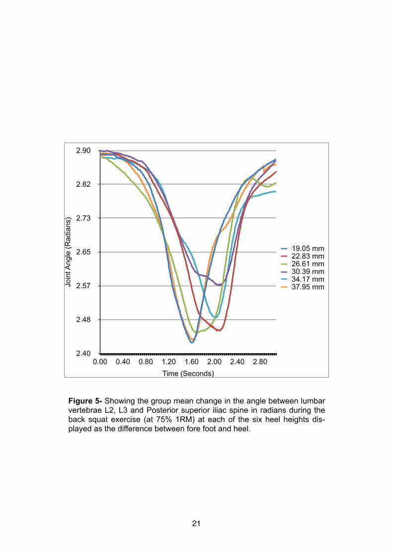
Figure 5- Showing the group mean change in the angle between lumbar vertebrae L2, L3 and Posterior superior iliac spine in radians during the back squat exercise (at 75% 1RM) at each of the six heel heights dis-played as the difference between fore foot and heel.
A repeated measures ANOVA was carried out comparing the different heel
heights (19.05 mm baseline, 22.83 mm, 26.61 mm, 30.39 mm, 34.17 mm,
and 37.95 mm) and grouped based on an ankle flexibility ratio (<0.20,
>0.2-<0.3, >0.3) during the back squat exercise.
!The global trunk angle, the main effect of heel height showed a non-signif-
icant result (F(5, 25) =0.353, P= 0.875). Meaning that heel height has no ef-
fect on trunk angle in the back squat exercise. The different groups found
a non-significant result (F(10, 25) =1.143, P= 0.390). Meaning ankle flexibility
has no effect on trunk ankle in the back squat.
!The angle between the L3, L5 and posterior superior iliac spine main ef-
fect of heel height showed a non-significant result (F(5, 25) =0.398, P=
0.846). Meaning that heel height has no effect on the angle between the
L3, L5 and posterior superior iliac spine. The different group a non-signifi-
cant result (F(10, 25) =0.269, P= 0.983). Meaning that ankle flexibility has no
effect on the angle between the L3, L5 and posterior superior iliac spine in
the back squat.
!!
�22
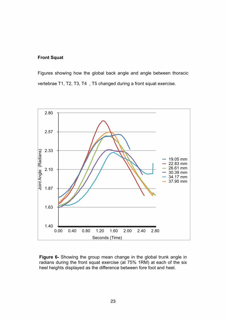
Front Squat
Figures showing how the global back angle and angle between thoracic
vertebrae T1, T2, T3, T4 , T5 changed during a front squat exercise.
�23
Join
t Ang
le (
Rad
ians
)
1.40
1.63
1.87
2.10
2.33
2.57
2.80
Seconds (Time)0.00 0.40 0.80 1.20 1.60 2.00 2.40 2.80
19.05 mm22.83 mm26.61 mm30.39 mm34.17 mm37.95 mm
Figure 6- Showing the group mean change in the global trunk angle in radians during the front squat exercise (at 75% 1RM) at each of the six heel heights displayed as the difference between fore foot and heel.
!!
�24
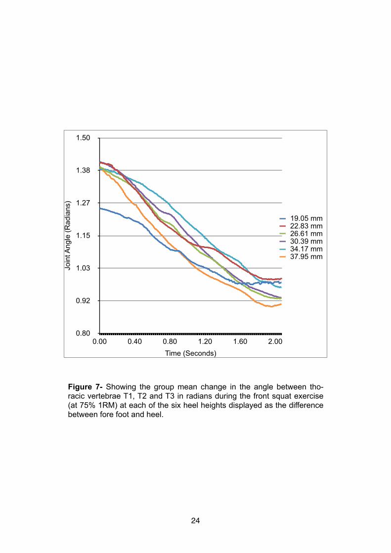
Figure 7- Showing the group mean change in the angle between tho-racic vertebrae T1, T2 and T3 in radians during the front squat exercise (at 75% 1RM) at each of the six heel heights displayed as the difference between fore foot and heel.
Join
t Ang
le (R
adia
ns)
0.80
0.92
1.03
1.15
1.27
1.38
1.50
Time (Seconds)0.00 0.40 0.80 1.20 1.60 2.00
19.05 mm22.83 mm26.61 mm30.39 mm34.17 mm37.95 mm
!
!
�25
Join
t Ang
le (R
adia
ns)
0.80
0.92
1.03
1.15
1.27
1.38
1.50
Time (Seconds)0.00 0.40 0.80 1.20 1.60 2.00
19.05 mm22.83 mm26.61 mm30.39 mm34.17 mm37.95 mm
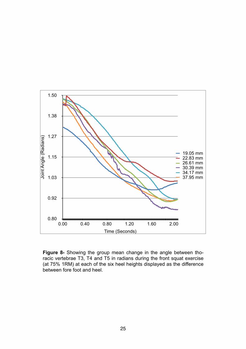
Figure 8- Showing the group mean change in the angle between tho-racic vertebrae T3, T4 and T5 in radians during the front squat exercise (at 75% 1RM) at each of the six heel heights displayed as the difference between fore foot and heel.
A repeated measures ANOVA was carried out comparing the different heel
heights (19.05 mm baseline, 22.83 mm, 26.61 mm, 30.39 mm, 34.17 mm,
and 37.95 mm) and grouped based on an ankle flexibility ratio (<0.20,
>0.2-<0.3, >0.3) during the front squat exercise.
The global trunk angle, the main effect of heel height showed a non-signif-
icant result (F(5,25) =1.298, P= 0.296). Meaning that heel height has no ef-
fect on trunk angle in the front squat exercise. The different groups found a
non-significant result (F(10,25) =1.303, P= 0.341). Meaning ankle flexibility
has no effect on trunk angle in the front squat.
!The angle between the T2, T3 and T4 main effect of heel height showed a
non-significant result (F(5, 25) =1.128, P= 0.371). Meaning that heel height
has no effect on the angle between the L3, L5 and posterior superior iliac
spine. The different group a non-significant result (F(10, 25) =1.377, P=
0.247). Meaning that ankle flexibility has no affect on the angle between
the L3, L5 and posterior superior iliac spine in the back squat.
!!
�26

Discussion
The hypothesises related to how changing the height of a weightlifting
shoe heel may affect the kinematics during the front and high bar back
squat exercises. Focusing on the kinematics within the thoracic and lum-
bar regions of the spine.
!The first hypothesis, found no significant differences within the trunk angle
and flexion/extension angle of the lumbar and thoracic regions across the
heel heights in both the front and back squat exercises. Meaning that
changing the heel height beyond that of standard weightlifting shoes
(19.05mm) has no effect on back kinematics during front and back squats.
The second hypothesis, found that ankle dorsiflexion flexibility has no ef-
fect on back kinematics during front and back squats, in relation to chang-
ing the heel height. Therefore from the rejection of both these hypothesis-
es, changing weightlifting shoe heel height has no effect on kinematics
within the spine, across different ankle flexibility groups in the front and
back squat.
!The main assumption was within the self reported one repetition maximum
of the back and front squat, because athletes may have over or under re-
�27
ported this weight compared to their current state of training or recovery.
This would therefore affect the amount they lifted which would have an in-
fluence on their technique. Ideally athletes would have their repetition
maximum to be tested. Although the benefit of having a true maximum
does not outweigh the time commitment and the significant fatigue caused
if a true maximum value is achieved. This would negatively affect the ath-
letes training (Häkkinen 1993).
!The limitations fall into two main categories; collection and analytical; and
sporting movements tested. Within the collection of data, only athletes
who have a sound squatting technique were used although this gave con-
sistent result this may not match athletic populations who use strength and
conditioning programs such as games players or track and field athletes
(Holt 2008) (Gleim 1997).
!The more technical area within the collection of data, is whether more pa-
rameters could have been recorded to gain a fuller understanding of the
movement. This more in depth understanding may have shown differences
between the heel heights. Examples of this include muscle activation lev
�28
els (electromyography) of the prime movers, force and centre of pressure
data. This would have allowed assessment of athlete weight distribution
and an insight into whether or not athletes weight shifted differently be-
tween each heel height, collecting force data would have also allowed for
inverse dynamics calculations to be used.
!Analysing electromyography data of each of the muscles throughout the
movement could benefit athletes by providing them with results that allow
them to focus their training on certain muscle groups. With the aim of re-
ducing tension on injured muscles or working other muscles more effi-
ciently in training for sporting performance. Using inverse dynamics calcu-
lations, allows a greater understanding of the movement by calculating
forces and moments around each joint. McGill (1997) findings points to-
ward trying to minimise the shear forces around the lower back because
they are linked to increased injury risk.
!Although measuring both of these parameters would improve the under-
standing of the movement they are both out of the scope of this report.
With the movement of squatting it would be impossible to measure EMG
with skin surface sensors because of the number and location of muscles
involved. Squatting is a three dimensional movement therefore three di-
�29
mensional inverse dynamics would be needed to get accurate and realistic
values. In 3D there are twice as many degrees of freedom than in 2D,
therefore it is significantly more complex (Robertson 2013).
!The use of additional wedges attached to weightlifting shoes, was found to
be ineffective in improving the front or back squat although they may still
be beneficial in other squatting type movements. Examples of movements
where this could be applied is overhead squat or single legged squat (pis-
tols), which both require higher levels of ankle mobility than the front or
back squat (Collins 2012). An other area of research would be applying
the heel heights more directly to the sport of weightlifting. Looking at the
catch phase of the clean and snatch which require a very stable squat be-
cause of the high dynamic loading. Although they add many addition vari-
ables such as the athletes technique during the triple extension, which
may effect how the bar moves and therefore lands in the catch phase.
!The findings were not as expected, although the research directly relating
to weightlifting shoes is limited therefore hard to predict the effect of differ-
ent implementations. Papers such as Sato (2012) found significant im-
provements when using weightlifting shoes. This means it can be as-
sumed that the standardised height of weightlifting shoes at 19.05mm, is
�30

the optimal height and that any additional height past this up to 37.95mm
does not have any benefit when front or back squatting. Further research
falls into two main area. Firstly whether or not the additional features with-
in weightlifting shoes such as the material properties (stiffness) of the sole,
stiff supporting upper, straps and flat sole are of more benefit than previ-
ously expected. Secondly if changing heel height would be of any benefit
in other squat movements (over heard or single leg) or in the weightlifting
movements (clean & jerk and snatch).
!!!!!!!!!Acknowledgements
The author thanks Dr Sam Allen for assistant and guidance during the project, Ciaran McElhinney with data collection and Dean Howard M.Eng with the design and production of the wedges.
�31
References
D Beniot. (2006). Effect of skin movement artefact on knee kinematics dur-ing gait and cutting motions. Gait posture. 24 (2),pp152-164.A Cappozzo. (1985). Lumbar spine loading during half-squat exercises. Medicine and Science in Sports and Exercise. 17 (5), pp613-620.
D R Clark. (2012). Muscle activation in the loaded free barbell squat: a brief review. Journal of strength and conditioning research . 26 (4), pp1169-1178.M Clark. (2010). NASM's Essentials of Sports Performance Training: Lip-pincott Williams & Wilkins. pp314-337.!H Clarkson. (2005). Joint Motion and Function Assessment: A Research-based Practical Guide: Lippincott Williams & Wilkins. pp353-355.
A Collins. (2012). The Complete Guide to Functional Training: A & C Black Publishers . pp286.
S Cotter. (2013). Kettle bell Training: Human Kinetics. pp19-22.A C Fry. (2003). Effect of Knee Position on Hip and Knee Torques During the Barbell Squat. Journal of Strength and Conditioning. 17 (4), pp629-633.
S Fukashiro. (1995). In vivo Achilles tendon loading during jumping in hu-mans. European Journal of Applied Physiology and Occupational Physiol-ogy. 71 (5), pp453-458.
K Häkkinen. (1993). Neuromuscular Fatigue and Recovery in Male and Female Athletes during Heavy Resistance Exercise. International Journal of Sports Medicine . 14 (2), pp53-59.
�32

H Herkowitz. (2004). The Lumbar Spine: Lippincott Williams & Wilkins. pp299-306.
L Holt (2008). Flexibility: A Concise Guide: To Conditioning, Performance Enhancement, Injury Prevention, and Rehabilitation. asa: Springer. pp 289-299.
International Weightlifting Federation. Technical and competition rules. Available at: http://www.iwf.net/doc/handbook/Handbook(2009)_techni-cal_and_competition_rules.pdf. Accessed 2nd April 2014.W Garrett. (2000). Exercise and Sport Science: Lippincott Williams & Wilkins. pp585-615.
G Gleim. (1997). Flexibility and Its Effects on Sports Injury and Perfor-mance. Sports Medicine . 24 (5), pp 289-299.
H Gray. (1973). Gray's Anatomy. 35th ed: Longman.J Gullett. (2009). A Biomechanical Comparison of Back and Front Squats in Healthy Trained Individuals. Journal of Strength & Conditioning Re-search. 23 (1), pp284-292.
M Kjaer. (2008). Textbook of Sports Medicine: Basic Science and Clinical Aspects of Sports Injury and Physical Activity: John Wiley & Sons. pp109-111.
W Kraemer. (2005). Strength Training for Young Athletes: Human Kinetics. pp135-137.
M Lehto. (2012). Introduction to Human Factors and Ergonomics for Engi-neers. 2nd ed: CRC Press. pp42-47.S McGill. (1997). The biomechanics of low back injury: Implications on cur-rent practice in industry and the clinic. Journal of Biomechanics. 30 (5), pp465–475.
P Merton. (1954). Voluntary strength and fatigue. The Journal of physiolo-gy. 123 (1), pp553-564.
�33
D Pincivero. (2000). Quadriceps-hamstring EMG activity during functional, closed kinetic chain exercise to fatigue. European Journal of Applied Phys-iology and Occupational Physiology. 81 (6), pp504-509.
G Robertson. (2004). Research Methods in Biomechanics: Human Kinet-ics. pp103-125. pp145-162, pp163-182.
G Robertson (2013). Research Methods in Biomechanics. 2nd ed: Human Kinetics . pp151-160.P Russell. (1989). A Preliminary Comparison of Front and Back Squat Ex-ercises. Research Quarterly for Exercise and Sport. 60 (3), pp201-208.
D Sanderson. (2006). Gastrocnemius and soleus muscle length, velocity, and EMG responses to changes in pedaling cadence. Journal of Elec-tromyography and Kinesiology. 16 (6), pp642-649.
D Sandler (2010). Fundamental Weight Training. 2nd ed: Human Kinetics. pp29-32.K Sato. (2012). Kinematic changes using weightlifting shoes on barbell back squat. Journal of Strength and Conditioning Research. 26 (1), pp28-33.G Shepard. (2009). Bigger, Faster, Stronger: Human Kinetics . pp76-79.
P Sparto. (1997). The Effect Of Fatigue On Multijoint Kinematics, Coordi-nation, And Postural Stability During A Repetitive Lifting Test. Journal of Orthopaedic & Sports Physical Therapy. 25 (3), pp3-12.
S Westing. (1991). Muscle activation during maximal voluntary eccentric and concentric knee extension. European Journal of Applied Physiology and Occupational Physiology . 62 (2), pp104-108.F Zajac. (1990). Modeling Musculoskeletal Movement Systems: Joint and Body Segmental Dynamics, Musculoskeletal Actuation, and Neuromuscu-lar Control. In: J Winters Multiple Muscle Systems Biomechanics and Movement Organisation: Springer.
C Zimmermann. (1993). Effects of seated posture on erector spinae EMG activity during whole body vibration. Ergonomics. 36 (6), pp667-675.
�34

!!!!!!!!!!!!!
!!!!!!!!!
�35
APPENDIX B
Extended Literature review
!In elite sport strength and conditioning is being utilised more in a wider
range of sports. This means a broader range of athletes are practicing ad-
vanced weightlifting movements with an aim of maximising performance at
the highest level. Within competitive sport specialised shoes are common
across a wide range of activities, for example light weight spiked shoes for
sprinting and cleated stiff soled shoes for cycling. They are designed to
optimise the athlete’s performance and reduce the chance of injury. More
specifically within strength and conditioning, specialised shoes can be
used which are designed to aid the performance of weightlifting move-
ments (clean & jerk and snatch). Although kinematics and weightlifting bar
(load) path are different for the two movements they are similar because
they both require the athlete to perform a stable weighted squat in the
catch phase. Achieving optimal technique within weightlifting is technically
hard, therefore more basic lifts are often used to build up the strength and
co-ordination in for example the front and back squat exercises. This is
also advantageous because it will allow for a more controlled comparison
of the effect different shoe designs on the kinematics and forces involved
within the body during these exercises (Clark 2010).
!
�36
�37
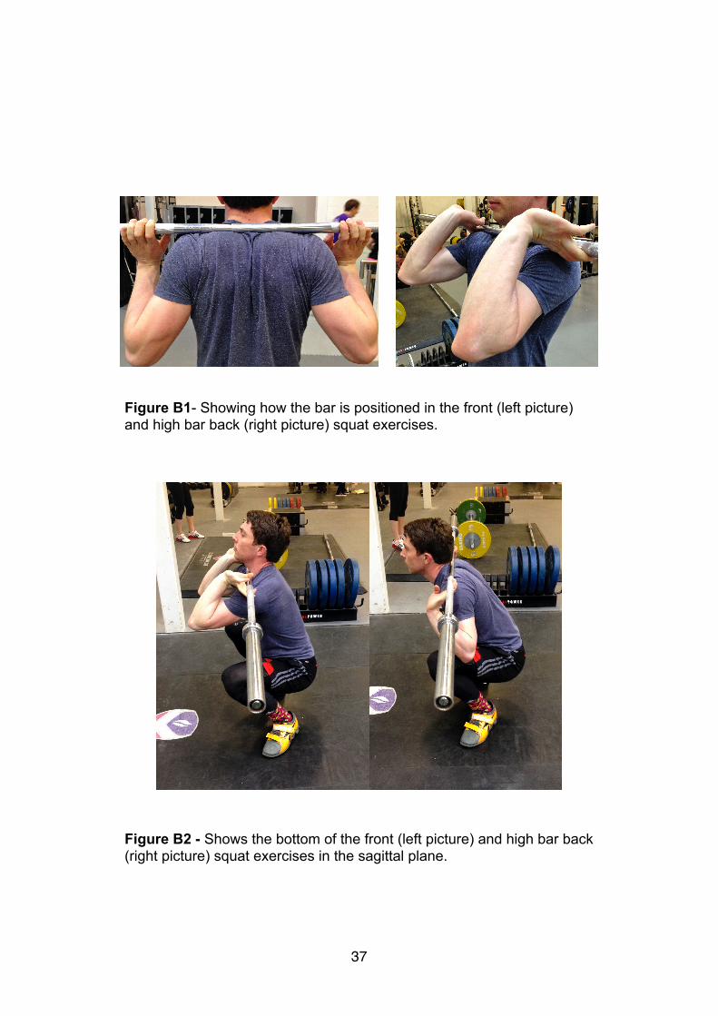
Figure B1- Showing how the bar is positioned in the front (left picture) and high bar back (right picture) squat exercises.
Figure B2 - Shows the bottom of the front (left picture) and high bar back (right picture) squat exercises in the sagittal plane.

Squatting Technique
The front and back squat exercises are overall similar movements, with
high compressive forces in the lumbar region of the back found in both lifts
(in excess of ten times body) (Cappozzo 1985, Russell 1989). The path of
the load and changes in joint angle and segment position should be similar
with the main difference relating to where and how the bar is positioned in
relation to the athlete. In a front squat the load is positioned as it would be
during the catch of the squat clean. This is on the front of the athlete’s
shoulders across the deltoid and clavicle area and it is stabilised by the
hands which are positioned so that the upper arm is parallel to the floor
throughout the movement and wrists fully extended so the fingers they are
under the bar. In contrast in a back squat, the load is positioned across the
trapezius muscle and supraspinatous fossa boney landmark on the upper
back and stabilised by the hands pushing the load into the body (Kraemer
2005). See fig. B1 for the positions of the hand during the two squatting
techniques, fig. B2 shows a sagittal plane view of the bottom of both
squatting techniques.
!In both movements the athlete should aim for a back angle closest to ver-
tical as possible throughout the movement, because greater trunk lean
has been linked to increased injury risk of the lumbar region of the spine
�38
because of increase flexion within the spine (Russell 1989). This means
that most of the movement should come from knee flexion rather than for-
ward flexion of the trunk at the hips, their is often more forward lean within
the back squat down to the positioning of the load. If performed correctly,
in both movements the athlete will sit straight down with the knees tracking
outwards from the centre over the feet and past the toes (often over 150
mm, at the bottom of the squat). The athlete then returns to the start posi-
tion, aiming for a similar movement as performed during the descent al-
though in heavy lifts it is often found that the hips tilt forward at the bottom
of the lift. This has the effect of increasing the moment at the hip and acti-
vation of the posterior chain (gluteus, back extensors and hamstring mus-
cle groups), which allows for greater torque around the hip joint. This
movement is easier to do in the back squat, because the load is more sta-
ble. Although this technique allows the athlete to lift more weight it causes
greater sheer forces within the spine, which are linked to lower back injury
(Gullett 2009). This explains why most athletes are able to lift more weight
in their back squat compared to the front squat exercise, with most articles
suggesting athletes should aim to be able to front squat 85% of their back
squat one repetition max (Shepard 2009)
!The position in which the athlete squats in is directly linked to the muscle
activation during the lift. With squatting positions falling into two main cat-
�39
egories, either where the athlete sits straight down with the knees tracking
past the feet compared to the more traditional back squat where most of
the movement comes from the hips therefore trunk lean and the shank
remain almost perpendicular to the floor. Fry (2003) found that in squats
where the knees were allowed to go past the feet there were significantly
(p<0.05) greater forces at the knee and higher muscle activation in the
knee extensors groups compared to squats where knee movement was
restricted.Here they found significantly (p<0.05) greater forces around the
hip and lumbar region of the spine with greater muscle activation within
the posterior chain. This research therefore points towards a squatting
technique which maximises forward knee tracking, with an aim to minimis-
ing the forces around the lower back. This is further backed by McGill
(1997) who found that larger forces around the lower back and hips are
related to an increased prevalence of lower back injury within the lumbar
region.
!The main limiting factors preventing the knees being able to track over the
feet are mobility issues in relation to flexion of the ankle and because of
this it would be logical to consider if the changes in heel height have dif-
ferent effects depending of dorsiflexion flexibility in the ankle. This will be
achieved by splitting the athletes into two groups based on how much they
�40

can flex their ankle whilst their foot is on the floor (Clarkson 2005). Equip-
ment that produces a greater angle between the shank and the floor would
be expected to aid in the knee going past the feet. This is because less
flexion at the ankle joint is needed to achieve the same shank ankle in re-
lation to the floor.
!Lumbar Region of the Spine
!Much of the previous research surrounding squatting technique focuses
on the lower back in relation to technique and injury. This research is to
going to review the basic structure of the spine and how that relates to
weightlifting. The spine’s main functions are to support the upper body,
muscle and ligament attachment and protect the spinal column. It is made
up of 33 vertebrae, which are irregularly shaped to fit together. They are
supported by a complex ligament structure which incases the spine, both
protecting it and allowing muscle attachment. The vertebrae are separated
by discs that allow the spine to move in a wide ROM and also compress to
allow for impacts and for it to be loaded. They are made of a fibro-cartilage
exterior and fluid interior.
!
�41
The spine is divided into five distinct sections based on the vertebrae
structure and position within the back. From top to bottom they are; the
cervical, thoracic, lumbar, sacral and coccygeal. Together they form an S
shaped curve that is designed to maximise the safe loading capacity. To
produce this the cervical and lumbar are extended, whereas the thoracic is
flexed when in the neutral position. This is the position of the spine at rest
and its best loading bearing orientation. From this structure and position
the spine is able to take large axial loads well in excess of 15 x body
weight, but is weak against shear forces which force it out of its neutral S
shape (Lehto 2012).
!The lumbar region contains five vertebrae and is located at the bottom of
the spine between the rib cage and the pelvis and it allows a large ROM in
multiple planes and supports much of the body’s weight. It contains the
largest non-fused segments in the spine, with the transverse process be-
ing a key attachment point for some of the muscles that support the lower
back and pelvis regions. This means that this area of the spine is suited to
high compressive loading as found within correct technique in weightlifting
movements. The erector spinae is one of the key muscles, extending and
rotating the spine therefore it is critical for maintaining a neutral spine pos-
ture during shear loading, such as in a squatting technique with large
�42
amount of trunk lean. Its origin is at the sacrum and it runs almost the full
length of the spine to the cervical. It consists of a large fleshy mass that
splits into three columns depending on their attachment points along the
spine (Gray 1973).
!Pain within the lumbar region has many possible causes, from a damaged
disc causing a nerve to be compressed often requiring surgery to muscle
or ligament strain that often heal with rest. The latter is the most common
cause of back pain within sporting movements. In most cases within mus-
cle or ligament damage the pain will resolve within a few days or weeks of
reduced load but although this isn't a chronic injury even a few days of
missed training or competition can be potentially serious for an elite ath-
lete. This sort of injury normally occurs because the shear force acting on
the spine is far greater than muscles can resist at that time meaning it is
forced to flex well outside its neutral posture during movement. This is why
weightlifting is potentially a high risk activity, because athletes are often
repeatedly handling loads in a dynamic fashion which are well in excess of
100 kg. This problem is compounded by research showing that as fatigue
increases, technique worsens and muscle strength reduces (Merton 1954)
(Sparto 1997). This is why training to strengthen the muscles within the
back and a consistent technique that promotes a neutral spine is so impor-
�43

tant. This has the effect of reducing the amount of work needed to be
done by these muscles during a movement thereby reducing chance of
injury if they are recruited as during a maximal lift. From this it is clear that
any equipment that is able to promote better technique, that reduces
forces causing the back to flex would aid in reducing injury and maximising
performance of an elite athlete (Herkowitz 2004). The previous research
looking at squatting technique has only measured the global trunk angle,
rather than specific changes within the lumbar region to the level of flexion
or extension during a movement. This could be achieved by placing mark-
ers on the spinous process on the lumbar segments thereby measuring
how much they flex or extend during the movement.
!Weightlifting Shoes
!Weightlifting shoes are designed to allow the athlete to squat as deeply
and efficiently as possible whilst still maintaining optimal technique
throughout the whole movement and a stable base. This is backed by
4.2.1 from the International Weightlifting Federation (IWF), technical and
competition rules which state that the purpose of weightlifting shoes is to
protect the lifters' feet and provide a stable, firm stance on the weightlifting
platform (IWF 2009). Research from Sato (2012) who compared back
�44
squatting in either running shoes or weightlifting shoes backs this. His find-
ings show that those in weightlifting shoes group, had significantly
(p<0.05) less trunk lean and greater foot segment angle. These changes
in technique are linked to reduced sheer forces within the lumbar region of
the spine which are linked to a reduced chance of injury (McGill 1997).
There was also greater knee extensor group activity in the weightlifting
shoe group during the squat. This crosses over into the front squat, there-
fore wearing weightlifting shoes might improve strength and stability in the
catch phase of the squat clean. The improvements in technique are relat-
ed to the characteristics of weightlifting shoes which fall into two main ar-
eas this is the construction of the upper and design of the sole.
!The upper is made of a stiff material, with straps across the fore foot and
mid foot that is designed to maximise the force that is applied to the sole
by not allowing the foot to move inside the shoe. Although this is thought
to improve performance there is little that can be done from a biomechani-
cal standpoint to analysis changes in technique. This review is therefore
going to focus on the second area, the design of the sole. The main fea-
tures of the midsole/outsole, are the flat outsole which allows a stable plat-
form to lift from. The midsole has a solid raised heel which is the main
benefit of weightlifting shoes and will form the main focus of this review.
�45

The raised heal, is made a non-compressible material such as wood or
injection moulded polymers which gives a much more stable platform
compared to the mid sole in running shoes which is designed to cushion
the foot. It has the effect of increasing the angle between the floor and the
shank, throughout the movement. Less flexion is needed at the ankle and
this collaborates research that found this to be the main limiting factor in
allowing the knees to track over the feet and therefore perform the correct
squatting technique. Therefore building on the research from Sato (2012)
that focused around comparing running or training shoes to weightlifting
shoes.When in squatting significantly (p<0.05) improvements in technique
�46
Figure B3- World class Iranian weightlifter Bahador Moulaei in 105 kg+ class in the 2013 world championship performing a 261 kg clean&jerk., using additional wedges attached to his shoes.
in those wearing weightlifting shoes were found. However this is not com-
pletely relevant to elite sport because most high level athletes will already
use weightlifting shoes during their strength and conditioning sessions. It
would therefore be more useful to compare differences within weightlifting
shoes design (Cotter 2012).
The raised heel is the feature that is the most important in changing tech-
nique compared to other shoe designs.. Manipulating it to see how the
height affects an athlete’s technique and therefore muscle activation and
internal forces during both the front and back squat will form the basis of
the research. The standard weightlifting heel height is 19.05 mm for all
sizes across most brands and designs. So it would be interesting to see
whether increasing the heel height would further aid performance. There
are no regulations according to the IWF regarding the use of additional
wedges attached to the shoe to increase heel height (IWF 2009). There
have been examples within elite weightlifting where athletes use additional
wedges to achieve heel heights of over 19.05 mm (fig. B3) but there has
been no published research to back up these modifications. This means it
can be thought that increasing the heel height would allow for better squat
technique (according to guidelines discussed above). The angle between
the floor and shank will be greater as the heel height increases leading to
less flexion at the ankle being required to achieve correct depth. This
�47

should allow for a straighter squat at maximum depth in both the front and
back squat.
!The changes in technique will be quantified by measuring the internal an-
gles of the hip, knee, ankle and shank at bottom of each of the lift. Inverse
dynamics will be applied to this model to calculate the internal forces at
the knee, hip and back because these have been identified as the key ar-
eas in regard to injury during weight training (McGill 1997). Finally elec-
tromyography will used to measure the muscle activity in the knee exten-
sor, hip extensors and lower back groups. These muscle groups were
chosen because they are the prime movers during a squat. Looking at
how their activation levels change as the athlete’s technique changes with
the different heel height is critical to fully understand the movements (Gar-
rett 2000).
!
�48
References!D Beniot. (2006). Effect of skin movement artifact on knee kinematics dur-ing gait and cutting motions. Gait Posture. 24 (2), pp152-164.A Cappozzo. (1985). Lumbar spine loading during half-squat exercises. Medicine and Science in Sports and Exercise. 17 (5), pp613-620.M Clark. (2010). NASM's Essentials of Sports Performance Training: Lip-pincott Williams & Wilkins. pp314-337.
H Clarkson. (2005). Joint Motion and Function Assessment: A Research-based Practical Guide: Lippincott Williams & Wilkins. pp353-355.
S Cotter. (2013). Kettle bell Training: Human Kinetics. pp19-22.A C Fry. (2003). Effect of Knee Position on Hip and Knee Torques During the Barbell Squat. Journal of Strength and Conditioning. 17 (4), pp629-633.
S Fukashiro. (1995). In vivo Achilles tendon loading during jumping in hu-mans. European Journal of Applied Physiology and Occupational Physiol-ogy. 71 (5), pp453-458.
H Herkowitz. (2004). The Lumbar Spine: Lippincott Williams & Wilkins. pp299-306.
International Weightlifting Federation. Technical and competition rules. Available at: http://www.iwf.net/doc/handbook/Handbook(2009)_techni-cal_and_competition_rules.pdf. Accessed 2nd April 2014.W Garrett. (2000). Exercise and Sport Science: Lippincott Williams & Wilkins. pp585-615.
H Gray. (1973). Gray's Anatomy. 35th ed: Longman.J Gullett. (2009). A Biomechanical Comparison of Back and Front Squats in Healthy Trained Individuals. Journal of Strength & Conditioning Re-search. 23 (1), pp284-292.
�49
M Kjaer. (2008). Textbook of Sports Medicine: Basic Science and Clinical Aspects of Sports Injury and Physical Activity: John Wiley & Sons. pp109-111.
W Kraemer. (2005). Strength Training for Young Athletes: Human Kinetics. pp135-137.
M Lehto. (2012). Introduction to Human Factors and Ergonomics for Engi-neers. 2nd ed: CRC Press. pp42-47.S McGill. (1997). The biomechanics of low back injury: Implications on cur-rent practice in industry and the clinic. Journal of Biomechanics. 30 (5), pp465–475.
P Merton. (1954). Voluntary strength and fatigue. The Journal of physiolo-gy. 123 (1), pp553-564.
D Pincivero. (2000). Quadriceps-hamstring EMG activity during functional, closed kinetic chain exercise to fatigue. European Journal of Applied Phys-iology and Occupational Physiology. 81 (6), pp504-509.
E Robertson. (2004). Research Methods in Biomechanics: Human Kinet-ics. pp103-125. pp145-162, pp163-182.P Russell. (1989). A Preliminary Comparison of Front and Back Squat Ex-ercises. Research Quarterly for Exercise and Sport. 60 (3), pp201-208.
D Sanderson. (2006). Gastrocnemius and soleus muscle length, velocity, and EMG responses to changes in pedaling cadence. Journal of Elec-tromyography and Kinesiology. 16 (6), pp642-649.
D Sandler (2010). Fundamental Weight Training. 2nd ed: Human Kinetics. pp29-32.K Sato. (2012). Kinematic changes using weightlifting shoes on barbell back squat. Journal of Strength and Conditioning Research. 26 (1), pp28-33.G Shepard. (2009). Bigger, Faster, Stronger: Human Kinetics . pp76-79.
P Sparto. (1997). The Effect Of Fatigue On Multijoint Kinematics, Coordi-nation, And Postural Stability During A Repetitive Lifting Test. Journal of Orthopaedic & Sports Physical Therapy. 25 (3), pp3-12.
�50

S Westing. (1991). Muscle activation during maximal voluntary eccentric and concentric knee extension. European Journal of Applied Physiology and Occupational Physiology . 62 (2), pp104-108.
M R Yeadon. (1990b). The simulation of ariel movement-II: a mathematical inertia model of the human body. Journal of Biomechanics. 23 (1), pp67-74.F Zajac. (1990). Modeling Musculoskeletal Movement Systems: Joint and Body Segmental Dynamics, Musculoskeletal Actuation, and Neuromuscu-lar Control. In: J Winters Multiple Muscle Systems Biomechanics and Movement Organisation: Springer.
C Zimmermann. (1993). Effects of seated posture on erector spinae EMG activity during whole body vibration. Ergonomics. 36 (6), pp667-675.
!!!!!!!!!
�51

!!!!
!!!!!
�52
APPENDIX C
Extended Method
!Participants and Equipment
!The participant group athletes (18-35 years) who have experience and
sound technique when performing the weightlifting movements and squat-
ting (snatch and clean & jerk). They will be split into two groups based on
their ankle dorsiflexion flexibility because this has shown to be one of the
main contributors in squatting technique. Eight athletes were tested with a
range of heights and leg lengths, this is in line with previous research in
the area (Sato 2012).
!To perform the squat an Olympic weightlifting training bar (20 kg, 28 mm
diameter) and incremental weighted discs will be used. T Co-ordinate
data will be recorded using a 10 camera Vicon T-series system which
tracks reflective markers attached to the body. A 31 passive reflective
marker set were used on the limbs and shoulders with an additional 7
smaller markers positioned on the lumbar region of the lower back. The
main set will be placed on boney landmarks, the medial and lateral posi-
tions of each joint of the limbs. These will be the legs, hips, shoulders and
�53
on each end of the load. With the additional seven markers, being placed
on each of the three lumbar spinous processes, four thoracic processes
and on the right and left psis to track lumbar flexion and extension
throughout the movement. No markers will be placed on the arms or ante-
rior shoulders because they are not directly related to squatting technique
and have little effect on technique. The athletes will wear their own
weightlifting shoes to ensure correct fit and to enable the athlete to feel
�54
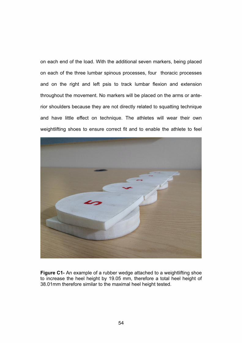
Figure C1- An example of a rubber wedge attached to a weightlifting shoe to increase the heel height by 19.05 mm, therefore a total heel height of 38.01mm therefore similar to the maximal heel height tested.
comfortable during the protocol. This will result in the technique being per-
formed as closely as possible to normal practice during training and com-
petition. The heel height will be incrementally increased in five randomly
ordered stages according to the testing protocol. A standard weight lifting
shoe has a baseline drop between heel and forefoot of 19.05mm, this will
be increased by attaching non compressible rubber wedges to the ath-
lete’s shoes to best mimic the shoes normal heel (see fig. C1) . They will
increase in five 3.78mm increments up to a maximum heel height 37.95
mm including the original heel (19.05 mm baseline, 22.83 mm, 26.61 mm,
30.39 mm, 34.17 mm, 37.95 mm).
!Protocol
They will then perform a standardised squatting warm up. This will start
with unloaded squats then working down in four sets increasing the
weight up to 75% of their self-reported 1RM for each exercise which is the
weight they will use during the testing protocol (5 repetitions at 50%, 3 at
repetitions 60%, 2 repetitions at 70% and 2 repetitions at 75% all percent-
ages of 1RM). A basic dorsiflexion flexibility test will then be performed
once the athlete has completed the warm up. They will then flex their an-
kle with the aim to flex it as far a possible, this ROM should be close to the
�55
expected ROM at the bottom the squat. Combined with thigh length a ratio
will be produced of which the groups will be based.
!Each athlete will then complete the testing protocol which involves per-
forming six front followed by six back squats at 75% of their self-reported
1RM. The different height wedges will be attached to the shoe (19.05 mm
baseline, 22.83 mm, 26.61 mm, 30.39 mm, 34.17 mm, 37.95 mm) in a
randomised order, to try and reduce any effect of fatigue on their tech-
nique. After each lift athletes will rest for upwards of three minutes to in-
sure they are full recovered, so they should be able to sustain a consistent
technique throughout the protocol (Sandler 2010).
!Post Processing
The focus will be within the hips/lower back and the knee joints because
they are key joints and loading areas which relate to injury and movement
within both of the squatting exercises. The marker locations will also be
used to calculate how internal angles change during the movement and
regarding minimum and maximum angles. These will be at the hip, knee
and ankle along with the angles of the shank and trunk in relation to the
floor. The markers on the lumbar region of the spine will be used to calcu-
�56
late flexion or extension of the spine compared to neutral (measured pre-
test with the subject standing in the anatomical position).Therefore their
position will be compared to neutral to see how much their alignment has
changed throughout the movement (additional extension being positive
and additional flexion being negative). All of these values will be compared
using statistical analysis.
!The statistical test used will be a repeated measures ANOVA. This will be
used to calculate group means, and standard deviations of the internal
forces, moments, internal/external angles and muscle activation levels. It
will also be used to identify whether and where the sig. differences and
interactions occur within each variable (p≤0.05) between each heel height
and the two groups. The statistical outputs and raw data will be compared
to previous literature to identify which heel heights allow the closest to op-
timal technique and minimal flexion within in the back comparing the three
flexibility groups.
!!!!!!!
�57

!!!!!!!!!!!!!!!
!!!!!!!!!!!!!
�58
APPENDIX D
Participant Information pack
My name is Andrew Brind-Surch, and I’m a Masters student studying Sports Biomechanics at Loughborough University . My project I am looking into the effect of changing weightlifting shoe heel height on squatting technique.
Testing Protocol
Please your own weightlifting shoes and tight lycra/compression shorts.
The whole protocol should take under 2hrs.
You will then be marked up with reflective markers and EMG sensors to track your movement (please bring tight Lycra shorts to wear). After this you will perform a standardised warm up, where you will lift up to 75% of your self-reported 1RM. Once you feel warmed up the testing protocol will start.
You will perform 12 squats at 75% of your self-reported 1RM (6 front, 6 back) at different weightlifting shoe heal heights (19.05 mm baseline, 22.83 mm, 26.61 mm, 30.39 mm, 34.17 mm, 37.95 mm). With at least three minutes between each one to make sure you as fully recovered.
You will be able to withdraw at any point if you decide you don’t want to take part in the study.
!!Health Screening Questionnaire !Please complete and if you answer yes to any of these questions please give details below each question. !1. Has your doctor ever said that you have a heart condition and that you
should only do physical activity recommended by a doctor? !2. Do you lose your balance because of dizziness or do you ever lose
consciousness? !3. Do you have any injury or condition that could affect your squatting
technique (such as back or knee problems)
�59

!4. Is your doctor currently prescribing drugs (for example, water pills) for
your blood pressure or heart condition? !5. Do you regular complete a weight training protocol, so understand the
technique involved? !Participant Name________________1RM Back________ Front______ !Age____ !!!I have read and fully understood the information concerning the test, and give full consent to take part. !Please contact me if you have any questions or concerns after the testing. !!!Participant signature !___________________________ !Researcher signature !___________________________ !Date !___________ !!Contact information
Andrew Brind-Surch
A . J . B r i n d - S u r c h 1 3 @ s t u d e n t . l-boro.ac.uk
�60
Tutor
Sam Allen