doc tinh nano
TRANSCRIPT

8/3/2019 Doc Tinh Nano
http://slidepdf.com/reader/full/doc-tinh-nano 1/15
R E V I E W Open Access
Risks from accidental exposures to engineerednanoparticles and neurological health effects:A critical reviewMyrtill Simkó1*, Mats-Olof Mattsson2
Abstract
There are certain concerns regarding the safety for the environment and human health from the use of engi-
neered nanoparticles (ENPs) which leads to unintended exposures, as opposed to the use of ENPs for medical pur-
poses. This review focuses on the unintended human exposure of ENPs. In particular, possible effects in the brain
are discussed and an attempt to assess risks is performed.
Animal experiments have shown that investigated ENPs (metallic nanoparticles, quantum dots, carbon nanotubes)
can translocate to the brain from different entry points (skin, blood, respiratory pathways). After inhalation or instil-
lation into parts of the respiratory tract a very small fraction of the inhaled or instilled ENPs reaches the blood and
subsequently secondary organs, including the CNS, at a low translocation rate. Experimental in vivo and in vitro
studies have shown that several types of ENPs can have various biological effects in the nervous system. Some of
these effects could also imply that ENPs can cause hazards, both acutely and in the long term. The relevance of
these data for risk assessment is far from clear. There are at present very few data on exposure of the general pub-
lic to either acute high dose exposure or on chronic exposure to low levels of air-borne ENPs. It is furthermore
unlikely that acute high dose exposures would occur. The risk from such exposures for damaging CNS effects is
thus probably very low, irrespective of any biological hazard associated with ENPs.
The situation is more complicated regarding chronic exposures, at low doses. The long term accumulation of ENPs
can not be excluded. However, we do not have exposure data for the general public regarding ENPs. Althoughtranslocation to the brain via respiratory organs and the circulation appears to be very low, there remains a possi-
bility that chronic exposures, and/or biopersistent ENPs, can influence processes within the brain that are triggering
or aggravating pathological processes.
In general, the present state of knowledge is unsatisfactory for a proper risk assessment in this area. Crucial deficits
include lack of exposure data, the absence of a proper dose concept, and that studies often fail in adequate
description of the investigated ENPs.
IntroductionThe purpose of the present review is to give a short
overview of how engineered nanoparticles (ENPs) can
translocate from the respiratory tract to the circulation,
pass the blood-brain-barrier (BBB), affect the brain, and
to discuss possible adverse health effects and associated
risks. We also suggest that there is a need for focused
research to support risk assessment. This research
should use standardized and proper methods and
experimental designs including the selection of the right
in vitro and/or in vivo models, controls, ENP character-
istics, doses, etc.
Nanoparticles (NPs) can be generated through both
natural (e.g., combustion by-products, volcano eruption
etc.) and synthetic processes. In the present article, we
focus on engineered nanoparticles and their unintended
exposure of the CNS.
In principal, researchers have agreed to use the term
nanoparticle if the material size is smaller than 100 nm
in three dimensions and are singular particles; although
different terms are still used in the literature, like nano-
sized materials, ultrafine particles (UFP), engineered
* Correspondence: [email protected] Academy of Sciences, Institute of Technology Assessment, Vienna,
Austria
Full list of author information is available at the end of the article
Simkó and Mattsson Particle and Fibre Toxicology 2010, 7:42
http://www.particleandfibretoxicology.com/content/7/1/42
© 2010 Simkó and Mattsson; licensee BioMed Central Ltd. This is an Open Access article distributed under the terms of the CreativeCommons Attribution License (http://creativecommons.org/licenses/by/2.0), which permits unrestricted use, distribution, andreproduction in any medium, provided the original work is properly cited.

8/3/2019 Doc Tinh Nano
http://slidepdf.com/reader/full/doc-tinh-nano 2/15
nanomaterials, manmade nanoparticles [1]. This shows
that the expression “nanomaterial” is related to the size
dimension only, but not to the material itself which can
contain any kind of substance. This is relevant from dif-
ferent perspectives, e.g. in political discussions and deci-
sions but also for dosimetry aspects. For the latter, it is
important to characterize the kind of the nanomaterial,
to define concentration(s), establish dose response rela-
tionships etc. Dosimetry is furthermore necessary for
risk estimation and for the establishment of thresholds
and/or limit values. The general use of the term nano-
material does not say much about the chemical condi-
tions. Therefore, the physico-chemical properties have
to be known for exposure calculations, including size,
shape and composition of the material.
ENPs and dose
For the calculation of the biological or chemical reactiv-ity of the material, knowledge about the physico-chemi-
cal properties, the number of molecules on the surface
of the nanosized material is needed, as well as the num-
ber of particles per cell. The number per cell is impor-
tant to determine the effective dose, since nanoparticles
have larger surface area than the corresponding bulk
material including a higher number of molecules on the
surface which can interact with the biological material,
and their larger number per mass allows their dispersion
into more cells. Information about the physico-chemical
properties including size and shape are important in
order to estimate ENP specific effective dose as well. In
in vitro studies it is difficult to estimate this dose
because NPs diffuse, settle, and agglomerate in cell cul-
ture media depending on different factors like media
density and viscosity, particle size, shape, charge and
density. Teeguarden et al. [2] developed a particokinetic
model to estimate cellular dose in vitro considering dif-
ferent factors like the dynamic precipitation rate in cell
culture media which depends on particle size and fol-
lows more the Brownian motion than gravitation.
Another important dose measure might be the rela-
tive biological effectiveness (RBE). If the physico-che-
mical properties and the number of molecules on the
surface of the nanosized material are known, as well asthe number of particles per cell, weighting factors might
be introduced as in dose calculation for ionizing radia-
tion, where RBE is calculated as a function of the quality
of the radiation. Thus, for the same absorbed dose,
alpha radiation is 20 times more biologically potent than
x-rays or gamma radiation. Accordingly, RBEs can then
be calculated for specific nanomaterials. The RBEs
would then be dependent of the material itself and the
number of internalized/taken up nanoparticles per cell.
The so called biological effective dose (BED) concept
describes oxygen radical generation, as an indirect
measure (or marker) for BED [3] considering the
physico-chemical properties of the material. This con-
cept is very useful. However, specific cell type dependent
redox potential capacities have also to be considered.
This is reviewed by Valco et al. [4] where pH dependent
effects are shown to be due to the specific redox capa-
city of the cell type in question, with cell type dependent
effects on e.g. cell cycle and developmental events. In
addition, the work by Sohaebuddin et al. [5] shows such
cell type dependent effects of various ENPs. Further-
more, for dose calculations relevant for both chronic
and for acute exposure, the dose rate has to be known,
which includes the time factor. To determine the reten-
tion time (how long an ENP is present in a cell or a
body) of a certain ENP, knowledge about the physico-
chemical properties, but also about the biological
deposition time in each site (deposition and retention
time are depending on deposition site) is needed. Inother words, knowledge about site dependent retention
time and bioavailability is needed to calculate the time
factor for dose rate. Other factors that will affect the
dose rate are that certain ENPs are biodegradable with a
relatively short half-life whereas others will not be meta-
bolized within the body. In addition, some ENPs may be
excreted, whereas others may accumulate over time.
The present knowledge regarding the different dose
concepts relevant for nanomaterials is however very lim-
ited, with possible exception of data from a few in vitro
studies. Relevant data from in vivo situations is mainly
absent.
Drug delivery systems and the blood-brain-barrierENPs have the potential to revolutionize medicine
because of their ability to reach and to affect target
organs and tissues, even “as distant” as tumours in the
brain, at the molecular and cellular levels. Medical and
pharmacological research is focused on applications of
nanosized materials, whereas side effects associated with
their use are generally not taken into consideration. In
fact, the knowledge about potential toxicity of ENPs is
far from comprehensive [6,7].
Drug delivery systems or nanocarriers should and may
overcome solubility or stability issues for the drug, andminimize drug induced side effects. However, the nano-
materials themselves can also induce significant toxic
effects (for reviews see [8,9]). Besides the chemical prop-
erties, this can be due to their electric, optical, and mag-
netic properties that are related to physical dimensions,
but also the surface of the material can be involved in
catalytic and oxidative reactions which themselves can
induce cytotoxicity. This toxicity can be greater than
that of a bulk material because the surface area-to-
volume ratio for nanomaterials is much greater. More-
over, some nanomaterials contain metals or compounds
Simkó and Mattsson Particle and Fibre Toxicology 2010, 7:42
http://www.particleandfibretoxicology.com/content/7/1/42
Page 2 of 15

8/3/2019 Doc Tinh Nano
http://slidepdf.com/reader/full/doc-tinh-nano 3/15
with known toxicity, and thus the breakdown of these
materials could elicit similar toxic responses.
A number of questions pertaining to the safety of
nanomaterials in this context are thus obvious. What is
the ultimate fate of the drug delivery systems/nanocar-
riers, and their components within the body? What hap-
pens with those which are not bio-degradable and those
which are functionalized, like carbon nanotubes, or
coated with different agents? Further on, what are the
consequences after long term exposure?
The blood-brain barrier (BBB) protects the central ner-
vous system from potentially harmful xenobiotics and
endogenous molecules (for review see [9]). The BBB,
formed by brain capillary endothelial cells linked together
by tight junctions, together with adjacent processes from
astrocytes, restricts the transfer of most substances from
the bloodstream into the brain. Therefore, substances
may gain access to the central nervous system by (lipid-mediated) free diffusion or potentially by receptor-
mediated endocytosis. Since tight junctions in the BBB
have a gap of only 4-6 nm, it has been suggested that
nanoparticles pass through the endothelial cell mem-
brane rather than via inter-endothelial junctions [10].
It has been shown that nanoparticles from the blood
circulation may influence endothelial cell membrane
integrity and/or disrupt the BBB [11], and may induce
vesicular transport to gain access into the CNS (see
below). Moreover, it seems to be accepted that nanopar-
ticles can induce oxidative stress leading to the genera-
tion of free radicals that could disrupt the BBB and cause
certain dysfunctions. It is also known that nanoparticles
without a surfactant coating are mainly internalized by
phagocytes and are thus unable to reach the brain in
desirable quantities, therefore almost no pharmaceutical
can reach the brain tissues by administering it with
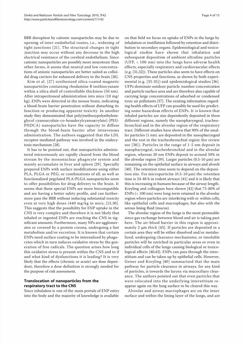
uncoated nanoparticles [12]. (See Figure 1 for a descrip-
tion of how ENPs can enter the cell and exert different
actions) However, surface modifications of nanoparticles
are presently intensely studied for nanomedicinal applica-
tions like diagnosis and therapy aiming to influence the
target-oriented pharmacokinetic behaviour of nanocar-
riers. Nanocarriers require surface modifications or other
forms of functional modifications for receptor-mediated
transport through the brain capillary endothelium to
deliver drugs to the central nervous system. Different
approaches to obtain suitable modifications are discussed
[13]. Kreuter et al. [14] demonstrated that polysorbate
80-coated polybutylcyanoacrylate (P80-PBCA) nanoparti-
cles can deliver the peptide “dalargin” into CNS to induce
its analgesic effects. Coating with alternative surfactants
did not produce the expected effects [15]. P80-PBCA
nanoparticles can thus deliver drugs to the brain, how-
ever these nanoparticles seem to have limitations due totheir potential toxicity [16]. Thus, Calvo et al. [17]
showed a drastic increase in sucrose permeability (as a
sign of BBB permeabilization) of the BBB in rats follow-
ing intravenous administration of P80-PBCA.
Other functional modifications of ENPs include for
example the conjugation of cell surface ligands or of
antibodies. In order to facilitate the crossing of the BBB,
apolipoprotein-E coating of nanoparticles for LDL
receptor mediated endocytosis in brain capillaries has
also been discussed [15,18]. Apolipoprotein, especially
ApoE combined with nanoparticles, behaves like low-
density lipoprotein (LDL) in the sense that LDL recep-
tor-mediated transcytosis enhances the drug delivery
together with nanoparticles across the BBB and is very
effective in drug delivery [19,20].
Another important factor in penetration of the BBB by
nanoparticles is their electrostatic charge. Cationic
charged molecules occupy anionic areas at the BBB
endothelium [21] and increase the endothelial cell per-
meability [22]. In in vitro studies, the cationized nano-
particles translocate more readily to the brain compared
with anionic or neutral nanoparticles [23]. Thus, both
the size and the charge of colloidal drug carriers are
important factors in determining drug or nanoparticle
delivery across the BBB or in brain parenchyma [24].However, there are little in vivo data regarding brain
permeability of cationized nanoparticles. One exception
is the work by Lockman et al. [25,26] who investigated
the effect of neutral, anionic and cationic charged ENPs
on the blood brain barrier (BBB) integrity and perme-
ability in situ with rat brain perfusion. Neutral ENPs
and low concentrations of anionic ENPs were found to
have no effect on BBB integrity, whereas high concen-
trations of anionic ENPs and cationic ENPs disrupted
the BBB structure. Especially cationic NPs displayed an
immediate toxic effect. It has been suggested that the
Unknownprocesses
e.g.diffusion
N s
ROS
Si nal transduction
[Ca2+]i
ENPsinvesicles
Nucleus
ENPsin
cytoplasm
ENPsinternalization/
activeuptake/
pinocytosis,
phagocytosis
Receptoractivation/
inactivation/Antioxidative
ff
DNArepair/
Apoptosis
DNA
internalization
Mediator release
Figure 1 Various ways for uptake of ENPs to mammalian cells
and the effects ENPs can have on intracellular processes. ROS:
reactive oxygen species.
Simkó and Mattsson Particle and Fibre Toxicology 2010, 7:42
http://www.particleandfibretoxicology.com/content/7/1/42
Page 3 of 15
8/3/2019 Doc Tinh Nano
http://slidepdf.com/reader/full/doc-tinh-nano 4/15
BBB disruption by cationic nanoparticles may be due to
opening of inter-endothelial routes, i.e., widening of
tight junctions [21]. The structural changes in tight
junction may occur without any decrease in the high
electrical resistance of the cerebral endothelium. Since
cationic nanoparticles are possibly more neurotoxic than
other forms, it seems that neutral and low concentra-
tions of anionic nanoparticles are better suited as colloi-
dal drug carriers for enhanced delivery to the brain [26].
Kim et al. [27] synthesized silica-coated magnetic
nanoparticles containing rhodamine B isothiocyanate
within a silica shell of controllable thickness (50 nm).
After intraperitoneal administration into mice (10 mg/
kg), ENPs were detected in the mouse brain, indicating
a blood-brain barrier penetration without disturbing its
function or producing apparent toxicity. In another
study they demonstrated that poly(methoxypolyethylene-
glycol cyanoacrylate-co-hexadecylcyanoacrylate) (PEG-PHDCA) nanoparticles have the capacity to diffuse
through the blood-brain barrier after intravenous
administration. The authors suggested that the LDL
receptor-mediated pathway was involved in the endocy-
tosis mechanism [28].
It has to be pointed out, that nanoparticles adminis-
tered intravenously are rapidly cleared from the blood
stream by the mononuclear phagocyte system and
mainly accumulate in liver and spleen [29]. Specially
prepared ENPs with surface modifications using either
PLA, PLGA or PEG, or combinations of all, as well as
functionalized pegylated PLA/PLGA nanoparticles seem
to offer possibilities for drug delivery to the brain. It
seems that these special ENPs are more biocompatible
and are having a better safety profile, and can further-
more pass the BBB without inducing substantial toxicity
even at very high doses (440 mg/kg in mice, [13,30].
This suggests that the possibility for ENP uptake in the
CNS is very complex and therefore it is not likely that
inhaled or ingested ENPs are reaching the CNS in sig-
nificant amounts. Furthermore, many NPs are agglomer-
ates or covered by a protein corona, undergoing a fast
metabolism and/or excretion. It is known that certain
ENPs need surface coating to be internalized by phago-
cytes which in turn induces oxidative stress by the gen-eration of free radicals. The question arises how long
this oxidative stress is present within the CNS and to if
and what kind of dysfunctions it is leading? It is very
likely that the effects (chronic or acute) are dose depen-
dent, therefore a dose definition is strongly needed for
the purpose of risk assessment.
Translocation of nanoparticles from therespiratory tract to the CNSSince inhalation is one of the main portals of ENP entry
into the body and the majority of knowledge is available
on that field we focus on uptake of ENPs in the lungs by
inhalation or instillation followed by retention and distri-
bution to secondary organs. Epidemiological and toxico-
log ical s tudies have s ho wn that inhalat io n and
subsequent deposition of ambient ultrafine particles
(UFP, < 100 nm) into the lungs have adverse health
effects, especially respiratory and cardiovascular effects
(e.g. [31,32]). These particles also seem to have effects on
CNS properties and functions, as shown by both experi-
mental (e.g. [33-35]) and epidemiological studies [36].
UFPs dominate outdoor particle number concentration
and particle surface area and are therefore also capable of
carrying large concentrations of adsorbed or condensed
toxic air pollutants [37]. The existing information regard-
ing health effects of UFP can possibly be used for predict-
ing some hazardous effects of ENPs. It is known that
inhaled particles are size dependently deposited in three
different regions, namely the nasopharyngeal, tracheo-bronchial and in the alveolar region of the respiratory
tract. Different studies have shown that 90% of the smal-
ler particles (1 nm) are deposited in the nasopharyngeal
and the rest in the tracheobronchial region (for review
see [38]). Particles in the range of 1-5 nm deposit in
nasopharyngeal, tracheobronchial and in the alveolar
region, whereas 20 nm ENPs deposit to around 50% in
the alveolar region [39]. Larger particles (0.5-10 μm) are
remaining on the epithelial surface in airways and alveoli
[40]. The retention time seem to depend on the deposi-
tion site. For microparticles (0.5-10 μm) the retention
time is 24-48 h in rodent airways [41] and it is likely that
this is increasing in humans because of the airway length.
Kreyling and colleagues have shown [42] that 75-80% of
ENPs (< 100 nm) were long-term retained in the alveolar
region where particles are interfering with or within cells,
like epithelial cells and macrophages, but also with the
serous lining fluid (mucus).
The alveolar region of the lungs is the most permeable
since gas exchange between blood and air is taking part
here. The air-blood barrier in this region is approxi-
mately 2 μm thick [43]. If particles are deposited in a
certain area they will be either dissolved and/or metabo-
lized, undergoing clearance mechanisms, or insoluble
particles will be enriched in particular areas or even inindividual cells of the lungs causing biological or toxico-
logical effects [40,43]. ENPs can pass through the inter-
stitium and can be taken up by epithelial cells. However,
Geiser and Kreyling [40] summarized that the main
pathway for particle clearance in airways, for any kind
of particles, is towards the larynx via mucociliary clear-
ance. The authors pointed out that even particles that
were relocated into the underlying interstitium re-
appear again on the lung surface to be cleared this way.
Alveolar and airway macrophages are on the inner
surface and within the lining layer of the lungs, and are
Simkó and Mattsson Particle and Fibre Toxicology 2010, 7:42
http://www.particleandfibretoxicology.com/content/7/1/42
Page 4 of 15

8/3/2019 Doc Tinh Nano
http://slidepdf.com/reader/full/doc-tinh-nano 5/15
constantly exposed to inhaled particles. Phagocytic
uptake is the main mechanism to remove insoluble
inhaled microsized particles. Monocytes/macrophages
are also circulating within the body to take part in the
main pathway for monocytes/macrophage-associated
particles clearance, which is the mucociliary transport. It
cannot be excluded that circulating particle-containing
macrophages may re-enter the interstitium and/or
lymph nodes and thus the lymphatic system. On the
other hand, it is suggested that inhaled and deposited
nanoparticles are not efficiently taken up by surface
macrophages. Therefore passive mechanisms like diffu-
sion, adhesive interaction, and also pinocytic uptake are
currently discussed for translocation [40,43]. Particles
which penetrate cells may enrich, interact with orga-
nelles, cause oxidative stress, induce different cellular
signalling pathways and leading to cellular effects like
the release of inflammatory intermediates such ascytokines and free radicals (see also Figure 1). Mühlfeld
et al. [44] have shown that inhaled aerosols of 20 nm
TiO2 in rats were distributed after one hour to all lung
compartments in proportion to the compartment
volume, and some particles were detected in erythro-
cytes within the pulmonary capillaries. Peters et al. [45]
hypothesized that the way how particles translocate to
secondary organs is by the blood circulation. Nemmar
et al. [46] documented by using technetium-labeled car-
bon NPs by an inhalation study in humans, that a cer-
tain amount of NPs diffuse rapidly into the systemic
circulation. However, certain published studies report
that translocation rates for NPs into the blood circula-
tion are very low [47].
Clearance mechanisms in airways and alveoli are redu-
cing the retention time of NPs in the lungs, therefore
only relatively few nanosized particles can translocate to
secondary organs. Chen et al. [ 48] have shown that
intratracheal-instilled polystyrene particles with an aver-
age diameter of 56.4 or 202 nm, are passing into the
blood circulation, but this translocation is between 1-
2.5% independently of the particle size. Liu et al. [49]
investigated the overall toxicity of nasally instilled
nanoscale copper particles (23.5 nm) in comparison
with micro-sized copper particles (17 μm) in mice andfound only in the high-dose group (40 mg/kg, three
times per week) significant pathological changes. It has
to be pointed out, that this is an enormously high dose
without any physiological significance. These kinds of
experiments are useful only for toxicity tests or for
hazard identification but not for risk assessment. There
are several studies performed using different nanomater-
ials and sizes and concentrations showing translocation
to secondary organs, basically to the liver, spleen and
kidney (detailed below). Kreyling et al. [50] performed
an iridium (2-4 nm) and/or carbon (5-10 nm) ENP
inhalation study with rats to learn about the transloca-
tion rate from lungs to blood circulation and secondary
organs. The authors detected from 0.1 to 1% Ir-NPs of
the retained fraction in liver, spleen, kidneys, heart, and
brain, and 1-5% in the remaining carcass (soft tissue
and bone). The mixed fraction of Ir with the carbon
ENP retained in secondary organs at significantly lower
levels than pure Ir-NP. Furthermore, 80 nm aggregates
translocated and accumulated significantly less than the
20 nm ones. In a recent review, Geiser and Kreyling
[40] summarized the evidence for translocation of cer-
tain ENPs like gold, silver, TiO2, polystyrene and carbon
nanoparticles in the size range of 5 - 100 nm across the
air-blood barrier from animal studies. In summary, the
translocation fraction out of the lung seems not to
exceed 5% for any of the investigated ENPs.
Mills et al. [51 ] investigated the extent to which
inhaled radioactive labelled carbon nanoparticles (Tech-negas, 99mTc, 4-20 nm, aggregates ca. 100 nm) were
able to access the systemic circulation on human volun-
teers. The authors detected more than 95% of Techne-
gas retention in the lungs, with no accumulation in liver
or spleen and concluded that the majority of carbon
nanoparticles remain within the lung up to 6 h after
inhalation and do not pass directly from the lungs into
the systemic circulation. Nemmar et al. [46] showed
that 20% of initial lung radioactive carbon nanoparticles
were detected in the liver, meaning that 80% remained
in the lung.
The translocation rate from the respiratory tract to
the central nervous system has been shown to be very
low. It is questionable if the amount of nanomaterials
which reaches the brain can cause hazardous effects.
However, Chen et al. [48] reported, that pulmonary
inflammation induced by instillation plays the major
role in enhancing the extrapulmonary translocation of
particles (using LPS coated particles). This fact is indi-
cating that at least LPS coated nanomaterials can induce
inflammatory effects which themselves are changing the
microenviroment leading to higher translocation rates to
secondary organs [35]. A systemic inflammation can
contribute to local inflammation in the brain which in
turn can lead to enhancement of ongoing inflammatory reactions in the brain [52].
If ENPs are injected or translocated to the blood cir-
culation, proteins are associating with the nanoparticles,
which in turn can lead to an in vivo response [ 53]. It
has been shown that a so called “corona” on the surface
of the ENPs is the result of the adsorption of different
serum/plasma proteins on ENPs. Cedervall and collea-
gues [53] reported that proteins compete for the nano-
particle “surface,” and the resulting “corona” largely
defines the biological identity of the particle. Lundqvist
et al. [54] have shown that the nature of the corona is
Simkó and Mattsson Particle and Fibre Toxicology 2010, 7:42
http://www.particleandfibretoxicology.com/content/7/1/42
Page 5 of 15

8/3/2019 Doc Tinh Nano
http://slidepdf.com/reader/full/doc-tinh-nano 6/15
determined by the local chemical property of the
nanomaterial including size and surface properties.
Therefore the kinetics of the ENPs are also depending
on the local corona-structure which is different in each
microenviroment. In drug delivery studies using polysor-
bate 80-coated nanoparticles, it was shown that the ENP
adsorbs apolipoproteins from the blood after injection.
These particles mimic lipoprotein particles which could
be taken up by the brain capillary endothelial cells via
LDL receptors [15].
In conclusion, the translocation rate of deposited
ENPs from the lung to the blood circulation and then
to secondary organs seems not to exceed 5%. Further-
more, the translocation from the blood to the CNS is
lower than 1% according to available studies (see [50,55]
and Table 1). Corona formation can change the translo-
cation rate and possibly increase the hazardous effects.
Axonal transport of ENPs to the brainAn important mechanism of particle endocytosis
involves the uptake by sensory nerve endings embedded
in airway epithelia. In the nasal region it is the olfactory
and trigeminus nerve system, and in the tracheobron-
chial region it is the extensive sensory nerve network.
Translocation to ganglia and the CNS can then be
accomplished by axonal transport.
The olfactory nerve pathway may be a critical portal
of ENP entry to the central nervous system of humans,
especially under high environmental or occupational
ENP exposures but also under chronic exposure.
Using colloidal gold particles (50 nm) that were intra-
nasally instilled in monkeys it was shown that particles
translocate in the axons of the olfactory nerves to the
olfactory bulbs, where nanoparticles were seen in the
mitochondria but not in the cytoplasm (as cited by
[56]). A study by Hunter and Dey [57] in rats demon-
strated the translocation of intranasally instilled rhoda-
mine-labeled microspheres (20-200 nm) to the
trigeminal ganglion inside the cranium via uptake into
the ophthalmic and maxillary branches of the trigeminus
nerve. In another study, Hunter and Undem [ 58]
instilled similar particles intratracheally into guinea pigs
and reported a neuronal translocation to the ganglion
nodosum in the neck area, which is integrated into the vagal system. More recent studies indicated that neuro-
nal translocation pathways are also operational for other
inhaled ENPs. Inhalation of elemental 13C ENPs (36 nm,
160 μg/m3) resulted in a significant accumulation of
these particles in the olfactory bulb of rats on the first
day, which constantly increased further throughout day
seven after the initial 6 h exposure [38]. Results from
ano ther inhalat io n s tudy w ith s olid nanos ized
Table 1 Translocation of various ENPs via respiratory pathways or via injection to blood and/or CNS
Material Administration ENP size (nm)** Translocation toblood Translocation toCNS Ref.*
Inha lation Na salinstillation
Injection
Carbonparticles
X 4-20 X [51]
X 100 X [46] (human)
X 36 X [38]
Cu X 23.5 X [49]
Ir X 2-4 X X [50]
MnO2 X 30 X X [56,59]
X 23 X X [62]
Polystyrene X 56.4 X X [48]
X 202 X X [48]
TiO2 X 20 X [48]
X 80, 155 (very highdoses)
X [61] (mouse)[64](mouse)
X 25-70 (s.c.) X [65]
X 25-70 (i.v.) X [66]
X 5 (i.p.) X [67]
Latex particles X 20-200 X [57,58] (guinea pigs)
Ag X 70-110 X [60]
* Studies were performed on rats unless otherwise indicated.
**s.c. = subcutaneous injection; i.v. = intra venous; i.p. = intraperitoneal
Simkó and Mattsson Particle and Fibre Toxicology 2010, 7:42
http://www.particleandfibretoxicology.com/content/7/1/42
Page 6 of 15

8/3/2019 Doc Tinh Nano
http://slidepdf.com/reader/full/doc-tinh-nano 7/15
manganese oxide particles (30 nm, 500 μg/m3) in rats
also demonstrated an increase of particles in the olfac-
tory bulb. Inhalation exposures were for 6 h/day, 5
days/week for up to 12 days. After 12 days of exposure
with both nostrils patent, Mn concentrations in the
olfactory bulb increased 3.5-fold (from 0.5 to 1.75 ng
Mn/mg tissue), whereas lung Mn concentrations
doubled; there were also increases in striatum, frontal
cortex, and cerebellum. When one nostril was occluded
during a 6-h exposure, the accumulation of Mn was
seen only in the olfactory bulb of the open nostril
[56,59]. These observations suggest that nanoparticles in
the air can enter into the CNS via the olfactory nerve
during accidental or prolonged environmental or occu-
pational exposure to humans.
Another study showed that inhaled 20 nm nanogold
particles (2 × 10⁶ particles/cm3) can accumulate in the
olfactory bulb of rats [60]. The exposure for 5 daysresulted in a significant increase of gold ENPs in the
olfactory bulb (8 ng Au/g body weight). After 15 days of
exposure, significant accumulations of gold particles
were detected in the septum and entorhinal cortex. Both
brain structures receive direct neuronal projections from
the olfactory bulb, and are important in attention and
new memory formation.
After nasal instillation (500 μg of TiO2 nanoparticle
suspension every other day for 30 days), the micro-
distributions of TiO2 NPs (80 nm) and fine TiO2 par-
ticles (155 nm) in the olfactory bulb of mice were
investigated by [61]. It could be demonstrated that
both types of investigated TiO2 particles were taken
up by the olfactory bulb via the primary olfactory neu-
rons and then accumulated in the olfactory nerve
layer, olfactory ventricle, and granular cell layer of the
olfactory bulb. The TiO2 content was increased in all
investigated brain regions (olfactory bulb, cerebral
cortex, hippocampus and cerebellum), with the most
significant increases seen in the hippocampus. The
presence of TiO2 in hippocampus was furthermore
accompanied by changes in neuron morphology and
increased amount of GFAP-positive cells in the CA4
region. Signs of oxidative stress were documented in
all regions of the brain. Interestingly, in general ana-tase TiO2 gave rise to stronger effects than the rutile
form.
Taken together, it seems that nanoparticles can trans-
locate to the nervous system through sensory nerves.
Translocation of 20 nm particles is 2-10 times higher in
the human olfactory bulb than in rats [6]. Thus, the
translocated nanoparticles in humans can enter into the
deeper brain structures in short exposure time. Based
on the limited data available, it is presently difficult to
assess to what extent accumulation in the brain via axo-
nal transport is a realistic possibility (see also Figure 2).
Neurobiological effects of ENPsIn vivo studies
Of the two principal cell types in the nervous system
(neurons and glia cells), the neurons have characteristics
that make them especially sensitive to various types of
stressors. The neurons have an especially vulnerable
anatomy due to their extensive and very thin and fragile
extensions (dendrites and especially axons). In addition,
these cells are metabolically very sensitive since they
rely solely on aerobic metabolism of glucose. The neu-
rons are extremely sensitive to oxidative stress, which in
many cases also is a contributing factor to a number of
neurodegenerative diseases. In addition, with very few
exceptions, neurons are not renewable in mammals,
making the nervous system functions very sensitive to
agents that cause cell death.
Studies performed on intact animals can specifically
address both exposure and dose requirements for poten-tial effects, as well as the specific effects on processes in
the brain (Table 2).
That nanosized MnO2 NP can translocate into the
brain after long-term inhalation/instillation has been
shown by Elder et al. [56] and Sarközi et al. [62]. In the
former study, rats were exposed to MnO2 ENP by inha-
lation for 12 days. Mn levels were seen to increase in
the olfactory bulb (3.5 times the background levels),
striatum, frontal cortex, and cerebellum. No functional
endpoints were investigated in this study, but molecular
signs of inflammatory changes were possible to detect.
ENP exposure: Inhalation
ungsNose
s l o c a t i o n
e C
n a l t r a n s p o r t
T r a n
Blood
e e x p o s u r h
r oni c ex
Secondary
organs
Reentry to
blood
A x o
B B B
p e n e t r a t i o n
A c u t o
s ur e
Brain / CNS
Figure 2 Overview of the routes by which ENPs can translocate
after inhalation through the nose or the lungs to the brain .
Note that inhalation through the nose represents the likelihood of
acute exposure effects whereas the inhalation pathway through the
lungs followed by translocation to secondary organs and possible
re-entry to the blood is showing the probability for chronic
exposure.
Simkó and Mattsson Particle and Fibre Toxicology 2010, 7:42
http://www.particleandfibretoxicology.com/content/7/1/42
Page 7 of 15

8/3/2019 Doc Tinh Nano
http://slidepdf.com/reader/full/doc-tinh-nano 8/15
In the work by Sarközi and co-workers [ 62], male
Wistar rats were subjected to MnO2 (23 nm) instillation
for 3, 6, or 9 weeks at 2.63 or 5.26 mg/kg bodyweight.
After the end of exposures, Mn was detected in the
brain by x-ray spectroscopy. The rats’ spontaneousmotility was negatively affected. In addition, electrophy-
siological changes in cortical activity and the conduction
velocity of the tail nerve were documented.
Chen et al. [11] infused rats intravenously with Al2O3
NP (8-12 nm; 29 mg/kg). The rats were sacrificed 20 h
after the infusion and the brains were subsequently
investigated with immunohistochemistry for certain
tight junction proteins normally present in the endothe-
lium of the BBB. The data indicate that the proteins
claudin-5 and occludin are down-regulated in the vessels
of treated animals, suggesting impairment of the BBB.
However, the data are only qualitatively expressed, no
proper quantification of the protein levels were made. It
is also unclear how many animals were investigated.
In a recent paper by Viswaprakash et al. [63] rat olfac-
tory epithelia were exposed to 1-2 nm zinc particles and
the responses to odorants were measured by electrool-
factogram and whole-cell patch clamp. The addition of
the Zn particles to the odorant suspension enhanced the
response to the odorant. Interestingly, this response was
seen to be specific for Zn particles, whereas neither Zn2
+ ions nor other metal particles (Cu, Ag, Au) elicited
similar responses.
The increased production and presence of nanosized
TiO2 particles in consumer products and in processeshas generated an interest into the possible effects of
these particles on human health. Regarding in vivo stu-
dies of nervous system function, a few recent articles
have rendered relevant data. Thus, Wang et al. [61] sub-
jected female mice to nasal instillation with TiO2 NP
(80 nm, rutile and 155 nm anatase; 500 μg every 2nd day
for 30 days) at an extremely high dose. Titanium parti-
cles were mainly accumulated in the cerebral cortex,
thalamus, olfactory bulb and hippocampus, (especially in
the CA1 and CA3 regions). There was an obviously dis-
persed arrangement of neurons in the hippocampal CA1
region after TiO2 exposure. Furthermore, the investiga-
tion of cell numbers in the stratum pyramidale of the
CA1 region indicated a drastic neuronal loss. There was
30% and 25% cell loss in the 80 and 155 nm TiO 2-
exposed groups, respectively. Apparent morphologicalchanges of hippocampal neurons and increased GFAP-
positive astrocytes in the CA4 region were also found,
which were in good agreements with the high TiO2 con-
tents in this hippocampus region. GFAP is best viewed
as a biomarker of early pathological effects, indicated by
the activation of astrocytes. Oxidative stress such as
lipid peroxidation, protein oxidation and increased activ-
ities of catalase, as well as excessive release of glutamic
acid and nitric oxide occurred in the whole brain of
exposed mice [61]. In a follow- up study, mice were
again intranasally instilled every second day with the
two types of TiO2 particles (80 nm, rutile or 155 nm,
anatase; purity > 99%, about 500 μg per mouse, respec-
tively). This time, brain tissues were collected at post-
instillation time points of 2, 10, 20 and 30 days and
evaluated for accumulation of TiO2, histopathology, oxi-
dative stress, and inflammatory markers. It is shown in
this study, that instilled TiO2 nanoparticles entered the
brain directly through the olfactory bulb during the
whole exposure period. In all brain parts and at all post-
exposure time periods, the measured concentrations of
both types of TiO2 particles were higher than in any of
the controls. The anatase form of the TiO2 exhibited
stronger effects on some of the investigated endpoints
in both these studies. In the olfactory bulb, TiO2 con-tents increased gradually with time. TiO2 particles were
mainly deposited in the hippocampus, where TiO2 con-
tents were significantly increased after exposure for 2
days, then stayed constant for 10 and 20 days, before
reaching the highest values after 30 days of exposure
[64]. After 30 days of exposure, pathological changes
were observed in olfactory bulb and hippocampus. Irre-
gular arrangements of neurons in the olfactory nerve
layers and dispersed arrangement and loss of neurons in
the CA1 region of hippocampus were demonstrated.
Hippocampal nerve cells were degenerated, together
Table 2 Experimental findings of neurobiological effects of specific ENPs
End-point In vivo In vitro References
Cell morphology changes Al2O3, TiO2 Fe2O3, QDs [11,61,67,69,70]
Increased inflammation signs an d markers MnO2 [56,62]
Increased oxidative stress TiO2, QDs Degussa P25, ferritin, C60, Ag [61,67,69,72]Antioxidative effects (inconsistent effects) CeO, YO, C60 [73-76]
Neuron function (inhibit ion and facil itation) MnO2, Zn, TiO2, QDs Mn, Ag, ZnO, CuO, CNTs, TiO2, QDs [61,67,68,77,88]
Behaviour (negative effect) MnO2 [62]
Development and differentiation (inconsistent effects) TiO2 Fe2O3, Ag, TiO2 [65,66,84]
Accelerated protein fibrillation TiO2, CNT, QDs, CeO, copolymer particles [86,87]
Simkó and Mattsson Particle and Fibre Toxicology 2010, 7:42
http://www.particleandfibretoxicology.com/content/7/1/42
Page 8 of 15
8/3/2019 Doc Tinh Nano
http://slidepdf.com/reader/full/doc-tinh-nano 9/15
with changes in the nuclear membrane, mitochondria,
rough endoplasmic reticulum, chromatin condensation,
and elevated amounts of free ribosomes [64].
Shimizu et al [65] studied effects of anatase TiO2 (the
particle size is not given in the article) that they injected
subcutaneously (s.c.) into pregnant mice. Male embryos
and pups were then investigated for certain gene expres-
sion patterns. The expression of genes associated with
brain development, motor activity, oxidative stress, and
apoptosis was changed compared to control animals
during various periods of investigation (embryonic day
16 to 21 days post partum). Also Takeda et al [ 66]
injected TiO2 (anatase, 25-70 nm) s.c. into pregnant
mice. The nanoparticles were found in the brains (cor-
tex, olfactory bulb) of the offspring. In addition, cells
expressing the apoptosis marker Caspase-3 increased in
the olfactory bulb of these animals. Abdominal injection
of high dose anatase TiO2 (5 nm; 5-150 mg/g) to micewere performed daily for 14 days in a recent study [67].
The TiO2 content of the brains increased with increas-
ing injection “doses”. Also changes in neuronal mor-
phology, transmitter levels and signs of oxidative stress
were seen to follow a dose-response relationship.
The effects of various types of quantum dots (QDs) in
the hippocampus of rats were investigated by Tang et al
[68]. They found that both unmodified (CdSe) and mod-
ified (streptavidin-CdSe/ZnS) QDs can negatively affect
synaptic transmission and plasticity in the rat hippocam-
pus. The QDs were directly applied into the hippocam-
pus and the effects on the electrophysiological
properties of the neurons in the area were recorded
after 20 min. Pair-pulse relation and long-term potentia-
tion were significantly decreased after treatments. The
effects were seen at two investigated concentrations of
QD, 0.5 and 10 nM. The authors also reported that
signs of oxidative stress were seen immediately after
completion of the electrophysiological measurements.
The results showed that SOD activity, GSH content and
MDA levels all increased in the animals treated with
QDs. These responses were stronger in the unmodified
(CdSe) QDs, and more pronounced at the higher inves-
tigated concentration. This finding is possibly due to the
toxic effects of Cd, which can be expected to be releasedfrom the unmodified QD. In a study by Maysinger et al
[69] intracortical injection (μM concentrations) of var-
ious types of PEGylated QDs, non-PEGylated CdTe
QDs, and CeO2 all caused activation of the glial cell
marker GFAP to various degrees in mice. The effects were
strongest in animals injected with CdTe QDs, and weakest
after CeO2 treatment. Furthermore, the study showed that
one of the PEGylated QDs, QD705, primarily accumulated
in glia, whereas a small fraction (0.5%) could be found in
neurons. Both these studies indicated that especially non-
PEGylated QDs can cause inflammation and possibly
gliosis in the brain. It is difficult to evaluate if the used
concentrations are relevant for any real exposure situation.
In conclusion, the referenced studies point to that the
investigated metallic nanoparticles all can translocate
from the point of application (respiratory tract, skin, cir-
culatory system) to the brains of the animals. However,
it is unclear from these studies under which specific
conditions this can be accomplished since the studies
have not investigated dose-response relationships, prop-
erties of the ENPs in question etc. Certain of the obser-
vations are furthermore made in experiments where
unrealistically high doses have been applied. A single
study also indicates that a high dose of TiO2 can pass
the placenta and be taken up into the brains of embryos.
Regarding the physiological effects of these exposures, it
is unclear to what extent, and at what exposure levels,
nervous system functions can be affected by ENPs.
However, the available data are suggestive of effects onneurotransmission, and possibly behaviour. Several signs
of changes in oxygen radical homeostasis were also
seen. The consequence of this could be that long-term
exposures cause permanent inflammatory states, which
can be a contributing factor in certain neurodegenera-
tive diseases.
In vitro studies
Synaptic transmission between neurons involves a num-
ber of structures and processes both in the pre- and the
postsynaptic neuron. The presynaptic neuron needs to
have the necessary machinery for synthesis of the neu-
ron-specific transmitter. Furthermore, it is necessary to
have structures for transmitter release and transmitter
re-uptake or enzymatic degradation of transmitter. The
post-synaptic neuron needs to express receptors for
transmitters and together with its synaptic partner it has
to express the structures that make the physical contacts
in the synapse. To some extent, the development and
function of neurotransmission in ENP exposed neurons
have been investigated, along with studies on the toxi-
city of ENP on nervous system components.
Several studies have dealt with toxicity and oxidative
stress due to ENP exposures. Thus, Pisanic et al. [ 70]
showed that iron oxide ENP (5-12 nm) have cytotoxiceffects on PC12 cells (a neuroendocrine cell line derived
from rat pheochromocytoma). At 1.5 and 15 mM iron
concentrations (but not at 0.15 mM) the ENP caused
decreased cell viability. The response to NGF (nerve
growth factor, inducing differentiation) in the PC12 cells
was also negatively affected, seen as diminished neurite
extension and number of neurites per cell. Also, the
level of the GAP43 protein, a marker for neuronal dif-
ferentiation, was decreased.
Maysinger et al. [69] showed that certain QDs were
taken up into differentiated PC12 cells, whereas other
Simkó and Mattsson Particle and Fibre Toxicology 2010, 7:42
http://www.particleandfibretoxicology.com/content/7/1/42
Page 9 of 15
8/3/2019 Doc Tinh Nano
http://slidepdf.com/reader/full/doc-tinh-nano 10/15
(non-PEGylated) QDs caused cell death. Increased oxi-
dative stress, measured as H2O2 production, was seen in
immortalized microglia cells exposed to Degussa P25
ENP that formed aggregates. The doses (2.5-120 ppm)
of the P25 aggregates were non-cytotoxic [71]. Alek-
seenko and coworkers [72] used rat brain synaptosomes
to test ferritin molecules that contain Fe3+ iron particles
(7 nm). The treatment caused ROS formation at high
doses (800 μg/ml), whereas the effects of 80 and 8 μg/
ml were not significantly different from controls. The
higher concentration could not induce glutamate
release, but inhibited uptake of glutamate. Consequently,
at 800 μg/ml, iron-based nanoparticles can cause condi-
tions that can lead to neurodegeneration. Schubert et al.
[73] showed that both CeO ENPs (6 and 12 nm) and
YO ENPs (12 nm) are neuroprotective in cultured hip-
pocampal neurons (HT22 cell line). The cells were trea-
ted with glutamate to generate ROS at levels that werecytotoxic, which was counteracted by addition of the
mentioned ENPs.
Conflicting results are available regarding the effects on
cytotoxicity and oxidative stress from fullerenes (C60).
Sayes et al. [74] used a water-soluble fullerene species,
nano-C60 that was cytotoxic to several human cell types,
including astrocytes. The cytotoxicity was mediated by
lipid peroxidation according to the authors. On the other
hand, polyhydroxylated C60 fullerenols at μM concentra-
tions acted as antagonists to glutamate receptors in a
study by Jin et al. [75]. The C60 particles blocked primar-
ily the AMPA-type glutamate receptor in neuronal cul-
tures from rat brain, and also to some extent NMDA and
KA receptors. The antagonistic behaviour on glutamate
receptors were not seen in GABA or taurine receptors. In
the absence of C60, higher concentrations of glutamate
were needed to elicit similar effect. The fullerenes could
also act as antioxidants, inhibiting effects of added H2O2
and Fe2+ . An earlier study by Dugan et al. [76] also
showed neuroprotective effects of C60 fullerenes on corti-
cal cell cultures exposed to NMDA or AMPA at concen-
trations that caused excitotoxicity.
The effects of Mn ENP on transmitter levels in PC12
cells were seen by Hussain et al. [77]. The Mn nanopar-
ticles specifically caused depletion of dopamine stores inthe PC12 cells. This occurred in a dose-dependent fash-
ion (concentrations ranging from 1-100 μg/ml) after
cells were exposed for 24 h to 40 nm particles. The
effect was similar to effects of added Mn2+ ions. How-
ever, the levels of ROS were much higher after Mn ENP
addition compared to Mn2+, or compared to Ag ENP
(15 nm). The latter ENP also caused dopamine deple-
tion, although to a lesser extent than Mn ENP.
In two studies Wang et al. have documented that ENP
inhibit the acetylcholine degrading enzymes acetylcho-
line esterase [78] and butyrylcholine esterase [79] in
solution. Several different types of ENP (MWCNT,
SWCNT, Cu, TiO2) could adsorb and thus inhibit
enzyme activities in a dose-dependent fashion. The
authors suggested that the inhibitory effects were caused
by ion dissolution from the ENPs.
The communication between neurons relies on trans-
mitter release which in turn is dependent on changes in
ion concentrations on the in and outside of the neuro-
nal membrane. Since such changes lead to displacement
of charged entities, it is possible to measure these events
by analyzing electric potentials that are present over the
membrane. Several studies have investigated whether
currents that pass through ion specific membrane chan-
nels are affected by ENP.
Tang et al. [68] studied the effects of CdSe QDs
(2.38 nm) on primary cultures of rat hippocampal neu-
rons. At 10 nM or higher concentrations, these particles
caused cell death, due to sustained increases in intracel-lular Ca2+ levels. The particles also had effects on vol-
tage gated Na-channels, where patch-clamp analyses
revealed enhanced activation and inactivation of the
sodium current, and also a prolonged activation time
and increased recovery time for the Na2+ current. Thus,
fewer Na-dependent potentials would occur in these
cells, interfering with normal synaptic transmission.
Another study revealed that silver particles (244.4 nm;
12.5 m2/g) could inhibit Na+ currents in rat hippocam-
pal slices [80]. This occurred at 10 μg/ml, but not at
lower concentrations. Zhao et al [81] could show that
ZnO ENP (20-80 nm; 2-3 crystal forms; 100 μg/ml, but
not at lower concentrations), increased amplitudes of
both Na+ and K+ currents, by increasing the number of
open Na+ channels, delaying rectifier K+ channels and
thus enhancing the excitability of neurons. Xu et al. [82]
have shown that CuO ENP (60.6 nm; 15.7 m2/g; 50 μg/
ml) could inhibit the rectifier K+ current in rat CA1 pyr-
amidal hippocampus neurons. Jakubek et al. [83]
demonstrated that carbon nanotubes could inhibit the
function of Ca2+ channels expressed in human embryo-
nic kidney tsA201 cells. This effect was probably due to
the release of Ni+ and Y + ions from the carbon nano-
tubes, and that these ions displaced Ca2+ from the chan-
nel pore.Taken together, these findings give some support for
the concept that several types of ENPs under specific in
vitro conditions can influence the electrophysiological
properties of neurons.
Exposure to silver is likely to increase due to the
increased use of silver nanoparticles. A recent study [84]
asked the question if silver ions (AgNO 3) can have
effects on the developing nervous system. The reason
why the authors investigated silver ions was that silver
nanoparticles are releasing ions according to the
authors. The experimental model was the mouse PC12
Simkó and Mattsson Particle and Fibre Toxicology 2010, 7:42
http://www.particleandfibretoxicology.com/content/7/1/42
Page 10 of 15

8/3/2019 Doc Tinh Nano
http://slidepdf.com/reader/full/doc-tinh-nano 11/15
neuroblastoma cell line which can be induced to differ-
entiate into neurons in the presence of NGF (nerve
growth factor). The cells were exposed for 1 h to Ag+ at
1 or 10 μM, or to control substances (chlorpyrifos,
which is a known developmental neurotoxicant; NaNO3
to investigate if effects were due to NO3- ions or to Ag
+). In undifferentiated cells, both concentrations of Ag+
inhibited DNA and protein synthesis. The higher con-
centration furthermore caused cell death and oxidative
stress, to an extent which was larger than the positive
control chlorpyrifos. Furthermore, it was clear from the
experiments that it was the Ag+ ion and not the NO3
-
that was responsible for the effects. Continuous expo-
sure to Ag+ in cells that were induced to differentiate
caused DNA synthesis inhibition and oxidative stress,
and also inhibition of the differentiated phenotype
(dopaminergic neurons), whereas cholinergic neuron dif-
ferentiation was favoured. This study suggests that Ag+
can exert a developmental neurotoxic effect at higher
concentrations that are even stronger than a known
neurotoxicant. Also at the lower Ag+ level, effects were
present, although less pronounced. However, one has to
keep in mind that this study was not dealing with silver
nanoparticles but instead was designed on the assump-
tion that silver ENPs would act as a depot for release of
silver ions. In the mouse neural stem cell line C17.2,
TiO2 ENP (50-250 μg/ml; 80-100 nm; rutile form) could
lower the proliferation rate and induce neuronal differ-
entiation [85 ]. The authors also performed protein
expression profiling and found that the induction of dif-
ferentiation by TiO2 was accompanied by changes in the
levels of nine of the investigated proteins. These data
suggest that TiO2 effects include modulation of the
PKC-epsilon pathway.
A common feature for several neurodegenerative dis-
eases is the formation of extra- or intracellular protein
complexes or aggregates. In e.g. Alzheimer’s disease, the
so-called amyloid hypothesis states that aggregates of
the beta-amyloid peptide are neurotoxic and cause local
inflammations that are detrimental for neurons. The
reasons for formation of these aggregates are manifold,
including both genetic and environmental factors. Since
so many patients are diagnosed with diseases like Alz-heimer’s every year, there is a constant interest into
potential aggravating factors. Thus, the question is if
ENPs can trigger or promote formation of beta-amyloid
aggregates. Wu et al. [86] have seen that TiO2 ENP (20
nm; 80:20 anatase: rutile) in a concentration dependent
manner (4-20 μM) accelerates the fibrillation, and thus
aggregate formation, of the beta-amyloid peptide in
solution. The proposed mechanism is that the nuclea-
tion process, which is rate limiting for fibril formation,
is shortened. Also other ENP were previously seen to
stimulate protein fibril formation. Thus, several types of
nanoparticles (copolymer particles, cerium oxide parti-
cles, QDs, carbon nanotubes) stimulated faster forma-
tion of fibrils of the beta2-microglobulin protein [87].
Both these studies thus suggest that formation of poten-
tially neurotoxic protein fibrils can be enhanced by
ENPs. However, these studies have to be treated with
caution, since the experiments were performed in solu-
tion, and not in any living system. Whether this is rele-
vant for any in vivo situation is unclear (Table 2).
Risk assessment and research needsA health risk assessment has to consider data from var-
ious lines of evidence (e.g. human epidemiological and
clinical studies, experimental animal and in vitro studies,
in silico studies) and integrate these into a cohesive eva-
luation. It is furthermore essential to have relevant
information on exposure. A risk can then be deduced
from exposure data together with the hazard assess-ment. Needless to say, the assessment becomes more
reliable when more relevant information is available.
Here we try to make an assessment of the risks for neu-
rological effects in humans that are subjected to unin-
tended air-borne exposures (i.e. non-clinical) of ENP
(see also Figure 3).
Data on assessment of human exposure to ENPs is
very sparse. However, there is at present very little rea-
son to expect that the general public is exposed to any
significant amounts of air-borne ENPs, although ENPs
are present in certain consumer products. It is more
likely that occupational exposures can be a factor in at
least some settings.
Besides the few data on exposure that makes risk
assessment difficult, the absence of a relevant dose con-
cept for quantification of hazards is an obstacle. We
consider this deficiency to be one of the biggest pro-
blems for risk assessment of ENPs today. There are dif-
ferent models available to study toxicological effects of
nanomaterials in the human body, like physiologically-
based pharmacotoxic and pharmacokinetic models, but
in addition the experience from radiobiology in generat-
ing dose concepts could be very valuable. Thus,
=
acute exposure:
low hazard expectedno data available low dose expectedchronic exposure:
risk is unknown
Figure 3 Risk assessment of ENPs to the brain has to be
considered for both acute and chronic exposure. The risk due to
acute exposure of ENPs is expected to be low based on current
knowledge. The lack of appropriate studies for chronic exposure
makes it impossible to assess the risk at present. A detailed
discussion is given in the text.
Simkó and Mattsson Particle and Fibre Toxicology 2010, 7:42
http://www.particleandfibretoxicology.com/content/7/1/42
Page 11 of 15
8/3/2019 Doc Tinh Nano
http://slidepdf.com/reader/full/doc-tinh-nano 12/15
knowledge about the retention time of the nanomaterial
in the body, and also half-life, (cf. radionuclides that
have dual effects; the effects of the element itself plus
the effect of the irradiation that the nuclide generates) is
essential to get an idea of both dose and unintended
reactions in vivo. Furthermore, the dose rate (the
kinetics of the uptake of ENPs per unit of time (acute
high dose exposure vs. chronic low dose exposure)) is
an important aspect of exposure, as well as the ENPs
physicochemical structure.
Even during clinical situations, where ENPs are cre-
ated to act as drug delivery systems, translocation to
CNS is difficult to assess. It has been shown that special
coverings and functional modifications of the surface of
ENPs are necessary for them to reach the target organ,
in this case the CNS.
Also experimental studies on animals suggest that
translocation even after instillation or inhalation of sub-stantial amounts is very low, but can occur (see also
Figure 2 for an overview of translocation routes). The
knowledge regarding the specific physico-chemical char-
acteristics that are important for translocation is sparse.
It is feasible that also in humans, translocation to at
least some degree can occur as a consequence of envir-
onmental and/or occupational exposure. Importantly,
there are no long term data available which could
demonstrate chronic exposure conditions. It has to be
pointed out that chronic exposure is relevant for non-
biodegradable and non-excreted ENPs, which can accu-
mulate over time within the brain leading to long term
(toxic) effects. In addition, long term and low “dose”
exposure to biodegradable ENPs can induce chronic
inflammation-like conditions by oxidative stress. Such a
condition can lead to pathological processes in the CNS.
Chronic exposure to ENPs within the CNS could possi-
bly also aggravate ongoing pathological processes.
Regrettably, this is presently only speculation since
knowledge about the effects of chronic and long term/
low dose exposure is entirely missing.
If ENPs are reaching the CNS through the olfactory
nerve after inhalation, the numbers of particles (dose)
can be higher (acute exposure) then by translocation
through the lungs. This circumstance can be relevantfor occupational exposure. Also chronic exposure in
occupational settings can lead to a brain exposure, both
through the lung and/or the olfactory nerve. However, if
a high CNS-exposure would occur, other parts of the
body would experience even higher exposures and thus
stronger toxic effects.
Table 2 summarizes in vivo and vitro data on effects
caused by ENPs on properties and functions of the
CNS. The noted effects suggest that several types of
ENPs can have various types of biological effects. Some
of these effects could also imply that ENPs can cause
hazards, both in an acute fashion and in the long term.
However, the relevance of these data for risk assessment
is far from clear. At issue is especially if these effects
would occur at levels that are relevant for environmental
or occupational exposure.
Since investigations into the possible harmful effects
of ENP have been performed only for a few years, it is
not surprising that many studies suffer from shortcom-
ings. It is nevertheless the view of these authors that it
is possible to improve the quality of the studies with a
few means, as outlined below.
It is essential
• that exposure assessments are performed so that
experimental studies can investigate effects of speci-
fic ENPs at relevant “doses”. Some of the presently
available studies are using enormously high doses of
ENPs without any relevance for risk assessment.However, it should also be mentioned that also
exposures to high doses can be informative, espe-
cially in the identification of possible hazards
• to perform more dose-response studies
• to use appropriate controls and studies should be
performed in a blinded manner. Very often the
informative value of a study would vastly improve if,
for toxicology studies, a relevant positive control was
applied. Positive controls are furthermore essential
for validation of the methodology used. Admittedly,
positive controls are sometimes difficult to identify,
but are basically physical or chemical agents with
known mechanisms of action.
• to correctly describe the physico-chemical proper-
ties of the ENP, which is sometimes missing. Essen-
tial information includes data on size, shape and
composition (which includes surface charge and
adsorbed species) and also redox-reactivity.
• to have knowledge about possible surface modifica-
tions, whether the ENP aggregates and their dissolu-
tion or degradation is needed (see e.g. [ 82] for
details).
• for risk assessment to report negative findings.
For adequate risk assessment of chronic exposure,information about metabolism of ENPs within the CNS,
accumulation, dose definition etc is needed. Obviously,
at the present state of knowledge, the risk assessment
needs to be performed on a case by case basis.
ConclusionThe aim of the present study is to assess if there is a
risk to especially the CNS after unintended exposure to
inhaled ENPs. A possible risk has two components, viz.
exposure and hazard. Regarding exposure, there are at
present very few if any data on exposure of the general
Simkó and Mattsson Particle and Fibre Toxicology 2010, 7:42
http://www.particleandfibretoxicology.com/content/7/1/42
Page 12 of 15
8/3/2019 Doc Tinh Nano
http://slidepdf.com/reader/full/doc-tinh-nano 13/15
public to either acute high dose exposure or on chronic
exposure to low dose levels of air-borne ENPs. It is
furthermore unlikely, with the exception of possibly a
few occupational situations that acute high dose expo-
sures would happen. The risks from such exposures for
damaging CNS effects is thus probably very low, irre-
spective of any biological hazards that ENPs could
constitute.
The situation is more complicated regarding chronic
exposures, at low doses. The long term accumulation of
ENPs can not be excluded. However, we do not have
access to exposure data for the general public regarding
ENPs. We also know that translocation to the brain via
respiratory organs and the circulation is very low, even
in cases where ENPs have such surface modifications as
to be able pass the BBB. At higher concentrations, ENP
can possibly enter the olfactory bulb via the olfactory
nerve, and then possibly distribute to other areas of thebrain. It is also shown in both in vivo and in vitro stu-
dies that several types of ENP have various types of bio-
logical effects. The relevance of these data is unclear.
However, a possibility remains that chronic exposures,
and/or biopersistent ENPs, can influence processes
within the brain that are triggering or aggravating
pathological processes.
In general, the present state of knowledge is unsatis-
factory for a proper risk assessment in this area.
Improvements of the study qualities as well as increased
number of relevant studies are strongly recommended.
Acknowledgements
This work is supported by the Federal Ministry for Transport, Innovation and
Technology, Austria.
Author details1Austrian Academy of Sciences, Institute of Technology Assessment, Vienna,
Austria. 2Health and Environment Department, Environmental Resources and
Technologies, Austrian Institute of Technology, Seibersdorf Austria.
Authors’ contributionsMS conceived of the study and participated in data collection and
screening, data analysis, drawing of conclusions. MOM participated in data
collection and screening, data analysis, drawing of conclusions. Both authors
drafted the manuscript, read and approved the final manuscript.
Competing interests The authors declare that they have no competing interests.
Received: 13 September 2010 Accepted: 21 December 2010
Published: 21 December 2010
References
1. SCENIHR: The scientific aspects of the existing and proposed definitionsrelating to products of nanoscience and nanotechnologies. 2007 [http://
ec.europa.eu/health/ph_risk/committees/04_scenihr/docs/scenihr_o_012.
pdf ].2. Teeguarden JG, Hinderliter PM, Orr G, Thrall BD, Pounds JG: Particokinetics
in vitro: dosimetry considerations for in vitro nanoparticle toxicity
assessments. Toxicol Sci 2007, 95:300-12.
3. Borm PJ, Kelly F, Kunzli N, Schins RP, Donaldson K : Oxidant generation by
particulate matter: from biologically effective dose to a promising, novel
metric. Occup Environ Med 2007, 64:73-4.
4. Valko M, Rhodes CJ, Moncol J, Izakovic M, Mazur M: Free radicals, metalsand antioxidants in oxidative stress-induced cancer. Chem Biol Interact
2006, 160:1-40.
5. Sohaebuddin SK, Thevenot PT, Baker D, Eaton JW, Tang L: Nanomaterialcytotoxicity is composition, size, and cell type dependent. Part Fibre
Toxicol 2010, 7:22.
6. Oberdorster G, Oberdorster E, Oberdorster J: Nanotoxicology: an emerging
discipline evolving from studies of ultrafine particles. Environ Health
Perspect 2005, 113:823-39.
7. Thomas K, Sayre P: Research strategies for safety evaluation of
nanomaterials, Part I: evaluating the human health implications of
exposure to nanoscale materials. Toxicol Sci 2005, 87:316-21.
8. De Jong WH, Borm PJ: Drug delivery and nanoparticles:applications and
hazards. Int J Nanomedicine 2008, 3:133-49.
9. Bhaskar S, Tian F, Stoeger T, Kreyling W, de la Fuente JM, Grazu V, Borm P,
Estrada G, Ntziachristos V, Razansky D: Multifunctional Nanocarriers for
diagnostics, drug delivery and targeted treatment across blood-brain
barrier: perspectives on tracking and neuroimaging. Part Fibre Toxicol 2010, 7:3.
10. Kniesel U, Wolburg H: Tight junctions of the blood-brain barrier. Cell Mol
Neurobiol 2000, 20:57-76.11. Chen L, Yokel RA, Hennig B, Toborek M: Manufactured aluminum oxide
nanoparticles decrease expression of tight junction proteins in brain
vasculature. J Neuroimmune Pharmacol 2008, 3:286-95.
12. Gao K, Jiang X: Influence of particle size on transport of methotrexate
across blood brain barrier by polysorbate 80-coated
polybutylcyanoacrylate nanoparticles. Int J Pharm 2006, 310:213-9.
13. Olivier JC: Drug transport to brain with targeted nanoparticles. NeuroRx
2005, 2:108-19.
14. Kreuter J, Alyautdin RN, Kharkevich DA, Ivanov AA: Passage of peptides
through the blood-brain barrier with colloidal polymer particles(nanoparticles). Brain Res 1995, 674:171-4.
15. Kreuter J, Shamenkov D, Petrov V, Ramge P, Cychutek K, Koch-Brandt C,
Alyautdin R: Apolipoprotein-mediated transport of nanoparticle-bound
drugs across the blood-brain barrier. J Drug Target 2002, 10:317-25.16. Tiwari SB, Amiji MM: A review of nanocarrier-based CNS delivery systems.
Curr Drug Deliv 2006, 3:219-32.17. Calvo P, Gouritin B, Chacun H, Desmaele D, D’Angelo J, Noel JP, Georgin D,
Fattal E, Andreux JP, Couvreur P: Long-circulating PEGylated
polycyanoacrylate nanoparticles as new drug carrier for brain delivery.
Pharm Res 2001, 18:1157-66.
18. Zensi A, Begley D, Pontikis C, Legros C, Mihoreanu L, Wagner S, Buchel C,
von Briesen H, Kreuter J: Albumin nanoparticles targeted with Apo E
enter the CNS by transcytosis and are delivered to neurones. J Control
Release 2009, 137:78-86.19. Kreuter J: Influence of the surface properties on nanoparticle-mediated
transport of drugs to the brain. J Nanosci Nanotechnol 2004, 4:484-8.
20. Zheng G, Chen J, Li H, Glickson JD: Rerouting lipoprotein nanoparticles toselected alternate receptors for the targeted delivery of cancer
diagnostic and therapeutic agents. Proc Natl Acad Sci USA 2005,
102:17757-62.
21. Nagy Z, Peters H, Huttner I: Charge-related alterations of the cerebral
endothelium. Lab Invest 1983, 49:662-71.
22. Hardebo JE, Kahrstrom J: Endothelial negative surface charge areas andblood-brain barrier function. Acta Physiol Scand 1985, 125:495-9.
23. Fenart L, Casanova A, Dehouck B, Duhem C, Slupek S, Cecchelli R,
Betbeder D: Evaluation of effect of charge and lipid coating on ability of
60-nm nanoparticles to cross an in vitro model of the blood-brain
barrier. J Pharmacol Exp Ther 1999, 291:1017-22.24. Sahagun G, Moore SA, Hart MN: Permeability of neutral vs. anionic
dextrans in cultured brain microvascular endothelium. Am J Physiol 1990,
259:H162-6.25. Lockman PR, Koziara JM, Mumper RJ, Allen DD: Nanoparticle surface
charges alter blood-brain barrier integrity and permeability. J Drug Target
2004, 12:635-41.
26. Koziara JM, Lockman PR, Allen DD, Mumper RJ: The blood-brain barrier
and brain drug delivery. J Nanosci Nanotechnol 2006, 6:2712-35.
Simkó and Mattsson Particle and Fibre Toxicology 2010, 7:42
http://www.particleandfibretoxicology.com/content/7/1/42
Page 13 of 15

8/3/2019 Doc Tinh Nano
http://slidepdf.com/reader/full/doc-tinh-nano 14/15
27. Kim JS, Yoon TJ, Yu KN, Kim BG, Park SJ, Kim HW, Lee KH, Park SB, Lee JK,
Cho MH: Toxicity and tissue distribution of magnetic nanoparticles in
mice. Toxicol Sci 2006, 89:338-47.
28. Kim HR, Gil S, Andrieux K, Nicolas V, Appel M, Chacun H, Desmaele D, Taran F, Georgin D, Couvreur P: Low-density lipoprotein receptor-
mediated endocytosis of PEGylated nanoparticles in rat brain
endothelial cells. Cell Mol Life Sci 2007, 64:356-64.29. Kreuter J: Drug targeting with nanoparticles. Eur J Drug Metab
Pharmacokinet 1994, 19:253-6.
30. Plard JP, Didier B: Comparison of the safety profiles of PLA50 and Me.
PEG-PLA50 nanoparticles after single dose intravenous administration to
rat. Colloids Surfaces B: Biointerfaces 1999, 16:173-183.
31. Wichmann HE, Spix C, Tuch T, Wolke G, Peters A, Heinrich J, Kreyling WG,
Heyder J: Daily mortality and fine and ultrafine particles in Erfurt,
Germany part I: role of particle number and particle mass. Res Rep Health
Eff Inst 2000, 5-86, discussion 87-94..
32. Delfino RJ, Sioutas C, Malik S: Potential role of ultrafine particles in
associations between airborne particle mass and cardiovascular health.
Environ Health Perspect 2005, 113:934-46.
33. Campbell A, Oldham M, Becaria A, Bondy SC, Meacher D, Sioutas C, Misra C,
Mendez LB, Kleinman M: Particulate matter in polluted air may increasebiomarkers of inflammation in mouse brain. Neurotoxicology 2005,
26:133-40.
34. Kleinman MT, Araujo JA, A Nel, Sioutas C, Campbell A, Cong PQ, H Li,Bondy SC: Inhaled ultrafine particulate matter affects CNS inflammatory
processes and may act via MAP kinase signaling pathways. Toxicol Lett
2008, 178:127-30.
35. Shin JA, Lee EJ, Seo SM, Kim HS, Kang JL, Park EM: Nanosized titanium
dioxide enhanced inflammatory responses in the septic brain of mouse.
Neuroscience 2010, 165:445-54.
36. Calderon-Garciduenas L, Franco-Lira M, Henriquez-Roldan C, Osnaya N,
Gonzalez-Maciel A, Reynoso-Robles R, Villarreal-Calderon R, Herritt L,
Brooks D, Keefe S, et al : Urban air pollution: influences on olfactory
function and pathology in exposed children and young adults. ExpToxicol Pathol 2010, 62:91-102.
37. Valavanidis A, Fiotakis K, Vlachogianni T : Airborne particulate matter and
human health: toxicological assessment and importance of size and
composition of particles for oxidative damage and carcinogenic
mechanisms. J Environ Sci Health C Environ Carcinog Ecotoxicol Rev 2008,
26:339-62.38. Oberdorster G, Sharp Z, Atudorei V, Elder A, Gelein R, Kreyling W, Cox C:
Translocation of inhaled ultrafine particles to the brain. Inhal Toxicol
2004, 16:437-45.
39. ICRP: Human respiratory tract model for radiological protection. A report
of a Task Group of the International Commission on Radiological
Protection. Ann ICRP 1994, 24:1-482.
40. Geiser M, Kreyling WG: Deposition and biokinetics of inhaled
nanoparticles. Part Fibre Toxicol 2010, 7:2.
41. Kreyling WG, Blanchard JD, Godleski JJ, Haeussermann S, Heyder J,Hutzler P, Schulz H, Sweeney TD, Takenaka S, Ziesenis A: Anatomic
localization of 24- and 96-h particle retention in canine airways. J Appl
Physiol 1999, 87:269-84.42. Moller W, Felten K, Meyer G, Meyer P, Seitz J, Kreyling WG: Corrections in
dose assessment of 99 mTc radiolabeled aerosol particles targeted to
central human airways using planar gamma camera imaging. J Aerosol
Med Pulm Drug Deliv 2009, 22:45-54.
43. Schmid O, Moller W, Semmler-Behnke M, Ferron GA, Karg E, Lipka J,Schulz H, Kreyling WG, Stoeger T: Dosimetry and toxicology of inhaled
ultrafine particles. Biomarkers 2009, 14(Suppl 1):67-73.
44. Muhlfeld C, Geiser M, Kapp N, Gehr P, Rothen-Rutishauser B: Re-evaluation
of pulmonary titanium dioxide nanoparticle distribution using the
“relative deposition index": Evidence for clearance throughmicrovasculature. Part Fibre Toxicol 2007, 4:7.
45. Peters A, Veronesi B, Calderon-Garciduenas L, Gehr P, Chen LC, Geiser M,
Reed W, Rothen-Rutishauser B, Schurch S, Schulz H: Translocation andpotential neurological effects of fine and ultrafine particles a critical
update. Part Fibre Toxicol 2006, 3:13.
46. Nemmar A, Hoet PH, Vanquickenborne B, Dinsdale D, Thomeer M,
Hoylaerts MF, Vanbilloen H, Mortelmans L, Nemery B: Passage of inhaled
particles into the blood circulation in humans. Circulation 2002, 105:411-4.
47. Kreyling WG, Semmler M, Erbe F, Mayer P, Takenaka S, Schulz H,
Oberdorster G, Ziesenis A: Translocation of ultrafine insoluble iridium
particles from lung epithelium to extrapulmonary organs is size
dependent but very low. J Toxicol Environ Health A 2002, 65:1513-30.48. Chen J, Tan M, Nemmar A, Song W, Dong M, Zhang G, Y Li: Quantification
of extrapulmonary translocation of intratracheal-instilled particles in vivo
in rats: effect of lipopolysaccharide. Toxicology 2006, 222:195-201.49. Liu Y, Gao Y, Zhang L, Wang T, Wang J, Jiao F, Li W, Liu Y, Li Y, Li B, et al :
Potential health impact on mice after nasal instillation of nano-sized
copper particles and their translocation in mice. J Nanosci Nanotechnol
2009, 9:6335-43.
50. Kreyling WG, Semmler-Behnke M, Seitz J, Scymczak W, Wenk A, Mayer P,
Takenaka S, Oberdorster G: Size dependence of the translocation of
inhaled iridium and carbon nanoparticle aggregates from the lung of
rats to the blood and secondary target organs. Inhal Toxicol 2009,
21:55-60.
51. Mills NL, Amin N, Robinson SD, Anand A, Davies J, Patel D, de la Fuente JM,
Cassee FR, Boon NA, Macnee W, et al : Do inhaled carbon nanoparticles
translocate directly into the circulation in humans? Am J Respir Crit Care
Med 2006, 173:426-31.
52. Perry VH: The influence of systemic inflammation on inflammation in thebrain: implications for chronic neurodegenerative disease. Brain Behav
Immun 2004, 18:407-13.
53. Cedervall T, Lynch I, Lindman S, Berggard T, Thulin E, Nilsson H,Dawson KA, Linse S: Understanding the nanoparticle-protein corona
using methods to quantify exchange rates and affinities of proteins for
nanoparticles. Proc Natl Acad Sci USA 2007, 104:2050-5.
54. Lundqvist M, Stigler J, Elia G, Lynch I, Cedervall T, Dawson KA: Nanoparticle
size and surface properties determine the protein corona with possible
implications for biological impacts. Proc Natl Acad Sci USA 2008,
105:14265-70.
55. Oberdorster G: Safety assessment for nanotechnology and
nanomedicine: concepts of nanotoxicology. J Intern Med 2010,
267:89-105.
56. Elder A, Gelein R, Silva V, Feikert T, Opanashuk L, Carter J, Potter R,Maynard A, Ito Y, Finkelstein J, et al : Translocation of inhaled ultrafine
manganese oxide particles to the central nervous system. Environ Health
Perspect 2006, 114:1172-8.57. Hunter DD, RD Dey: Identification and neuropeptide content of
trigeminal neurons innervating the rat nasal epithelium. Neuroscience1998, 83:591-9.
58. Hunter DD, Undem BJ: Identification and substance P content of vagal
afferent neurons innervating the epithelium of the guinea pig trachea.
Am J Respir Crit Care Med 1999, 159:1943-8.
59. Elder A, Oberdorster G: Translocation and effects of ultrafine particles
outside of the lung. Clin Occup Environ Med 2006, 5:785-96.
60. Yu LE, Lanry Yung L-Y, Ong C-N, Tan Y-L, Suresh Balasubramaniam K,
Hartono D, Shui G, Wenk MR, Ong W-Y: Translocation and effects of goldnanoparticles after inhalation exposure in rats. Nanotoxicology 2007,
1:235-242.
61. Wang J, Liu Y, Jiao F, Lao F, Li W, Gu Y, Li Y, Ge C, Zhou G, Li B, et al : Time-dependent translocation and potential impairment on central nervous
system by intranasally instilled TiO(2) nanoparticles. Toxicology 2008,
254:82-90.
62. Sarkozi L, Horvath E, Konya Z, Kiricsi I, Szalay B, Vezer T, Papp A: Subacute
intratracheal exposure of rats to manganese nanoparticles: Behavioral,
electrophysiological, and general toxicological effects. Inhal Toxicol 2009,21:83-91.
63. Viswaprakash N, Dennis JC, Globa L, Pustovyy O, Josephson EM, Kanju P,
Morrison EE, Vodyanoy VJ: Enhancement of odorant-induced responses in
olfactory receptor neurons by zinc nanoparticles. Chem Senses 2009,
34:547-57.64. Wang J, Chen C, Liu Y, Jiao F, Li W, Lao F, Li Y, Li B, Ge C, Zhou G, et al :
Potential neurological lesion after nasal instillation of TiO(2)
nanoparticles in the anatase and rutile crystal phases. Toxicol Lett 2008,183:72-80.
65. Shimizu M, Tainaka H, Oba T, Mizuo K, Umezawa M, Takeda K: Maternal
exposure to nanoparticulate titanium dioxide during the prenatal period
alters gene expression related to brain development in the mouse. Part
Fibre Toxicol 2009, 6:20.
Simkó and Mattsson Particle and Fibre Toxicology 2010, 7:42
http://www.particleandfibretoxicology.com/content/7/1/42
Page 14 of 15

8/3/2019 Doc Tinh Nano
http://slidepdf.com/reader/full/doc-tinh-nano 15/15
66. Takeda K, Suzuki K, Ishihara A, Kubo-Irie M, Fujimoto R, Tabata M, Oshio S,
Nihei Y, Ihara T, Sugamata M: Nanoparticles transferred from pregnant
mice to their offspring can damage the genital and cranial nerve
systems. J Health Sci 2009, 55:95-102.67. Ma L, Liu J, Li N, Wang J, Duan Y, Yan J, Liu H, Wang H, Hong F: Oxidative
stress in the brain of mice caused by translocated nanoparticulate TiO2
delivered to the abdominal cavity. Biomaterials 2010, 31:99-105.68. Tang M, Li Z, Chen L, Xing T, Hu Y, Yang B, Ruan DY, Sun F, Wang M: The
effect of quantum dots on synaptic transmission and plasticity in the
hippocampal dentate gyrus area of anesthetized rats. Biomaterials 2009,
30:4948-55.
69. Maysinger D, Behrendt M, Lalancette-Hebert M, Kriz J: Real-time imaging of
astrocyte response to quantum dots: in vivo screening model system for
biocompatibility of nanoparticles. Nano Lett 2007, 7:2513-20.
70. Pisanic TR, Blackwell JD, Shubayev VI, Finones RR, Jin S: Nanotoxicity of
iron oxide nanoparticle internalization in growing neurons. Biomaterials
2007, 28:2572-81.
71. Long TC, Tajuba J, Sama P, Saleh N, Swartz C, Parker J, Hester S, Lowry GV,
Veronesi B: Nanosize titanium dioxide stimulates reactive oxygen species
in brain microglia and damages neurons in vitro. Environ Health Perspect
2007, 115:1631-7.72. Alekseenko AV, Waseem TV, Fedorovich SV : Ferritin, a protein containing
iron nanoparticles, induces reactive oxygen species formation and
inhibits glutamate uptake in rat brain synaptosomes. Brain Res 2008,1241:193-200.
73. Schubert D, Dargusch R, Raitano J, Chan SW: Cerium and yttrium oxide
nanoparticles are neuroprotective. Biochem Biophys Res Commun 2006,
342:86-91.
74. Sayes CM, Gobin AM, Ausman KD, Mendez J, West JL, Colvin VL: Nano-C60
cytotoxicity is due to lipid peroxidation. Biomaterials 2005, 26:7587-95.
75. Jin H, Chen WQ, Tang XW, Chiang LY, Yang CY, Schloss JV, Wu JY:
Polyhydroxylated C(60), fullerenols, as glutamate receptor antagonists
and neuroprotective agents. J Neurosci Res 2000, 62:600-7.
76. Dugan LL, Gabrielsen JK, Yu SP, Lin TS, Choi DW: Buckminsterfullerenolfree radical scavengers reduce excitotoxic and apoptotic death of
cultured cortical neurons. Neurobiol Dis 1996, 3:129-35.
77. Hussain SM, Javorina AK, Schrand AM, Duhart HM, Ali SF, Schlager JJ: The
interaction of manganese nanoparticles with PC-12 cells induces
dopamine depletion. Toxicol Sci 2006, 92:456-63.
78. Wang Z, Zhao J, Li F, Gao D, Xing B: Adsorption and inhibition of acetylcholinesterase by different nanoparticles. Chemosphere 2009,
77(1):67-73.
79. Wang Z, Zhang K, Zhao J, Liu X, Xing B: Adsorption and inhibition of
butyrylcholinesterase by different engineered nanoparticles.
Chemosphere 2010, 79:86-92.
80. Liu Z, Ren G, Zhang T, Yang Z: Action potential changes associated with
the inhibitory effects on voltage-gated sodium current of hippocampal
CA1 neurons by silver nanoparticles. Toxicology 2009, 264:179-84.81. Zhao J, Xu L, Zhang T, Ren G, Yang Z: Influences of nanoparticle zinc
oxide on acutely isolated rat hippocampal CA3 pyramidal neurons.
Neurotoxicology 2009, 30:220-30.
82. Xu LJ, Zhao JX, Zhang T, Ren GG, Yang Z: In vitro study on influence of nano particles of CuO on CA1 pyramidal neurons of rat hippocampus
potassium currents. Environ Toxicol 2009, 24:211-7.
83. Jakubek LM, Marangoudakis S, Raingo J, Liu X, Lipscombe D, Hurt RH: The
inhibition of neuronal calcium ion channels by trace levels of yttrium
released from carbon nanotubes. Biomaterials 2009, 30:6351-7.84. Powers CM, Wrench N, Ryde IT, Smith AM, Seidler FJ, Slotkin TA: Silver
impairs neurodevelopment: studies in PC12 cells. Environ Health Perspect
2010, 118:73-9.
85. Liu X, Ren X, Deng X, Huo Y, Xie J, Huang H, Jiao Z, Wu M, Liu Y, Wen T: A
protein interaction network for the analysis of the neuronaldifferentiation of neural stem cells in response to titanium dioxide
nanoparticles. Biomaterials 2010, 31:3063-70.
86. Wu WH, Sun X, Yu YP, Hu J, Zhao L, Liu Q, Zhao YF, Li YM: TiO2nanoparticles promote beta-amyloid fibrillation in vitro. Biochem Biophys
Res Commun 2008, 373:315-8.
87. Linse S, Cabaleiro-Lago C, Xue WF, Lynch I, Lindman S, Thulin E, Radford SE,
Dawson KA: Nucleation of protein fibrillation by nanoparticles. Proc Natl
Acad Sci USA 2007, 104:8691-6.
88. Tang M, Wang M, Xing T, Zeng J, Wang H, Ruan DY: Mechanisms of
unmodified CdSe quantum dot-induced elevation of cytoplasmic
calcium levels in primary cultures of rat hippocampal neurons.
Biomaterials 2008, 29:4383-91.
doi:10.1186/1743-8977-7-42
Cite this article as: Simkó and Mattsson: Risks from accidental exposuresto engineered nanoparticles and neurological health effects: A criticalreview. Particle and Fibre Toxicology 2010 7:42.
Submit your next manuscript to BioMed Centraland take full advantage of:
• Convenient online submission
• Thorough peer review
• No space constraints or color figure charges
• Immediate publication on acceptance
• Inclusion in PubMed, CAS, Scopus and Google Scholar
• Research which is freely available for redistribution
Submit your manuscript atwww.biomedcentral.com/submit
Simkó and Mattsson Particle and Fibre Toxicology 2010, 7:42
http://www.particleandfibretoxicology.com/content/7/1/42
Page 15 of 15