do alterations in muscle strength, flexibility, range of
TRANSCRIPT

REVIEW Open Access
Do alterations in muscle strength, flexibility,range of motion, and alignment predictlower extremity injury in runners: asystematic reviewShefali M. Christopher1,2* , Jeremy McCullough3, Suzanne J. Snodgrass2 and Chad Cook4
Abstract
Background: Injury is common in running and seen to impact up to 94% of recreational runners. Clinicians oftenuse alterations from normal musculoskeletal clinical assessments to assess for risk of injury, but it is unclear if theseassessments are associated with future injury.
Objectives: To identify alterations in muscle strength, flexibility, range of motion, and alignment that may predictlower extremity injury in runners.
Methods: Articles were selected following a comprehensive search of PubMed, Embase, CINAHL, and SPORTDiscus fromdatabase inception to May 2018. Included articles were prospective cohort studies, which specifically analyzed musculoskeletalimpairments associated with future running-related injury. Two authors extracted study data, assessed the methodologicalquality of each study using the Critical Appraisal Tool and assessed the overall quality using the GRADE approach.
Results: Seven articles met the inclusion criteria. There was very low quality of evidence for the 7 identified clinical assessmentalteration categories. Strong hip abductors were significantly associated with running-related injury in one study. Increased hipexternal-to-internal rotation strength and decreased hip internal range of motion were protective for running injury, each inone study. Decreased navicular drop in females had a protective effect for running-related injury in one study.
Conclusions: Due to very low quality of evidence for each assessment, confounders present within the studies, a limitednumber of studies, different measurement methods among studies, measurement variability within clinical assessments,inconsistent definitions of injury and runner, different statistical modeling, and study bias, caution is suggested in interpretingthese results.
Keywords: Running, Examination, Injury
BackgroundInjury in runners is common, affecting 19.4 to 94.4%of runners annually [1, 2]. A high incidence of lowerextremity running injuries such as Achilles tendino-pathy, anterior and/or lateral knee pain, hamstringinjury, stress fractures, or medial tibial stress syndrome, isreported commonly in the scientific literature [1, 3]. Des-pite widespread research on running injuries and theirtreatment, there are few long-term strategies or guidelines
for preventing injuries in runners [4]. Alterations in ob-jective musculoskeletal clinical assessments that predictwhether a runner is at risk of injury might potentially formthe basis of long-term prevention strategies.A method for identifying those at risk for future running-
related injuries is necessary in clinical or communitywellness settings. Recently, researchers have focused ondeveloping models to predict running-related injury(RRI) by examining the interaction of factors such astraining related characteristics (i.e. work load) [5] andacute to chronic workload ratios (i.e. changes in weeklyrunning distance) [6, 7]. Several studies [8–15] have inves-tigated running gait and formally evaluated kinematic and
* Correspondence: [email protected] of physical therapy Education, Elon University, Elon, NC 27244, USA2School of Health Sciences, The University of Newcastle, Callaghan, AustraliaFull list of author information is available at the end of the article
© The Author(s). 2019 Open Access This article is distributed under the terms of the Creative Commons Attribution 4.0International License (http://creativecommons.org/licenses/by/4.0/), which permits unrestricted use, distribution, andreproduction in any medium, provided you give appropriate credit to the original author(s) and the source, provide a link tothe Creative Commons license, and indicate if changes were made. The Creative Commons Public Domain Dedication waiver(http://creativecommons.org/publicdomain/zero/1.0/) applies to the data made available in this article, unless otherwise stated.
Christopher et al. Archives of Physiotherapy (2019) 9:2 https://doi.org/10.1186/s40945-019-0054-7

kinetic factors that may predict or differentiate an injuredrunner from an uninjured runner. However, kinematicmeasures used in laboratories are not readily transferableto clinical practice, as they require complex equipmentsuch as force plates and motion analysis systems.In practice, clinicians use objective assessments to deter-
mine alterations in muscle strength, muscle flexibility,joint range of motion, and alignment during evaluation ofrunners. Clinicians use results of these tests to explainRRI to patients [16] as these assessments have been hy-pothesized to be associated with running injuries [17–19].They often rely on the results of single studies reportingindividual tests as well as studies that use cross sectionaldesigns. To our knowledge, alterations in objective mus-culoskeletal clinical assessments have not been formallyinvestigated for their ability to predict injury in runners ina systematic review. Therefore, the objective of this reviewis to identify alterations in muscle strength, flexibility,joint range of motion, and alignment that may predictlower extremity injury in runners in order to improve fu-ture statistical modeling for injury risks in runners. Syn-theses of clinical assessments’ utility may assist clinicianswho commonly use stand-alone findings from singlecross-sectional studies to evaluate risk in athletes.
MethodsStudy designThis study used the Preferred Reporting Items for System-atic Reviews and Meta-Analyses (PRISMA) statementduring the search and reporting phase of this systematicreview [20]. The systematic review was also registeredwith PROSPERO International prospective register ofsystematic reviews (CRD42016020087).
Search strategyPubMed, Cumulative Index of Nursing and AlliedHealth Literature (CINAHL), Embase, and SPORTDis-cus databases were searched in consultation with a bio-medical librarian to identify studies reporting the use ofobjective musculoskeletal clinical assessments predictinglower extremity injury in runners from database incep-tion to May 2018. Keywords and standardized vocabu-lary (e.g. medical subject headings (MeSH) for PubMed)were combined with Boolean operators to build thesearches. The search terms for PubMed are included inAppendix 1. The searches for CINAHL, Embase, andSPORTDiscus were built from the PubMed search usingcontrolled vocabulary for each database. A detailed handsearch involving references from the selected articlesand gray literature was conducted, as computerizedsearches can occasionally omit relevant articles. Searcheswere limited to humans.
Inclusion/exclusion criteriaWe included only prospective cohort studies with longitu-dinal designs examining the relationship between musculo-skeletal clinical assessments of the lower extremity assessedin a baseline cohort of runners who were uninjured andwere followed over time to identify occurrence of an RRI.This inclusion criteria assisted our aim of predictive model-ing, as the included studies “predict the output value fornew observations given their input value” [21]. We only in-cluded studies that reported on strength of association (i.e.,odds, hazard, or risks ratios in either bivariable or multivar-iable models) to assist predictive modelling. Odds ratio isused to compare the odds of an outcome when exposed tothe variable of interest [22], hazard ratio measures the riskof complication given different event rates [23], and risk ra-tio measures risk of an event happening in one group com-pared to another group [24].Running-related injury was operationally-defined in this
review by at least one of the following: 1) diagnosed by amedical physician, athletic trainer or physical therapist, 2)presence of pain with duration of symptoms > 24 h, 3) de-creased running mileage, or 4) missed workouts. Lower ex-tremity was defined as any anatomic structure caudal to thelumbar spine. Included studies had to report on RRI. Weexcluded studies that did not mention clinical assessments,as well as studies using 3D analysis (camera/video) for in-terpretation. We excluded studies investigating 3D runningkinematics (3D biomechanical risk factors) as this reviewfocused on factors evaluated by clinicians. Due to time andexpense, 3D is not regularly used by clinicians. We alsoexcluded 2D video analysis as the validity and reliability ofthis evaluative method is still being established and thefocus of this review was objective assessments that arefrequently used by clinicians [25–27]. We also excludedmilitary studies as the running conditions (e.g. footwear,carrying load, clothing) are usually different from recre-ational or competitive runners that would be seen in acommunity-based setting. Our inclusion criteria allowedfor a variety of runner characteristics and follow-up points.
Study selectionTwo authors (SC and JM) reviewed abstracts and se-lected full text articles independently. Disagreements onwhether to include an article were resolved by consult-ing a third author (CC).
Data extractionData regarding study population (e.g., gender), definition ofinjury, clinical assessment measure investigated, strength ofassociation statistics, methodological quality of studies andoverall quality of the evidence were extracted from full textarticles by one reviewer (SC), and confirmed by a secondreviewer (JM). Included studies presented all needed data
Christopher et al. Archives of Physiotherapy (2019) 9:2 Page 2 of 14

in the manuscript; therefore, no authors were contacted forfurther information.
Quality of studiesIncluded full text articles were each assessed independently bytwo authors (SC and JM) using the Critical Appraisal Tool(CAT), adapted form of the Critical Appraisal Form for Quan-titative Studies to evaluate the methodological quality of theselected papers [28, 29]. This tool was chosen because a simi-lar study investigating biomechanical risk factors in runnerswith defined injuries also used the adapted CAT [29]. The toolis designed to evaluate study quality based on the sample,measures, methods, and outcomes. Items that met criteria, ‘+’,were added to the total score, with the best quality score of 16.A CAT score of > 75% was deemed good quality, 50–75%moderate quality, and lower than 50% poor quality [29].To evaluate the overall quality of evidence and strength of
the findings for of the each clinical assessment alteration cat-egory, the GRADE approach (Grading of RecommendationsAssessment, Development and Evaluation) [30] was used.The quality of each specific clinical assessment alteration cat-egory (Low or very low, as these were observational studies)was based on the performance of the studies against five do-mains: Risk of bias (methodological quality of each clinicalassessment test alteration) [31], inconsistency (heterogeneitywithin assessment test categories) [32], indirectness (applic-ability of the findings in terms of population and outcomes)[33], imprecision (the number of participants and events andwidth of confidence level for each assessment) [34], and pub-lication bias (the probability of selective publication) [35].
ResultsSearch resultsInitially, before 189 duplicates were removed, the searchyielded 916 results (PubMed 317, Embase 379, SPORT-Discus 33, CINAHL 179, and 8 via hand search)(Fig. 1).After the first screening, 50 full-text articles were re-trieved. Following a consensus meeting, seven articleswere included in this review. Reference checking did notfind any additional studies.A Patient, Exposure, Outcomes (PEO) table, which de-
scribes attributes of each study (author, population, exposure,and injury definition) is included in Appendix 2. Descriptionsof the objective musculoskeletal clinical assessments identifiedin the included studies and their methods of measurementhave been outlined in Appendix 2. The number of runnersincluded in each study sample ranged from 59 to 532.
Quality of studiesThe results of the assessment of quality of each studyusing the critical appraisal tool are reported in Table 1.Among the seven studies included in this review, per
the CAT, two were of good methodological quality (>75%) [36, 37] and five were of moderate quality (50–
75%) [16, 38–41]. The majority of methodological short-comings were observed in the following items: samplebias (7/7 studies) [16, 36–41], reporting validity of mea-sures (5/7 studies) [16, 38–41], justification of samplesize (5/7 studies) [16, 38–41], and reporting reliability ofmeasures (5/7 studies) [16, 38–41].The included studies in this review were all observational
design, and therefore per the GRADE approach were consid-ered of low quality of evidence overall [31]. When evaluatingeach domain, the clinical assessment alterations categorieswere downgraded either for imprecision, indirectness, incon-sistency or all three, resulting in very low quality evidence foreach clinical assessment alteration investigated in this review[33, 34, 42]. Publication bias refers to the probability of select-ive publishing and due to the limited amount of studies foreach the clinical assessment alterations(up to three) this itemwas not used to downgrade evidence in this review [35]. Theresults of GRADE are reported in Table 2.
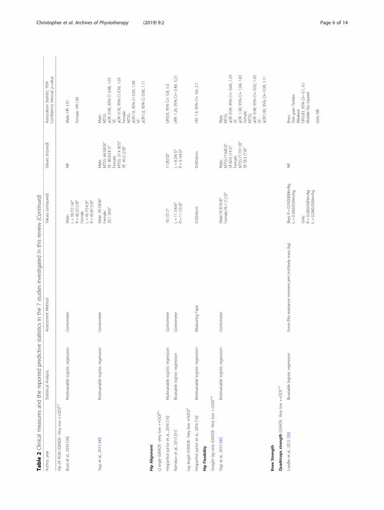
Objective musculoskeletal clinical assessments (Table 2)Hip strengthEvidence for hip strength was of very low quality (hip abduc-tion strength downgraded due to indirectness, inconsistency,and imprecision whereas the rest were downgraded due to in-directness and imprecision). Of the two studies investigatinghip abduction strength, one study [39] reported that strongerhip abduction strength was significantly associated with injuredrunners (OR=5.35, 95% CI= 1.46, 19.53) whereas the otherstudy [38] found no significant association. Finnoff et al. [39],also reported a significant protective association with increasedhip external rotation to internal rotation strength ratio RRI(OR=0.01, 95% CI= <0.01, 0.44). There were no significantassociations between hip adduction, abduction to adduc-tion ratio, external rotation, internal rotation, flexion, ex-tension, flexion-to-extension strength ratio and RRI [39].
Hip joint range of motionEvidence for hip joint range of motion was of very lowquality (downgraded due to indirectness and inconsist-ency). Two studies [36, 40] investigated hip internal andexternal range of motion, of which one study [40] foundthat increased hip internal rotation was protective againstRRI in females that developed medial tibial stress syn-drome (aOR = 0.91, 95% CI= 0.85, 0.99) [40].
Hip alignmentEvidence for hip alignment was of very low quality (Q angledowngraded for indirectness and inconsistency, and leglength downgraded for imprecision). Two studies [16, 40]investigated Q angle and one study [16] investigated leglength. The studies were unable to find significant relation-ships between hip alignment tests investigated and RRI.
Christopher et al. Archives of Physiotherapy (2019) 9:2 Page 3 of 14

Hip flexibilityEvidence for hip flexibility was of very low quality (down-graded for indirectness and imprecision). One study [40]investigated straight leg raise and did not find significant as-sociation between straight leg raise test and RRI.
Knee strengthEvidence for knee strength was of very low quality (down-graded for indirectness and imprecision). One study [38]
investigated knee strength using a HHD and did not finda significant association between quadriceps strength orhamstring strength and RRI.
Ankle alignmentEvidence for ankle alignment was of very low quality (navicu-lar drop downgraded for indirectness and inconsistency, andfoot posture index downgraded for indirectness and impreci-sion). Three studies [36, 37, 40] investigated navicular drop
Table 1 Quality assessment of included studies – adapted from the Critical Appraisal Form (CAT) for Quantitative Studies [28, 29]
Author I-1 I- 2 I-3 I-4 I-5 I- 6 I- 7 I-8 I-9 I-10 I-11 I-12 I-13 I-14 I-15 I-16 T.S T.%
Buist et al., 2010 [36] + + – + + + – + + – + + + + + + 13 81.25
Finnoff et al., 2011 [39] + + – + + + – + + – – + + + + + 12 75.0
Hespanhol Junior et al., 2016 [16] + + – + + + – + + – – + + + + + 12 75.0
Luedke et al., 2015 [38] + + – + + – + + + + – + – + – + 11 68.75
Plisky et al., 2007 [37] + + – + + + + + + + + + + + + + 15 93.75
Ramskov et al., 2013 [41] + + – – + + – + + – – + + + + + 11 68.75
Yagi et al., 2013 [40] + + – + + + – + + – – + + + + + 12 75.0
Note. Item 1: Purpose of the study was clearly stated, Item 2: Study design was appropriate, Item 3: Study detected sample bias, Item 4: Measurement biaseswere detected in the study, Item 5: Sample size was stated, Item 6: The sample was described in detail, Item 7: Sample size was justified, Item 8: Outcomeswere clearly stated and relevant, Item 9: Method of measurement was described sufficiently, Item 10: The measures used were reliable, Item 11: Themeasures used were valid, Item 12: The results were reported in terms of statistical significance, Item 13: The analysis methods used were appropriate, Item14: Clinical importance was reported, Item 15: Missing data were reported when appropriate, Item 16: Conclusions were relevant and appropriate givenmethods and results of the studyAbbreviations I- Item, T.S- total score, T%- total CAT %, meets criteria ‘+’, does not meet criteria ‘-’
Fig. 1 PRISMA flow diagram of studies in systematic review
Christopher et al. Archives of Physiotherapy (2019) 9:2 Page 4 of 14

Table
2Clinicalmeasuresandtherepo
rted
pred
ictivestatisticsin
the7stud
iesinvestigated
inthisreview
Autho
r,year
StatisticalAnalysis
Assessm
entMetho
dValues
(uninjured
)Values
(injured)
Associatio
nStatistic,95%
Con
fiden
ceInterval;p-value
Hip
Streng
th
Hip
abduc
tion
(GRA
DE-
Very
low
+++O)b,c,d
Finn
offet
al.,2011
[39]
Bivariablelogisticregression
(%BW
xheigh
t)=Torque(Nxm
)×100/[BW(N)xhe
ight(m
)]2.57(0.53)%
3.14(0.63)%
OR:5.35
,95%
CI=
1.46
,19.53
;p:<.01
Lued
keet
al.,2015
[38]
Bivariablelogisticregression
Force(N)xresistance
mom
entarm
(m)/bo
dymass(kg).
Boys:
R=0.25(0.07)
Nm/Kg
L=0.25(0.08)
Nm/Kg
Girls:
R=0.25(0.08)
Nm/Kg
L=0.26(0.07)
Nm/Kg
NR
Boys:Shinpain
tertiles
Weakest:OR:1.25,95%
C=I0.2,9.9.
Middle:OR1.00,N
AGirls:Shin
pain
tertiles
WeakestOR:1.23,95%
CI=
0.7,21.6,
Middle:OR2.28,95%
CI=
0.2,28.0
Hip
adduc
tion
(GRA
DE-
Very
low
++OO)c,d
Finn
offet
al.,2011
[39]
Bivariablelogisticregression
(%BW
xheigh
t)=Torque(Nxm
)×100/[BW(N)xhe
ight(m
)]2.79
(0.61)%
2.87
(0.45)%
OR:1.23,95%
CI=
0.48,3.17
Hip
abduc
tion
toad
duc
tion
ratio(GRA
DE-
Very
low
++O)c,d
Finn
offet
al.,2011
[39]
Bivariablelogisticregression
NR
1.12
(0.28)%
1.06
(0.25)%
OR:14.14,95%
CI=
0.90,221.06
Hip
internal
rotation
(GRA
DE-
Very
low
++OO)c,d
Finn
offet
al.,2011
[39]
Bivariablelogisticregression
(%BW
xheigh
t)=Torque(Nxm
)×100/[BW(N)xhe
ight(m
)]1.68
(0.40)%
1.88
(0.68)%
OR:2.75,95%
CI=
0.33,23.17
Hip
external
rotation
(GRA
DE-
Very
low
++OO)c,d
Finn
offet
al.,2011
[39]
Bivariablelogisticregression
(%BW
xheigh
t)=Torque(Nxm
)×100/[BW(N)xhe
ight(m
)]1.44
(0.31)%
1.34
(0.26)%
OR:0.35,95%
CI=
0.03,4.48
Hip
external
tointernal
rotation
streng
th(GRA
DE-
Very
low
++OO)c,d
Finn
offet
al.,2011
[39]
Bivariablelogisticregression
NR
0.87
(0.17)%
0.74
(0.13)%
OR:
0.01
,95%
CI=
<0.01
,0.44;p:0.02
Hip
flexion
(GRA
DE-
Very
low
++OO)c,d
Finn
offet
al.,2011
[39]
Bivariablelogisticregression
(%BW
xheigh
t)=Torque(Nxm
)×100/[BW(N)xhe
ight(m
)]2.84
(0.61)%
2.49
(0.92)%
OR:0.40,95%
CI=
0.05,3.09
Hip
extension(GRA
DE-
Very
low
++OO)c,d
Finn
offet
al.,2011
[39]
Bivariablelogisticregression
(%BW
xheigh
t)=Torque(Nxm
)×100/[BW(N)xhe
ight(m
)]3.15
(0.79)%
2.87
(0.79)%
OR:0.64,95%
CI=
0.21,1.90
Hip
flexion
toextensionstreng
th(GRA
DE-
Very
low
++OO)c,d
Finn
offet
al.,2011
[39]
Bivariablelogisticregression
NR
0.86
(0.15)%
0.96
(0.13)%
OR:0.17,95%
CI=
0.021,5.61
Hip
Rang
eof
Motion
Hip
IRRO
M(GRA
DE-
Very
low
++OO)b,c
Buistet
al.,2010
[36]
Multivariablelogisticregression
Gon
iometer
Male
L=30.6(8.1)°
R=31.1(8.8)°
Female
L=35.9(9.5)°
R=37.7(8.3)°
NR
Male:HR:1.00
FemaleHR0.98
aHR:
0.99
,95%
CI=
0.97
,1.01;
P:0.08
Yagi
etal.,2013
[40]
Multivariablelogisticregression
Gon
iometer
Male:12.4(8.7)°
Female:25.5(9.5)°
Male:
MTSS:12.9(5.8)°
SF:7.5(3.5)°
Female:
MTSS:31.1(9.9)°
SF:20.7(7.6)°
Male
MTSS:
aOR:0.99,95%
CI0.91,1.08
SF:
aOR:1.26,95%
CI0.81,1.96
Female
MTSS:
aOR0.91
,95%
CI0
.85,
0.99
;p:0.02
SF:
aOR:1.00,95%
CI0.88,1.12
Christopher et al. Archives of Physiotherapy (2019) 9:2 Page 5 of 14
Table
2Clinicalmeasuresandtherepo
rted
pred
ictivestatisticsin
the7stud
iesinvestigated
inthisreview
(Con
tinued)
Autho
r,year
StatisticalAnalysis
Assessm
entMetho
dValues
(uninjured
)Values
(injured)
Associatio
nStatistic,95%
Con
fiden
ceInterval;p-value
HipER
ROM
(GRA
DE-
Very
low
++OO)b,c
Buistet
al.,2010
[36]
Multivariablelogisticregression
Gon
iometer
Male:
L=39.7(11.6)°
R=40.2(12.9)°
Female
L=45.7(14.3)°
R=45.8(13.9)°
NR
Male:HR:1.01
Female:HR:1.00
Yagi
etal.,2013
[40]
Multivariablelogisticregression
Gon
iometer
Male:39.7(8.8)°
Female:
35.1(9.0)°
Male:
MTSS:44.5(8.9)°
SF:40.0(14.1)°
Female:
MTSS:37.4(8.5)°
SF:43.3(2.9)°
Male:
MTSS:
aOR:0.96,95%
CI0.88,1.03
SF:
aOR:0.76,95%
CI0.56,1.03
Female
MTSS:
aOR:1.0,95%
CI0.93,1.08
SF:
aOR:1.0,95%
CI0.90,1.11
Hip
Alig
nmen
t
Qan
gle(GRA
DE-
Very
low
++OO)b,c
Hespanh
oljunior
etal.,2016
[16]
Multivariablelogisticregression
Gon
iometer
10.1(5.1)°
11.8(5.0)°
OR:0.9,95%
CI=
0.8,1.0
Ramskov
etal.,2013
[41]
Bivariablelogisticregression
Gon
iometer
L=11.1(4.4)°
R=11.1(5.0)°
L=8.2(4.5)°
R=9.1(4.5)°
cRR:1.26,95%
CI=
0.49,3.23
Legleng
th(GRA
DE-
Very
low
+OOO)d
Hespanh
oljunior
etal.,2016
[16]
Multivariablelogisticregression
Measurin
gTape
0.5(0.6)cm
0.4(0.6)cm
OR:1.3,95%
CI=
0.6,2.7
Hip
Flexibility
Straight
legraise
(GRA
DE-
Very
low
++OO)c,d
Yagi
etal.,2013
[40]
Multivariablelogisticregression
Gon
iometer
Male:74.3(10.4)°
Female:76.1(12.5)°
Male:
MTSS:77.6(8.5)°
SF:60.0(14.1)°
Female:
MTSS:77.7(11.0)°
SF:78.3(7.6)°
Male
MTSS:
aOR:0.99,95%
CI=
0.60,1.29
SF:
aOR:1.38,95%
CI=
1.04,1.83
Female
MTSS:
aOR:0.98,95%
CI=
0.92,1.05
SF:
aOR:1.00,95%
CI=
0.90,1.11
Kne
eStreng
th
Qua
dricepsstreng
th(GRA
DE-
Very
low
++OO)c,d
Lued
keet
al.,2015
[38]
Bivariablelogisticregression
Force(N)xresistancemom
entarm
(m)/bo
dymass(kg).
Boys:R
=0.31(0.06)Nm/kg
L=0.30(0.05)Nm/kg
Girls:
R=0.28(0.04)Nm/kg
L=0.28(0.05)Nm/kg
NR
Boys:
Shin
pain
Tertiles
Weakest
OR:0.83,95%
CI=
0.1,6.1
Middle:Noinjured
Girls:NR
Christopher et al. Archives of Physiotherapy (2019) 9:2 Page 6 of 14

Table
2Clinicalmeasuresandtherepo
rted
pred
ictivestatisticsin
the7stud
iesinvestigated
inthisreview
(Con
tinued)
Autho
r,year
StatisticalAnalysis
Assessm
entMetho
dValues
(uninjured
)Values
(injured)
Associatio
nStatistic,95%
Con
fiden
ceInterval;p-value
Ham
string
streng
th(GRA
DE-
Very
low
++OO)c,d
Lued
keet
al.,2015
[38]
Bivariablelogisticregression
Force(N)xresistancemom
entarm
(m)/bo
dymass(kg).
Boys:
R=0.22(0.06)
Nm/kg
L=0.21(0.06)
Nm/kg
Girls:
R=0.20(0.03)
Nm/kg
L=0.20(0.04)
Nm/kg
NR
Boys:
Shin
pain
Tertiles
Weakest
OR:1.20,95%
CI=
0.2,8.8,Middle:
OR:0.40,95%
CI=
0.1,5.2
Girls:
Shin
pain
Tertiles
Weakest:O
R:1.33,95%
CI=
0.2,16.7
Middle:OR:0.55,95%
CI=
0.1,9.9
Ank
leAlig
nmen
t
Nav
icular
drop(GRA
DE-
Very
low
++OO)b,c
Buistet
al.,2010
[36]
Multivariablelogisticregression
NR
Male:
L=6.6(3.5)mm
R=6.7(3.5)mm
Female:
L=6.0(3.1)mm
R=6.2(2.8)mm
NR
MaleHR1.02
FemaleHR0.92
aHR-
0.87
,95%
CI=
0.77
,0.98;
p:0.01
Plisky
etal.,2007
[37]
Bivariablelogisticregression
Rulerpe
rpen
dicularto
thefloor
>10
mm
NBo
ys:20(43.5)
NGirls:2
4(40.7)
<10
mm
NBo
ys:26(56.5)
NGirls:2
5(59.3)
N15.8
N14.9
OR:1.0
OR:0.9,95%
CI=
0.3,2.8
Yagi
etal.,2013
[40]
Multivariablelogisticregression
Gon
iometer
Male:4.5(3.4)mm
Female:4.2(2.4)mm
Male
MTSS:4.9(3.0)
mm
SF:2.4(3.1)m
m
Female
MTSS:4.9(3.0)mm
SF:3.4(2.9)m
m
Male
MTSS:
aOR:0.93,95%
CI=
0.75,1.14
SF:
aOR:1.00,95%
CI=
0.71,1.42
Female
MTSS:
aOR:0.90,95%
CI=
0.70,1.19
SF:
aOR:1.5,95%
CI=
0.95,2.51
Foot
posture
index
(GRA
DE-
Very
low
++OO)c,d
Ramskov
etal.,2013
[41]
Bivariablelogisticregression
Metho
dby
Redm
ondet
alN:
Very
pron
ated
:1Pron
ated
:14
Neutral:79
Supinated:
0Very
supinated:
0
N:
Very
pron
ated
:3Pron
ated
:4Neutral:14
Supinated:
0Very
supinated:
0
cRR:1.65,95%
CI=
0.65,4.17
Christopher et al. Archives of Physiotherapy (2019) 9:2 Page 7 of 14

Table
2Clinicalmeasuresandtherepo
rted
pred
ictivestatisticsin
the7stud
iesinvestigated
inthisreview
(Con
tinued)
Autho
r,year
StatisticalAnalysis
Assessm
entMetho
dValues
(uninjured
)Values
(injured)
Associatio
nStatistic,95%
Con
fiden
ceInterval;p-value
Ank
leRa
ngeof
Motion
Ank
ledorsifle
xion
(GRA
DE-
Very
low
+OOO)c
Buistet
al.,2010
[36]
Multivariablelogisticregression
Gon
iometer
Male:
L= KB-104.7(7.8)°
KS-99.2(8.2)°
R= KB-104.6(7.5)°
KS-99.2(7.8)°
Female:
L= KB-103.6(11.5)°
KS-99.0(10.9)°
R= KB-103.8(8.7)°
KS-99.1(9.2)°
NR
Male
HR:1.01(KB)
HR:1.01
(KS)
Female
HR:1.00(KB)
HR:1.00
(KS)
ORod
dsratio
,aORad
justed
odds
ratio
,HRHazardratio
n,aH
Rad
justed
hazard
ratio
,RRriskratio
,cRR
cumulativerelativ
erisk,SF
stress
fracture,M
TSSmed
ialtibialstresssynd
rome,KB
knee
bent,K
Skn
eestraight
GRA
DEworking
grou
pgrad
esof
eviden
ce:(bo
lded
?he
adingforbe
low
items)
Low
quality
:Further
research
islikelyto
have
anim
portan
tim
pact
onou
rfin
ding
sVe
rylow
quality
:Weareun
certainab
outthefin
ding
sa.Ite
mwas
downg
rade
ddu
eto
riskof
bias
inmetho
ds,recruitm
ent,follow
upor
selectiverepo
rting
b.Ite
mwas
downg
rade
dueto
inconsistencysuch
asdifferen
cein
measuremen
tmetho
d,po
pulatio
n,injury
defin
ition
with
inthestud
iesinclud
edin
theou
tcom
ec.Ite
mwas
downg
rade
ddu
eto
indirectne
ssan
dthereforeap
plicab
ility
offin
ding
srega
rdingpo
pulatio
nor
outcom
esd.
Item
was
downg
rade
ddu
eto
imprecision(i.e.sm
allsam
plesize
<30
0)
Christopher et al. Archives of Physiotherapy (2019) 9:2 Page 8 of 14

and the development of running injuries. One study [36]found a significant protective relationship between decreasednavicular drop amount in females and injury (HR= 0.92); twostudies did not find a significant relationship between navicu-lar drop and injured runners. One study [41] investigated theFoot Posture Index [43] and did not find a significant rela-tionship between foot posture and injured runners.
Ankle joint range of motionEvidence for ankle range of motion was of very lowquality (downgraded for indirectness). One study [36] in-vestigated ankle dorsiflexion range of motion and didnot report a significant association between ankle dorsi-flexion (in knee straight and bent) and RRI.
DiscussionFindings within the studiesThe goal of this study was to summarize the results ofstand-alone studies that have investigated clinical assessmentand risk of injury. Synthesizing the work should improve anunderstanding of which factors may be transferable to aclinical environment. Stand-alone findings such as increasedhip external to internal rotation strength ratio and decreasednavicular drop were protective of injury, but only in a fewstudies. We also found that increased hip abduction strengthwas predictive of injury and decreased hip internal rotationwas protective of injury in runners, largely contradictingclinical thought and results from non-longitudinal studies ofassociation [44]. In no cases did we find compelling evi-dence from multiple studies of common predictors of injuryrisk in running. Also, all clinical assessment alteration cat-egories had very low quality of evidence; therefore, cliniciansshould be very cautious interpreting the results below.As stated, increased hip external to internal strength ratio
was seen to be protective for injury in runners that developedpatella femoral pain syndrome. This finding was reported inone study by Finnoff et al. [39] Although the authors did notoperationally define this ratio, it is assumed that an increasein hip external rotator strength when compared to internalrotator strength would be protective for runners. The hip ex-ternal rotators muscles control femoral internal rotation anda lack of control may be linked with running injury [45, 46].It is important to note there were several confounders in thisstudy. The study did not report running distance per week(mileage) nor did it report any injury history, both of whichhave been associated as risk factors for injury. Because theseathletes were high school runners, these factors could havesignificantly influenced results [1].Decreased navicular drop was seen to be protective of injury
in this review. This finding was reported in one study [36];however, it was not significant among the two other studies[25, 28] that did investigate this measure. Excessive pronationof the foot causes tibial rotation and has been seen to be re-lated to medial stress syndrome in runners [47]. This finding
was investigated in novice runners participating in a 13-weektraining program for a 4-mile running event and thereforecannot be applied to all running populations in general.Increased hip abduction strength was found to be predict-
ive of injury in one cohort study. The finding that runnerswith stronger hip abductors were more associated with RRImay have been due to a number of confounders. The partic-ipants included in the study were high school athletes, pos-sibly novice runners. As mentioned before, weekly trainingmileage and injury history were not reported. Finnoff et al.[39], theorized that the injured subjects in the group hadhigher body mass index (BMI), which could have led tohigher hip abduction moments. To compensate for theselarger moments, the runners may have developed increasedhip abductor (eccentric) strength over time [39]. This find-ing shows that some injured runners may have increasedstrength, specifically if they are younger or novice runnerswith a higher BMI. Caution should be used when interpret-ing this result with all running populations.Decreased hip internal rotation was found to be protect-
ive in one cohort study [40]. Excessive hip internal rota-tion has been associated with injury during jump landingtasks and lack of control of the lower extremity in thefrontal and transverse planes has also been hypothesizedas a cause for injury in runners [48, 49]. Decreased mobil-ity could therefore be beneficial and protective for run-ners, as it would require less neuromuscular control. Thisfinding shows that stiffness in runners may not be an im-pairment as previously thought [50, 51], specifically if theyare young and may not have developed the neuromuscularcontrol needed to stabilize the limb. Caution should beused while interpreting the findings of this study as partic-ipants were high school runners. Shin pain was the onlyinjury reported. Mileage of the runners was not reported;however, frequency of training was. Experience was notedas national, state, or entry level, however no history ofrunning injury or amount of running miles was reported.
Findings between the studiesThe GRADE level of evidence quality was very low for allobjective assessment alteration categories included in thisreview. Studies were downgraded for either indirectness, in-consistency, imprecision or all three. There were no com-mon predictors across a number of studies in this review.There may be several reasons for the lack of commonalityor the occasional findings that are contradictory to clinicalthought, such as differences in subject demographics, dif-ferent measurement methods, measurement variabilitywithin clinical assessments, inconsistent definitions of in-jury and runner, different statistical models, and study bias.These issues have been further addressed below.There were a wide range of different assessments used to
compare clinical assessment alterations and future injurywithin the seven prospective studies, and studies used
Christopher et al. Archives of Physiotherapy (2019) 9:2 Page 9 of 14
different methods when measuring the same construct. Forexample, ankle alignment was measured with naviculardrop [36, 37, 40] or Foot Posture Index [41]. This lack ofhomogeneity between studies resulted in difficulties com-paring clinical assessments between studies, even whenstudies focused on a similar construct (e.g., alignment).A variety of methods was used to define and report the
clinical assessments, even when the same testing devicewas used. For instance, weakness in hip HHD assessmentwas often reported by asymmetry between left and rightsides [39, 40]. However, another study [38] divided strengthinto three tertiles (weakest, middle and strong) across par-ticipants and used the strongest strength values as the com-parator. One study [38] multiplied the HHD reading by themoment arm and then normalized it to the participant’sbody mass. The other studies normalized HHD values tobody mass and height [39]. This variability in the reportingof muscle strength assessments made it difficult to comparestudies, perform meta-analyses, or identify common pat-terns of muscle strength in included prospective studies.Population and injury definitions were also heterogeneous
among studies. Running populations in studies varied fromnovice to recreational, with more males than females in theQ angle studies [13, 29]. Running related injury has been de-fined many ways in the literature, as evidenced by the widevariability of injury incidence rates reported in various stud-ies [1, 2, 52]. When defining an injury, studies used: 1) evalu-ation by a medical physician, athletic trainer or physicaltherapist [39], 2) presence of pain with duration of symp-toms > 24 h [37], 3) decrease in running mileage, 4) missedworkouts [16] or, 5) a combination of the variables listed [36,38, 40, 41] all which were included in our study. Consistentreporting about injury severity, the course of treatment, pre-vious injury, or whether the runner had sought assistancefrom a health care provider was lacking. Difference in levelsof injury severity would likely alter associational modelingand influence the statistical significance of the findings.Lastly, statistical modeling was different among studies.
Three studies used a multivariable model, whereas fourstudies used a bivariable model. Among the three studiesthat used a multivariable model, measures of independentvariables such as age [36], other clinical tests [16] and BMI[40] were also included in the regression analysis model.This could have influenced the relationship between singu-lar clinical test (such as navicular drop) [36] and RRI.Previous reviews investigating the risk of RRI have also
reported similar criticisms [53, 54]. Winter et al. [53] inves-tigated fatigue and RRI, and were unable to find conclusivepatterns of associations due to a lack of homogeneity of therunners, small sample sizes, and the distances that wererun to determine fatigue. A systematic review studying ver-tical ground reaction force and injury was also unable tomake recommendations due to a lack of prospective studiesinvestigating this variable and its association with injury
[54]. When reviewing biomechanical risk factors, Aderemand colleagues [29] concluded that shod female runnerswith iliotibial band syndrome (ITBS) may have associatedincreased peak knee internal rotation and peak hip adduc-tion during stance (based on one prospective cohort study),but because of limitations in effect size and the number ofstudies and methods, the authors did not make any add-itional recommendations. In the one review that investi-gated alterations to the musculoskeletal system, similar tothe current study, i.e., plantar pressures, the authors con-cluded there was inconsistency among studies and sug-gested improved methodology for future research [55].
LimitationsThere are several limitations to this review. Studies withpost-operative populations were excluded from the study, so itis possible the runners included in the selected studies had lesssevere injuries, which potentially influenced the clinical assess-ment alterations between baseline and future injury. This wasperformed to better generalize the results to the population ofrunners commonly seen in outpatient community-basedclinics, who often present without having seen a surgeon [56].
ConclusionThis review suggests that objective assessments that measurealterations in muscle strength, flexibility, alignment, and rangeof motion of the lower extremity had very low quality of evi-dence. Within the studies there were several confounderssuch as participant’s experience, unknown injury history, andunknown weekly running mileage, all of which have beenseen to be associated with RRI [1]. Among the studies, therewere a limited number of studies investigating each assess-ment, inconsistent results, different measurement methodsamong studies, measurement variability within clinical assess-ments, inconsistent definitions of injury and runner, differentstatistical modeling, and study bias. Future studies should aimto improve the quality of the studies as well as use standard-ized assessments and minimize confounders when conduct-ing clinical research to predict injury in runners.
Appendix 1Search terms used in PubMed databaseInjury[tiab] OR Injuries[tiab] OR “physiopathology” [Sub-
heading] OR “injuries” [Subheading] OR “Wounds andInjuries”[Mesh]) AND (Runner[tiab] OR Runners[tiab] ORRunning) AND (Muscle Strength OR Muscle WeaknessOR Strength[tiab] OR Weakness[tiab]) AND (sensitive[tiab]OR sensitivity[tiab] OR specificity[tiab] OR sensitivity andspecificity[MeSH] OR diagnosis[tiab] OR diagnostic[tiab]OR diagnosed[tiab] OR diagnosis[MeSH] OR diagnosis[sh]OR cross-sectional studies[Mesh] OR cross-sectional[tiab])NOT (review[ptyp] OR Editorial[ptyp] OR Letter[ptyp]OR Case Reports[ptyp] OR Comment[ptyp]) NOT (ani-mals[mh] NOT humans[mh]
Christopher et al. Archives of Physiotherapy (2019) 9:2 Page 10 of 14

Appendix 2Table 3 PEO (Population, Exposure, Observation) Table; description of included articles
Author, Year ofpublication
PopulationN (gender)Follow up
Exposure (Clinical Measure) Observation (Injury Definition)
Buist et al., 2010 [36] 532 novice runners(226 male, 306 female);8 or 13-week program
Range of motion with universal goniometer:Internal and external ROM of the hip: assessed insupine and the tested hip and knee flexed to 90°Ankle dorsal flexion- measured both with theknee fully extended and flexed to 90° passively,in supine position.Alignment: Navicular drop- assessed bymeasuring the change in the height of thenavicular tuberosity between sitting with thesubtalar joint in neutral position and standing,weight-bearing with the subtalar joint in relaxedstance, measurements were made twice for eachfoot, results were averaged
Self-reported musculoskeletalpain of the lower extremity orback causing a restriction ofrunning for at least 1 week, i.e.3 scheduled consecutivetraining sessions.
Finnoff et al., 2011 [39] 98 high school cross countryand track athletes(53 male and 45 female);Cross country and/or trackseason
Leg Length- measuring from anterior superioriliac spine (ASIS) to a point 2 cm proximal to theapex of medial malleolusMuscle strength with HHD for break test:Hip flexion- seated hip flexion to 120° with HHDon distal aspect of thighHip Extension- extend test hip to a neutralposition with the knee extended whilemaintaining neutral hip rotation with HHDagainst the subject’s posterior calcaneusHip External Rotation- seated knees were alsoflexed 90° with the hip in neutral rotation withHHD positioned 2 cm proximal to the apex ofthe medial malleolusHip Internal rotation- position identical to theone used for hip external rotation strengthtesting with HHD positioned 2 cm proximal tothe apex of the lateral malleolusHip Abduction- 30° abduction with neutral hipflexion, extension, rotation) HHD positioned 2 cmproximal to the apex of lateral malleolusHip Adduction- neutral flexion, extension,rotation (subject allowed to grasp table for trunkstability). Strength test was performed with theHHD placed 2 cm proximal to the medial malleolusPain- Visual Analogue Scale (10 cm)
ATC monitored and evaluatedby physician investigators:ITBS suspected with lateral kneepain, local tenderness overlateral knee where ITB crossesover condyle, exacerbated byflexion and extension whileapplying pressurePFP suspected with anteriorknee pain, exacerbated bydeep knee flexion and/orclimbing stairs, and byreproduction of pain with atleast one of following: 1)pressure over distal quadricepswith active contraction and 2)direct palpation of medial andlateral patellar facets
Hespanhol Junior et al.,2016 [16]
89 recreational runners (68male/21 female);12 weeks
Leg Length: in a supine position, lower limbsrelaxed. Measuring tape was used to determinethe real length of the lower limbs i.e., the lengthbetween the ASIS of the hemipelvis to the centerof the ipsilateral medial malleolus of both lowerlimbs. The lower limb length discrepancy wasconsidered normal when lower than 1.0 cmQ-angle: In sports clothes and standing barefoot inan orthostatic position. A straight line was tracedusing a ruler from the ASIS to the center of thepatella, and a second line was traced from the centerof the patella to the tibial tuberosity. The angleformed by the intersection of these two linesconstitutes the Q-angle, which was measured by auniversal goniometer. Values between 10° and 15°were considered normal for both genders
Missed at least one trainingsession due to musculoskeletalpain(Biweekly questionnairereporting musculoskeletal pain,number of training sessionsmissed, pain intensity (10 pointnumerical pain rating scale),description (type and anatomicallocation) of new injury)
Luedke et al., 2015 [38] 68 High school runners (16male, 47 female);Interscholastic cross-countryseason
Muscle strength with HHD for bilateral peakisometric strength (2 trials):Hip abduction- sidelying, non-test limb waspositioned in 30–45° of hip flexion and 90° of kneeflexion, pelvis was stabilized to the table using astrap, test hip was in 0° of extension and abductedto parallel with the table and HHD was placed justproximal to the lateral malleolus on the test limb
Injury- required athlete to beremoved from practice orcompetitive event, or miss asubsequent practice/competitive eventPT or LAT determine injury:Knee pain 1. Pain around antaspect of knee 2) insidious
Christopher et al. Archives of Physiotherapy (2019) 9:2 Page 11 of 14

Table 3 PEO (Population, Exposure, Observation) Table; description of included articles (Continued)
Author, Year ofpublication
PopulationN (gender)Follow up
Exposure (Clinical Measure) Observation (Injury Definition)
Knee Extension: seated at the end of a table withthe test knee at 45° of flexion, stabilizing strapwas placed around the thighs and table,resistance applied to the anterior aspect of thetibia 5 cm proximal to the ankle jointKnee Flexion - prone and the test knee flexed to45°, stabilizing strap was placed around the pelvisand table with resistance applied to the posterioraspect of the tibia 5 cm proximal to the ankle joint
onset 3) no incidence oftraumaShin injury 1) continuous orintermittent shin pain 2)exacerbated by weightbearing activities 3) local painwith palpation along tibia
Plisky et al., 2007 [37] 105 high school cross countryrunners (59 male, 46 female);13 week cross country season
Alignment:Navicular drop (normalized to full foot lengthand truncated foot length) - in unilateralstanding position, the runner’s foot placedsubtalar neutral, ruler was placed next to themedial foot perpendicular to the floor and wasread (mm) at the height of the navicular tubercle,2 measurements were recorded, relaxing inbetween, and the difference value wasdocumented as navicular drop (Runners wereallowed to maintain their balance by placing ahand on a handrail during unilateral stance)
PT and ATC examined runnerfor MTSS criteria 1) continuousor intermittent pain in thetibial region, exacerbated byweight bearing activities 2)local pain with palpationalong distal 2/3 of posteriormedial tibia
Ramskov et al., 2013 [41] 59 novice runners(31 male, 28 female);10 weeks
Alignment: Foot Posture Index [43].Q angle- center of the goniometer placed uponthe middle of the patella, one arm of thegoniometer placed along the line connectingASIS with the middle of patella, other arm wasplaced along the line connecting the middle ofpatella and the tibial tuberosity
Injury: Any running-relatedinjury to lower extremity orlower back that causes at leastone week of restricted runningDiagnoses by physiotherapist~ 1 week after injury; ifextensive exam neededreferred to university hospitalmedical center division ofsports traumatology
Yagi et al., 2013 [40] 230 high school runners (134male, 96 females); 3 years
Range of motion:Hip rotation- measured with the hip and kneeflexed at 90° in the sitting position; the hip and kneewere rotated internally and externally to firm endfeel with the angles relative to the initial position.Ankle dorsiflexion-measure in two positions withknee in extension and 90° flexion; ankle was passivelymoved into dorsiflexion from a neutral-startingposition until a firm end feel was elicited (examinerfirst identified the neutral position of the subtalar jointand then kept the neutral position while dorsiflexingthe foot until a firm end point was felt)Flexibility:Straight leg raising – supine, passively into hipflexion until firm resistance was felt and thepelvis tilted posteriorlyAlignment (knee varus or valgus and ankleeversion inversion in standing closed feet),Navicular drop test-distance between the naviculartuberosity and the floor during [1] quiet tandemstance with the subtalar joint placed in neutral,and no load on the foot, and [2] relaxed tandemstance with full load on the footQ angle- center axis of a long-arm goniometerplaced over the center of the patella, proximal tibiawas palpated, and the lower goniometer arm wasaligned along the patellar tendon to the tibialtubercle, upper arm of the goniometer was pointeddirectly at the anterior superior iliac spineStrength: Hip abduction isometric break test withHHD
Could not run for 7 days due toshin pain - radiographs taken (ifreinjured counted in study asadditional subject) anddiagnosis by sports physician
NR not reported, m/wk. miles per week, yr. year, ROM range of motion, HHD Hand held dynamometer, MTSS medial tibial stress syndrome, SF stress fracture
Christopher et al. Archives of Physiotherapy (2019) 9:2 Page 12 of 14

AbbreviationsaOR: Adjusted odds ratio; aRR: Adjusted risk ratio; HHD: Hand held Dynamometer;HR: Hazard ratio; OR: Odds ratio; RR: Risk Ratio; RRI: Running-related Injury
AcknowledgementsThe authors would like to thank Leila Ledbetter (biomedical librarian) for herhelp with the literature search.
FundingThis research did not receive any specific grant from funding agencies in thepublic, commercial, or not-for-profit sectors.
Availability of data and materialsNot applicable.
Declaration of interestsThe authors declare that there is no conflict of interest.
IRBNone
Authors' contributionsSMC provided idea, design, writing, review of manuscript and overall contentof material; JM provided review of articles and quality tool; SS and CCprovided writing, review and overall content of manuscript. All authors readand approved final manuscript.
Authors informationShefali Christopher has been a sports physical therapist for 10 years. As aclinician, she predominantly treated runners and used musculoskeletal clinicalassessments to evaluate and treat injured runners. As part of her PhD, from theUniversity of Newcastle in Australia, she wanted to investigate the utility of thetests she was using and see if they had any predictive capability.
Ethics approval and consent to participateNot applicable.
Consent for publicationNot applicable.
Competing interestsThe authors declare that they have no competing interests.
Publisher’s NoteSpringer Nature remains neutral with regard to jurisdictional claims inpublished maps and institutional affiliations.
Author details1Department of physical therapy Education, Elon University, Elon, NC 27244, USA.2School of Health Sciences, The University of Newcastle, Callaghan, Australia.3Pivot Physical Therapy, Culpeper, VA 22701, USA. 4Division of Physical Therapy,Duke University, 2200 W. Main Street, Durham, NC 27705, USA.
Received: 25 July 2018 Accepted: 20 January 2019
References1. van Gent RN, Siem D, van Middelkoop M, van Os AG, Bierma-Zeinstra SM, Koes
BW. Incidence and determinants of lower extremity running injuries in longdistance runners: a systematic review. Br J Sports Med. 2007;41(8):469–80.
2. Reinking MF. Exercise-related leg pain in female collegiate athletes: theinfluence of intrinsic and extrinsic factors. Am J Sports Med. 2006;34(9):1500–7.
3. Lopes AD, Hespanhol Júnior LC, Yeung SS, Costa LO. What are the mainrunning-related musculoskeletal injuries? A Systematic Review Sports Med.2012;42(10):891–905.
4. Barton CJ, Bonanno DR, Carr J, Neal BS, Malliaras P, Franklyn-Miller A,et al. Running retraining to treat lower limb injuries: a mixed-methodsstudy of current evidence synthesised with expert opinion. Br J SportsMed. 2016.
5. Malisoux L, Nielsen RO, Urhausen A, Theisen D. A step towards understandingthe mechanisms of running-related injuries. J Sci Med Sport. 2015;18(5):523–8.
6. Hulme A, Thompson J, Nielsen RO, Read GJM, Salmon PM. Towards a complexsystems approach in sports injury research: simulating running-related injurydevelopment with agent-based modelling. Br J Sports Med. 2018. https://doi.org/10.1136/bjsports-2017-098871. [Epub ahead of print] Review. PubMedPMID: 2991512.
7. Damsted C, Glad S, Nielsen RO, Sorensen H, Malisoux L. Is there evidencefor an association between changes in training load and running-relatedinjuries? A systematic review. Int J Sports Phys Ther. 2018;13(6):931–42.
8. Messier SP, Edwards DG, Martin DF, Lowery RB, Cannon DW, James MK, etal. Etiology of iliotibial band friction syndrome in distance runners. Med SciSports Exerc. 1995;27(7):951–60.
9. Noehren B, Davis I, Hamill J. ASB clinical biomechanics award winner 2006prospective study of the biomechanical factors associated with iliotibialband syndrome. Clin Biomech (Bristol, Avon). 2007;22(9):951–6.
10. Ferber R, Noehren B, Hamill J, Davis IS. Competitive female runners with ahistory of iliotibial band syndrome demonstrate atypical hip and kneekinematics. J Orthop Sports Phys Ther. 2010;40(2):52–8.
11. Phinyomark A, Osis S, Hettinga BA, Leigh R, Ferber R. Gender differences ingait kinematics in runners with iliotibial band syndrome. Scand J Med SciSports. 2015;25(6):744–53.
12. Foch E, Milner CE. Frontal plane running biomechanics in female runnerswith previous iliotibial band syndrome. J Appl Biomech. 2014;30(1):58–65.
13. Foch E, Milner CE. The influence of iliotibial band syndrome history on runningbiomechanics examined via principal components analysis. J Biomech. 2014;47(1):81–6.
14. Foch E, Reinbolt JA, Zhang S, Fitzhugh EC, Milner CE. Associations betweeniliotibial band injury status and running biomechanics in women. GaitPosture. 2015;41(2):706–10.
15. Grau S, Krauss I, Maiwald C, Axmann D, Horstmann T, Best R. Kinematicclassification of iliotibial band syndrome in runners. Scand J Med Sci Sports.2011;21(2):184–9.
16. Hespanhol Junior LC, de Carvalho AC, Costa LO, Lopes AD. Lower limbalignment characteristics are not associated with running injuries in runners:prospective cohort study. Eur J Sport Sci. 2016;16(8):1137–44.
17. Meininger AK, Koh JL. Evaluation of the injured runner. Clin Sports Med.2012;31(2):203–15.
18. Plastaras CT, Rittenberg JD, Rittenberg KE, Press J, Akuthota V.Comprehensive functional evaluation of the injured runner. Phys MedRehabil Clin N Am. 2005;16(3):623–49.
19. O'Connor FG, Wilder RP. Evaluation of the injured runner. J BackMusculoskelet Rehabil. 1995;5(4):281–94.
20. Moher D, Liberati A, Tetzlaff J, Altman DG. Preferred reporting items forsystematic reviews and meta-analyses: the PRISMA statement. PLoS Med.2009;6(7):e1000097.
21. Shmueli G. To explain or predict. Stat Sci. 2010;25(3):289–310.22. Szumilas M. Explaining odds ratios. J Can Acad Child Adolesc Psychiatry.
2010;19(3):227–9.23. Spruance SL, Reid JE, Grace M, Samore M. Hazard ratio in clinical trials.
Antimicrob Agents Chemother. 2004;48(8):2787–92.24. Zhang J, Yu KF. What's the relative risk? A method of correcting the odds
ratio in cohort studies of common outcomes. JAMA. 1998;280(19):1690–1.25. Maykut JN, Taylor-Haas JA, Paterno MV, DiCesare CA, Ford KR. Concurrent
validity and reliability of 2d kinematic analysis of frontal plane motionduring running. Int J Sports Phys Ther. 2015;10(2):136–46.
26. Creaby MW, Le Rossignol S, Conway ZJ, Ageberg E, Sweeney M,Franettovich Smith MM. Frontal plane kinematics predict three-dimensionalhip adduction during running. Phys Ther Sport. 2017;27:1–6.
27. Dingenen B, Staes FF, Santermans L, Steurs L, Eerdekens M, Geentjens J, et al. Aretwo-dimensional measured frontal plane angles related to three-dimensionalmeasured kinematic profiles during running? Phys Ther Sport. 2018;29:84–92.
28. Law M, Stewart D, Letts L, Pollock N, Bosch J, Westmorland M. Guidelinesfor critical review of qualitative studies. McMaster University occupationaltherapy evidence-based practice research Group; 1998.
29. Aderem J, Louw QA. Biomechanical risk factors associated with iliotibialband syndrome in runners: a systematic review. BMC Musculoskelet Disord.2015;16:356.
30. Guyatt G, Oxman AD, Akl EA, Kunz R, Vist G, Brozek J, et al. GRADEguidelines: 1. Introduction-GRADE evidence profiles and summary offindings tables. J Clin Epidemiol. 2011;64(4):383–94.
Christopher et al. Archives of Physiotherapy (2019) 9:2 Page 13 of 14
31. Ryan R, Hill S. How to GRADE the quality of the evidence. CochraneConsumers and Communication Group, available at http://cccrg.cochrane.org/author-resources. Version 3.0. December 2016. Accessed 1 Dec 2018.
32. Guyatt GH, Oxman AD, Kunz R, Woodcock J, Brozek J, Helfand M, et al.GRADE guidelines: 7. Rating the quality of evidence--inconsistency. J ClinEpidemiol. 2011;64(12):1294–302.
33. Guyatt GH, Oxman AD, Kunz R, Woodcock J, Brozek J, Helfand M, et al.GRADE guidelines: 8. Rating the quality of evidence--indirectness. J ClinEpidemiol. 2011;64(12):1303–10.
34. Guyatt GH, Oxman AD, Kunz R, Brozek J, Alonso-Coello P, Rind D, et al.GRADE guidelines 6. Rating the quality of evidence--imprecision. J ClinEpidemiol. 2011;64(12):1283–93.
35. Guyatt GH, Oxman AD, Montori V, Vist G, Kunz R, Brozek J, et al. GRADEguidelines: 5. Rating the quality of evidence--publication bias. J ClinEpidemiol. 2011;64(12):1277–82.
36. Buist I, Bredeweg SW, Lemmink KA, van Mechelen W, Diercks RL. Predictors ofrunning-related injuries in novice runners enrolled in a systematic trainingprogram: a prospective cohort study. Am J Sports Med. 2010;38(2):273–80.
37. Plisky MS, Rauh MJ, Heiderscheit B, Underwood FB, Tank RT. Medial tibialstress syndrome in high school cross-country runners: incidence and riskfactors. J Orthop Sports Phys Ther. 2007;37(2):40–7.
38. Luedke LE, Heiderscheit BC, Williams DS, Rauh MJ. Association of isomentricstrength of hip and knee muscles with injury risk in high school crosscountry runners. Int J Sports Phys Ther. 2015;10(6):868–76.
39. Finnoff JT, Hall MM, Kyle K, Krause DA, Lai J, Smith J. Hip strength and kneepain in high school runners: a prospective study. PM and R. 2011;3(9):792–801.
40. Yagi S, Muneta T, Sekiya I. Incidence and risk factors for medial tibial stresssyndrome and tibial stress fracture in high school runners. Knee Surg SportsTraumatol Arthrosc. 2013;21(3):556–63.
41. Ramskov D, Jensen ML, Obling K, Nielsen RO, Parner ET, Rasmussen S. Noassociation between q-angle and foot posture with running-related injuries: a10 week prospective follow-up study. Int J Sports Phys Ther. 2013;8(4):407–15.
42. Guyatt GH, Oxman AD, Vist G, Kunz R, Brozek J, Alonso-Coello P, et al.GRADE guidelines: 4. Rating the quality of evidence--study limitations (riskof bias). J Clin Epidemiol. 2011;64(4):407–15.
43. Redmond AC, Crosbie J, Ouvrier RA. Development and validation of a novelrating system for scoring standing foot posture: the foot posture index. ClinBiomech (Bristol, Avon). 2006;21(1):89–98.
44. Mucha MD, Caldwell W, Schlueter EL, Walters C, Hassen A. Hip abductorstrength and lower extremity running related injury in distance runners: asystematic review. J Sci Med Sport. 2016.
45. Ireland ML, Willson JD, Ballantyne BT, Davis IM. Hip strength in females withand without patellofemoral pain. J Orthop Sports Phys Ther. 2003;33(11):671–6.
46. Mizuno Y, Kumagai M, Mattessich SM, Elias JJ, Ramrattan N, Cosgarea AJ, etal. Q-angle influences tibiofemoral and patellofemoral kinematics. J OrthopRes. 2001;19(5):834–40.
47. Loudon JK, Jenkins W, Loudon KL. The relationship between static posture andACL injury in female athletes. J Orthop Sports Phys Ther. 1996;24(2):91–7.
48. Souza RB, Powers CM. Predictors of hip internal rotation during running: anevaluation of hip strength and femoral structure in women with andwithout patellofemoral pain. Am J Sports Med. 2009;37(3):579–87.
49. Snyder KR, Earl JE, O'Connor KM, Ebersole KT. Resistance training isaccompanied by increases in hip strength and changes in lower extremitybiomechanics during running. Clin Biomech (Bristol, Avon). 2009;24(1):26–34.
50. Boutris N, Byrne RA, Delgado DA, Hewett TE, McCulloch PC, Lintner DM,Harris JD. Is there an association between noncontact anterior cruciateligament injuries and decreased hip internal rotation or radiographicFemoroacetabular impingement? A Systematic Review Arthroscopy. 2018;34(3):943-50.
51. VandenBerg C, Crawford EA, Sibilsky Enselman E, Robbins CB, Wojtys EM,Bedi A. Restricted hip rotation is correlated with an increased risk foranterior cruciate ligament injury. Arthroscopy. 2017;33(2):317–25.
52. Yamato TP, Saragiotto BT, Lopes AD. A consensus definition of running-related injury in recreational runners: a modified Delphi approach. J OrthopSports Phys Ther. 2015;45(5):375–80.
53. Winter S, Gordon S, Watt K. Effects of fatigue on kinematics and kinetics duringoverground running: a systematic review. J Sports Med Phys Fitness. 2017;57(6):887-99.
54. van der Worp H, Vrielink JW, Bredeweg SW. Do runners who suffer injurieshave higher vertical ground reaction forces than those who remain injury-free?A systematic review and meta-analysis. Br J Sports Med. 2016;50(8):450–7.
55. Mann R, Malisoux L, Urhausen A, Meijer K, Theisen D. Plantar pressuremeasurements and running-related injury: a systematic review of methodsand possible associations. Gait Posture. 2016;47:1–9.
56. Hodges PW, Richardson CA. Inefficient muscular stabilization of the lumbarspine associated with low back pain. A motor control evaluation oftransversus abdominis. Spine (Phila Pa 1976). 1996;21(22):2640–50.
Christopher et al. Archives of Physiotherapy (2019) 9:2 Page 14 of 14