dnasequencesproximaltohumanmitochondrialdna ... · ... cause genome instability by perturbing...
TRANSCRIPT
DNA Sequences Proximal to Human Mitochondrial DNADeletion Breakpoints Prevalent in Human Disease FormG-quadruplexes, a Class of DNA Structures InefficientlyUnwound by the Mitochondrial Replicative Twinkle Helicase*
Received for publication, March 19, 2014, and in revised form, August 18, 2014 Published, JBC Papers in Press, September 5, 2014, DOI 10.1074/jbc.M114.567073
Sanjay Kumar Bharti‡, Joshua A. Sommers‡, Jun Zhou§¶1, Daniel L. Kaplan�, Johannes N. Spelbrink**‡‡2,Jean-Louis Mergny§¶3, and Robert M. Brosh, Jr.‡4
From the ‡Laboratory of Molecular Gerontology, NIA, National Institutes of Health, NIH Biomedical Research Center, Baltimore,Maryland 21224, the §ARNA Laboratory, University of Bordeaux, F-33000 Bordeaux, France, ¶INSERM U869, Institut Europeen deChimie et Biologie (IECB), F-33600 Pessac, France, the �Department of Biomedical Sciences, Florida State University College ofMedicine, Tallahassee, Florida 32312, the **FinMIT Centre of Excellence, BioMediTech and Tampere University Hospital, PirkanmaaHospital District, University of Tampere, FI-33014 Tampere, Finland, and the ‡‡Department of Pediatrics, Nijmegan Centre forMitochondrial Disorders, Radboud University Medical Centre, Geert Grooteplein 10, P. O. Box 9101, 6500 HB Nijmegen, The Netherlands
Background: Mitochondrial DNA deletions are prominent in human genetic disorders and cancer.Results: Predicted mitochondrial G-quadruplex-forming sequences map in close proximity to known deletion breakpoints andform G-quadruplexes in vitro.Conclusion: The mitochondrial replicative helicase Twinkle inefficiently unwinds intra- and intermolecular G-quadruplexes.Significance: Mitochondrial G-quadruplexes are likely to cause genome instability by perturbing replication machinery.
Mitochondrial DNA deletions are prominent in humangenetic disorders, cancer, and aging. It is thought that stalling ofthe mitochondrial replication machinery during DNA synthesisis a prominent source of mitochondrial genome instability; how-ever, the precise molecular determinants of defective mitochon-drial replication are not well understood. In this work, we per-formed a computational analysis of the human mitochondrialgenome using the “Pattern Finder” G-quadruplex (G4) predic-tor algorithm to assess whether G4-forming sequences reside inclose proximity (within 20 base pairs) to known mitochondrialDNA deletion breakpoints. We then used this information tomap G4P sequences with deletions characteristic of representa-tive mitochondrial genetic disorders and also those identified invarious cancers and aging. Circular dichroism and UV spectralanalysis demonstrated that mitochondrial G-rich sequencesnear deletion breakpoints prevalent in human disease formG-quadruplex DNA structures. A biochemical analysis of puri-fied recombinant human Twinkle protein (gene product ofc10orf2) showed that the mitochondrial replicative helicaseinefficiently unwinds well characterized intermolecular andintramolecular G-quadruplex DNA substrates, as well as a uni-
molecular G4 substrate derived from a mitochondrial sequencethat nests a deletion breakpoint described in human renal cellcarcinoma. Although G4 has been implicated in the initiation ofmitochondrial DNA replication, our current findings suggestthat mitochondrial G-quadruplexes are also likely to be a sourceof instability for the mitochondrial genome by perturbing thenormal progression of the mitochondrial replication machin-ery, including DNA unwinding by Twinkle helicase.
The human mitochondrial genome consists of a 16,569-bpcircular DNA molecule that encodes essential subunits of mito-chondrial complexes I, II, III, IV, and V in oxidative phosphor-ylation, a process responsible for the generation of the majorityof the cellular ATP pool (1). Therefore, energy production isintimately linked to an intact and correctly coding mitochon-drial DNA (mtDNA) sequence. Because of the utmost impor-tance of the mitochondrion, it naturally follows that the accu-mulation of mitochondrial DNA mutations leading tomitochondrial dysfunction are implicated in human geneticdisorders (2), neurodegeneration (3), cancer (4), and aging (5).Understanding the molecular basis for mitochondrial DNAmutations has attracted considerable interest in the clinical andbasic science fields.
mtDNA deletions are prominent in genetic disorders such asPearson marrow-pancreas syndrome (PMPS),5 Kearns-Sayresyndrome (KSS), and progressive external ophthalmoplegia
* This research was supported, in whole or in part, by the National Institutesof Health, NIA, Intramural Research Program.
1 Recipient of a European Union Marie Curie International Incoming Fellow-ship (IIF).
2 Supported by the Academy of Finland (Centre of Excellence funding) andthe Netherlands Organization for Scientific Research (NWO), VICI Grant865.10.004.
3 Supported by Fondation ARC, the Aquitaine Regional Council, and theAgence Nationale de la Recherche (ANR Grants Quarpdiem andOligoswitch).
4 To whom correspondence should be addressed: Laboratory of MolecularGerontology, National Institutes of Health, NIA, NIH Biomedical ResearchCenter, 251 Bayview Blvd., Baltimore, MD 21224. Tel.: 410-558-8578; Fax:410-558-8162; E-mail: [email protected].
5 The abbreviations used are: PMPS, Pearson marrow-pancreas syndrome;CSBII, conserved sequence block II; FANCJ, Fanconi anemia complementa-tion group J; G4, G-quadruplex; G4P, potential G4-forming (sequence); H,heavy; L, light; HRCC, human renal cell carcinoma; KSS, Kearns-Sayre syn-drome; PEO, progressive external ophthalmoplegia; PNA, peptide-nucleic acid;ssDNA, single-stranded DNA; ATP�S, adenosine 5�-O-(thiotriphosphate).
THE JOURNAL OF BIOLOGICAL CHEMISTRY VOL. 289, NO. 43, pp. 29975–29993, October 24, 2014Published in the U.S.A.
OCTOBER 24, 2014 • VOLUME 289 • NUMBER 43 JOURNAL OF BIOLOGICAL CHEMISTRY 29975
by guest on June 11, 2018http://w
ww
.jbc.org/D
ownloaded from
(PEO) as well as various cancers and age-related diseases (6).mtDNA deletions are often located in regions of the genomeflanked by tandem repeat sequences. It is proposed that at leastone mechanism likely to underlie the mitochondrial genomicinstability that leads to deletions is the stalling of the mitochon-drial DNA synthesis machinery during replication. Mitochon-drial replication stalling can lead to replication slippage, aber-rant DNA structures, and double strand breaks, which can inturn promote mtDNA deletions. Although this model is attrac-tive, the molecular determinants of mitochondrial replicationstalling are not well understood.
The mitochondrial genome consists of a heavy (H) strand,which is rich in guanine bases, and a complementary light (L)strand, which is rich in cytosine bases. Because of its chemicalnature, the guanine-rich (G-rich) strand is prone to formG-quadruplex (G4) DNA, an alternative DNA structure char-acterized by planar stacks of four guanines interacting by non-conventional Hoogsteen hydrogen bonds. G4 DNA is stabilizedby a monovalent cation (typically Na� or K�) that resides in thecentral barrel of the G-tetrad. There has been growing interestin nuclear G-quadruplex DNA metabolism in recent years withincreasing evidence that G4 exists in vivo (7, 8) (for review seeRefs. 9 –11). However, it has been only very recently thatresearchers have begun to focus on the mitochondrial genomeand its propensity to form G-quadruplexes or other alternateDNA structures. The Zakian laboratory (12) performed the firstanalysis of G4 DNA motifs in yeast mitochondria, which indi-cates a 10-fold greater density of G4 motifs in mitochondrialversus nuclear genomes of Saccharomyces cerevisiae. Damas etal. (13) conducted a computational analysis of human mtDNAdeletions and correlated these with non-B DNA conformationsincluding hairpin, cruciform, and cloverleaf-like elements.Their findings suggest that non-B form conformations arelikely to contribute to the formation of mitochondrial deletions.In 2013, Oliveira et al. (14) described their findings from meta-analysis of human mtDNA sequence leading them to proposethat noncanonical DNA structures, including helix-distortingintrinsically curved regions and long G-quadruplex elements,are likely to drive mtDNA rearrangements.
In the current study, we employed “Pattern Finder,” a G4predictor algorithm, to map potential G4-forming (G4P)sequences throughout the human mitochondrial genome andchart them to known mitochondrial deletion breakpoints. Weutilized this information to correlate G4P sequences with dele-tions characteristic of representative mitochondrial geneticdisorders and also those identified in various cancers and aging.We performed physical analysis of representative predictedG4-forming sequences residing adjacent to clinically relevantmitochondrial deletions to substantiate that they do indeedform G-quadruplexes. Finally, we tested G-quadruplex DNAsubstrates with various topologies including a unimolecular G4derived from human mitochondrial sequence for unwinding bythe human mitochondrial DNA helicase Twinkle. Our resultsprovide insight into the hypothesis that mitochondrial G-qua-druplexes are likely to be a source of instability for the mito-chondrial genome by perturbing the normal progression of themitochondrial replication machinery.
EXPERIMENTAL PROCEDURES
Mitochondrial G-quadruplex Motif Prediction and Correlationwith Mitochondrial Deletions—Mitochondrial G4P sequenceswere searched using Pattern Finder software to predict G4 in auser-defined quadruplex pattern for a given input sequence(15). Human mtDNA sequences used for analysis were takenfrom accession number NC_012920 (formerly, AC_000021).Mitochondrial L-strand (C-rich) and H-strand (G-rich) DNAwas subjected to Pattern Finder analysis to assign G4 motifswith a stem size of 2–5 nucleotides (nt) and loop size of 1–7 nt.The mitochondrial deletion breakpoints are reported based onL-strand numbering. The mitochondrial deletion data wereobtained from MitoMap and MitoBreak.
Circular Dichroism Spectroscopy, UV Melting Curves, andThermal Difference Spectra—Oligonucleotides used to prepareG4 DNA substrates for biophysical analysis were purchasedfrom Eurogentec (Seraing, Belgium) without further purifica-tion. The oligonucleotides derived from human mitochondrialsequence were: KSS, 5�-GGGGAGGGGTGTTTAAGGGGTT-GGCTAGGG-3�; human renal cell carcinoma (HRCC), 5�-GGG-GGTTGGGTATGGGGAGGGGGG-3�; and PMPS, 5�-GGGAC-GCGGGCGGGGGATATAGGG-3�. Strand concentrations weredetermined by UV absorbance using the extinction coefficientsprovided by the manufacturer. All of the chemicals used were pur-chased from Sigma unless otherwise stated. The samples wereheated to 95 °C for 5 min, cooled slowly to room temperature, andthen stored at 4 °C before use unless otherwise stated (the concen-tration of each strand was 5 �M, and the buffer was prepared with10 mM cacodylic acid buffered to pH 7.0 with LiOH containing 100mM K� or Na�).
Circular dichroism (CD) spectra were recorded on a JascoJ-815 equipped with a Peltier temperature control accessory(Jasco, Hachioji, Japan). Each CD spectrum was obtained byaveraging five scans at a speed of 100 nm/min, and the collec-tion temperature was set at 20 °C. UV absorbance measure-ments were performed on an Uvikon XS (Secomam) equippedwith a circulating water bath. Temperature was measured withan inert glass sensor immersed into a quartz cell. UV absor-bance was monitored at 295 nm with a temperature gradient of0.5 °C/min. For each sample, the thermal difference spectrumwas obtained by subtracting the absorbance spectrum at lowertemperature from the one at higher temperature as describedpreviously (16).
Recombinant Helicase Proteins—Recombinant human Twin-kle protein (gene product of c10orf2) with a C-terminal hexa-histidine tag was expressed in bacterial cells and purified asdescribed previously (17). Recombinant human Fanconi ane-mia complementation group J (FANCJ) protein with a C-termi-nal FLAG tag was expressed in insect cells and purified asdescribed previously (18). DnaB was purified from Escherichiacoli as described previously (19).
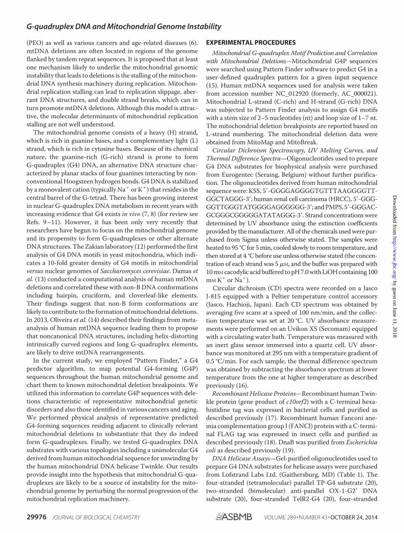
DNA Helicase Assays—Gel-purified oligonucleotides used toprepare G4 DNA substrates for helicase assays were purchasedfrom Lofstrand Labs Ltd. (Gaithersburg, MD) (Table 1). Thefour-stranded (tetramolecular) parallel TP-G4 substrate (20),two-stranded (bimolecular) anti-parallel OX-1-G2� DNAsubstrate (20), four-stranded TelR2-G4 (20), four-stranded
G-quadruplex DNA and Mitochondrial Genome Instability
29976 JOURNAL OF BIOLOGICAL CHEMISTRY VOLUME 289 • NUMBER 43 • OCTOBER 24, 2014
by guest on June 11, 2018http://w
ww
.jbc.org/D
ownloaded from
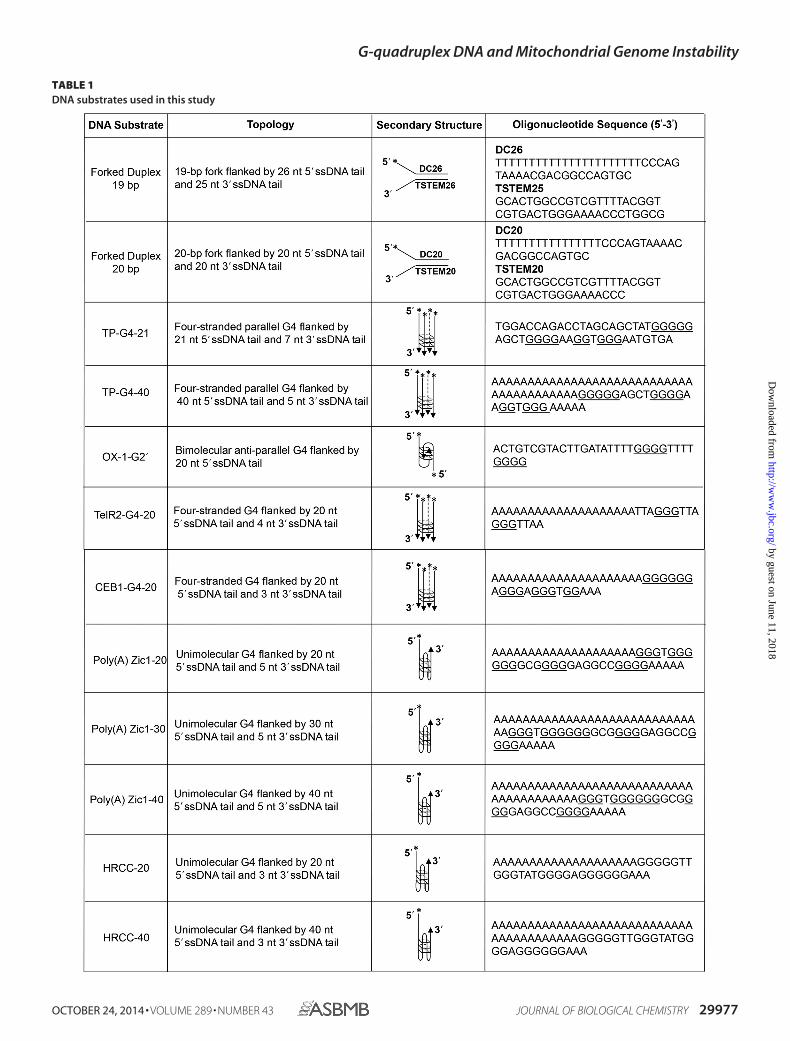
TABLE 1DNA substrates used in this study
G-quadruplex DNA and Mitochondrial Genome Instability
OCTOBER 24, 2014 • VOLUME 289 • NUMBER 43 JOURNAL OF BIOLOGICAL CHEMISTRY 29977
by guest on June 11, 2018http://w
ww
.jbc.org/D
ownloaded from
CEB1-G4 (21, 22), and unimolecular poly(A) Zic1-G4 substratewith 5� single-stranded DNA (ssDNA) tail lengths of 20, 30, or40 nt were prepared from gel-purified oligonucleotides asdescribed previously (23). The 19-bp forked duplex DNA sub-strate was prepared by annealing DC26 and TSTEM25 asdescribed earlier (24).
Standard helicase reaction mixtures (20 �l) containing 5fmol of the indicated TP-G4 or OX-1-G2� DNA substrate or theforked duplex DNA substrate (0.25 nM DNA substrate concen-tration) and the indicated concentration of FANCJ (20) orDnaB hexamer (25) were as described previously unless statedotherwise. For the Twinkle hexamer, the reaction conditionsfor the forked duplex were as described previously (26) exceptfor the inclusion of 25 mM KCl and the additional presencethroughout the incubation period of a 100-fold excess of oligo-nucleotide of the same sequence as the labeled strand in thepartial duplex substrate to serve as a displaced strand trap (17).Forked duplex helicase reactions were terminated as describedpreviously and resolved on nondenaturing 12% polyacrylamide(19:1 acrylamide-bisacrylamide) gels (24). Twinkle reactionconditions for the intermolecular G4 substrates (TP-G4, OX-1-G2�, TelR2-G4, and CEB1-G4) were the same as for theforked duplex except there was no additional oligonucleotidepresent during the reaction. For the unimolecular poly(A)Zic1-G4 or HRCC-G4 DNA substrate (5 fmol), helicase reac-tions (20 �l) were performed in a reaction buffer containing a
20-fold excess of peptide-nucleic acid complementary oligonu-cleotide (23). G4 helicase reactions were terminated by theaddition of 20 �l of stop buffer (74% glycerol, 0.01% xylenecyanol, 0.01% bromphenol blue, 10 mm KCl, and 20 mmEDTA). Reaction products for intermolecular and unimolecu-lar G4 DNA substrates were resolved on 10 and 15% nondena-turing polyacrylamide (19:1 acrylamide-bisacrylamide) gels,respectively. Gels were scanned by PhosphorImager and quan-titated as described previously (24).
RESULTS
Bioinformatic Analysis of Potential G-quadruplex-formingSequences in the Human Mitochondrial Genome and TheirLocations with Respect to Deletion Breakpoints—The humanmitochondrial genome contains G-rich sequences predicted toform G-quadruplex DNA (Fig. 1); G4P sequences can be foundin both the L-strand (top) and H-strand (bottom). However, agreater number of G4P sequences are present in the H-strand,which is also the G-rich strand. G4P sequences reside withinthe coding sequences of mitochondrial genes and also the tRNAgenes. G4P sequences can also be found to a lesser extent innoncoding regions of the mitochondrial genome.
We compared the G4P sequences in human mitochondrialDNA with other mammalian species to determine the degree ofconservation. The number of G4 motifs in the mitochondrialgenomes of mouse, rat, and monkey was markedly less than
FIGURE 1. Graphical representation of predicted G4 motifs and deletion breakpoints in the human mitochondrial genome coding and noncodingsequences. Mitochondrial genes, tRNA, and G-rich sequences predicted to form G4 are shown in the L-strand (top) and the H-strand (bottom). The 5� and 3�deletion breakpoints, collected from MitoMap and MitoBreak, residing within or adjacent to predicted G4-forming sequences, are shown. The number ofguanines (2–5 G) and loop size (1–7 nt) were employed for search criteria using QuadBase Pattern Finder. The predicted G4-forming sequences with param-eters �G3 and loop size 1–7 nt are also shown by bold dots. RNR1, 12 S ribosomal RNA; RNR2, 16 S ribosomal RNA; ND1, NADH dehydrogenase 1; ND2, NADHdehydrogenase 2; COX1, cytochrome c oxidase I; COXII, cytochrome c oxidase II; ATP8, ATP synthase 8; ATP6, ATP synthase 6; COXIII, cytochrome c oxidase III;ND3, NADH dehydrogenase 3; ND4L, NADH 4L dehydrogenase; ND4, NADH dehydrogenase 4; ND5, NADH dehydrogenase 5; ND6, NADH dehydrogenase 6;CYTB, cytochrome b.
G-quadruplex DNA and Mitochondrial Genome Instability
29978 JOURNAL OF BIOLOGICAL CHEMISTRY VOLUME 289 • NUMBER 43 • OCTOBER 24, 2014
by guest on June 11, 2018http://w
ww
.jbc.org/D
ownloaded from
found in human (Table 2). The number of conserved G4Psequences identified by the Pattern Finder parameters betweenthese species and humans was greatest in monkey followed byrat and then mouse.
The human mitochondrial genome deletions (collected fromMitoMap and MitoBreak) that reside adjacent to the G4Psequences derived from our analysis are shown in Fig. 1. Weidentified both 5� and 3� deletion breakpoints that reside in theG4P sequence or adjacent (within 20 bp) to the G4P sequence.The mitochondrial genome deletion breakpoints were mappedproximal to G4P sequences located within the coding regions of
mitochondrial genes and tRNA genes, as well as noncodingregions to a lesser extent.
To ascertain the relative abundance of deletion breakpointsin predicted mitochondrial G4-forming sequences, we com-pared the distribution and density of unique mitochondrialdeletion breakpoints in G4P sequences, tRNA genes, and theD-loop region (Table 3). The total number of mitochondrial 5�or 3� deletion breakpoints was greater in G4P as compared withtRNA or the D-loop region. We next determined the deletionbreakpoint density (deletions/kb) for each region of mitochon-drial DNA. The 5� mitochondrial deletion breakpoint densitywas �1.4- and 7.6-fold greater in G4P as compared with tRNAgenes and the D-loop, respectively. The 3� deletion breakpointdensity of mitochondrial G4P was 1.4- greater and 0.8-fold lessthan that of tRNA genes and D-loop, respectively (Table 3).
Association of Potential G-quadruplex-forming Sequenceswith Mitochondrial DNA Deletion Breakpoints Prevalent inHuman Disease—Our computational analysis revealed thatG4P sequences reside proximal to mtDNA deletions found in anumber of genetic diseases (Table 4). Among the genetic disor-ders associated with mitochondrial deletions near G4Psequences is progressive external ophthalmoplegia. PEO ischaracterized by the adult onset of muscular deficiencies,including those of the external eye and skeletal system (27). G4Psequences were also located adjacent to mitochondrial deletion
TABLE 2Evolutionary conservation of G4 motifs in mitochondrial genomes ofhuman and selected mammals
OrganismaMitochondrial
genomeSequencesimilarity
Total G4motifsb
Conserved G4motifsc
bp %Human 16,569 100 270 270Monkey 16,564 78 148 81Rat 16,313 67 108 46Mouse 16,299 66 59 34
a Accession numbers: human (NC_012920 AC_000021); monkey (NC_005943);rat (NC_001665); mouse (NC_005089).
b Total G4P motifs obtained based on stem size of 2–5 nt and loop size of 1–7 ntthrough Pattern Finder analysis.
c Number of predicted G4 motifs that are common in the indicated mammal andhuman.
TABLE 3Distribution and density of 5� and 3� deletion breakpointsThe number of unique deletion breakpoints � 620 5� deletion breakpoints and 497 3� deletion breakpoints (13).
Deletion breakpoint
G4Pa tRNAb D-loop (AT-rich)c
Deletion breakpoints Deletion breakpoints Deletion breakpoints
Total no.d %e Density (deletions/kb)f Total no.d %e Density (deletions/kb)f Total no.d %e Density (deletions/kb)f
5� deletion breakpoints 194 31 38 57 9 28 6 1 53� deletion breakpoints 125 25 25 36 7 18 37 7 31
a Number of deletion breakpoints in G4P excluding those overlapping in tRNA and D-loop region.b Number of deletion breakpoints in tRNA excluding those overlapping in G4P or D-loop region.c Number of deletion breakpoints in D-loop excluding those overlapping in G4P or tRNA.d Total number of unique deletion breakpoints in the case of G4P, tRNA, and D-loop.e Number of deletion breakpoints in percentage (%) with respect to either 620 5� deletions or 497 3� deletions for G4P, tRNA, and D-loop.f Deletion breakpoint density is based on 5071, 2017, and 1180 bp, respectively, for G4P, tRNA, and D-loop.
TABLE 4Selected disease-associated mitochondrial deletion breakpoints in proximity to predicted G4-forming sequences
Deletionbreakpointa G4P neighboring sequenceb Genetic disease References
5� 10190 (10187) tata2tcccccgcccgcgtcccttt (10210) Pearson marrow-pancreassyndrome
(29, 84)3� 13753 (13751) tt2tcccccgcatcccccttccaaacaacaatccccctcta (13790)5� 12113 (12081) tatcccccattctcctcctatccctcaaccccg2ac (12115)
5� 12103 (12078) cctatcccccattctcctcctatcc2ctc (12106) Kearns-Sayre syndrome, mitochondrialencephalomyopathy
(85–87)3� 14414 (14412) cc2tcaacccctgacccccatgcc (14435)
5� 4398 (4374) ccgtgccacctatcacaccccatcc2ta (4400) Diabetes mellitus (88, 89)3� 14822 (14806) cctccccaccccatcc2aacatctcc (14830)
5� 7974 (7942) ctcctacatacttcccccattattcctagaacc2agg (7977) Kearns-Sayre syndrome, mitochondrialencephalomyopathy, lactic acidosis,stroke-like episodes syndrome
(90, 91)3� 15496 (15495) t2aggcgacccagacaattataccctagccaaccccttaaacacccctccccacatc (15550)
5� 6325 (6325) c2ctccgtagacctaaccatcttctcct (6351) Mitochondrial myopathy (92)3� 13989 (13971) ccaaaacctgcccctact2cctcctagacctaacc (14004)
5� 6329 (6311) ctcccaccctggagcctc2cgtagacc (6336) Chronic progressive externalophthalmoplegia
(93)3� 13994 (13970) gccaaaacctgcccctactcctcc2tagacctaacc (14004)
5� 6330 (6311) ctcccaccctggagcctccg2tagacctaacc (6341) Kearns-Sayre syndrome, Pearsonmarrow-pancreas syndrome
(87)3� 13994 (13970) gccaaaacctgcccctactcctcc2tagacctaacc (14004)
5� 7491 (7365) accccccaaagctggtttcaagccaac2cccatggcctcc (7503) Kearns-Sayre syndrome, progressiveexternal ophthalmoplegia
(85)3� 11004 (10995) caagccaac2gccacttatccagtgaacc (11022)
a Deletion breakpoints reside within the G4P sequence or �20 nt away from G4P. The breakpoint is indicated by a down arrow.b L-strand sequence is shown (5� to 3�). The predicted G4-forming sequence (G4P) is underlined.
G-quadruplex DNA and Mitochondrial Genome Instability
OCTOBER 24, 2014 • VOLUME 289 • NUMBER 43 JOURNAL OF BIOLOGICAL CHEMISTRY 29979
by guest on June 11, 2018http://w
ww
.jbc.org/D
ownloaded from
breakpoints associated with KSS, a clinical subgroup of mito-chondrial encephalomyopathies associated with PEO and skel-etal muscle defects (28).
Our analysis revealed that a G4P sequence is located in abreakpoint region of a mitochondrial DNA deletion associatedwith PMPS, a fatal disease of early infancy affecting the hema-topoietic system and pancreas exocrine system (29) (Table 4).The molecular features of PMPS are attributed to a large-scaledeficiency in oxidative phosphorylation. The site-specific mito-chondrial deletions associated with PMPS and other mitochon-drial disorders (KSS and PEO) are characterized by directrepeats of 8 –13 bp present in the normal mitochondrialgenome, which flank the deletion breakpoints (29 –31). It hasbeen proposed that homologous recombination may beresponsible for the direct repeat signature pattern characteris-tic of PMPS and other sporadic mtDNA deletion disorders (32).The common 4977-bp deletion flanked by a 13-bp repeat in thenormal mtDNA sequence frequently identified in sporadicPEO and other mitochondrial disorders (33) is characterized bya 5� breakpoint at mtDNA position 8468 bp, which resides in apredicted G4-forming sequence according to the PatternFinder algorithm. Unusual secondary DNA structures such asG-quadruplexes may be prone to form in the mitochondrialgenome at loci such as the 5� breakpoint of the common
4977-bp deletion and interfere with normal mitochondrialDNA metabolic processes such as replication or repair, leadingto double strand breaks.
Association of Potential G-quadruplex-forming Sequenceswith Mitochondrial DNA Deletion Breakpoints Found inCancer—We identified a number of cases in which G4Psequences flanked mtDNA deletion breakpoints reportedlyassociated with cancer (Table 5). G4P sequences flankingmtDNA deletions in thyroid tumors were prominent. G4P-as-sociated mitochondrial genome instability may be relevant toprevious observations that a subset of thyroid tumors displayelevated mtDNA deletions (34). A relatively uncommon andsmall 264-bp deletion in the mitochondrial coding sequence forthe first subunit (ND1) of NADH:ubiquinone oxidoreductaseof the electron transport chain was reported in a HRCC (35).This deletion is flanked by G4P sequences according to ourcomputational analysis (Table 5). Biophysical studies of theG4P flanking sequence confirmed its ability to form G4 (see Fig.2A and “Biophysical Analysis of Selected G4P MitochondrialSequences” below). A 6-bp direct repeat flanked the deletedregion (35), suggesting that replication slippage due to mtDNAsynthesis stalling at G4 DNA may play a role.
Using the G4P algorithm, we determined that a G4Psequence flanked a 7079-bp mtDNA deletion site reported to
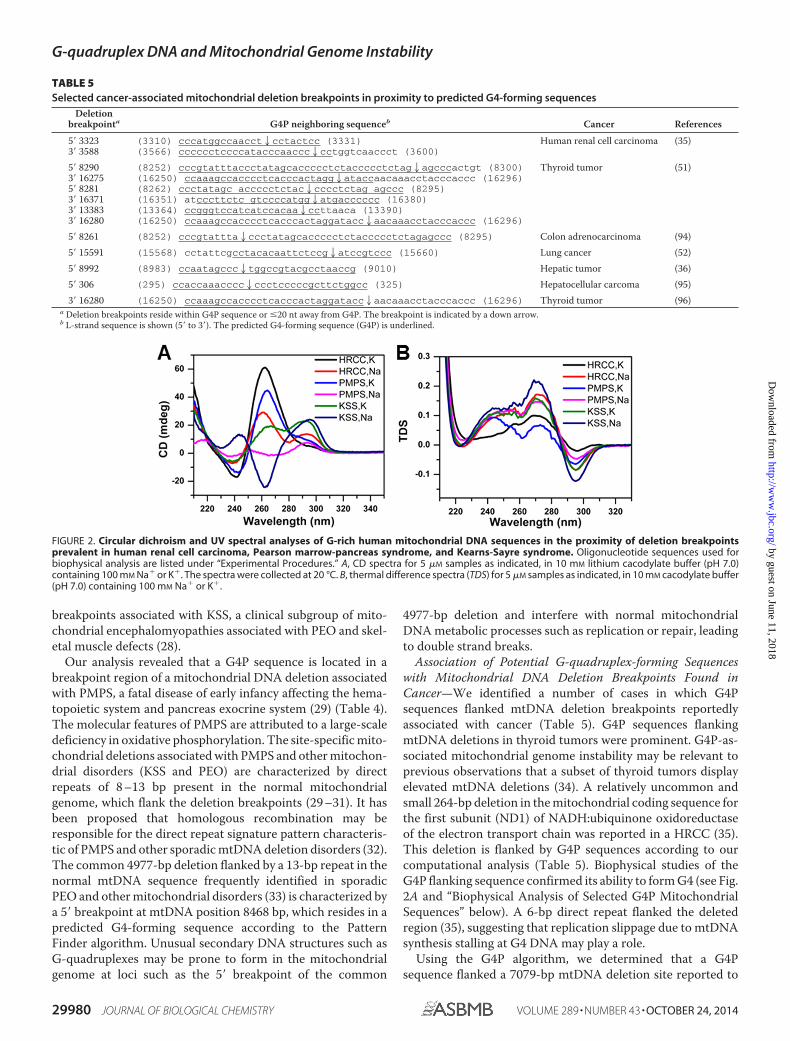
FIGURE 2. Circular dichroism and UV spectral analyses of G-rich human mitochondrial DNA sequences in the proximity of deletion breakpointsprevalent in human renal cell carcinoma, Pearson marrow-pancreas syndrome, and Kearns-Sayre syndrome. Oligonucleotide sequences used forbiophysical analysis are listed under “Experimental Procedures.” A, CD spectra for 5 �M samples as indicated, in 10 mM lithium cacodylate buffer (pH 7.0)containing 100 mM Na� or K�. The spectra were collected at 20 °C. B, thermal difference spectra (TDS) for 5 �M samples as indicated, in 10 mM cacodylate buffer(pH 7.0) containing 100 mM Na� or K�.
TABLE 5Selected cancer-associated mitochondrial deletion breakpoints in proximity to predicted G4-forming sequences
Deletionbreakpointa G4P neighboring sequenceb Cancer References
5� 3323 (3310) cccatggccaacct2cctactcc (3331) Human renal cell carcinoma (35)3� 3588 (3566) cccccctccccatacccaaccc2cctggtcaaccct (3600)
5� 8290 (8252) cccgtatttaccctatagcaccccctctaccccctctag2agcccactgt (8300) Thyroid tumor (51)3� 16275 (16250) ccaaagccacccctcacccactagg2ataccaacaaacctacccaccc (16296)5� 8281 (8262) ccctatagc accccctctac2cccctctag agccc (8295)3� 16371 (16351) atcccttctc gtccccatgg2atgacccccc (16380)3� 13383 (13364) ccgggtccatcatccacaa2ccttaaca (13390)3� 16280 (16250) ccaaagccacccctcacccactaggatacc2aacaaacctacccaccc (16296)
5� 8261 (8252) cccgtattta2ccctatagcaccccctctaccccctctagagccc (8295) Colon adrenocarcinoma (94)5� 15591 (15568) cctattcgcctacacaattctccg2atccgtccc (15660) Lung cancer (52)5� 8992 (8983) ccaatagccc2tggccgtacgcctaaccg (9010) Hepatic tumor (36)5� 306 (295) ccaccaaacccc2ccctcccccgcttctggcc (325) Hepatocellular carcoma (95)3� 16280 (16250) ccaaagccacccctcacccactaggatacc2aacaaacctacccaccc (16296) Thyroid tumor (96)
a Deletion breakpoints reside within G4P sequence or �20 nt away from G4P. The breakpoint is indicated by a down arrow.b L-strand sequence is shown (5� to 3�). The predicted G4-forming sequence (G4P) is underlined.
G-quadruplex DNA and Mitochondrial Genome Instability
29980 JOURNAL OF BIOLOGICAL CHEMISTRY VOLUME 289 • NUMBER 43 • OCTOBER 24, 2014
by guest on June 11, 2018http://w
ww
.jbc.org/D
ownloaded from
exist in cirrhotic liver that surrounds hepatic tumors (36)(Table 5). This mtDNA deletion was not characterized by directrepeat flanking sequences, suggesting that a mechanism otherthan replication slippage may be involved. Interestingly, the7079-bp mtDNA deletion was detected only in the cirrhoticliver surrounding the hepatic tumor but not in the tumor itself,suggesting that mitochondrial dysfunction contributes to livercirrhosis, which in turn serves as a risk factor for liver cancer.Next generation sequencing of mtDNA from tumor-derivedtissues should provide a more comprehensive analysis of themtDNA mutation spectra, which may be informative forunderstanding the molecular mechanism underlying the originof the mtDNA deletions associated with cancer and the poten-tial role of G4P sequences.
Association of Potential G-quadruplex-forming Sequenceswith Mitochondrial DNA Deletion Breakpoints Observed inAging—Using the computational analysis to map G4P sequencesflanking mitochondrial deletion breakpoints, we sought todetermine whether G4P sequences might play a role in themitochondrial genome instability prevalent in aged tissues ofthe human body based on a survey of the literature. A compar-ison of the mitochondrial G4P sequences with reportedmtDNA deletions associated with aging suggest this to be thecase (Table 6). We will briefly discuss several examples to illus-trate the potential importance of G4-forming sequences inmitochondrial DNA deletions found in aging tissues. An anal-ysis of photo-aged tumor-free skin of nonmelanoma skin can-cer patients reveals an increase in mitochondrial DNA dele-tions that correlates with patient age (37). Most of the identifiedmtDNA deletions contained repeat sequences, suggesting areplication slippage model. This would be consistent with theproposal that mtDNA photoadducts interfere with normalmtDNA replication; however, G4 DNA may also contribute toreplication stalling and mitochondrial genomic instability.Understanding the potential role of G-quadruplexes in replica-tion slippage or other aspects of replication mismanagement isan important area of study that deserves greater attention.Aberrant DNA structures such as G4 DNA, in addition to bulky
adducts imposed by chronic UV exposure, may contribute tothe accumulation of mtDNA deletions in skin during chrono-logical aging.
A decline in mitochondrial function is prevalent in age-re-lated disorders characterized by neurodegeneration (6). Kray-tsberg et al. (38) found that aged human substantia nigra con-tain a great abundance of mitochondrial DNA deletions, someof which we determined to be in close proximity to G4Psequences (Table 6). It was observed that mtDNA deletionswere enriched in cytochrome c oxidase-deficient neurons, sug-gesting that the mitochondrial genome instability was respon-sible for impaired cellular respiration and functional impair-ment of the aged neurons of the substantia nigra (38). Furtherwork in this area to explore the potential role of G-quadru-plexes in brain mtDNA deletions may be informative for age-related disorders such as Parkinson disease, which is character-ized by primary neurodegeneration in the substantia nigra (39).
Biophysical Analysis of Selected G4P Mitochondrial Sequences—To determine whether representative mitochondrial G4Psequences residing adjacent to known deletion breakpointsform G-quadruplex structures, the classical spectroscopicmethods of CD and UV light absorption were used. The oligo-nucleotides selected for physical analysis corresponded to G4Psequences flanking mitochondrial DNA deletion breakpointsdetected in PMPS, KSS, and HRCC. CD spectral analysis dem-onstrated that HRCC and PMPS showed a positive peak around265 nm and negative peak at 240 nm in the presence of 100 mM
K� (Fig. 2A), suggesting that parallel G-quadruplex structureswere formed (40). However, PMPS and KSS exhibited positiveand negative peaks around 295 and 260 nm, respectively, in thepresence of 100 mM Na�, which is in agreement with the for-mation of nonparallel G-quadruplexes (40). Interestingly, bothHRCC in Na� and KSS in K� displayed two positive peaksaround 295 and 260 nm. These spectra are similar to thosereported previously for the G-rich hTERT promoter, implyingthe coexistence of two different G-quadruplex conformations(41). Furthermore, the thermal difference spectra of all threesequences showed a negative peak around 295 nm and two
TABLE 6Selected age-associated mitochondrial deletion breakpoints in proximity to predicted G4-forming sequences
Deletionbreakpointa G4P neighboring sequenceb Aging features References
5� 540 (535) ccccata2ccccgaaccaaccaaaccccaaagacacccccc (573) Heart tissue (97)3� 13764 (13751) tttcccccgc atc2ccccttcc ( 13771)5� 5433 (5421) gaacatacaaaa2cccaccccattcctcccc (5450)3� 13023 (13010) cccaattaggtct2ccacccctgactcccctcagcc (13044)
5� 1491 (1482) cacaccgcccgtc2accctcctc (1500) Skin tissue (37)3� 5206 (5191) cacccttaattccat2ccaccctcctctccc (5220)
5� 6329 (6311) ctcccaccctggagcctcc2gtagacctaac catcttctcc (6350) Skeletal muscle (98)3� 13994 (13971) ccaaaacctgcccctactcctcc2tagacctaacc (14004)
5� 6437 (6420) ccccctgccataacccaa2taccaaacgcccc (6450) Substantia nigra neurons (99)3� 14077 (14043) ccaaatct ccacctccat catcacctcaacccaa2aaag (14080)
5� 6501 (6463) ccgtcctaatcacagcagcctatctctcc2cagtcctagc (6511) Substantia nigra neurons (38)3� 13802 (13781) tccccctctacctaaaactca2cagccctcg (13810)
3� 16284 (16250) ccaaagccacccctcacccactaggataccaaca2aacctacccaccc (16296) Cochlear tissue (100)
5� 7409 (7396) gccccccaccctac2cacacattcga (7420) Substantia nigra neurons of agedindividual with Parkinson’sdisease
(101)
3� 5447 (5333) cccaccccattcct2ccccacactcatcgcccttaccac gctactccta cctatctcccc (5491) Skeletal muscle (102)a Deletion breakpoints reside within G4P sequence or �20 nt away from G4P. The breakpoint is indicated by a down arrow.b L-strand sequence is shown (5� to 3�). The predicted G4-forming sequence (G4P) is underlined.
G-quadruplex DNA and Mitochondrial Genome Instability
OCTOBER 24, 2014 • VOLUME 289 • NUMBER 43 JOURNAL OF BIOLOGICAL CHEMISTRY 29981
by guest on June 11, 2018http://w
ww
.jbc.org/D
ownloaded from
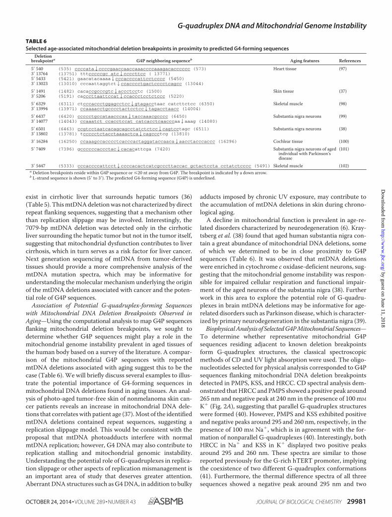
positive peaks around 275 and 243 nm (Fig. 2B), which is atypical feature of G-quadruplex DNA (42). In addition, UVmelting profiles of all these sequences exhibited a hypochromictransition at 295 nm, which is in agreement with the formationof G-quadruplex structures (42) (Fig. 3). Interestingly, Tmdepends on the nature of the cation (compare left- and right-hand panels in Fig. 3), which is strong evidence for G-quadru-plex formation (42).
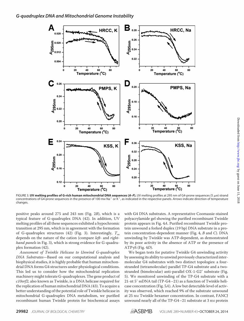
Assessment of Twinkle Helicase to Unwind G-quadruplexDNA Substrates—Based on our computational analysis andbiophysical studies, it is highly probable that human mitochon-drial DNA forms G4 structures under physiological conditions.This led us to consider how the mitochondrial replicationmachinery might tolerate G-quadruplexes. The gene product ofc10orf2, also known as Twinkle, is a DNA helicase required forthe replication of human mitochondrial DNA (43). To acquire abetter understanding of the potential role of Twinkle helicase inmitochondrial G-quadruplex DNA metabolism, we purifiedrecombinant human Twinkle protein for biochemical assays
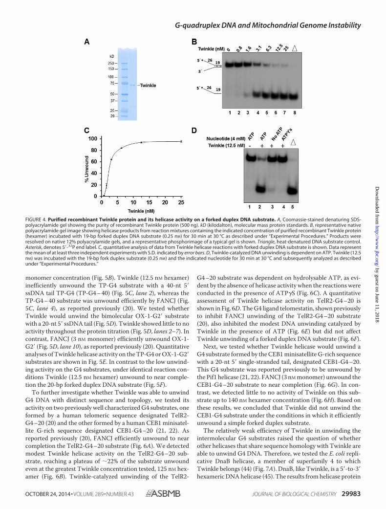
with G4 DNA substrates. A representative Coomassie-stainedpolyacrylamide gel showing the purified recombinant Twinkleprotein appears in Fig. 4A. Purified recombinant Twinkle pro-tein unwound a forked duplex (19 bp) DNA substrate in a pro-tein concentration-dependent manner (Fig. 4, B and C). DNAunwinding by Twinkle was ATP-dependent, as demonstratedby its poor activity in the absence of ATP or the presence ofATP�S (Fig. 4D).
We began tests for putative Twinkle G4 unwinding activityby assessing its ability to unwind previously characterized inter-molecular G4 substrates with two distinct topologies: a four-stranded (tetramolecular) parallel TP-G4 substrate and a two-stranded (bimolecular) anti-parallel OX-1-G2� substrate (Fig.5). We monitored unwinding of the TP-G4 substrate with a21-nt 5� ssDNA tail (TP-G4 –21) as a function of Twinkle heli-case concentration (Fig. 5A). A low but detectable level of activ-ity was observed, which reached 9% of the substrate unwoundat 25 nM Twinkle hexamer concentration. In contrast, FANCJunwound nearly all of the TP-G4 –21 substrate at 3 nM protein
FIGURE 3. UV melting profiles of G-rich human mitochondrial DNA sequences (A–F). UV melting profiles at 295 nm of G4-prone sequences (5 �M) strandconcentrations of G4-prone sequences in the presence of 100 mM Na� or K�, as indicated in the respective panels. Arrows indicate direction of temperaturechanges.
G-quadruplex DNA and Mitochondrial Genome Instability
29982 JOURNAL OF BIOLOGICAL CHEMISTRY VOLUME 289 • NUMBER 43 • OCTOBER 24, 2014
by guest on June 11, 2018http://w
ww
.jbc.org/D
ownloaded from
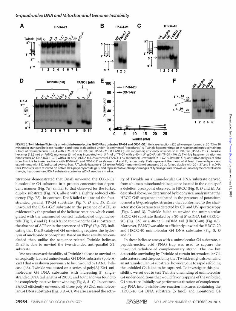
monomer concentration (Fig. 5B). Twinkle (12.5 nM hexamer)inefficiently unwound the TP-G4 substrate with a 40-nt 5�ssDNA tail TP-G4 (TP-G4 – 40) (Fig. 5C, lane 2), whereas theTP-G4 – 40 substrate was unwound efficiently by FANCJ (Fig.5C, lane 4), as reported previously (20). We tested whetherTwinkle would unwind the bimolecular OX-1-G2� substratewith a 20-nt 5� ssDNA tail (Fig. 5D). Twinkle showed little to noactivity throughout the protein titration (Fig. 5D, lanes 2–7). Incontrast, FANCJ (3 nM monomer) efficiently unwound OX-1-G2� (Fig. 5D, lane 10), as reported previously (20). Quantitativeanalyses of Twinkle helicase activity on the TP-G4 or OX-1-G2�substrates are shown in Fig. 5E. In contrast to the low unwind-ing activity on the G4 substrates, under identical reaction con-ditions Twinkle (12.5 nM hexamer) unwound to near comple-tion the 20-bp forked duplex DNA substrate (Fig. 5F).
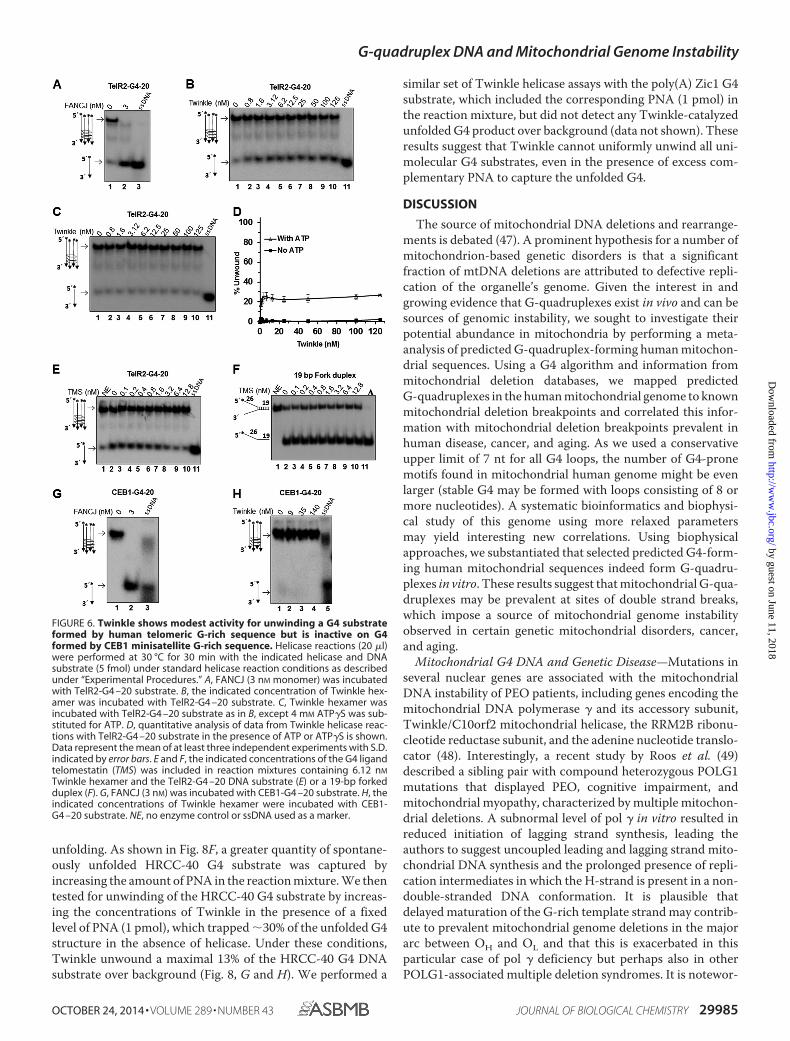
To further investigate whether Twinkle was able to unwindG4 DNA with distinct sequence and topology, we tested itsactivity on two previously well characterized G4 substrates, oneformed by a human telomeric sequence designated TelR2-G4 –20 (20) and the other formed by a human CEB1 minisatel-lite G-rich sequence designated CEB1-G4 –20 (21, 22). Asreported previously (20), FANCJ efficiently unwound to nearcompletion the TelR2-G4 –20 substrate (Fig. 6A). We detectedmodest Twinkle helicase activity on the TelR2-G4 –20 sub-strate, reaching a plateau of �22% of the substrate unwoundeven at the greatest Twinkle concentration tested, 125 nM hex-amer (Fig. 6B). Twinkle-catalyzed unwinding of the TelR2-
G4 –20 substrate was dependent on hydrolysable ATP, as evi-dent by the absence of helicase activity when the reactions wereconducted in the presence of ATP�S (Fig. 6C). A quantitativeassessment of Twinkle helicase activity on TelR2-G4 –20 isshown in Fig. 6D. The G4 ligand telomestatin, shown previouslyto inhibit FANCJ unwinding of the TelR2-G4 –20 substrate(20), also inhibited the modest DNA unwinding catalyzed byTwinkle in the presence of ATP (Fig. 6E) but did not affectTwinkle unwinding of a forked duplex DNA substrate (Fig. 6F).
Next, we tested whether Twinkle helicase would unwind aG4 substrate formed by the CEB1 minisatellite G-rich sequencewith a 20-nt 5� single-stranded tail, designated CEB1-G4 –20.This G4 substrate was reported previously to be unwound bythe Pif1 helicase (21, 22). FANCJ (3 nM monomer) unwound theCEB1-G4 –20 substrate to near completion (Fig. 6G). In con-trast, we detected little to no activity of Twinkle on this sub-strate up to 140 nM hexamer concentration (Fig. 6H). Based onthese results, we concluded that Twinkle did not unwind theCEB1-G4 substrate under the conditions in which it efficientlyunwound a simple forked duplex substrate.
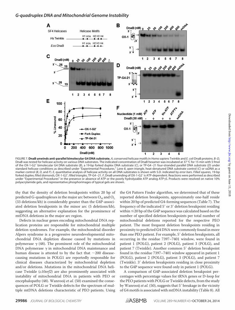
The relatively weak efficiency of Twinkle in unwinding theintermolecular G4 substrates raised the question of whetherother helicases that share sequence homology with Twinkle areable to unwind G4 DNA. Therefore, we tested the E. coli repli-cative DnaB helicase, a member of superfamily 4 to whichTwinkle belongs (44) (Fig. 7A). DnaB, like Twinkle, is a 5�-to-3�hexameric DNA helicase (45). The results from helicase protein
FIGURE 4. Purified recombinant Twinkle protein and its helicase activity on a forked duplex DNA substrate. A, Coomassie-stained denaturing SDS-polyacrylamide gel showing the purity of recombinant Twinkle protein (500 ng). kD (kilodalton), molecular mass protein standards. B, representative nativepolyacrylamide gel image showing helicase products from reaction mixtures containing the indicated concentration of purified recombinant Twinkle protein(hexamer) incubated with 19-bp forked duplex DNA substrate (0.25 nM) for 30 min at 30 °C as described under “Experimental Procedures.” Products wereresolved on native 12% polyacrylamide gels, and a representative phosphorimage of a typical gel is shown. Triangle, heat-denatured DNA substrate control.Asterisk, denotes 5�-32P end label. C, quantitative analysis of data from Twinkle helicase reactions with forked duplex DNA substrate is shown. Data representthe mean of at least three independent experiments with S.D. indicated by error bars. D, Twinkle-catalyzed DNA unwinding is dependent on ATP. Twinkle (12.5nM) was incubated with the 19-bp fork duplex substrate (0.25 nM) and the indicated nucleotide for 30 min at 30 °C and subsequently analyzed as describedunder “Experimental Procedures.”
G-quadruplex DNA and Mitochondrial Genome Instability
OCTOBER 24, 2014 • VOLUME 289 • NUMBER 43 JOURNAL OF BIOLOGICAL CHEMISTRY 29983
by guest on June 11, 2018http://w
ww
.jbc.org/D
ownloaded from
titrations demonstrated that DnaB unwound the OX-1-G2�bimolecular G4 substrate in a protein concentration-depen-dent manner (Fig. 7B) similar to that observed for the forkedduplex substrate (Fig. 7C), albeit with a slightly reduced effi-ciency (Fig. 7E). In contrast, DnaB failed to unwind the four-stranded parallel TP-G4 substrate (Fig. 7, D and E). DnaBunwound the OX-1-G2� substrate in the presence of ATP, asevidenced by the product of the helicase reaction, which comi-grated with the unannealed control radiolabeled oligonucleo-tide (Fig. 7, B and F). DnaB failed to unwind the G4 substrate inthe absence of ATP or in the presence of ATP�S (Fig. 7F), indi-cating that DnaB-catalyzed G4 unwinding requires the hydro-lysis of nucleoside triphosphate. Based on these results, we con-cluded that, unlike the sequence-related Twinkle helicase,DnaB is able to unwind the two-stranded anti-parallel G2�substrate.
We next assessed the ability of Twinkle helicase to unwind anentropically favored unimolecular G4 DNA substrate (poly(A)Zic1) that was shown previously to be unwound by FANCJ heli-case (46). Twinkle was tested on a series of poly(A) Zic1-uni-molecular G4 DNA substrates with increasing 5� single-stranded DNA tail lengths of 20, 30, and 40 nt and was found tobe completely inactive for unwinding (Fig. 8, A–C). In contrast,FANCJ efficiently unwound all three poly(A) Zic1 unimolecu-lar G4 DNA substrates (Fig. 8, A–C). We also assessed the activ-
ity of Twinkle on a unimolecular G4 DNA substrate derivedfrom a human mitochondrial sequence located in the vicinity ofa deletion breakpoint observed in HRCC (Fig. 8, D and E). Asdescribed above, we determined by biophysical analysis that theHRCC G4P sequence incubated in the presence of potassiumformed a G-quadruplex structure that conformed to the char-acteristic G4 parameters detected by CD and UV spectroscopy(Figs. 2 and 3). Twinkle failed to unwind the unimolecularHRCC G4 substrate flanked by a 20-nt 5� ssDNA tail (HRCC-20) (Fig. 8D) or a 40-nt 5� ssDNA tail (HRCC-40) (Fig. 8E).Moreover, FANCJ was able to efficiently unwind the HRCC-20and HRCC-40 unimolecular G4 DNA substrates (Fig. 8, Dand E).
In these helicase assays with a unimolecular G4 substrate, apeptide-nucleic acid (PNA) trap was used to capture theunwound radiolabeled complementary strand. The low butdetectable unwinding by Twinkle of certain intermolecular G4substrates raised the possibility that Twinkle might also unwindan intramolecular G4 substrate; however, due to rapid refoldingthe unfolded G4 failed to be captured. To investigate this pos-sibility, we set out to test Twinkle unwinding of unimolecularG4 under conditions that would favor trapping of the unfoldedG4 structure. Initially, we performed a titration of complemen-tary PNA into Twinkle-free reaction mixtures containing theHRCC-40 G4 DNA substrate (5 fmol) and monitored G4
FIGURE 5. Twinkle inefficiently unwinds intermolecular G4 DNA substrates TP-G4 and OX-1-G2�. Helicase reactions (20 �l) were performed at 30 °C for 30min under standard helicase reaction conditions as described under “Experimental Procedures.” A, Twinkle hexamer titration in reaction mixtures containing5 fmol of tetramolecular TP-G4 with a 21-nt 5� ssDNA tail (TP-G4 –21). B, FANCJ (3 nM monomer) efficiently unwinds 5� ssDNA tail (TP-G4 –21). C, Twinklehexamer (12.5 nM) or FANCJ monomer (3 nM) was incubated with 5 fmol of TP-G4 with a 40-nt 5� ssDNA tail (TP-G4 – 40). D, Twinkle hexamer titration onbimolecular G4 DNA (OX-1-G2�) with a 20-nt 5� ssDNA tail. As a control, FANCJ (3 nM monomer) unwound OX-1-G2� substrate. E, quantitative analysis of datafrom Twinkle helicase reactions with TP-G4 –21 and OX-1-G2� as shown in A and D, respectively. Data represent the mean of at least three independentexperiments with S.D. indicated by error bars. F, Twinkle hexamer (12.5 nM) or FANCJ monomer (3 nM) unwound 20 bp forked duplex with 20 nt 5� and 3� ssDNAtails. Products were resolved on native 10% polyacrylamide gels, and representative phosphorimages of typical gels are shown. NE, no enzyme control; opentriangle, heat-denatured DNA substrate control or ssDNA used as a marker.
G-quadruplex DNA and Mitochondrial Genome Instability
29984 JOURNAL OF BIOLOGICAL CHEMISTRY VOLUME 289 • NUMBER 43 • OCTOBER 24, 2014
by guest on June 11, 2018http://w
ww
.jbc.org/D
ownloaded from
unfolding. As shown in Fig. 8F, a greater quantity of spontane-ously unfolded HRCC-40 G4 substrate was captured byincreasing the amount of PNA in the reaction mixture. We thentested for unwinding of the HRCC-40 G4 substrate by increas-ing the concentrations of Twinkle in the presence of a fixedlevel of PNA (1 pmol), which trapped �30% of the unfolded G4structure in the absence of helicase. Under these conditions,Twinkle unwound a maximal 13% of the HRCC-40 G4 DNAsubstrate over background (Fig. 8, G and H). We performed a
similar set of Twinkle helicase assays with the poly(A) Zic1 G4substrate, which included the corresponding PNA (1 pmol) inthe reaction mixture, but did not detect any Twinkle-catalyzedunfolded G4 product over background (data not shown). Theseresults suggest that Twinkle cannot uniformly unwind all uni-molecular G4 substrates, even in the presence of excess com-plementary PNA to capture the unfolded G4.
DISCUSSION
The source of mitochondrial DNA deletions and rearrange-ments is debated (47). A prominent hypothesis for a number ofmitochondrion-based genetic disorders is that a significantfraction of mtDNA deletions are attributed to defective repli-cation of the organelle’s genome. Given the interest in andgrowing evidence that G-quadruplexes exist in vivo and can besources of genomic instability, we sought to investigate theirpotential abundance in mitochondria by performing a meta-analysis of predicted G-quadruplex-forming human mitochon-drial sequences. Using a G4 algorithm and information frommitochondrial deletion databases, we mapped predictedG-quadruplexes in the human mitochondrial genome to knownmitochondrial deletion breakpoints and correlated this infor-mation with mitochondrial deletion breakpoints prevalent inhuman disease, cancer, and aging. As we used a conservativeupper limit of 7 nt for all G4 loops, the number of G4-pronemotifs found in mitochondrial human genome might be evenlarger (stable G4 may be formed with loops consisting of 8 ormore nucleotides). A systematic bioinformatics and biophysi-cal study of this genome using more relaxed parametersmay yield interesting new correlations. Using biophysicalapproaches, we substantiated that selected predicted G4-form-ing human mitochondrial sequences indeed form G-quadru-plexes in vitro. These results suggest that mitochondrial G-qua-druplexes may be prevalent at sites of double strand breaks,which impose a source of mitochondrial genome instabilityobserved in certain genetic mitochondrial disorders, cancer,and aging.
Mitochondrial G4 DNA and Genetic Disease—Mutations inseveral nuclear genes are associated with the mitochondrialDNA instability of PEO patients, including genes encoding themitochondrial DNA polymerase � and its accessory subunit,Twinkle/C10orf2 mitochondrial helicase, the RRM2B ribonu-cleotide reductase subunit, and the adenine nucleotide translo-cator (48). Interestingly, a recent study by Roos et al. (49)described a sibling pair with compound heterozygous POLG1mutations that displayed PEO, cognitive impairment, andmitochondrial myopathy, characterized by multiple mitochon-drial deletions. A subnormal level of pol � in vitro resulted inreduced initiation of lagging strand synthesis, leading theauthors to suggest uncoupled leading and lagging strand mito-chondrial DNA synthesis and the prolonged presence of repli-cation intermediates in which the H-strand is present in a non-double-stranded DNA conformation. It is plausible thatdelayed maturation of the G-rich template strand may contrib-ute to prevalent mitochondrial genome deletions in the majorarc between OH and OL and that this is exacerbated in thisparticular case of pol � deficiency but perhaps also in otherPOLG1-associated multiple deletion syndromes. It is notewor-
FIGURE 6. Twinkle shows modest activity for unwinding a G4 substrateformed by human telomeric G-rich sequence but is inactive on G4formed by CEB1 minisatellite G-rich sequence. Helicase reactions (20 �l)were performed at 30 °C for 30 min with the indicated helicase and DNAsubstrate (5 fmol) under standard helicase reaction conditions as describedunder “Experimental Procedures.” A, FANCJ (3 nM monomer) was incubatedwith TelR2-G4 –20 substrate. B, the indicated concentration of Twinkle hex-amer was incubated with TelR2-G4 –20 substrate. C, Twinkle hexamer wasincubated with TelR2-G4 –20 substrate as in B, except 4 mM ATP�S was sub-stituted for ATP. D, quantitative analysis of data from Twinkle helicase reac-tions with TelR2-G4 –20 substrate in the presence of ATP or ATP�S is shown.Data represent the mean of at least three independent experiments with S.D.indicated by error bars. E and F, the indicated concentrations of the G4 ligandtelomestatin (TMS) was included in reaction mixtures containing 6.12 nM
Twinkle hexamer and the TelR2-G4 –20 DNA substrate (E) or a 19-bp forkedduplex (F). G, FANCJ (3 nM) was incubated with CEB1-G4 –20 substrate. H, theindicated concentrations of Twinkle hexamer were incubated with CEB1-G4 –20 substrate. NE, no enzyme control or ssDNA used as a marker.
G-quadruplex DNA and Mitochondrial Genome Instability
OCTOBER 24, 2014 • VOLUME 289 • NUMBER 43 JOURNAL OF BIOLOGICAL CHEMISTRY 29985
by guest on June 11, 2018http://w
ww
.jbc.org/D
ownloaded from
thy that the density of deletion breakpoints within 20 bp ofpredicted G-quadruplexes in the major arc between OH and OL(33 deletions/kb) is considerably greater than the G4P-associ-ated deletion breakpoints in the minor arc (5 deletions/kb),suggesting an alternative explanation for the prominence ofmtDNA deletions in the major arc region.
Defects in nuclear genes encoding mitochondrial DNA rep-lication proteins are responsible for mitochondrial multipledeletion syndromes. For example, the mitochondrial disorderAlpers syndrome is a progressive neurodevelopmental mito-chondrial DNA depletion disease caused by mutations inpolymerase � (48). The prominent role of the mitochondrialDNA polymerase � in mitochondrial DNA maintenance andhuman disease is attested to by the fact that �200 disease-causing mutations in POLG1 are reportedly responsible forclinical diseases characterized by mitochondrial depletionand/or deletions. Mutations in the mitochondrial DNA heli-case Twinkle (c10orf2) are also prominently associated withinstability of mitochondrial DNA in patients with PEO orencephalopathy (48). Wanrooij et al. (50) examined the conse-quences of POLG or Twinkle defects for the spectrum of mul-tiple mtDNA deletions characteristic of PEO patients. Using
the G4 Pattern Finder algorithm, we determined that of thesereported deletion breakpoints, approximately one-half residewithin 20 bp of predicted G4-forming sequences (Table 7). Thefrequency of the indicated 5� or 3� deletion breakpoint residingwithin �20 bp of the G4P sequence was calculated based on thenumber of specified deletion breakpoints per total number ofmitochondrial deletions reported for the respective PEOpatient. The most frequent deletion breakpoints residing inproximity to predicted G4 DNA were commonly found in morethan one PEO patient. For example, 5� deletion breakpoints, alloccurring in the residue 7397–7401 window, were found inpatient 1 (POLG), patient 2 (POLG), patient 3 (POLG), andpatient 7 (Twinkle). Another common 5� deletion breakpointfound in the residue 7397–7401 window appeared in patient 1(POLG), patient 2 (POLG), patient 3 (POLG), and patient 7(Twinkle). 3� deletion breakpoints residing in close proximityto the G4P sequence were found only in patient 1 (POLG).
A comparison of G4P-associated deletion breakpoint per-centages with percentage values for tRNA genes or D-loop forthe PEO patients with POLG or Twinkle defects, from the studyby Wanrooij et al. (50), suggests that 5� breakage in the vicinityof G4 motifs is associated with mtDNA instability (Table 8). All
FIGURE 7. DnaB unwinds anti-parallel bimolecular G4 DNA substrate. A, conserved helicase motifs in Homo sapiens Twinkle and E. coli DnaB proteins. B–D,DnaB was tested for helicase activity on various DNA substrates. The indicated concentration of DnaB hexamer was incubated at 37 °C for 15 min with 5 fmolof the OX-1-G2� bimolecular G4 DNA substrate (B), a 19-bp forked duplex DNA substrate (C), or TP-G4 –21 four-stranded parallel DNA substrate (D) understandard helicase conditions as described under “Experimental Procedures.” Lane 8, open triangle, heat-denatured DNA substrate control (C) or ssDNA as amarker control (B, D, and F). E, quantitative analysis of helicase activity on all DNA substrates is shown with S.D. indicated by error bars. Filled squares, 19-bpforked duplex; filled diamonds, OX-1-G2�; filled triangles, TP-G4 –21. F, DnaB unwinding of OX-1-G2� is ATP-dependent. Reactions were performed as describedunder “Experimental Procedures” in the presence or absence of ATP or the poorly hydrolyzable ATP analog ATP�S. Products were resolved on native 10%polyacrylamide gels, and representative phosphorimages of typical gels are shown.
G-quadruplex DNA and Mitochondrial Genome Instability
29986 JOURNAL OF BIOLOGICAL CHEMISTRY VOLUME 289 • NUMBER 43 • OCTOBER 24, 2014
by guest on June 11, 2018http://w
ww
.jbc.org/D
ownloaded from
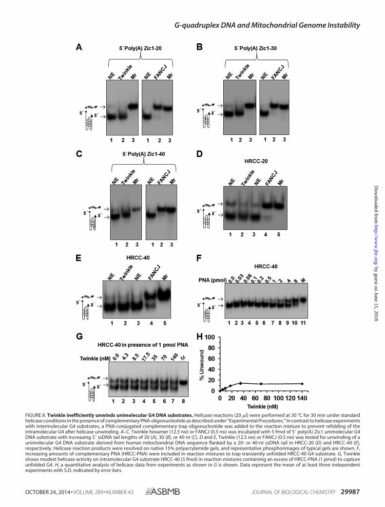
FIGURE 8. Twinkle inefficiently unwinds unimolecular G4 DNA substrates. Helicase reactions (20 �l) were performed at 30 °C for 30 min under standardhelicase conditions in the presence of complementary PNA-oligonucleotide as described under “Experimental Procedures.” In contrast to helicase experimentswith intermolecular G4 substrates, a PNA-conjugated complementary trap oligonucleotide was added to the reaction mixture to prevent refolding of theintramolecular G4 after helicase unwinding. A–C, Twinkle hexamer (12.5 nM) or FANCJ (0.5 nM) was incubated with 5 fmol of 5� poly(A) Zic1 unimolecular G4DNA substrate with increasing 5� ssDNA tail lengths of 20 (A), 30 (B), or 40 nt (C). D and E, Twinkle (12.5 nM) or FANCJ (0.5 nM) was tested for unwinding of aunimolecular G4 DNA substrate derived from human mitochondrial DNA sequence flanked by a 20- or 40-nt ssDNA tail in HRCC-20 (D) and HRCC-40 (E),respectively. Helicase reaction products were resolved on native 15% polyacrylamide gels, and representative phosphorimages of typical gels are shown. F,increasing amounts of complementary PNA (HRCC-PNA) were included in reaction mixtures to trap transiently unfolded HRCC-40 G4 substrate. G, Twinkleshows modest helicase activity on intramolecular G4 substrate HRCC-40 (5 fmol) in reaction mixtures containing an excess of HRCC-PNA (1 pmol) to captureunfolded G4. H, a quantitative analysis of helicase data from experiments as shown in G is shown. Data represent the mean of at least three independentexperiments with S.D. indicated by error bars.
G-quadruplex DNA and Mitochondrial Genome Instability
OCTOBER 24, 2014 • VOLUME 289 • NUMBER 43 JOURNAL OF BIOLOGICAL CHEMISTRY 29987
by guest on June 11, 2018http://w
ww
.jbc.org/D
ownloaded from
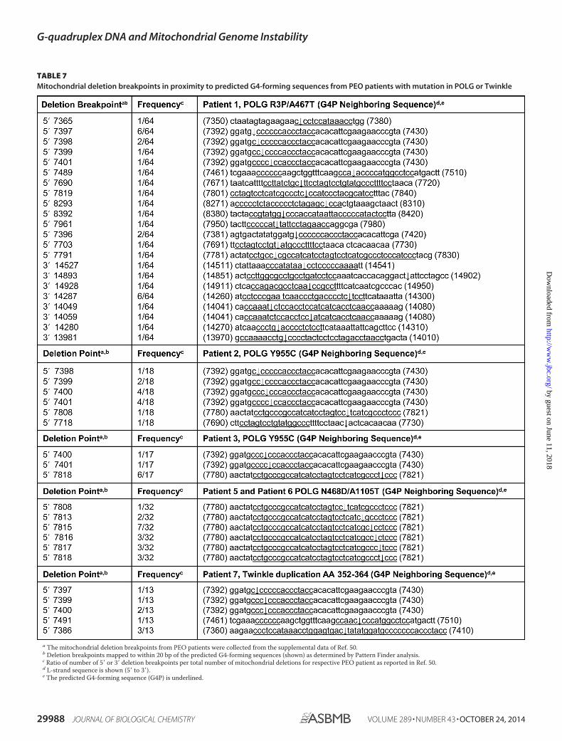
TABLE 7Mitochondrial deletion breakpoints in proximity to predicted G4-forming sequences from PEO patients with mutation in POLG or Twinkle
a The mitochondrial deletion breakpoints from PEO patients were collected from the supplemental data of Ref. 50.b Deletion breakpoints mapped to within 20 bp of the predicted G4-forming sequences (shown) as determined by Pattern Finder analysis.c Ratio of number of 5� or 3� deletion breakpoints per total number of mitochondrial deletions for respective PEO patient as reported in Ref. 50.d L-strand sequence is shown (5� to 3�).e The predicted G4-forming sequence (G4P) is underlined.
G-quadruplex DNA and Mitochondrial Genome Instability
29988 JOURNAL OF BIOLOGICAL CHEMISTRY VOLUME 289 • NUMBER 43 • OCTOBER 24, 2014
by guest on June 11, 2018http://w
ww
.jbc.org/D
ownloaded from
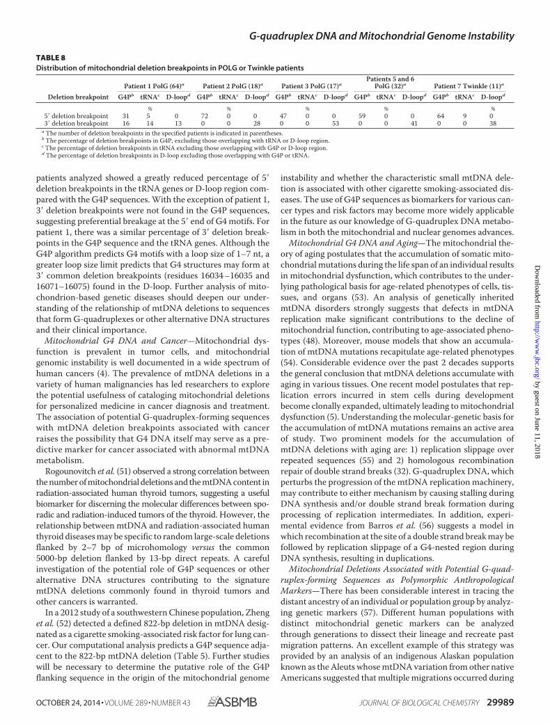
patients analyzed showed a greatly reduced percentage of 5�deletion breakpoints in the tRNA genes or D-loop region com-pared with the G4P sequences. With the exception of patient 1,3� deletion breakpoints were not found in the G4P sequences,suggesting preferential breakage at the 5� end of G4 motifs. Forpatient 1, there was a similar percentage of 3� deletion break-points in the G4P sequence and the tRNA genes. Although theG4P algorithm predicts G4 motifs with a loop size of 1–7 nt, agreater loop size limit predicts that G4 structures may form at3� common deletion breakpoints (residues 16034 –16035 and16071–16075) found in the D-loop. Further analysis of mito-chondrion-based genetic diseases should deepen our under-standing of the relationship of mtDNA deletions to sequencesthat form G-quadruplexes or other alternative DNA structuresand their clinical importance.
Mitochondrial G4 DNA and Cancer—Mitochondrial dys-function is prevalent in tumor cells, and mitochondrialgenomic instability is well documented in a wide spectrum ofhuman cancers (4). The prevalence of mtDNA deletions in avariety of human malignancies has led researchers to explorethe potential usefulness of cataloging mitochondrial deletionsfor personalized medicine in cancer diagnosis and treatment.The association of potential G-quadruplex-forming sequenceswith mtDNA deletion breakpoints associated with cancerraises the possibility that G4 DNA itself may serve as a pre-dictive marker for cancer associated with abnormal mtDNAmetabolism.
Rogounovitch et al. (51) observed a strong correlation betweenthe number of mitochondrial deletions and the mtDNA content inradiation-associated human thyroid tumors, suggesting a usefulbiomarker for discerning the molecular differences between spo-radic and radiation-induced tumors of the thyroid. However, therelationship between mtDNA and radiation-associated humanthyroid diseases may be specific to random large-scale deletionsflanked by 2–7 bp of microhomology versus the common5000-bp deletion flanked by 13-bp direct repeats. A carefulinvestigation of the potential role of G4P sequences or otheralternative DNA structures contributing to the signaturemtDNA deletions commonly found in thyroid tumors andother cancers is warranted.
In a 2012 study of a southwestern Chinese population, Zhenget al. (52) detected a defined 822-bp deletion in mtDNA desig-nated as a cigarette smoking-associated risk factor for lung can-cer. Our computational analysis predicts a G4P sequence adja-cent to the 822-bp mtDNA deletion (Table 5). Further studieswill be necessary to determine the putative role of the G4Pflanking sequence in the origin of the mitochondrial genome
instability and whether the characteristic small mtDNA dele-tion is associated with other cigarette smoking-associated dis-eases. The use of G4P sequences as biomarkers for various can-cer types and risk factors may become more widely applicablein the future as our knowledge of G-quadruplex DNA metabo-lism in both the mitochondrial and nuclear genomes advances.
Mitochondrial G4 DNA and Aging—The mitochondrial the-ory of aging postulates that the accumulation of somatic mito-chondrial mutations during the life span of an individual resultsin mitochondrial dysfunction, which contributes to the under-lying pathological basis for age-related phenotypes of cells, tis-sues, and organs (53). An analysis of genetically inheritedmtDNA disorders strongly suggests that defects in mtDNAreplication make significant contributions to the decline ofmitochondrial function, contributing to age-associated pheno-types (48). Moreover, mouse models that show an accumula-tion of mtDNA mutations recapitulate age-related phenotypes(54). Considerable evidence over the past 2 decades supportsthe general conclusion that mtDNA deletions accumulate withaging in various tissues. One recent model postulates that rep-lication errors incurred in stem cells during developmentbecome clonally expanded, ultimately leading to mitochondrialdysfunction (5). Understanding the molecular-genetic basis forthe accumulation of mtDNA mutations remains an active areaof study. Two prominent models for the accumulation ofmtDNA deletions with aging are: 1) replication slippage overrepeated sequences (55) and 2) homologous recombinationrepair of double strand breaks (32). G-quadruplex DNA, whichperturbs the progression of the mtDNA replication machinery,may contribute to either mechanism by causing stalling duringDNA synthesis and/or double strand break formation duringprocessing of replication intermediates. In addition, experi-mental evidence from Barros et al. (56) suggests a model inwhich recombination at the site of a double strand break may befollowed by replication slippage of a G4-nested region duringDNA synthesis, resulting in duplications.
Mitochondrial Deletions Associated with Potential G-quad-ruplex-forming Sequences as Polymorphic AnthropologicalMarkers—There has been considerable interest in tracing thedistant ancestry of an individual or population group by analyz-ing genetic markers (57). Different human populations withdistinct mitochondrial genetic markers can be analyzedthrough generations to dissect their lineage and recreate pastmigration patterns. An excellent example of this strategy wasprovided by an analysis of an indigenous Alaskan populationknown as the Aleuts whose mtDNA variation from other nativeAmericans suggested that multiple migrations occurred during
TABLE 8Distribution of mitochondrial deletion breakpoints in POLG or Twinkle patients
Deletion breakpointPatient 1 PolG (64)a Patient 2 PolG (18)a Patient 3 PolG (17)a
Patients 5 and 6PolG (32)a Patient 7 Twinkle (11)a
G4Pb tRNAc D-loopd G4Pb tRNAc D-loopd G4Pb tRNAc D-loopd G4Pb tRNAc D-loopd G4Pb tRNAc D-loopd
% % % % %5� deletion breakpoint 31 5 0 72 0 0 47 0 0 59 0 0 64 9 03� deletion breakpoint 16 14 13 0 0 28 0 0 53 0 0 41 0 0 38
a The number of deletion breakpoints in the specified patients is indicated in parentheses.b The percentage of deletion breakpoints in G4P, excluding those overlapping with tRNA or D-loop region.c The percentage of deletion breakpoints in tRNA excluding those overlapping with G4P or D-loop region.d The percentage of deletion breakpoints in D-loop excluding those overlapping with G4P or tRNA.
G-quadruplex DNA and Mitochondrial Genome Instability
OCTOBER 24, 2014 • VOLUME 289 • NUMBER 43 JOURNAL OF BIOLOGICAL CHEMISTRY 29989
by guest on June 11, 2018http://w
ww
.jbc.org/D
ownloaded from
the habitation of the New World (58). From the G4P computa-tional analysis and a survey of the literature, we identified areported 9-bp mitochondrial deletion used as a polymorphicanthropological marker for native North American groups (59)as residing within 20 bp of a G4P sequence (5�-GGGGGTAGA-GGGGGTGCTATAGGGTAAATACGGG-3� H-strand). Thedeletion has its highest frequency in the American Southwest andis absent in the Arctic and Subarctic regions (59). The distributionof the G4P-associated mitochondrial deletion marker hasprovided some insight into the migration patterns of East Asians tothe New World. Genographic projects such as this will benefitfrom further genetic analysis of indigenous populations by usingnovel genetic markers as important tools. Understanding the roleof G-quadruplex metabolism and other molecular mechanismsthat affect chromosomal stability and mutations, thereby influen-cing the appearance of mitochondrial genetic polymorphisms, willhelp in deciphering the origin of polymorphic markers. Theseefforts will in turn be useful for anthropological studies such as thehistory of human migration patterns and other fields such asforensic science (60).
Implications of Twinkle G4 Studies for Mitochondrial DNAReplication—Because the Twinkle helicase is required forhuman mtDNA replication, we assessed whether Twinkle wasable to unwind various uni-, bi-, and tetramolecular G4 DNAsubstrates. We found that the helicase is not efficient inunwinding the various topological forms of G4 DNA testedunder the same conditions in which it efficiently unwound con-ventional forked duplex substrates. Therefore, it is reasonableto suggest that G4 DNA structures are likely to persist in themitochondrial genome and may contribute to double strandbreaks in regions nearby G4, ultimately leading to the mito-chondrial deletions prevalent in human disease. Although thisstudy does not definitively support the proposed model for arole of G4 DNA in mitochondrial genome instability, it shedslight on a novel aspect of mtDNA metabolism and provides areasonable platform for its further study in biological systems.
An important biochemical observation made in this study isthat Twinkle helicase poorly unwound all of the G-quadruplexsubstrates tested. This is paradoxical given that the humanmitochondrial genome is very G4-prone. It was recently dem-onstrated that close coupling of T7 DNA polymerase with gp4helicase during leading strand synthesis results in a synergisticinteraction in which DNA synthesis drives fork unwinding (61).If Twinkle functions in a similar manner with the mitochon-drial replicative polymerase �, then the helicase might morereadily melt G-quadruplex structures that impede mitochon-drial DNA replication. Furthermore, auxiliary mitochondrialhelicases are likely to assist in resolving G4 structures thatimpede mtDNA replication and other processes such asmtDNA repair. The most likely candidate for the role of G4resolution in mitochondria is PIF1 helicase. Studies from theNicolas and Zakian laboratories have implicated yeast PIF in G4metabolism (21, 22, 62– 66). Although the recombinant nuclearform of human Pif1 has been shown to unwind G4 DNA sub-strates in vitro (67), a definitive role for human PIF1 in mito-chondrial G4 metabolism remains to be determined. OtherDNA helicases with substantial residence time in mitochondriainclude the SUV3 helicase (68, 69) and DNA2 helicase-nuclease
(70, 71), the latter of which has been shown to process G-quad-ruplex DNA in vitro (72, 73). Mutations in DNA2 were identi-fied recently as associated with progressive myopathy in a mul-tiple deletion syndrome (74).
In addition to helicases, a number of other G4 DNA-bindingproteins are known to exist that may play a role in mitochon-drial G4 metabolism (11). The demonstration that nucleartopoisomerase I interacts with G4 DNA structures (75) raisesthe possibility that the mitochondrion-specific topoisomerase I(76) plays a role in G-quadruplex metabolism as well. G4-bind-ing and G4-metabolizing proteins may affect not only mtDNAreplication during the elongation phase but also replication ini-tiation in which G-quadruplexes have recently been implicated.Mitochondrial transcription creates primers in a CG-rich ele-ment, generally termed conserved sequence block II (CSBII),required for the initiation of leading strand DNA replication.G-quadruplexes formed by RNA result in transcription termi-nation and dictate primer formation for DNA synthesis initia-tion (77). More recently, Wanrooij et al. (78) reported that themitochondrial RNA primer required for the leading strand ori-gin of mtDNA replication forms a hybrid G-quadruplexbetween RNA and DNA in CSBII and that this quadruplex for-mation plays a critical role in determining the rate of DNAsynthesis initiation in human mitochondria by influencing thearchitecture and persistence of an RNA-DNA hybrid (R-loop)residing at the leading strand origin of DNA replication.Because CSBII-like sequences are widespread and found inboth yeast and mammals (79) (but not, for example, in Drosoph-ila), G-quadruplexes are now believed to play an important andoften evolutionarily conserved role in mitochondrial replica-tion initiation. Current models of mtDNA replication place therole of Twinkle helicase to unwind duplex DNA downstream ofnascent DNA primer synthesized by polymerase � (80). Thus,our experimental data suggest that Twinkle may be specializedto leave intact the RNA primer-associated G-quadruplex andfocus its work on facilitating polymerase � strand displacementDNA synthesis extension with the help of the mitochondrialssDNA-binding protein.
Future Directions—The recent development of G4-specificprobes (G4-directed antibodies (7, 8, 81) and ligands (82)) hasmade it more realistic to study the abundance and relevance ofG4 mitochondrial structures in vivo. There has been a growinginterest in nuclear G4 DNA metabolism to further the under-standing of the biological roles of G4 structure, and it may alsobe a potential target for anticancer agents and therapies. Thisnow applies to mitochondrial G4 metabolism as well. Recently,a discovery was made that guanine-rich oligonucleotides,which may be useful anti-cancer drugs, can be introduced intolung cancer cells where they persist as a topologically distinctclass of G4 structures in the mitochondria (83). As theseauthors indicate, a more refined understanding of the cellulartracking and localization of guanine-rich oligonucleotides mayprovide greater insight into design optimization for cancertherapies (83). From a broader perspective, G-quadruplexesthat form in the human mitochondrial genome are likely tohave major consequences for mtDNA replication, transcrip-tion, and repair, impacting health and longevity.
G-quadruplex DNA and Mitochondrial Genome Instability
29990 JOURNAL OF BIOLOGICAL CHEMISTRY VOLUME 289 • NUMBER 43 • OCTOBER 24, 2014
by guest on June 11, 2018http://w
ww
.jbc.org/D
ownloaded from
REFERENCES1. Anderson, S., Bankier, A. T., Barrell, B. G., de Bruijn, M. H., Coulson,
A. R., Drouin, J., Eperon, I. C., Nierlich, D. P., Roe, B. A., Sanger, F.,Schreier, P. H., Smith, A. J., Staden, R., and Young, I. G. (1981) Sequenceand organization of the human mitochondrial genome. Nature 290,457– 465
2. Wallace, D. C. (1999) Mitochondrial diseases in man and mouse. Science283, 1482–1488
3. Pinto, M., and Moraes, C. T. (2013) Mitochondrial genome changes andneurodegenerative diseases. Biochim. Biophys. Acta 1842, 1198 –1207
4. Wallace, D. C. (2012) Mitochondria and cancer. Nat. Rev. Cancer 12,685– 698
5. Bratic, A., and Larsson, N. G. (2013) The role of mitochondria in aging.J. Clin. Invest. 123, 951–957
6. Greaves, L. C., Reeve, A. K., Taylor, R. W., and Turnbull, D. M. (2012)Mitochondrial DNA and disease. J. Pathol. 226, 274 –286
7. Henderson, A., Wu, Y., Huang, Y. C., Chavez, E. A., Platt, J., Johnson,F. B., Brosh, R. M., Jr., Sen, D., and Lansdorp, P. M. (2014) Detection ofG-quadruplex DNA in mammalian cells. Nucleic Acids Res. 42, 860 – 869
8. Lam, E. Y., Beraldi, D., Tannahill, D., and Balasubramanian, S. (2013)G-quadruplex structures are stable and detectable in human genomicDNA. Nat. Commun. 4, 1796
9. Bochman, M. L., Paeschke, K., and Zakian, V. A. (2012) DNA secondarystructures: stability and function of G-quadruplex structures. Nat. Rev.Genet. 13, 770 –780
10. Maizels, N., and Gray, L. T. (2013) The G4 genome. PLoS Genet. 9,e1003468
11. Wu, Y., and Brosh, R. M., Jr. (2010) G-quadruplex nucleic acids andhuman disease. FEBS J. 277, 3470 –3488
12. Capra, J. A., Paeschke, K., Singh, M., and Zakian, V. A. (2010) G-quadru-plex DNA sequences are evolutionarily conserved and associated withdistinct genomic features in Saccharomyces cerevisiae. PLoS Comput.Biol. 6, e1000861
13. Damas, J., Carneiro, J., Goncalves, J., Stewart, J. B., Samuels, D. C., Amo-rim, A., and Pereira, F. (2012) Mitochondrial DNA deletions are associ-ated with non-B DNA conformations. Nucleic Acids Res. 40, 7606 –7621
14. Oliveira, P. H., da Silva, C. L., and Cabral, J. M. (2013) An appraisal ofhuman mitochondrial DNA instability: new insights into the role of non-canonical DNA structures and sequence motifs. PLoS One 8, e59907
15. Yadav, V. K., Abraham, J. K., Mani, P., Kulshrestha, R., and Chowdhury,S. (2008) QuadBase: genome-wide database of G4 DNA-occurrence andconservation in human, chimpanzee, mouse and rat promoters and 146microbes. Nucleic Acids Res. 36, D381–D385
16. Mergny, J. L., Li, J., Lacroix, L., Amrane, S., and Chaires, J. B. (2005)Thermal difference spectra: a specific signature for nucleic acid struc-tures. Nucleic Acids Res. 33, e138
17. Sen, D., Nandakumar, D., Tang, G. Q., and Patel, S. S. (2012) The humanmitochondrial DNA helicase TWINKLE is both an unwinding and anannealing helicase. J. Biol. Chem. 287, 14545–14556
18. Wu, Y., Sommers, J. A., Loiland, J. A., Kitao, H., Kuper, J., Kisker, C., andBrosh, R. M. (2012) The Q motif of FANCJ DNA helicase regulates itsdimerization, DNA binding, and DNA repair function. J. Biol. Chem.287, 21699 –21716
19. Kaplan, D. L., and O’Donnell, M. (2002) DnaB drives DNA branch mi-gration and dislodges proteins while encircling two DNA strands. Mol.Cell 10, 647– 657
20. Wu, Y., Shin-ya, K., and Brosh, R. M., Jr. (2008) FANCJ helicase defectivein Fanconia anemia and breast cancer unwinds G-quadruplex DNA todefend genomic stability. Mol. Cell. Biol. 28, 4116 – 4128
21. Piazza, A., Boule, J. B., Lopes, J., Mingo, K., Largy, E., Teulade-Fichou,M. P., and Nicolas, A. (2010) Genetic instability triggered by G-quadru-plex interacting Phen-DC compounds in Saccharomyces cerevisiae. Nu-cleic Acids Res. 38, 4337– 4348
22. Piazza, A., Serero, A., Boule, J. B., Legoix-Ne, P., Lopes, J., and Nicolas, A.(2012) Stimulation of gross chromosomal rearrangements by the humanCEB1 and CEB25 minisatellites in Saccharomyces cerevisiae depends onG-quadruplexes or Cdc13. PLoS Genet. 8, e1003033
23. Giri, B., Smaldino, P. J., Thys, R. G., Creacy, S. D., Routh, E. D., Hantgan,R. R., Lattmann, S., Nagamine, Y., Akman, S. A., and Vaughn, J. P. (2011)G4 resolvase 1 tightly binds and unwinds unimolecular G4-DNA. Nu-cleic Acids Res. 39, 7161–7178
24. Brosh, R. M., Jr., Waheed, J., and Sommers, J. A. (2002) Biochemicalcharacterization of the DNA substrate specificity of Werner syndromehelicase. J. Biol. Chem. 277, 23236 –23245
25. Suhasini, A. N., Sommers, J. A., Yu, S., Wu, Y., Xu, T., Kelman, Z., Kaplan,D. L., and Brosh, R. M., Jr. (2012) DNA repair and replication fork heli-cases are differentially affected by alkyl phosphotriester lesion. J. Biol.Chem. 287, 19188 –19198
26. Farge, G., Holmlund, T., Khvorostova, J., Rofougaran, R., Hofer, A., andFalkenberg, M. (2008) The N-terminal domain of TWINKLE contributesto single-stranded DNA binding and DNA helicase activities. NucleicAcids Res. 36, 393– 403
27. Van Goethem, G., Martin, J. J., and Van Broeckhoven, C. (2003) Progres-sive external ophthalmoplegia characterized by multiple deletions of mi-tochondrial DNA: unraveling the pathogenesis of human mitochondrialDNA instability and the initiation of a genetic classification. Neuromo-lecular Med. 3, 129 –146
28. Zeviani, M., Moraes, C. T., DiMauro, S., Nakase, H., Bonilla, E., Schon,E. A., and Rowland, L. P. (1988) Deletions of mitochondrial DNA inKearns-Sayre syndrome. Neurology 38, 1339 –1346
29. Rotig, A., Cormier, V., Koll, F., Mize, C. E., Saudubray, J. M., Veerman, A.,Pearson, H. A., and Munnich, A. (1991) Site-specific deletions of themitochondrial genome in the Pearson marrow-pancreas syndrome.Genomics 10, 502–504
30. Mita, S., Rizzuto, R., Moraes, C. T., Shanske, S., Arnaudo, E., Fabrizi,G. M., Koga, Y., DiMauro, S., and Schon, E. A. (1990) Recombination viaflanking direct repeats is a major cause of large-scale deletions of humanmitochondrial DNA. Nucleic Acids Res. 18, 561–567
31. Samuels, D. C., Schon, E. A., and Chinnery, P. F. (2004) Two directrepeats cause most human mtDNA deletions. Trends Genet. 20, 393–398
32. Chen, X. J. (2013) Mechanism of homologous recombination and impli-cations for aging-related deletions in mitochondrial DNA. Microbiol.Mol. Biol. Rev. 77, 476 – 496
33. Chen, T., He, J., Huang, Y., and Zhao, W. (2011) The generation of mi-tochondrial DNA large-scale deletions in human cells. J. Hum. Genet. 56,689 – 694
34. Rogounovitch, T., Saenko, V., and Yamashita, S. (2004) MitochondrialDNA and human thyroid diseases. Endocr. J. 51, 265–277
35. Horton, T. M., Petros, J. A., Heddi, A., Shoffner, J., Kaufman, A. E., Gra-ham, S. D., Jr., Gramlich, T., and Wallace, D. C. (1996) Novel mitochon-drial DNA deletion found in a renal cell carcinoma. Genes ChromosomesCancer 15, 95–101
36. Yamamoto, H., Tanaka, M., Katayama, M., Obayashi, T., Nimura, Y., andOzawa, T. (1992) Significant existence of deleted mitochondrial DNA incirrhotic liver surrounding hepatic tumor. Biochem. Biophys. Res. Com-mun. 182, 913–920
37. Eshaghian, A., Vleugels, R. A., Canter, J. A., McDonald, M. A., Stasko, T.,and Sligh, J. E. (2006) Mitochondrial DNA deletions serve as biomarkersof aging in the skin, but are typically absent in nonmelanoma skin can-cers. J. Invest. Dermatol. 126, 336 –344
38. Kraytsberg, Y., Kudryavtseva, E., McKee, A. C., Geula, C., Kowall, N. W.,and Khrapko, K. (2006) Mitochondrial DNA deletions are abundant andcause functional impairment in aged human substantia nigra neurons.Nat. Genet. 38, 518 –520
39. Levy, G. (2007) The relationship of Parkinson disease with aging. Arch.Neurol. 64, 1242–1246
40. Kypr, J., Kejnovska, I., Renciuk, D., and Vorlíckova, M. (2009) Circulardichroism and conformational polymorphism of DNA. Nucleic AcidsRes. 37, 1713–1725
41. Lim, K. W., Amrane, S., Bouaziz, S., Xu, W., Mu, Y., Patel, D. J,, Luu, K. N.,and Phan, A. T. (2009) Structure of the human telomere in K� solution:a stable basket-type G-quadruplex with only two G-tetrad layers. J. Am.Chem. Soc. 131, 4301– 4309
42. Mergny, J. L., Phan, A. T., and Lacroix, L. (1998) Following G-quartetformation by UV-spectroscopy. FEBS Lett. 435, 74 –78
G-quadruplex DNA and Mitochondrial Genome Instability
OCTOBER 24, 2014 • VOLUME 289 • NUMBER 43 JOURNAL OF BIOLOGICAL CHEMISTRY 29991
by guest on June 11, 2018http://w
ww
.jbc.org/D
ownloaded from
43. Rajala, N., Gerhold, J. M., Martinsson, P., Klymov, A., and Spelbrink, J. N.(2014) Replication factors transiently associate with mtDNA at the mi-tochondrial inner membrane to facilitate replication. Nucleic Acids Res.42, 952–967
44. Matsushima, Y., Farr, C. L., Fan, L., and Kaguni, L. S. (2008) Physiologicaland biochemical defects in carboxyl-terminal mutants of mitochondrialDNA helicase. J. Biol. Chem. 283, 23964 –23971
45. Patel, S. S., and Picha, K. M. (2000) Structure and function of hexamerichelicases. Annu. Rev. Biochem. 69, 651– 697
46. Bharti, S. K., Sommers, J. A., George, F., Kuper, J., Hamon, F., Shin-ya, K.,Teulade-Fichou, M. P., Kisker, C., and Brosh, R. M., Jr. (2013) Speciali-zation among iron-sulfur cluster helicases to resolve G-quadruplexDNA structures that threaten genomic stability. J. Biol. Chem. 288,28217–28229
47. Krishnan, K. J., Reeve, A. K., Samuels, D. C., Chinnery, P. F., Blackwood,J. K., Taylor, R. W., Wanrooij, S., Spelbrink, J. N., Lightowlers, R. N., andTurnbull, D. M. (2008) What causes mitochondrial DNA deletions inhuman cells? Nat. Genet. 40, 275–279
48. Copeland, W. C. (2012) Defects in mitochondrial DNA replication andhuman disease. Crit. Rev. Biochem. Mol. Biol. 47, 64 –74
49. Roos, S., Macao, B., Fuste, J. M., Lindberg, C., Jemt, E., Holme, E., Mos-lemi, A. R., Oldfors, A., and Falkenberg, M. (2013) Subnormal levels ofPOL � cause inefficient initiation of light-strand DNA synthesis and leadto mitochondrial DNA deletions and progressive external ophthalmo-plegia. Hum. Mol. Genet. 22, 2411–2422
50. Wanrooij, S., Luoma, P., van Goethem, G., van Broeckhoven, C., Suom-alainen, A., and Spelbrink, J. N. (2004) Twinkle and POLG defects en-hance age-dependent accumulation of mutations in the control region ofmtDNA. Nucleic Acids Res. 32, 3053–3064
51. Rogounovitch, T. I., Saenko, V. A., Shimizu-Yoshida, Y., Abrosimov,A. Y., Lushnikov, E. F., Roumiantsev, P. O., Ohtsuru, A., Namba, H., Tsyb,A. F., and Yamashita, S. (2002) Large deletions in mitochondrial DNA inradiation-associated human thyroid tumors. Cancer Res. 62, 7031–7041
52. Zheng, S., Qian, P., Li, F., Qian, G., Wang, C., Wu, G., Li, Q., Chen, Y., Li,J., Li, H., He, B., and Ji, F. (2012) Association of mitochondrial DNAvariations with lung cancer risk in a Han Chinese population from south-western China. PLoS One 7, e31322
53. Harman, D. (1956) Aging: a theory based on free radical and radiationchemistry. J. Gerontol. 11, 298 –300
54. Trifunovic, A., Wredenberg, A., Falkenberg, M., Spelbrink, J. N., Rovio,A. T., Bruder, C. E., Bohlooly-Y, M., Gidlof, S., Oldfors, A., Wibom, R.,Tornell, J., Jacobs, H. T., and Larsson, N. G. (2004) Premature ageing inmice expressing defective mitochondrial DNA polymerase. Nature 429,417– 423
55. Madsen, C. S., Ghivizzani, S. C., and Hauswirth, W. W. (1993) In vivo andin vitro evidence for slipped mispairing in mammalian mitochondria.Proc. Natl. Acad. Sci. U.S.A. 90, 7671–7675
56. Barros, P., Boan, F., Blanco, M. G., and Gomez-Marquez, J. (2009) Effectof monovalent cations and G-quadruplex structures on the outcome ofintramolecular homologous recombination. FEBS J. 276, 2983–2993
57. Rubicz, R., Melton, P. E., and Crawford, M. H. (2007) Molecular markersin anthropological genetic studies, in Anthropological Genetics, pp.141–186, Cambridge University Press, Cambridge, UK
58. Rubicz, R., Schurr, T. G., Babb, P. L., and Crawford, M. H. (2003) Mito-chondrial DNA variation and the origins of the Aleuts. Hum. Biol. 75,809 – 835
59. Lorenz, J. G., and Smith, D. G. (1994) Distribution of the 9-bp mitochon-drial DNA region V deletion among North American Indians. Hum. Biol.66, 777–788
60. Zapico, S. C., and Ubelaker, D. H. (2013) mtDNA mutations and theirrole in aging, diseases and forensic sciences. Aging Dis. 4, 364 –380
61. Pandey, M., and Patel, S. S. (2014) Helicase and polymerase move to-gether close to the fork junction and copy DNA in one-nucleotide steps.Cell Rep. 6, 1129 –1138
62. Lopes, J., Piazza, A., Bermejo, R., Kriegsman, B., Colosio, A., Teulade-Fichou, M. P., Foiani, M., and Nicolas, A. (2011) G-quadruplex-inducedinstability during leading-strand replication. EMBO J. 30, 4033– 4046
63. Paeschke, K., Capra, J. A., and Zakian, V. A. (2011) DNA replication
through G-quadruplex motifs is promoted by the Saccharomyces cerevi-siae Pif1 DNA helicase. Cell 145, 678 – 691
64. Paeschke, K., Bochman, M. L., Garcia, P. D., Cejka, P., Friedman, K. L.,Kowalczykowski, S. C., and Zakian, V. A. (2013) Pif1 family helicasessuppress genome instability at G-quadruplex motifs. Nature 497,458 – 462
65. Ribeyre, C., Lopes, J., Boule, J. B., Piazza, A., Guedin, A., Zakian, V. A.,Mergny, J. L., and Nicolas, A. (2009) The yeast Pif1 helicase preventsgenomic instability caused by G-quadruplex-forming CEB1 sequences invivo. PLoS Genet. 5, e1000475
66. Sabouri, N., McDonald, K. R., Webb, C. J., Cristea, I. M., and Zakian, V. A.(2012) DNA replication through hard-to-replicate sites, including bothhighly transcribed RNA Pol II and Pol III genes, requires the S. pombePfh1 helicase. Genes Dev. 26, 581–593
67. Sanders, C. M. (2010) Human Pif1 helicase is a G-quadruplex DNA-binding protein with G-quadruplex DNA-unwinding activity. Biochem. J.430, 119 –128
68. Golik, P., Szczepanek, T., Bartnik, E., Stepien, P. P., and Lazowska, J.(1995) The S. cerevisiae nuclear gene SUV3 encoding a putative RNAhelicase is necessary for the stability of mitochondrial transcripts con-taining multiple introns. Curr. Genet. 28, 217–224
69. Stepien, P. P., Margossian, S. P., Landsman, D., and Butow, R. A. (1992)The yeast nuclear gene suv3 affecting mitochondrial post-transcriptionalprocesses encodes a putative ATP-dependent RNA helicase. Proc. Natl.Acad. Sci. U.S.A. 89, 6813– 6817
70. Duxin, J. P., Dao, B., Martinsson, P., Rajala, N., Guittat, L., Campbell, J. L.,Spelbrink, J. N., and Stewart, S. A. (2009) Human Dna2 is a nuclearand mitochondrial DNA maintenance protein. Mol. Cell. Biol. 29,4274 – 4282
71. Zheng, L., Zhou, M., Guo, Z., Lu, H., Qian, L., Dai, H., Qiu, J., Yakubovs-kaya, E., Bogenhagen, D. F., Demple, B., and Shen, B. (2008) HumanDNA2 is a mitochondrial nuclease/helicase for efficient processing ofDNA replication and repair intermediates. Mol. Cell 32, 325–336
72. Lin, W., Sampathi, S., Dai, H., Liu, C., Zhou, M., Hu, J., Huang, Q., Camp-bell, J., Shin-Ya, K., Zheng, L., Chai, W., and Shen, B. (2013) MammalianDNA2 helicase/nuclease cleaves G-quadruplex DNA and is required fortelomere integrity. EMBO J. 32, 1425–1439
73. Masuda-Sasa, T., Polaczek, P., Peng, X. P., Chen, L., and Campbell, J. L.(2008) Processing of G4 DNA by Dna2 helicase/nuclease and replicationprotein A (RPA) provides insights into the mechanism of Dna2/RPAsubstrate recognition. J. Biol. Chem. 283, 24359 –24373
74. Ronchi, D., Di Fonzo, A., Lin, W., Bordoni, A., Liu, C., Fassone, E., Pa-gliarani, S., Rizzuti, M., Zheng, L., Filosto, M., Ferro, M. T., Ranieri, M.,Magri, F., Peverelli, L., Li, H., Yuan, Y. C., Corti, S., Sciacco, M., Moggio,M., Bresolin, N., Shen, B., and Comi, G. P. (2013) Mutations in DNA2 linkprogressive myopathy to mitochondrial DNA instability. Am. J. Hum.Genet. 92, 293–300
75. Arimondo, P. B., Riou, J. F., Mergny, J. L., Tazi, J., Sun, J. S., Garestier, T.,and Helene, C. (2000) Interaction of human DNA topoisomerase I withG-quartet structures. Nucleic Acids Res. 28, 4832– 4838
76. Zhang, H., Barcelo, J. M., Lee, B., Kohlhagen, G., Zimonjic, D. B.,Popescu, N. C., and Pommier, Y. (2001) Human mitochondrial topo-isomerase I. Proc. Natl. Acad. Sci. U.S.A. 98, 10608 –10613
77. Wanrooij, P. H., Uhler, J. P., Simonsson, T., Falkenberg, M., and Gustafs-son, C. M. (2010) G-quadruplex structures in RNA stimulate mitochon-drial transcription termination and primer formation. Proc. Natl. Acad.Sci. U.S.A. 107, 16072–16077
78. Wanrooij, P. H., Uhler, J. P., Shi, Y., Westerlund, F., Falkenberg, M., andGustafsson, C. M. (2012) A hybrid G-quadruplex structure formed be-tween RNA and DNA explains the extraordinary stability of the mito-chondrial R-loop. Nucleic Acids Res. 40, 10334 –10344
79. Xu, B., and Clayton, D. A. (1995) A persistent RNA-DNA hybrid isformed during transcription at a phylogenetically conserved mitochon-drial DNA sequence. Mol. Cell. Biol. 15, 580 –589
80. Kasiviswanathan, R., Collins, T. R., and Copeland, W. C. (2012) Theinterface of transcription and DNA replication in the mitochondria.Biochim. Biophys. Acta 1819, 970 –978
81. Schaffitzel, C., Postberg, J., Paeschke, K., and Lipps, H. J. (2010) Probing
G-quadruplex DNA and Mitochondrial Genome Instability
29992 JOURNAL OF BIOLOGICAL CHEMISTRY VOLUME 289 • NUMBER 43 • OCTOBER 24, 2014
by guest on June 11, 2018http://w
ww
.jbc.org/D
ownloaded from
telomeric G-quadruplex DNA structures in cells with in vitro generatedsingle-chain antibody fragments. Methods Mol. Biol. 608, 159 –181
82. Largy, E., Granzhan, A., Hamon, F., Verga, D., and Teulade-Fichou, M. P.(2013) Visualizing the quadruplex: from fluorescent ligands to light-upprobes. Top. Curr. Chem. 330, 111–177
83. Tseng, T. Y., Wang, Z. F., Chien, C. H., and Chang, T. C. (2013) In-celloptical imaging of exogenous G-quadruplex DNA by fluorogenic ligands.Nucleic Acids Res. 41, 10605–10618
84. Rotig, A., Bourgeron, T., Chretien, D., Rustin, P., and Munnich, A. (1995)Spectrum of mitochondrial DNA rearrangements in the Pearson mar-row-pancreas syndrome. Hum. Mol. Genet. 4, 1327–1330
85. Degoul, F., Nelson, I., Lestienne, P., Francois, D., Romero, N., Duboc, D.,Eymard, B., Fardeau, M., Ponsot, G., and Paturneau-Jouas, M. (1991)Deletions of mitochondrial DNA in Kearns-Sayre syndrome and ocularmyopathies: genetic, biochemical, and morphological studies. J. Neurol.Sci. 101, 168 –177
86. Harding, A. E., and Hammans, S. R. (1992) Deletions of the mitochon-drial genome. J. Inherit. Metab. Dis. 15, 480 – 486
87. Yamashita, S., Nishino, I., Nonaka, I., and Goto, Y. (2008) Genotype andphenotype analyses in 136 patients with single large-scale mitochondrialDNA deletions. J. Hum. Genet. 53, 598 – 606
88. Ballinger, S. W., Shoffner, J. M., Hedaya, E. V., Trounce, I., Polak, M. A.,Koontz, D. A., and Wallace, D. C. (1992) Maternally transmitted diabetesand deafness associated with a 10.4 kb mitochondrial DNA deletion. Nat.Genet. 1, 11–15
89. Gerbitz, K. D., van den Ouweland, J. M., Maassen, J. A., and Jaksch, M.(1995) Mitochondrial diabetes mellitus: a review. Biochim. Biophys. Acta1271, 253–260
90. Nakase, H., Moraes, C. T., Rizzuto, R., Lombes, A., DiMauro, S., andSchon, E. A. (1990) Transcription and translation of deleted mitochon-drial genomes in Kearns-Sayre syndrome: implications for pathogenesis.Am. J. Hum. Genet. 46, 418 – 427
91. Zupanc, M. L., Moraes, C. T., Shanske, S., Langman, C. B., Ciafaloni, E.,and DiMauro, S. (1991) Deletion of mitochondrial DNA in patients withcombined features of Kearns-Sayre and MELAS syndromes. Ann. Neu-rol. 29, 680 – 683
92. Larsson, N. G., and Holme, E. (1992) Multiple short direct repeats asso-ciated with single mtDNA deletions. Biochim. Biophys. Acta 1139,
311–31493. Johns, D. R., Rutledge, S. L., Stine, O. C., and Hurko, O. (1989) Directly
repeated sequences associated with pathogenic mitochondrial DNA de-letions. Proc. Natl. Acad. Sci. U.S.A. 86, 8059 – 8062
94. Savre-Train, I., Piatyszek, M. A., and Shay, J. W. (1992) Transcription ofdeleted mitochondrial DNA in human colon adenocarcinoma cells.Hum. Mol. Genet. 1, 203–204
95. Lee, H. C., Li, S. H., Lin, J. C., Wu, C. C., Yeh, D. C., and Wei, Y. H. (2004)Somatic mutations in the D-loop and decrease in the copy number ofmitochondrial DNA in human hepatocellular carcinoma. Mutat. Res.547, 71–78
96. Mkaouar-Rebai, E., Chamkha, I., Kammoun, T., Chabchoub, I., Aloulou,H., Fendri, N., Hachicha, M., and Fakhfakh, F. (2010) A case of Kearns-Sayre syndrome with two novel deletions (9.768 and 7.253 kb) of themtDNA associated with the common deletion in blood leukocytes, buc-cal mucosa and hair follicles. Mitochondrion 10, 449 – 455
97. Bodyak, N. D., Nekhaeva, E., Wei, J. Y., and Khrapko, K. (2001) Quanti-fication and sequencing of somatic deleted mtDNA in single cells: evi-dence for partially duplicated mtDNA in aged human tissues. Hum. Mol.Genet. 10, 17–24
98. Bua, E., Johnson, J., Herbst, A., Delong, B., McKenzie, D., Salamat, S., andAiken, J. M. (2006) Mitochondrial DNA-deletion mutations accumulateintracellularly to detrimental levels in aged human skeletal muscle fibers.Am. J. Hum. Genet. 79, 469 – 480
99. Reeve, A. K., Krishnan, K. J., Elson, J. L., Morris, C. M., Bender, A., Light-owlers, R. N., and Turnbull D. M. (2008) Nature of mitochondrial DNAdeletions in substantia nigra neurons. Am. J. Hum. Genet. 82, 228 –235
100. Markaryan, A., Nelson, E. G., and Hinojosa, R. (2008) Detection of mito-chondrial DNA deletions in the cochlea and its structural elements fromarchival human temporal bone tissue. Mutat. Res. 640, 38 – 45
101. Bender, A., Krishnan, K. J., Morris, C. M., Taylor, G. A., Reeve, A. K.,Perry, R. H., Jaros, E., Hersheson, J. S., Betts, J., Klopstock, T., Taylor,R. W., and Turnbull, D. M. (2006) High levels of mitochondrial DNAdeletions in substantia nigra neurons in aging and Parkinson disease.Nat. Genet. 38, 515–517
102. Katayama, M., Tanaka, M., Yamamoto, H., Ohbayashi, T., Nimura, Y.,and Ozawa, T. (1991) Deleted mitochondrial DNA in the skeletal muscleof aged individuals. Biochem. Int. 25, 47–56
G-quadruplex DNA and Mitochondrial Genome Instability
OCTOBER 24, 2014 • VOLUME 289 • NUMBER 43 JOURNAL OF BIOLOGICAL CHEMISTRY 29993
by guest on June 11, 2018http://w
ww
.jbc.org/D
ownloaded from
Spelbrink, Jean-Louis Mergny and Robert M. Brosh, Jr.Sanjay Kumar Bharti, Joshua A. Sommers, Jun Zhou, Daniel L. Kaplan, Johannes N.
Inefficiently Unwound by the Mitochondrial Replicative Twinkle HelicasePrevalent in Human Disease Form G-quadruplexes, a Class of DNA Structures DNA Sequences Proximal to Human Mitochondrial DNA Deletion Breakpoints
doi: 10.1074/jbc.M114.567073 originally published online September 5, 20142014, 289:29975-29993.J. Biol. Chem.
10.1074/jbc.M114.567073Access the most updated version of this article at doi:
Alerts:
When a correction for this article is posted•
When this article is cited•
to choose from all of JBC's e-mail alertsClick here
http://www.jbc.org/content/289/43/29975.full.html#ref-list-1
This article cites 101 references, 22 of which can be accessed free at
by guest on June 11, 2018http://w
ww
.jbc.org/D
ownloaded from