dna structure
TRANSCRIPT
DNA StructureDNA Structure

How do cells “know” how to produce ATP?How do cells “know” how to produce
other chemicals like enzymes?How do cells “know” how to reproduce themselves?
What are genes?How do genes work?
How do genes determine the characteristics of an organism?
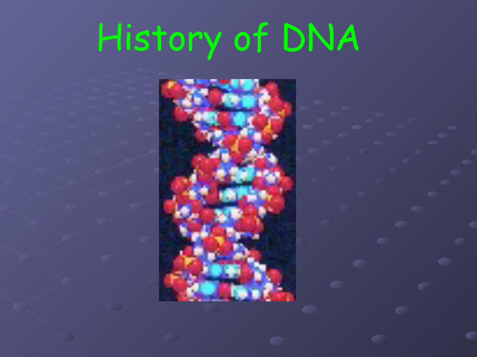
History of DNA
In the 1900’s biologists were trying to find these answers.
In order to understand genetics, biologists had to figure out the chemical structure of a gene.
(what are the parts & pieces?)

Friedrich Meischer – 1870’sFirst to isolate DNA

Frederick Griffith - 1928
-Studied two types of bacteria (smooth/rough)-cause of pneumonia (lung disease)
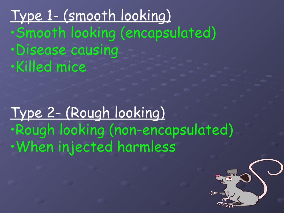
Type 2- (Rough looking)•Rough looking (non-encapsulated)•When injected harmless
Type 1- (smooth looking)•Smooth looking (encapsulated)•Disease causing•Killed mice
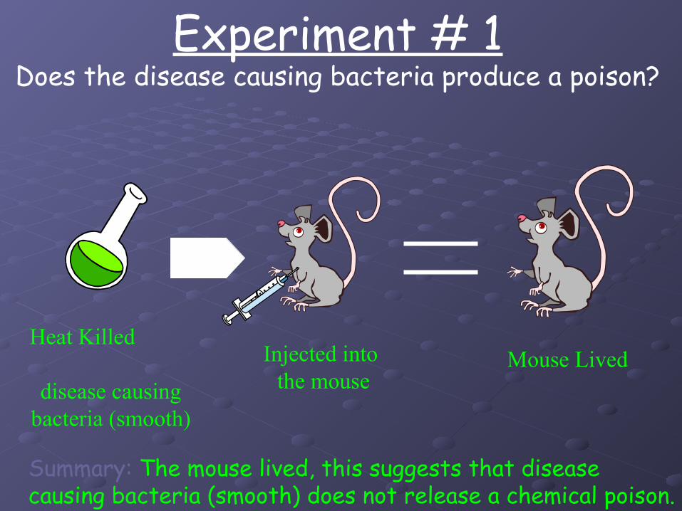
Experiment # 1Does the disease causing bacteria produce a poison?
Heat Killed
disease causingbacteria (smooth)
Mouse LivedInjected into the mouse
Summary: The mouse lived, this suggests that diseasecausing bacteria (smooth) does not release a chemical poison.
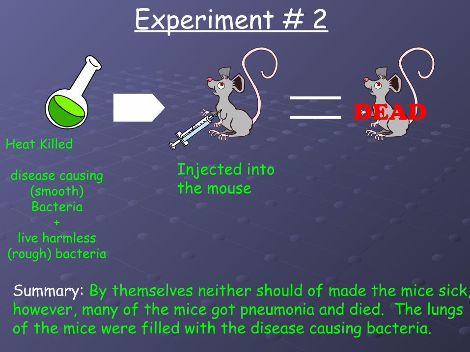
Heat Killed
disease causing (smooth) Bacteria
+live harmless
(rough) bacteria
Injected into the mouse
Experiment # 2
Summary: By themselves neither should of made the mice sick,however, many of the mice got pneumonia and died. The lungsof the mice were filled with the disease causing bacteria.
DEAD
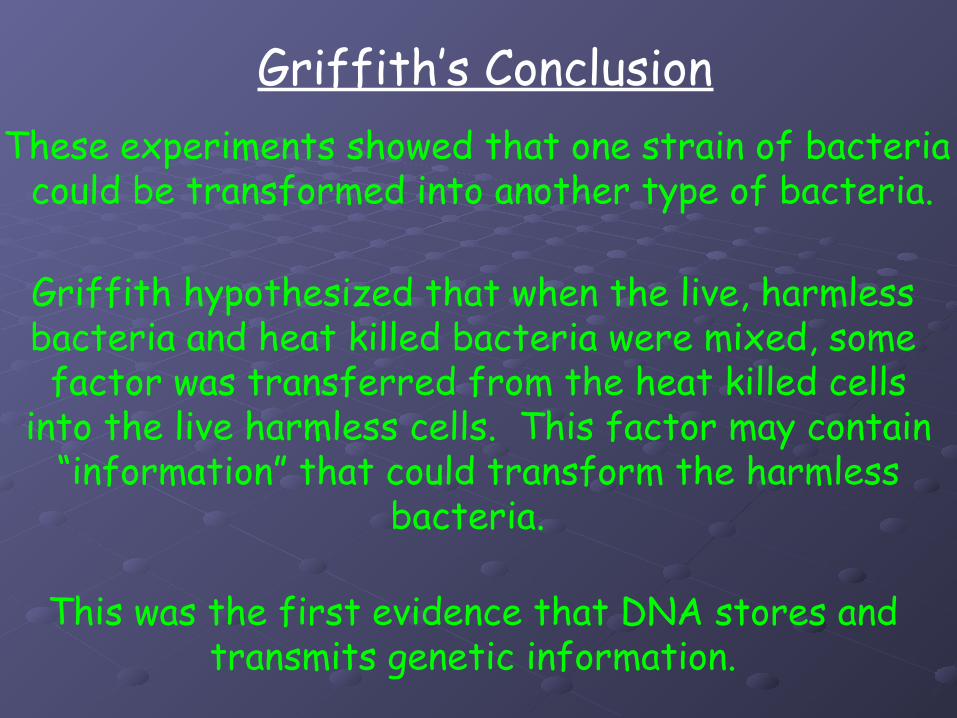
These experiments showed that one strain of bacteria could be transformed into another type of bacteria.
Griffith’s Conclusion
Griffith hypothesized that when the live, harmless bacteria and heat killed bacteria were mixed, some factor was transferred from the heat killed cells
into the live harmless cells. This factor may contain“information” that could transform the harmless
bacteria.
This was the first evidence that DNA stores and transmits genetic information.

Oswald Avery - 1944
Oswald Avery led a group of scientists to furtherinvestigate the work of Griffith. They wanted find out whichmolecule in the heat killed bacteria was the most important
for transformation.
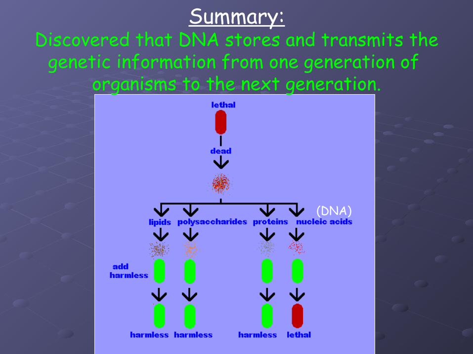
Summary:Discovered that DNA stores and transmits the
genetic information from one generation of organisms to the next generation.
(DNA)
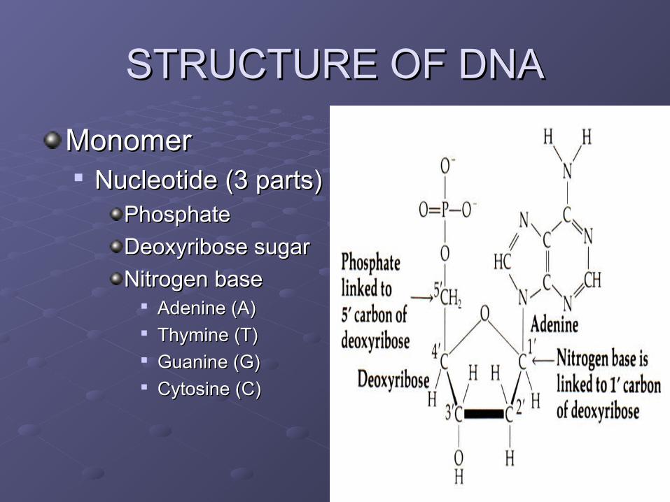
STRUCTURE OF DNASTRUCTURE OF DNA
MonomerMonomer Nucleotide (3 parts)Nucleotide (3 parts)
PhosphatePhosphate
Deoxyribose sugarDeoxyribose sugar
Nitrogen baseNitrogen base Adenine (A)Adenine (A) Thymine (T)Thymine (T) Guanine (G)Guanine (G) Cytosine (C)Cytosine (C)
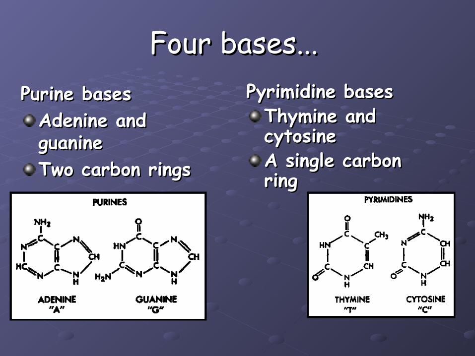
Four bases...Four bases...Purine basesPurine bases
Adenine and Adenine and guanineguanineTwo carbon ringsTwo carbon rings
Pyrimidine basesPyrimidine basesThymine and Thymine and cytosinecytosineA single carbon A single carbon ringring
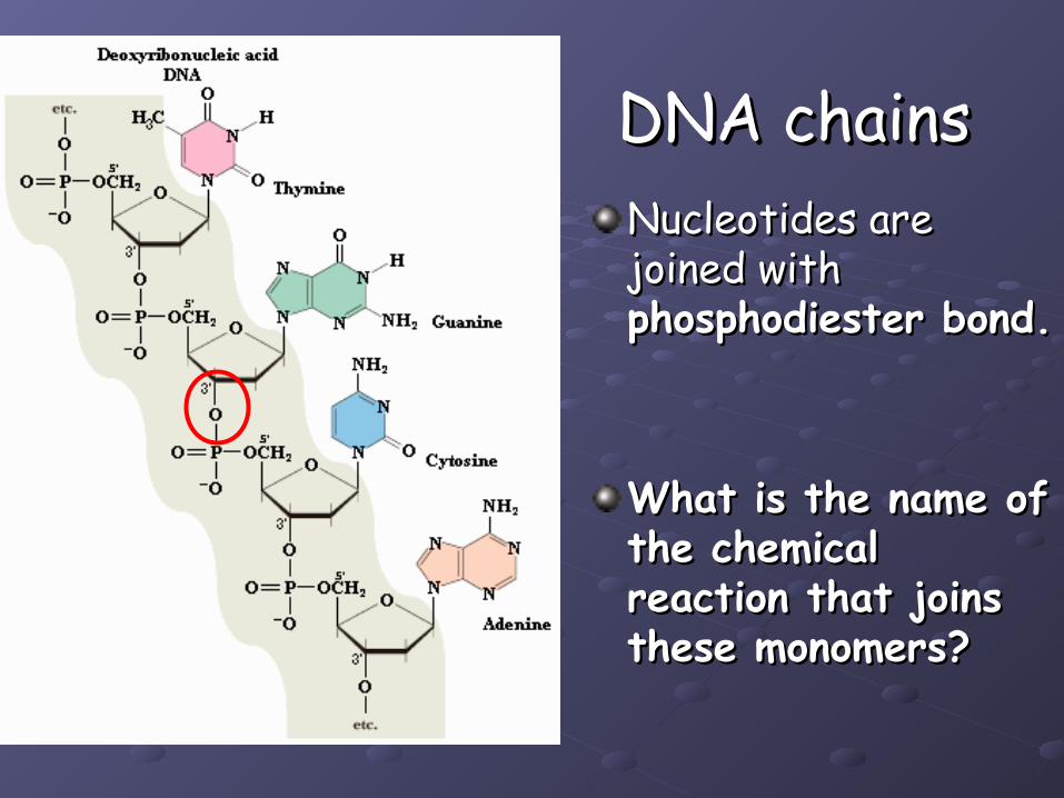
DNA chainsDNA chainsNucleotides are Nucleotides are joined with joined with phosphodiester bond.phosphodiester bond.
What is the name of What is the name of the chemical the chemical reaction that joins reaction that joins these monomers?these monomers?
Edwin Chargaff - 1947Edwin Chargaff - 1947
Studied DNA in various species.Studied DNA in various species.
Found that nitrogen-containing bases are Found that nitrogen-containing bases are proportionate within each species.proportionate within each species.
The proportions hold true across species.The proportions hold true across species.
Came up with rules for complementary Came up with rules for complementary base-pairing.base-pairing.

Chargaff’s RulesChargaff’s Rules
Adenine and Thymine are found in Adenine and Thymine are found in proportionate amounts.proportionate amounts.
%A = %T%A = %T
Cytosine and Guanine are found in Cytosine and Guanine are found in proportionate amounts.proportionate amounts.
%C = %G%C = %G
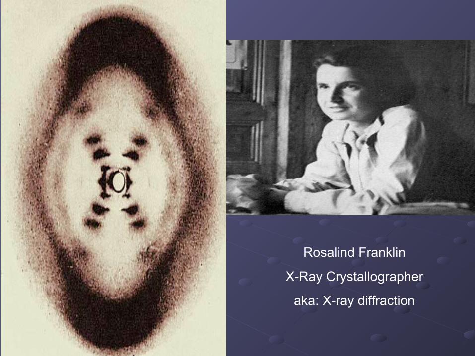
Rosalind Franklin
X-Ray Crystallographer
aka: X-ray diffraction
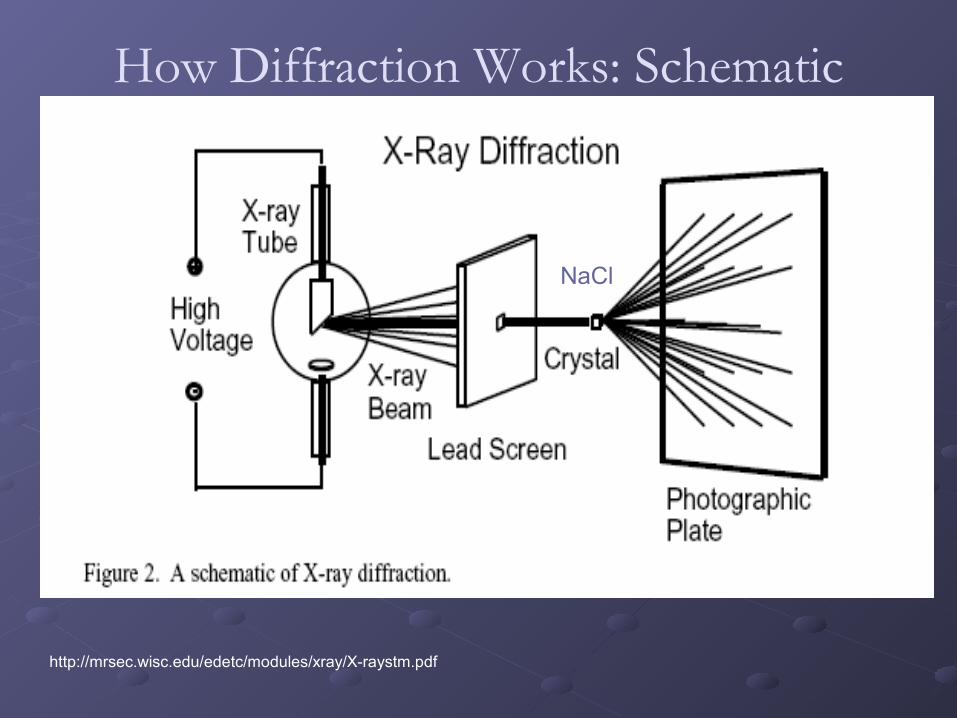
How Diffraction Works: Schematic
http://mrsec.wisc.edu/edetc/modules/xray/X-raystm.pdf
NaCl

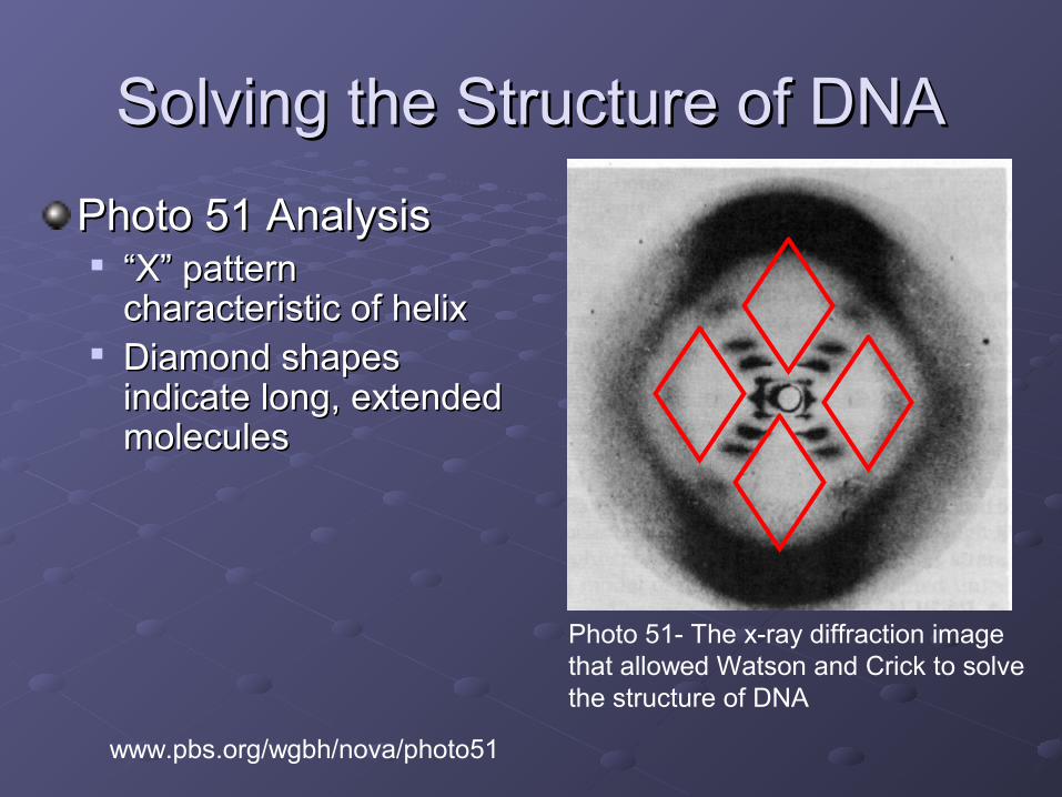
Solving the Structure of DNASolving the Structure of DNA
Photo 51 AnalysisPhoto 51 Analysis
Photo 51- The x-ray diffraction image that allowed Watson and Crick to solve the structure of DNA
www.pbs.org/wgbh/nova/photo51

Solving the Structure of DNASolving the Structure of DNA
Photo 51- The x-ray diffraction image that allowed Watson and Crick to solve the structure of DNA
Photo 51 AnalysisPhoto 51 Analysis ““X” pattern X” pattern
characteristic of helixcharacteristic of helix
www.pbs.org/wgbh/nova/photo51
Solving the Structure of DNASolving the Structure of DNA
Photo 51- The x-ray diffraction image that allowed Watson and Crick to solve the structure of DNA
Photo 51 AnalysisPhoto 51 Analysis ““X” pattern X” pattern
characteristic of helixcharacteristic of helix Diamond shapes Diamond shapes
indicate long, extended indicate long, extended moleculesmolecules
www.pbs.org/wgbh/nova/photo51

Solving the Structure of DNASolving the Structure of DNA
Photo 51- The x-ray diffraction image that allowed Watson and Crick to solve the structure of DNA
Photo 51 AnalysisPhoto 51 Analysis ““X” pattern X” pattern
characteristic of helixcharacteristic of helix Diamond shapes Diamond shapes
indicate long, extended indicate long, extended moleculesmolecules
Smear spacing reveals Smear spacing reveals distance between distance between repeating structuresrepeating structures
Missing smears Missing smears indicate interference indicate interference from second helixfrom second helix
www.pbs.org/wgbh/nova/photo51

Information Gained from Photo 51Information Gained from Photo 51 Double HelixDouble Helix Radius: 10 angstromsRadius: 10 angstroms Distance between bases: 3.4 angstroms Distance between bases: 3.4 angstroms Distance per turn: 34 angstromsDistance per turn: 34 angstroms
Combining Data with Other Information Combining Data with Other Information DNA made from:DNA made from:
sugarsugarphosphates phosphates
4 bases (A,C,G,T)4 bases (A,C,G,T) Chargaff’s RulesChargaff’s Rules
%A=%T%A=%T%G=%C%G=%C
Molecular ModelingMolecular Modeling
Solving the Structure of DNASolving the Structure of DNA
Watson and Crick’s model
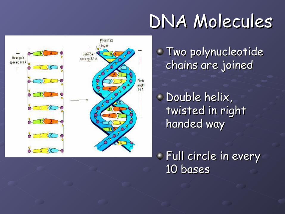
DNA MoleculesDNA MoleculesTwo polynucleotide Two polynucleotide chains are joinedchains are joined
Double helix, Double helix, twisted in right twisted in right handed wayhanded way
Full circle in every Full circle in every 10 bases10 bases
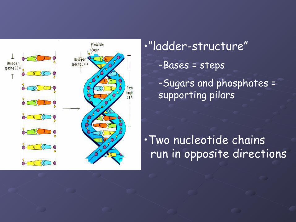
•”ladder-structure”–Bases = steps
–Sugars and phosphates = supporting pilars
•Two nucleotide chains run in opposite directions
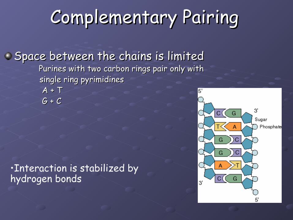
Complementary PairingComplementary Pairing
Space between the chains is limited Space between the chains is limited Purines with two carbon rings pair only with Purines with two carbon rings pair only with single ring pyrimidinessingle ring pyrimidines A + TA + T G + CG + C
•Interaction is stabilized by hydrogen bonds
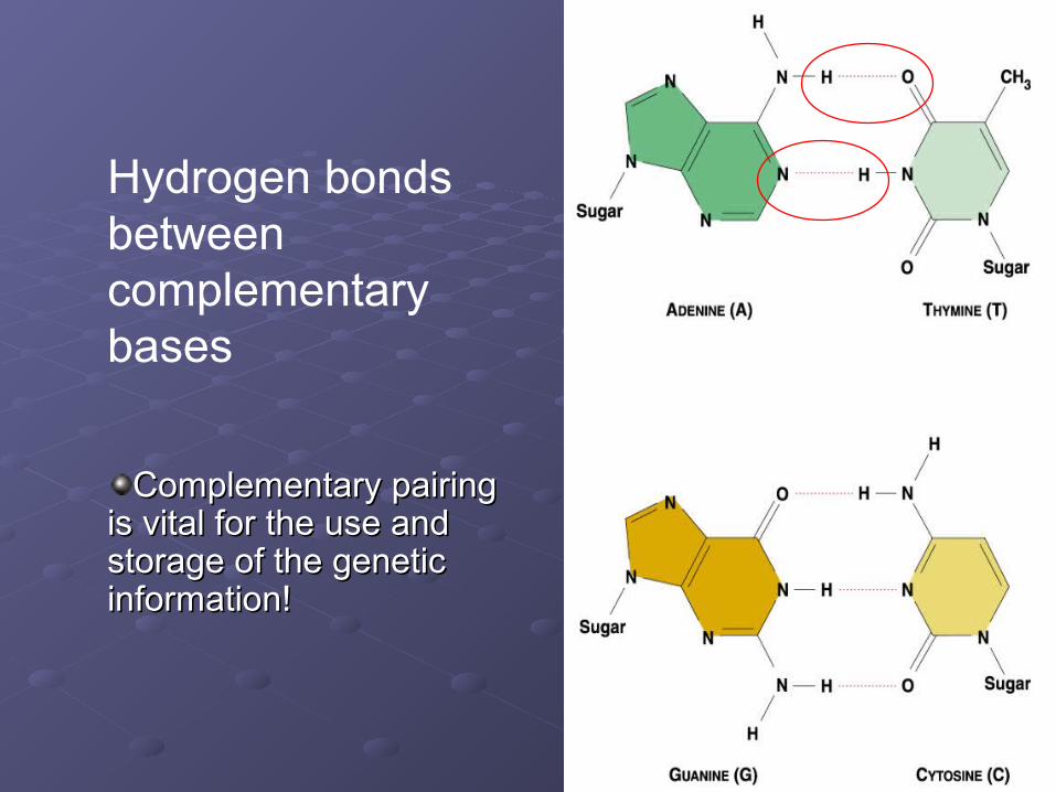
Hydrogen bonds between complementary bases
Complementary pairing Complementary pairing is vital for the use and is vital for the use and storage of the genetic storage of the genetic information!information!