dna damage response -...
TRANSCRIPT

DNA Damage Response
Giuseppina Giglia-Mari1,2,3, Angelika Zotter1,4, and Wim Vermeulen1
1Department of Genetics, Erasmus University Medical Center, Dr Molewaterplein 50, 3015 GE Rotterdam,The Netherlands
2Department of Cancer Biology, Institute of Pharmacology and Structural Biology, CNRS,205 route de Narbonne, 31077 Toulouse, France
3Universite de Toulouse Paul Sabatier, 31000, Toulouse, France4Core Research Laboratory, Istituto Toscano Tumori, Villa delle Rose, Via Cosimo il Vecchio 2,50139 Firenze, Italy
Correspondence: [email protected]
Structural changes to DNA severely affect its functions, such as replication and transcription,and play a major role in age-related diseases and cancer. A complicated and entanglednetwork of DNA damage response (DDR) mechanisms, including multiple DNA repair path-ways, damage tolerance processes, and cell-cycle checkpoints safeguard genomic integrity.Like transcription and replication, DDR is a chromatin-associated process that is generallytightly controlled in time and space. As DNA damage can occur at any time on anygenomic location, a specialized spatio-temporal orchestration of this defense apparatus isrequired.
Selective advantage by random mutations inthe genetic material has driven evolution of
terrestrial life. Despite this obvious advantagefor biological diversity, genome instability hasin most cases adverse effects on organismallife. Preservation of genomic integrity is a pre-requisite for proper cell function and faith-ful transmission of the genome to progeny.However, environmental factors and the chem-ical properties of DNA do not guarantee life-long stability and proper functioning of thegenome.
Genomic insults arise from side effects ofDNA metabolizing processes, such as replicationerrors, uncontrolled recombination, off-targetmutation induction by somatic hypermutation
during antigen production, and inaccurate VDJrecombination (Liu and Schatz 2009; Mahaneyet al. 2009). The biggest genomic burden is,however, induced by processes that directlydamage DNA. DNA lesions are derived fromthree main sources (Lindahl 1993; Friedberget al. 2006): environmental agents such as ultra-violet light, ionizing radiation, and numerousgenotoxic chemicals; reactive oxygen species(ROS) generated by respiration and lipidperoxidation; and spontaneous hydrolysis ofnucleotide residues, inducing abasic sites anddeamination of C, A, G, or 5methyl-C. It is esti-mated that each cell is confronted with approx-imately 104–105 lesions per day, indicatingthat clearance of genomic injuries constitutes
Editors: Tom Misteli and David L. Spector
Additional Perspectives on The Nucleus available at www.cshperspectives.org
Copyright # 2011 Cold Spring Harbor Laboratory Press; all rights reserved; doi: 10.1101/cshperspect.a000745
Cite this article as Cold Spring Harb Perspect Biol 2011;3:a000745
1
on June 28, 2018 - Published by Cold Spring Harbor Laboratory Press http://cshperspectives.cshlp.org/Downloaded from

a demanding task to maintain proper genomefunction.
Essential genome processes, such as tran-scription and replication, are severely affectedby DNA lesions. Replication over damaged DNAinduces mutations, which may initiate and pro-pagate carcinogenesis. Acute effects arise whenlesions block transcription causing cellularsenescence or apoptosis, resulting in damage-induced accelerated aging (Mitchell et al. 2003;Akbari and Krokan 2008; Sinclair and Oberdo-erffer 2009).
THE DNA DAMAGE RESPONSE
To deal with the fundamental problem of geno-mic erosion, a sophisticated network of DNAdamage-response (DDR) systems has evolved.These include a set of DNA repair mecha-nisms, damage tolerance processes, and cell-cycle
checkpoint pathways. The biological signifi-cance of a functional DDR for human healthis clearly illustrated by the severe consequencesof inherited defects in DDR factors resultingin various diseases, including immune defi-ciency, neurological degeneration, prematureaging, and severe cancer susceptibility (Hoeij-makers 2001; Hoeijmakers 2009).
DNA Repair Mechanisms
The heart of the cellular defense against DNAinjuries is formed by a variety of DNA repairmechanisms (Hoeijmakers 2001; Hoeijmakers2009), each with their own damage specifi-city (Table 1). Together, they are able to removethe vast majority of injuries from the genome.The simplest solution that emerged in evolutionis the direct reversal of lesions by specializedactivities, such as photolyases that selectively
Table 1. Induction of DNA lesions and corresponding repair pathway.
Lesion Cause Repair process(es)
CPD, 6-4PP(1) Sunlight NERBulky adducts(2) Food, cigarette smoke NERIntrastrand crosslinks Chemotherapy (e.g., Cis-Pt) NER8-oxo-dG(3) ROS(4), respiration BERThymineglycol(3) ROS(4), respiration BERN7-Alkyl-dG, N3-Alkyl-dA Food, pollutants BERO6-Alkyl-dG Food, pollutants DR(5), BER?5-methyl-dC DNMT(6) BER/AID-BER/NER?(7)
Uracil, (Hypo)Xanthine Spontaneous deamination BERAbasic site Spontaneous hydrolysis BERSingle-strand breaks Ionizing radiation, ROS Ligation, BERDouble-strand breaks Ionizing radiation, ROS, VDJ-rec HR, NHEJTyrosyl-30DNA(8) Topo-I inhibition, ROS SSBRMismatches Replication errors MMRSmall insertion/deletions Replication slippage MMRInterstrand crosslinks Chemotherapy ICLR/ HR?
1. CPD: cyclobutane pyrimidine dimer; 6-4 PP: 6-4 pyrimidine-pyrimidone photo-product.
2. A large group of chemicals conjugated to bases that cause DNA helix destabilization such as: Benzo[a]pyrene (a polycylic
aromatic hydrocarbon); Aflatoxins (present in fungal food contaminations); and Nitrosamines (tobacco smoke).
3. A large group of different oxidation products affecting either the base or the phosphate-sugar backbone of which
8-oxo-dG is the most abundant.
4. ROS: reactive oxygen species, produced as side-product of respiration/metabolism and ionizing radiation.
5. DR: direct reversal, involving the suicide enzyme MGMT.
6. DNMT: DNA methyltransferase, functions in epigenetic gene-expression control (e.g., at CpG islands).
7. The mechanism of 5-Me-C repair/conversion is a matter of debate. Recently, a GADD45a-dependent NER reaction was
suggested (Barreto et al. 2007).
8. Proteolytic degradation of conjugated Topo-I to 30DNA termini creates tyrosyl-30DNA bonds, resolved by TDP1
(El-Khamisy et al. 2009).
G. Giglia-Mari, A. Zotter, and W. Vermeulen
2 Cite this article as Cold Spring Harb Perspect Biol 2011;3:a000745
on June 28, 2018 - Published by Cold Spring Harbor Laboratory Press http://cshperspectives.cshlp.org/Downloaded from

reverse UV-induced DNA damage (Weber2005) and the suicide enzyme O6-methylgua-nine transferase (MGMT) that transfers themethyl group from DNA by covalently couplingit to an internal cysteine residue of MGMT,thereby destroying the enzymatic activity(Friedberg et al. 2006). Photolyases are notconserved into the mammalian branch andmammals have to rely on a more complexmechanism to remove UV injuries: nucleotideexcision repair (NER) (see below).
Base Excision Repair (BER)
Bases with small chemical alterations that donot strongly disturb the DNA double-helixstructure are substrates for Base Excision Repair(BER) (Almeida and Sobol 2007; Hegde et al.2008) (Table 1). These damages, or group oflesions, are targeted by lesion-specific DNA gly-cosylases that both recognize and remove thedamaged base from the sugar-phosphate back-bone. The resulting abasic (AP) site is incisedby AP-endonucleases and the single nucleotidegap is filled-in by the BER-specific DNA poly-meraseb and finally sealed by the XRCC1/LigaseIII complex. Single strand breaks (SSBs) arerepaired by a specialized BER mechanism, desig-nated single-strand break repair (SSBR). Theabundant nuclear protein Poly-ADP-Ribose-Polymerase (PARP) is rapidly activated by SSBsand causes auto-poly-ADP-ribosylation, whichrecruits the XRCC1/ligase III complex as wellas end-processing enzymes such as aprataxin(Gueven et al. 2004) and TDP1 (tyrosyl-DNA-phosphodiesterase) to create ligatable DNAends (Caldecott 2007; El-Khamisy et al. 2009).
Nucleotide Excision Repair (NER)
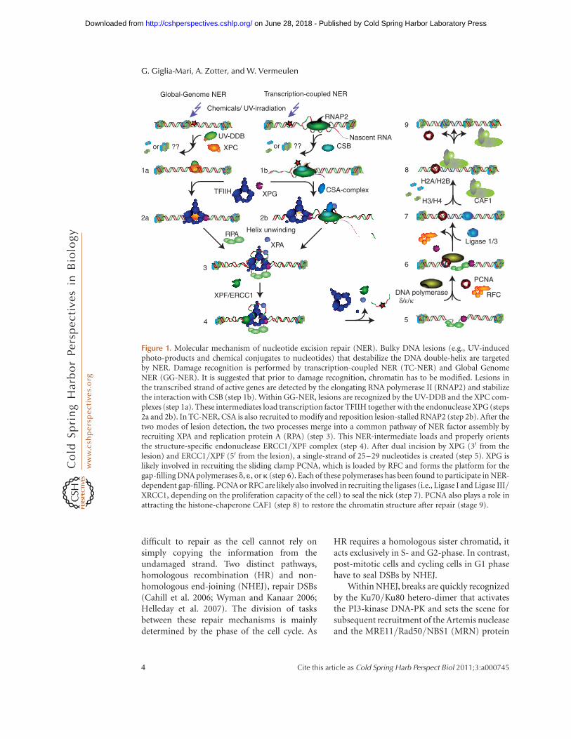
NER removes a broad spectrum of single-strandlesions that cause local helix-destabilization(Table 1). NER is a complex multi-step process,involving the concerted action of at least 25 dif-ferent polypeptides (Hoeijmakers 1993; Gilletand Scharer 2006) (Fig. 1). Two different modesof damage detection are operational in NER:transcription-coupled NER (TC-NER), whichefficiently removes transcription-stalling
lesions and allows quick resumption of tran-scription (Bohr et al. 1986; Hanawalt 1994),and global genome NER (GG-NER), whichlocalizes lesions anywhere in the genome. InTC-NER, damage sensing is performed by thestalled RNA polymerase, and the Cockayne syn-drome factors A and B (CSA and CSB) playessential roles in TC-NER complex assembly(Fousteri et al. 2006; Fousteri and Mullenders2008). Lesion discrimination in GG-NER is exe-cuted by the concerted action of two complexes:XPC/hHR23B (Masutani et al. 1994) and UV-DDB (DDB1 and DDB2/XPE) (Chu and Chang1988; Keeney et al. 1994; Sugasawa et al. 2009).The subsequent steps of TC-NER and GG-NERconverge into a common mechanism in whichfirst the NER/basal transcription factor TFIIH(Egly 2001) is recruited (Yokoi et al. 2000; Volkeret al. 2001). The bi-directional helicase of TFIIHopens the damaged DNA segment over a stretchof approximately 30 nucleotides (Sugasawa et al.2009). The unwound DNA is stabilized by XPAand RPA (Replication Protein A) that also orient(de Laat et al. 1998) the two structure-specificendonucleases XPG (O’Donovan et al. 1994)and the ERCC1-XPF complex (Sijbers et al.1996), which respectively incise the damagedstrand 30 and 50 with respect to the lesion. Theresulting 25–30 nucleotide single strand gap isfilled in by normal DNA replication proteins,including replication factor C (RFC), PCNA,RPA, and the DNA polymerases d, 1, or k (Ogiet al. 2010). Finally, the gap is sealed by DNAligases I or III, dependent on the proliferationstatus of the cell (Moser et al. 2007) (Fig. 1).
DNA Double-Strand Break Repair (DSBR)
Lesions that are substrates for NER and BER arelocated in one of the strands of DNA and areremoved in a “cut-and-patch”-mechanism. Inboth cases, the undamaged complementarystrand serves as a faithful template for the repairof the damaged strand. Some damaging agents,however, affect both strands, such as ionizingradiation that induces DNA double-strand breaks(DSBs) and agents that produce inter-strandcross-links (ISCLs) (Table 1). These lesions areextremely cytotoxic because they are more
DNA Repair
Cite this article as Cold Spring Harb Perspect Biol 2011;3:a000745 3
on June 28, 2018 - Published by Cold Spring Harbor Laboratory Press http://cshperspectives.cshlp.org/Downloaded from
difficult to repair as the cell cannot rely onsimply copying the information from theundamaged strand. Two distinct pathways,homologous recombination (HR) and non-homologous end-joining (NHEJ), repair DSBs(Cahill et al. 2006; Wyman and Kanaar 2006;Helleday et al. 2007). The division of tasksbetween these repair mechanisms is mainlydetermined by the phase of the cell cycle. As
HR requires a homologous sister chromatid, itacts exclusively in S- and G2-phase. In contrast,post-mitotic cells and cycling cells in G1 phasehave to seal DSBs by NHEJ.
Within NHEJ, breaks are quickly recognizedby the Ku70/Ku80 hetero-dimer that activatesthe PI3-kinase DNA-PK and sets the scene forsubsequent recruitment of the Artemis nucleaseand the MRE11/Rad50/NBS1 (MRN) protein
1a
2a
3
UV-DDB
XPC
TFIIH
Helix unwinding
XPA
RPA
4
6
7
PCNA
DNA polymerase δ/ε/κ
Ligase 1/3
XPF/ERCC1
XPG
Chemicals/ UV-irradiation
CSB
Transcription-coupled NERGlobal-Genome NER
1b
2b
CSA-complex
Nascent RNA
RFC
CAF1
H2A/H2B
H3/H4
or ?? or ??
8
9
5
RNAP2 RNAP2
Figure 1. Molecular mechanism of nucleotide excision repair (NER). Bulky DNA lesions (e.g., UV-inducedphoto-products and chemical conjugates to nucleotides) that destabilize the DNA double-helix are targetedby NER. Damage recognition is performed by transcription-coupled NER (TC-NER) and Global GenomeNER (GG-NER). It is suggested that prior to damage recognition, chromatin has to be modified. Lesions inthe transcribed strand of active genes are detected by the elongating RNA polymerase II (RNAP2) and stabilizethe interaction with CSB (step 1b). Within GG-NER, lesions are recognized by the UV-DDB and the XPC com-plexes (step 1a). These intermediates load transcription factor TFIIH together with the endonuclease XPG (steps2a and 2b). In TC-NER, CSA is also recruited to modify and reposition lesion-stalled RNAP2 (step 2b). After thetwo modes of lesion detection, the two processes merge into a common pathway of NER factor assembly byrecruiting XPA and replication protein A (RPA) (step 3). This NER-intermediate loads and properly orientsthe structure-specific endonuclease ERCC1/XPF complex (step 4). After dual incision by XPG (30 from thelesion) and ERCC1/XPF (50 from the lesion), a single-strand of 25–29 nucleotides is created (step 5). XPG islikely involved in recruiting the sliding clamp PCNA, which is loaded by RFC and forms the platform for thegap-filling DNA polymerases d, 1, or k (step 6). Each of these polymerases has been found to participate in NER-dependent gap-filling. PCNA or RFC are likely also involved in recruiting the ligases (i.e., Ligase I and Ligase III/XRCC1, depending on the proliferation capacity of the cell) to seal the nick (step 7). PCNA also plays a role inattracting the histone-chaperone CAF1 (step 8) to restore the chromatin structure after repair (stage 9).
G. Giglia-Mari, A. Zotter, and W. Vermeulen
4 Cite this article as Cold Spring Harb Perspect Biol 2011;3:a000745
on June 28, 2018 - Published by Cold Spring Harbor Laboratory Press http://cshperspectives.cshlp.org/Downloaded from
complex. These proteins are involved in DNAend-processing, preceding ligation performedby the XRCC4/LigaseIV complex (Weteringsand van Gent 2004; Burma et al. 2006; vanGent and van der Burg 2007). During DNAend-processing, loss or changes of a few nu-cleotides may occur. For this reason, NHEJ,although it very rapidly seals DSBs, is an error-prone repair process.
However, when cells do have a homologoustemplate, as in the S- and G2-phase of the cycle,DSBs can be repaired by HR. Homologousrecombination is initiated by binding of theMRN complex to a DSB and functions to holdthe broken pieces together (de Jager et al.2001) and provides the structural bases for theCtIP nuclease. The MRN-CtIP complex cata-lyzes end resection at the break in concertwith exonuclease I (EXO1) (Limbo et al. 2007;Sartori et al. 2007; Takeda et al. 2007). Subse-quently, RPA binds to the newly created single-strand region and through a complicatedhandoff mechanism, the RPA-filament is ex-changed into a RAD51 nucleo-protein filament.This RAD51-filament is crucial for strand inva-sion into the homologous sister, creating a tem-porarily triplex-DNA structure in which strandexchange occurs (Wyman et al. 2004). Themolecular details of these complex transactionsare as yet enigmatic, although genetic studieshave revealed a whole list of proteins that playan important role in these transactions (Lisbyand Rothstein 2009). The biggest challengewithin HR-driven DSBR is, however, the ques-tion on how homologous regions are identifiedwithin the complex nuclear environment.
DNA Damage Tolerance
Persisting lesions not removed by any of therepair mechanism will interfere with DNA rep-lication. Lesion-stalled replication forks canlead to highly cytotoxic DSBs and require aprompt response. At least two DNA damagetolerance mechanisms have evolved: translesionsynthesis (TLS) and recombination-dependentdaughter-strand gap repair (DSGR) (Scullyet al. 2000; Li et al. 2002). These processes donot actually remove lesions, but serve as a
temporary solution to overcome stalled DNAreplication machines. Upon lesion-induced rep-lication blockage, the regular high-fidelity DNApolymerases (pold/1 or a) are temporarily ex-changed with translesion polymerase (pol z-k)(Friedberg et al. 2005; Lehmann 2006) to syn-thesize across the lesion. Although TLS can cir-cumvent lesion-induced replication stalling, thereduced fidelity of the alternative polymerasescauses generally enhanced mutagenesis.
Damage Signaling
To create an extended time window to allowcompletion of lesion removal prior to replica-tion or cell division, damage sensing is linkedto an intricate signal transduction cascade thatinduces cell cycle arrest (Bartek et al. 2007; Call-egari and Kelly 2007). Depending on the natureof the DNA injury and the phase of the cell cyclein which the lesion is encountered, the cell cyclecan be arrested at the G1/S transition, withinthe S-phase, or at the G2/M transition (Zhouand Elledge 2000). Alternatively, when toomany injuries are encountered, apoptosis istriggered in order to protect the organism frompotentially harmful cells (Bernstein et al. 2002).The phosphatidylinositol 3-kinase (PI3) ATM(Ataxia Telangiectasia mutated) is directly re-cruited and activated by the DSB-recognizingprotein complex MRN. This initiating kinasetransduces phosphorylation to a high numberof adapter/transducer proteins, carrying theATM-consensus sequence (Matsuoka et al.2007). Finally, downstream effector kinases, suchas the checkpoint kinase Chk2, are activated(Falck et al. 2002). Bulky lesions cause replica-tion collapse that induces single-strand DNAby the retraction of the replication fork. RPAbinds to ssDNA and recruits ATR (ATM-related) via its association with ATRIP (ATRinteracting protein) and activates the check-point protein Chk1 (Tibbetts et al. 2000; Chenand Sanchez 2004). RPA covered ssDNA alsotriggers the Rad17-dependent loading of theRAD9-HUS1-RAD1 (9-1-1 complex), which isan important transducer of checkpoint activa-tion upon DNA damage (Smits et al. 2010). Athird PI3 kinase, DNA-PK (DNA-dependent
DNA Repair
Cite this article as Cold Spring Harb Perspect Biol 2011;3:a000745 5
on June 28, 2018 - Published by Cold Spring Harbor Laboratory Press http://cshperspectives.cshlp.org/Downloaded from

protein kinase, composed of its catalytic subu-nit DNA-PKcs and a regulatory Ku70/80 heter-odimer), is also activated by IR-induced DSBs.DNA-PKcs is essential for NHEJ in highereukaryotes (Burma et al. 2006) and additionallyfunctions in telomere maintenance and induc-tion of apoptosis (Burma and Chen 2004). Intotal, a complicated set of different emergencystrategies are called into action when genomicinsults are encountered. Although many of theindividual players are identified and the down-stream signaling cascades have been dissected,their respective interactions and communica-tion is far from resolved.
Intertwined DNA-transacting Processes
The different repair processes are generally con-sidered separate entities. However, in recentyears, it has become clear that most of theseDDR processes are part of an intricate networkwith significant overlap, often sharing specificessential components.
Several DDR factors appeared to act indiverse DNA maintenance systems. One typicalexample is the hetero-dimeric ERCC1/XPFcomplex. This structure-specific endonucleasewas originally identified as the nuclease thatincises 50 of the DNA lesions within NER (West-erveld et al. 1984; Sijbers et al. 1996). Furtheranalysis revealed additional functions for thiscomplex in HR (Adair et al. 2000; Niedernhoferet al. 2001), interstrand cross-link repair (DeSilva et al. 2000; Niedernhofer et al. 2004), andtelomere maintenance (Zhu et al. 2003).
Besides overlap between distinct repair pro-cesses, DDR is also linked to other essentialDNA transacting mechanisms, such as tran-scription and replication. A prime example ofsuch a link is the tight connection betweenNER and transcription, illustrated by the exis-tence of a specialized transcription-coupledNER pathway (TC-NER) (Bohr et al. 1985; Fou-steri and Mullenders 2008). The chromatin re-modeling protein Cockayne Syndrome B (CSB)(Citterio et al. 2000) is essential for TC-NERand is implicated in transcription elongation(van den Boom et al. 2004). Moreover, the basalRNA polymerase II transcription factor TFIIH
is also a pivotal factor in NER (Fig. 1) (Schaefferet al. 1993; Drapkin et al. 1994; Egly 2001;Hoogstraten et al. 2002). Finally, the essentialreplication factor A (RPA) is implicated in basi-cally all DDR mechanisms, including NER, HR,and damage signaling.
Different strategies are used to control themulti-functionality of these factors: (1) distinctspatial organization, (2) incorporation intodiverse functional complexes, and/or (3) dy-namic sharing of these components. Regulationof pleiotropic functionality of proteins is com-monly achieved by distinct posttranslationalmodifications (PTMs). Within DDR, differentPTMs were identified, ranging from phosphor-ylation, acetylation, methylation, neddylation,mono- and poly-ubiquitylation, and sumoy-lation to poly-ADP-ribosylation (Harper andElledge 2007; Huen and Chen 2008). One ofthe most common PTMs involved in DDR isdifferential phosphorylation, mainly driven bythe ATM, ATR, and DNA-PKcs kinases (Mat-suoka et al. 2007). Recent research indicatesalso that differential ubiquitination plays animportant role in DDR regulation (Berginket al. 2007; Harper and Elledge 2007; Reed andGillette 2007; Huen and Chen 2008; Alpi andPatel 2009; Panier and Durocher 2009).
STRUCTURAL AND FUNCTIONALORGANIZATION OF DDR
Chromatin and DDR
The nucleus is highly structured and function-ally compartmentalized in part due to areas ofvarious degrees of chromatin compaction,creating possible obstacles for DDR factor ac-cessibility. Decompaction and subsequent res-toration of the starting chromatin structure inconjunction with DDR thus creates anotherlevel of complexity in genome maintenance reg-ulation. Chromatin-associated processes suchas transcription, replication, and DNA repairare regulated by a complex set of structuralchanges in chromatin (Groth et al. 2007; Liet al. 2007). Control of chromatin functionsand its compaction occurs by at least fourknown processes: (1) active ATP-consuming
G. Giglia-Mari, A. Zotter, and W. Vermeulen
6 Cite this article as Cold Spring Harb Perspect Biol 2011;3:a000745
on June 28, 2018 - Published by Cold Spring Harbor Laboratory Press http://cshperspectives.cshlp.org/Downloaded from

remodeling machines of the SWI/SNF-super-family of DNA-dependent ATPases (Neves-Costa and Varga-Weisz 2006; Saha et al. 2006)that slide or physically evict core histones orentire nucleosomes from active sites; (2) incor-poration of diverse histone variants by histonechaperones (Loyola and Almouzni 2007; Altafet al. 2009); (3) differential binding of abundantnon-core histone proteins, such as the linkerhistone H1, the family of high mobility groupproteins (HMG), or different isoforms of thehetero-chromatin protein 1 (HP1); and (4)covalent modifications or PTMs of the corehistones, such as acetylation, methylation,phosphorylation, and ubiquitylation (He andLehming 2003). Accordingly, recently a largenumber of chromatin modifications and re-modeling events were shown to be linked toDDR (Groth et al. 2007; Dinant et al. 2008;Misteli and Soutoglou 2009; Nag and Smerdon2009; van Attikum and Gasser 2009).
Access, Repair, and Restore
Despite increasing knowledge of the role ofchromatin in DDR, a general mode of actionor detailed mechanistic insight is lacking.Already in 1991 a hypothetical three step modelfor DNA repair in chromatin was postulated(Smerdon 1991), the so-called “ARR-model,”for Access, Repair, and Restore, based on anal-ogy to transcription regulation in chromatin.In this model, it was postulated that chromatinremodeling would be required to provide “ac-cess” of damage-recognition factors to initiate“DNA repair” and, when the job is finished,“restoration” of the chromatin structure. Sincethen, clear indications have been found thatthe H3/H4 chaperone CAF1, likely in conjunc-tion with Asf1 (anti-silencing function 1), isimplicated in restoring chromatin after NER(Green and Almouzni 2002; Mello et al. 2002;Polo et al. 2006). Additional studies implicatedAsf1 and FACT (facilitating transcription fac-tor) (Chen et al. 2008; Heo et al. 2008) in his-tone exchange near DSBs. However, less directevidence for chromatin remodeling factors forthe first step (access) was found. Althoughseveral chromatin remodelers, such as INO80
(Downs et al. 2000; van Attikum et al. 2004),facilitate DDR factor recruitment by movinghistones away from the break, they appear toact after the initial damage recognition. In addi-tion, chromatin modifications in yeast occurafter UV-irradiation by Gcn5-induced H3 ace-tylation and are dependent on the Swi/SnfDNA translocase Rad16/Rad7/Abf1. Despitethe more open chromatin structure by hyper-acetylation, it is not directly clear whether thismodification facilitates the recruitment of theRad4 (yeast ortholog of XPC) DNA damagerecognition protein (Waters et al. 2009).
Phosphorylation of the Histone H2AVariant H2AX
The most prominent DDR-associated covalenthistone modification is the phosphorylation ofthe histone H2A-variant H2AX in response toDNA damage by the checkpoint kinases ATM,ATR, and DNA-PKcs (Rogakou et al. 1998;O’Driscoll et al. 2003; Falck et al. 2005).H2AX is incorporated into approximately5%–25% of histone octamers, although itsphosphorylation (gH2AX) is constrained tomicroscopically discernable structures, the ion-izing irradiation-induced foci (IRIF) (Fig. 2).Phosphorylation of H2AX is a relatively earlyevent after damage, immediately following MRNbinding and ATM activation. These gH2AX focico-localize with most of the DSB-associatedDDR factors (see below) and are thought to serveas docking sites for recruiting and retainingDNA repair and signaling factors to DSBs.g-H2AX spreads over several megabases aroundDSBs and appears condensed into IRIFs (Roga-kou et al. 1999), suggesting a dominant struc-tural role in DSB-DDR. Surprisingly, however,although mice lacking H2AX are radiation-sensi-tive and exhibit several features associated withdefective DDR, they are only partially defectivein DSB repair and are not fully compromisedin checkpoint activation (Celeste et al. 2002).This notion argues, contrary to expectation,that this impressive structural organization intolarge molecular assemblies only makes theDDR process more efficient but is not essentialfor DDR.
DNA Repair
Cite this article as Cold Spring Harb Perspect Biol 2011;3:a000745 7
on June 28, 2018 - Published by Cold Spring Harbor Laboratory Press http://cshperspectives.cshlp.org/Downloaded from
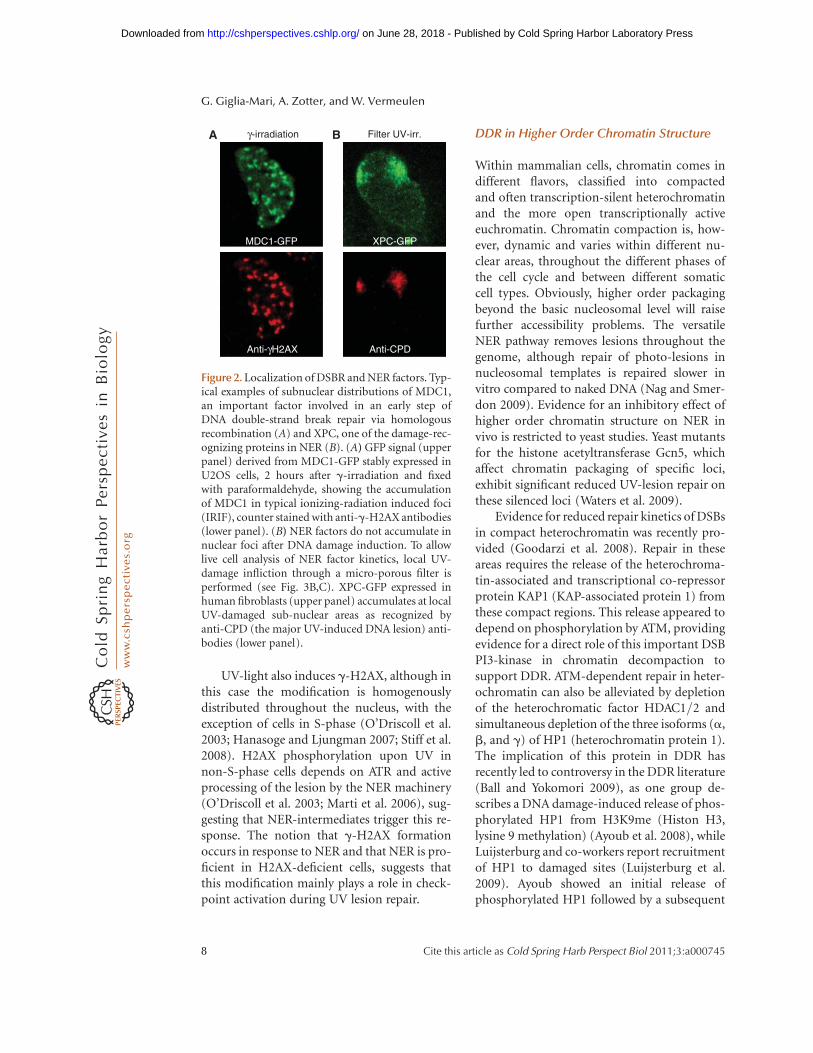
UV-light also induces g-H2AX, although inthis case the modification is homogenouslydistributed throughout the nucleus, with theexception of cells in S-phase (O’Driscoll et al.2003; Hanasoge and Ljungman 2007; Stiff et al.2008). H2AX phosphorylation upon UV innon-S-phase cells depends on ATR and activeprocessing of the lesion by the NER machinery(O’Driscoll et al. 2003; Marti et al. 2006), sug-gesting that NER-intermediates trigger this re-sponse. The notion that g-H2AX formationoccurs in response to NER and that NER is pro-ficient in H2AX-deficient cells, suggests thatthis modification mainly plays a role in check-point activation during UV lesion repair.
DDR in Higher Order Chromatin Structure
Within mammalian cells, chromatin comes indifferent flavors, classified into compactedand often transcription-silent heterochromatinand the more open transcriptionally activeeuchromatin. Chromatin compaction is, how-ever, dynamic and varies within different nu-clear areas, throughout the different phases ofthe cell cycle and between different somaticcell types. Obviously, higher order packagingbeyond the basic nucleosomal level will raisefurther accessibility problems. The versatileNER pathway removes lesions throughout thegenome, although repair of photo-lesions innucleosomal templates is repaired slower invitro compared to naked DNA (Nag and Smer-don 2009). Evidence for an inhibitory effect ofhigher order chromatin structure on NER invivo is restricted to yeast studies. Yeast mutantsfor the histone acetyltransferase Gcn5, whichaffect chromatin packaging of specific loci,exhibit significant reduced UV-lesion repair onthese silenced loci (Waters et al. 2009).
Evidence for reduced repair kinetics of DSBsin compact heterochromatin was recently pro-vided (Goodarzi et al. 2008). Repair in theseareas requires the release of the heterochroma-tin-associated and transcriptional co-repressorprotein KAP1 (KAP-associated protein 1) fromthese compact regions. This release appeared todepend on phosphorylation by ATM, providingevidence for a direct role of this important DSBPI3-kinase in chromatin decompaction tosupport DDR. ATM-dependent repair in heter-ochromatin can also be alleviated by depletionof the heterochromatic factor HDAC1/2 andsimultaneous depletion of the three isoforms (a,b, and g) of HP1 (heterochromatin protein 1).The implication of this protein in DDR hasrecently led to controversy in the DDR literature(Ball and Yokomori 2009), as one group de-scribes a DNA damage-induced release of phos-phorylated HP1 from H3K9me (Histon H3,lysine 9 methylation) (Ayoub et al. 2008), whileLuijsterburg and co-workers report recruitmentof HP1 to damaged sites (Luijsterburg et al.2009). Ayoub showed an initial release ofphosphorylated HP1 followed by a subsequent
MDC1-GFP XPC-GFP
Anti-CPDAnti-γH2AX
γ-irradiationA B Filter UV-irr.
Figure 2. Localization of DSBR and NER factors. Typ-ical examples of subnuclear distributions of MDC1,an important factor involved in an early step ofDNA double-strand break repair via homologousrecombination (A) and XPC, one of the damage-rec-ognizing proteins in NER (B). (A) GFP signal (upperpanel) derived from MDC1-GFP stably expressed inU2OS cells, 2 hours after g-irradiation and fixedwith paraformaldehyde, showing the accumulationof MDC1 in typical ionizing-radiation induced foci(IRIF), counter stained with anti-g-H2AX antibodies(lower panel). (B) NER factors do not accumulate innuclear foci after DNA damage induction. To allowlive cell analysis of NER factor kinetics, local UV-damage infliction through a micro-porous filter isperformed (see Fig. 3B,C). XPC-GFP expressed inhuman fibroblasts (upper panel) accumulates at localUV-damaged sub-nuclear areas as recognized byanti-CPD (the major UV-induced DNA lesion) anti-bodies (lower panel).
G. Giglia-Mari, A. Zotter, and W. Vermeulen
8 Cite this article as Cold Spring Harb Perspect Biol 2011;3:a000745
on June 28, 2018 - Published by Cold Spring Harbor Laboratory Press http://cshperspectives.cshlp.org/Downloaded from

spreading to neighboring chromatin. This dis-crepancy is difficult to explain, besides possibledifference in DNA damage induction and slightdifferences in kinetic measurements. A possibleexplanation for the apparent initial disappear-ance prior to the observed accumulation atdamaged sites might be the sudden highly local-ized damage induction. The extreme high localconcentration of light might in addition toDNA damage also induce chromatin-proteindamage. The next wave of HP1 accumulationreflects then the more physiological responseto DNA damage induction. Despite the conflict-ing data and interpretations, it is clear that com-paction of chromatin and HP1 play an as yet notentirely understood role in DDR, as disruptionof HP1 orthologs in C. elegans induces a diversespectrum of DNA damage sensitivities (Ball andYokomori 2009; Luijsterburg et al. 2009).
Tools to Analyze DDR in Living Cells
The dynamic interactions with chromatin andthe multiple engagements of DDR factors indi-cate that analysis of each of the separate pro-cesses in vitro is not sufficient to fully uncovermechanistic details, and demands cellularbiological approaches. The possibility togenetically tag proteins with the autofluores-cent protein GFP has revolutionized cell biology(Tsien and Miyawaki 1998). The simultaneoustechnological advances in microscopy anddevelopment of quantitative fluorescent meas-urements and sophisticated photo-bleachingprocedures (White and Stelzer 1999; Houts-muller and Vermeulen 2001; Lippincott-Schwartz et al. 2001) have provided spectacularnew insights into the regulation and dynamicorganization of chromatin-associated processes(Houtsmuller et al. 1999; Phair and Misteli2000). In particular, the development of severalsystems to locally introduce DNA damage orimmobilize DDR factors in cultured living cellshas been beneficial (Figs. 2 and 3): (1) irradia-tion through a filter or mask that partly shieldthe cells (Nelms et al. 1998; Katsumi et al.2001; Mone et al. 2001); (2) micro-beam laserirradiation, with or without photo-sensitizers,at sub-nuclear areas (Cremer et al. 1980; Tashiro
et al. 2000; Lukas et al. 2003; Meldrum et al.2003; Lan et al. 2004; Dinant et al. 2007); (3)guided a-particle and heavy iron radiation(Jakob et al. 2003; Aten et al. 2004; Hauptneret al. 2004); (4) integration of rare-cuttingendonucleases (Lisby et al. 2004; Rodrigue et al.2006; Soutoglou et al. 2007); and (5) DDR pro-tein tethering to specific integrated amplifiedsequences (Soutoglou and Misteli 2008).
Organization of DNA Double-StrandBreak Response
In situ studies revealed that next to g-H2AX, ahigh number of DDR proteins relocalize intoIRIF foci upon genomic stress (Bekker-Jensenet al. 2006). This is particularly pronounced forproteins implicated in the repair and signalingof DSBs by homologous recombination (HR)(Fig. 2). A systematic analysis of the spatial dis-tribution of DSB-DDR factors using a methodto locally introduce DSBs in cultured living cells(Lukas et al. 2003) resulted in a localization clas-sification of DDR factors (Bekker-Jensen et al.2006). In this procedure, cells were cultured inthe presence of photo-sensitizing nucleotide-analogs (Iodo-deoxyuridine) prior to micro-beam laser irradiation with 337 nm that inducesDSB in a user-defined sub-nuclear area (Lukaset al. 2003). Several subclasses of repair proteinswere found based on their recruitment proper-ties: (1) The major checkpoint mediators, suchas Mdc1 (mediator of DNA damage checkpointprotein 1), 53BP (p53 binding protein), BRCA1(breast cancer protein 1), ATM, and the MRNcomplex, co-localize in IRIF with g-H2AX-decorated chromatin, termed “DSB-flankingchromatin”. Assembly of these proteins at theDSB-flanking chromatin appeared to occurthroughout the cell cycle. It has been estimatedthat these foci contain several hundred copiesof each of the participating DDR factors. (2)Another group of DSB-activated proteinsassembles in much smaller ssDNA micro-com-partments that are most likely formed by 50
resections at DSB, an important HR intermedi-ate. These “microfoci” are only formed in S- andG2-phase cells and typically accumulate next toRPA, factors directly involved in HR repair, like
DNA Repair
Cite this article as Cold Spring Harb Perspect Biol 2011;3:a000745 9
on June 28, 2018 - Published by Cold Spring Harbor Laboratory Press http://cshperspectives.cshlp.org/Downloaded from

Rad51, Rad52, BRCA1, and FANCD2 (Fanconianemia complementation group D2). They alsocontain the ssDNA-activated checkpoint kinaseATR and the 9-1-1 complex (Warmerdam et al.2009). Also MRN and BRCA1 were found inthese microfoci independent of g-H2AX orMdc1, while their recruitment to DSB-flankingchromatin is dependent on these proteins. (3)Several DSB repair factors, particularly thoseinvolved in NHEJ, such as DNA-PKcs andKu70/80, could not be found to re-localize
into microscopically discernible foci. It is likelythat this process proceeds much faster than HRand that NHEJ factors do not need to be loadedin such large molecular assemblies to executetheir function. However, using procedures thatintroduce high local concentrations of breaksin living cells, with the aid of multi-photonmicro-beam laser irradiation, microscopicallydiscernible accumulations of NHEJ could befound (Mari et al. 2006; Uematsu et al. 2007).These accumulations likely reflect high local
TFIIH-GFP MergeHoechst
GFP-XPA MergeHoechst
XPC-GFP MergeHoechst
A B
C
D
Time (s)0R
elat
ive
fluor
esce
nceE
Figure 3. Live cell analysis of NER. (A) Distribution of three different NER factors tagged with GFP in living cells;DNA is stained by the DNA stain Hoechst. The damage recognition factor XPC is concentrated in nuclear areas(top panel) that also contain high DNA concentrations when the XPC-GFP (Hoogstraten et al. 2008) is expressedin mouse-embryonal fibroblasts that exhibit the species-specific dense DNA-containing areas. This inhomoge-neous distribution contrasts to other NER factors such as XPA (Rademakers et al. 2003), which are homoge-nously distributed (lower panel) and the repair/transcription factor TFIIH that is enriched in nuclei(Hoogstraten et al. 2002). (B) Schematic cartoon of the procedure to locally inflict UV-damage in living culturedcells by irradiation through a microporous filter (Volker et al. 2001). (C) Local accumulation of XPB-GFP(TFIIH subunit) in UV-damaged areas used to determine the dwell time of this NER factor in the damagedarea by fluorescence recovery after photo bleaching (FRAP). (D) Human fibroblasts expressing XPC-GFP locallydamaged at the indicated position (purple flash, middle panel) by UV-C laser (Dinant et al. 2007). The rightpanel shows a clear accumulation of this protein as soon as 30 seconds after irradiation. (E) Schematic repre-sentation of the quantification of NER factor time-dependent accumulation at local UV-damage in living cellsto determine the assembly kinetics of these factors within the chromatin-bound NER complex.
G. Giglia-Mari, A. Zotter, and W. Vermeulen
10 Cite this article as Cold Spring Harb Perspect Biol 2011;3:a000745
on June 28, 2018 - Published by Cold Spring Harbor Laboratory Press http://cshperspectives.cshlp.org/Downloaded from

concentrations of breaks and repair factorsrather than a specific chromatin structure. (4)Other factors implicated in DSB processing donot exhibit discernible accumulation at sites ofdamage, since these proteins are omnipresenton chromatin and simply get post-translation-ally modified at or near breaks. One of themis Smc1 (Structural Maintenance of Chromo-somes 1), a structural component of the cohe-sin complex required for sister chromatidcohesion during S-phase and also implicatedin DSB repair. Smc1 is phosphorylated on Ser-ine 957 (a canonical ATM target site) by ATMand ATR after exposure to a broad array ofstimuli including IR, HU, and UV-light (Kimet al. 2002). (5) While many of the DDR factorsare recruited or retained at the site of damage,proteins like the effector kinases Chk1 andChk2 are released from chromatin in responseto DNA damage. Activated checkpoint prote-ins distribute through to nucleus to activatesoluble pan-nuclear targets such as p53 andCdc25A (Kastan and Bartek 2004). Also, theseeffectors, crucial for efficient DNA damage-induced gene expression (p53) and cell-cycle ar-rest (Cdc25A, p53), do not accumulate at DNAdamage sites.
Dynamics and Function of IRIF
One obvious question is: What is the functionof IRIFs? Although they are certainly associatedwith DSBs, this seemingly easy question is, how-ever, difficult to answer, and different modelscan be envisaged; for example, foci may (1) rep-resent sites of active DSB repair or (2) sitesrefractory or difficult to repair. Determining thedynamics of these structures might shed somelight. Dwell time measurements of HR proteinsin IRIFs have revealed a highly dynamic interac-tion of some of the factors (Rad54 and Rad52)with these apparent long-lasting structures(Essers et al. 2002). The more structural pro-tein Rad51 that forms nucleo-protein filamentsexhibits much longer residence times in thesefoci. Real time imaging in living cells of GFP-tagged DSB-DDR protein distribution in re-sponse to local damage induction alloweddetermination of the assembly kinetics of the
different factors (Bekker-Jensen et al. 2005).One of the most striking findings in these stud-ies is that assembly occurs in two kinetically sep-arable waves, i.e., an immediate loading of, forexample, MRN and MDC1, followed by a sec-ond wave of loading of, for example, 53BP1and BRCA1. This secondary, slower wave hasbeen suggested to retain and concentrate therepair factors near the insult.
It is surprising to note that particularly theDNA repair proteins (Rad51, Rad54, etc.) areonly found in the micro-foci, whereas DDRproteins implicated in damage recognition andsignaling appear to accumulate in larger struc-tures. Recently, it was shown that H2A andH2AX ubiquitination occurs in response to DSBand that these modified histones as well as theenzymes RNF8, Ubc13, RNF168, and HERC2accumulate in large foci (Huen et al. 2007; Mai-land et al. 2007; Doil et al. 2009; Stewart et al.2009; Bekker-Jensen et al. 2010). This histonemodification appears to play an importantrole in recruiting the signaling proteins 53BP1and BRCA1. It seems that the larger structuresare particularly important for transducing andamplifying damage signaling.
Despite intensive research, the exact molec-ular function of IRIFs remains enigmatic. Themost popular model is that IRIFs serve to locallyconcentrate the enzymes required for DSB. Ifthat is indeed their prime function, it is surpris-ing to note that such huge amounts of activitiesare required. The high number of proteins andthe long-lasting presence of foci argue that thereaction catalyzed by these enzymes is ineffi-cient. In light of this reasoning, the optionthat foci represent breaks refractory or difficultto break remains open. Recently, a hint towardthe possible molecular function of the largechromatin depositions was revealed by directlytargeting DDR factors to specific artificialgenomic positions in the absence of actuallesions (Soutoglou and Misteli 2008). Immobi-lizing repair factors to chromatin elicits a dam-age signaling response without the actualpresence of DNA damage. These data suggestthat prolonged binding of repair factors is suffi-cient to trigger, sustain, and amplify the DNAdamage signaling.
DNA Repair
Cite this article as Cold Spring Harb Perspect Biol 2011;3:a000745 11
on June 28, 2018 - Published by Cold Spring Harbor Laboratory Press http://cshperspectives.cshlp.org/Downloaded from

Another interesting debate in the field withrespect to structure and nuclear distribution ofDSBs is on the choreography of DSBs in thenuclear space and the issue of how ends of dif-ferent breaks find each other. Chromosomaltranslocations are initiated by DSBs and it hasbeen shown that translocations between differ-ent chromosomes occur in a cell-type specificmanner (Meaburn et al. 2007), a phenomenonlikely driven by the non-random spatial organ-ization of the genome (Roix et al. 2003; Lanctotet al. 2007; Meaburn and Misteli 2007). Theseobservations favor a so-called “contact-first”model, i.e., that chromosome fibers should bein close proximity to allow translocations, asopposed to a “breakage-first” model in whichbreaks are mobile and roam the nucleus forinteractions. Soutoglou et al. developed an ele-gant procedure to investigate this enigma byspecifically generating a single DSB, using a spe-cific endonuclease site located between tworepetitive sequences of distinct repressor bind-ing sites, which can be visualized by differentfluorescently-tagged repressors (Soutoglou et al.2007). With this procedure, very limited move-ment over time of the DSB was observed, thussupporting the “contact-first” model. In con-trast, however, Aten et al. found evidence forthe “breakage-first” model, using a-particaltracks to inflict DSB (Aten et al. 2004), in whichthey observed limited movement and fusion offoci. These contradictory findings might bederived from the different experimental proce-dures and cell-cycle phase in which the analysesare performed. In addition, both models maynot be mutually exclusive and both processesmay play a role in the process of chromosomaltranslocations.
Organization of Nucleotide Excision Repair
In the absence of DNA damage, NER factors aregenerally homogenously distributed through-out the nucleoplasm (Fig. 3A); however, XPCand TFIIH are exceptions to this rule. XPCappears to co-localize with dense or high DNAconcentrations (Hoogstraten et al. 2008) andTFIIH is enriched in the nucleolus (Hoog-straten et al. 2002). Unlike DSB repair, NER
factors do not exhibit re-localization into mi-croscopically discernible subnuclear structuresupon DNA damage induction, making it diffi-cult to unravel the structural organization ofNER-dependent damage response. Despite theabsence of microscopically discernable repairfoci within NER, live cell studies on NER pro-teins were, however, the first to reveal the highlydynamic character and mobility of chroma-tin-transacting proteins in mammalian cells(Houtsmuller et al. 1999). GFP-based studiesshowed that the NER-specific 50-endonucleaseERCC1/XPF (Houtsmuller et al. 1999), thedamaged DNA binding proteins DDB2 (Luij-sterburg et al. 2007) and XPC (Hoogstratenet al. 2008), the damage verification factor XPA(Rademakers et al. 2003), the 30 endonucleaseXPG (Zotter et al. 2006), and the multifunc-tional TFIIH complex (Hoogstraten et al. 2002;Giglia-Mari et al. 2006) each move with theirown unique rate through the nucleus. Thisnotion contrasts to an earlier model, based onisolation of NER factors from cell nuclei, inwhich it was postulated that an assembly orcomplex of most NER-factors, i.e., the so-called“nucleotide excision repairosome,” forms thefunctional unit within NER (Svejstrup et al.1995). Further application of cell lines stablyexpressing these biologically active GFP-taggedNER factors have allowed detailed analysis ofthe kinetic properties of each of these factorswhen actively engaged in NER. The develop-ment of a procedure to locally inflict NER-spe-cific DNA damage in mammalian cells at thesingle cell level, using UV-C light irradiationthrough a microporous filter (Katsumi et al.2001; Mone et al. 2001) (Figs. 2 and 3B) andlater by the development of UV-C laser micro-irradiation set-up (Dinant et al. 2007) (Fig. 3D),provided detailed insight into how the differentNER factors assemble into NER complexes(Mone et al. 2004; Zotter et al. 2006; Luijster-burg et al. 2007; Alekseev et al. 2008; Hoog-straten et al. 2008; Dinant et al. 2009; Nishiet al. 2009) (Fig. 3E). Additional FRAP studieson a series of NER factors, using differentdoses of UV (correlating with different concen-trations of photo-lesions, which are a prime tar-get for NER), variable repair times and in cell
G. Giglia-Mari, A. Zotter, and W. Vermeulen
12 Cite this article as Cold Spring Harb Perspect Biol 2011;3:a000745
on June 28, 2018 - Published by Cold Spring Harbor Laboratory Press http://cshperspectives.cshlp.org/Downloaded from

lines with distinct NER-efficiencies furtherprovided insight into the kinetic framework ofNER in living mammalian cells (Politi et al.2005; Luijsterburg et al. 2010). Most of the NERfactors, with the exception of the DNA damagesensor XPC (Hoogstraten et al. 2008; Nishi et al.2009), freely diffuse through the nuclear spaceand only assemble into functional repair com-plexes at the site of the damage. Advanced mod-eling based on NER kinetic studies favored amodel of kinetic-proofreading to achieve highspecificity of lesion recognition by proteins witha relatively low discrimination of damaged sitesversus non-damaged DNA (Luijsterburg et al.2010).
NER and Damage Signaling
In spite of detailed knowledge on the NERmechanism, the connection with UV-inducedDNA damage signaling is less well character-ized. The ATR kinase and loading of the 9-1-1complex involving the RAD17 clamp-loadercertainly play a role in UV-damage signaling(Niida and Nakanishi 2006). However, the con-founding effect of UV-induced replication stress(Zou and Elledge 2003; Falck et al. 2005) makesit difficult to disentangle NER-related signal-ing from replication stress-induced signaling.Nevertheless, a direct relationship betweenNER and checkpoint signaling was identified(Giannattasio et al. 2004). In addition, NER-dependent ATR activation and H2AX phos-phorylation occurred in non S-phase cells(O’Driscoll et al. 2003; Hanasoge and Ljung-man 2007), likely caused by ssDNA-containingNER-intermediates (Stiff et al. 2008). NER-processing and ATR are also required for UV-induced H2A ubiquitination (Bergink et al.2006). A similar chromatin mark was found inresponse to DSBs (Huen et al. 2007; Ikuraet al. 2007; Mailand et al. 2007; Nicassio et al.2007). Strikingly, the enzymes involved in DSB-induced H2A ubiquitination, such as UBC13and RNF8, were also responsible for the NER-dependent H2A-ubiquitination (Marteijn et al.2009). This UV-induced chromatin markfurther triggers the recruitment of MDC1,BRCA1, and 53BP1, factors previously known
to function in DSB-induced DDR. These find-ings suggest highly conserved chromatin mod-ification and loading of signaling factorsbetween entirely distinct DDR pathways, DSBrepair, and NER. This notion further corrobo-rates the suggestion that large-scale chromatinmodifications in response to DNA damageand local concentration of DDR factors playan important function in damage signal main-tenance and amplification (Soutoglou andMisteli 2008; Marteijn et al. 2009).
Dynamic Organization in Somatic Cells
It is important to keep in mind that all des-cribed live cell studies in DNA repair havebeen conducted on cultured cells. Cultured cellsare under constant stress (e.g., atmospheric oxy-gen) and usually in a highly replicative status.Moreover, physiological processes critically de-pend on the cellular context or micro-environ-ment (cell-cell contacts with neighboring cells,extracellular matrix, etc.). Within larger ani-mals, more than 90% of the somatic cells arein a non-proliferative status, thus making ex-trapolations to the actual in vivo situationeven more delicate. To acquire an integral viewon DDR in different post-mitotic highly differ-entiated cells, knock-in mouse models express-ing endogenously fluorescently tagged crucialproteins have been generated. In the first exam-ple of such a mouse model, the yellow variant ofGFP was fused to the XPB subunit of the repair/transcription factor TFIIH, by targeted integra-tion into the endogenous Xpb gene locus(Giglia-Mari et al. 2009). Previous studies incultured cells showed that TFIIH interacts fora few seconds with transcription initiation sites(Hoogstraten et al. 2002). A similar dynamicbehavior was observed in highly proliferativecells in mouse tissue, e.g., skin keratinocytes(Giglia-Mari et al. 2009). Surprisingly, tran-scription-dependent chromatin binding takeson the order of minutes/hours in post-mitoticcells, such as neurons. This suggests that a well-known and extensively studied cellular pathway,such as transcription, can have a completelydifferent dynamic organization in differentcells. The mechanistic reason for this dynamic
DNA Repair
Cite this article as Cold Spring Harb Perspect Biol 2011;3:a000745 13
on June 28, 2018 - Published by Cold Spring Harbor Laboratory Press http://cshperspectives.cshlp.org/Downloaded from

behavioral change remains enigmatic and willbe the next challenge to reveal.
In view of these observations, it remainsquestionable whether current concepts of DDRfunctioning are applicable to all cell types andtissues. Is a keratinocyte repairing DNA damagedifferently than a neuron? Do all DDR factorsplay similar functions in different cells? Arethere also development- and differentiation-driven variations in DDR, and if so, how arethey regulated? Intriguingly, differential repairkinetics and damage sensitivities have beenfound in somatic cultured cells and embryonicstem cells (ESC) (de Waard et al. 2008). Partof these differences can be attributed to amore open chromatin structure in ESC, as fur-ther reduction of chromatin compaction byreducing the amount of the linker histone H1increased the damage response (Murga et al.2007).
CONCLUSION
With the availability of protein taggingtechnology and advanced confocal imaging,spectacular novel insight in the dynamicinterplay of DDR factors with damaged DNAhas been gained. These studies have revealed ageneral minimal model of freely diffusing con-stituents that assemble in a stochastic fashionwith damaged DNA to create dynamic assem-blies of multiple factors at these sites to finallyexert their function (Dinant et al. 2009; Luijster-burg et al. 2010). This view of the dynamicorganization of complex pathways in the mam-malian cell nucleus has challenged the currenttextbook models that give the impression ofstable structures containing large complexes,in which all constituents are present at all times.
Live cells studies on DDR have revealed thatpathways intermingle and share components.Controlling this complex interplay requiresperfect coordination in time and space offunctions to ensure stability and maintenanceof functions. But differently from man-mademachines, the dynamic organization of nuclearfunctions is not the result of a predefined masterplan, but, fascinatingly, is the result of a longevolution process selecting for a subtle mix of
stochastic diffusion and protein affinities foroptimal performance.
Dynamic studies in living cells and, recently,in living animals, allow us to study repair mech-anisms in action. Together with the current“omics” approaches (proteomic, genomic andtranscription arrays, deep-sequencing, etc.)and the emerging systems biological proce-dures, these new tools and techniques providetremendous opportunities to reach a full under-standing of DDR, the biological consequencesof inefficient DDR in patients and in the generalpopulation, in cancer protection, and in age-related diseases.
REFERENCES
Adair GM, Rolig RL, Moore-Faver D, Zabelshansky M,Wilson JH, Nairn RS. 2000. Role of ERCC1 in removalof long non-homologous tails during targeted homolo-gous recombination. Embo J 19: 5552–5561.
Akbari M, Krokan HE. 2008. Cytotoxicity and mutagenicityof endogenous DNA base lesions as potential cause ofhuman aging. Mech Age Dev 129: 353–365.
Alekseev S, Luijsterburg MS, Pines A, Geverts B, Mari PO,Giglia-Mari G, Lans H, Houtsmuller AB, MullendersLH, Hoeijmakers JH, et al. 2008. Cellular concen-trations of DDB2 regulate dynamic binding of DDB1at UV-induced DNA damage. Mol Cell Biol 28: 7402–7413.
Almeida KH, Sobol RW. 2007. Aunified view of base excisionrepair: lesion-dependent protein complexes regulatedby post-translational modification. DNA Repair 6:695–711.
Alpi AF, Patel KJ. 2009. Monoubiquitylation in the Fanconianemia DNA damage response pathway. DNA Repair 8:430–435.
Altaf M, Auger A, Covic M, Cote J. 2009. Connectionbetween histone H2Avariants and chromatin remodelingcomplexes. Biochem Cell Biol 87: 35–50.
Aten JA, Stap J, Krawczyk PM, van Oven CH, Hoebe RA,Essers J, Kanaar R. 2004. Dynamics of DNA double-strand breaks revealed by clustering of damaged chromo-some domains. Science 303: 92–95.
Ayoub N, Jeyasekharan AD, Bernal JA, Venkitaraman AR.2008. HP1-beta mobilization promotes chromatinchanges that initiate the DNA damage response. Nature453: 682–686.
Ball AR Jr, Yokomori K. 2009. Revisiting the role of hetero-chromatin protein 1 in DNA repair. J Cell Biol 185:573–575.
Barreto G, Schafer A, Marhold J, Stach D, Swaminathan SK,Handa V, Doderlein G, Maltry N, Wu W, Lyko F, et al.2007. Gadd45a promotes epigenetic gene activation byrepair-mediated DNA demethylation. Nature 445:671–675.
G. Giglia-Mari, A. Zotter, and W. Vermeulen
14 Cite this article as Cold Spring Harb Perspect Biol 2011;3:a000745
on June 28, 2018 - Published by Cold Spring Harbor Laboratory Press http://cshperspectives.cshlp.org/Downloaded from

Bartek J, Bartkova J, Lukas J. 2007. DNA damage signallingguards against activated oncogenes and tumour progres-sion. Oncogene 26: 7773–7779.
Bekker-Jensen S, Lukas C, Kitagawa R, Melander F, KastanMB, Bartek J, Lukas J. 2006. Spatial organization of themammalian genome surveillance machinery in responseto DNA strand breaks. J Cell Biol 173: 195–206.
Bekker-Jensen S, Lukas C, Melander F, Bartek J, Lukas J.2005. Dynamic assembly and sustained retention of53BP1 at the sites of DNA damage are controlled byMdc1/NFBD1. J Cell Biol 170: 201–211.
Bekker-Jensen S, Rendtlew Danielsen J, Fugger K, GromovaI, Nerstedt A, Lukas C, Bartek J, Lukas J, Mailand N. 2010.HERC2 coordinates ubiquitin-dependent assembly ofDNA repair factors on damaged chromosomes. NatCell Biol 12: 80–86.
Bergink S, Jaspers NG, Vermeulen W. 2007. Regulation ofUV-induced DNA damage response by ubiquitylation.DNA Repair 6: 1231–1242.
Bergink S, Salomons FA, Hoogstraten D, Groothuis TA, deWaard H, Wu J, Yuan L, Citterio E, Houtsmuller AB,Neefjes J, et al. 2006. DNA damage triggers nucleotideexcision repair-dependent monoubiquitylation of his-tone H2A. Genes Dev 20: 1343–1352.
Bernstein C, Bernstein H, Payne CM, Garewal H. 2002. DNArepair/pro-apoptotic dual-role proteins in five majorDNA repair pathways: fail-safe protection against carci-nogenesis. Mut Res 511: 145–178.
Bohr VA, Okumoto DS, Hanawalt PC. 1986. Survival ofUV-irradiated mammalian cells correlates with efficientDNA repair in an essential gene. Proc Natl Acad Sci 83:3830–3833.
Bohr VA, Smith CA, Okumoto DS, Hanawalt PC. 1985.DNA repair in an active gene: removal of pyrimidinedimers from the DHFR gene of CHO cells is muchmore efficient than in the genome overall. Cell 40:359–369.
Burma S, Chen BP, Chen DJ. 2006. Role of non-homologousend joining (NHEJ) in maintaining genomic integrity.DNA Repair 5: 1042–1048.
Burma S, Chen DJ. 2004. Role of DNA-PK in the cellularresponse to DNA double-strand breaks. DNA Repair 3:909–918.
Cahill D, Connor B, Carney JP. 2006. Mechanisms ofeukaryotic DNA double strand break repair. Front Biosci11: 1958–1976.
Caldecott KW. 2007. Mammalian single-strand break repair:mechanisms and links with chromatin. DNA Repair 6:443–453.
Callegari AJ, Kelly TJ. 2007. Shedding light on the DNAdamage checkpoint. Cell Cycle 6: 660–666.
Celeste A, Petersen S, Romanienko PJ, Fernandez-CapetilloO, Chen HT, Sedelnikova OA, Reina-San-Martin B,Coppola V, Meffre E, Difilippantonio MJ, et al. 2002.Genomic instability in mice lacking histone H2AX.Science 296: 922–927.
Chen CC, Carson JJ, Feser J, Tamburini B, Zabaronick S,Linger J, Tyler JK. 2008. Acetylated lysine 56 on histoneH3 drives chromatin assembly after repair and signalsfor the completion of repair. Cell 134: 231–243.
Chen Y, Sanchez Y. 2004. Chk1 in the DNA damageresponse: conserved roles from yeasts to mammals.DNA Repair 3: 1025–1032.
Chu G, Chang E. 1988. Xeroderma pigmentosum group Ecells lack a nuclear factor that binds to damaged DNA.Science 242: 564–567.
Citterio E, Van Den Boom V, Schnitzler G, Kanaar R, BonteE, Kingston RE, Hoeijmakers JH, Vermeulen W. 2000.ATP-Dependent Chromatin Remodeling by the Cock-ayne Syndrome B DNA Repair-Transcription-CouplingFactor. Mol Cell Biol 20: 7643–7653.
Cremer C, Cremer T, Fukuda M, Nakanishi K. 1980. Detec-tion of laser–UV microirradiation-induced DNA photo-lesions by immunofluorescent staining. Hum Genet 54:107–110.
de Jager M, van Noort J, van Gent DC, Dekker C, Kanaar R,Wyman C. 2001. Human Rad50/Mre11 is a flexiblecomplex that can tether DNA ends. Mol Cell 8:1129–1135.
de Laat WL, Appeldoorn E, Sugasawa K, Weterings E, Jas-pers NG, Hoeijmakers JH. 1998. DNA-binding polarityof human replication protein A positions nucleases innucleotide excision repair. Genes Dev 12: 2598–2609.
de Silva IU, McHugh PJ, Clingen PH, Hartley JA. 2000.Defining the roles of nucleotide excision repair andrecombination in the repair of DNA interstrand cross-links in mammalian cells. Mol Cell Biol 20: 7980–7990.
de Waard H, Sonneveld E, de Wit J, Esveldt-van Lange R,Hoeijmakers JH, Vrieling H, van der Horst GT. 2008.Cell-type-specific consequences of nucleotide excisionrepair deficiencies: Embryonic stem cells versus fibro-blasts. DNA Repair 7: 1659–1669.
Dinant C, de Jager M, Essers J, van Cappellen WA, Kanaar R,Houtsmuller AB, Vermeulen W. 2007. Activation of mul-tiple DNA repair pathways by sub-nuclear damage induc-tion methods. J Cell Sci 120: 2731–2740.
Dinant C, Houtsmuller AB, Vermeulen W. 2008. Chromatinstructure and DNA damage repair. Epig & C 1: 9.
Dinant C, Luijsterburg MS, Hofer T, von Bornstaedt G,Vermeulen W, Houtsmuller AB, van Driel R. 2009.Assembly of multiprotein complexes that control genomefunction. J Cell Biol 185: 21–26.
Doil C, Mailand N, Bekker-Jensen S, Menard P, Larsen DH,Pepperkok R, Ellenberg J, Panier S, Durocher D, Bartek J,et al. 2009. RNF168 binds and amplifies ubiquitin conju-gates on damaged chromosomes to allow accumulationof repair proteins. Cell 136: 435–446.
Downs JA, Lowndes NF, Jackson SP. 2000. A role for Saccha-romyces cerevisiae histone H2A in DNA repair. Nature408: 1001–1004.
Drapkin R, Reardon JT, Ansari A, Huang JC, Zawel L, Ahn K,Sancar A, Reinberg D. 1994. Dual role of TFIIH in DNAexcision repair and in transcription by RNA polymeraseII. Nature 368: 769–772.
Egly JM. 2001. The 14th Datta Lecture. TFIIH: from tran-scription to clinic. FEBS Lett 498: 124–128.
El-Khamisy SF, Katyal S, Patel P, Ju L, McKinnon PJ,Caldecott KW. 2009. Synergistic decrease of DNA single-strand break repair rates in mouse neural cells lackingboth Tdp1 and aprataxin. DNA Repair 8: 760–766.
DNA Repair
Cite this article as Cold Spring Harb Perspect Biol 2011;3:a000745 15
on June 28, 2018 - Published by Cold Spring Harbor Laboratory Press http://cshperspectives.cshlp.org/Downloaded from

Essers J, Houtsmuller AB, van Veelen L, Paulusma C, NiggAL, Pastink A, Vermeulen W, Hoeijmakers JH, KanaarR. 2002. Nuclear dynamics of RAD52 group homologousrecombination proteins in response to DNA damage.Embo J 21: 2030–2037.
Falck J, Coates J, Jackson SP. 2005. Conserved modes ofrecruitment of ATM, ATR and DNA-PKcs to sites ofDNA damage. Nature 434: 605–611.
Falck J, Petrini JH, Williams BR, Lukas J, Bartek J. 2002. TheDNA damage-dependent intra-S phase checkpoint isregulated by parallel pathways. Nat Genet 30: 290–294.
Fousteri M, Mullenders LH. 2008. Transcription-couplednucleotide excision repair in mammalian cells: molecularmechanisms and biological effects. Cell Res 18: 73–84.
Fousteri M, Vermeulen W, van Zeeland AA, Mullenders LH.2006. Cockayne syndrome A and B proteins differentiallyregulate recruitment of chromatin remodeling and repairfactors to stalled RNA polymerase II in vivo. Mol Cell 23:471–482.
Friedberg E, Walker G, Siede W, Wood R, Schultz R,Ellenberg T. 2006. DNA Repair and Mutagenesis. ASMPress, Washington, DC.
Friedberg EC, Lehmann AR, Fuchs RP. 2005. Trading places:how do DNA polymerases switch during translesionDNA synthesis? Mol Cell 18: 499–505.
Giannattasio M, Lazzaro F, Longhese MP, Plevani P, Muzi-Falconi M. 2004. Physical and functional interactionsbetween nucleotide excision repair and DNA damagecheckpoint. Embo J 23: 429–438.
Giglia-Mari G, Miquel C, Theil AF, Mari PO, Hoogstraten D,Ng JM, Dinant C, Hoeijmakers JH, Vermeulen W. 2006.Dynamic interaction of TTDAwith TFIIH is stabilized bynucleotide excision repair in living cells. PLoS Biology 4:e156.
Giglia-Mari G, Theil AF, Mari PO, Mourgues S, NonnekensJ, Andrieux L, de Wit J, Miquel C, Wijgers N, Maas A,et al. 2009. Differentiation Driven Changes in theDynamic Organization of Basal Transcription Initiation.PLoS Biology 7: e1000220.
Gillet LC, Scharer OD. 2006. Molecular mechanisms ofmammalian global genome nucleotide excision repair.Chem Rev 106: 253–276.
Goodarzi AA, Noon AT, Deckbar D, Ziv Y, Shiloh Y, LobrichM, Jeggo PA. 2008. ATM signaling facilitates repair ofDNA double-strand breaks associated with heterochro-matin. Mol Cell 31: 167–177.
Green CM, Almouzni G. 2002. When repair meets chroma-tin. First in series on chromatin dynamics. EMBO Rep 3:28–33.
Groth A, Rocha W, Verreault A, Almouzni G. 2007. Chroma-tin challenges during DNA replication and repair. Cell128: 721–733.
Gueven N, Becherel OJ, Kijas AW, Chen P, Howe O, RudolphJH, Gatti R, Date H, Onodera O, Taucher-Scholz G, et al.2004. Aprataxin, a novel protein that protects againstgenotoxic stress. Hum Mol Gen 13: 1081–1093.
Hanasoge S, Ljungman M. 2007. H2AX phosphorylationafter UV irradiation is triggered by DNA repair inter-mediates and is mediated by the ATR kinase. Carcinogen-esis 28: 2298–2304.
Hanawalt PC. 1994. Transcription-coupled repair andhuman disease. Science 266: 1957–1958.
Harper JW, Elledge SJ. 2007. The DNA damage response:ten years after. Mol Cell 28: 739–745.
Hauptner A, Dietzel S, Drexler GA, Reichart P, Krucken R,Cremer T, Friedl AA, Dollinger G. 2004. Microirradiationof cells with energetic heavy ions. Radiat Environ Biophys42: 237–245.
He H, Lehming N. 2003. Global effects of histone modifica-tions. Brief Funct Genomic Proteomic 2: 234–243.
Hegde ML, Hazra TK, Mitra S. 2008. Early steps in the DNAbase excision/single-strand interruption repair pathwayin mammalian cells. Cell Res 18: 27–47.
Helleday T, Lo J, van Gent DC, Engelward BP. 2007. DNAdouble-strand break repair: from mechanistic under-standing to cancer treatment. DNA Repair 6: 923–935.
Heo K, Kim H, Choi SH, Choi J, Kim K, Gu J, Lieber MR,Yang AS, An W. 2008. FACT-mediated exchange of his-tone variant H2AX regulated by phosphorylation ofH2AX and ADP-ribosylation of Spt16. Mol Cell 30:86–97.
Hoeijmakers JH. 2001. Genome maintenance mechanismsfor preventing cancer. Nature 411: 366–374.
Hoeijmakers JH. 2009. DNA damage, aging, and cancer. NEngl J Med 361: 1475–1485.
Hoeijmakers JHJ. 1993. Nucleotide excision repair II: fromyeast to mammals. Trends in Gen 9: 211–217.
Hoogstraten D, Bergink S, Ng JM, Verbiest VH, LuijsterburgMS, Geverts B, Raams A, Dinant C, Hoeijmakers JH,Vermeulen W, et al. 2008. Versatile DNA damage detec-tion by the global genome nucleotide excision repair pro-tein XPC. J Cell Sci 121: 2850–2859.
Hoogstraten D, Nigg AL, Heath H, Mullenders LH, vanDriel R, Hoeijmakers JH, Vermeulen W, HoutsmullerAB. 2002. Rapid Switching of TFIIH between RNA Poly-merase I and II Transcription and DNA Repair In Vivo.Mol Cell 10: 1163–1174.
Houtsmuller AB, Rademakers S, Nigg AL, Hoogstraten D,Hoeijmakers JHJ, Vermeulen W. 1999. Action of DNArepair endonuclease ERCC1/XPF in living cells. Science284: 958–961.
Houtsmuller AB, Vermeulen W. 2001. Macromoleculardynamics in living cell nuclei revealed by fluorescenceredistribution after photobleaching. Histochem Cell Biol115: 13–21.
Huen MS, Chen J. 2008. The DNA damage response path-ways: at the crossroad of protein modifications. Cell Res18: 8–16.
Huen MS, Grant R, Manke I, Minn K, Yu X, Yaffe MB, ChenJ. 2007. RNF8 transduces the DNA-damage signal via his-tone ubiquitylation and checkpoint protein assembly.Cell 131: 901–914.
Ikura T, Tashiro S, Kakino A, Shima H, Jacob N,Amunugama R, Yoder K, Izumi S, Kuraoka I, Tanaka K,et al. 2007. DNA damage-dependent acetylation andubiquitination of H2AX enhances chromatin dynamics.Mol Cell Biol 27: 7028–7040.
Jakob B, Scholz M, Taucher-Scholz G. 2003. Biologicalimaging of heavy charged-particle tracks. Radiat Res159: 676–684.
G. Giglia-Mari, A. Zotter, and W. Vermeulen
16 Cite this article as Cold Spring Harb Perspect Biol 2011;3:a000745
on June 28, 2018 - Published by Cold Spring Harbor Laboratory Press http://cshperspectives.cshlp.org/Downloaded from

Kastan MB, Bartek J. 2004. Cell-cycle checkpoints and can-cer. Nature 432: 316–323.
Katsumi S, Kobayashi N, Imoto K, Nakagawa A, YamashinaY, Muramatsu T, Shirai T, Miyagawa S, Sugiura S,Hanaoka F, et al. 2001. In situ visualization ofultraviolet-light-induced DNA damage repair in locallyirradiated human fibroblasts. J Inves Derma 117:1156–1161.
Keeney S, Eker APM, Brody T, Vermeulen W, Bootsma D,Hoeijmakers JHJ, Linn S. 1994. correction of the DNArepair defect in xeroderma pigmentosum group E byinjection of a DNA damage-binding protein. Proc NatlAcad Sci 91: 4053–4056.
Kim JS, Krasieva TB, LaMorte V, Taylor AM, Yokomori K.2002. Specific recruitment of human cohesin to laser-induced DNA damage. J Biol Chem 277: 45149–45153.
Lan L, Nakajima S, Oohata Y, Takao M, Okano S, MasutaniM, Wilson SH, Yasui A. 2004. In situ analysis of repairprocesses for oxidative DNA damage in mammalian cells.Proc Natl Acad Sci 101: 13738–13743.
Lanctot C, Cheutin T, Cremer M, Cavalli G, Cremer T. 2007.Dynamic genome architecture in the nuclear space: regu-lation of gene expression in three dimensions. Nat RevGenet 8: 104–115.
Lehmann AR. 2006. Translesion synthesis in mammaliancells. Exp Cell Res 312: 2673–2676.
Li B, Carey M, Workman JL. 2007. The role of chromatinduring transcription. Cell 128: 707–719.
Li Z, Xiao W, McCormick JJ, Maher VM. 2002. Identifica-tion of a protein essential for a major pathway used byhuman cells to avoid UV- induced DNA damage. ProcNatl Acad Sci 99: 4459–4464.
Limbo O, Chahwan C, Yamada Y, de Bruin RA, WittenbergC, Russell P. 2007. Ctp1 is a cell-cycle-regulated proteinthat functions with Mre11 complex to control double-strand break repair by homologous recombination.Mol Cell 28: 134–146.
Lindahl T. 1993. Instability and decay of the primary struc-ture of DNA. Nature 362: 709–715.
Lippincott-Schwartz J, Snapp E, Kenworthy A. 2001. Study-ing protein dynamics in living cells. Nature Reviews 2:444–456.
Lisby M, Barlow JH, Burgess RC, Rothstein R. 2004. Chor-eography of the DNA damage response: spatiotemporalrelationships among checkpoint and repair proteins.Cell 118: 699–713.
Lisby M, Rothstein R. 2009. Choreography of recombina-tion proteins during the DNA damage response. DNARepair 8: 1068–1076.
Liu M, Schatz DG. 2009. Balancing AID and DNA repairduring somatic hypermutation. Trends in Imm 30:173–181.
Loyola A, Almouzni G. 2007. Marking histone H3 variants:how, when and why? Trends in Bioch Sci 32: 425–433.
Luijsterburg MS, Dinant C, Lans H, Stap J, Wiernasz E,Lagerwerf S, Warmerdam DO, Lindh M, Brink MC,Dobrucki JW, et al. 2009. Heterochromatin protein 1 isrecruited to various types of DNA damage. J Cell Biol185: 577–586.
Luijsterburg MS, Goedhart J, Moser J, Kool H, Geverts B,Houtsmuller AB, Mullenders LH, Vermeulen W, van
Driel R. 2007. Dynamic in vivo interaction of DDB2 E3ubiquitin ligase with UV-damaged DNA is independentof damage-recognition protein XPC. J Cell Sci 120:2706–2716.
Luijsterburg MS, von Bornstaedt G, Gourdin AM, Politi AZ,Mone MJ, Warmerdam DO, Goedhart J, Vermeulen W,van Driel R, Hofer T. 2010. Stochastic and reversibleassembly of a multiprotein DNA repair complex ensuresaccurate target site recognition and efficient repair. J CellBiol 189: 445–463.
Lukas C, Falck J, Bartkova J, Bartek J, Lukas J. 2003. Distinctspatiotemporal dynamics of mammalian checkpoint reg-ulators induced by DNA damage. Nat Cell Biol 5:255–260.
Mahaney BL, Meek K, Lees-Miller SP. 2009. Repair of ioniz-ing radiation-induced DNA double-strand breaks bynon-homologous end-joining. Biochem J 417: 639–650.
Mailand N, Bekker-Jensen S, Faustrup H, Melander F, Bar-tek J, Lukas C, Lukas J. 2007. RNF8 ubiquitylates histonesat DNA double-strand breaks and promotes assembly ofrepair proteins. Cell 131: 887–900.
Mari P-O, Florea BI, Persengiev SP, Verkaik NS, Bruggen-wirth HT, Modesti M, Giglia-Mari G, Bezstarosti K,Demmers JAA, Luider TM, et al. 2006. Dynamic assem-bly of end-joining complexes requires interaction be-tween Ku70/80 and XRCC4. Proc Natl Acad Sci 103:18597–18602.
Marteijn JA, Bekker-Jensen S, Mailand N, Lans H, Schwert-man P, Gourdin AM, Dantuma NP, Lukas J, VermeulenW. 2009. Nucleotide Excision Repair-induced H2A ubiq-uitination is dependent on MDC1 and RNF8 and revealsa universal DNA damage response. J Cell Biol 186:835–847.
Marti TM, Hefner E, Feeney L, Natale V, Cleaver JE. 2006.H2AX phosphorylation within the G1 phase after UVirradiation depends on nucleotide excision repair andnot DNA double-strand breaks. Proc Natl Acad Sci 103:9891–9896.
Masutani C, Sugasawa K, Yanagisawa J, Sonoyama T, Ui M,Enomoto T, Takio K, Tanaka K, van der Spek PJ, BootsmaD, et al. 1994. Purification and cloning of a nucleotideexcision repair complex involving the xeroderma pig-mentosum group C protein and a human homolog ofyeast RAD23. EMBO J 13: 1831–1843.
Matsuoka S, Ballif BA, Smogorzewska A, McDonald ER, 3rdHurov, Luo J, Bakalarski CE, Zhao Z, Solimini N, Leren-thal Y, et al. 2007. ATM and ATR substrate analysis revealsextensive protein networks responsive to DNA damage.Science 316: 1160–1166.
Meaburn KJ, Misteli T. 2007. Cell biology: chromosome ter-ritories. Nature 445: 379–781.
Meaburn KJ, Misteli T, Soutoglou E. 2007. Spatial genomeorganization in the formation of chromosomal translo-cations. Semin Cancer Biol 17: 80–90.
Meldrum RA, Botchway SW, Wharton CW, Hirst GJ. 2003.Nanoscale spatial induction of ultraviolet photoproductsin cellular DNA by three-photon near-infrared absorp-tion. EMBO Rep 4: 1144–1149.
Mello JA, Sillje HH, Roche DM, Kirschner DB, Nigg EA,Almouzni G. 2002. Human Asf1 and CAF-1 interactand synergize in a repair-coupled nucleosome assemblypathway. EMBO Rep 3: 329–334.
DNA Repair
Cite this article as Cold Spring Harb Perspect Biol 2011;3:a000745 17
on June 28, 2018 - Published by Cold Spring Harbor Laboratory Press http://cshperspectives.cshlp.org/Downloaded from
Misteli T, Soutoglou E. 2009. The emerging role of nucleararchitecture in DNA repair and genome maintenance.Nature Reviews 10: 243–254.
Mitchell JR, Hoeijmakers JH, Niedernhofer LJ. 2003. Divideand conquer: nucleotide excision repair battles cancerand ageing. Curr Opin Cell Biol 15: 232–240.
Mone MJ, Bernas T, Dinant C, Goedvree FA, Manders EM,Volker M, Houtsmuller AB, Hoeijmakers JH, VermeulenW, van Driel R. 2004. In vivo dynamics of chromatin-associated complex formation in mammalian nucleotideexcision repair. Proc Natl Acad Sci 101: 15933–15937.
Mone MJ, Volker M, Nikaido O, Mullenders LH, vanZeeland AA, Verschure PJ, Manders EM, van Driel R.2001. Local UV-induced DNA damage in cell nucleiresults in local transcription inhibition. EMBO Rep 2:1013–1017.
Moser J, Kool H, Giakzidis I, Caldecott K, Mullenders LH,Fousteri MI. 2007. Sealing of chromosomal DNA nicksduring nucleotide excision repair requires XRCC1 andDNA ligase III alpha in a cell-cycle-specific manner.Mol Cell 27: 311–323.
Murga M, Jaco I, Fan Y, Soria R, Martinez-Pastor B,Cuadrado M, Yang SM, Blasco MA, Skoultchi AI,Fernandez-Capetillo O. 2007. Global chromatin compac-tion limits the strength of the DNA damage response.J Cell Biol 178: 1101–1108.
Nag R, Smerdon MJ. 2009. Altering the chromatin land-scape for nucleotide excision repair. Mut Res 682: 13–20.
Nelms BE, Maser RS, MacKay JF, Lagally MG, Petrini JH.1998. In situ visualization of DNA double-strand breakrepair in human fibroblasts. Science 280: 590–592.
Neves-Costa A, Varga-Weisz P. 2006. The roles of chromatinremodelling factors in replication. Results Probl Cell Differ41: 91–107.
Nicassio F, Corrado N, Vissers JH, Areces LB, Bergink S,Marteijn JA, Geverts B, Houtsmuller AB, Vermeulen W,Di Fiore PP, et al. 2007. Human USP3 is a chromatinmodifier required for S phase progression and genomestability. Curr Biol 17: 1972–1977.
Niedernhofer LJ, Essers J, Weeda G, Beverloo B, de Wit J,Muijtjens M, Odijk H, Hoeijmakers JH, Kanaar R.2001. The structure-specific endonuclease Ercc1-Xpf isrequired for targeted gene replacement in embryonicstem cells. Embo J 20: 6540–6549.
Niedernhofer LJ, Odijk H, Budzowska M, van Drunen E,Maas A, Theil AF, de Wit J, Jaspers NG, Beverloo HB,Hoeijmakers JH, et al. 2004. The structure-specific endo-nuclease Ercc1-Xpf is required to resolve DNA inter-strand cross-link-induced double-strand breaks. MolCell Biol 24: 5776–5787.
Niida H, Nakanishi M. 2006. DNA damage checkpoints inmammals. Mutagenesis 21: 3–9.
Nishi R, Alekseev S, Dinant C, Hoogstraten D, Houts-muller AB, Hoeijmakers JH, Vermeulen W, Hanaoka F,Sugasawa K. 2009. UV-DDB-dependent regulation ofnucleotide excision repair kinetics in living cells. DNARepair 8: 767–776.
O’Donovan A, Davies AA, Moggs JG, West SC, Wood RD.1994. XPG endonuclease makes the 30 incision in humanDNA nucleotide excision repair. Nature 371: 432–435.
O’Driscoll M, Ruiz-Perez VL, Woods CG, Jeggo PA, Good-ship JA. 2003. A splicing mutation affecting expressionof ataxia-telangiectasia and Rad3-related protein (ATR)results in Seckel syndrome. Nat Genet 33: 497–501.
Ogi T, Limsirichaikul S, Overmeer RM, Volker M, TakenakaK, Cloney R, Nakazawa Y, Niimi A, Miki Y, Jaspers NG,et al. 2010. Three DNA polymerases, recruited by differ-ent mechanisms, carry out NER repair synthesis inhuman cells. Mol Cell 37: 714–727.
Panier S, Durocher D. 2009. Regulatory ubiquitylation inresponse to DNA double-strand breaks. DNA Repair 8:436–443.
Phair RD, Misteli T. 2000. High mobility of proteins in themammalian cell nucleus. Nature 404: 604–609.
Politi A, Mone MJ, Houtsmuller AB, Hoogstraten D, Ver-meulen W, Heinrich R, van Driel R. 2005. Mathematicalmodeling of nucleotide excision repair reveals efficiencyof sequential assembly strategies. Mol Cell 19: 679–690.
Polo SE, Roche D, Almouzni G. 2006. New histone incorpo-ration marks sites of UV repair in human cells. Cell 127:481–493.
Rademakers S, Volker M, Hoogstraten D, Nigg AL, MoneMJ, Van Zeeland AA, Hoeijmakers JH, HoutsmullerAB, Vermeulen W. 2003. Xeroderma pigmentosum groupA protein loads as a separate factor onto DNA lesions.Mol Cell Biol 23: 5755–5767.
Reed SH, Gillette TG. 2007. Nucleotide excision repair andthe ubiquitin proteasome pathway–do all roads lead toRome? DNA Repair 6: 149–156.
Rodrigue A, Lafrance M, Gauthier MC, McDonald D,Hendzel M, West SC, Jasin M, Masson JY. 2006. Interplaybetween human DNA repair proteins at a unique double-strand break in vivo. EMBO J 25: 222–231.
Rogakou EP, Boon C, Redon C, Bonner WM. 1999. Mega-base chromatin domains involved in DNA double-strandbreaks in vivo. J Cell Biol 146: 905–916.
Rogakou EP, Pilch DR, Orr AH, Ivanova VS, Bonner WM.1998. DNA double-stranded breaks induce histoneH2AX phosphorylation on serine 139. J Biol Chem 273:5858–5868.
Roix JJ, McQueen PG, Munson PJ, Parada LA, Misteli T.2003. Spatial proximity of translocation-prone gene lociin human lymphomas. Nat Genet 34: 287–291.
Saha A, Wittmeyer J, Cairns BR. 2006. Mechanisms fornucleosome movement by ATP-dependent chromatinremodeling complexes. Results Probl Cell Differ 41:127–148.
Sartori AA, Lukas C, Coates J, Mistrik M, Fu S, Bartek J, BaerR, Lukas J, Jackson SP. 2007. Human CtIP promotes DNAend resection. Nature 450: 509–514.
Schaeffer L, Roy R, Humbert S, Moncollin V, Vermeulen W,Hoeijmakers JHJ, Chambon P, Egly J. 1993. DNA repairhelicase: a component of BTF2 (TFIIH) basic transcrip-tion factor. Science 260: 58–63.
Scully R, Puget N, Vlasakova K. 2000. DNA polymerasestalling, sister chromatid recombination and the BRCAgenes. Oncogene 19: 6176–6183.
Sijbers AM, De Laat WL, Ariza RR, Biggerstaff M, Wei YF,Moggs JG, Carter KC, Shell BK, Evans E, De Jong MC,et al. 1996. Xeroderma pigmentosum group F caused
G. Giglia-Mari, A. Zotter, and W. Vermeulen
18 Cite this article as Cold Spring Harb Perspect Biol 2011;3:a000745
on June 28, 2018 - Published by Cold Spring Harbor Laboratory Press http://cshperspectives.cshlp.org/Downloaded from

by a defect in a structure-specific DNA repair endonu-clease. Cell 86: 811–822.
Sinclair DA, Oberdoerffer P. 2009. The ageing epigenome:damaged beyond repair? Age Res Rev 8: 189–198.
Smerdon MJ. 1991. DNA repair and the role of chromatinstructure. Curr Opin Cell Biol 3: 422–428.
Smits VA, Warmerdam DO, Martin Y, Freire R. 2010. Mech-anisms of ATR-mediated checkpoint signalling. FrontBiosci 15: 840–853.
Soutoglou E, Dorn JF, Sengupta K, Jasin M, Nussenzweig A,Ried T, Danuser G, Misteli T. 2007. Positional stability ofsingle double-strand breaks in mammalian cells. Nat CellBiol 9: 675–682.
Soutoglou E, Misteli T. 2008. Activation of the cellular DNAdamage response in the absence of DNA lesions. Science320: 1507–1510.
Stewart GS, Panier S, Townsend K, Al-Hakim AK, Kolas NK,Miller ES, Nakada S, Ylanko J, Olivarius S, Mendez M,et al. 2009. The RIDDLE syndrome protein mediates aubiquitin-dependent signaling cascade at sites of DNAdamage. Cell 136: 420–434.
Stiff T, Cerosaletti K, Concannon P, O’Driscoll M, Jeggo PA.2008. Replication independent ATR signalling leads toG2/M arrest requiring Nbs1, 53BP1 and MDC1. HumMol Gen 17: 3247–3253.
Sugasawa K, Akagi J, Nishi R, Iwai S, Hanaoka F. 2009. Two-step recognition of DNA damage for mammalian nu-cleotide excision repair: Directional binding of the XPCcomplex and DNA strand scanning. Mol Cell 36: 642–653.
Svejstrup JQ, Wang Z, Feaver WJ, Wu X, Bushnell DA,Donahue TF, Friedberg EC, Kornberg RD. 1995. Differentforms of TFIIH for transcription and DNA repair:holo-TFIIH and a nucleotide excision repairosome. Cell80: 21–28.
Takeda S, Nakamura K, Taniguchi Y, Paull TT. 2007. Ctp1/CtIPand the MRN complex collaborate in the initial stepsof homologous recombination. Mol Cell 28: 351–352.
Tashiro S, Walter J, Shinohara A, Kamada N, Cremer T. 2000.Rad51 accumulation at sites of DNA damage and in post-replicative chromatin. J Cell Biol 150: 283–291.
Tibbetts RS, Cortez D, Brumbaugh KM, Scully R, Living-ston D, Elledge SJ, Abraham RT. 2000. Functional interac-tions between BRCA1 and the checkpoint kinase ATRduring genotoxic stress. Genes Dev 14: 2989–3002.
Tsien RY, Miyawaki A. 1998. Seeing the machinery of livecells. Science 280: 1954–1955.
Uematsu N, Weterings E, Yano K, Morotomi-Yano K, JakobB, Taucher-Scholz G, Mari PO, van Gent DC, Chen BP,Chen DJ. 2007. Autophosphorylation of DNA-PKCS reg-ulates its dynamics at DNA double-strand breaks. J CellBiol 177: 219–229.
van Attikum H, FritschO, Hohn B,GasserSM. 2004. Recruit-ment of the INO80 complex by H2A phosphorylationlinks ATP-dependent chromatin remodeling with DNAdouble-strand break repair. Cell 119: 777–788.
van Attikum H, Gasser SM. 2009. Crosstalk between histonemodifications during the DNA damage response. TrendsCell Biol 19: 207–217.
van den Boom V, Citterio E, Hoogstraten D, Zotter A, EglyJM, van Cappellen WA, Hoeijmakers JH, HoutsmullerAB, Vermeulen W. 2004. DNA damage stabilizes interac-tion of CSB with the transcription elongation machinery.J Cell Biol 166: 27–36.
van Gent DC, van der Burg M. 2007. Non-homologous end-joining, a sticky affair. Oncogene 26: 7731–7740.
Volker M, Mone MJ, Karmakar P, Hoffen A, Schul W, Ver-meulen W, Hoeijmakers JHJ, van Driel R, Zeeland AA,Mullenders LHF. 2001. Sequential Assembly of theNucleotide Excision Repair Factors In Vivo. Mol Cell 8:213–224.
Warmerdam DO, Freire R, Kanaar R, Smits VA. 2009. Cellcycle-dependent processing of DNA lesions controlslocalization of Rad9 to sites of genotoxic stress. Cell Cycle8: 1765–1774.
Waters R, Teng Y, Yu Y, Yu S, Reed SH. 2009. Tilting at wind-mills? The nucleotide excision repair of chromosomalDNA. DNA Repair 8: 146–152.
Weber S. 2005. Light-driven enzymatic catalysis of DNArepair: a review of recent biophysical studies on photo-lyase. Biochim Biophys Acta 1707: 1–23.
Westerveld A, Hoeijmakers JH, van Duin M,deWit J, Odijk H,Pastink A, Wood RD, Bootsma D. 1984. Molecular cloningof a human DNA repair gene. Nature 310: 425–429.
Weterings E, van Gent DC. 2004. The mechanism of non-homologous end-joining: a synopsis of synapsis. DNARepair 3: 1425–1435.
White J, Stelzer E. 1999. Photobleaching GFP reveals proteindynamics inside live cells. Trends Cell Biol 9: 61–65.
Wyman C, Kanaar R. 2006. DNA double-strand break repair:all’s well that ends well. Annu Rev Genet 40: 363–383.
Wyman C, Ristic D, Kanaar R. 2004. Homologous recombi-nation-mediated double-strand break repair. DNA Repair3: 827–833.
Yokoi M, Masutani C, Maekawa T, Sugasawa K, Ohkuma Y,Hanaoka F. 2000. The xeroderma pigmentosum group Cprotein complex XPC-HR23B plays an important role inthe recruitment of transcription factor IIH to damagedDNA. J Biol Chem 275: 9870–9875.
Zhou BB, Elledge SJ. 2000. The DNA damage response: put-ting checkpoints in perspective. Nature 408: 433–439.
Zhu XD, Niedernhofer L, Kuster B, Mann M, HoeijmakersJH, de Lange T. 2003. ERCC1/XPF removes the 3’ over-hang from uncapped telomeres and represses formationof telomeric DNA-containing double minute chromo-somes. Mol Cell 12: 1489–1498.
Zotter A, Luijsterburg MS, Warmerdam DO, Ibrahim S,Nigg A, van Cappellen WA, Hoeijmakers JH, van DrielR, Vermeulen W, Houtsmuller AB. 2006. Recruitmentof the nucleotide excision repair endonuclease XPG tosites of UV-induced dna damage depends on functionalTFIIH. Mol Cell Biol 26: 8868–8879.
Zou L, Elledge SJ. 2003. Sensing DNA damage throughATRIP recognition of RPA-ssDNA complexes. Science300: 1542–1548.
DNA Repair
Cite this article as Cold Spring Harb Perspect Biol 2011;3:a000745 19
on June 28, 2018 - Published by Cold Spring Harbor Laboratory Press http://cshperspectives.cshlp.org/Downloaded from
October 27, 20102011; doi: 10.1101/cshperspect.a000745 originally published onlineCold Spring Harb Perspect Biol
Giuseppina Giglia-Mari, Angelika Zotter and Wim Vermeulen DNA Damage Response
Subject Collection The Nucleus
Developments in RNA Splicing and Disease
Charizanis, et al.Michael G. Poulos, Ranjan Batra, Konstantinos
DNA Damage Response
VermeulenGiuseppina Giglia-Mari, Angelika Zotter and Wim
Nuclear Stress BodiesGiuseppe Biamonti and Claire Vourc'h
Gene Positioning
Lavitas, et al.Carmelo Ferrai, Inês Jesus de Castro, Liron
Nuclear Functions of ActinNeus Visa and Piergiorgio Percipalle
Lamin-binding ProteinsKatherine L. Wilson and Roland Foisner
Organization of DNA Replication
CardosoVadim O. Chagin, Jeffrey H. Stear and M. Cristina
The Nucleus IntroducedThoru Pederson
The NucleolusThoru Pederson
Nuclear SpecklesDavid L. Spector and Angus I. Lamond
RNA Processing and Export
GrünwaldSami Hocine, Robert H. Singer and David
Biogenesis of Nuclear BodiesMiroslav Dundr and Tom Misteli
DiseaseHigher-order Genome Organization in Human
Tom Misteli
The Budding Yeast Nucleus
GasserAngela Taddei, Heiko Schober and Susan M.
Organization of TranscriptionLyubomira Chakalova and Peter Fraser
Orphan Nuclear Bodies
Miguel LafargaMaria Carmo-Fonseca, Maria T. Berciano and
http://cshperspectives.cshlp.org/cgi/collection/ For additional articles in this collection, see
Copyright © 2011 Cold Spring Harbor Laboratory Press; all rights reserved
on June 28, 2018 - Published by Cold Spring Harbor Laboratory Press http://cshperspectives.cshlp.org/Downloaded from