division plane orientation in plant cells
TRANSCRIPT
1
DIVISION PLANE ORIENTATION IN PLANT CELLS
Amanda J. Wright and Laurie G. Smith
Section of Cell and Developmental Biology,
University of California, San Diego,
9500 Gilman Dr., La Jolla, CA 92093-0116
[email protected], [email protected] (corresponding author)
2
1 INTRODUCTION Plants are sessile organisms composed of non-motile cells locked into position by rigid
cell walls. Plants grow by a combination of cell elongation and cell division with no cell
migration, making a cell’s initial position relative to that of its neighbors difficult to adjust.
Consequently, proper orientation of new cell walls during cell division is key to ensuring robust
plant form and function. In contrast to animal cells, where cytokinesis is achieved via
contraction of the plasma membrane between daughter nuclei (cleavage), plant cells divide by
building a new cell wall between the daughter cells. Thus, it is perhaps not surprising that the
mechanisms used by plant cells to orient their division planes also appear to be different from
those of animal cells, where the division plane is determined by spindle position. In somatic
plant cells, the division plane is established in the cell cortex prior to mitosis, and the new cell
wall is inserted at this site upon completion of cytokinesis.
Initial establishment of the division plane is marked by a cytoskeletal structure unique to
plant cells called the preprophase band (PPB), which appears during G2 and disappears before
metaphase. The PPB is a cortical band of parallel microtubules (MTs) and microfilaments (MFs)
that encircles the cell at the future division plane. The PPB breaks down upon formation of the
mitotic spindle, which segregates chromosomes to daughter nuclei. Normally the spindle forms
so that its axis is perpendicular to the plane delineated by the PPB. Upon completion of
chromosome segregation, a plant-specific, cytokinetic apparatus called a phragmoplast forms
between the daughter nuclei. A cytoskeleton-based structure containing both MTs and MFs, the
phragmoplast acts as scaffolding for the building of a new cell wall (cell plate) during
cytokinesis. Following its initiation between the daughter nuclei, the phragmoplast expands
laterally, taking on the shape of a donut or torus as MTs and MFs are disassembled from its
interior, where the cell plate has already been deposited, and assembled at its exterior.
Cytokinesis is completed when the phragmoplast has expanded to the cell periphery where the
cell plate attaches to the mother cell wall at the former PPB site.
Understanding the spatial control of cytokinesis requires a complete knowledge of how
the PPB, spindle, and phragmoplast are formed and the forces that influence their positioning.
The last major reviews related to this subject were published prior to 2002 and contain many

3
valuable references (Mineyuki 1999; Kumagai and Hasezawa 2001; Brown and Lemmon 2001;
Smith 2001). In this review, we emphasize discussion of work published in the past five years.
During this period, imaging of GFP fusion proteins in living cells has confirmed and extended
previous discoveries and has permitted new and informative observations regarding the spatial
control of plant cell division. Further characterizations of new and old mutants and the
corresponding gene products have also provided new insights into the mechanisms by which
plant cells orient their division planes.
2 SELECTION AND ESTABLISHMENT OF THE DIVISION PLANE The PPB was originally described as a cortical band of MTs that encircles the mother cell
perimeter at the future site of cell plate insertion (Pickett-Heaps and Northcote 1966a, 1966b;
Fig. 1c). Formed during an arbitrarily defined portion of the cell cycle called preprophase that
corresponds to G2 or early prophase, the PPB persists throughout prophase (Wick and Duniec
1984; Venverloo and Libbenga 1987; Mineyuki et al. 1988). During preprophase and prophase,
cortical MTs are found only within the PPB (Mineyuki et al. 1991; Granger and Cyr, 2000).
More recently, MFs have been identified as a component of the PPB. Prior to the onset of
mitosis, MFs become co-aligned with PPB MTs while the density of MFs elsewhere in the cortex
is reduced (Palevitz 1987; Traas et al. 1987; McCurdy and Gunning 1990; Sano et al. 2005). In
large, vacuolated cells, formation of the PPB coincides with a global reorganization of the
cytoplasm to form the phragmosome, a plate-like arrangement of transvacuolar cytoplasmic
strands connecting the cortex/plasma membrane to the nucleus (Sinnott and Bloch 1940;
Venverloo and Libbenga 1987). A variety of other cellular components and activities are
enriched in the PPB/phragmoplast zone including Golgi (Nebenführ et al. 2000; Dixit and Cyr
2002a), endocytosis (Dhonukshe et al. 2005b), and endoplasmic reticulum in gymnosperms only
(Zachariadis et al. 2001; Zachariadis et al. 2003). Thus, while the PPB is often thought of as a
MT structure, it is really a complex assembly associated with many local changes in cellular
organization.
In the wide variety of cell types where PPBs have been observed, they faithfully predict
the future division plane (for review, Mineyuki 1999). Moreover, pharmacological (Hoshino et
al. 2003; Vanstraelen et al. 2006) or genetic (Traas et al. 1995) disruption of PPBs causes cells
4
to divide in aberrant orientations, supporting the conclusion that the PPB plays a key role in
determining division planes. The PPB has long been thought to function during prophase to
establish a cortical “division site” that somehow guides the expanding phragmoplast (Pickett-
Heaps and Northcote 1966a, 1966b; Gunning 1982), but it is still a mystery how its position is
determined, how it forms, and how it marks the cell cortex so that the expanding phragmoplast
can be guided to its former location during cytokinesis.
2.1 Selection of the division plane While selection of the division plane is not well understood, the preprophase nucleus, cell
geometry, cell polarity, and extrinsic signals all appear to play a role.
2.1.1 A role for the nucleus
In addition to PPB/phragmosome formation, another important, early event in division
plane establishment is migration of the nucleus into the division plane, if it is not already located
there. Nuclear migration in symmetrically dividing cells is dependent on intact MTs, while most
studies show little or no effect of actin depolymerizing drugs (Venverloo and Libbenga 1987;
Mineyuki and Furuya 1986; Katsuta et al. 1990; but also contrasting report of Miyake et al.
1997). MTs grow out from the nuclear surface to the cortex during preprophase/prophase,
initially in all directions but becoming gradually restricted to the future division plane as the
nucleus is centered and the PPB forms (Fig. 1c). This arrangement of cytoplasmic MTs is
observed in both large, vacuolated cells with phragmosomes, where the MTs are present within
phragmosomal strands, and also in small cells with no recognizable phragmosome (Fig. 1d;
Wick and Duniec 1983; Katsuta et al. 1990; Kutsuna and Hasezawa 2002; Dhonukshe et al.
2005b). As MTs are relatively stiff polymers, they may center the nucleus simply by pushing it
away from the cell periphery as they extend from the nuclear surface and “hit” the plasma
membrane. Maintaining the new nuclear position initially requires both MTs and MFs, but
following the breakdown of the connecting cytoplasmic MTs during mitosis, MFs alone are
sufficient to maintain the position of the spindle (Venverloo and Libbenga 1987; Lloyd and
Traas 1988; Katsuta et al. 1990). Interestingly, in contrast to symmetrically dividing cells,
nuclear migration to the division plane in asymmetrically dividing cells requires actin, but not
MTs (Mineyuki and Palevitz 1990; Kennard and Cleary 1997).
5
Clearly the position of the PPB and premitotic nucleus are interrelated, but what is the
cause and effect relationship between them? When the premitotic nucleus of Adiantum
protonema cells was displaced by centrifugation from its normal, apical position, the majority of
cells formed a PPB around the displaced nucleus instead of at the apical location (Murata and
Wada 1991). Similar results were previously reported for centrifuged wheat root cells (Burgess
and Northcote 1968). These experiments clearly indicate an important role for the premitotic
nucleus in dictating the site of PPB formation in some cells (Murata and Wada 1991). In
contrast, when the premitotic nucleus was displaced in asymmetrically dividing cells, the PPB
still formed in the usual location (Pickett-Heaps 1969; Galatis et al. 1984). Moreover, in
asymmetrically dividing cells, the premitotic nucleus can occasionally be observed outside of the
future division plane delineated by the PPB without experimental manipulation (e.g. Panteris et
al. 2006). Thus, whether the nucleus leads or follows the PPB may depend on cell type, with the
nucleus leading in symmetrically dividing cells and following in asymmetrically dividing cells.
In this regard, it is interesting that nuclear migration to the division plane appears to depend
primarily on MTs in symmetrically dividing cells and MFs in asymmetrically dividing cells.
While the centering of the nucleus by a MT-based mechanism appears to be an important
part of the division plane selection process in some cells, nuclear position alone cannot be
sufficient to determine the division plane. For example, an elongated cell may divide
symmetrically in either transverse or longitudinal planes – in either case, the premitotic nucleus
will be located centrally. As discussed in the next sections, cell geometry and cell polarity may
also be important factors in division plane selection.
2.1.2 A role for cell geometry
Most plant cell divisions appear to be constrained by cell geometry in two key ways
(reviewed by Lloyd 1991). First, cell plates do not attach to the mother cell wall at the same
point as a mature, neighboring cell wall, preventing the formation of 4-way junctions (Fig. 2a;
Sinnot and Bloch 1941). Second, the plane of cell division is often aligned with the shortest axis
of the cell, although many exceptions to this rule exist (Hofmeister 1863). Experimental
treatments in which round cells were forced to adopt an elongated shape by externally applied

6
pressure have further reinforced the notion that cell geometry can be an important determinant in
division plane selection (Lynch and Lintilhac 1997). Since it is known that the PPB and
phragmosome predict the future plane of division, it is the position of their formation, and not
that of the cell plate itself, that must be influenced by these rules.
How might cells “read” their geometries in order to follow these division plane rules?
Cytoplasmic strands, and presumably the MTs and MFs they contain, are known to be under
tension in preprophase (Hahne and Hoffman 1984; Goodbody et al. 1991). Since elements under
tension with mobile attachment points will tend to adopt the shortest path across the cell, tension
could provide a simple explanation for alignment of the division plane with the cell’s short axis.
Tension could also explain avoidance of four-way junctions. As a new cell wall ages and
strengthens, it creates an inward-protruding vertex in its own cell file and a corresponding
outward-protruding vertex in the neighboring files (Fig. 2b). Thus, cytoskeletal elements may
simply avoid these vertexes because attaching at a vertex would mean spanning a longer distance
(Flanders et al. 1990; Lloyd 1991). Since PPB position is correlated with the MTs extending
from the nucleus to the cortex (often contained within phragmosomal strands), the connecting
MTs may interpret the geometry via tension and then influence the position of the forming PPB
(Flanders et al. 1990).
2.1.3 A role for cell polarity
While it seems that geometrical rules can explain division plane selection in many cells,
they may be descriptive rather than instructive. In section 2.1.4, below, we discuss asymmetric
divisions where cell polarity clearly influences division plane selection. However, it is
interesting to consider the possibility that symmetric division planes are governed by polarizing
cues as well. Hofmeister’s rule states that new cell walls are typically formed perpendicular to
the mother cell’s axis of elongation (Hofmeister 1863). The connection between the plane of
division and the growth polarity of a cell may be mediated in part by the hormone auxin
(Dhonukshe et al. 2005a). This connection is suggested by the observation that mutations in
genes encoding proteins involved in polar auxin transport often have disrupted planes of division
(Mayer et al. 1993; Shevell et al. 1994; Willemsen et al. 2003). Additionally, application of
auxin efflux inhibitors causes abnormal division planes, which are predicted by abnormally
7
oriented PPBs, in elongated cells that would normally divide symmetrically and transversely
(Petrásek et al. 2002; Dhonukshe et al. 2005b). Polarized auxin flow may link the plane of cell
division with the polarity of cell growth, allowing for changes in growth and division patterns in
response to developmental and environmental cues. Moreover, differing patterns of polarized
auxin transport might explain why some symmetrically dividing, elongated cells divide
transversely while others divide longitudinally.
2.1.4 A role for extracellular signals
Some cells do not abide by the geometrical rules that predict the division planes of most
plant cells. Divisions that occur in response to wounding are a clear example. Upon injury, cells
close to the wound are induced to divide and the plane of division parallels the wound even if
that plane is oblique relative to the axis of the dividing cell or the growth axis of the plant
(Sinnott and Bloch 1941; Venverloo and Libbenga 1987; Goodbody and Lloyd 1990). It is
unknown what signals orient wound-induced division planes. An intriguing recent study
suggests that during wound-induced divisions in Coleus, both nuclear migration and PPB
formation follow other, earlier determinants of the division plane that respond more directly to
wound-induced extracellular cues. The cortical cytoplasmic ring, or CCR, is a ring of cytoplasm
containing MFs and ER that encircles the future division plane following wounding of Coleus
pith tissue (Panteris et al. 2004). Nuclear migration into the division plane occurs after CCR
formation coincident with a proliferation of transvacuolar strands, some of which connect the
nucleus to the CCR, forming a phragmosome. The MT component of the PPB forms in
alignment with the CCR and phragmosome. It would be very interesting to know whether
factors directing CCR formation have any role in more conventional modes of plant cell division.
Asymmetric divisions are another clear example where dividing cells do not follow
standard geometrical rules in selecting their division planes (see Bisgrove and Kropf, this
volume). In some cases, these divisions appear to be oriented in relation to extracellular cues. A
good example is the asymmetric division of the subsidiary mother cells (SMCs) during stomatal
development in grasses. Prior to dividing, SMCs become polarized towards the adjacent guard
mother cell (GMC) as evidenced by the formation of a localized F-actin patch at a site on the
plasma membrane adjacent to the GMC, migration of the SMC nucleus to this actin patch, and

8
formation of an asymmetric PPB predicting the asymmetric division plane that will later form a
small subsidiary cell adjacent to the GMC and a much larger sister cell (for review, Smith 2001).
The orientation of the SMC division clearly violates the geometry rules because the division wall
is curved, does not form perpendicular junctions with the mother cell, and is not orientated
perpendicular to the axis of cell growth. However, in no case has a cell division-orienting signal
been clearly identified, nor are there examples where the response to such a signal is understood
as to how it positions the nucleus, PPB, or phragmosome.
2.2 PPB formation Studies of PPB formation have focused primarily on understanding the assembly of the
MT component of this array. Prior to the decision to undergo mitosis and the formation of the
PPB, interphase cells contain a cortical MT array (Fig. 1a, b). In elongated cells, this array
consists of ordered, parallel MTs that are mostly orientated perpendicular to the axis of
elongation. In isodiametric cells, the cortical MT array can be ordered or random. One of the
mysteries of PPB formation is how PPB MTs are confined to a band at the cortex when
previously the entire cortex was capable of supporting cortical MT growth. It is known that the
MT PPB is created from newly polymerized MTs, not a rearrangement of pre-existing MTs
(Cleary et al. 1992; Panteris et al. 1995). The lack of lateral MT movement also suggests that
MT rearrangement via the action of motor proteins is not required for MT PPB formation (Shaw
et al. 2003; Vos et al. 2004).
In highly vacuolated cells the PPB forms at the same time as the phragmosome
(Venverloo and Libbenga 1987). Additionally, incomplete or complete PPBs were associated
with incomplete or complete phragmosomes, respectively, suggesting interdependence in their
formation (Venverloo and Libbenga 1987; Flanders et al. 1990). Unlike the PPB, the
phragmosome is maintained throughout mitosis, suggesting that it may aid in phragmoplast
guidance later during cell division.
2.2.1 Insights from studies of MT dynamics in living cells
One theory to account for the spatially restricted appearance of the PPB is that its
formation results from local changes in MT dynamics. To quantify differences in MT dynamics

9
between interphase and preprophase cells, fluorescently labeled MTs were observed in live BY-2
cells. In one study, PPB MTs labeled at their plus ends with mammalian CLIP170:GFP had a
faster growth rate and an increased catastrophe frequency when compared to similarly labeled
interphase cortical MTs (Dhonukshe and Gadella 2003). Another study using a different
fluorescently tagged MT binding protein (MAP4:GFP) also observed an increase in cortical MT
growth rate and catastrophe frequency during preprophase as well as an increase in MT rescue
frequency and a decrease in the shrinkage rate (Vos et al. 2004). These results of both studies
suggest that alterations to MT dynamics during G2 could be a key force behind PPB formation.
In one proposed model, the observed increase in catastrophe frequency selectively depolymerizes
cortical MTs outside of the PPB zone, increasing the amount of tubulin available for the
polymerization of PPB MTs and promoting the observed increase in growth rate (Dhonukshe and
Gadella 2003). An alternative model proposes that changes in MT dynamics during preprophase
actually increase average MT lengths throughout the cortex, and that MT bundling selectively
stabilizes MTs within the PPB zone (Vos et al. 2004). This model is similar to the proposal for
how MTs form aligned cortical arrays during interphase (Dixit et al. 2006).
Although the factors responsible for spatially restricted alterations to MT dynamics/stability
needed to bring about PPB formation by either mechanism are largely unknown, members of the
MAP65 family of MT bundling proteins, which associate with PPBs in preprophase/prophase
cells (Smertenko et al. 2004) may be important for stabilization of PPB MTs.
2.2.2 Insights from genetic and pharmacological perturbations
One advance in our understanding of how MT lengths are regulated during PPB
formation has come from studies of Arabidopsis MOR1. Cells of mor1 mutants have
disorganized interphase MTs, short, disorganized spindles, short phragmoplasts that tend to
fragment, and often lack PPBs (Kawamura et al. 2006; Whittington et al. 2001). The MOR1
gene encodes a member of the well-characterized MAP215 family of MT binding proteins
(Whittington et al. 2001). Alleles that give the previously described phenotypes are temperature-
sensitive point mutations, while lesions expected to result in a complete loss of function cause
sterility due to defects in cytokinesis following pollen mitosis I (Twell et al. 2002). Two
different localization patterns for MOR1 in Arabidopsis have been reported. One antibody only
recognizes the plus ends of MTs (Twell et al. 2002) while another associates along the entire

10
length of MTs (Kawamura et al. 2006). Kawamura et al. suggests this discrepancy can be
explained by the epitope recognized by the first antibody being masked everywhere except at the
MT plus end. Together, the phenotypic and protein localization data suggest that MOR1, like its
animal homologues, is required for the formation of long MTs (Whittington et al. 2001;
Kawamura et al. 2006). The observation that only half of mor1 cells form PPBs prior to mitosis
suggests that MOR1-dependent MT lengthening is critical for PPB formation.
Analysis of another Arabidopsis mutant, ton2, suggests that regulation of MT stability by
phosphorylation/dephosphorylation is essential for PPB formation. ton2 mutants are sterile,
dwarfed plants made up of misshapen cells with irregular planes of division (Traas et al. 1995).
Interphase cortical MTs in these mutants are randomly oriented instead of aligned, and PPBs fail
to form, although spindles and phragmoplasts appear relatively normal (Traas et al. 1995;
McClinton and Sung 1997). The C terminal half of the TON2 protein is similar to a B’’
regulatory subunit of the serine/threonine phosphatase, PP2A (Camilleri et al. 2002). In general,
B’’ subunits provide specificity to the PP2A holoenzymes they associate with by targeting the
enzyme to specific substrates or subcellular localizations (for review, Janssens and Goris 2001).
TON2 binds a member of the Arabidopsis PP2A holoenzyme family in a yeast two-hybrid assay,
suggesting that TON2 may act as a PP2A regulator (Camilleri et al. 2002).
Consistent with this hypothesis, studies with kinase and phosphatase inhibitors indicate
that the phosphorylation status of (unknown) proteins influencing MT stability plays a key role
in the formation of PPBs and other MT arrays. Treatment with the PP2A inhibitor endothall
results in disorganized cortical MTs, poorly assembled PPBs, premature spindle organization,
multipolar spindles, and disrupted phragmoplasts in cultured alfalfa cells (Ayaydin et al. 2000).
Similarly, after treatment with cantharidin, another phosphatase inhibitor, exposed cortical MTs
in sectioned maize root cells depolymerized, while treatment with kinase inhibitors promoted
MT stabilization (Tian et al. 2004). The implication of both studies is that dephosphorylation
stabilizes MTs while phosphorylation de-stabilizes MTs. Thus, these studies are consistent with
the conclusion that phosphatase activity promoted by TON2 could be critical for MT
stabilization needed for the formation of PPBs and ordered MT arrays. In a survey of GFP
fusions to proteins involved in cell division, TON2:GFP was reported to be in the nucleus and
11
cytoplasm during interphase and localized to the phragmoplast during cytokinesis, but
localization during preprophase/prophase was not described (Van Damme et al. 2004). Thus, it
remains unclear where TON2 acts in the cell to influence PPB formation. An important advance
for understanding TON2 function would be the identification of substrate(s) of the TON2-
associated phosphatase.
2.3 PPB maturation and influence on spindle assembly/orientation After initial formation of a broad PPB in G2, the MT PPB progressively narrows
throughout prophase (Fig. 1c, d; Wick and Duniec 1983; Marcus et al. 2005). Like PPB
formation, PPB narrowing requires de novo tubulin polymerization (Panteris et al. 1995). The
MF component of the PPB does not undergo conspicuous narrowing, remaining much wider than
the final MT PPB throughout prophase (McCurdy and Gunning 1990; Mineyuki and Palevitz
1990). While MTs are required for formation of the MF PPB, MFs are not required for the
formation of the MT PPB (Palevitz 1987; McCurdy and Gunning 1990). However, application
of actin depolymerizing drugs does inhibit MT PPB narrowing (in fact the MT PPB widens in
this situation) suggesting MFs are needed both to initiate and to maintain narrow MT PPBs
(Mineyuki and Palevitz 1990). Actin depolymerizing drugs also result in misaligned division
planes, suggesting a possible role for PPB narrowing in division plane alignment (Venverloo and
Libbenga 1987; Mineyuki and Palevitz 1990). To test this hypothesis, a MT depolymerizing
drug was applied to BY-2 cells expressing a fluorescent MT marker after the initial formation of
the PPB but before narrowing (Marcus et al. 2005). The drug was then washed out to allow for
spindle re-formation, mitosis, and cytokinesis. While the reforming spindle was not always
perpendicular to the plane of the former PPB, the phragmoplast was able to successfully track
back to the former PPB site, resulting in normally oriented cell division (Marcus et al. 2005).
These results suggest that the key function(s) of the MT PPB in “marking” the division site for
subsequent phragmoplast guidance are accomplished early, prior to PPB narrowing, but that the
presence and/or narrowing of the PPB later in prophase is important for consistent spindle
orientation (Marcus et al. 2005).
Other recent studies further support the notion that the PPB influences spindle assembly
and orientation. The spindle normally forms so that its axis is perpendicular to the plane of
12
division predicted by the PPB (Gunning et al. 1978a). Since spindle poles begin to form on the
surface of the prophase nucleus while the PPB is still present and MTs connect the spindle poles
and PPB, it has long been thought that the PPB may influence the initial position of the spindle
poles (Fig. 1e; Wick and Duniec 1984; Mineyuki et al. 1991; Nogami et al. 1996). To
investigate this possibility, spindle behavior was examined in a unique population of Arabidopsis
suspension cells expressing a MT marker, EB1:GFP. In this population, a significant percentage
of cells fail to produce a PPB (Chan et al. 2005). In cells with a PPB, spindle poles marked by
an accumulation of EB1-GFP formed prior to PPB and nuclear envelope disassembly, and the
spindles that subsequently formed were always perpendicular to the plane of the PPB. In
contrast, in cells that did not form a PPB, spindle formation was delayed until after nuclear
envelope breakdown and spindle orientations were unpredictable (Chan et al. 2005).
Investigating the same issue by a different approach, Yoneda et al. (2005) examined
spindle formation in BY-2 cells expressing a fluorescent tubulin marker that sometimes formed
two PPBs after synchronization with aphidicolin. While cells with a single PPB formed the
expected bipolar spindle, cells with two PPBs formed multipolar spindles. Extra spindle poles
were most likely to be formed in the region between the double PPBs instead of directly
underneath them. Based on this and other observations, it has been suggested that the bipolar
nature of MT accumulations on the surface of the nuclear envelope in cells with a PPB could be
due to inhibition by the PPB of MT polymerization or bundling in adjacent areas of the nuclear
surface (Granger and Cyr 2001; Yoneda et al. 2005; Chan et al. 2005). Bipolar spindle
formation also requires the kinesin ATK1 since Arabidopsis cells lacking this protein have multi-
polar spindles (Marcus et al. 2003). Through its influence on initiation of spindle poles, the PPB
also appears to determine the initial orientation of the spindle.
Although the spindle does not determine the division plane in plant cells as it does in
animal cells, its position can impact the final position of the cell plate. Since the phragmoplast
arises from the remnants of the spindle, initiation and maintenance of the spindle in an
orientation perpendicular to the former PPB ensures that the newly initiated phragmoplast is
already aligned with the division plane, thereby facilitating attachment of the cell plate at the
former PPB site (see further discussion, Section 4).
13
2.4 PPB breakdown Although it faithfully predicts the future division plane, the PPB disappears during
mitosis well before the initiation of cytokinesis. The signal that initiates the breakdown of the
PPB is unknown, but one hypothesis is that some factor released from the nucleus upon nuclear
envelope breakdown is responsible (Mineyuki et al. 1991; Murata and Wada 1991). This
hypothesis was suggested by experiments with centrifuged Adiantum protonemata. After PPB
formation, centrifugation of these elongated cells to displace the nucleus from the site of the PPB
caused PPB breakdown to be delayed until metaphase or after mitosis was complete (Murata and
Wada 1991). Similar results were seen in centrifuged onion cells (Mineyuki et al. 1991).
Following up on these results, Dixit and Cyr (2002b) determined precisely the relationship
between nuclear envelope breakdown and PPB disappearance in BY-2 cells. By observing cells
expressing fluorescently tagged MTs and nuclear envelopes, they found that nuclear envelope
breakdown precedes PPB breakdown by approximately 2.3 minutes. Interestingly, if nuclei were
asymmetrically positioned within the plane of the PPB, the region of the PPB closest to the
nucleus broke down first suggesting a signaling connection between the nucleus and the PPB that
influences MT stability (Dixit and Cyr 2002b). Given the evidence discussed earlier in 2.2.2 that
phosphorylation destabilizes PPB MTs, an interesting possibility to explain these findings is that
release of a kinase from the nucleus upon nuclear envelope breakdown plays an important role in
PPB disassembly.
3 HOW DO CELLS REMEMBER THE DIVISION
PLANE FOLLOWING PPB DISASSEMBLY? In a wide variety of plant cell types, the phragmoplast attaches to the cell plate at the
former location of the PPB despite the fact that the PPB was disassembled upon entry into
mitosis. As discussed earlier, it has been suggested that the PPB leaves a mark on the plasma
membrane during its brief existence that is later recognized by the phragmoplast. The nature of
this mark remains to be fully elucidated, but several components have been identified.
14
3.1 Vesicles/membrane trafficking In the vicinity of the PPB, vesicles and vesicle budding/fusion at the plasma membrane
were observed using electron microscopy suggesting some form of vesicle transport might be
required for establishment of the cortical mark recognized during cytokinesis (Gunning et al.
1978b; Galatis and Mitrakos 1979; Galatis et al. 1982; Eleftheriou 1996; Dhonukshe et al.
2005b). Testing a potential role for Golgi-dependent secretion, Dixit and Cyr (2002a) examined
BY-2 cells expressing fluorescently-labeled tubulin and N-acetylglucosaminyltransferase I, a
resident Golgi enzyme. As previously observed by Nebenführ et al. (2000), they saw a small
increase in the number of Golgi stacks in the PPB zone. However, treatment during PPB
formation with brefeldin A, an inhibitor of Golgi-dependent secretion, followed by washing out
to permit cytokinesis to occur, did not result in disrupted cell plate orientation. This result
suggests that Golgi activity is not necessary for the formation of the PPB mark (Dixit and Cyr
2002a).
To probe a role for endocytosis, Dhonukshe et al. (2005b) applied FM-64, a dye that
marks endocytotic vesicles, to BY-2 cells and observed a belt of labeled vesicles that co-
localized with the PPB. One possible interpretation of this result is that endocytosis may be
involved in establishing the cortical mark, perhaps by be altering the characteristics of the
plasma membrane in the vicinity of the PPB.
3.2 Actin depleted zone (ADZ) While the MF component of the PPB breaks down along with the MT component,
cortical actin is retained elsewhere, creating a zone of local F-actin depletion corresponding to
the former PPB site which persists and “negatively” marks the division site throughout mitosis
and cytokinesis. This so-called actin-depleted zone (ADZ) was first observed in living
Tradescantia stamen hair cells injected with fluorescent phalloidin (Cleary et al. 1992) and in
fixed root cells (Liu and Palevitz 1992). More recently, a similar F-actin distribution was
observed in BY-2 cells expressing an actin-binding domain from fimbrin fused to GFP (Sano et
al. 2005). However, in these cells local accumulations of MF fluorescence were observed at both
edges of the ADZ creating MF “twin peaks” (Sano et al. 2005). Although this feature was not
pointed out, other published images of ADZs in living cells also show local enrichments of F-
15
actin at the edges of the ADZ (e.g. Valster and Hepler 1997; Cleary 1995; Vanstraelen et al.
2006). Thus, MF twin peaks may be a general property of ADZs in living cells, but difficult to
preserve by chemical fixation. In any case, a key question is what is the significance of the
ADZ? To address this question, semi-synchronized cultures of BY-2 cells were treated with
actin depolymerizing drugs then the drugs were washed out at various times during cell division
(Hoshino et al. 2003; Sano et al. 2005). The drugs had a maximum disruption to cell plate
orientation when applied for a period spanning the peak of ADZ formation, while drug treatment
during cytokinesis had little effect. These experiments suggest that the presence of the ADZ
during cytokinesis is not essential for phragmoplast guidance, but that either the ADZ or PPB F-
actin plays an important role in establishment or maintenance of the division site prior to
cytokinesis.
3.3 KCA1 depleted zone (KDZ) A kinesin, KCA1, has recently been shown to serve as a second negative marker of the
division site (Vanstraelen et al. 2006). KCA1 shows a strong increase in plasma membrane
localization in dividing cells and also localizes to the newly forming cell plate during cytokinesis
(Vanstraelen et al. 2004; Vanstraelen et al. 2006). Interestingly, from the time the PPB forms
until the completion of cytokinesis, KCA1 localization is reduced at the future division site
relative to the rest of the plasma membrane. The KCA1 depleted zone (KDZ) forms earlier than
the ADZ but then co-localizes with it following PPB disassembly (Vanstraelen et al. 2006).
Drug studies showed that while actin and MTs are not required for KDZ maintenance during
mitosis and cytokinesis, MTs are required for KDZ formation (Vanstraelen et al. 2006). In cells
where PPBs were destroyed by MT inhibitors and failed to reform upon drug wash out, no KDZ
was present and cell plates failed to insert at the position of the former PPB (Vanstraelen et al.
2006). This experiment is consistent with the conclusion that KDZs play an important role in
phragmoplast guidance but does not prove this point since MT PPBs were also destroyed by drug
treatment. A definitive demonstration of the role of KCA1 in the spatial control of cytokinesis
awaits further studies.

16
3.4 Tangled, POK1, and POK2 Recently, the first positive marker of the division site has been discovered. The tangled1
(tan1) gene was originally studied in maize, where mutations in this gene cause a high frequency
of misoriented cell divisions (Smith et al. 1996). This defect was attributed mainly to the failure
of most phragmoplasts to be guided to former PPB sites, suggesting a role for TAN1 in division
plane establishment and/or phragmoplast guidance (Cleary and Smith 1998). Tan1 was cloned
and found to encode a highly basic protein weakly similar to MT binding domains of vertebrate
adenomatous polyposis coli (APC) proteins, and in vitro assays showed that TAN1 is capable of
binding MTs (Smith et al. 2001). tan1 is expressed in tissues where cells are actively dividing,
but a specific protein localization could not be determined since anti-TAN1 antibodies
apparently cross-reacted with other, TAN1-related proteins (Smith et al. 2001).
To overcome this difficulty, the localization pattern of the Arabidopsis TAN1 homolog
(AtTAN) was determined by fusing it to YFP. Startlingly, AtTAN localizes to a ring coincident
with the PPB in preprophase/prophase cells, which remains in position throughout mitosis and
cytokinesis suggesting that AtTAN serves as a positive memory of the division site after the PPB
breaks down (Walker et al. submitted). Confirming an important role for AtTAN in spatial
control of cytokinesis, root tips of plants with mutations in this gene have misoriented divisions
attributable to misguided phragmoplasts. While maintenance of the AtTAN ring was found to be
MT independent, ton2 mutants, which lack PPBs, also lack AtTAN rings (Walker et al.
submitted).
PHRAGMOPLAST ORIENTING KINESIN 1 and 2 (POK1 and POK2) were originally
identified in maize as interactors with TAN1 via a yeast two-hybrid screen (Müller et al. 2006).
Arabidopsis mutants for both genes were isolated, and while neither single mutant has a
phenotype, the pok1;2 double mutant is dwarfed with misorientated planes of cell division
suggesting a role for these kinesins in the orientation of the plane of division (Müller et al. 2006).
As in maize tan1 mutants, phragmoplasts often fail to be guided to former PPB sites in pok1;2
double mutants. Additionally, POK1/2 are needed for effective recruitment of AtTAN to the site
of the PPB suggesting that the functionally redundant motor activities of POK1 and POK2 are
used to localize AtTAN during preprophase. Since TON2, the putative phosphatase regulatory

17
subunit, is also required for AtTAN localization this suggests that the phosphorylation status of
AtTAN, POK1, or POK2 may be relevant to the formation of the AtTAN ring. Alternatively, the
dependence of AtTAN ring formation on TON2 maybe related to its role in PPB formation via
hypothesized dephosphorylation of proteins needed for MT stability as previously discussed
(Section 2.2.2).
4 PHRAGMOPLAST GUIDANCE TO THE DIVISION SITE After spindle breakdown, the phragmoplast arises from the remnants of the spindle and
begins to expand, ultimately attaching the cell plate at the division site predicted by the former
PPB. As discussed earlier, the spindle is initially suspended within the division plane with its
long axis perpendicular to this plane and is usually maintained in this position throughout
mitosis, apparently via cytoplasmic actin cables (Traas et al. 1987; Lloyd and Traas 1988; Sano
et al. 2005). Thus, in most cells, the phragmoplast is already aligned with the division plane
upon inception and relatively little guidance of its expansion may be needed for cell plate
attachment at the former PPB site. This may be especially true if the phragmoplast expands
within a phragmosome. However, if the spindle or early phragmoplast becomes misaligned with
the division plane, the expanding phragmoplast can be guided to the former PPB site (Galatis et
al. 1984; Granger and Cyr 2001). For example, in some cell types, spindles normally rotate to an
oblique orientation during mitosis. When this occurs, the phragmoplast is initially oriented
obliquely, but rotates as cytokinesis proceeds so that the cell plate attaches at the former PPB site
(Palevitz and Hepler 1974a; Palevitz 1986; Cleary and Smith 1998). Furthermore,
phragmoplasts displaced from the division plane via centrifugation can often migrate back to the
former PPB site as they expand (Ôta 1961; Gunning and Wick 1985). However, under
experimental conditions that result in the spindle forming far away from the former PPB site, the
subsequent phragmoplast fails to be guided to this site. Thus, it appears that phragmoplast
guidance can only operate within a relatively short distance of the cortical division site (Galatis
et al. 1984; Granger and Cyr 2001). Very little is known about how the expanding phragmoplast
“finds” the division site, but most investigation has focused on defining roles for MFs and MTs.
Before discussing these studies, we first discuss two alternate modes of cytokinesis with different
implications for mechanisms of phragmoplast guidance.
18
4.1 Symmetric versus polarized cytokinesis According to the “textbook” view of cytokinesis just discussed, the phragmoplast and
associated nuclei are initially positioned in the middle of the cell and the phragmoplast expands
extensively before making contact with the cell cortex (Fig. 1i). In this situation, initial guidance
of the expanding phragmoplast would have to involve a long-range interaction with the cortex
(e.g., as discussed later, perhaps mediated by MFs or MTs connecting the phragmoplast to the
cell cortex at this stage). Subsequently, when the expanded phragmoplast makes contact with the
cortex, phragmoplast position could be “fine tuned” via direct interactions between the
phragmoplast edges and the cortical mark.
In contrast to these ideas, a novel form of cytokinesis, termed asymmetric or polarized
cytokinesis, is common in large, vacuolated cells as well as smaller, meristemic cells (Venverloo
and Libbenga 1987; Cutler and Ehrhardt 2002; Panteris et al. 2004; Chan et al. 2005). In
polarized cytokinesis, the prophase nucleus is found at the edge of the cell resulting in a lateral
location of the subsequent spindle and phragmoplast (Cutler and Ehrhardt 2002). After the
initial anchoring of the cell plate to the mother cell cortex, the phragmoplast continues to expand
across the mother cell creating a plane that bisects it (Fig. 1j; Cutler and Ehrhardt 2002). In this
mode of cytokinesis, the edges of the expanding phragmoplast may follow a track suggested by
the cortical mark at the former location of the PPB without any need for long-range interactions
between the phragmoplast and cortex. What causes the initial asymmetric positioning of the
prophase nucleus that initiates polarized cytokinesis is of great interest (Cutler and Ehrhardt
2002). While the studies to be discussed in Sections 4.2 and 4.3 have mostly not considered the
question of whether the cells being examined were undergoing symmetric or polarized
cytokinesis, future studies should consider this issue carefully because of possible mechanistic
differences in phragmoplast guidance in these two types of divisions.
4.2 Role of actin in phragmoplast guidance MFs are a major component of the phragmoplast and they co-align with MTs.
Nevertheless, phragmoplast MFs appear not to play as critical a role in cell plate formation as
MTs because actin depolymerizing drugs cause distortion or misalignment of cell plates rather
than disrupting cell plate formation per se (Palevitz and Helper 1974b; Gallagher and Smith

19
1999; Granger and Cyr 2001; Baluska et al. 2001). Actin bundles that could potentially be
involved in phragmoplast guidance have been observed to connect the edge of the phragmoplast
to the cortex in fixed cells, cells injected with florescent phalloidin, and cells expressing
fluorescent actin binding proteins (Traas et al. 1987; Lloyd and Traas 1988; Valster and Hepler
1997; Sano et al. 2005). However, there are other MF populations that may be important for
division plane control (e.g. the MF PPB or ADZ), and experiments in which drugs are applied
continuously do not reveal which MF population(s) are critical for cell plate orientation.
Experiments employing timed treatments with actin depolymerizing drugs have been
more informative as to the potential role of MFs in division plane determination. Palevitz and
Hepler (1974b) applied cytochalasin to dividing cells after prophase was complete and found that
the nuclear/phragmoplast complex was positioned correctly in a majority of Allium guard mother
cells examined. Actin inhibitor experiments previously discussed in Section 3.2 also suggest that
the important role played by MFs in division plane control is early in the cell cycle and that MFs
do not play a large role in phragmoplast guidance during cytokinesis (Hoshino et al. 2003; Sano
et al. 2005).
In addition to studies focusing on actin itself, other investigations have examined the
potential role of myosin, an actin-based motor protein. Two different myosin inhibitors delayed
or inhibited the lateral expansion of the phragmoplast beyond the width of the nuclei in
Tradescantia stamen hair cells (Molchan et al. 2002). Application of BDM, an inhibitor
suspected to affect a greater array of myosins, also resulted in tilting of the nuclear/
phragmoplast complex (Molchan et al. 2002). These results suggest a role for myosins in lateral
expansion of the phragmoplast and perhaps also in phragmoplast guidance, although drug
application throughout the cell cycle limits the interpretation of these results since it is possible
that myosins are needed for division plane establishment and/or maintenance. It was also
observed that application of one of the inhibitors disrupted the structure of the phragmoplast,
suggesting that the effects of myosin inhibitors on phragmoplast expansion/guidance may be
indirect (Molchan et al. 2002). Thus, further work will be needed to clarify when and how
myosins may be involved in cytokinesis or its spatial regulation. Genetic studies investigating

20
the roles of myosins would be very helpful in this regard but are complicated by the large
number of myosin genes, whose products may act redundantly.
4.3 Role of MTs in phragmoplast guidance Phragmoplast MTs play an essential role in cell plate formation. Vesicles travel along
phragmoplast MTs, fuse with the developing cell plate, and provide membrane and cell wall
materials needed for its continuous expansion. Since MTs are essential for cell plate formation,
destruction of phragmoplast MTs with MT depolymerizing drugs does not offer any clues about
the roles of MTs in phragmoplast guidance. However, recent observations of MTs radiating out
from the phragmoplast/nuclear complex suggest that MTs may indeed play a role. Chan et al.
(2005) reported that in Arabidopsis cultured cells, EB1:GFP-labeled MTs link phragmoplast-
associated nuclei to the cell poles, while Dhonukshe et al. (2005b) reported that in tobacco BY-2
cells, MTs labeled with either EB1:GFP or MAP4:GFP connect phragmoplast-associated nuclei
to the former PPB site and other areas of the cortex (Fig. 1g, h). We have also observed MTs
connecting the phragmoplast/nuclear complex to the cortex in fixed Arabidopsis root tips cells
labeled with an anti-tubulin antibody (L.G. Smith, unpublished), alleviating concern that the GFP
fusion proteins used in live cell studies might be stabilizing MTs to create a MT population that
does not normally exist. Most likely, MT-based connections between the phragmoplast and the
cell cortex have not been previously reported because they are easily overlooked in the presence
of the dense, brightly labeled phragmoplast MT array.
MTs connecting the phragmoplast/nuclear complex to the cell cortex (referred to as
“astral MTs” or “endoplasmic MTs”) have been proposed to play a role in orienting the
expanding phragmoplast, although evidence of the functional importance of this MT population
has not yet been reported (Chan et al. 2005; Dhonukshe et al. 2005b). Certainly, this is an
attractive idea, particularly given the lack of evidence that MFs can perform this function, and
the well-established role of astral MTs in orienting the mitotic spindle via interactions with the
cell cortex in animal cells, where spindle orientation determines the plane of division (cleavage).
Interestingly, one mechanism for spindle orientation in animal cells involves a direct interaction
of EB1 at MT plus ends with local, cortical accumulations of adenomatous polyposis coli (APC)
protein (Lu et al. 2001; McCartney et al. 2001; Yamashita et al. 2003). The TANGLED protein,

21
which is localized at the former PPB site in the cell cortex throughout mitosis and cytokinesis
(Walker et al. submitted), is distantly related to the basic domain of APC, though it lacks EB1-
binding and other domains (Smith et al. 2001). Nonetheless, an intriguing model for future
investigation is that EB1 at the plus ends of MTs radiating from the phragmoplast/nuclear
complex interact (perhaps via an unknown linker protein) with TAN at the division site to help
orient phragmoplast expansion, thereby aiding attachment of the cell plate at the former PPB site.
5 CONCLUSIONS
While the roles of MTs and MFs in the process of cell division generally and the
orientation of the plane of division specifically are becoming ever more clear, there is still
relatively little known about the identity and function of other proteins that participate in
specifying the orientation of cell division. The challenge for the future will be to discover the
proteins that regulate the formation and positioning of the PPB/phragmosome, spindle, and
phragmoplast and determine how these proteins interpret polarity, external cues, and cell
geometry to specify the correct positioning of cytoskeletal structures governing cell plate
orientation.
Acknowledgements
Work on cytokinesis in the authors’ laboratory was supported by NIH R01 GM53137.
22
Figure Legends
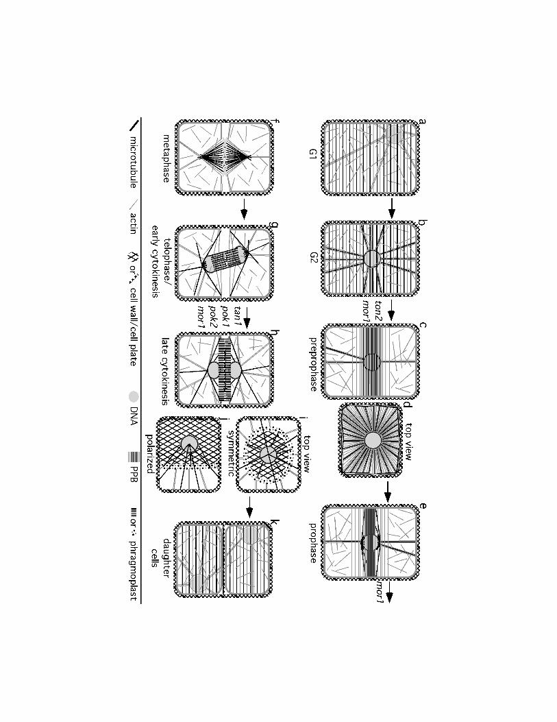
Figure 1
A transverse division in a “typical” plant cell is diagrammed. a, b, c, e, f, g, h, and k
represent two dimensional projections of the outer half of the cell, while d, g, and h represent
midplane views. Genes important for the transition to each stage are indicated under the arrows.
a) Cells in interphase/G1 have peripheral nuclei, ordered cortical MT and MF arrays, cortical
MFs arranged in a meshwork, and MFs that tether the actin-coated nucleus (as indicated by the
grey circle around the nucleus) to the cortex. b) G2 occurs after nuclear migration to the center
of the cell in preparation for mitosis. MTs are nucleated from the nuclear surface (as indicated
by the black circle around the nucleus) and connect the nucleus to the cortex in alignment with
MFs. c) In preprophase, breakdown of the ordered cortical MT and MF arrays occurs in concert
with PPB formation. The PPB consists of a cortical band of MTs and MFs. MTs and MFs
extend from the nucleus to the PPB and to the poles of the cell. d) This top down view
highlights the cortical nature of the PPB and the MTs and MFs that connect the nucleus to the
cortex. In vacuolated cells, these cytoskeleton components are contained within the
transvacuolar strands that comprise the phragmosome. e) During prophase, the MT PPB narrows
while the MF PPB remains the same width. By late prophase, MTs nucleated at nuclear surface
have begun to organize themselves into a bipolar spindle. MTs extend from the new spindle
poles to the PPB. f) During metaphase, the PPB disappears leaving behind an actin-depleted
zone at the cortex. The MT spindle is surrounded by MFs that extend to the cortex at the former
PPB site and to the poles to stabilize spindle position. g) During telophase, the phragmoplast
forms from the remnants of the spindle and is composed of two anti-parallel arrays of MTs and
MFs. The new cell plate is deposited where the two arrays meet. MTs nucleated at the former
spindle poles extend to the cortex, including the former site of the PPB. MFs connect the
phragmoplast to the cortex, including the former site of the PPB. h) During late cytokinesis, the
phragmoplast makes adjustments so that it aligns with the former PPB site. MTs nucleated from
the nucleus contribute to the expansion of the phragmoplast and continue to probe the cortex, but
begin to focus on the former PPB site. MFs continue to connect the phragmoplast to PPB site. i-
j) Top down view of cytokinesis. Only one nucleus is visible since the other one is hidden
beneath the expanding cell plate. i) During symmetric cytokinesis, the phragmoplast is initiated

23
in the center of the cell and expands to create a donut shape while depositing new cell plate. j)
Polarized cytokinesis begins off center so the new cell wall first becomes anchored to one side of
the mother cell. The phragmoplast continues to expand along the other side. k) After formation
of the new cell plate, the cytoskeletal arrangements in the daughter cells resemble that seen in G1
cells. This figure is based predominantly on results reported in Wick and Duniec 1984; Lloyd
and Traas 1988; Katsuta et al. 1990; Mineyuki et al. 1991; Cleary 1995; Nogami et al. 1996;
Cutler and Ehrhardt 2002; Dhonukshe et al. 2005b; Chan et al. 2005; Sano et al. 2005.
Figure 2
Geometry and plant cell division. a) A new cell wall (dashed line) avoids connecting to the
mother cell wall at a pre-existing cell wall junction and forming a four-way junction (on the left).
Instead the connection between the new cell wall and the mother cell will often form a three-way
junction (on the right). b) The avoidance of four-way junctions is hypothesized to be due to the
cytoskeleton elements extending from the nucleus to the cortex seeking the shortest distance due
to tension during PPB/phragmosome formation. Previous junctions have an out-pocketing which
places the cortex in that area farther way from the nucleus.

24
References
Ayaydin F, Vissi E, Meszaros T, Miskolczi P, Kovacs I, Feher A, Dombradi V, Erdodi F,
Gergely P, Dudits D (2000) Inhibition of serine/threonine-specific protein phosphatases causes
premature activation of cdc2MsF kinase at G2/M transition and early mitotic microtubule
organisation in alfalfa. Plant J 23: 85-96
Baluska F, Jasik J, Edelmann HG, Salajová T, Volkmann D (2001) Latrunculin B-induced plant
dwarfism: Plant cell elongation is F-actin-dependent. Dev Biol 231: 113-124
Brown RC, Lemmon BE (2001) The cytoskeleton and spatial control of cytokinesis in the plant
life cycle. Protoplasma 215: 35-49
Burgess J, Northcote DH (1968) The relationship between the endoplasmic reticulum and
microtubular aggregation and disaggregation. Planta 80: 1-14
Camilleri C, Azimzadeh J, Pastuglia M, Bellini C, Grandjean O, Bouchez D (2002) The
Arabidopsis TONNEAU2 gene encodes a putative novel protein phosphatase 2A regulatory
subunit essential for the control of the cortical cytoskeleton. Plant Cell 14: 833-845
Chan J, Calder G, Fox S, Lloyd C (2005) Localization of the microtubule end binding protein
EB1 reveals alternative pathways of spindle development in Arabidopsis suspension cells. Plant
Cell 17: 1737-1748
Cleary AL (1995) F-actin redistributions at the division site in living Tradescantia stomatal
complexes as revealed by microinjection of rhodamine-phalloidin. Protoplasma 185: 152-165
Cleary AL, Gunning BES, Wasteneys GO, Hepler PK (1992) Microtubule and F-actin dynamics
at the division site in living Tradescantia stamen hair cells. J Cell Sci 103: 977-988
Cleary AL, Smith LG (1998) The Tangled1 gene is required for spatial control of cytoskeletal

25
arrays associated with cell division during maize leaf development. Plant Cell 10: 1875-1888
Cutler SR, Ehrhardt DW (2002) Polarized cytokinesis in vacuolate cells of Arabidopsis. Proc
Natl Acad Sci U S A 99: 2812-2817
Dhonukshe P, Gadella TWJ (2003) Alteration of microtubule dynamic instability during
preprophase band formation revealed by yellow fluorescent protein-CLIP170 microtubule plus-
end labeling. Plant Cell 15: 597-611
Dhonukshe P, Kleine-Vehn J, Friml J (2005a) Cell polarity, auxin transport, and cytoskeleton-
mediated division planes: who comes first? Protoplasma 226: 67-73
Dhonukshe P, Mathur J, Hulskamp M, Gadella TWJ (2005b) Microtubule plus-ends reveal
essential links between intracellular polarization and localized modulation of endocytosis during
division-plane establishment in plant cells. BMC Biol 3: 11
Dixit R, Chang E, Cyr R (2006) Establishment of polarity during organization of the
acentrosomal plant cortical microtubule array. Mol Biol Cell 17: 1298-1305
Dixit R, Cyr R (2002a) Golgi secretion is not required for marking the preprophase band site in
cultured tobacco cells. Plant J 29: 99-108
Dixit R, Cyr RJ (2002b) Spatio-temporal relationship between nuclear-envelope breakdown and
preprophase band disappearance in cultured tobacco cells. Protoplasma 219: 116-121
Eleftheriou EP (1996) Developmental features of protophloem sieve elements in roots of wheat
(Triticum aestivum L.). Protoplasma 193: 204-212
Flanders DJ, Rawlins DJ, Shaw PJ, Lloyd CW (1990) Nucleus-associated microtubules help
determine the division plane of plant epidermal cells: avoidance of four-way junctions and the
role of cell geometry. J Cell Biol 110: 1111-1122
26
Galatis B, Apostolakos P, Katsaros C (1984) Experimental studies on the function of the cortical
cytoplasmic zone of the preprophase microtubule band. Protoplasma 122: 11-26
Galatis B, Mitrakos K (1979) On the differential divisions and preprophase microtubule bands
involved in the development of stomata of Vigna sinensis L. J Cell Sci 37: 11-37
Galatis P, Apostolakos P, Katsaros C, Loukari H (1982) Pre-prophase microtubule band and
local wall thickening in guard cell mother cells of some Leguiminosae. Ann Bot 50: 779-791
Gallagher K, Smith LG (1999) discordia mutations specifically misorient asymmetric cell
divisions during development of the maize leaf epidermis. Development 126: 4623-4633
Goodbody KC, Lloyd CW (1990) Actin filaments line up across Tradescantia epidermal cells,
anticipating wound-induced division planes. Protoplasma 157: 92-101
Goodbody KC, Venverloo CJ, Lloyd CW (1991) Laser microsurgery demonstrates that
cytoplasmic strands anchoring the nucleus across the vacoule of premitotic plant cells are under
tension. Implications for division plane alignment. Development 113: 931-939
Granger CL, Cyr RJ (2000) Microtubule reorganization in tobacco BY-2 cells stably expressing
GFP-MBD. Planta 210: 502-509
Granger CL, Cyr RJ (2001) Use of abnormal preprophase bands to decipher division plane
determination. J Cell Sci 114: 599-607
Gunning, BES (1982) The cytokinetic apparatus: Its development and apatial regulation. In:
Lloyd CW (ed) The Cytoskeleton in Plant Growth and Development. Academic Press, London,
pp 229-292
Gunning BES, Hardham AR, Hughes JE (1978a) Pre-prophase bands of microtubules in all
27
categories of formative and proliferative cell division in Azolla roots. Planta 143: 145-160
Gunning BES, Hardham AR, Hughes JE (1978b) Evidence for initiation of microtubules in
discrete regions of the cell cortex in Azolla root tip cells, and a hypothesis on the development of
cortical arrays of microtubules. Planta 134: 161-179
Gunning BES, Wick SM (1985) Preprophase bands, phragmoplasts, and spatial control of
cytokinesis. J Cell Sci Suppl 2: 157-179
Hahne G, Hoffman F (1984) The effect of laser microsurgery on cytoplamic strands and
cytoplasmic streaming in isolated plant protoplasts. Europ J Cell Biology 33: 175-179
Hofmeister W (1863) Zusatze und Berichtigungen zu den 1851 veroffentlichen
Untersuchungengen der Entwicklung hoherer Kryptogamen. Jahrbucher fur Wissenschaft und
Botanik 3: 259-293
Hoshino H, Yoneda A, Kumagai F, Hasezawa S (2003) Roles of actin-depleted zone and
preprophase band in determining the division site of higher-plant cells, a tobacco BY-2 cell line
expressing GFP-tubulin. Protoplasma 222: 157-165
Janssens V, Goris J (2001) Protein phosphatase 2A: a highly regulated family of serine/threonine
phosphatases implicated in cell growth and signalling. Biochem J 353: 417-439
Katsuta J, Hashiguchi Y, Shibaoka H (1990) The role of the cytoskeleton in positioning of the
nucleus in premitotic tobacco BY-2 cells. J Cell Sci 95: 413-422
Kawamura E, Himmelspach R, Rashbrooke MC, Whittington AT, Gale KR, Collings DA,
Wasteneys GO (2006) MICROTUBULE ORGANIZATION 1 regulates structure and function of
microtubule arrays during mitosis and cytokinesis in the Arabidopsis root. Plant Physiol 140:
102-114
28
Kennard JL, Cleary AL (1997) Pre-mitotic nuclear migration in subsidiary mother cells of
Tradescantia occurs in G1 of the cell cycle and requires F-actin. Cell Motil Cytoskeleton 36: 55-
67
Kumagai F, Hasezawa S (2001) Dynamic organization of microtubules and microfilaments
during cell cycle progression in higher plant cells. Plant Biol 3: 4-16
Kutsuna N, Hasezawa S (2002) Dynamic organization of vacuolar and microtubule structures
during cell cycle progression in synchronized tobacco BY-2 cells. Plant Cell Physiol 43: 965-973
Liu B, Palevitz BA (1992) Organization of cortical microfilaments in dividing root cells. Cell
Motil Cytoskeleton 23: 252-264
Lloyd CW (1991) How does the cytoskeleton read the laws of geometry in aligning the division
plane of plant cells? Dev Suppl 1: 55-65
Lloyd CW, Traas JA (1988) The role of F-actin in determining the division plane of carrot
suspension cells. Drug studies. Development 102: 211-221
Lu B, Roegiers F, Jan LY, Jan YN (2001) Adherens junctions inhibit asymmetric division in the
Drosophila epithelium. Nature 409: 522-525
Lynch TM, Lintilhac PM (1997) Mechanical signals in plant development: a new method for
single cell studies. Dev Biol 181: 246-256
Marcus AI, Dixit R, Cyr RJ (2005) Narrowing of the preprophase microtubule band is not
required for cell division plane determination in cultured plant cells. Protoplasma 226: 169-174
Marcus AI, Li W, Ma H, Cyr RJ (2003) A kinesin mutant with an atypical bipolar spindle
undergoes normal mitosis. Mol Biol Cell 14: 1717-1726

29
Mayer U, Büttner G, Jürgens G (1993) Apical-basal pattern formation in the Arabidopsis
embryo: studies on the role of the gnom gene. Development 117: 149-162
McCartney BM, McEwen DG, Grevengoed E, Maddox P, Bejsovec A, Peifer M (2001)
Drosophila APC2 and Armadillo participate in tethering mitotic spindles to cortical actin. Nat
Cell Biol 3: 933-938
McClinton RS, Sung ZR (1997) Organization of cortical microtubules at the plasma membrane
in Arabidopsis. Planta 201: 252-260
McCurdy DW, Gunning BES (1990) Reorganization of cortical actin microfilaments and
microtubules at preprophase and mitosis in wheat root-tip cells: A double label
immunofluorescence study. Cell Motil Cytoskeleton 15: 76-87
Mineyuki Y (1999) The preprophase band of microtubules: Its function as a cytokinetic
apparatus in higher plants. Int Rev Cytol 187: 1-49
Mineyuki Y, Furuya M (1986) Involvement of colchicine-sensitive cytoplasmic element in
premitotic nuclear positioning of Adiantum protonemata. Protoplasma 130: 83-90
Mineyuki Y, Palevitz BA (1990) Relationship between preprophase band organization, F-actin,
and the division site in Allium. J Cell Sci 97: 283-295
Mineyuki Y, Marc J, Palevitz BA (1991) Relationship between the preprophase band, nucleus,
and spindle in dividing Allium cotyledon cells. J Plant Physiol 138: 640-649
Mineyuki Y, Wick SM, Gunning BES (1988) Preprophase bands of microtubules and the cell
cycle: Kinetics and experimental uncoupling of their formation from the nuclear cycle in onion
root-tip cells. Planta 174: 518-526
Miyake T, Hasezawa S, Nagata T (1997) Role of cytoskeletal components in the migration of
30
nuclei during the cell cycle transistion from G1 phase to S phase of tobacco BY-2 cells. J Plant
Physiol 150: 528-536
Molchan TM, Valster AH, Hepler PK (2002) Actomyosin promotes cell plate alignment and late
lateral expansion in Tradescantia stamen hair cells. Planta 214: 683-693
Müller S, Han S, Smith LG (2006) Two kinesins are involved in the spatial control of cytokinesis
in Arabidopsis thaliana. Curr Biol 16: 888-894
Murata T, Wada M (1991) Effects of centrifugation on preprophase-band formation in Adiantum
protonemata. Planta 183: 391-398
Nebenführ A, Frohlick JA, Staehelin LA (2000) Redistribution of Golgi stacks and other
organelles during mitosis and cytokinesis in plant cells. Plant Physiol 124: 135-151
Nogami A, Suzaki T, Shigenaka Y, Nagahama Y, Mineyuki Y (1996) Effects of cycloheximide
on preprophase bands and prophase spindles in onion (Allium cepa L.) root tip cells. Protoplasma
192: 109-121
Ôta T (1961) The role of cytoplasm in cytokinesis of plant cells. Cytologia 26: 428-447
Palevitz BA (1986) Division plane determination in guard mother cells of Alluim: Video time-
lapse analysis of nuclear movements and phragmoplast rotation in the cortex. Dev Biol 177: 644-
654
Palevitz BA (1987) Actin in the preprophase band of Allium cepa. J Cell Biol 104: 1515-1519
Palevitz BA, Hepler PK (1974a) The control of the plane of division during stomatal
differentiation in Allium. I. Spindle Reorientation. Chromosoma 46: 297-326
Palevitz BA, Hepler PK (1974b) The control of the plane of division during stomatal

31
differentiation in Allium II. Drug Studies. Chromosoma 46: 327-341
Panteris E, Apostolakos P, Galatis B (1995) The effect of taxol on Triticum preprophase root
cells: preprophase microtubule band organization seems to depend on new microtubule
assembly. Protoplasma 186: 72-78
Panteris E, Apostolakos P, Galatis B (2006) Cytoskeletal asymmetry in Zea mays subsidiary cell
mother cells: a monopolar prophase microtubule half-spindle anchors the nucleus to its polar
position. Cell Motil Cytoskeleton 63: 696-709
Panteris E, Apostolakos P, Quader H, Galatis B (2004) A cortical cytoplasmic ring predicts the
division plane in vacuolated cells of Coleus: the role of actomyosin and microtubules in the
establishment and function of the division site. New Phytol 163: 271-286
Petrásek J, Elckner M, Morris DA, Zazímalová E (2002) Auxin efflux carrier activity and auxin
accumulation regulate cell division and polarity in tobacco cells. Planta 216: 302-308
Pickett-Heaps JD (1969) Preprophase microtubules and stomatal differentation; Some effects of
centrifugation on symmetrical and asymmetrical cell division. J Ultrastruct Res 27: 24-44
Pickett-Heaps JD, Northcote DH (1966a) Organization of microtubules and endoplasmic
reticulum during mitosis and cytokinesis in wheat meristems. J Cell Sci 1: 109-120
Pickett-Heaps JD, Northcote DH (1966b) Cell division in the formation of the stomatal complex
of the young leaves of wheat. J Cell Sci 1: 121-128
Sano T, Higaki T, Oda Y, Hayashi T, Hasezawa S (2005) Appearance of actin microfilament
'twin peaks' in mitosis and their function in cell plate formation, as visualized in tobacco BY-2
cells expressing GFP-fimbrin. Plant J 44: 595-605
Shaw SL, Kamyar R, Ehrhardt DW (2003) Sustained microtubule treadmilling in Arabidopsis
32
cortical arrays. Science 300: 1715-1718
Shevell DE, Leu WM, Gillmor CS, Xia G, Feldmann KA, Chua NH (1994) EMB30 is essential
for normal cell division, cell expansion, and cell adhesion in Arabidopsis and encodes a protein
that has similarity to Sec7. Cell 77: 1051-1062
Sinnott EW, Bloch R (1940) Cytoplasmic behavior during division of vacuolate plant cells. Proc
Natl Acad Sci USA 26: 223-227
Sinnott EW, Bloch R (1941) The relative position of cell walls in developing plant tissues. Am J
Bot 28: 607-617
Smertenko AP, Chang H-Y, Wagner V, Kaloriti D, Fenyk S, Sonobe S, Lloyd C, Hauser M-T,
Hussey PJ (2004) The Arabidopsis microtubule associated protein AtMAP65-1: Molecular
analysis of its microtubule bundling activity. Plant Cell 16: 2035-2047
Smith LG (2001) Plant cell division: building walls in the right places. Nat Rev Mol Cell Biol 2:
33-39
Smith LG, Gerttula SM, Han S, Levy J (2001) TANGLED1: A microtubule binding protein
required for the spatial control of cytokinesis in maize. J Cell Biol 152: 231-236
Smith LG, Hake S, Sylvester AW (1996) The tangled-1 mutation alters cell division orientations
throughout maize leaf development without altering leaf shape. Development 122: 481-489
Tian GW, Smith D, Gluck S, Baskin TI (2004) Higher plant cortical microtubule array analyzed
in vitro in the presence of the cell wall. Cell Motil Cytoskeleton 57: 26-36
Traas JA, Doonan JH, Rawlins DJ, Shaw PJ, Watts J, Lloyd CW (1987) An actin network is
present in the cytoplasm throughout the cell cycle of carrot cells and associates with the dividing
nucleus. J Cell Biol 105: 387-395

33
Traas J, Bellini C, Nacry P, Kronenberger J, Bouchez D, Caboche M (1995) Normal
differentiation patterns in plants lacking microtubular preprophase bands. Nature 375: 676-677
Twell D, Park SK, Hawkins TJ, Schubert D, Schmidt R, Smertenko A, Hussey PJ (2002)
MOR1/GEM1 has an essential role in the plant-specific cytokinetic phragmoplast. Nat Cell Biol
4: 711-714
Valster AH, Hepler PK (1997) Caffeine inhibition of cytokinesis: effect on the phragmoplast
cytoskeleton in living Tradescantia stamen hair cells. Protoplasma 196: 155-166
Van Damme D, Bouget FY, Van Poucke K, Inzé D, Geelen D (2004) Molecular dissection of
plant cytokinesis and phragmoplast structure: a survey of GFP-tagged proteins. Plant J 40: 386-
398
Vanstraelen M, Torres Acosta JA, De Veylder L, Inzé D, Geelen D (2004) A plant-specific
subclass of C-terminal kinesins contains a conserved A-type cyclin-dependent kinase site
implicated in folding and dimerization. Plant Physiol 135: 1417-1429
Vanstraelen M, Van Damme D, De Rycke R, Mylle E, Inze D, Geelen D (2006) Cell cycle-
dependent targeting of a kinesin at the plasma membrane demarcates the division site in plant
cells. Curr Biol 16: 308-314
Venverloo CJ, Libbenga KR (1987) Regulation of the plane of cell division in vacuolated cells I.
The function of nuclear positioning and phragmosome formation. J Plant Physiol 131: 267-284
Vos JW, Dogterom M, Emons AMC (2004) Microtubules become more dynamic but not shorter
during preprophase band formation: a possible "search-and-capture" mechanism for microtubule
translocation. Cell Motil Cytoskeleton 57: 246-258
Walker KL, Muller S, Allen D, Ehrhardt DW, Smith LG (submitted) Arabidoposis TANGLED

34
marks the division site throughout mitosis and cytokinesis.
Whittington AT, Vugrek O, Wei KJ, Hasenbein NG, Sugimoto K, Rashbrooke MC, Wasteneys
GO (2001) MOR1 is essential for organizing cortical microtubules in plants. Nature 411: 610-
613
Wick SM, Duniec J (1983) Immunofluorescence microscopy of tubulin and microtubule arrays
in plant cells. I. Preprophase band development and concomitant appearance of nuclear
envelope-associated tubulin. J Cell Biol 97: 235-243
Wick SM, Duniec J (1984) Immunofluorescence microscopy of tubulin and microtubule arrays
in plant cells. II. Transition between the pre-prophase band and the mitotic spindle. Protoplasma
122: 45-55
Willemsen V, Friml J, Grebe M, van den Toorn A, Palme K, Scheres B (2003) Cell polarity and
PIN protein positioning in Arabidopsis require STEROL METHYLTRANSFERASE1 function.
Plant Cell 15: 612-625
Yamashita YM, Jones DL, Fuller MT (2003) Orientation of asymmetric stem cell division by the
APC tumor suppressor and centrosome. Science 301: 1547-1550
Yoneda A, Akatsuka M, Hoshino H, Kumagai F, Hasezawa S (2005) Decision of spindle poles
and division plane by double preprophase bands in a BY-2 cell line expressing GFP-tubulin.
Plant Cell Physiol 46: 531-538
Zachariadis M, Quader H, Galatis B, Apostolakos P (2001) Endoplasmic reticulum preprophase
band in dividing root-tip cells of Pinus brutia. Planta 213: 824-827
Zachariadis M, Quader H, Galatis B, Apostolakos P (2003) Organization of the endoplasmic
reticulum in dividing cells of the gymnosperms Pinus brutia and Pinus nigra, and of the
pterophyte Asplenium nidus. Cell Biol Int 27: 31-40