diversity of taxol producing endophytic fungi from taxus ... · the completion of my dissertation...
TRANSCRIPT

Diversity of taxol producing endophytic fungi
from Taxus baccata and process optimization
for taxol production
A Thesis
Submitted in fulfillment of the requirement
for the award of the degree of
DOCTOR OF PHILOSOPHY
IN
BIOTECHNOLOGY
By
Sanjog Garyali
(Reg. No. 900900003)
Department of Biotechnology
Thapar University, Patiala –147004
Punjab (India)
June 2014


Diversity of taxol producing endophytic fungi
from Taxus baccata and process optimization for
taxol production
A Thesis
Submitted in fulfillment of the requirement
for the award of the degree of
DOCTOR OF PHILOSOPHY
IN
BIOTECHNOLOGY
By
Sanjog Garyali
(Reg. No. 900900003)
Department of Biotechnology
Thapar University, Patiala –147004
Punjab (India)
July 201

i

ii

iii
ACKNOWLEDGEMENT
The completion of my dissertation and subsequent Ph.D. has been a long journey. It’s
true that “Life is what happens” when you are completing your dissertation. Life doesn’t
stand still, nor wait until you are finished and have time to manage it but one of the joys
of completion is to look over the journey past and remember all the friends and family
who have helped and supported me along this long but fulfilling road.
My dissertation has always been a priority. At any rate, I have finished, but not alone,
and am elated. I could not have succeeded without the invaluable support of several
people around me. Without these supporters, especially the selected few I’m about to
mention, I may not have gotten to where I am today, at least not sanely.
First of all I bow my head to "Almighty God" to give me strength to complete this work.
I’d now like to give special thanks and gratitude to my major advisor Dr. M. S. Reddy,
Professor, Department of Biotechnology, Thapar University, Patiala who stepped in as
my supervisor and helped push me through all the phases of this beautiful journey. I
could not have asked for a better role model, supportive & prudent advisor who was
always ready to help me whenever I was struck in any doubt. His intelligent ideas,
thought provoking discussions and pertinent guidance helped me sail forward in every
obstacle during my work. I sincerely thank him for his confidence and faith on me
throughout my research.
I am highly thankful to my second research advisor Dr. Anil Kumar Dutta, Associate
Professor, Department of Biotechnology, Thapar University, Patiala for his guidance for
successful completion of my work. I could not be prouder of my academic roots and hope
that I can in turn pass on the research values and the dreams that he has given to me. His

iv
dedication for research rendered to me during my work. I extend my heartfelt thanks to
him forever.
I would like to express my deep appreciation to my committee members Dr. Sanjai
Saxena and Dr. M. Chhibber for their valuable suggestions, patient advice and
motivation during every progress presentation of my PhD course. I could always look
back on them for any support during for my study.
I am also indebted to all faculty members of the Department of Biotechnology and
Environmental Sciences for their help and suggestions during my experimental work.
I extend my sincere thanks to Ms. M Vasundhara for her constant help and guidance
during the various stages of my work.
I extend my sincere word of thanks to Dr. K. K. Raina, Deputy Director, Thapar
University, Dr. P.K. Bajpai, Dean (Research and Sponsored projects) and Dr. Dinesh
Goyal, Professor, Department of Biotechnology and Environmental Sciences for their
encouragement and support during the course of my PhD.
My sincere thanks are to my lab seniors Dr. Diwakar Aggarwal and Dr. Deepika Kumari
for their guidance and support at every step I needed.
I would also like to thank my friends Dr. Puja Tandon, Ritam Garg, Gurpreet Kaur
Khaira, Seema Bhanwar, Rahul Rathore, Tumul Shyam Verma, Gurudutt, Aman
Sandher, Fatehbir Singh, Rishi, Neha Lohia, Taranpreet Kaur, Mahiti, Vineet, Prerna,
Charu, Rajneesh Verma, Kunal Garg, Hiteshi and Swapnil for their support when I was
low with tough times and fighting back to get back on the track. I really find myself lucky
to have friends like them in my life.

v

vi
List of Publications
The following publications are the outcome of the present research work:
1. Garyali S, A Kumar, M S Reddy (2013). Taxol production by an endophytic fungus,
Fusarium redolens, isolated from Himalayan yew. Journal of Microbiology and
Biotechnology 23: 1372-1380.
2. Garyali S, A Kumar, M S Reddy (2013). Diversity and antimitotic activity of taxol-
producing endophytic fungi isolated from Himalayan yew. Annals of Microbiology
(DOI: 10.1007/s13213-013-0786-7).
3. Garyali S, A Kumar, M S Reddy (2014). Enhancement of taxol production from
endophytic fungus Fusarium redolens. Biotechnology and Bioprocess Engineering
(Accepted)
Papers presented in conferences
Garyali S, Kumar A, Reddy MS. Diversity of taxol producing endophytic fungi from
Taxus baccata and process optimization for taxol production. National Conference
on Emerging Trends in Biopharmaceuticals: Relevance to Human Health,
November 11-13, 2010, Thapar University, Patiala, Punjab.
Garyali S, Kumar A, Reddy MS. Diversity of taxol producing endophytic fungi from
Taxus baccata growing in Northern Himalaya. National seminar on current
perspectives of fungi in health care and environment, March 13-14, 2013,
Department of Biotechnology, Bangalore University, Bangalore.

vii
Garyali S, Kumar A, Reddy MS. Screening of taxol producing endophytic fungi using
molecular markers. International conference on Plant Biotechnology, Molecular
medicine and Human Health, October 18-20, 2013, Department of Genetics,
University of Delhi, South Campus New Delhi.
Garyali S, Bansal M, Dhami N, Kumar A, Reddy MS. Response surface methodology as
a tool for enhanced production of secondary metabolites and extracellular enzymes.
National symposium on Emerging trends in botanical sciences, February 17-18,
2014, Department of botany, Punjabi University, Patiala, Punjab.
Garyali S, Kumar A, Reddy MS. Optimization of taxol production from endophytic
fungus Fusarium redolens. National conference on: Fungal Diversity and
biotechnology for food and chemicals, February 27-28, 2014, Centre of advanced
study in marine biology, Annamalai University, Tamil Nadu.

viii
ABSTRACT
Endophytic fungi represent an under explored resource of novel lead compounds and
have the capacity to produce diverse classes of plant secondary metabolites. In the
present study, we investigated the diversity of taxol-producing endophytic fungi from
Taxus baccata L. subsp. wallichiana (Zucc.) Pilger. A total of 60 fungal endophytes were
isolated from the inner bark (phloem-cambium) of T. baccata, collected from different
locations of the northern Himalayan region of India. Two key genes, DBAT (10-
deacetylbaccatin III-10-O-acetyl transferase) and BAPT (C-13 phenylpropanoid side
chain-CoA acyltransferase), involved in taxol biosynthesis were used as molecular
markers for screening of the taxol producing strains. Five different endophytic species
gave positive amplification hits by molecular marker screening with the bapt gene. These
fungi were characterized based on morphological characters and internal transcribed
spacer (ITS) sequence analysis and identified as: Fusarium redolens (TBPJ-B),
Gibberella avenacea (C-1), Paraconiothyrium brasiliense (TBPJ-13), Microdiplodia sp.
(TBPJ-A) and Fusarium tricinctum (B-7),
The taxol-producing capability of these endophytic fungi was validated by HPLC-
MS. The highest yield of taxol was found to be 66.25 μg/L by Fusarium redolens
compared to other four strains. The amount of taxol produced by other four strains in S-7
liquid medium are TBPJ-A (27.40 µg/L), B-7 (23.47 µg/L), TBPJ-13 (19.60 µg/L) and
C-1 (11.03 µg/L). The antitumour activity of the fungal taxol was tested by potato disc
tumor induction assay using Agrobacterium tumefaciens as the tumor induction agent.
This assay depicted that fungal taxol from all the endophytes successfully inhibited tumor
formation in potato discs just like authentic taxol while it did not affect bacterial viability

ix
in any case. These results confirmed that all fungal extracts tested in this study had
similar antineoplastic activity as that of paclitaxel.
The medium components and different growth parameters were optimized for
production of taxol from the endophytic fungus Fusarium redolens. Optimization of
medium components was performed using Plackett-Burman (PB) design and response
surface methodology (RSM). Different carbon and nitrogen sources were compared and
then PB design was employed for screening the important trace elements. Results showed
that NH4NO3, MgSO4 and NaOAc were the most important components which were
further investigated by Box-Behnken design. Optimal concentrations of NH4NO3, MgSO4
and NaOAc achieved for maximum taxol production were 6.25, 0.63 and 1.25 g/L,
respectively. The predicted response in Box-Behnken experimental design for taxol
production gave a value of 195 µg/L, while the actual experimental value was 198 µg/L,
suggesting that experimental and predicted values were in good agreement. About three
fold increase of taxol production was observed after optimization of fermentation
conditions and medium components by RSM. These results suggested the success of
RSM in enhancing the production of fungal taxol.
In the present investigation, potential of pharmaceutically important endophytes
has been investigated. This study has shed light on endophyte biotechnology for its
applications in production of anticancerous drug taxol. PCR amplification of genes
involved in taxol biosynthesis (DBAT and BAPT) is an efficient and reliable method for
pre-screening the taxol-producing fungi.
F. redolens and F. verticillioides are the first ever reports of endophytic fungi capable of
taxol production obtained from Taxus baccata subsp. wallichiana. Paraconiothyrium

x
brasiliense, Microdiplodia sp. and Phomopsis sp. reported from other Taxus plants but
not reported from the Himalayan yew. This study offers important information and a new
source for the production of the important anticancer drug taxol by endophytic fungus
fermentation. The endophytic taxol was found to be equally effective as that of authentic
paclitaxel which paves way for exploitation of these potent endophytic fungi for
industrial bioprocesses. Enhanced production of fungal taxol by RSM encouraged the use
of these statistical tools for large scale production of this bioactive compound. Though
further research at the molecular level is requisite for better understanding of the host
endophyte interactions involved in production of this immensely demanding antitumour
drug, the current work is a step forward to encourage the use of these fungal endophytes
as alternative to the conventional sources.

xi
TABLE OF CONTENTS
Chapters Page No.
Acknowledgement iii
List of publications vi
Abstract viii
Contents xi
Abbreviations xvii
1. Introduction 1-12
1.1 General Introduction 1
1.2 Endophytes as biological factories of functional metabolites 3
1.3 Taxol - history, clinical impact 4
1.4 Gap in studies 6
1.5 Present study 9
1.6 Specific objectives of the present investigations 12
2. Review of literature 13-58
2.1 Endophyte vs. host: the relationship between endophytic fungi and
host plant
15
2.2 The plausibility of Horizontal gene transfer (HGT) hypothesis 18
2.3 Biodiversity of endophytes 21
2.4 Isolation and identification of endophytic fungi 22
(a) Isolation of endophytic fungi 22
(b) Identification of endophyte fungi 24
(i) Internal transcribed spacers 24

xii
(ii) Morphological identification 26
2.5 Endophytes as biological factories of functional metabolites 26
2.5.1 Endophytic natural products as drugs and novel drug leads 28
(a) Endophytes as producers of antibiotics 28
(b) Endophytes and there antiviral compounds 30
(c) Antioxidant compounds produced by endophytes 31
(d) Other natural products by endophytes 32
(1) Antidiabetic agents 32
(2) Immunosuppressive compounds 32
(3) Insecticidal activities 33
(e) Endophytic fungal products as anticancer agents 34
(i) Camptothecin 35
(ii) Vinca alkaloids 36
(iii) Podophyllotoxin 36
(iv) Taxol 37
2.6 Biosynthesis of taxol 43
2.7 Extraction and determination of fungal taxol 47
(i) Choice of different solvents and extraction methods 49
(ii) Determination of taxol in fungal fermentation products 49
2.8 Antimitotic activity of fungal taxol 50
2.9 Optimization of conditions for maximum taxol production by
statistical tool Response Surface Methodology (RSM)
53
3. Materials and methods 59-82
3.1 Sample collection areas and geographic features 59
3.2 Isolation and cultivation of endophytic fungi from Taxus baccata bark 59

xiii
3.3 Screening of taxol producing fungi based on PCR amplification 60
3.3.1 Isolation of fungal genomic DNA 60
3.3.1.1 CTAB method for DNA extraction 60
3.3.1.2 DNA Purification 61
3.3.1.3 Checking of DNA (Agarose Gel Electrophoresis) 61
3.3.1.4 Quantification of DNA using nanodrop 62
3.3.1.5 Ethidium bromide fluorescent DNA quantification 62
3.3.2 Ribonucleic acid (RNA) isolation and complementary DNA
(cDNA) synthesis
63
3.3.2.1 RNA isolation 63
3.3.2.2 cDNA synthesis 63
3.3.3 Molecular screening of endophytes using gene specific primers 63
3.3.3.1 Cloning and characterization of BAPT gene 65
3.4 Identification of endophytic fungi 66
3.4.1 Morphological characterization 66
3.4.2 Molecular phylogenetic analysis [Amplification of Internal
Transcribed Spacer (ITS) region]
66
3.4.2.1 Purification of PCR products 67
3.4.2.2 Ligation in pTZ57R/T vector 67
3.4.2.3 Genetic Transformation of ITS products into E. coli
DH5 cells
68
3.4.2.4 Blue/white screening for recombinant plasmids 69
3.4.2.5 Isolation and purification of plasmid DNA from
recombinant bacteria by alkaline lysis method
70
3.4.2.6 Size screening for recombinant plasmids 71
3.4.2.7 Sequencing 71

xiv
3.4.2.8 Analysis of sequence data 71
3.5 Biochemical screening of fungal taxol 72
3.5.1 Fungal cultivation 72
3.5.2 Extract preparation 72
3.5.3 Column chromatography 73
3.5.4 Thin layer chromatography (TLC) 73
3.5.5 High performance liquid chromatography (HPLC) 74
3.5.6 High performance liquid chromatography-Mass spectroscopy
(HPLC-MS)
74
3.6 Antitumorigenic Activity Assay 75
3.7 Optimization of process parameters and media components for taxol
production by Response surface methodology
76
3.7.1 Optimization of basic parameters 76
3.7.1.1 Estimation of radial growth 76
3.7.1.2 Estimation of fungal biomass (Growth kinetics) 76
3.7.1.3 Optimization of medium-to-flask volume ratio (Vm/Vf
ratio)
76
3.7.1.4 Estimation of optimal pH and temperature 77
3.7.2 Experimental design 77
3.7.2.1 One-factor-at-a-time strategy 77
3.7.2.1.1 Effect of carbon and nitrogen sources on
biomass and taxol production
77
3.7.2.2 Statistical methods 78
3.7.2.2.1 Plackett-Burman design (PB design) 78
3.7.2.2.2 Response surface methodology (RSM) 79
3.7.3 Analytical methods 81

xv
3.8 Statistical Analysis 82
4. Results 83-160
4.1 Isolation and characterization of taxol producing endophytic fungi 83
4.1.1 Isolation of endophytic fungi 83
4.1.2 Molecular screening (Primary screening of taxol-producing
endophytic fungi)
83
4.1.2.1 Screening for taxol-producing endophytic fungi using
dbat and bapt specific primers
83
4.1.2.2 Cloning and characterization of BAPT gene 89
4.1.3 Molecular phylogenetics 96
4.1.3.1 PCR amplification of ITS region 96
4.1.3.2 Phylogenetic analyses 103
4.1.4 Fungal morphology 106
4.1.4.1 Macroscopic characteristics 106
4.1.4.2 Microscopic characteristics 108
4.2 Biochemical screening of taxol production by endophytic fungi 118
4.2.1 Thin layer chromatography (TLC) 120
4.2.2 High performance liquid chromatography (HPLC; Perkin Elmer) 121
4.2.3 Liquid chromatography mass spectroscopy (LC-MS; Waters) 124
4.3 Anti tumorous activity of fungal taxol 129
4.3.1 Potato disc tumor induction assay 129
4.4 Process optimization for taxol production from endophytic fungus
Fusarium redolens
134
4.4.1 One-factor-at-at-a-time (OFAT) 135
4.4.1.1 Standardization of basic parameters 135

xvi
(a) Estimation of radial growth 135
(b) Estimation of fungal biomass (Growth kinetics) 135
(c) Optimization of medium volume (Vm) to flask volume (Vf)
ratio
135
(d) Estimation of optimal pH and temperature for biomass and
taxol production
136
4.4.1.2 Medium optimization (Effect of carbon and nitrogen
sources on biomass and taxol production)
142
4.4.2 Plackett-Burman design 147
4.4.3 Response surface methodology 151
5. Discussion 161-171
5.1 Isolation and characterization of taxol producing endophytic fungi 161
5.2 Biochemical screening of taxol production by endophytic fungi 165
5.3 Anti tumorous activity of fungal taxol 167
5.4 Process optimization for taxol production from endophytic fungus
Fusarium redolens
168
6. Summary 172-176
References 177-225
Appendix I 226-233
Appendix II 234-243

xvii
ABBREVIATIONS
Abbreviations Description
µm micrometer
aa
amsl
amino acid
above mean sea level
ACN acetonitrile
AD Anno Domini
AIDS acquired immuno deficiency syndrome
BLAST Basic Local Alignment Search Tool
bp base pair
cDNA complementary DNA
CHCl3 chloroform
cm centimeter
CTAB cetyltrimethylammonium bromide
DAD diode array detector
DCM dichloromethane
dd double distillation
DMSO dimethyl sulfoxide
E. coli Escherichia coli
et.al. et alii
F forward
FW fresh weight
g gram

xviii
Inc. Incorporation
IPTG isopropyl β-D-1-thiogalactopyranoside
kg kilogram
kV kilo volt
L Litre
L. Linnaean
M molar
m/z mass-to-charge ratio
MeOH methanol
mg milligram
min minute
mM millimolar
NCBI National Center for Biotechnology Information
ng nanogram
nm nanometer
No. number
ºC degree celsius
OD optical density
PBS phosphate buffered saline
PCR polymerase chain reaction
pH negative decimal logarithm of the hydrogen ion
activity in a solution
R reverse
RNA ribonucleic acid
rpm revolutions per minute

xix
SD standard deviation
sec seconds
sp. specie
spp. species
T. baccata Taxus baccata
μg micro gram
μL micro liter
Tm melting temperature
V volt
Vm/Vf medium-to-flask volume
v/v volume per volume
w/v weight per volume

1
CHAPTER 1
Introduction
1.1 General Introduction
Pharmaceutical biology perceives plants as ‘bio-factories’ of therapeutic compounds with
high potential. Even in the present era of modern technology, it is not possible to estimate the
impact of plant derived products as pharmaceutical relevant lead compounds. With expanding
realization of the health hazards and toxicity related with the indiscriminate use of synthetic
drugs and antibiotics, interest in the use of biogenic drugs has resuscitated throughout the
world (Nalawade et al. 2003). Even the world’s most universally used drug “AspirinTM
” is
derivative of salicylic acid which was originally obtained from white willow (Salix alba).
Plants, like all other organisms, live in thriving community of microbes and provide a
unique environment for endophytes. Diverse fungal endophytes exist within plant tissues,
with a global approximation of up to a million species (Petrini 1991). Endophytes (Gr. endon,
within; phyton, plant), the microbes that colonize living, internal tissues of plants without
causing any immediate, overt negative effects have attracted considerable attention of many
scientists worldwide as potential producers of novel and biologically active metabolites (Cai
et al. 2004; Strobel et al. 2004; Cragg and Newman 2006). The term Endophyte was first
coined by de Bary in 1866 and has become deeply embedded in the literature ever since.
Endophytic fungi delineate an important and quantifiable component of fungal
biodiversity and are well known to affect plant community diversity and structure
(Manoharachary et al. 2005; Krings 2007). Endophytes are the important components of plant
micro-ecosystems (Tan et al. 2001; Zhang et al. 2006; Rodrigues et al. 2009; Suryanarayanan
et al. 2011) and it has been suggested that fungi can influence the distribution, ecology,
physiology, and biochemistry of the host plants (Sridhar 1995; Ramesh et al. 2009). To

2
endophytes, land plants present a complex, spatially and temporally diverse ecological
habitat. Of nearly 3,00,000 plant species that inhibit our planet, each individual one is host to
several to hundreds of endophytic fungi (Tan and Zou 2001; Damon et al. 2012), creating a
vast biodiversity: a myriad of undescribed species, a rich source of novel natural products
there from and an unknown genetic background of all the affiliations thus implied.
Single endophyte can invade a wide host range and can be isolated from different plants
belonging to different families and classes growing under different ecological and
geographical conditions (Petrini 1996). These endophytes are found virtually in every plant
on earth. These endophytic fungi reside in the living tissue of host plant and do so in a variety
of relationships ranging from symbiotic to pathogenic (Perotto et al. 2002; Strobel et al. 2004:
Deshmukh and Verekar 2008; Verekar et al. 2014). They receive nutrition and protection
from the host plant, while the host plant may benefit from enhanced competitive abilities and
increased resistance to herbivores, pathogens, and various abiotic stresses by attaining the
metabolic substances of endophytes (Saikkonen et al. 1998; Tan and Zou 2001; Zhang et al.
2006; Daghino et al. 2009; Girlanda et al. 2009; Achal et al. 2011). The endophytes have
been investigated to be very rich source of novel biological active secondary metabolites
(Strobel et al. 2004; Deshmukh and Verekar 2008; Jagadish et al. 2009; Verma et al. 2009;
Suryanarayanan et al. 2009; Kharwar et al. 2011; Verekar et al. 2014). In 2008, Moricca and
Ragazzi showed that the type of interaction between an endophyte and a plant is administered
by the genes of both organisms and modulated by the environment. Endophytes can be
transmitted from one generation to the next through the tissue of host seed or vegetative
propagules (Carroll 1988). Apparently, acquired chemical defense appears to be a common
basis for endophytic association between a plant and its particular endosymbiont (Carroll
1988; Clay 1988). These endophytes are metabolically more active than their free analogues
due to their specific functions in nature and activation of various metabolic pathways to

3
survive in the host tissue (Strobel and Daisy 2003). In particular, the capability to produce a
large number of chemically different secondary metabolites is related mainly with the
filamentous fungi for the eukaryotes (Donadio et al. 2002).
1.2 Endophytes as biological factories of functional metabolites
Drug discovery and development has a long history and dates back to the early days of
human civilization. The Mayans used endophytic fungi grown on roasted green corn to treat
intestinal sickness (Buss and Hayes 2000). Later, the Benedictine monks (800 AD) began to
apply Papaver somniferum belonging to the poppy family (Papaveraceae) as an anesthetic
and analgesic agent. In 1928 Alexander Fleming, a Scottish scientist found penicillin from
the mold Penicillium notatum. Although this result was the milestone of the beginning of the
antibiotic era, the importance of Alexander Fleming's discovery was not known (Bellis 2012).
Ever since the discovery of penicillin, the attention of pharmaceutical companies and
research laboratories was focused more on fungi as a source of lead compounds. Since then,
people have been engaged to search and discover new microbial metabolites with activity
against plant, animals and human pathogens.
Endophytes play a significant role in the daily life of human being besides they may
produce over abundance of compounds with a wide range of properties, including antibiotics,
antivirals, antimycotics, immunosuppressant, antidiabetic agents, antioxidants, insecticidal
products and anticancer agents (Demain 1999; Strobel and Daisy 2003; Strobel et al. 2004;
Keller et al. 2005; Strobel 2006; Mitchell 2008; Verma et al. 2009; Aly et al. 2010 and 2011;
Porras-Alfaro and Bayman 2011). The various secondary metabolites including active
principles produced by endophytes possess unique structures and great bioactivities,
representing a huge reservoir which offers an immense potential for exploitation for
medicinal, agricultural and industrial uses (Tan 2001; Zhang 2006).

4
The new technologies offer distinctive opportunities in screening of the natural
products which serves as a major source for drug discovery. Microorganisms act as absolute,
readily renewable, reproducible and inexhaustible source of enduring pharmaceutical
potential. It is hypothesized that many microorganisms (99%) have yet to be discovered
(Davis et al. 2005). In the recent past, endophytes have been enormously studied for their
potential as novel sources for various bioactive compounds. The diversity and specialized
habituation makes these endophytes an exhilarating field of study in the search for new
medicines or novel drugs.
1.3 Taxol - history, clinical impact
Functional metabolites of endophytic origin have displayed a considerable potential to impact
the pharmaceutical sector (Tan and Zou 2001; Strobel 2003; Strobel and Daisy 2003; Strobel
et al. 2004; Gunatilaka 2006). Among the bioactive compounds of endophytic fungi origin
described to date, the ones notable for their antitumour activity have drawn special attention,
with paclitaxel (Taxol) as the most striking example (Figure 1). It is one of the most
successful and widely used anticancer drugs developed in the past 50 years. This anticancer
drug was first found in the bark of Pacific yew tree; Taxus brevifolia (Wani et al. 1971). All
the species of Taxus are known to produce taxol; a chemical substance of tetracyclic
diterpene lactam which is highly efficient, has low toxicity and broad-spectrum natural
anticancer drug (Georg et al. 1995). In India it is represented by Taxus baccata subsp.
wallichiana (Himalayan yew).

5
Fig. 1 Structure of Paclitaxel (Taxol)
Paclitaxel represents a new class of antineoplastic agent as it has a distinctive mode of action.
Unlike other antimicrotubule agents like podophyllotoxin, colchicine, vinca alkaloids, and
combretastatin which impede microtubule assembly, paclitaxel stabilizes microtubules
against depolymerization; it promotes the polymerization of microtubules but inhibits
depolymerization (Schiff et al. 1978; Horowitz et al. 1986). This atypical stability blocks the
cells ability to disassemble the mitotic spindle during cell division; cells are blocked in the
G2/M phase of the cell cycle (Schiff et al. 1978, 1980) and this finally leads to cell death.
Taxol underwent clinical trials in the 1980s and was approved by the Food and Drug
Administration (FDA) in 1992 as a very important chemotherapeutic agent for treatment of a
variety of cancers (Suffness and Wall 1995). While the number of cancers treated by taxol is
expanding, to date it has been chiefly used to treat metastatic carcinomas of the ovary, cell
lung cancer and metastatic breast cancer as well as in second-line treatment of AIDS-related
Kaposi’s sarcoma (McGuire et al. 1989; Rowinsky et al.1990; Holmes et al. 1991; Yuan et al.
2000). It has also been investigated for the treatment of diseases not related with cancer such
as Alzheimer’s or Parkinsonism (Zhang 2005).

6
1.4 Gap in studies
Since the discovery of taxol, substantial energy has been invested in trying to increase its
extraction, but supply has been a crucial challenge throughout the clinical development of
this drug. Taxol makes up only a minor proportion of the total taxoid content of Taxus trees.
The conventional source of taxol relies mainly on Yew trees as the most reliable source but
most of these species are rare, endangered and slow growing. Even the amount of taxol
produced in them is comparatively low in relation to other taxoids; 0.001-0.05% of taxol
found even in the most productive species Taxus brevifolia (Wheeler et al. 1992). The natural
sources for taxol do not represent reliable production system therefore. The commercial
isolation of 1 kg of taxol from T. brevifolia requires 10,000 kg of Taxus bark or 2000 - 3000
very slow-growing yew trees (Hartzell 1991; Croom 1995; Suffness and Wall 1995;
Schippmann 2001). Normal cancer patient needs approximately 2.5 - 3.0 gm of Paclitaxel for
a full regimen of antitumor treatment (Bedi 1996). Furthermore, the yield of taxol is highly
dependent on the Taxus species. While species such as Taxus baccata (European yew tree)
produce scarcely any taxol at all (Nadeem et al. 2002). Additionally, extraction of taxol from
yew trees requires a complex system and purification techniques are very expensive. So,
considering these facts, together with the high demand for the drug, there is an urgent need to
find other alternative sources and new ways of taxol production. One of the ways is to
develop sustainable harvesting protocols of yews in natural stands, transforming elite
cultivars of the wild species into a commercially reared crop (Smith 2002). Other way is to
generate taxol by chemical synthesis which was achieved by Holton and Nicolau in 1994.
However, the complexity of the biosynthetic pathway and low yield limit its applicability. An
alternative approach is production by semisynthesis, which requires intermediates such as
baccatin III or 10-deacetylbaccatin III, extracted from renewable needles of Taxus without

7
destroying the trees (Holton et al. 1995). This production system still relies on yew trees for
precursor molecules and therefore depends on epigenetic and environmental factors.
An alternative production strategy is the use of Taxus cell suspension cultures, acquired
from the species T. brevifolia (Gibson et al. 1993), T. baccata (Srinivasan et al. 1995) and T.
canadensis (Ketchum et al. 1999). These cell cultures yield biomass faster than Taxus trees
and can be grown under reproducible environments. Under optimized culture conditions and
induction of production with methyl jasmonate, it is possible to generate up to 23 mg/L/d of
taxanes with a taxol content of 13-20% (Ketchum et al. 1999). These yields signify the
majestic biosynthetic capacity of Taxus cell cultures. However, upholding such high rates of
secondary metabolite production in plant cell culture is very strenuous (Deus-Neumann and
Zenk 1984; Hall and Yeoman 1987; Morris et al. 1989; Parr et al. 1990; Schripsema and
Verpoorte 1992). Additionally several total synthesis routes have been developed too,
however at best providing a maximum yield of 2% of taxol, hence not representing a useful
alternative production platform (Holton et al. 1994a; Holton et al. 1994b; Nicolaou et al.
1994; Danishefsky et al. 1996; Xiao et al. 2003).
All together the production methods used today have improved a lot over the times, still
are very difficult and costly regarding not only the production itself but also with concern of
purification of either taxol or late precursors (Baccatin III) from complex taxane mixtures.
This discrepancy between demand and supply is the biggest confrontation in clinical
application of this drug. It has propelled research into new production strategies, such as
metabolic engineering of the yeast Saccharomyces cerevisiae (Jennewein et al. 2005; Dejong
et al. 2006; Engels et al. 2008), Escherichia coli (Huang et al. 1998; Ajikumar et al. 2010)
and different plant systems like Arabidopsis thaliana (Besumbes et al. 2004) and the moss
Physcomitrella patens (Anterola et al. 2009). However, metabolic engineering of yeast for
the total biosynthesis of taxol or other advanced taxoids is extremely complicated and still in

8
its infancy. Today, the total fermentation of taxadiene has been attained in significant
amounts in Saccharomyces cerevisiae (Engels et al. 2008) and E. coli (Ajikumar et al. 2010).
Hence, the venture of recombinant microorganisms, like yeast or bacteria, offers great
prospects not only for the production of taxol but also for other complex natural products and
derivatives thereof (Chang and Keasling 2006).
Ultimately to lower the price of taxol and make it more widely available, an extensive
search for alternative sources for taxol and related taxanes was initiated. This approach led to
the isolation of endophytic fungi, surprisingly having been shown to contain the identical
natural products after cultivation independently from their plant host. The discovery of novel
taxol-producing endophytes: Taxomyces andreanae and Pestalotiopsis microspora,
demonstrated that organisms other than Taxus spp. could produce taxol (Stierle 1993 and
Strobel 1996). Since then scientists throughout the world have isolated and identified a
number of “taxol-synthesizing” endophytes from various sources (Li et al. 1996; Li et al.
1998; Kim et al. 1999; Wang et al. 2000; Huang et al. 2001; Guo et al. 2006; Sun et al. 2008;
Kumaran et al. 2010; Wang and Tang 2011). It is remarkable that taxol produced by these
endophytes is identical to that produced by Taxus spp., both chemically and biologically
(Stierle et al. 1993). Presently, isolation and utilization of taxol-producing fungi have made
significant progress worldwide (Lin et al. 2003; Zhao et al. 2008). It is generally agreed that
endophytic fungi grow rapidly and are easy to culture (Lin et al. 2003). So, biotechnological
methods could be used to improve taxol-producing capability of fungi and eventually
industrial production of taxol would be possible. But there is an urgent need to develop a
reliable screening system for the identification of endophytes capable of taxol production
based on the presence of important genes involved in taxol biosynthesis. Such screening
system might facilitate the handling of large number of samples altogether and help in
identification of taxol producing endophytes. Optimizing fermentation conditions may also

9
increase the yield of taxol by endophytic fungi. Taking into account the above possibility the
available diversity of endophytic fungi from the Taxus baccata needs to be investigated and
their potential for the production of the drug needs to be assessed.
1.5 Present study
In the present work, we investigated the taxol producing endophytic fungal diversity of Taxus
baccata L. subsp. wallichiana (Zucc.) Pilger (Himalayan yew) as no work to study the
diversity of taxol-producing endophytic fungi from this yew species growing in the northern
Himalayan region of India has been reported to date. Taxus baccata subsp. wallichiana is the
only species of Taxus which is found in the temperate Himalayas and in the hills of
Meghalaya, Nagaland and Manipur at altitudes of 1,800-3,300 m amsl. It is a medium-sized,
slow-growing, nonresinous, evergreen conifer that undergoes cross-pollination and has been
found to grow best in well-drained moist areas, in cool temperate to sub-tropical climates.
Common names for Taxus baccata L. subsp. wallichiana
(Zucc.) Pilger (Himalayan yew)
Dogri Birmi, Brammi, Postul, Thuneer
Hindi Gallu
Marathi Barmi
Sanskrit Manduparni

10
Scientific Classification
Kingdom Plantae
Division Pinophyta
Class Pinopsida
Order Taxales
Family Taxaceae
Genus Taxus
Species baccata
Sub-species L. wallichiana
Traditional method for screening the taxol producing capability of endophytic fungi
include thin layer chromatography (TLC), high performance liquid chromatography (HPLC)
and liquid chromatography mass spectroscopy (LC-MS). Undoubtedly these chromatographic
techniques are a necessary to authenticate that compound produced by endophytic isolates is
exactly the same that of our interest, but on the same context screening all the isolated
endophytes biochemically is uneconomical, cumbersome and sometimes it may even be
impossible. Thus, the primary separation of endophytic fungi from the plant material was
comparatively simple operational process, but the detection process of taxol-producing
endophytic fungi was laborious and uneconomical. Consequently, the objective of this study
was to develop an efficient protocol for screening taxol producing fungal isolates from
diverse endophytic fungi with the ultimate purpose of simplifying the screening process and
reduce workload. So, in the present study to explore a simple and efficient protocol for
screening of taxol generating endophytes, key genes involved in the taxol biosynthesis were
brought into consideration i.e. DBAT and BAPT genes (Zhang et al. 2008). DBAT (10-

11
deacetylbaccatin III-10-O-acetly transferase) catalyzes the formation of baccatin III, which is
the immediate diterpenoid precursor of taxol (Walker and Croteau 2000) and BAPT (C-13
phenylpropanoid side chain-CoA acyltransferase) as the acyl donor, to form N-debenzoyl-2´-
deoxytaxol, that is, it catalyzes the attachment of the biologically important taxol side chain
precursor (Walker et al. 2002). Gene specific PCR primers were used to screen the isolated
endophytic isolates harbouring the key genes involved in taxol biosynthesis. This PCR
amplification based protocol is an efficient, reliable and economical method for pre-screening
taxol-producing fungi.
Verification of any claimed biological compound requires testing in bioassay to
validate its activity before undergoing clinical testing. Bioassay methods used to assess the
antitumor activity of various extracts have varied over years and have led to discoveries of
important compounds. In the present work, potato disc tumor induction assay was used to
validate the antineoplastic activity of fungal taxol using Agrobacterium tumefaciens as tumor
inducing agent. This bioassay is a simple, inexpensive and fast screen for antitumor
compounds and validates the antitumor activity of test compound regardless of mode of
action on tumor formation.
Optimization of medium components is regarded as the most effective measure to
improve fermentation productivity of secondary metabolites i.e., designing an appropriate
medium and investigating the most suitable conditions such as pH, temperature, incubation
time, medium-to-flask volume ratio etc. This operation relates to several methods of
statistical experimental design. The traditional method of optimization is based on one factor
at a time (OFAT) approach. Plackett-Burman design and Response surface methodology are
found to be most efficient tools for process optimization. These statistical tools have been
widely and successfully used in optimizing the critical factors affecting secondary metabolite
production in different organisms and systems (Xu et al. 2006; Luo et al. 2004;

12
Saravanakumar and Kaviyarasan 2010; Srivastava et al. 2012; Wang et al. 2013). Present
study was aimed to optimize the fermentation medium by Plackett-Burman (PB) design and
response surface methodology (RSM) for the enhanced production of taxol by Fusarium
redolens.
Present study will shed light on diversity of taxol producing fungi from different
locations. Taxol production by different fungi will be screened by biochemical as well as
molecular methods based on DBAT and BAPT gene. Anti tumour effect of the fungal taxol
will be assessed by potato disc tumour induction assay and an attempt will be made to
enhance the production of taxol by novel endophytic fungus with aid of statistical tools based
on RSM. The objectives of the present work aim to isolate and characterize taxol producing
endophytic fungi which will be screened for efficient taxol production so as to harness it for
various pharmaceutical applications.
1.6 Specific objectives of the present investigations
1. Isolation and characterization of endophytic fungi from Taxus baccata subsp.
wallichiana growing at different locations of Indian Himalayan region (IHR)
2. Molecular screening of endophytic isolates to screen and select capable taxol
producing endophytes based on PCR amplification of DBAT and BAPT gene
3. Biochemical screening of taxol produced by endophytic fungi
4. Evaluating the antitumorous efficacy of fungal taxol
5. Optimization of parameters and media components for efficient production of fungal
taxol by application of Response surface methodology

13
Chapter 2
Review of literature
Endophytes are the organisms which spend whole or part of their life cycle colonizing inter
and/or intracellulary within tissues of host plant (Wilson 1995; Sturza et al. 2000). As per
Petrini (1991), these are “the organisms which inhibit plant organs that at some time in their
life colonize internal plant tissues without causing apparent harm to the host”. The term
“endophyte” was introduced by De Bary (de Bary 1866) and included “Fungi and bacteria
which, for all or part of their life cycle, invade the tissues of living plants and cause
unapparent, asymptomatic infections entirely within the plant tissues but cause no symptoms
of disease” (Wilson 1995). In literal translation, the word endophyte is derived from Greek
‘endon’ meaning within and ‘phyton’ meaning plant. These endophytes are found to be very
important components of the plant micro-ecosystems (Zhao et al. 2010). They have been
found to influence distribution, ecology, physiology and biochemistry of host plants (Sridhar
and Raviraja 1995).
The plant endophytes spend the whole or part of their life cycle inside the healthy
tissues of the host plants (Stone et al. 2000; Tan and Zou 2001) (Fig. 2.1). The first report
describing these microbes antecede to the turn of 19th
and 20th
century (Freeman 1904). Most
oftenly encountered endophytes are from fungi; however, the existence of many endophytic
bacteria has also been been documented. Plant endophytic fungi have been found in each
plant species examined and it is estimated that there are over one million fungal endophytes
in nature (Petrini 1991). Endophytes represent vast diversity of microbial adaptations which
have developed in special environments. A variety of associations exist between fungal
endophytes and their host plants, varying from mutualistic or symbiotic to antagonistic or
slightly pathogenic (Perotto et al. 2002; Schulz 2005; Arnold 2007; Cingeetham et al. 2014).

14
The diversity and distinctive habituation makes these organisms important in the search for
novel molecules. Endophytic fungi have been acknowledged as important and novel resource
of natural bioactive products. These endophytes produce a number of bioactive compounds
for helping the host plant to endure external biotic and abiotic stresses, and benefit the host
growth in return (Silvia et al. 2007; Rodriguez et al. 2009). Some endophytic fungi have
developed the potential to produce the same/similar bioactive compounds as those generated
from the host plans, and to develop a substitutable approach for efficiently synthesizing these
scarce and valuable bioactive compounds (Gunatilaka 2006; Zhou et al. 2009). Many
medicinal plants have been reported to be storehouse of fungal endophytes with metabolites
of pharmaceutical importance (Padhi et al. 2013). Several reviewers have shed light onto
natural bioactive products produced by these endophytes with potential applications in
agriculture, medicine and food industry (Tan and Zou 2001; Strobel and Daisy 2003; Owen
& Hundley 2004; Schulz and Boyle 2005; Gunatilaka 2006).
Fig. 2.1 Endophyte asexual life cycle (Tan and Zou, 2001)
The aim of the present study is to deliver a comprehensive overview of the current
knowledge on the subject and considerable emphasis is put on exploiting the astounding
diversity of the endophytic world for its pharmaceutical potential.

15
2.1 Endophyte vs. host: the relationship between endophytic fungi and host plant
Endophytes develop special mechanisms to penetrate and inhibit the host tissues in close
association. They possess the exoenzymes compulsory to colonize their hosts and grow well
in the apoplastic washing fluid of the host. To truly define the interaction between endophyte
and the host plant seems to be quite a task (Kusari et al. 2012; Padhi et al. 2013). Though a
variety of relationships have been studied to exist between fungal endophytes and their host
plants, varying from mutualistic or symbiotic to antagonistic or slightly pathogenic, based on
a fine-tuned equilibrium between the demands of the invader and the plant response (Arnold
2007; Schulz and Boyle 2005; Kogel et al. 2006). It has been found that the concentrations of
some plant defense metabolites are lower than in the control when the host is infected with a
pathogen than with an endophyte (Schulz et al. 2002). There exists equilibrium between
fungal virulence and plant defense. Schulz and co-workers, documented an elegant
hypothesis postulating this relationship to be a ‘balanced antagonism’ (Figure 2.2) (Schulz
and Boyle 2005).
Fig. 2.2 Balanced antagonism hypothesis of endophyte vs. host plant relationship (Schulz and
Boyle 2005)

16
The said preception can be interpreted as equilibrium under environmental,
physiological and genetic control, resulting in fitness benefits for both partners. If this
balance is interrupted by either a decrease in plant defense or an increase in fungal virulence,
disease develops. On one hand, the theory portrays fungal endophytes as masters of
phenotypic plasticity, capable of infecting as endosymbionts, colonize cryptically and finally
sporulate as pathogens or saprophytes. This creative variability insinuates evolutionary
potential. On the other hand, it does not exclude the possibility of secondary metabolites
being a contribution of endophytic partner to a mutualistic relationship. There is always a
conflict of interests at all stages of relationships between endophytes and plant partners
(Smith and Read 1997). However, the development of tools for non-invasive monitoring of
sub-cellular activities during the establishment of mutualistic interactions will contribute to a
deeper understanding of the mechanism that balances virulence against defence; hostility
against hospitality (Kogel et al. 2006). Endophytes synthesize metabolites in order to
compete with epiphytes and/or pathogens to colonize the host and to regulate host
metabolism in balanced association. Selection of host plant, screening, and utilization of
potential endophytes involves studies on plant diversity, ethnobotany and fungal taxonomy.
The metabolic interactions of the endophyte with its host through mutualism may favor the
synthesis of some similar secondary metabolites (Preeti et al. 2009; Kusari et al. 2013).
Endophytes undergo long-term symbiotic relationships with their host plants and many of
them may produce bioactive substances as part of these relationships. They exist in the same
habitat, through long-term coexistence and through direct contact, they might exchange
genetic material (Wang and Dai 2011; Nadeem et al. 2012).
In order to adjust to the ecological environment, plants have developed several
mechanisms to overcome microbial diseases including production of several toxic substances.
Some are available in healthy plants and some are synthesized during pathogenesis.

17
Endophytes have a sturdy tolerance toward host’s unique metabolites. The detoxification of
these highly bioactive defense compounds is an important transformation ability of many
endophytes which to a certain extent decides the colonization span of their hosts (Wang and
Dai 2011). Biotransformation abilities of endophytes help in detoxification effects towards
toxic metabolites produced by host plant and production of some novel bioactive secondary
metabolites (Zikmundova et al. 2002; Saunders and Kohn 2009). Only with excellent
biotransformation abilities, they can face the external environments directly. It is believed
that the types of active compounds produced by endophytes have been much more than those
produced by their host plants (Wang and Dai 2011). The former have become an important
origin of novel biologically active secondary metabolites with potential in different sectors of
agriculture, medicine and food. As several microorganisms have developed resistance to
some of current drugs, there has been a hunt for new drugs and endophytes have been found
to produce several novel drug like molecules. Improvement of existing drugs by modifying
them with endophytes is one of the novel ways of exploiting these bioactive metabolites.
Because of their effective biotransformation enzymes, endophytic fungi have been employed
to change the three dimensional conformation of compounds. Some researchers have
attempted to use endophytes to obtain more active substances. Studies of Borges et al. (2008),
Agusta et al. (2005) and Verza et al. (2009) showed that divergent metabolites could be
obtained by using different types of fungi and those metabolite productions were
stereoselective. Utilization of endophytes for region and stereoselective production of novel
products allows us to procure novel compounds which cannot be synthesized by chemical
ways. So, natural product drugs generated as microbial secondary metabolites exhibit a
number of properties that make them excellent candidates for industrial processes (Tejesvi et
al. 2007). The endophytes in culture also offer higher yield of secondary metabolites upon
subjecting to strain improvement program (Penalva et al. 1998). These factors encourage us

18
to explore and study different groups of fungi from different biotopes in order to utilize their
biotechnological promise. Documented plant species should also be evaluated from the point
of their distribution and taxonomy and also for their chemical and microbial profile.
2.2 The plausibility of Horizontal gene transfer (HGT) hypothesis
The presence of bacterial genes in phagotrophic eukaryotes was initially described by the
‘you are what you eat hypothesis’ (Doolittle 1998). However, the presence of bacterial genes
in nonphagotrophic organisms (including members of the fungal kingdom) has exhibited that
mechanisms other than phagocytosis are responsible. While enthusiasts call it ‘the essence of
the phylogenetic process and the driving force in a new paradigm for evolution’ (Doolittle
1999), sceptics delineate it as no more than one of many phylogenetic anomalies (Kurland et
al. 2003). Horizontal (or lateral) gene transfer is defined as the exchange and stable
integration of genetic material between different strains or species (Doolittle 1999).
Horizontal gene transfer (HGT) differs from vertical gene transfer, which is the normal
transmission of genetic material from parent to offspring. HGT is oftenly observed in
prokaryotes and until recently was supposed to be of limited importance to eukaryotes.
However, there is an increasing body of evidence to suggest that HGT is an important
mechanism in eukaryotic genome evolution, particularly in unicellular organisms. The
transfer of individual genes, gene clusters or entire chromosomes can have significant
influence on niche specification, disease emergence or shift in metabolic capabilities. In
terms of genomic sequencing, the fungal kingdom is one of the most densely sampled
eukaryotic lineages and is at the forefront of eukaryote comparative genomics and equips us
to use fungi to study eukaryotic evolutionary mechanisms including HGT. Though highly
controversial, the hypothesis of horizontal gene transfer (HGT) seems quite fascinating. Two
intriguing examples from endophyties have been well studied. Both instances deal with the
occurrence of identical natural products in unrelated taxa, namely: the host and the invader.

19
The potent cytotoxic agents, maytansinoids were first detected in the Ethiopian shrub,
Maytenus serrata (Kupchan et al. 1972). Further, investigations revealed occurrence of
maytansinoids not only from higher plants (Wani et al. 1973; Ahmed et al. 1981; Powel et al.
1982), but also in mosses (Sakai et al. 1988; Suwanborirux et al. 1990) and in Gram positive
Actinomycetes (Higashide et al. 1977; Asai et al. 1978). One can assume that the biosynthesis
of these natural products has been repeatedly developed during evolution. However, the fact
that approximately 48 genes are involved in the bacterial synthesis of maytansinoids makes it
highly unlikely (Yu et al. 2002). Similarly, the aforementioned ubiquity of paclitaxel
manifestation in yews as well as in taxonomically distant fungi raises questions. Therefore, it
seems possible that in the course of evolution a horizontal gene transfer took place between
different, taxonomically unrelated species, thus describing the distant distribution of the
antineoplastic secondary metabolites.
Nevertheless, before supplicating horizontal gene transfer, alternative and equally
possible explanations need to be thoroughly considered. In case of maytansinoids, all
affirmations seem to suggest them that plant associated microorganisms are being ultimately
producing these compounds. Maytansine, the unique parent compound, was found neither in
cell suspension cultures from Maytenus buchananii (Kutney et al. 1981) nor in callus cultures
raised from Maytenus wallichiana (Dymowski and Furmanowa 1990) and Putterlickia
verrucosa (Pullen et al. 2003). This is in line with the result of an in-depth search for unique
gene involved in maytansinoid biosynthesis, encoding for 3-amino-5-hydroxybenzoic acid
(AHBA) synthase, in Putterlickia verrucosa cell cultures. A comprehensive PCR based
homology screen gave negative results (Pullen et al. 2003). These observations indicate that
plants do not produce maytansinoids ab initio. However, an active role of the plant in an
overall biosynthesis can not be eliminated as the host converts a bacterially synthesized
precursor into the final biologically active compound. Secondly, maytansine is only produced

20
as a consequence of a pathogen attack on the plant. Hence the plants may contain a
biologically inactive bacterially produced precursor, which is converted into the potent final
product only in response to a signal resulting from the pathogen attack. Alternatively, the
bacterial production of the maytansinoid precursor could be activated by a plant signal in
response to the pathogen aggression (Cassady et al. 2004).
On the contrary, the synthesis of paclitaxel appears to be a genuine feature of the yew
host, as adequate evidence supporting the production of the diterpenoid by sterile cell
suspension cultures of Taxus species (e.g. Ketchum and Gibson 1996; Ketchum and Croteau
1998; Yukimune et al. 2000; Wu and Lin 2003; Naill and Roberts 2005; Khosroushahi et al.
2006; Vongpaseuth and Roberts 2007). This is further validated by the above mentioned work
of Croteau and his associates who succeefully isolated paclitaxel biosynthetic genes of plant
origin (Hezari et al. 1995). The taxadiene synthase (ts) gene which has a long N-terminal
targeting sequence play a role in localization and processing in the plastids, indicating that
this gene is plant derived rather than of a fungal origin (Koepp et al. 1995; Walker and
Croteau 2001). Accordingly, an extensive PCR based screening for microbial ts, dbat
(encoding 10-deacetylbaccatin III-10-O-acetyltransferase) and bapt (encoding C-13
phenylpropanoyl side chain-CoA acyltransferase) using the designed PCR primer based on
the conserved regions of these key genes of taxol biosynthetic pathway in yew provides
essential evidence for the molecular blueprint of taxol biosynthesis being an inherent genetic
trait of endophytic fungi (Flores-Bustamante et al. 2010; Zhou et al. 2007; Zhang et al. 2008;
Staniek et al. 2009). Moreover, the biochemical detection of taxol production affords
definitive proof for the presence of taxol biosynthetic pathway in endophytic fungi,
supporting the fact that evolutionary trajectory of taxol gene cluster between microbial and
plant origin might be coexisting.

21
Very meager incidences of eukaryote (nonfungal) to fungal HGT have been located and
current evidence suggests that rates of HGT into and between fungi are relatively low;
therefore, reconstructing the fungal tree of life (FTOL) is a viable endeavour (Fitzpatrick
2012). Furthermore, there is not much evidence yet to suggest that fungal HGT has been so
rampant that it undermines a tree of life outlook, replacing it with a web of life hierarchy
similar to what is observed in prokaryotes. To recapitulate the evidence for lateral gene
transfer in eukaryotes remains largely anecdotal (Rosewich and Kistler 2000). Although
tempting and attractive, the HGT hypothesis has yet to give route to a more plausible
alternative to postulate the endophyte-host co-evolution.
2.3 Biodiversity of endophytes
Many theoretical models and experimental tests described the important functions of
diversity (Naeem 2002) including the enhancement of primary productivity (Tilman et al.
1997a), nutrient retention (Tilman et al. 1997), nutrient flow (Cardinale et al. 2002), water
availability and resistance to pathogen invasion (Levine et al. 1999). The diversity of
endophytes is exhibited not only in the specificity of the hosts and their morphology, but also
the type of benefits, which they provide to the host (Bacon and White 2000). Nowadays,
endophytes have been isolated from diverse groups of plants ranging from large trees
(Gonthier et al. 2006; Oses et al. 2008), palms (Taylor et al. 1999; Frohlich et al. 2000), sea
grasses (Alva et al. 2002) and even from lichens (Lie et al. 2007). Most endophytes isolated
till date belongs to ascomycetes and their anamorphs; however, Rungjindamai et al. (2008)
reported several endophytes of basidiomycetes. However, the colonization rate and the
isolation rate of these fungi from plants vary considerably; some medicinal plants harbour
more endophytes than others (Huang et al. 2008). Some of the common endophytes not only
prevailed in more plant hosts but also with high relative frequencies within host. In contrast,

22
some other endophytic fungi were perceived in only one given plant host (Arnold et al. 2001;
Bettucci et al. 2004; Huang et al. 2008; Kusari et al. 2012).
2.4 Isolation and identification of endophytic fungi
(a) Isolation of endophytic fungi
Endophytic fungi colonize in living, internal tissues of plants without inducing any
immediate, overtly negative effects (Hirsch and Braun, 1992). These are mainly composed of
sac fungi in its anamorphic state and also small numbers of basidiomycetes and zygomycetes.
There are two main procedures of isolating endophytic fungi from host plants. The frequently
used method is to surface sterilize the plant tissue and plant materials are carefully inoculated
in water agar medium. Individual hyphal tips emerging from the tissue are excised and placed
on potato dextrose agar (PDA) and the growth of mycelium is observed (Strobel et al. 1996;
Li et al. 1996). In the second method, the outer bark of each sample is removed and cut into
pieces under sterile conditions. Then, bark pieces of each sample are ground into paste and
bark paste is added to melted PDA medium, poured into Petri plate and cultured at 25°C.
After hypha grows from the medium, individual hyphal tips of the various fungi are
transferred to new PDA plates and incubated at 25°C for at least 2 weeks. Each fungal culture
is then checked for purity and transferred to another agar plate by the hyphal tip method
(Huang et al. 2001).
The plant materials used in separation of endophytic fungi include different tissues and
organs of yew trees, such as roots, stems, leaves, and fruits (Zhu et al. 2008; Venkatachalam
et al. 2008). The initial separation of endophytic fungi from plant materials is a comparatively
simple operational process, but screening is laborious and time consuming (Zhou et al. 2007).
Compared to biochemical screening methods (traditional screening), molecular marker
screening is a rapid and efficient alternative method for the detection of endophytes capable

23
of taxol production. Primers based on two key genes of the taxol biosynthetic pathway, 10-
deacetylbaccatin III-10-Oacetyl transferase (DBAT) and C-13 phenylpropanoid side chain-
CoA acyltransferase (BAPT), have been applied in the primary screening of taxol-producing
endophytic fungi (Zhang et al. 2008). The molecular screening results of taxol producing
isolates can further be authenticated by various chromatographic techniques (TLC, HPLC,
LC-MS) (Cheng et al. 2007; Dai and Tao 2008; Zhang et al. 2009). In the past two decades,
several endophytic microorganisms isolated from different geographical settings have been
reported to produce taxol through biochemical or molecular marker screening (Flores-
Bustamante et al. 2010). By now, at least 19 genera of endophytic fungi (i.e. Alternaria,
Aspergillus, Botryodiplodia, Botrytis, Cladosporium, Ectostroma, Fusarium, Metarhizium,
Monochaetia, Mucor, Ozonium, Papulaspora, Periconia, Pestalotia, Pestalotiopsis,
Phyllosticta, Pithomyces, Taxomyces, Tubercularia) have been screened, which have the
ability to produce paclitaxel and its analogues (i.e. baccatin III, 10-deacetylbaccatin III)
(Stierle et al. 1993; Gangadevi and Muthumary 2008; Zhao et al. 2008, 2009; Zhou et al.
2010). The hosts of paclitaxel-producing fungi predominantly include Taxus (i.e. T. baccata,
T. cuspidata, T. media, and T. yunnanensis) and non-Taxus species (i.e. Cardiospermum
helicacabum, Citrus medica, Cupressus sp., Ginkgo biloba, Hibiscus rosa-sinensis,
Podocarpus sp., Taxodium distichum, Terminalia arjuna, Torreya grandifolia, and Wollemia
nobilis). Such a prominent number and wide range implies that both paclitaxel-producing
fungi and their hosts have biological diversity. These outcomes showed a promising way that
the endophytic fungi would be an alternative paclitaxel-producing resource. Although the
amount of taxol found in most of the Taxus-associated endophytic fungi is small compared to
that of trees, the short generation time and high growth rate of the fungi make it worthwhile
to investigate these species for taxol production (Liu et al. 2009).

24
(b) Identification of endophyte fungi
Identification of endophyte fungi is a task that requires judging the taxonomic status of the
endophyte by its morphological or molecular characteristics (Lin et al. 2003; Zhou et al.
2009). Therefore, the classification of fungi is convoluted and chaotic (Miu and Hong 2007).
Like other biological classification systems, morphological identification of endophytic
fungal strains is established on the morphology of the fungal culture colony (or hyphae), the
characteristic of the spore and whether the reproductive structures of these features are
distinguishable (Wei 1979; Carrnichael et al. 1980, Barnett and Hunter 1998). Specialized
skills are needed to accurately classify fungi at the species level in the conventional manner.
To overcome this limitation, it is effective to use the comparatively conservative 18S
ribosomal DNA (rDNA) and internal-transcribed spacer (ITS) sequence to conduct
phylogenetic clustering analysis and to research fungi diversity. Some researchers classify
and identify fungi by analyzing 18S ribosomal RNA gene homology (Si et al. 2008).
Internal transcribed spacers (ITS): The most favoured locus for DNA-based mycological
studies at the subgeneric level for species identification is the internal transcribed spacer
(ITS) region of the nuclear ribosomal repeat unit (Horton and Bruns 2001; Pandey et al.
2003; Bridge et al. 2005). The nucleotide sequence of Ribosomal DNA (rDNA) changes very
slowly and in eukaryotes it is arranged in tandemly repeated units containing the coding
regions for highly conserved regions and variable regions such as internal transcribed spacer
(ITS) regions and 18S, 5.8S, and 28S ribosomal RNA separated by spacers (Fig. 2.3). Fungal
rRNA operons contain two ITS regions (Fig. 2.4). One is located between the 18S and 5.8S
rRNA genes (ITS1) and the other exists between the 5.8S and 28S rRNA genes (ITS2). The
sequence of the two ITS regions accumulate mutations at a faster rate than the 5.8S, 18S, and
28S rRNA genes because the two ITS sequences are excised and not required for any
functional purpose after the transcription of rRNA operon. Hence analysis of ITS regions

25
(variation in the spacers) has proven useful for distinguishing among a wide diversity of
difficult-to-identify taxa. The ITS region is now conceivably the most widely sequenced
DNA region in fungi. It has typically been most functional for molecular systematics at the
species level, and even within species. Gardes and Bruns (1993) designed two taxon selective
primers, ITS1-F and ITS4-R, intended to be specific for fungi and basidiomycetes,
respectively.
Fig. 2.3 Schematic representation of the fungal ribosomal 18S rRNA gene and ITS regions
with primer binding locations (Embong et al. 2008)
With frequent improvements and better access to technology, molecular biology
procedures are introduced to identify taxol-producing endophytic fungi. For example, in the
process of classifying Fusarium species, Yli-Mattila et al. (2004) reported that it is difficult to
distinguish Fusarium species and its varieties because the divergence among the interspecific
modalities within the Fusarium species are tiny (Yli-Mattila et al. 2004; Konstantinova and
Yli-Mattila 2004; Seifert and Lvesque 2004; Guadet et al. 1989). Such classifications should
be augmented by molecular biology methods. Cheng et al. (2007) used not only traditional
morphological methods but also molecular biology methods, such as sequence alignment of
18S rDNA, ITS, and β-tubulin gene (TB), to identify Y1117 as a new endophytic fungus of
Fusarium, a taxol-producing fungus. While molecular biology method shares high

26
specificity, high accuracy and simplicity, they cannot completely substitute conventional
methods of fungi identification (Yang and Zhou 2004).
Morphological identification: It is based on observing the fungal growth conditions in the
culture media. Selected hyphae are put through liquid culture or solid culture and observing
the mycelium under the microscope. The fungus is identified by the mycelium, spore
structure, hygiene conditions of the conidiophore, spore morphology, color and other
characteristics in culture. The examination of the colony include its shape and height, growth
rate, surface characteristics, edge characteristics, texture, color, medium color, smell, and so
on (Wei 1979).
2.5 Endophytes as biological factories of functional metabolites
Diverse endophytic fungi inhabit the plants, representing abundant resource of bioactive
natural compounds with potential for exploitation in pharmaceutical and agricultural field
(Schulz et al. 2002). However, it is observed that most of the endophytic fungal diversity
remains uncovered (Huang et al. 2008). Many of compounds produced by these fungi are
biologically active and comprises of alkaloids, flavonoids, steroids, terpenoids, peptides,
polyketones, quinols and phenols as well as some chlorinated compounds. Until 2003,
approximately 4,000 secondary metabolites with biological activity have been reported from
fungi (Dreyfuss et al. 1994). Most of these metabolites are produced by species of
Acremonium, Aspergillus, Fusarium and Penicillium. Schulz et al. (2002) isolated around
6,500 endophytic fungi and tested their biological potential. They analyzed 135 secondary
metabolites and found that 51% of bioactive compounds (38% for soil isolates) isolated from
endophytic fungi were new natural products. These workers deduced that endophytic fungi
are a good source of novel compounds and that “screening is not a random walk though a
forest”.

27
A large number of secondary metabolites have been extracted and characterized from
different endophytic microbes (Dreyfuss et al. 1994; Tan and Zou 2001; Kumar et al. 2004;
Strobel et al. 2004; Tejesvi et al. 2007; Suryanarayanan et al. 2012). In some cases, plant-
associated fungi are able to make the identical bioactive metabolites as the host plant itself.
One of the best examples of this is the discovery of phytohormones “gibberellins” in
Fusarium fujikuroi in the early 1930s (Kharwar et al. 2008). Another crucial example is that
of taxol (currently used as an anti-cancer agent) which was first found in the bark of the
Pacific yew tree (Wani et al. 1971). The discovery that taxol could be produced by
endophytes of the Yew tree (Taxus sp.) by Strobel et al. (1996) lead to an outburst of
endophyte studies worldwide on a number of other medicinal plants (Tan and Zou 2001;
Strobel et al. 2004; Tejesvi et al. 2007; Huang et al. 2008).
Fungal endophytes render important roles in the biosynthesis of secondary metabolites.
Combination of inducing factors, including both plants and endophytic fungi increased the
accumulation of secondary metabolites in plants and fungi, respectively (Zhang et al. 2009;
Li et al. 2009). Biosynthetic pathway studies unveil that plants and endophytic fungi have
similar but distinct metabolic pathways for production of secondary metabolites (Jennewein
et al. 2001). Independent production of taxol by endophytic fungi has been shown by the
isolation of the gene 10-deacetylbaccatin-III-10-O-acetyl transferase from the endophytic
fungus Clasdosporium cladosporiodes MD2 isolated from Taxus media (Zhang et al. 2009).
This gene is involved in the taxol biosynthetic pathway and shares 99 % identity with T.
media and 97 % identity with Taxus wallichiana var. marirei. A few studies showed that
endophytes associated with non taxol producing plants (Yew species) have also been found to
produce taxol. An endophytic taxol-producing fungus Colletotrichum gloeosporioides
isolated from the leaves of a medicinal plant, Justicia gendarussa, produced high amount of
taxol (Gangadevi and Muthumary 2008). While studies of Wang et al. (2008) revealed the

28
endophytic association of Colletotrichum species as endophytes most frequently isolated
from T. mairei, have not yet been reported as endophytes of Taxus though they have been
reported as usual endophytes from other plants (Frohlich et al. 2000; Larran et al. 2001;
Photita et al. 2001; Cannon and Simmons 2002; Arnold et al. 2003). Several other bioactive
natural products of industrial importance have been found to be produced by different
endophytic fungi (Wang et al. 2001, 2002; Zhin-Lin et al. 2007; Debbab et al. 2009).
2.5.1 Endophytic natural products as drugs and novel drug leads
Over 20 natural-product-derived drugs have been flinged onto the worldwide market from
2001 to 2005 and approximately 140 have undergone various stages of clinical trials (Butler
2005; Lam 2007). Functional metabolites of endophytic origin have already exhibited
considerable potential to influence the pharmaceutical arena (Tan and Zou 2001; Strobel
2003; Strobel and Daisy 2003; Strobel et al. 2004; Gunatilaka 2006). A few examples are
mentioned below with the focus on their presumed therapeutic significance.
(a) Endophytes as producers of antibiotics
A number of antibacterial and antifungal compounds have been isolated from several
endophytic fungi (Strobel and Daisy 2003; Strobel et al. 2004; Verma et al. 2009; Deshmukh
and Verekar 2012). As the anti-infective branch is experiencing an inadequacy of lead
compounds progressing into clinical trials, pristine antibacterial templates with novel
mechanisms of action have several benifits over known antibiotics, especially in the fight
against multi-drug resistant bacteria and emerging pathogens. Guanacastepene (Figure 2.4), a
novel diterpenoid produced by a fungus isolated from the branch of Daphnopsis americana
growing in Guanacaste, Costa Rica, has been reported to be evocative of a potentially new
class of antibacterial agents presenting activity against methicillin-resistant Staphylococcus
aureus and vancomycin-resistant Enterococcus faecium (Singh et al. 2000).

29
Fig. 2.4 Structure of Guanacastepene
The endophytic fungus Chloridium sp. produces Javanicin (Verma et al. 2007). This highly
functionalized napthaquinone displays strong antibacterial activity against Pseudomonas sp.
Three metabolites Phomoenamide, Phomonitroester and Deacetylphomoxanthone B have
been reported from endophytic fungus Phomopsis sp. PSU-D15 (Ally et al. 2008; Sappapan
et al. 2008). Phomoenamide was found to manifest moderate antimycobacterial activity
against Mycobacterium tuberculosis H37Ra (Rukachaisirikul et al. 2008). Two polyketides,
Clavatol and Patulin (a mycotoxin, commonly found in rotting apples and produced by
variety of molds like Aspergillus and Penicillium) (Figure 2.5) produced by an endophytic
fungus Aspergillus clavatonanicus isolated from Taxus mairei may be involved in the
protection of T. mairei against attack by plant pathogens (Leuchtman 2003; Zhang et al.
2008).
Fig. 2.5 Structure of Patulin

30
Endophytic fungus Phomopsis sp. YM 311483 produced four new ten membered lactones
which showed antifungal activity against Aspergillus niger, Botrytis cinere and Fusarium sp.
(Huang et al. 2008). These lactones were isolated from the extract of endophytic fungus
Xylaria sp. PSU-D14 and exhibited two compounds: Sordaricin (Figure 2.6) and 2, 3-
dihydro-5 hydroxy-2 methyl-4H-1-banzopyron-4-1, venaceum (Poncharoen et al. 2008).
Endophytic Fusarium sp. from plant Selaginella pollescens collected in the Guanacaste
conservation area of Costa Rica was screened for antifungal activity and showed potent
activity against Candida albicans in agar diffusion assay (Strobel and Daisy 2003). Jesterone
is another antifungal compound which was isolated from fungus Pestalotiopsis jesteri. It is
the only compound from the endophytes in which total synthesis has been reported (Li and
Strobel 2001).
Fig. 2.6 Structure of Sordarcin derivative
(b) Endophytes and there antiviral compounds
Although apparently the probability for the discovery of endophytic compounds having
antiviral activity is in its infancy, probably due to absence of suitable antiviral screening
systems in most compound discovery programmes, only some propitious fungal metabolites
have been found till now. Initially, two novel tridepside human cytomegalovirus (hCMV)
protease inhibitors, cytonic acids A and B were isolated from Cytonaema sp. inhabiting the
internal tissues of Quercus sp. (Guo et al. 2000). Further, research of the microbial flora

31
characteristic of oak trees resulted in the isolation of a prospectively valuable fungal
specimen from the leaves of Quercus coccifera. This endophyte turned out to be a synthesizer
of hinnuliquinone (Figure 2.7) - a potent inhibitor of the HIV-1 protease (Singh et al. 2004).
Fig. 2.7 Structure of Hinnuliquinone
(c) Antioxidant compounds produced by endophytes
Two isobenzofuranones, pestacin and isopestacin (Figure 2.8), with structural resemblance to
the flavonoids - a well-established group of free-radical-scavengers, demonstrated to exceed
the anti-oxidant activity of trolox (a vitamin E derivative) by at least one order of magnitude,
as measured by the total oxyradical scavenging capacity (TOSC) assay. These new potent
antioxidants were acquired from Pestalotiopsis microspora, an endophyte of Terminalia
morobensis inhabiting the Sepik River drainage of Papua New Guinea (Strobel et al. 2002;
Harper et al. 2003).
Fig. 2.8 Structure of (a) Pestacin (b) Isopestacin
(a) (b)

32
(d) Other natural products by endophytes
(1) Antidiabetic agents
Pseudomassaria sp., a fungal endophyte convalescented from leaves of an undetermined
plant collected near Kinshasa (Democratic Republic of Congo) was revealed to be a
biofactory of nonpeptidal insulin mimetic L-783,281 (Figure 2.9). Its discovery encouraged a
revolutionary notion in the therapy of diabetes, namely: an orally administered activator of
the human insulin receptor (Zhang et al. 1999). Moreover, this fascinating endophytic
metabolite was reported capable of triggering the Trk family of tyrosine kinase receptors,
leading to the activation of multiple signalling cascades, culminating in neuroregenerative
effects, involving neuronal survival and neurite outgrowth (Wilkie et al. 2001). Several other
researchers have also reported anti-diabetic and anti-inflammatory activities from different
endophytic fungi (Mishra et al. 2013).
Fig. 2.9 Structure of L-783,281
(2) Immunosuppressive compounds
The quest for new immunosuppressants from endophytes resulted in the isolation of
subglutinol A (Figure 2.10) and subglutinol B. These diterpene pyrones from Fusarium
subglutinans, harboured by Tripterygium wilfordii (perennial twining vine), displayed
substantial immunosuppressive activity while inducing none of the detrimental cytotoxic
effects characteristic of cyclosporine A (Lee et al. 1995; US Patent 5648376, 1997).

33
Fig. 2.10 Structure of Subglutinol A
(3) Insecticidal activities
Several endophytic fungi are known to synthesize secondary metabolites having insecticidal
properties. A novel compound with insecticidal activity was isolated and identified as
phomosine G, obtained from Phomopsis sp. an endophyte of Adenocarpus foliolosus (Daija et
al. 2005). Nodulisporic acid, a novel diterpene that displayed potent insecticidal properties
against the larvae of the blowfly, is the introductory nodulisporium compound isolated from
endophytes Nodulispotium sp. and Bontia daphnoides (Verma et al. 2007). This novel indole
diterpene exhibits the insecticidal properties by activating insect glutamategated chloride
channels (Strobel and Daisy 2003). Ergot alkaloids consist of distinct kinds of alkaloids and
comprise of ergovaline, ergotamine, ergosine, ergostine and ergonine. Metabolites from
endophytic fungi Geotrichum sp. AL4, isolated from leaves of Azadirachta indica were found
to have nematicidal/insecticidal activities. The fungal metabolites were found to be
chlorinated, empimeric compound 1, 3 oxazinane derivatives and were assessed for their
nematicidal activity against nematodes. Another endophytic fungus, Muscodor vitigenus,
isolated from Paullina paullinioides, yields naphthalene which is a widely harnessed insect
repellent (Daisy et al. 2002). In recent years, a number of alkaloids have been discovered
from endophytic fungi in plants, which showcased excellent insecticidal activities (Zhang et

34
al. 2012). Recently, Biils et al. (2012) isolated Hypoxylon pulicicidum, a novel fungal species
capable of producing a pantropical insecticide.
(e) Endophytic fungal products as anticancer agents
The hunt for natural products as potential anticancer agents precedes to 1550 BC, but the
scientific era of this search is much more recent, emerging in the 1950s with the discovery
and development of the vinca alkaloids, vinblastine and vincristine, and the isolation of the
cytotoxic podophyllotoxins (Strobel et al. 1996; Strobel and Daisy 2003; Cragg and Newman
2004; Srivastava et al. 2005; Deshmukh and Verekar 2008; Deshmukh et al. 2009; Kharwar
et al. 2011; Giridharan et al. 2012; Verekar et al. 2014). Plant-derived compounds have
played an important part in the development of several clinically useful anticancer drugs e.g.
vinblastine, vincristine, selerotiorin, the camptothecin (CPT) derivatives, topotecan and
irinotecan, etoposide, derived from epipodophyllotoxin, with paclitaxel (Taxol) as the most
impactful example (Figure 2.11).
Taxol Podophyllotoxin

35
Fig. 2.11 Structures of some industrially relevant secondary metabolites used as anticancer
compounds
(i) Camptothecin
CPT is a pentacyclic quinoline alkaloid, a potent antineoplastic agent. It has a unique
mechanism of action involving interference with eukaryotic DNA. It primarily targets the
intranuclear enzyme DNA topoisomerase I required for the swiveling and relaxation of DNA
during DNA replication and transcription. It was first isolated by Wall et al. (1966) from the
wood of C. acuminata Decaisne (Nyssaceae), a plant native to mainland China. This alkaloid
has been proclaimed from several plant species e.g. Ervatamia heyneana, Merrilliodendron
megacarpum and Ophiorrhiza species, with the extortionate yield found in Nothapodytes
nimmoniana (Govindachari and Viswanathan 1972). CPT inhibits the replication of human
Camptothecin Vinblastine
Vincristine

36
immunodeficiency virus in vitro and is also known to be effective in the complete abeyance
of breast, cervical, lung and uterine cancer (Kusari et al. 2009). Puri et al. (2005) first
reported an endophytic fungus Entrophospora infrequens obtained from Nothapodytes
foetida that had the potential to produce CPT. Amna et al. (2006) conducted the kinetic
studies on the growth and CPT accumulation of the endophyte E. infrequens in suspension
culture and established that this endophytic fungus would be a potential alternate microbial
source to produce CPT.
(ii) Vinca alkaloids
Vinblastine and vincristine are two natural bisindole alkaloids from Catharanthus roseus or
Vinca rosea used as major drugs in the treatment of lymphoma and leukemia, respectively
(Barnett et al. 1978). The two anticancerous drugs (vinblastine and vincristine) prevent
mitosis in metaphase and bind to tubulin, thus preventing the cell from making the spindles it
needs to divide.C. roseus L. (Apocynaceae) Madagascar Periwinkle is found to contain a very
large number of alkaloids, about 100 of which have been isolated so far (Verpoorte et al.
1997; Samuelsson 1999; Hughes and Shanks 2002). Studies by Kharwar et al. (2008) on
Catharanthus roseus endophytes (from two different ecosystems in North India) revealed
that, Alternaria sp. and Fusarium oxysporum isolated from phloem of the plant material were
capable for production of vinca alkaloids.
(iii) Podophyllotoxin
Podophyllotoxin is a pharmaceutically active natural drug belonging to the chemical group of
lignans. It functions as a mitotic inhibitor by binding reversibly to tubulin and inhibiting
microtubule assembly (Cragg and Suffness 1988). Etoposide and teniposide (its thiophene
analog) are structurally affiliated to podophyllotoxin (Patel et al. 2010). So, podophyllotoxin
is used as a precursor for the synthesis of these important antitumour drugs like etoposide

37
(VP-16-213) and teniposide (VM-26) which are used in the treatment of lung cancer,
testicular cancer, a variety of leukemias and other solid tumors (Majumder and Jha 2009).
Several reports are available showing production of podophyllotoxin from endophytes of
Podophyllum hexandrm, P. peltatum, Juniperus recurva and J. communis L. Horstmann (Puri
and Chawla 2006; Eyberger et al. 2006; Cao et al. 2007; Kour et al. 2008; Kusari et al. 2009;
Nadeem et al. 2012).
(iv) Taxol
In comparison with other antineoplastic agents such as camptothecin, vinca alkaloids,
podophyllotoxin and colchicine, the binding dynamics of taxol with tubulin are peculiar. The
former enhance microtubule disassembly where as taxol promotes assembly and stabilization
(Roberts and Hyams 1979). Because of its unique ability to bind specifically to β-tubulin and
its cytotoxicity at lower concentrations, taxol is being used for the treatment of several
classical tumors. Taxol has been successfully employed in clinical treatments of advanced,
progressive and drug refractory ovarian cancer (McGuire et al. 1989; Einzig et al. 1991;
Markman 1991) and breast cancer (Holmes et al. 1991). Presently, it is also used for the
treatment of lung (Ettinger 1992), head and neck (Forastiere et al. 1993), renal, prostrate,
colon, cervix, gastric and pancreatic cancers (Einzig et al. 1991; Arbuck et al. 1993; Roth et
al. 1993; Brown et al. 1993).
Production of a plant-based natural drug is incessantly not up to the desired level.
Moreover, it is generated at a specific developmental stage or under specific environmental
condition, stress or nutrient availability. Also the plants may be very slow growing drawing
several years to acquire a suitable growth phase for product accumulation and extraction.
Same is the case with taxol, whose supply from the bark is limited (0.01-0.05 %) (Wheeler et
al. 1992) because the plant is not abundantly found in nature (Cragg et al. 1993), grows
slowly taking several decades to increase a few inches in diameter (Flores and Sgrignoli

38
1991), and contains trace amounts of Paclitaxel (0.01 % of dry weight o the bark) (Banerjee
et al. 1996). Thus the supply of this potent antineoplastic agent was soon to become the
matter of scarcity, as even early estimations indicated that the demand of paclitaxel for
clinical treatment of cancer(s) might exceed 300 Kg, which would amount to 750000 yew
trees per year (Stierle et al. 1995; Sohn and Okos 1998) i.e. 1 Kg of paclitaxel is produced
after extraction from 10000 Kg of Taxus bark (Schippmann 2001) and a cancer patient needs
approximately 2.5-3.0 gm of paclitaxel for the treatment (Bedi et al. 1996). Additionally,
extraction of taxol from yew trees requires a complex system and specific purification
techniques using advanced and expensive technology. The supply catastrophe, as well as the
ecological implications resulting in the plant-endangered distinction, prompted the search for
the development of alternative resource and potential strategy to fulfill the growing demand
of the market and make the drug more widely available.
In the last 40 years, many effective approaches such as field cultivation, plant cell and
tissue culture, chemical synthesize for paclitaxel production have been developed and much
progression has been achieved (Table 2.1 ) (Fett-Neto et al. 1992; Holton and Nicolau 1994;
Wang and Zhong 2002; Croteau et al. 2006; Zhou and Wu 2006). However, these approaches
for the production of taxol are non realistic as these come with a number of problems like; are
time consuming, product yield is lower and even the processes are non-economical.

39
Table 2.1 Various biotechnological approaches for the production of taxol other than
obtaining it from yew bark and other plant parts.
Plant species Culture type Taxol yield References
Taxus cuspidata Callus 0.020 % DW Fett-Neto et al. (1992)
Taxus brevifolia In vivo 0.01 % of dry weight of
the bark
Banerjee et al. (1996)
Taxus media Cell suspension 115.2 mg/L Yukimune et al. (1996)
Taxus chinensis Elicitors
(Aspergillus niger)
2-fold increase to control Wang et al. (2001)
Taxus x media
Hairy root culture Twice the amount of taxol
than that in the bark of
T. brevifolia
Furmanowa and
Syklowska-Baranek
(2000)
Taxus chinensis Cell cultures in bioreactors 612 mg/L Wang and Zhong (2002)
After several years of continuous efforts, a novel paclitaxel-producing endophytic
fungus Taxomyces andreanae was successfully discovered from the Pacific yew in 1993
(Stierle et al.). This colossal finding firstly revealed that the plant endophytic fungi also had
the potential to produce paclitaxel, giving us a novel and promising approach to produce this
valuable compound. Since then many scientists throughout the globe have been expanding
their interests in exploring fungal endophytes as potential candidates for producing paclitaxel.
Comprehensive research for Paclitaxel-producing endophytic fungi from Taxus species as
well as from other related plant species, microbial fermentation processes and genetic
engineering for improving paclitaxel production has been developed and much advancement

40
has been achieved during the past two decades. Table 2.2 shows varying yields of taxol
production by various endophytic fungi. In order to deal with low taxol yield, current studies
need to be focused on isolation and identification of high taxol-producing cell lines, stable
yield of taxol and optimization of fermenting conditions. Strain improvement and
optimization of the media of taxol-producing fungus Fusarium maire has been discussed by
Xu et al. (2006). The establishment of efficient transformation system of taxol producing
endophytic fungus EFY-21 (Ozonium sp.) from T. chinensis var. mairei led to improved taxol
production. Table 2.2 summarizes a list of taxol-producing endophytic fungi isolated from
year 1993 till 2013.
Table 2.2 A list of taxol producing endophytic fungi isolated from yew associated and non-
associated plants (1993-2013)
Endophytic fungus Host Taxol yield References
Taxomyces andreanae Taxus brevifolia 24-50 ng/L Stierle et al. (1993)
Unidentified Taxus yunnanensis - Qiu et al. (1994)
Pestalotiopsis microspora Taxus wallichiana 60-70 µg/L Strobel et al. (1996)
Pestalotiopsis microspora Taxodium distichum 0.05-1.49 µg/L Li et al. (1996)
Monochaetia sp. &
Fusarium lateritium
Taxus baccata 102 & 130 ng/L
Strobel et al. (1996)
Alternaria sp. & P.
microspora
Taxus cuspidata 157 & 268 ng/L
Pestalotiopsis microspora Taxus wallichiana 500 ng/L
Pithomyces Taxus sumatrana 95 ng/L
Pestalotia bicilia Taxus baccata 1081 ng/L
Pestalotiopsis guepinii Wollemia nobilis 0.17 µg/L Strobel et al. (1997)
Periconia sp. Torreya grandifolia 30-830 ng/L Li et al. (1998)
Seimatoantlerium tepuiense Venezuelan Guyana 250-350 ng/L Strobel et al. 1999
Alternaria sp. Ginkgo biloba 0.12-0.26 µg/L Kim et al. (1999)

41
Tubercularia sp. Taxus chinensis var.
mairei
185 µg/L Wang et al. (2000)
Taxomyces sp. - 2.3 µg/L Wan et al. (2001)
Rhizoctonia sp. - 1.43 µg/L
Chen et al. (2003)
Phoma - 32.93 µg/L
Botrytig - 4.092 µg/L
Penicillium - 8.24 µg/L
Trichoderma - 19.586 µg/L
Mucor - 1.08 µg/L
Chaetomium - 21.1 µg/L
Alternaria alternate - 84.5 µg/L Tian (2006)
Ozonium sp. T. chinensis var.
mairei
4-18 µg/L Guo et al. (2006)
Pestalotiopsis - 8.5 µg/L Li (2006)
Fusarium - 2.7 µg/L Cheng et al. (2007)
Fusarium mairei - 20 µg/L Dai and Tao (2008)
Botrytis sp. - 206.34 µg/L Zhao et al. (2008a, b)
Bartalinia robillardoides Tassi Aegle marmelos
Correa ex
Roxb.
187.6 µg/L Gangadevi et al.
(2008)
Colletotrichum gloeosporioides Justicia gendarussa 163.4 µg/L Gangadevi et al.
(2008)
Pestalotiopsis pauciseta - 113.3 µg/L Gangadevi et al.
(2008)
Fusarium solani Taxus celebica 1.6 µg/L Chakravarthi et al.
(2008)
Botryodiplodia theobromae - 280.50 µg/L Venkatachalam et al. (2008)
Cladosporium cladosporioides Taxus media 800 µg/L Zhang et al. (2009)
Phyllosticta dioscoreae Hibiscus rosa-
senensis
298 µg/L Kumaran et al. (2009)
Fusarium solani, Tax-3 Taxus chinensis 163.35 µg/L Deng et al. (2009)
Phyllosticta tabernaemontanae Wrightia tinctoria 461 µg/L Kumaran et al. (2009)

42
Aspergillus niger var. taxi Taxus cuspidata 273.46 µg/L Zhao et al. (2009)
Metarhizium anisopliae Taxus chinensis 846 µg/L Liu et al. (2009)
Gliocladium sp. Taxus baccata 10 µg/L Sreekanth et al. (2009)
Pestalotiopsis versicolor Taxus cuspidata 478 µg/L Kumaran et al. (2010)
Ozonium sp. (EFY-21) T. chinensis var.
mairei
- Wei et al. (2010)
Nigrospora sp. Taxus globosa 0.142-0.221 µg/L
Ruiz-Sanchez et al. (2010)
Botryodiplodia theobromae
Pat.
Morinda citrifolia
Linn.
- Pandi et al. (2010)
Pestalotiopsis breviseta - 0.064 mg/L Kathiravan and
Sri Raman (2010)
Lasiodiplodia theobromae M. citrifolia 245 µg/L Pandi et al. (2011)
Phoma species Aloe vera 73.66 µg/L Rebecca et al. (2011)
Pestalotiopsis malicola Plant debris
(Yunnan, China)
186 µg/L Bi et al. (2011)
Colletotrichum capsici Capsicum annuum 687 µg/L Kumaran et al. (2011)
Didymostilbe sp. T. chinensis var.
mairei
8-15 µg/L Wang and Tang (2011)
Stemphylium sedicola Taxus baccata 6.9 µg/L Mirjalili et al. (2012)
Fusarium oxysporum Rhizophora
annamalayana
172.3 µg/L Elavarasi et al. (2012)
Guignardia mangiferae,
Fusarium proliferatum &
Colletotrichum gloeosporioides
Taxus x media
(Anglojap yew)
720, 240 & 120
ng/L
Xiong et al. (2013)

43
2.6 Biosynthesis of taxol
As a natural diterpenoid, taxol is formed exclusively from geranylgeranyl diphosphate
(GGPP), which is synthesized from three isopentenyl diphosphate (IPP) molecules and the
isomer dimethyl diphsophate (DMAPP) by the enzyme geranylgeranyl diphosphate synthase
(Fig. 2.12). This enzyme is of special interest as it leads to the formation of a branched point
progenitor of a variety of diterpenoids and tetraterpenoids. According to Eisenreich et al.
(1996), the IPP involved in the biosynthesis of the taxane ring is formed by the plastidic
route. However, other studies (Lansing et al. 1991; Zamir et al 1992; Cusido et al. 2007;
Exposito et al. 2009) have shown the involvement of the cytosolic pathway. Srinivasan et al.
(1996) suggested that cytosolic IPP could play a role in taxol production in the initial growth
phase of Taxus cells. Additionally, Wang et al. (2003) after supplementing T. chinensis cell
suspensions with two inhibitors of metabolite translocation, suggested that translocation of
IPP through the plastidic membrane only occurs during late growth phase of the culture. A
recent study in T. baccata cell cultures showed that while taxol biosynthesis was blocked by
the addition of fosmidomycin (an inhibitor of the plastidic pathway), it was also reduced by
mevinolin (an inhibitor of the cytosolic pathway), indicating that both pathways could be
involved in the biosynthesis of taxol (Cusido et al. 2007).
The first committed step of taxol biosynthesis is the cyclization of geranylgeranyl
diphosphate (GGPP) to the taxa-(4,5),(11,12)-diene, a reaction catalyzed by taxadiene
synthase (TS), amonomeric protein of 79 kDa (Fig. 2.13). The enzyme was purified and
characterized by Hezari et al. (1995) and the gene that codifies for TS has been cloned and
functionally expressed in E. coli by Wildung and Croteau (1996). Afterwards, oxygen and
acyl groups are added to the taxane core by oxygenation at multiple positions mediated by
cytochrome P450 mono-oxygenases. The hydroxylation at the C5 position of the taxane ring
by the enzyme cytochrome P450 taxadiene-5α-hydroxylase (T5αH) results in the formation

44
of taxa-4(20),11(12)-dien-5α-ol, which is the second step in taxol biosynthesis (Jennewein et
al. 2004). T5αH is a protein of 56 kDa with an N-terminal sequence of insertion in the
membrane of endoplasmic reticulum. This enzyme, apart from its hydroxylating activity, also
conditions the migration of the double bond from 4(5) to 4(20). Although these two
metabolic steps, cyclization and hydroxylation; are slow, they do not seem to be rate-limiting
in taxol biosynthesis (Hefner et al. 1996).
The next step in the pathway is catalyzed by a specific taxadiene-5α-ol-O-acetyl
transferase (TDAT) that acylates taxa-4(20),11(12)-dien-5α-ol at the C5 position to form
taxa- 4(20),11(12)-dien-5α-yl-acetate. This enzyme is a protein of 50 kDa that bears no N-
terminal organellar targeting information (Walker et al. 2000). The product of this reaction is
then hydroxylated by taxoid 10β-hydroxylase (T10βH) at C10. T10βH is a P450-dependent
monooxygenase cloned and functionally characterized in yeast (Schoendorf et al. 2001).
Another Cyt P450-dependent hydroxylase leading to the formation of taxa-4(20), 11(12)-
dien-5α-13α-diol has been found (Jennewein et al. 2001). The fact that this enzyme uses the
same substrate as TDAT, the taxa-4(20), 11(12)-dien-5α-ol suggests that taxol biosynthesis is
not a linear pathway and that there are branch points that can lead to other related taxoids. It
has been observed that this alternative step is especially frequent in cell cultures elicited with
methyl jasmonate (Wheeler et al. 2001). The taxoid 14β-hydroxylase (T14βH) is responsible
for the formation of taxa-4(20), 11(12)-dien-5α-acetoxy-10β-14β-diol (Jennewein et al.
2003). This enzyme does not use substrates already hydroxylated at the C13 position but only
those hydroxylated at the C10 position, suggesting that T14βH cannot be involved in the
production of taxol, which does not present any hydroxylation at the C14 position.

45
Fig. 2.12 Prevalent consensus biosynthetic route for Taxol in Taxus species. Abbreviations: MVA: mevalonic acid; MEP: 2-C-methyl-D-erythritol-4-phosphate; GGPPS:
geranylgeranyldiphosphate synthase; TS: taxa-4(5),11(12)-diene synthase that catalyzes the committed step of this pathway; T5αH: taxa-4(5),11(12)-diene-5α-hydroxylase; TAT: taxa-
4(5),11(12)-diene-5α-ol-O-acetyltranseferase; T10βH: taxane-10β-hydroxylase; ‡: ‘oxetane ring’ formation and branch migration enzymes including taxane 2α-O-benzoyltransferase (T2BT or
DBBT = debenzoyltaxane-2´-α-O-benzoyltransferase) as well as C-13 hydroxylation and steps taking pathway flux towards non-Taxol-type molecules; DBAT: 10-deacetylbaccatin III-O-
acetyltransferase; BAPT: baccatin III 13-O-(3-amino-3-phenylpropanoyl) transferase; DBTNBT: 3´-N-debenzoyl-2´-deoxytaxol-N-benzoyltransferase which follows hydroxylation in the side
chain by an unknown enzyme; PAM: phenylalanineaminomutase; *: β-phenylalanine coenzyme A ligase. Multiple arrows imply more than one biosynthetic step. The Taxol biosynthetic
pathway is proposed to have about 20 different enzymatic steps in Taxus plants (Kusari et al. 2014)

46
The last steps in the taxol biosynthetic pathway, after the formation of taxa-
4(20),11(12)-dien-5α,10β-diol 5-acetate, include several hydroxylations at the C1, C2, C4
and C7 positions, oxidation of C9 and epoxidation at the C4C5 double bond. It is known that
the hydroxylations are mediated by Cyt P450 enzymes but not exactly in which order. Taking
into account the oxidation frequency of the taxoids found in cell cultures, a probable
sequence validated by phylogenetic analyses of previously cloned taxoid P450 oxigenases
could be: C5, C10, C2, C9, C13, C7 and finally C1 (Vongpaseuth et al. 2007). However,
rather than intermediates in taxol biosynthesis, some of these taxoids might be commodities
of the in vitro cultures.
Although different mechanisms for the oxetane ring formation have been proposed
(Floss et al. 1995; Walker et al. 1999; Jennewein et al. 2001), it is currently accepted that the
process involves epoxidation of the 4(20) double bond followed by migration of the α-
acetoxy group from the C5 to the C4 position together with the expansion of the oxirane to
the oxetane group. It is possible that this step precedes hydroxylation at C1 in taxol
biosynthesis, and in this case the hypothetical polyhydroxylated intermediate would be a
taxadien-hexaol rather than a heptaol hydroxylated at C1 (Croteau et al. 2006). The enzyme
that epoxidates the C4-C20 double bond has not yet been functionally characterized and the
expansion of the oxirane-to-oxetane ring is also an incompletely known step. After the
formation of the hypothetical polyhydroxylated precursor by the activity of the enzyme 2α-O-
benzoyl transferase (DBT), a protein of 50 kDa, the next compound obtained is 10-
deacetylbaccatin III. Another identified transacetylation reaction in the taxol biosynthetic
pathway involves hydroxylation at the C10 position of the 10-DAB (10-deacetylbaccatin III),
which is catalyzed by the enzyme 10-deacetyl-baccatin III-10-O-acetyl transferase (DBAT).
It leads to the formation of a diterpene intermediate, baccatin III, using 10-DAB and acetyl
CoA as substrates (Fig. 2.13).

47
An essential step in the taxol biosynthesis is the esterification of the C13 hydroxyl
group of baccatin III with the β-phenylalanoyl-CoA side chain. The side chain is obtained
from the amino acid β-phenyalanine by the action of phenylalanine aminomutase (PAM)
(Walker et al. 2004). An unknown ester CoA ligase probably activates the compound so it
can bind to baccatin III. The enzyme that catalyzes the conjugation of the β-phenylalanoyl-
CoA side chain to baccatin III is C-13-phenylpropanoyl-CoA transferase (BAPT), yielding
the compound 3´-N-debenzoyl-2´-deoxytaxol (Fig. 2.13). This compound, by the action of an
unknown Cyt P450-dependent hydroxylase that hydroxylates the C2´ position and the
enzyme 3´-N-debenzoyl-2´-deoxytaxol N-benzoyl transferase (DBTNBT) that conjugates
benzoyl-CoA to 3´-N-debenzoyl-2´-deoxytaxol, yields taxol as the final compound. This
enzyme can be exploited to improve the production of taxol in genetically engineered
systems (Walker et al. 2002).
2.7 Extraction and determination of fungal taxol
Fermentation of endophytic fungi: Endophytic fungi fermentation is always carried out by a
two-stage culture process. Fungi are first inoculated into basal liquid medium for several days
to ensure sufficient mycelium growth and then are transferred to fermentation medium to
harvest good yields of secondary metabolites. For example, Xiang et al. (2002) inoculated
several fungal mycelia, preserved in PDA solid media, into 250 mL flasks containing 50 mL
basal liquid medium. The mycelium was cultured at 28°C for 3 days and then transferred to
500 mL fermentation liquid medium and cultured at 28°C for another 8-10 days in a rotary
shaker. The conditions of the fermentation greatly affected the yield. Different factors include
temperature, pH value, amounts of inoculation, volumes of fermentation liquid medium, and
fermentation time affect the final yield (Dai and Tao 2008). When different taxol precursors
such as; sodium benzoate, special fungal regulators, extracts of Taxus needles, and different
saccharines were added to fungal fermentation broth at different phases of the fermentation

48
Fig. 2.13 Key genes involved in biosynthesis for Taxol in Taxus species. Abbreviations: GGPPS: geranylgeranyldiphosphate synthase; TS: taxa-4(5),11(12)-diene synthase that catalyzes the
committed step of this pathway; DBAT: 10-deacetylbaccatin III-O-acetyltransferase; BAPT: baccatin III 13-O-(3-amino-3-phenylpropanoyl) transferase. Multiple arrows imply more than one
biosynthetic step (Xiong et al. 2013)

49
process, taxol yields were increased (Stierle et al. 1995). Studying the growth conditions of
different fungi is very important because it could help to increase the taxol yield.
Choice of different solvents and extraction methods: Until now, different organic solvents
were used to extract taxol, such as ethyl acetoacetate, dichloromethane (DCM), chloroform,
and methyl alcohol (Dai and Tao 2008; Stierle et al. 1995; Chen et al. 2004). At the same
time, different extraction methods were adopted by various study groups for several fungal
species. After the specified fermentation period, some researchers centrifuged the culture and
collected the supernatant. The mycelium were homogenized for 15-20 min to release the
target material and centrifuged again. The two supernatant were collected together, filtered,
evaporated at 50°C to one third of its original volume and then extracted with two equal
volumes of chloroform chloride. All the organic extracts were evaporated at 35°C and the
residue is resuspended in a certain amount of methanol and subjected for taxol detection (Dai
and Tao 2008). Others use an easier method that extracts the mycelium directly. Mycelia are
dried overnight (35-40°C), crushed and extracted thrice in methanol. The methanolic fraction
is centrifuged and supernatant is reconstituted with an equal volume of distilled water and
then portioned with DCM. The culture broth is also extracted with three equal volumes of
DCM. The DCM fractions are collected, combined and evaporated under pressure at 35°C.
Then, the obtained residue is resuspended with a small quantity of chloroform and purified
with silica gel column filled with silica particles (Zhou et al. 2001; Chen et al. 2004). The
advantage of this method is that it can efficiently extract all the taxol in the fungal cultures. In
summary, to extract taxol from fungal culture, pre-treatment, solvent selection and
evaporation temperature must be considered critically.
Determination of taxol in fungal fermentation products: The methods that can detect taxol
in fungal fermentation products include thin-layer chromatography (TLC) (Chen et al. 2004),
high performance liquid chromatography (HPLC) (Huang et al. 2001; Chen et al. 2004),

50
liquid chromatography-mass spectroscopy (LC-MS) (Zhou et al. 2007; Zhu et al. 2008),
ultraviolet (UV) immunity analysis and many others (Oehme et al. 2002). In addition to the
above-mentioned techniques, competitive inhibition enzyme immunoassay (CIEIA) is a
method that uses mAb3C6 (McAb possesses specificity to taxol) and mAb8A10 (McAb
possesses specificity to taxol analogues) to bioassay taxol; CIEIA is easy to use and sensitive
(Grothaus et al. 1993). However, up to now HPLC remains the most popular method to be
used for determining concentration and also for tracing and isolating taxol (Nadeem et al.
2002; Zhou et al. 2004; Wang et al. 2007; Gangadevi et al. 2008; Zhao et al. 2009).
2.8 Antimitotic activity of fungal taxol
Traditional medicine based on legends, beliefs, customs, and casual observations or
indications has produced leads for new antitumor and antibiotic drugs (Wedge and Camper
1999), Vinca rosea was traditionally used for diabetes and was found to decrease white blood
cell counts (Lippincott and Lippincott 1975). Vinca alkaloids are used to treat pediatric
leukemia and other neoplasms today. Podophyllum peltatum has been reported to be used by
the Penobscot Indians to treat cancer. Others used the resin to remove venereal warts and it is
still the treatment of choice (Gordaliza et al. 1994). Derivatives of Podophyllum are used
today to treat small lung and testicular cancer. Native American Indians used Taxus spp. as
an abortifacient as well as for skin cancers. Taxol from Taxus brevifolia is used for ovarian
and breast cancer. The development of a pharmaceutical of ethnobotanical, anecdotal or
folkloric origin can be expensive and require years of experimentation. Verification of
claimed biological activity requires testing in bioassays and ultimately clinical testing.
Bioassays can provide information about the biological activity of a plant and fungal extracts
and can also be used to direct fractionation of the extract to identify active components.
Bioassay methods used in assessing the antitumor activity of plant and fungal extracts
have varied over the years. These methods have yielded important discoveries including

51
vincristine, vinblastine, the podophyllotoxins derivatives, 10-hydroxy-camptothecin and
Taxol (Suffness and Douors 1979; Wani et al. 1980). The 3PS (P388) (methylcholanthrene-
induced) leukemic mouse assay and the in vitro screening for 9KB (human nasopharyngeal
carcinoma) cytotoxicity (Jackson et al. 1984; Wall and Wani 1977) came to be the assays of
choice. New primary in vitro screening assays were developed by developmental therapeutics
program of National Cancer Institute (NCI) and consist of a panel of over 50 human cancer
cell lines (McLaughlin and Rogers 1998). These assays identify agents having cell-type
selective toxicity (Paclitaxel). However, they are costly, require specialized laboratories and
equipment, and may present problems in interpreting data obtained with plant extracts.
The inhibition of Agrobacterium tumefaciens-induced tumors (or Crown Gall) in potato
disc tissue is an assay based on antimitotic activity and can detect a broad range of known
and novel antitumor effects (McLaughlin and Rogers 1998). Crown Gall is a neoplastic plant
disease caused by A. tumefaciens (Kahl and Schell 1982; Lippincott and Lippincott 1975).
The validity of this bioassay is predicted on the observation that certain tumorigenic
mechanisms are similar in plants and animals (Becker 1975; Braun 1972; Karpas 1982). It
has been shown that the inhibition of Crown Gall tumor initiation on potato discs and
subsequent growth showed good correlation with compounds and extracts active in the 3PS
leukemic mouse assay (Galsky et al. 1980; Galsky et al. 1981). Ferrigni et al. (1982) showed
that the potato disc tumor assay was statistically more predictive of 3PS activity than either
the 9KB or the 9PS cytotoxicity assays. Podophyllin, Taxol, Camptothecin, Vincristine and
Vinblastine has all shown significant tumor inhibition in the 3PS (also referred to as P-388)
leukemic mouse assay (Gordaliza et al. 1994; Kahl and Schell 1982; Lewis and Elvin- Lewis
1977; Riley 1999). The bacterium, A. tumefaciens, is a gram-negative rod that is the causative
agent of Crown Gall Disease. It is a disease in which a mass of tissue bulging from stems and
roots of woody and herbaceous plants is produced. These masses (tumors) may be spongy or

52
hard, and may or may not have a deleterious effect on the plant. The tumors produced are
histologically similar to those tumors found in humans and animals (Agrios 1997). During
infection of plant material with A. tumefaciens, a tumor-producing plasmid (Ti-plasmid),
found in the bacterial DNA, is incorporated into the plant’s chromosomal DNA. When plant
tissue is wounded it releases phenols, etc., which will activate the Ti plasmid in A.
tumefaciens. The Ti-plasmid causes the plant’s cells to multiply rapidly without going
through apoptosis, resulting in tumor formation similar in nucleic acid content and histology
to human and animal cancers (Agrios 1997). McLaughlin (1991) concluded that the Crown
Gall tumor (potato disc) assay could be used as a fairly rapid, inexpensive and reliable pre-
screen for antitumor activity.
Mechanism-based assays detect potential anticancer agents that interfere with
neoplastic growth, or focus on target receptors that were discovered as the mechanism of
drug action (McLaughlin, 1991). Crown Gall is a neoplastic disease of plants induced by
specific strains of A. tumefaciens. Galsky et al. (1980) first demonstrated that inhibition of
Crown Gall tumor distribution on potato discs correlated with compounds and plant extracts
known to be active in the 3PS leukemic mouse tumor assay. The antitumor effect is
independent of antibiosis (Galsky et al. 1980). Thus, as a pre-screen, antibiosis is not a
problem.
The Potato Disc Tumor Assay is a simple, inexpensive and fast screen for antitumor
agents. It was used to identify the antitumor activity of ellagic acid and an extract of Meliva
volkensii fruit (Wedge and Camper 1999). Also, Coker et al. (2003) conducted this assay
using camptothecin, podophyllin, taxol, vinblastine and vincristine as the test drugs.
Camptothecin served as a positive control and inhibited tumor production at all
concentrations tested. There was a significant difference in activity between camptothecin,
podophyllin and vincristine at 0.001 ppm; but camptothecin was not significantly different

53
from paclitaxel at the same concentration. Camptothecin was significantly more active at 0.01
ppm than podophyllin, vinblastine or vincristine at the same concentration. No significant
difference was observed between camptothecin at 0.1 ppm and podophyllin, vinblastine, or
taxol at the same rate. Camptothecin at 0.1 ppm was significantly more inhibiting than
vincristine at 0.1 ppm. All compounds tested (Podophyllin, taxol, vinblastine and vincristine),
inhibited tumor production in potato disc tumor assay at all tested concentrations. The study
reported herein affirms the use of this assay as a first general screen in the search for new
antitumor agents, whether their mode of action is inhibition of topoisomerase, interference
with tubulin function, or prevention of microtubule reorganization.
2.9 Optimization of conditions for maximum taxol production by statistical tool
Response Surface Methodology (RSM)
Production of various biotechnological products required at large scale often needs to be
optimized for conditions which provide their maximum level. It is prerequisite to find all the
factors and conditions involved in a process and optimize the parameters so that their
response reaches the optimum level. This operation relates to several methods of statistical
experimental design. Several conventional methods for optimization of a process exist e.g.
‘one variable at time’ (OVAT) approach. This strategy has the advantage that the individual
effects of medium components can be seen on a graph, without the need to revert to statistical
analysis.
But there are several drawbacks with OVAT, as:
1. They are time consuming
2. Require huge sets of experiments to be conducted
3. Are uneconomical
4. Lack mutual interactions among variables (Rathi et al. 2002; Kunamneni 2005)

54
Orthogonal and Plackett-Burman (PB) designs are often applied to reduce the number of
trials and determine the critical variables. PB design (Plackett and Burman 1946) is a well-
established and widely used statistical technique for selecting the most effective component
with high significance levels for further optimization, while ignoring interactions among
variables (Chattopadhyay et al. 2002; Naveena et al. 2005; Prakesh and Srivastava 2005;
Srivastava and Srivastava, 2005; Chauhan et al. 2006; Sayyad et al. 2007; Jin et al. 2008;
Rajendran et al. 2008; Lu et al. 2009; Kiran et al. 2010). PB design is a class of fractional
two-level factorial designs, which does not describe interaction among factors (nutrient
components) and is used only to screen and evaluate important factors influencing the
response (Hymavathi et al. 2010). It is a resolution III design which confounds main effects
with two-factor interactions. However, despite the compounded alias structure, in contrast to
fractional factorial designs, PB design is advantageous as the effects are not fully confounded
and in certain circumstances even allow interactions to be estimated simultaneously
(Tyssedal and Samset 1997). Factor sparsity can be an appropriate assumption at an early
stage of an investigation; this assumption states that only a few factors are responsible for
variation in the data (Box and Meyer 1985). Hence, based on the assumption of factor
sparsity and the principle of effect heredity, which says that for an interaction to be
significant, at least one of the corresponding factors main effects also needs to be significant
(Tyssedal and Samset 1997), a 12-run (non-geometric) PB design was used for screening of
significant nutrients affecting growth and taxol production (Luo and He 2004; Xu et al.
2006).
Based on the results of the PB design, a process optimization tool, response surface
methodology (RSM), was then used to determine the optimum concentrations of selected
significant factors (independent variables, i.e. medium nutrients) affecting the desired
response (dependent variable, i.e. biomass or taxol production) (Theodore and Panda 1995;

55
Luo et al. 2004; Xu et al. 2006). RSM may be summarized as a collection of experimental
strategies, mathematical methods, and statistical inference methods for exploring the
functional relationship between a response value and a set of design variables (Han et al.
2008; Kumar and Gupta 2008). In this way, the interactive effects of possible influencing
parameters can be evaluated and taken into consideration within a limited number of planned
experiments, thus enhancing productivity. A Box-Behnken design (BB design) or Central
Composite Design (CCD), using the Design-Expert software, is usually used to acquire data
that are then fitted to an empirical, polynomial model. The experimental design coupled with
a polynomial model is a powerful combination that usually provides an adequate
representation of most continuous-response surfaces over a relatively broad factor domain
(Srivastava and Srivastava 2005).
A lot of studies have proved the successful application of RSM as a powerful statistical
tool for rapid search of key factors from a multivariable system, minimizing the error in
determining the effect of various parameters and the results are achieved in an economical
manner. Over the years RSM has been widely used for enhanced production of various
enzymes, fermentation products, biomolecules and secondary metabolites in different
organisms and systems using computer software such as Design-Expert (Vohra and
Satyanarayana 2002; Xiong et al. 2004; Lv et al. 2008; Singh and Bishnoi 2012). Luo and He
(2004) employed RSM for optimizing elicitors and precursors for paclitaxel production in
cell suspension culture of Taxus chinensis which resulted in 2 times higher paclitaxel
production (54 mg/L) in comparison to unoptimized medium. Combination of orthogonal
design and RSM was used for optimization of medium for the production of secondary
metabolite squalestatin S1 by Phoma sp. (Parra et al. 2005). Confirmatory experiments of
optimal composition produced 434 mg/L (60% improvement in maximum litre production) of
the metabolite. Chang et al. (2006) worked on optimization of medium components for

56
submerged culture of Ganoderma lucidum using statistical tool RSM. When the strain was
grown in optimized medium under optimal operating conditions, improvement in mycelium
formation from 1.70 to 18.70 g/L was recorded along with the increase in polysaccharide
production from 0.140 to 0.420 g/L.
Xu et al. (2006) employed RSM for enhancement of taxol production by endophytic
fungus Fusarium maire. After optimization of media components the yield of taxol increased
from 20 to 225 µg/L. Similarly, enhancement in cordycepin production by Cordyceps
militaris CCRC 32219 was attained by use of RSM strategy (Shih et al. 2007). Promising
results were obtained by Banik and Pandey (2008) while optimizing medium components for
enhancing oleanolic acid production from Lantana camara (ornamental plant). Yin and Dang
(2008) employed RSM for enhancing the yield of polysaccharides from Lycium barbarum
and found that predicted values coincided with that of the experimental results. Niladevi et al.
(2009) applied RSM for optimization of different nutritional and physical parameters for
production of laccase by a novel filamentous bacterial strain Streptomyces psammoticus
MTCC 7334 in submerged fermentation and reported 3 fold increases in optimized medium.
Enhanced production of butyric acid (maximum yield of 12.05 g/L) by Clostridium
thermobutyricum in shake flask culture using RSM was reported by Zhang et al. (2009).
Similarly, Majumder et al. (2009) applied the statistical tool RSM for enhancing glucan
production by Leuconostoc dextranium and found that gulcan produced experimentally (1015
± 4.5 mg/L) was in aggrement with the predicted amount. Production of aspergiolide A by
the marine-derived fungus Aspergillus glaucus HB1-19 and in (+)-terrein by Aspergillus
terreus PF26 has been reported via medium optimization using statistical methodology (Cai
et al. 2009; Yin et al. 2012). Guo et al. (2010) reported optimization of β-galactan,
polysaccharide of Phellinus igniarius by RSM and maxium yield (50.39 mg/g) of crude
intracellular polysaccharide (IPS) from mycelia was obtained in optimized medium. Wu et al.

57
(2011) also implied RSM for enhancing the production of fumigaclavine C (FC) and helvolic
acid (HA) by Aspergillus fumigatus CY018 and found higher yields of 17.20 mg/L for FC
and 16.88 mg/L for HA under optimized medium conditions. Srivastava and Srivastava
(2012) employed statistical tool RSM for media optimization for enhanced azadirachtin
production in hairy root culture of Azadirachta indica. The optimized medium composition
yielded a root biomass production of 14.2 gm/L and azadirachtin accumulation of 5.2 mg/g
(68% more than unoptimized medium). RSM was also used for media optimization by Bajaj
et al. (2012) for enhancing the production of sophorolipids (SLs) by Starmerella bombicola
NRRL Y-17069 and maximum yield of SLs was found to be 54.16 g/L which was in
aggrement with the predicted value. Varshney et al. (2013) performed statistical optimization
of medium components to increase the manganese peroxide productivity (MnP) by
Phanerochaete chrysosporium NCIM 1197 and reported 70.20 U/ml enhancements in MnP
production. Hedge et al. (2013) reported 6 fold increase (BC concentration 1.72% w/v) in
bacterial cellulase (BC) production by Gluconacetobacter persimmonis with the implications
of RSM. Enhancement of palmarumycin C13 production in liquid culture of endophytic fungi
Berkleasmium sp. Dzf12 after treatment with metal ions was reported by Mou et al. (2013). A
6 fold increase (203.85 mg/L) was obtained when the endophytic fungus was grown in liquid
medium with optimized nutritional parameters. Similarly, Baadhe et al. (2013) used RSM for
enhancing production of amorphadiene (amorpha-4,11-diene; precursor of antimalarial drug)
by an engineered yeast strain Saccharomyces cerevisiae (YCF-AD1) and the experimental
yield was found to be in agreement with the predicted values. Wang et al. (2013) employed
RSM for optimizing media for 2´,4´-dihydroxy-6´-methoxy-3´,5´-dimethylchalcone
production by Ceriporia lacerata. Compared to unoptimized medium, the production of
DCM was increased 6 folds in optimum medium. Madhusudhan et al. (2014) used the
statistical tool RSM for enhanced production of insoluble melanin by Streptomyces lusitanus

58
DMZ-3. Vijayaraghavan and Vincent (2014) reported statistical optimization of fibrolytic
enzyme produced by Pseudoalteromonas sp. IND11 and an enzyme activity of 1573 U/ml
was obtained under optimized medium parameters which was three times higher than
unoptimized medium.

59
CHAPTER 3
Material and Methods
3.1 Sample collection areas and geographic features
Bark samples of Taxus baccata L. subsp. wallichiana (Zucc.) Pilger (Himalayan yew) were
collected from different locations of Northern Indian Himalayan Region (IHR). It extends
from Kashmir to Uttrakhand with latitudes 30º-38º N and longitudes 78º-89º E and includes
parts of Jammu and Kashmir, Himachal Pradesh and Uttrakhand. In the present study,
samples were collected from Bhaderwah (district Doda, Jammu and Kashmir) which is
situated 32.98º N 75.72º E, Shimla (Himachal Pradesh) situated 31.61º N 77.10º E, and
Almora (Uttrakhand) situated 29.62º N 79.67º E. Bark samples were collected from
Bhadrewah in the month of October 2009, from Shimla in the month of November 2009 and
Almora in the month April 2010.
3.2 Isolation and cultivation of endophytic fungi from Taxus baccata bark
Bark samples (1 × 3 cm) were harvested from the stem of relatively young T. baccata subsp.
wallichiana growing at different locations of the northern Himalayan region. Collected bark
pieces were placed in a sealed plastic bag, transported to the laboratory, and stored at 4ºC. For
isolation of endophytic fungi, bark pieces were washed meticulously under running tap water,
followed by final washing with sterile distilled water under aseptic conditions. The bark
samples were surface sterilized by treating with 70% aqueous ethyl alcohol (v/v) for 60 s to
kill epiphytic micro-organisms, followed by washing in 4 % sodium hypochlorite for 60-90
sec and rinsing twice in sterilized distilled water. The excess moisture on the bark surface
was blotted using sterile filter paper. The outer bark was teased apart with the help of flame
sterilized sharp blade in order to procure inner bark. Surface disinfected small pieces (~ 0.5 ×

60
0.5 × 0.5 cm) of inner bark were excised and placed on the surface of potato dextrose agar
(PDA) medium supplemented with ampicillin (50 μg/ml) in Petri plates. The plates were
incubated at 28ºC for 5-10 days to permit the growth of endophytic fungi. The plates were
periodically checked for the growth of endophytic fungal colonies and culture purity. Pure
fungal cultures of endophytic isolates were acquired by the hyphal tip method (Strobel et al.
1996). All the fungal isolates were coded and stock cultures were preserved by subculturing
at monthly intervals. For long term preservation, isolates were stored in sterile distilled water
as agar plugs. From an actively growing stock culture, sub-cultures were made on fresh plates
and after 7 days of incubation at 25ºC, these were used as the commencing material for
molecular screening and biochemical detection of taxol.
3.3 Screening of taxol producing fungi based on PCR amplification
3.3.1 Isolation of fungal genomic DNA
The fungal isolates were inoculated aseptically and individually in 20 ml of potato dextrose
broth in 150 ml Erlenmeyer flasks. Cultures were incubated at 25-28°C at 120 rpm for 3-5
days and the mycelium of each fungus was harvested by centrifugation at 12,000 rpm for 10
min. Genomic DNA was extracted from the mycelia using the CTAB method (Zhang et al.
1996).
3.3.1.1 CTAB method for DNA extraction
1. Taken 3 gm of mycelium in an autoclaved mortar, ground in liquid nitrogen to fine
powder. Immediately transferred (do not allow to thaw) to 50 ml oak ridge tube and added
preheated CTAB buffer to make slurry along with 200 µL β-mercaptoethanol. Incubated at
60ºC for 1h in water bath with mixing at regular intervals.
2. Added equal volume of chloroform:iso-amyl alcohol (24:1), mixed for about 5 min. and
centrifuged for 10 min at 5000 rpm.

61
3. Removed aqueous phase with wide-bore pipette (cut off tip from mouth) to clean oak
ridge tube. Repeated chloroform extraction if extract was still colored.
4. Precipitated DNA with 0.66 volumes of cold isopropanol and incubated for 1 h at -20ºC.
5. Centrifuged at 10000x g for 15 min.
6. Supernatant was discarded and dissolved the pellet in 1 mL TE buffer (Appendix I) and
taken solution in microfuge tube.
7. 2 µL RNase solution (10 mg/mL stock) was added and incubated at 37ºC for 1 h. RNase
stock solution was preheated for 5 min at 60ºC before its use.
8. Added equal volume of phenol:chloroform (1:1 v/v) and shaken slowly. Centrifuged (1000
x g, 10 min) and retained aqueous phase.
9. Added 0.3 volume of 3M sodium acetate and 0.6 volume of chilled iso-propanol.
Incubated for 1 h at -20ºC.
10. Centrifuged (10000 x g, 8 min) and retained pellet. Washed pellet with 30 µL of 70%
EtOH and air-dried pellet.
11. Dissolved pellet in TE buffer and stored at -20ºC.
3.3.1.2 DNA Purification
The DNA was purified by elution through the Wizard DNA Clean up system (Promega,
USA) according to manufacturer’s instructions in order to remove contaminants, which can
hamper in manipulation of DNA.
3.3.1.3 Checking of DNA (Agarose Gel Electrophoresis)
1. Made 0.8 % (w/v) gel in TBE buffer (Appendix I) by boiling in microwave. Added
ethidium bromide (1µL/30mL) after cooling down and poured the gel in mini gel tray with
comb. Allowed the gel to solidify.

62
2. Placed gel tray in electrophoresis unit, filled the unit with 0.5X TBE buffer (Appendix I)
and removed the comb carefully.
3. Prepared samples by adding tracking dye to DNA samples and mixed.
4. After loading the samples in wells, electrophoresis was carried out at 60 V for 30-45
minutes and visualized on a U.V. transilluminator.
3.3.1.4 Quantification of DNA using nanodrop
The concentration of extracted DNA in suspension was estimated by using a nanodrop
spectrophotometer (Thermo Fisher Scientific Inc. USA). For DNA quantification 1 µl of
sample was loaded onto the lower optical surface and then the lever arm was lowered. The
upper optical surface engages with the sample, establishing a liquid column with the path
length determined by the gap between the two optical surfaces. The sample is analysed at
both 1 mm and 0.2 mm path, allocating a large dynamic range of nucleic acid detection. The
quality of the DNA was evaluated by measurement of the A260/A280 and the A230/A260 ratios.
Ideally, the A260/A280 ratio should be 1.8-2.0 while the A230/A260 ratio should be 0.3-0.9.
Ratios (A260/A280) less than 1.8 indicate protein or phenol contamination, while ratios greater
than 2.0 indicate the presence of RNA.
3.3.1.5 Ethidium bromide fluorescent DNA quantification
DNA was migrated electrophoretically in an agarose gel (0.8% w/v) containing ethidium
bromide (1µL/30mL). The quantity of DNA was visually determined with reference to a
known DNA concentration of lambda phage (Fermentas, USA) by comparing the intensity of
fluorescence.

63
3.3.2 Ribonucleic acid (RNA) isolation and complementary DNA (cDNA) synthesis
3.3.2.1 RNA isolation (Chuang et al. 1993)
1. One gm of mycelium was taken in an autoclaved mortar, ground in liquid nitrogen to fine
powder. Immediately transferred (do not allow to thaw) to sterile eppendorf and added
preheated extraction buffer (CTAB buffer) to make slurry along with 40 µL β-
mercaptoethanol. Incubated at 65ºC for 20 min in water bath with mixing at regular
intervals.
2. Added equal volume of chloroform:iso-amyl alcohol (24:1), mixed for about 5 min and
centrifuged at room temperature for 10 min at 12000 g. Transferred aqueous phase to a
clean eppendorf. This step was repeated twice.
3. Added ¼ volumes of 10M Lithium chloride to the supernatant and mixed well.
4. RNA was allowed to precipitate overnight at 4ºC and harvested by centrifugation at 12000
g for 30 min.
5. Washed pellet with 200 µL of 75% EtOH (12000 g at 4ºC for 10 min) and air-dried pellet.
6. Dissolved pellet into 25µl of DEPC water and stored at -20ºC
3.3.2.2 cDNA synthesis
Extracted RNA was quantified using a nanodrop spectrophotometer (Thermo Fisher
Scientific Inc. USA). cDNA synthesis was performed using a RevertAid First Strand cDNA
synthesis kit (Thermo Fisher Scientific Inc. USA) as per the manufacturer’s instructions.
3.3.3 Molecular screening of endophytes using gene specific primers
The conserved sequences of two key genes involved in the taxol biosynthetic pathway, DBAT
(10-deacetylbaccatin III-10-O-acetyl transferase) and BAPT (C-13 phenylpropanoid side
chain-CoA acyltransferase), were used as molecular markers for the primary screening of
taxol producing fungi. The following specific primers (Table 3.1) as described by Li et al.

64
(2006) were used for PCR amplification, which was accomplished in the GeneAmp® PCR
system 2700 (Applied Biosystems, USA).
Table 3.1 Specific primer sequences for DBAT and BAPT genes
Gene Primer sequences
DBAT dbat-F 5′-GGGAGGGTGCTCTGTTTG-3′
dbat-R 5′-GTTACCTGAACCACCAGAGG-3′
BAPT bapt-F 5′-CCTCTCTCCGCCATTGACAA-3′
bapt-R 5′-TCGCCATCTCTGCCATACTT-3′
First, the fungal isolates were screened for the existence of the DBAT gene. Amplification
was done using dbat-F and dbat-R primers in a 20 μl PCR mixture. The PCR reaction
mixture consisted of 1X PCR buffer (Fermentas, USA), each dNTPs at a concentration of 200
µM, 1.5 mM MgCl2, each primer at a concentration of 0.1 µM, 2.5 units of Taq DNA
polymerase (Fermentas, USA) and 50 ng DNA in a final volume of 20 µl. The PCR
programme consisted of the following primer extension conditions (Table 3.2) as mentioned
in Zhang et al. (2008).
Table 3.2 Primer extension conditions for amplification with dbat primers
Cycle No. Reaction step Time Temperature
1 Denaturation of template DNA 6 min 95ºC
35 Denaturation 50 sec 94ºC
Annealing of primers 30 sec 50ºC
Elongation 50 sec 68ºC
1 Final elongation 10 min 68ºC

65
The amplified DNA fragments were examined by agarose gel electrophoresis, and fungi
showing amplification for DBAT gene were further subjected to BAPT gene amplification.
PCR amplification was done using bapt-F and bapt- R primers in a 20 μl reaction mixture,
with the following primer extension conditions (Table 3.3) as mentioned in Zhang et al.
(2008). Reaction mixture for the PCR contained 1X PCR buffer (Fermentas, USA), each
dNTPs at a concentration of 200 µM, 1.5 mM MgCl2, each primer at a concentration of 0.1
µM, 2.5 units of Taq DNA polymerase (Fermentas, USA) and 50 ng DNA in a final volume
of 20 µl.
Table 3.3 Primer extension conditions for amplification with bapt primers
Cycle No. Reaction step Time Temperature
1 Denaturation of template DNA 6 min 95ºC
35 Denaturation 50 sec 94ºC
Annealing of primers 50 sec 55ºC
Elongation 50 sec 68ºC
1 Final elongation 10 min 68ºC
The amplified DNA fragments were examined by agarose gel electrophoresis. Those fungal
isolates which gave PCR positive results for both the molecular markers (DBAT and BAPT)
were first identified and then subjected to biochemical screening.
3.3.3.1 Cloning and characterization of BAPT gene
The PCR amplified product of BAPT gene of positive fungal isolates were purified using a
QIAquick® PCR purification kit (Qiagen Inc., USA). The purified PCR products were
ligated to pTZ 57R/T vector (Thermo scientific) and transformed into E. coli DH5α
(Invitrogen). Transformed colonies were carefully picked and the inserts were sequenced.
The BAPT gene sequences of endophytic isolates were compared by using BLASTx and

66
aligned with the protein sequences of the BAPT gene of various Taxus species using Clustal
W software (Larkin et al. 2007).
3.4 Identification of endophytic fungi
3.4.1 Morphological characterization
The fungal isolates which showed positive results in molecular screening were characterized
based on its morphological characters. For examining the cultural and morphological
characters, endophytic isolates were subcultured onto fresh media (PDA) and incubated at
28ºC for 2 weeks. Cultural characters such as colour and nature of the growth of the colony
were intented by visual observation. Morphological characteristics of the fungal endophytes
like mycelia, conidiophores and conidia were microscopically (Nikon Eclipse E200)
examined (Barnett et al. 1998; Wei 1979).
3.4.2 Molecular phylogenetic analysis [Amplification of Internal Transcribed Spacer
(ITS) region]
ITS regions of the rDNA from genomic DNA of selected endophytic fungi together with the
5.8S rRNA gene were amplified using the universal primers, ITS1 and ITS4 (Table 3.4) as
described by White et al. (1990).
Table 3.4 Primer sequences for ITS 1 and ITS 4 genes
Universal primers
ITS1 5′-TCCGTAGGTGAAC CTGCGG-3′
ITS4 5′-TCCTCCGCTTATTGATATGC-3′
Reaction mixture for the PCR contained 1X PCR buffer (Fermentas, USA), each dNTPs at a
concentration of 200 µM, 1.5 mM MgCl2, each primer at a concentration of 0.1 µM, 2.5 units
of Taq DNA polymerase (Fermentas, USA) and 50 ng DNA in a final volume of 50 µl. The

67
amplification of ITS region from isolates was carried out with a GenAmp thermocycler
(Applied Biosystem, USA). The PCR programme consisted of primer extension conditions as
described in table 3.5.
Table 3.5 Primer extension conditions for amplification with ITS universal primers
Cycle No. Reaction step Time Temperature
1 Denaturation of template DNA 5 min 94ºC
35 Denaturation 1 min 94ºC
Annealing of primers 1 min 54ºC
Elongation 1 min 72ºC
1 Final elongation 10 min 72ºC
Controls containing no DNA template were included for verifying that there is no
contamination of reagents and reaction buffer. Successful amplifications were confirmed by
agarose gel (1.5% w/v) electrophoresis and visualization on a U.V. transilluminator.
3.4.2.1 Purification of PCR products
Amplified ITS products were purified by agarose gel (0.8%) electrophoresis prior to cloning.
The DNA fragment was excised from the gel, using the QIAquick gel extraction kit (Qiagen
Inc., USA) following the instructions of the manufacturer. Purified PCR products were eluted
with 30 μl TE buffer (pH 8.0). Purified PCR products were then straightway used for cloning
in T-vector.
3.4.2.2 Ligation in pTZ57R/T vector
The ITS PCR products were cloned using the restriction independent InsTA Cloning Kit,
following the manufacturer’s protocol (Fermentas, USA). The fungal ITS amplicons were
ligated into pTZ57R/T vector. The reaction mixture was prepared as described in table 3.6.

68
The ligation reaction mixture was incubated overnight at 4°C, analyzed on 0.8% agarose gel
and was used for transformation.
Table 3.6 Ligation reaction mixture
Plasmid pTZ57R/T (55ng/μl) 3 μl
Insert (75ng/μl) 4 μl
Buffer (5X) 6 μl
T4 Ligase 1 μl
H2O 16 μl
3.4.2.3 Genetic Transformation of ITS products into E. coli DH5 cells
1. A single colony of E. coli DH5 from a freshly grown plate was inoculated into 25 ml of
LB broth in a 250 ml flask and incubated for 16-20 hrs at 37°C under shaking condition
(120 rpm). Aseptically 200 l of the above-saturated culture was transferred into 25 ml of
fresh LB broth in a 250 ml flask. The culture was further incubated with vigorous shaking
at 37°C for 2-3 hrs. To monitor the growth of the culture, the OD590 was determined at
every one-hour (OD590 should be ~ 0.5).
2. The above culture was transferred to sterile, disposable, ice-cold 50 ml polypropylene
tubes. The culture was cooled to 0°C by storing the tubes on ice for 10 min. The cells were
harvested by centrifugation at 8,000 rpm for 10 minutes at 4°C.
3. The pellet was resuspended in 10 ml of ice-cold 0.1 M CaCl2 and stored on ice for 15 min.
Further, the cells were recovered by centrifugation at 8,000 rpm for 10 min at 4°C.
4. The cell pellet was resuspended in 1 ml of ice-cold 0.1 M CaCl2. CaCl2 treatment for 2
hours induces considerably a transient state of “competence” in the E. coli cells.

69
5. One hundred micro litre of the suspension of competent cells were transferred to a sterile
and prechilled microfuge tube (1.5 ml capacity).
6. The plasmid DNA sample (~100 ng in a volume of 5 l or less) was added to each tube.
The content of the tubes were mixed gently and stored the tubes on ice for 30 min.
7. The tubes were incubated in a circulating water bath that has been preheated to 42°C for
exactly 2 min without shaking.
8. The tubes were rapidly transferred to an ice bath and chilled the cells for 1-2 min. One ml
of LB broth was added to each tube and incubated the cultures for 45-60 min at 37°C to
allow the bacteria to recover and to express the antibiotic resistance marker encoded by
the plasmid.
9. One hundred micro liter of transformed cells were spread on Luria agar-Ampicillin-X-Gal-
IPTG plates and incubated at 37°C. Transformed colonies appeared in 12-16 hrs.
3.4.2.4 Blue/white screening for recombinant plasmids
For the selection of transformants, the E. coli DH5 α (LacZ-) bacterial host cells were plated
on Luria Agar medium containing 50 μg/ml ampicillin. X-Gal and IPTG were used to screen
colonies containing a recombinant plasmid. The cloning site in the pTZ57R/T vector is
located in the multiple cloning site (MCS) of the plasmid’s lacZα gene; if no insert is present,
functional β-galactosidase is produced, and the transformed bacterial colony is blue. These
few blue colonies occur due to the presence of supercoiled vector molecules, which have
escaped linearization. However, if the host cell receives a recombinant plasmid containing an
ITS insert in the lacZα gene, the resulting transformant colony is white (Lac Z-).

70
3.4.2.5 Isolation and purification of plasmid DNA from recombinant bacteria by
alkaline lysis method
1. A single transformed E. coli white colony was transferred into 2 ml of Luria broth
containing appropriate antibiotic (ampicillin, used in a final concentration of 50 g/ml) in
a loosely capped 15 ml tube and incubated the culture overnight at 37°C with vigorous
shaking.
2. 1.5-2.0 ml of the above-saturated culture was poured into a microfuge tube and cells were
harvested by centrifugation at 8,000 rpm for 1 min.
3. The bacterial pellet was resuspended in 200 l of ice-cold Solution I (Appendix I) by
vigorous vortexing to ensure that the bacterial pellet is completely dispersed in this
solution.
4. Further, 200 l of freshly prepared Solution II (Appendix I) was added and the contents
were mixed by gentle inversion of the tubes, five to ten times. Vortexing is avoided here.
The tubes were stored on ice for 5 min.
5. Finally, 300 l of ice-cold Solution III (Appendix I) was added and mixed by inversion to
disperse Solution III (Appendix I) through the viscous bacterial lysate. The tubes were
stored on ice for 10 min.
6. The tubes were centrifuged at 12,000 rpm for 10 min in a microfuge.
7. The upper aqueous phase was then extracted with an equal volume of
phenol:chloroform:isoamyl alcohol (25:24:1).
8. To precipitate extracted plasmid DNA, 0.7 volumes isopropanol were added to the
aqueous phase, followed by 10 min centrifugation at 12,000 rpm.
9. The DNA pellets were washed with 750 μl ethyl alcohol (70%) and microfuged for
another 10 min.

71
10. Finally, the pellets were resuspended in 40 μl TE buffer/milliQ water and stored at -20°C
for further use.
3.4.2.6 Size screening for recombinant plasmids
Clones containing ITS inserts of approximately 500-750 bp were identified by PCR
screening; using the rapid protocol for preparation of template DNA from single bacterial
colony, using M13-forward (5'-GTAAAACGACGGCCAGT-3') M13-reverse (5'-CAGGAA
ACAGCTATGAC-3') plasmid primers. The amplification products were checked by agarose
gel (1.0% w/v) electrophoresis.
3.4.2.7 Sequencing
The ITS inserts were sequenced for both strands using M13 forward and reverse primers,
used for pTZ57R/T vectors. The sequence was generated by chain termination method
(Sanger et al. 1977) using an Applied Biosystems automatic sequencer (DNA Sequencing
Facility, Department of Biochemistry, South Campus, Delhi University, New Delhi, India).
3.4.2.8 Analysis of sequence data
The ITS nrDNA sequences of all the isolates were compared with those available in GenBank
databases using BLAST search program (Altschul et al. 1997) to find the possible
homologous sequences of the newly sequenced taxa for each fungus. The sequences of
closely related strains and uncultured fungi retrieved from GenBank were aligned to
minimize the number of inferred gaps. The sequences were edited with BioEdit 5.0.6 (Hall
1999) and aligned using multiple alignments ClustalW software (Larkin et al. 2007). The
phylogenetic analysis was carried out by the maximum parsimony method and the Kimura
two-parameter distance calculation by MEGA5.1 software. In computational phylogenetics,
maximum parsimony delineates an appropriate non-parametric statistical method for the
construction of phylogenies. The favoured phylogenetic tree, in this application is the tree

72
that assumes the least evolutionary change to explain observed data i.e. is maximally
parsimonious. On the other hand, Kimura’s two parameter model corrects for multiple hits,
taking into account transitional and transversional substitution rates, while presuming that the
four nucleotide frequencies are the same and the rates of substitution do not vary among sites.
The bootstrap was 1,000 replications to assess the reliable level for the nodes of the tree
(Tamura et al. 2011). All sequences were annotated using Sequin software and submitted to
the NCBI GenBank database. Cultures of selected fungal endophytes were deposited in the
Microbial Type Culture Collection and Gene Bank, Institute of Microbial Technology,
Chandigarh, India.
3.5 Biochemical screening of fungal taxol
3.5.1 Fungal cultivation
Production of taxol by the five fungal endophytes with positive results of primary screening
was studied by a two stage fermentation procedure. In the first phase, these fungi were grown
in submerged culture and in the second phase they were grown as stationary culture. Flasks
with 100 ml of sterilized S7 medium (Appendix I) were inoculated with agar plugs containing
mycelium from 7 days old cultures of the five selected endophytes, incubated at 28ºC on a
rotary shaker (250 rpm) for 5 days (Stierle et al. 1993). These cultures were used as seed
cultures (First stage). For taxol production, 10-20 ml seed cultures were transferred to 2 litre
Erlenmeyer flasks containing 500 ml of modified S7 medium as described by Stierle et al.
(1993). The flasks were incubated at 28ºC for three weeks (21 days) as stationary culture
(Second stage).
3.5.2 Extract preparation
After 3 weeks of incubation the cultures were harvested and passed through four layers of
muslin cloth to part the mycelial mat from the culture filtrate. Then, 0.2 mg Na2CO3 was

73
added to the culture filtrates with frequent shaking to minimize the amount of fatty acids that
might contaminate the taxol in the broths. The harvested mycelia were frozen by liquid
nitrogen, crushed rigorously in a mortar and extracted 3 times in 10 ml of methanol. Then,
the fermentation broths were extracted with three equal volumes of dichloromethane (DCM),
and the methanol fractions were reconstituted with an equal volume of distilled water and
portioned with DCM. The DCM fractions were combined and the solvents were removed
from the organic extracts by rotary evaporation under reduced pressure at ~ 35ºC (Strobel et
al. 1994). The residues were re-dissolved in chloroform for column chromatography.
3.5.3 Column chromatography
Column chromatography was carried out on a glass column packed with silica gel to a 12 cm
bed volume (Silica gel 60-120 mesh; Merck). The column was equilibrated in chloroform
(CHCl3) and rinsed several times before loading the sample. Sample previously dissolved and
stored in CHCl3 was loaded on the column and again rinsed 2-3 times with chloroform.
Taxanes were eluted in 20 ml of acetonitrile and the elute was dried in vacuo and finally
dissolved in 1 ml of HPLC grade methanol (MeOH) for TLC, HPLC, and LC-MS analyses.
3.5.4 Thin layer chromatography (TLC)
Thin layer chromatography (TLC) analysis was carried out on a 0.5 mm (20 × 20 cm) silica
gel (lab prepared) preparative TLC plate (Silica gel GF 254; Merck). The methanolic extracts
from column chromatography as well as the methanolic solution of Paclitaxel standard
(Paclitaxel Taxus brevifolia; Sigma Chemicals Co., St. Louis, USA) were chromatographed
on the TLC plate, and developed in chloroform:methanol 7:1 (v/v). A spray reagent that
consisted of 1% vanillin (w/v) in sulfuric acid was used to detect the compound of interest on
the TLC plate (Cardellina 1991; Wang et al. 2000). Another TLC plate loaded with the same
fungal sample and authentic taxol was also exposed to iodine-potassium iodide fumes in a

74
glass chamber for confirming the presence of the compound. The fungal taxol was identified
by comparison with authentic taxol, on the basis of its chromatographic mobility and Rf
values. The TLC plates were also visualized under short-wave UV light at 254 nm to
visualize the bands of desired compound. After TLC, the area of plate containing crude
compound was rigorously removed by scrapping off silica at the appropriate Rf and eluting it
with pure methanol.
3.5.5 High performance liquid chromatography (HPLC)
Taxol identification was carried out by injecting 20 μl of putative samples to HPLC (Acquity
HPLC; Waters, USA) equipped with a reverse phase column (RP-C18 pre-packed column;
Waters company) and detected with online DAD (diode array detector) set at a wavelength of
232 nm. Elution was carried out in an isocratic mode with mobile phase
methanol:acetonitrile:water (20:40:40 v/v) at a flow rate of 1 ml/min and run time of 30 min,
at room temperature.
3.5.6 High performance liquid chromatography-Mass spectroscopy (HPLC-MS)
A Waters Acquity HPLC-MS apparatus with atmospheric pressure electrospray ionisation
and triple-quadrupole tandem mass spectrometer operating in the positive mode was used for
LC-MS analysis. The instrument was equipped with a HPLC microgradient pump (Waters,
USA) and an autosampler. Elution was in an isocratic mode with acetonitrile:water (49:51) as
mobile phase. The samples in 100 % methanol were infused into the mass spectrometer
through a reverse phase C18 column and separated at a flow rate of 0.3 ml/min with column
temperature of 25ºC and spray voltage of 2.2 kV by the loop injection method. The MS
scanning ranged from 100 to 1,000 m/z and the mass spectral fragment ions of taxol were
observed. Nitrogen was used both as the nebulizer and curtain gas at a pressure of 0.7 Torr
and a flow rate of 13 mL/min.

75
3.6 Antitumorigenic Activity Assay
The antimitiotic activity of fungal taxol was assayed by potato disc tumor induction as
described in Coker et al. (2003) using Agrobacterium tumefaciens as the tumor causing agent.
In this assay:
1. Healthy potatoes were surface sterilized, and using an autoclaved and flame-sterilized cork
borer (10 mm), cylinders were cut from the potato. The cylinders were given a final wash
in sterile distilled water, and 0.5 cm thick discs were cut from the cylinders using a flame-
sterilized blade.
2. The discs were placed aseptically in Petri plates containing 15% water agar.
3. Agrobacterium tumefaciens (MTCC No. 431) grown on yeast extract medium (YEM) for
48 h at 28ºC was used for inoculation. The cell suspension was centrifuged and suspended
in phosphate buffer saline (PBS: 0.043% KH2PO4, 0.148% Na2PO4, and 0.72% NaCl) to
attain the absorbance of 0.96 ± 0.02 at 600 nm.
4. Paclitaxel (Sigma Chemicals) was dissolved in dimethylsulfoxide (DMSO) at a
concentration of 1 mg/ml and then further diluted to 0.1, 0.01, and 0.001 μg/μl,
respectively. Standard taxol served as positive inhibitory control. Other controls included
DMSO with PBS, DMSO without bacterium, and DMSO with the bacterium.
5. The test solutions consisted of 400 μl of the drug (Paclitaxel) or fungal extract + 100 μl of
sterile water + 400 μl of standardized bacterium suspension. Each disc in the petriplate
was overlaid with 50 μl of the appropriate extract/water/bacterial mix, incubated at room
temperature for 15-20 days, and observed regularly.
6. After the incubation time, the discs were stained with Lugol’s reagent (I2KI: 5 % I2 + 10%
KI in distilled water). Stained potato discs were viewed under a dissecting microscope and
tumors were counted. The experiment with fungal extracts of selected endophytic fungi
was repeated thrice at all dilutions and the results were analyzed.

76
7. Bacterial viability was determined by incubating the drug (Paclitaxel: 0.001 mg/ml) and
extracting with bacterial suspension (in PBS solution) in YEM broth. After 3, 6, 9, and 12
h of inoculation, the growth was monitored by taking absorbance at 600 nm. All the
experiments were performed in triplicate.
3.7 Optimization of process parameters and media components for taxol production by
Response surface methodology
Fusarium redolens (TBPJ-B), which showed maximum taxol production, was selected for
process optimization of medium components and growth conditions.
3.7.1 Optimization of basic parameters
3.7.1.1 Estimation of radial growth
Mycelial disc of 5 mm cut from the edge of actively growing culture was inoculated at the
centre of potato dextrose agar (PDA) in Petri plates. The plates were incubated at 28ºC for 10
days and the growth [diameter (in cm)] was measured at daily intervals.
3.7.1.2 Estimation of fungal biomass (Growth kinetics)
To determine the biomass produced by F. redolens, it was grown in S7 liquid medium in 250
mL Erlenmeyer flasks and incubated at 28ºC. After 5, 10, 15, 20, 25 and 30 days of
incubation, the mycelium was harvested and the fresh weight (FW) was recorded.
3.7.1.3 Optimization of medium-to-flask volume ratio (Vm/Vf ratio)
The impact of medium-to-flask volume ratio in the range of 0.01 to 0.03 was tested on
mycelial growth and taxol production. The fungus was grown in 250 mL Erlenmeyer flasks
with different volumes of S7 medium analogous to each volumetric ratio. Mycelium was
harvested from the fermentation broth after 20 days. The incubation was carried out under
dark conditions, temperature maintained at 28ºC and medium pH of 6.8. The fresh weight of

77
the mycelium was recorded and taxol production was determined from culture filtrate and the
mycelium as described in Nadeem et al. (2002).
3.7.1.4 Estimation of optimal pH and temperature
The culture was grown in 50 ml of S7 liquid medium in 250 mL Erlenmeyer flasks at
different pH (3.5, 4.5, 5.5, 6.5, 7.5, 8.5 and 9.5) and temperature (5, 15, 25 and 35 ºC). The
pH of medium was adjusted with 0.5 M NaOH and 0.5 M HCl. After 20 days of incubation,
the fresh weights of mycelium and taxol production were determined.
3.7.2 Experimental design
Statistical design strategies were implemented to optimize the fermentation medium. The
one-factor-at-a-time strategy was first used to ascertain the basic medium composition. Then,
a two-level Plackett-Burman design was selected to analyse the significance of each factor.
Finally, a Box-Behnken design was used to locate the optimum values for the screened
factors given by response prediction.
3.7.2.1 One-factor-at-a-time strategy (OFAT)
3.7.2.1.1 Effect of carbon and nitrogen sources on biomass and taxol production
The culture was grown in 250 mL Erlenmeyer flasks containing 50 ml of S7 medium
supplemented with different concentrations of carbon sources (4%, 6%, 8%, 10% and 12%)
such as sucrose, glucose and fructose. Similarly, liquid medium was supplemented with
varying concentrations of nitrogen sources (0.4%, 0.6%, 0.8% and 1%) such as ammonium
nitrate (NH4NO3), peptone and calcium nitrate [Ca(NO3)2·4H2O]. The flasks were incubated
at 25ºC with an initial medium pH of 6.5 for 20 days. After 20 days the fresh weight of the
mycelium was recorded and taxol production was determined as described in Nadeem et al.
(2002).

78
3.7.2.2 Statistical methods
3.7.2.2.1 Plackett-Burman design (PB design)
The Plackett-Burman design was the statistical approach chosen for this optimization study to
determine the effect of trace elements. PB design permits the investigation of up to N-1
variables with N experiments. This design eases to identify the most significant nutrients and
their concentrations from a group for further optimization. This design is effective, especially
when the investigator is encountered with a number of factors and is unassertive which
settings are likely to produce optimal or near-optimal response. In this investigation, eight
micronutrients [ferric chloride (FeCl3·6H2O), potassium dihydrogen phosphate (KH2PO4),
manganese chloride (MnCl2·4H2O), magnesium sulphate (MgSO4·7H2O), zinc sulphate
(ZnSO4·7H2O), sodium acetate (NaOAc), thiamine and phenylalanine] were evaluated. Trial
experimental protocols were framed using Design-Expert version 8.0.7.1 software (Stat-Ease
Corporation, Minneapolis, MN). Each variable was analysed at two levels, a high (+) and a
low (-) level of concentration i.e. the higher level of the components was chosen to equal four
times their lower levels, as shown in table 3.7. Twelve experiments were formulated using
PB design and the response was measured in terms of taxol production. Experiments were
performed in 250 mL Erlenmeyer flasks containing 50 mL of S7 medium (optimized Vm/Vf
ratio at 0.20) at 25ºC with medium pH of 6.5 as still cultures in dark. After 20 days of
incubation, the mycelium was harvested from the fermentation broth for biomass analysis
(FW) and taxol production. The scrutiny of the experimental results was performed based on
the first-order model assumption to compute the coefficient value of each selected
constituent. A high positive coefficient value (t value) implies that the analysed factor has a
major influence on response and a low p value of variables (p<0.05) implies a significant
effect. The components giving high positive t value and p value less than 0.05 were chosen
for subsequent concentration optimization.

79
Table 3.7 The two levels of medium components used in the Plackett-Burman design
Components Symbol Low level
(-1) (g/l)
High level
(+1) (g/l)
FeCl3 A 0.001 0.004
KH2PO4 B 0.2 0.8
MnCl2 C 0.001 0.004
MgSO4 D 0.25 1
NaOAc E 0.001 0.004
ZnSO4 F 0.5 2
Thiamine (Vit. B1) G 0.05 0.2
Phenylalanine H 0.002 0.008
3.7.2.2.2 Response surface methodology (RSM)
Selected pertinent factors i.e. those having high t value and low p value, were subjected to
RSM for determining their optimum concentrations while the rest of the medium components
were retained at constant level. A three coded level Box-Behnken design (BB design) using
the Design-Expert software was employed for determining the optimal concentrations of the
screened factors in PB design. Seventeen experiments were formulated using BB design
(Table 3.8) for the three independent variables [NH4NO3 (conc. range 2.5 to 10 gm/l),
MgSO4·7H2O (conc. range 0.25 to 1 gm/l) and NaOAc (conc. range 0.5 to 2 gm/l)] each at
three levels of concentrations used to optimize the taxol production. The rest of the
parameters and media components were kept at constant level. The experiments were carried
out in 250 ml Erlenmeyer flasks containing 50 ml of S-7 medium at 25ºC with initial pH of
6.5 as still cultures in dark. After 20 days of incubation, the biomass (FW) was harvested and

80
Table 3.8 Experimental recipe in Box-Behnken experimental design protocol for medium
optimization
Trials Actual values (g/L)
NH4NO3 MgSO4 NaOAc
1 2.50 0.63 0.50
2 2.50 0.63 2.00
3 10.00 0.63 0.50
4 10.00 0.63 2.00
5 6.25 0.25 0.50
6 6.25 0.25 2.00
7 6.25 1.00 0.50
8 6.25 1.00 2.00
9 2.50 0.25 1.25
10 10.00 0.25 1.25
11 2.50 1.00 1.25
12 10.00 1.00 1.25
13 6.25 0.63 1.25
14 6.25 0.63 1.25
15 6.25 0.63 1.25
16 6.25 0.63 1.25
17 6.25 0.63 1.25

81
taxol production was determined. The data on taxol production obtained from RSM was
subjected to analysis of variance (ANOVA). The behaviour of the system was explained by
the following quadratic equation:
Y is the predicted response, βο the offset term, βi the linear offset, βii the squared offset, βij the interaction effect
and χi is the dimensionless coded value of Xi.
The statistical significance of the above model equation was determined by Fisher’s F-
test value and the proportion of variance explained by the model was given by the multiple
coefficient of determination, R2 value. Contour plots (2D) were generated by the statistical
Design-Expert software on the premise of the response (taxol production) analysis to
visualize the interactive effect of the significant factors on taxol production. Unless otherwise
mentioned all experiments were performed in triplicates.
3.7.3 Analytical methods
After 20 days of incubation, the fermentation broth was filtered through 4 layers of cheese
cloth to remove the mycelia. The mycelium was blotted on a filter paper to depose excess of
medium and weighed for fresh weight (FW) estimation. For the extraction of taxol, the
mycelium was dried at 30-35ºC (until a constant weight was obtained), crushed in liquid
nitrogen and extracted thrice with methanol. The methanolic fraction was reconstituted with
equal volume of distilled water and portioned with dichloromethane (DCM). The
fermentation broth was also extracted with three equal volumes of DCM. The DCM fractions
were carefully pooled and evaporated under vacuum at 35ºC. The residue was taken up in
HPLC grade methanol. The amount of taxol present in each sample was estimated by using
HPLC (Waters, USA) equipped with a reverse-phase C18 column (column temperature
maintained at 35ºC) as stationary phase, while methanol:acetonitrile:water (20:40:40 v/v) was
Y = βο + Σ βiχi+ Σ βijχi χj+ Σ βii χi²

82
employed as the mobile phase with a flow rate of 1 ml/min and run time of 30 min. The
absorbance of taxol was measured at 232 nm by a diode array detector (DAD).
3.8 Statistical Analysis
The data were expressed as mean values and standard deviation (SD). The data were
analyzed by analysis of variance (ANOVA) and the means were compared with Tukey’s test
at P < 0.05. All the analyses were performed by using Graph Pad Prism 5.1 software
(GraphPad software, Inc. USA).

83
Chapter 4
Results
4.1 Isolation and characterization of taxol producing endophytic fungi
4.1.1 Isolation of endophytic fungi
Bark samples of Taxus baccata L. subsp. wallichiana (Zucc.) collected from various sites of
Indian Himalayan region (IHR) (Fig. 4.1a, b) were carefully packed and transported to
laboratory. The bark pieces before inoculating on PDA were surface disinfected and the
plates were sealed and incubated at 28ºC (Fig. 4.2). Pure fungal cultures obtained after
several sub-cultures were coded, maintained by sub-culturing at regular intervals and stored
in sterile dd H2O as agar plugs for long term preservation (Fig. 4.3). In total, 60 endophytic
fungi with different morphotypes harbouring in the bark of Taxus baccata subsp. wallichiana
(Himalayan yew) were isolated.
4.1.2 Molecular screening (Primary screening of taxol-producing endophytic fungi)
4.1.2.1 Screening for taxol-producing endophytic fungi using dbat and bapt specific
primers
Genomic DNA of 60 isolated endophytic fungi was extracted from fungal mycelium using
the CTAB method. Purified fungal DNA of all endophytes were quantified using a nanodrop
and were accordingly diluted to get a final template concentration of 50-100 ng/µl. All the
fungal morphotypes were screened for the presence of dbat and bapt genes. Firstly
endophytes were screened for the presence of dbat gene. Eight out of the sixty fungi showed
positive for dbat gene amplification with approximately 200 bp amplified fragments of this
gene (Fig. 4.4 a). These eight endophytes were further screened for the presence of bapt gene.

84
Fig. 4.1a Taxus baccata bark sample collection sites (Source: Google images and Google maps)
BHADERWAH
JAMMU & KASHMIR
(Source: Google Maps)
JAGESHWAR
UTTRAKHAND
(Source: Google Maps)
SHIMLA
HIMACHAL PRADESH
(Source: Google Maps)
UTTRAKHAND
HIMACHAL PRADESH

85
Fig. 4.1b Taxus baccata bark samples collected from various sites of northern Himalayan
region
Bhaderwah
(Jammu & Kashmir)
Shimla
(Himachal Pradesh)
Almora
(Uttrakhand)
Taxus baccata subsp.
wallichiana leaves
Taxus baccata subsp.
wallichiana seed cones
Natural strands of Taxus baccata L. subsp.
wallichiana (Zucc.) Pilger

86
Fig. 4.2 Inoculation of surface sterilized bark pieces on potato dextrose agar (PDA)

87
Fig. 4.3 Isolated pure cultures of endophytic fungi
Out of 8 endophytes screened, five gave positive result for bapt gene amplification of
about 500 bp and these isolates were designated as TBPJ-B, TBPJ-A, TBPJ-13, B-7 and C-1
(Fig. 4.4 b). For the above mentioned molecular screening, genomic DNA was used as
template and out of 60 endophytic fungi 8 isolates were harbouring dbat gene and 5 isolates
harboured bapt gene. But, it is a well known fact that introns (intervening sequences) are
present in genomic DNA of eukaryotes. So to clarify that the amplification results obtained
with genomic DNA is reproducible and also those fungal isolates which gave negative results
for dbat and bapt gene presence is not because of the interference of introns, complimentary
DNA (cDNA) for all 60 endophytes was synthesized using ribonucleic acid (RNA) isolated
from the same. cDNA of all endophytes was subjected for dbat and bapt gene screening. The
results obtained from screening of cDNA coincided with that of genomic DNA. So, in
primary screening of isolated endophytes for the selection of promising isolates harbouring
genes involved in taxol biosynthetic pathway, five fungi gave positive hits for both dbat and
bapt genes.
TBS2b TBPJ-X

88
Lane: (1) Ladder 100 bp; (2) Taxus baccata DNA; (3) TBPJ-X; (4) TBPJ-6; (5) TT-21a; (6)
TBPJ-B; (7) TBPJ-A; (8) TBPJ-13; (9) B-7; (10) C-1
Lane: (1) Ladder 1kb; (2) Taxus baccata DNA; (3) B-7; (4) TBPJ-A; (5) TBPJ-B; (6) TBPJ-13;
(7) C-1; (8) TT-21a
Fig. 4.4 Representative PCR analysis for the presence of (a) DBAT gene amplification and (b) BAPT
gene amplification in fungal isolates
1000
750
500
250
1 kb
Ladder
1 2 3 4 5 6 7 8
1 2 3 4 5 6 7 8 9 10
500
400
300
200
100
100 bp
Ladder
(b)
(a)

89
4.1.2.2 Cloning and characterization of BAPT gene
After pre-screening for the presence of BAPT gene, those endophytes which showed positive
PCR amplification results were subjected for BAPT gene characterization. The amplified
DNA fragments of bapt gene from all 5 endophytic isolates were bulk amplified and purified
(PCR purification kit, Qiagen). The purified products were ligated into pTZ 57R/T vector
(Fermentas, Thermo Fisher Scientific Inc. USA) as per the manufacturer’s instructions and
transformed into E. coli DH5α cells and sequenced. The received sequences were screened
for the presence of vector sequences using VecScreen (NCBI) online software and were
deleted. BAPT gene nucleotide sequence for each fungus sized approximately 500 bp and
each sequence was individually analysed for sequence identity in GenBank database using
blastx (NCBI) search program. Each fungal gene sequence showed similarity with existing
bapt gene sequences of various Taxus species in NCBI database (Fig. 4.5 A-E). The partial
protein sequences of bapt gene of all five endophytes along with the protein sequences of
bapt gene of various related Taxus species were edited (using BioEdit) and aligned using
Clustal W software (Larkin et al. 2007). The Clustal W alignment results depicted in figure
4.6, clearly showed high homology between protein sequences of fungal and plant (Taxus
species) bapt gene. These results confirmed the presence of taxol biosynthetic machinery in
all the five endophytic fungal strains. The partial protein sequences of bapt gene of all 5
endophytic strains were deposited in the GenBank of NCBI data library under the accession
numbers: KC924919 for TBPJ-B, KF010845 for TBPJ-A, KF010844 for TBPJ-13,
KF010842 for B-7 and KF010843 for C-1 (Table 4.6).

90
(A) Blastx result for TBPJ-B

91
(B) Blastx result for TBPJ-A

92
(C) Blastx result for TBPJ-13

93
(D) Blastx result for B-7

94
(E) Blastx result for C-1
Fig. 4.5 A-E Screen shots: BAPT gene sequence identity with the existing sequences in GenBank DNA database for 5 endophytic fungi

95
T.xmedia (AAT73200) PLSAIDNMARAFS---NVLLVYAANMDRVSADPAKVIREALSKVLVYYYPFAGRLRNKENGELEVECTGQGVLFLEAMADSDLSVLTDLDNYNPSFQQLI
T.mairei (AEJ84001) PLSAIDNMARAFS---NVLLVYAANMDRVSADPAKVIREALSKVLVYYYPFAGRLRNKENGELEVECTGQGVLFLEAMADSDLSVLADLDNYNPSFQQLI
T.cuspidate (AAL92459) PLSAIDNMARAFS---NVLLVYAANMDRVSADPAKVIREALSKVLVYYYPFAGRLRNKENGELEVECTGQGVLFLEAMADSDLSVLTDLDNYNPSFQQLI
T.sumatrana (ACN62085) PLSAIDNMARAFS---NVLLVYTADMDRVSADPAKVIREALSKVLVYYYPFAGRLRNKEDGELEVECTGQGVLFLEAMADSDLSVLTDLDDYKPSFQQLI
G.avenacea PLSAIDNIARAFS---NILLVYSA-SHRVSADPAKVIREALSKVLVYYYPFAGRLRKKENGELEVECTGQGVLFVEAMADSDLSVLTDLDDYNPSFQQLL
P.brasiliense PLSAIDNIARAFS---NILLVYSA-SHRVSADPAKVIREALSKVLVYYYPFAGRLRKKENGELEVECTGQGVLFVEAMADSDLSVLTDLDDYNPSFQQLL
F.tricinctum PLSAIDNIARAFS---NILLVYSA-SHRVSADPAKVIREALSKVLVYYYPFAGRLRKKENGELEVECTGQGVLFVEAMADSDLSVLTDLDDYNPSFQQLL
Microdiplodia sp. PLSAIDNIARAFS---NILLVYSA-SHRVSADPAKVIREALSKVLVYYYPFAGRLRKKENGELEVECTGQGVLFVEAMADSDLSVLTDLDDYNPSFQQLL
F.redolens PLSAIDNIARAFS---NILLVYSA-SHRVSADPAKVIREALSKVLVYYYPFAGRLRKKENGELEVECTGQGVLFVEAMADSDLSVLTDLDDYNPSFQQLL
T.baccata (AGJ71919) QLSTLDNLPGVRENIFNTLLVYNA-SDRVSADPAKVIRQALSKVLVYYSPFAGRLRKKENGDLEVECTGEGALFVEAMADTDLSVLGDLDDYSPSLEQLL
T.canadensis (ABW84235) QLSTLDNLPGVRENIFNTLLVYNA-SDRVSADPAKVIRQALSKVLVYYSPFAGRLRKKENGDLEVECTGEGALFVEAMADTDLSVLGDLDDYSPSLEQLL
T.globosa (ABW84246) QLSTLDNLPGVRENIFNTLLVYNA-SDRVSADPAKVIRQALSKVLVYYSPFAGRLRKKENGDLEVECTGEGALFVEAMADTDLSVLGDLDDYSPSLEQLL
**::**:. . . * **** * .***********:********* *******:**:*:*******:*.**:*****:***** ***:*.**::**:
T.xmedia (AAT73200) FSLPQDTDIEDLHLLIVQ----VTRFTCGGFVVGANVYGSTCDAKGFGQFLQGMAEMA
T.mairei (AEJ84001) FSLPQDTDIEDLHLLIVQ----VTRFTCGGFVVGTNVYGSVCDAKGFGQFLQGMAEMA
T.cuspidate (AAL92459) FSLPQDTDIEDLHLLIVQ----VTRFTCGGFVVGANVYGSACDAKGFGQFLQSMAEMA
T.sumatrana (ACN62085) FSLPQDTDIEDLHLLIVQ----VTRFTCGGFVVGANVYGSVCDAKGFGQFLQGMAEMA
G.avenacea FSVPQDADIEDLHLFIVQVFMQVTRFTCGGFVVGANVYGSVCDGKGFGQFLQSMAEMA
P.brasiliense FSVPQDADIEDLHLFIVQVFMQVTRFTCGGFVVGANVYGSVCDGKGFGQFLQSMAEMA
F.tricinctum FSVPQDADIEDLHLFIVQVFMQVTRFTCGGFVVGANVYGSVCDGKGFGQFLQSMAEMA
Microdiplodia sp. FSVPQDADIEDLHLFIVQVFMQVTRFTCGGFVVGANVYGSVCDGKGFGQFLQSMAEMA
F.redolens FSVPQDADIEDLHLFIVQVFMQVTRFTCGGFVVGANVYGSVCDGKGFGQFLQSMAEMA
T.baccata (AGJ71919) FCLPPDTDIEDIHPLVVQ----VTRFTCGGFVVGVSFCHGICDGLGAGQFLIAMGEMA
T.canadensis (ABW84235) FCLPPDTDIEDIHPLVVQ----VTRFTCGGFVVGVSFCHGICDGLGAGQFLIAMGEMA
T.globosa (ABW84246) FCLPPDTDIEDVHPLVVQ----VTRFTCGGFVVGVSFCHGICDGLGAGQFLIAMGEMA
*.:* *:****:* ::** ************... . **. * **** .*.***
Fig 4.6 Multiple sequence alignment of bapt protein of various Taxus species aligned with bapt protein of different endophytic fungi. NCBI
accession numbers of bapt gene of fungal isolates are as follows: Gibberella avenacea (KF010843), Paraconiothyrium brasiliense (KF010844),
Fusarium tricinctum (KF010842), Microdiplodia sp. (KF010845) and Fusarium redolens (KC924919)

96
4.1.3 Molecular phylogenetics
4.1.3.1 PCR amplification of ITS region
Genomic DNA of the 5 endophytic isolates was used for PCR amplification of ITS region of
the nrDNA using universal primers (ITS1 and ITS4). After completion of PCR cycles the
resultant PCR products were subjected to agarose gel electrophoresis and amplicons of size
ranging from 500-750 bp (approx.) were observed under UV transilluminator (Fig. 4.7). ITS
products of all 5 endophytes were bulk amplified and purified using a PCR purification kit
(Qiagen, Inc. USA). The purified ITS amplicons were ligated into pTZ 57R/T vector,
transformed into E. coli DH5α cells and were sequenced. ITS sequences of the five
endophytic fungi were found to be of lengths: 567 bp for TBPJ-B, 752 bp for TBPJA, 643
bp for TBPJ-13, 517 bp for B-7 and 590 bp for C-1 (Appendix II). Individually ITS
sequences were searched for sequence identity in GenBank DNA database using BlastN
(NCBI) search program (Altschul et al. 1997). The Blast results revealed that the ITS
sequences of all 5 endophytic fungi had 99-100% similarity with the existing sequences of
NCBI database (Table 4.1). TBPJ-B showed similarity with Fusarium redolens strain Ppf2
(EF495234) with 99% percentage identity (Fig. 4.8). Similarly, TBPJ-A showed similarity
with Microdiplodia sp. G16A (EF432267) with 100% percentage identity, TBPJ-13 had first
hit with Paraconiothyrium brasiliense isolate F01 (JF439492) with 100% identity, B-7 had
first hit with Fusarium tricinctum isolate 6477 (JQ846085) with 100% identity and C-1 with
Gibberella avenacea isolate FA08 (EU255798) with 99% percentage identity (Fig. 4.8). The
ITS sequences of all 5 endophytic strains were deposited in the GenBank of NCBI data
library under the accession numbers: KC924920 for TBPJ-B, KF010841 for TBPJ-A,
KF010840 for TBPJ-13, KF010839 for B-7 and KF010838 for C-1 (Table 4.2).

97
Fig. 4.7 ITS amplification of different fungal isolates of the present study. Lane (1) Ladder
(1 kb); (2) TBPJ-B; (3) TBPJA; (4) TBPJ-13; (5) B-7; (6) C-1
Table 4.1 Analysis and putative taxonomic affinities of fungal endophytes isolated from
Taxus baccata (Himalayan yew)
Strain and site of isolation Length of
sequenced ITS
region (bp)
Molecular identification
Blast and first hit % identity
TBPJ-B
(Bhaderwah, J & K)
567 Fusarium redolens stain
Ppf2
(EF495234)
99%
TBPJ-A
(Almora, Uttrakhand)
752 Microdiplodia sp. G16A
(EF432267)
100%
TBPJ-13
(Shimla, H.P.)
643 Paraconiothyrium
brasiliense isolate F01
(JF439492)
100%
B-7
(Shimla, H.P.)
517 Fusarium tricinctum
isolate 6477
(JQ846085)
100%
C-1
(Almora, Uttrakhand)
590 Gibberella avenacea isolate
FA08
(EU255798)
99%
1 2 3 4 5 6
2000
1500
1000
750
500
250
1 kb
Ladder

98
(A) BlastN result for TBPJ-B

99
(B) BlastN result for TBPJ-A

100
(C) BlastN result for TBPJ-13

101
(D) BlastN result for B-7

102
(E) BlastN result for C-1
Fig. 4.8 A-E Screen shots: ITS sequence identity with the existing sequences in GenBank DNA database for 5 endophytic fungi

103
4.1.3.2 Phylogenetic analyses
The ITS sequence for each of the 5 endophytic fungi was individually compared with those
available in GenBank database using Blast search program and sequences of closely related
fungal strains retrieved from the database were edited (BioEdit software) and aligned (Clustal
W software) before the construction of phylogenetic tree using molecular evolutionary
genetics analysis (MEGA 5) software. The phylogenetic tree reconstructed using ITS
sequences of TBPJ-B, TBPJ-A, TBPJ-13, B-7 and C-1 is shown in Fig. 4.9.
Phylogenetic tree constituted of four clades. Strain TBPJ-B formed as clade 1 with a
boot-strap value of 99% was clustered with Fusarium redolens (KC924920), B-7 was
clustered with Fusarium tricinctum (KF010839) in clade 2 (boot-strap support 96%) and C-1
with Gibberella avenacea (KF010838) in clade 3 (boot-strap support 93%). Similarly, TBPJ-
A clustered with Microdiplodia sp. G16A (KF010841) with a boot-strap value of 74% and
TBPJ-13 with Paraconiothyrium brasiliense (KF010840), were clustered in clade 4. Strain
TBPJ-13 with a low boot-strap value of 42% shared sequence similarity of 100% with
Paraconiothyrium brasiliense.

104
Fig. 4.9 Phylogenetic tree of different endophytic fungi based on ITS nrDNA sequence data using
parsimony analysis. Number at nodes are bootstrap scores (above 50%) obtained from 1,000
replications. Neurospora crassa is used as an out-group. The scale bar indicates the number of
nucleotide substitutions per site

105
Table 4.2 Accession Numbers of fungal cultures, ITS sequences and partial protein
sequences of bapt gene
Endophytic
Fungal Code
Location of
Bark Collection
MTCC
Accession No.
(Fungal
Cultures)
NCBI
GenBank
Accession No.
(ITS
Sequences)
NCBI
GenBank
Accession No.
(Partial protein
sequence of
bapt gene)
TBPJ-B Bhaderwah,
Jammu &
Kashmir
MTCC11742 KC924920 KC924919
TBPJ-A Almora,
Uttrakhand
MTCC11759 KF010841 KF010845
TBPJ-13 Shimla,
Himachal
Pradesh
MTCC11758 KF010840 KF010844
B-7 Shimla,
Himachal
Pradesh
MTCC11743 KF010839 KF010842
C-1 Almora,
Uttrakhand
MTCC11754 KF010838 KF010843

106
4.1.4 Fungal morphology
4.1.4.1 Macroscopic characteristics
(a) TBPJ-13 colonies grew on PDA with a radial growth rate of 2.5 cm per week in dark
conditions. The endophytic fungus grew giving a pure white, creamish centre from front
while creamish dark brown color on the reverse of colony (Fig. 4.10). The fungus grew with
raised elevation on media and had wooly appearance.
(b) TBPJ-A colony gave whitish color from front and creamish dark brown centered when
viewed from reverse of culture plate (Fig. 4.11). Margins were regular, smooth and
submerged, and the radial growth on PDA at 25°C under dark conditions was 3.5 cm per
week.
(c) C-1 on PDA under dark conditions produced less abundant white mycelium. The fungus
filled the Petri plate completely in 8 days. Colony front appeared dull beige in color while the
reverse appeared black with light brownish edges (Fig. 4.12).
(d) Aerial mycelia of TBPJ-B on PDA under dark conditions appeared pink white pigmented
were abundant and densely fluffy. Colony reverse appeared pink at centre with orangish
towards edges (Fig. 4.13). The culture had some unique fragrance when culture plate was
opened.
(e) On PDA media at 25°C under dark conditions, B-7 aerial mycelia produced violet pinkish
pigments (Fig. 4.14). The fungus grew with a radial growth rate of 3.5 cm per week,
appearing cottony with flat spreading.
The colonial morphology of five fungal endophytes is summarized in Table 4.3.

107
Table 4.3 Colonial morphology of five fungal endophytes
Sl. No. Fungus
code
Color Appearance Growth
(on PDA at 25°C)
Elevation Special characters
Front Centre Edges Back
1 TBPJ-13 Pure white Creamish White Creamish, dark
brown
Round, wooly 5 cm/14 days;
slow, compact but
dense with broken
edges
Raised -
2 TBPJ-A Whitish Dull white Rich white Creamish, dark
brown centered
Cottony, round 7 cm/14 days;
moderate, non
dense, compact,
margins regular
and smooth
Flat but slightly
raised
Growth is circular like co
centric rings with
creamish towards centre
and gets whitish as
moving towards edges
3 C-1
Dull beige Beige Beige Black with light
brownish edges
Cottony, round Filled Petri plate
completely in 8
days; dense,
broken edges
Raised Fast growing fungi
4 TBPJ-B Pink with
whitish tinge
Pinkish Dull white Pink centre with
orangish towards
edges
Cottony, round 8 cm/week;
moderate, broken
edges
Flat spreading Culture having some
unique fragrance when
culture plate is opened
5 B-7 Violet,
pinkish
White-pinkish Pinkish Dark pinkish
centre, creamish
towards edges
Round, cottony 7 cm/14 days;
moderate, non
dense, broken
edges
Complete flat
spreading
Unique pinkish color

108
4.1.4.2 Microscopic characteristics
1. TBPJ-13
100 µm
Fig. 4.10 Macroscopic and microscopic characteristics of Paraconiothyrium brasiliense
(TBPJ-13). A-B: colony features on PDA upper and lower surface respectively; C:
conidiomatal formation; D: conidia
Under microscope, conidiomatal initials were evident as small knots of fungal hyphae (Fig.
4.10). Also intertwining of several hyphae was observed which resulted in primodium
formation. One-celled, ellipsoid to short-cylindrical conidia were observed. Conidia were
rounded at both ends containing minutely granular conidial mass which appeared dark brown
to black in color and measured (3.3-5.6) × (2.2-3.6) µm in size.
C D
TBPJ-13
A B

109
2. TBPJ-A
10 µm
Fig. 4.11 Macroscopic and microscopic characteristics of Microdiplodia species (TBPJ-A). A-
B: colony features on PDA upper and lower surface respectively; C: conidiophores; D:
brown, one septate conidia (a) and hyaline, aseptate conidium (b)
When viewed under microscope the aerial mycelium was sparse and conidiogenous cells
were found lining the inner conidiomatal cavity (Fig. 4.11). Conidiophores of aerial
mycelium mostly monophialidic but occasionally polyphialidic in nature were observed.
Ellipsoidal to subcylinderical conidia with obtuse apex and obtusely rounded base were
present. 1/2 septate thick walled conidia, initially hyaline, smooth becoming golden-brown
measured (10-12) × (4-5) µm in size.
(a)
(b)
C D
TBPJ-A
A B

110
3. C-1
20 µm
20 µm
Fig. 4.12 Macroscopic and microscopic characteristics of Phomopsis species (C-1). A-B:
colony features on PDA upper and lower surface respectively; asexual conidia of
Phomopsis sp. C: α-conidial spores (elliptical); D: hair-like filamentous β-conidial spores
(needlelike)
Microscopic morphologies indicated presence of colorless, elliptical, single α-spores with 2
oil drops measuring (4.5-7.0) × (2.0-2.8) µm and needle/hair-like, filamentous, colorless β-
spores, curved at one end measuring (15-25) × (0.5-1.5) µm in size (Fig. 4.12).
C D
C-1 (TB12-A1a)
A B

111
4. TBPJ-B
10 µm
10 µm
10 µm
10 µm
10 µm
Fig. 4.13 Macroscopic and microscopic characteristics of Fusarium redolens (TBPJ-B). A-B:
colony features on PDA upper and lower surface respectively; C: tubular, thick walled,
septate hyphae; D: polyphialidic conidiophores (a) and monophialidic conidiophores (b) of
the aerial mycelium; E: long sporodochial conidia; F: microconidia
(a)
(b)
C D
E F
A B
TBPJ-B

112
Microscopic morphologies indicated presence of tubular, thick walled, septate hyphae, simple
or branched (Fig. 4.13). Conidiophores consisting of short monophialides up to 15 µm long 4
µm wide and polyphialides up to 32 µm long and 4 µm wide, appeared in the culture
incubated in dark. Microconidia were sparse, one celled, smooth, ovoid, septate, present
solitary and measured (1.75-2.5) × (1.0-1.5) µm in size. Macroconidia borne on branched
conidiophores were abundant, thin walled, typically fusiform with slightly curved apical cell
and a foot-shaped basal cell, bending equally towards both ends. Two-septate conidia
measured (17.0-29) × (3.5) µm, three-septate conidia measured (22-35) × (4) µm and five-
septate ones measured (55-60) × (3.5-4.6) µm in size. Chlamydospores were not present.
5. B-7
Under microscope both macro and micro conidia were observed (Fig. 4.14). Macroconidia
produced were long, slender, straight and thin walled measuring about (36.5-50.0) × (3.7-4.0)
µm with three-septates. The apical cell of the macroconidia was curved and tapered, and the
basal cell was notched. Microconidia were typically single celled, small, hyaline, oval with a
flat base containing a single nucleus. Microconidia were present 0-2 septate measuring (3-4)
× (2.5- 3.0) µm, and were found in long catenate chains formed from morphologically simple
monophialides that occur in V-shape pairs. Phialides bearing the microconidia chains
measured (15-29) × (3-4.5) mm in size. No chlamydospores were observed.
Table 4.4 summarizes the microscopic characteristics of fungal isolates of the present study.

113
Fig. 4.14 Macroscopic and microscopic characteristics of Fusarium verticillioides (B-7). A-
B: colony features on PDA upper and lower surface respectively; C: macroconidia; D:
microconidia; E: microconidia forms long chains (a) and false head (b); F: branched
monophialides
C D
E F
(a)
(b)
A B
B-7

114
Table 4.4 Microscopic characteristics of different fungal isolates of the present study
Sl. No. Fungus code Microscopic characteristics
1 TBPJ-13 Conidiomatal initials evident as small knots of fungal hyphae;
intertwining of several hyphae resulted in primodium
formation; one-celled, ellipsoid to short-cylindrical conidia
were observed.
2 TBPJ-A Aerial mycelium were sparse; conidiogenous cells lining the
inner conidiomatal cavity; conidiophores mostly
monophialidic but occasionally polyphialidic in nature;
ellipsoidal to subcylinderical, golden brown conidia.
3 C-1
Colorless, elliptical, single spores of α-conidia; needle like,
filamentous spores of β-conidia were observed.
4 TBPJ-B Tubular, thick walled, septate hyphae; monophialidic and
polyphialidic conidiophores present; one celled ovoid
microconidia were sparse; thin walled fusiform macroconidia
were abundant; chlamydospores absent.
5 B-7 Long 3-septate macroconidia; small single celled 0-2 septate
microconidia; microconidia formed long chains originating
from phialides.
Table 4.5 Identification of endophytic fungal isolates based on molecular analysis and
morphological studies
Strain
code
Molecular identification
(ITS sequence analysis)
Morphological identification
(Based on macroscopic and
microscopic features)
TBPJ-13 Paraconiothyrium brasiliense isolate F01
(JF439492)
Paraconiothyrium brasiliense
TBPJ-A Microdiplodia sp. G16A
(EF432267)
Microdiplodia species
C-1
Gibberella avenacea isolate FA08
(EU255798)
Phomopsis species
TBPJ-B
Fusarium redolens stain Ppf2
(EF495234)
Fusarium redolens/
verticillioides
B-7
Fusarium tricinctum isolate 6477
(JQ846085)
Fusarium verticillioides

115
Table 4.6 A-E Scientific classification of 5 endophytic fungi
(A) Scientific classification TBPJ-13
Kingdom Fungi
Phylum Ascomycota
Subphylum Pezizomycotina
Class Dothideomycetes
Subclass Pleosporomycetidae
Order Pleosporales
Family Montagnulaceae
Genus Paraconiothyrium
Paraconiothyrium brasiliense
(B) Scientific classification TBPJ-A
Kingdom Fungi
Phylum Ascomycota
Class Dothideomycetes
Order Botryosphaeriales
Family Botryosphaeriaceae
Genus Microdiplodia
Microdiplodia species
(C) Scientific classification C-1
Kingdom Fungi
Phylum Ascomycota
Class Sordariomycetes
Subclass Sordariomycetidae
Order Diaporthales
Family Diaporthaceae
Genus Phomopsis
Phomopsis species

116
(D) Scientific classification TBPJ-B
Kingdom Fungi
Subkingdom Dikarya
Phylum Ascomycota
Subphylum Pezizomycotina
Class Sordariomycetes
Order Hypocreales
Family Nectriaceae
Genus Fusarium
Fusarium redolens
(E) Scientific classification B-7
Kingdom Fungi
Subkingdom Dikarya
Phylum Ascomycota
Subphylum Pezizomycotina
Class Sordariomycetes
Order Hypocreales
Family Nectriaceae
Genus Fusarium
Fusarium verticillioides

117
Conclusions and salient findings
Sixty different endophytic fungi were isolated from the bark and samples of T. baccata. Out of
these, 8 fungi showed positive amplification with DBAT gene. These were further screened for
the presence of BAPT gene and 5 showed positive results. Presence of both these genes
suggested that these endophytic isolates may produce taxol. Based on preliminary molecular
screening, the different taxol producing fungal isolates were selected and characterized
morphologically as well as on the basis of ITS rDNA sequence analysis. These fungi were
identified as Fusarium redolens, Microdiplodia sp, Paraconiothyrium brasiliense, Fusarium
tricinctum and Gibberella avenacea.

118
4.2 Biochemical screening of taxol production by endophytic fungi
In order to detect the taxol producing capability of selected endophytic isolates, Fusarium
redolens (TBPJ-B), Microdiplodia sp. (TBPJ-A), Paraconiothyrium brasiliense (TBPJ-13),
Fusarium verticillioides (B-7) and Phomopsis sp. (C-1), extracts of all the five species with
positive results in the primary screening were put through chromatographic analysis (TLC,
HPLC and LC-MS) for the detection of fungal taxol. Briefly, all five endophytes were
individually cultured in S7 liquid medium (Appendix II) for 3 weeks at 28º as still cultures
(Fig. 4.15 a, b). After 21 days of incubation, the fungal mycelium was separated from broth
by filtration through cheese cloth. Both mycelium and fermentation broth were processed
separately and extraction was done with DCM (Fig. 4.16). DCM fractions were evaporated
under reduced pressure by rotary evaporator and organic extracts re-dissolved in chloroform
were purified by column chromatography.
Fig. 4.15a Fungal isolates grown in S7 media for 21 days for taxol detection
TBPJ-B TBPJ-13
B-7

119
Fig. 4.15b Fungal isolates grown in S7 media (for 21 days)
Fig. 4.16 DCM extraction of fermentation broth and mycelia extract

120
4.2.1 Thin layer chromatography (TLC)
In order to gain first insight into the presence of taxol in the extracts of 5 fungal endophytes
(TBPJ-13, B-7, TBPJ-A, TBPJ-B and C-1) obtained after column chromatography, TLC was
employed. Fungal extract samples along with standard taxol were equidistantly loaded on
silica gel TLC plate, samples allowed to air dry and then the plate was introduced in TLC
chamber having chloroform:methanol as the mobile phase. After the mobile phase had
covered around 80% of TLC plate through capillary action it was carefully removed, air dried
completely and sprayed on with vanillin-sulphuric acid spray reagent. After reacting with the
spray reagent on gentle heating, the fungal taxol and standard taxol appeared as bluish spots,
which after 24 h turned grey in color (Fig. 4.17). Authentic taxol which was used as positive
control gave a Rf value of 0.36, corresponding to which the taxol in the fungal extracts
yielded the same intense blue color reaction with the spray reagent.
TLC plates with fungal samples and standard taxol were also viewed under UV
illumination at a wavelength of 254 nm. The active principle on thin layer plate appeared as
dark bands.TLC thus provided the initial confirmation for the presence of taxol in the organic
extracts of fungal isolates (combind result of medium filtrate and mycelium extract).
Fig. 4.17 Thin layer chromatography; compounds obtained after silica gel column
chromatography. Lane T standard taxol; 1-5 fractions obtained from the gradient of
chloroform, acetonitrile (1: TBPJ-B, 2: TBPJ-A, 3: TBPJ-13, 4: B-7, 5: C-1)
T 1 2 3 4 5

121
4.2.2 High performance liquid chromatography (HPLC; Perkin Elmer)
After the initial confirmation for the presence of taxol in fungal extracts through TLC, all
extracts were further subjected to HPLC analysis. HPLC was performed to authenticate the
presence of taxol in the fungal extracts and if present, then to quantify the amount in each
sample. Fungal extract samples in pure HPLC grade methanol, obtained after processing
culture filtrate and mycelium from 21 day old culture in S7 semi synthetic medium was
subjected to HPLC. Before loading samples for HPLC detection, all fungal extracts were
filtered through sterile 0.2 µm syringe filter individually (Millipore, USA). Purified fungal
extracts as well as standard taxol were subjected to HPLC detection. 20 µL of purified extract
was injected for each fungal endophyte in triplicates and peak detected at a wavelength range
of 232 nm. HPLC chromatogram analysis of fungal extracts and authentic taxol gave a single
peak under the same chromatographic conditions when eluted from a reverse phase C18
column (Fig. 4.18). The HPLC peak positions and peak shapes of taxol from fungal extracts
had same retention time as that of standard taxol i.e. retention time of standard taxol 13.27
min and fungal taxol; C-1: 13.17 min, TBPJ-13: 13.30 min, B-7: 13.14 min, TBPJ-A: 13.08
min and TBPJ-B: 13.05 min (Fig. 4.18). Standard curve prepared by using peak values of
different concentrations of authentic taxol served as the basis for quantification of taxol
produced by the endophytic fungi. Among the 5 taxol-producing fungi, TBPJ-B had the
highest taxol yield of 66.25 µg/L, in comparison with those of TBPJ-A which produced 27.40
µg/L, B-7 produced 23.47 µg/L, TBPJ-13 produced 19.60 µg/L and C-1 produced 11.03 µg/L
of taxol in S-7 semi synthetic liquid medium (Table 4.7). Quantitative HPLC analysis of the
fungal taxol showed that TBPJ-B (Fusarium redolens) produced the maximum amount of
taxol compared to other isolates. Thus, HPLC was successfully implied for the detection and
quantification of taxol in all five fungal extracts.

122
Table 4.7 Detection and quantification of fungal taxol by HPLC
Sl. No. Sample
codes
Samples Retention
time
(in min)
Amount of
taxol
(µg/L)
1 Standard taxol
(a) 0.1 µg/µl
(b) 0.01 µg/µl
(c) 0.001 µg/µl
Authentic taxol
13.27
13.27
13.27
2 C-1 Phomopsis sp. 13.17 11.03 ± 0.52e
3 TBPJ-13 Paraconiothyrium brasiliense 13.30 19.60 ± 1.3d
4 B-7 Fusarium verticillioides 13.14 23.47 ± 2.1c
5 TBPJ-A Microdiplodia sp. 13.08 27.40 ± 2.43c
6 TBPJ-B Fusarium redolens 13.05 66.25 ± 3.9a
Values sharing a common letter within the column are not significant at (P<0.05). All values
are mean ± SD (n = 3)

123
Fig. 4.18 Identification and quantification of taxol by HPLC (a) elution profile of standard taxol,
(b–d) elution profile of taxol from the extracts of fungal isolates: (b) TBPJ-13; (c) TBPJ-A; (d)
TBPJ-B
13.27
(b) TBPJ-13 taxol peak (retention time: 13.30)
13.30
13.08
(d) TBPJ-B taxol peak (retention time: 13.05)
13.05
(a) Standard taxol peak (retention time: 13.27)
(c) TBPJ-A taxol peak (retention time: 13.05)

124
4.2.3 Liquid chromatography mass spectroscopy (LC-MS; Waters)
Convincing evidence for the identity of the fungal taxol was obtained by high resolution LC-
MS and was carried out using Waters Acquity HPLC-MS apparatus (Fig. 4.19). The
parameters followed for LC-MS analysis are as described in table 4.8.
Fig. 4.19 Set up for Liquid chromatography mass spectroscopy
Table 4.8 Parameters followed for LC-MS analysis
Mobile phase Methanol:Acetonitrile:Water
Sample dissolved in Methanol
Absorbance 232 nm
Spray flow 2 µL/min (autosampler)
Spray voltage 2.2kV (loop injection method)
Run time 15 min
Detector Photodiode Array(PDA)
Column Reverse phase C18
Column temperature 25°C
Data type Accurate Mass

125
The elution profile of HPLC portion in LC-MS clearly showed that the authentic taxol and
taxol in fungal extracts had the same retention time i.e. retention time of standard taxol was
3.43 min. while those of fungal extracts was: 3.44 min for TBPJ-B, 3.42 min for TBPJ-13,
3.43 min for TBPJ-A (Fig. 4.20). Characteristically, mass spectra of authentic taxol yielded
an (M+H)+ peak at m/z 854.7 and an (M+Na)
+ peak at m/z 876.8 (Fig. 4.21). By comparison,
the fungal taxol also yielded a peak MH+ at m/z 854.3 and MNa
+ at m/z 876.3 with
characteristic fragments at m/z 569, 551, 509, 286 and 268 (Fig. 4.21; Table 4.9). Major
fragment ions observed in the mass spectrum of taxol can be placed into three categories
which represents the major portions of the molecule (McClure et al. 1992). Spectral peaks of
fungal taxol gave m/z ratios similar to the molecular ions of standard taxol, verifying that the
5 endophytic strains can produce taxol in vitro.
Table 4.9 LC-MS analysis of purified fungal taxol
Sl.
No.
Sample
Codes
LC-MS
Liquid
chromatography
retention time
(in min)
Mass spectroscopy
Characteristic ions
[(M+H)+ at m/z 854 &
(M+Na) at m/z 876]
Specific ions
(m/z)
1 Authentic
taxol
(conc. 0.1 µg/µl)
3.43 854, 876
569, 551, 509,
286, 268
2 TBPJ-13 3.42 854 569, 509, 286
3 TBPJ-A 3.43 854, 876 569, 551, 286
4 TBPJ-B 3.44 854, 876 569, 551, 509,
286

126
Fig. 4.20 HPLC portion in LC-MS (a) elution profile of standard taxol, (b–d) elution profile
of taxol from the extracts of fungal isolates: (b) TBPJ-B; (c) TBPJ-13; (d) TBPJ-A
(a)
(b)
(c)
(d)
3.43
3.44
3.42
3.43

127
Fig. 4.21 Mass spectra of (a) standard taxol and taxol isolated from fungal strain (b) TBPJ-B; (c)
TBPJ-A; (d) TBPJ-13. Characteristic ions at m/z 854 (M+H)+ and m/z 876 (M+Na)
+ were recorded
along the characteristic fragmentation pattern; specific ions were recorded at m/z 569, 551, 509, 286,
and 268 both in standard and fungal taxol
(a)
(b)
(c)
(d)
551

128
Conclusions and salient findings
TLC and HPLC analysis depicted that fungal extracts from Fusarium redolens,
Microdiplodia sp., Paraconiothyrium brasiliense, Fusarium verticillioides and Phomopsis sp. had
similar peaks of taxol as was in the case of standard taxol. F. redolens produced significantly
higher amount of taxol compared to other fungal isolates. The results were further verified by
LC-MS which confirmed the production of taxol by all these endophytes. So, based on this
study, we have been successful in isolating novel and promising strains of taxol producing
endophytic fungi from Taxus baccata.

129
4.3 Anti tumorous activity of fungal taxol
4.3.1 Potato disc tumor induction assay
Results of chromatographic techniques mentioned in the previous section clearly showed
presence of fungal taxol in the extracts of all five fungal endophytes obtained from the bark
of Taxus baccata (Himalayan yew). The very next task was to test whether the fungal taxol
bears the same antineoplastic activity as that of standard paclitaxel. So to test the anti-
tumorigenic activity of fungal taxol an immunoassay “Potato disc tumor induction assay” was
performed.
Fig. 4.22 Potato disc tumor induction assay with taxol producing fungal endophytes
Agrobacterium tumefaciens strain MTCC 431 was used as the tumor inducing agent.
Potato discs placed aseptically in Petri plates containing 15% water agar were overlaid with
50 µL of appropriate fungal extract/water/bacterial mix (Fig. 4.22). After 20 days of
incubation at room temperature, the potato discs were stained with Lugol’s reagent. Starch in
the potato tissue took up the strain and appeared dark brown in color, but tumors produced by
A. tumefaciens did not take up the stain and appeared creamy to orange (Mc Laughlin and
Rogers 1998). In this study three internal control treatments were used (Table 4.10). A.
TBPJ-13
TBPJ-A
TBPJ-B
B-7

130
tumefaciens with DMSO induced at least 10 tumors per potato disc (Fig. 4.23 A-D). DMSO
alone did not induce any tumor. These results clearly showed that DMSO as a solvent neither
interfered with the activity of the bacterium nor induced tumor itself (Table 4.10). Authentic
taxol served as positive control and inhibited tumor production at all the tested concentrations
(Fig. 4.23 E). Fungal taxol from 5 endophytes (TBPJ-B, TBPJ-A, TBPJ-13, B-7 and C-1) also
inhibited tumor formation the same way the authentic taxol did (Fig. 4.23 F). These results
justified that all fungal extracts tested in this study had similar antineoplastic activity as that
of paclitaxel and thus inhibited tumor formation at all tested concentrations in potato disc
tumor induction assay.
Viability test conducted to check whether there was any effect of the standard drug
(paclitaxel) and drug in the fungal extracts on the viability of the bacterium (A. tumefaciens).
The bacterial viability was determined by incubating standard paclitaxel and fungal extract
individually with 1×10⁹
colony-forming units (CFU) of A. tumefaciens suspension (in PBS;
three tubes per test). Paclitaxel was used at a concentration of 0.001 mg/ml and fungal extract
was used undiluted. Bacterial suspension and standard paclitaxel/fungal extract where given
an exposure time of 3, 6, 9 and 12 h. After specified time intervals 100 µL of solution from
each tube was transferred to fresh YEM medium and incubated for 24 h. Growth of bacterium
was determined spectrophotometrically by taking readings at O.D. 600 nm (Table 4.11). The
results of the viability test in the present study depicted that action of drugs tested is on the
formation of tumors and not on the bacterial viability.

131
Fig. 4.23 Potato disc tumor induction assay. (A-D) Tumor formation was observed on potato
disc treated only with Agrobacterium tumefaciens, and no tumor formation was observed on
potato disc treated with (E) A. tumefaciens + standard taxol or (F) A. tumefaciens + fungal
extract (5 endophytic fungi, respectively)
A B
C D
E F

132
Table 4.10 Potato disc tumor induction assay by Agrobacterium tumefaciens at different
concentrations of standard taxol and fungal extracts
Sl.
No.
Potato discs treated with
Tumors
produced
Results
1 DMSO or pure methanol
(Solvent control: without
Agrobacterium or fungal extract)
-
No effect of solvent control on
potato discs
2 Agrobacterium tumefaciens
(tumor initiation control:
Agrobacterium used without drug)
+
Tumor formation observed (5-7
tumors produced per disc)
3 A. tumefaciens + DMSO or
Methanol
+
No effect of solvent control on
the viability of bacterium and
tumor induction (5-7 tumors
produced per disc)
4 Taxol (1mg/ml stock)
(a) 0.1 µg/µl
(b) 0.01 µg/µl
(c) 0.001 µg/µl
-
No tumor development at all
tested concentrations in the
positive control
Different concentrations of fungal extract of sample no. 5-9 used were: undiluted sample, ½
dilution, ¼ dilution, 1/8 dilution, 1/16 dilution.
5 TBPJ-13 -
No tumors developed at all tested
concentrations; fungal extract
having antitumor effect
6 TBPJ-A -
7 TBPJ-B -
8 B-7 -
9 C-1 -
- ve indicates no formation of tumors; + ve indicates formation of visible tumors; assay was
performed in triplicates.

133
Table 4.11 Growth of Agrobacterium tumifaciens on YEM medium (OD at 600 nm) as a
function of incubation time in phosphate buffered saline (0.001 mg/ml of paclitaxel and
undiluted fungal extract was used)
Values sharing a common letter within the column are not significant at (P<0.05). All values
are mean ± SD (n = 3)
Conclusions and salient findings
In this part of work, the anti-tumourogenic activity of fungal taxol was assessed in
comparison to the standard taxol. Potato disc tumour induction assay depicted that fungal
taxol from all the endophytes of present study successfully inhibited tumor formation in
potato discs just like authentic taxol while it did not affect bacterial viability in any case.
Current work proved the significant potential of fungal taxol without any negative effect on
the bacterial viability.
Exposure time
(in hours)
Growth
Negative control
(Bacterium
inoculum)
Positive control
(DMSO
+Bacterium)
Paclitaxel
(Taxol)
Fungal extract
3 0.20 ± 0.03d 0.16 ± 0.031
c 0.18 ± 0.03
c 0.16 ± 0.02
c
6 0.33 ± 0.03c 0.25 ± 0.032
c 0.25 ± 0.04
c 0.23 ± 0.04
c
9 0.45 ± 0.04b 0.40 ± 0.025
b 0.37 ± 0.05
b 0.35 ± 0.03
b
12 0.58 ± 0.07a 0.54 ± 0.070
a 0.53 ± 0.06
a 0.47 ± 0.06
a

134
4.4 Process optimization for taxol production from endophytic fungus Fusarium
redolens
As it was envisaged that fungal taxol is novel and promising alternative for producing large
amounts of the drug in very efficient and economical way in short periods of time, an attempt
was made to enhance the production of taxol from efficient taxol producing endophyte
Fusarium redolens by optimizing the media components through Response Surface
Methodology. The different parameters to be optimized were as under:
Process optimization for taxol production from endophytic fungus
Fusarium redolens
Sr. No. Parameters
1. Radial Growth
2. Growth kinetics (No. of days)
3. Media-to-flask volume (Vm/Vf)
4. Optimal pH
5. Optimal temperature
6. Carbon sources (Sucrose/Glucose/Fructose)
5. Nitrogen sources (NH4NO3/Peptone/CaNO3)
6. Plackett-Burman Design: Screening of important factors
for biomass and taxol production
7. Response Surface Methodology: Determining the optimal
concentrations of selected significant factors (independent
variables)

135
4.4.1 One-factor-at-at-a-time (OFAT)
4.4.1.1 Standardization of basic parameters
(a) Estimation of radial growth
This factor was studied to determine how much time does F. redolens takes on PDA plate to
cover the maximum media surface with mycelia. The results (Table 4.12) showed that
maximum radial growth of the fungus on PDA was observed on 7th
day of culturing i.e. the
fungal mycelium covered approximately 8.9 cm media surface and figure 4.24 is the
graphical representation for the same. Statistical analysis revealed that time period had
significant effect on growth of the fungus. Growth was lowest on day 1 while it was found to
be highest on 7th
day.
(b) Estimation of fungal biomass (Growth kinetics)
To study the growth kinetics of F. redolens, the fungus was grown in S7 liquid medium in
250 ml flasks, incubated at 28º for varying time intervals (in days). The mycelial growth and
product profile for the fungus showed that the biomass increased exponentially till the
experimental period of 20 days after which it started decreasing (Fig. 4.25). On the other
hand taxol production also increased from 5.02-67.72 µg/L in 20 days before falling down to
60 µg/ L at 30 days (Table 4.13). The growth and secondary metabolite production remained
almost static after 20 days of incubation. It was found that time period had significant effect
on biomass and taxol production. Highest taxol production (67.72 µg/L) and biomass
production (12.8 gm/250 ml) was obtained on day 20th
of growth cycle (Fig. 4.25).
(c) Optimization of medium volume (Vm) to flask volume (Vf) ratio
While optimizing the ratio of Vm to Vf, it was observed that the biomass increased from 4.4
gm/250 ml at Vm/Vf ratio of 0.10 to a reasonably high value of 12.5 gm/ 250 ml at Vm/Vf
ratio of 0.20, which thereafter decreased to 7.2 gm/250 ml at Vm/Vf ratio of 0.30 (Fig. 4.26).
On the other hand taxol production (as combined effect of mycelium and culture filtrate)

136
continuously increased from 14-70 µg/L with an increase in Vm/Vf ratio from 0.10-0.20,
which thereafter decreased to 60.9 µg/L at Vm/Vf = 0.30 (Table 4.14). Statistical analysis
revealed that Vm/Vf had significant effect on biomass as well as taxol production. Hence the
optimal value of Vm/Vf for successful development of S7 medium for maximum biomass and
taxol production was found to be 0.20.
(d) Estimation of optimal pH and temperature for biomass and taxol production
The initial pH of 6.5 of S7 medium was found to be optimal for growth (12.7 gm/250 ml) and
taxol production (69.4 µg/L) by F. redolens (Fig. 4.27). Neutral pH also supported the growth
and taxol production of the strain (Table 4.15). No growth was observed at pH <3.0 and pH
>11.0. Although pH had significant effect on biomass and taxol production, but an
insignificant effect was observed in pH range of 7.5-8.5.
Maximum biomass (12.9 gm/ 250 ml) and taxol production (68.12 µg/L) by the fungus
was recorded at incubation temperature of 25ºC (Fig.4.28) compared to low (15ºC) and high
(35ºC) temperatures. Growth was almost ceased at < 10 ºC and > 40 ºC (Table 4.16).
Statistical analysis revealed that biomass and taxol production varied significantly over the
temperature range.

137
Table 4.12 Estimation of radial growth of Fusarium redolens
No. of days Growth (cm)
1 1.3 ± 0.06e
2 2.8 ± 0.09d
3 3.5 ± 0.14c
4 5.0 ± 0.18bc
5 7.2 ± 0.26b
6 8.4 ± 0.35a
7 8.9 ± 0.42a
Values bearing different letters in the same column differ significantly (P<0.05). All values
are mean ± SD (n = 3)
1 2 3 4 5 6 70
2
4
6
8
10
Days
Gro
wth
rate
(cm
)
Fig. 4.24 Estimation of radial growth of taxol producing endophytic fungus Fusarium
redolens. Bars represent mean ± SD (n = 3)

138
Table 4.13 Effect of different time intervals on biomass and taxol yield in taxol producing
endophytic fungus Fusarium redolens
Time
(days)
Biomass FW
(gm)
Taxol *
(µg/L)
0 0.94 ± 0.03fg
5.02 ± 0.11i
5 1.9 ± 0.08f 11.5 ± 1.9
fg
10 6.2 ± 0.73d 26.43 ± 2.4
e
15 9.2 ± 0.86b 54.86 ± 2.8
bc
20 12.8 ± 0.94a 67.72 ± 3.4
a
25 10.8 ± 0.87b 64.4 ± 2.7
a
30 7.2 ± 0.79c 60.9 ± 3.1
b
* Taxol production from mycelium and culture filtrate
Values bearing different letters in the same column differ significantly (P<0.05). All values
are mean ± SD (n = 3)
Fig. 4.25 Effect of different time intervals on biomass and taxol yield in endophytic fungus
Fusarium redolens. Bars represent mean ± SD (n = 3)
Tax
ol
pro
du
ctio
n (
µg
/ L
)
0 5 10 15 20 25 300
5
10
15
20
0
20
40
60
80
Biomass Taxol
Bio
mas
s (g
/ 2
50
ml)
Time (days)

139
Table 4.14 Effect of medium to flask volume ratio on biomass and taxol yield in endophytic
fungus Fusarium redolens
Medium
to flask
volume
(Vm/Vf)
Amount of
liquid medium
(250 ml flask)
Biomass FW
(gm/250ml)
Taxol*
(µg/L)
0.10 25 4.4 ± 0.2fg
14 ± 0.8fg
0.15 37.5 8.7 ± 0.7c 48 ± 2.3
c
0.20 50 12.5 ± 1.0a 70 ± 2.8
a
0.25 62.5 10.9 ± 0.8b 59 ± 2.6
b
0.30 75 7.2 ± 0.6d 25 ± 1.3
de
* Taxol production from mycelium and culture filtrate
Values bearing different letters in the same column differ significantly (P<0.05). All values
are mean ± SD (n = 3)
0.10 0.15 0.20 0.25 0.300
10
20
30
0
20
40
60
80Biomass Taxol
Medium to Flask volume (Vm/Vf) ratio
Bio
mass
(g
/ 2
50
ml)
Taxo
l (µg
/l)
Fig. 4.26 Effect of medium/flask volume ratio on biomass and taxol yield in endophytic
fungus Fusarium redolens. Bars represent mean ± SD (n = 3)

140
Table 4.15 Effect of different pH on biomass and taxol yield in endophytic fungus Fusarium
redolens
pH range Biomass FW
(gm/250ml)
Taxol *
(µg/L)
3.5 4.8 ± 0.31f 13.47 ± 1.01
f
4.5 7.2 ± 0.90d 28.78 ± 1.9
d
5.5 10.0 ±0.82b 54.32 ± 2.5
b
6.5 12.7 ± 0.96a 69.4 ± 3.1
a
7.5 10.8 ± 0.91b 47.12 ± 2.6
c
8.5 10.5 ± 0.87b
31.83 ± 2.2d
9.5 8.8 ± 0.72c 18.71 ± 1.3
ef
* Taxol production from mycelium and culture filtrate
Values bearing different letters in the same column differ significantly (P<0.05). All values
are mean ± SD (n = 3)
Fig. 4.27 Effect of different pH on biomass and taxol yield in endophytic fungus Fusarium
redolens. Bars represent mean ± SD (n = 3)
Bio
mas
s (g
/ 2
50
ml)
Tax
ol
pro
du
ctio
n (
µg
/L
)
3.5 4.5 5.5 6.5 7.5 8.5 9.50
10
20
30
0
20
40
60
80Biomass Taxol
pH range

141
Table 4.16 Effect of different temperature on biomass and taxol yield in endophytic fungus
Fusarium redolens
Temperature
range (ºC)
Biomass FW
(gm/250 ml)
Taxol* (µg/L)
5 0.5 ± 0.02f 1.7 ± 0.08
g
15 6.2 ± 0.3bc
6.78 ± 0.49f
25 12.9 ± 1.01a 68.12 ± 2.9
a
35 7.5 ± 0.7b 9.4 ± 0.52
f
* Taxol production from mycelium and culture filtrate
Values bearing different letters in the same column differ significantly (P<0.05). All values
are mean ± SD (n = 3)
5 15 25 350
5
10
15
20
25
30
0
20
40
60
80
Biomass (g/ 250 mL) Taxol (µg/ L)
Bio
mass (
g/
250 m
L)
Taxo
l (µ
g/
L)
Temperature (ºC)
Fig. 4.28 Effect of different temperature on biomass and taxol yield of taxol producing
endophytic fungus Fusarium redolens. Bars represent mean ± SD (n = 3)

142
4.4.1.2 Medium optimization
Effect of carbon and nitrogen sources on biomass and taxol production
Higher biomass and taxol production was observed in sucrose amended medium (80 gm/L)
compared to other carbon sources. ANOVA test revealed that carbon source had significant
effect on fungal biomass as well as taxol production. Among the carbon sources, medium
supplemented with sucrose showed significantly higher (P<0.05) fresh weight, dry weight as
well as taxol production compared to glucose and fructose (Table 4.17). Although some
statistical difference was found in taxol production in fructose amended media, but relatively
lesser biomass and taxol was produced in both fructose and glucose amended media. Least
taxol production was noticed in glucose amended media. Maximum taxol yield of 133 µg/L
was observed when the concentration of sucrose was 8% (w/v). The biomass increased with
the increasing concentration of sucrose, suggesting that higher concentration of sucrose did
not affect the growth of the fungus (Fig. 4.29, 4.30).
Maximum biomass and taxol production was observed in NH4NO3 amended medium (8
gm/L) compared to other nitrogen sources (Table 4.18). ANOVA test revealed that nitrogen
source had significant effect on fungal biomass as well as taxol production. Culture amended
with NH4NO3 showed significantly higher (P<0.05) fresh weight, dry weight as well taxol
production (Table 4.18). Although some statistical difference was found in taxol production
in peptone amended media, but relatively lesser biomass and taxol was produced in both
CaNO3 and peptone amended media. Least taxol production was noticed in CaNO3 amended
media. Maximum taxol yield of 145 µg/L was observed when the concentration of NH4NO3
was 0.8% (w/v) (Fig. 4.31, 4.32).
Table 4.19 summarizes the optimized media components using OFAT approach.

143
Table 4.17a Effect of different concentrations of carbon sources on fresh weight, dry weight and taxol yield in Fusarium redolens
* Taxol production as a combined effect of mycelium and culture filtrate; Values sharing a common letter in the column (lower case) and rows (uppercase) are not significant (P < 0.05); All
values are mean ± SD (n = 3)
Table 4.17b Two-way ANOVA results for carbon sources
df: degree of freedom; SS: sum-of-squares; MSS: mean square; ns: not significant; *** P < 0.0001
Conc. (%) Fresh Weight (FW) Dry Weight (DW) Taxol*
Sucrose Glucose Fructose Sucrose Glucose Fructose Sucrose Glucose Fructose
4 16.31 ± 0.86
aA 10.71 ± 1.02
bC 13.03 ± 0.76
cB 1.52 ± 0.18
dA 0.91 ± 0.03
dC 1.21 ± 0.03
cB 120.3 ± 4.3
aA 89.0 ± 4.7
aC 98.25 ± 3.6
cB
6 16.9 ± 0.93aA
11.90 ± 1.07aC
14.68 ± 0.82bB
1.67 ± 0.22cA
0.94 ± 0.08cdC
1.15 ± 0.02cB
127.0 ± 3.8aA
93.2 ± 4.3aC
107.0 ± 5.1bcB
8 17.9 ± 1.02aA
12.93 ± 1.04aC
15.8 ± 1.03aB
1.89 ± 0.31aA
1.36± 0.09aC
1.51 ± 0.07aB
133.0 ± 4.5aA
98.6 ± 4.9aC
120.2 ± 6.1aB
10 17.2 ± 0.94aA
12.5 ± 0.92aC
15.4 ± 0.92abB
1.85 ± 0.29aA
1.08± 0.05bC
1.43 ± 0.04bB
130.16 ± 4.1aA
96.16 ± 3.7aC
115.85 ± 5.2abB
12 17.0 ± 0.79aA
12.1 ± 0.81aC
15.25 ± 0.87abB
1.71 ± 0.24aA
0.95 ±0.03cC
1.40 ± 0.05bB
128.7 ± 3.2aA
91.32 ± 3.1aC
110.5 ± 4.3abB
Source of Variation
Fresh weight Dry weight Taxol
df SS MSS F P -value df SS MSS F P -value df SS MSS F P -value
Carbon sources 2 190.2 95.08 337.5 *** 2 3.480 1.740 1093 *** 2 8872 4436 190.1 ***
Conc. (%) 4 22.96 5.739 20.37 *** 4 0.8664 0.2166 136.1 *** 4 1123 280.8 12.03 ***
Interaction 8 1.652 0.2065 0.7331 ns 8 0.1314 0.01642 10.32 *** 8 177.1 22.14 0.9489 ns
Residual (error) 30 8.451 0.2817 30 0.04773 0.001591 30 700.1 23.34

144
4 6 8 10 120
5
10
15
20Sucrose Glucose Fructose
Carbon source (%)
Bio
mass
(g
/ 2
50
ml)
4 6 8 10 120
50
100
150
Sucrose Glucose Fructose
Carbon source (%)
Taxo
l (µ
g/l)
Fig. 4.29, 4.30 Effect of different carbon sources on biomass and taxol yield in endophytic fungus Fusarium redolens. Bars represent mean ± SD
(n = 3)

145
Table 4.18a Effect of different concentrations of nitrogen sources on fresh weight, dry weight and taxol yield in Fusarium redolens
* Taxol production as a combined effect of mycelium and culture filtrate; Values sharing a common letter in the column (lower case) and rows (uppercase) are not significant (P < 0.05); All
values are mean ± SD (n = 3)
Table 4.18b Two-way ANOVA results for nitrogen sources
df: degree of freedom; SS: sum-of-squares; MSS: mean square; ns: not significant; ** P < 0.01; *** P < 0.0001
Conc. (%) Fresh Weight (FW) Dry Weight (DW) Taxol*
NH4NO3 Peptone CaNO3 NH4NO3 Peptone CaNO3 NH4NO3 Peptone CaNO3
0.4 15.37 ± 1.3cA
14.56 ± 1.7cA
13.12 ± 1.4cA
1.61 ± 0.08bA
1.54 ± 0.09bA
1.39 ± 0.03dB
114 ± 3.1cA
95 ± 3.3cB
89 ± 3.9cB
0.6 17.5 ± 1.5bA
17.7 ± 1.3aA
16.75 ± 1.3aA
2.07 ± 0.09aA
1.86 ± 0.07aB
1.74 ± 0.09aC
132 ± 3.9bA
121 ± 3.9aAB
111 ± 3.4aB
0.8 18.3 ± 1.2aA
16.83 ± 1.6bAB
15.43 ± 1.6bB
2.10 ± 0.04aA
1.84 ± 0.10aB
1.65 ± 0.08bC
145 ± 5.2aA
107 ± 2.7bB
97 ± 3.5bB
1.0 18.7 ± 1.7aA
17.9 ± 1.1aA
13.57 ± 1.2cB
2.05 ± 0.09aA
1.8 ±0.04aB
1.37 ± 0.06dC
124 ± 4.3cdA
106 ± 3.5bB
84 ± 4.1cC
Source of Variation
Fresh weight Dry weight Taxol
df SS MSS F P -value df SS MSS F P -value df SS MSS F P -value
Nitrogen sources 2 48.54 24.27 37.45 *** 2 1.245 0.6227 353.0 *** 2 7024 3512 90.96 ***
Conc. (%) 3 56.92 15.64 24.13 *** 3 0.8203 0.2734 155.0 *** 3 2811 937.1 24.27 ***
Interaction 6 18.69 3.115 4.806 ** 6 0.2047 0.03412 19.35 *** 6 949.1 158.2 4.097 **
Residual (error) 24 15.55 0.6481 24 0.04233 0.001764 24 926.7 38.61

146
0.4 0.6 0.8 1.00
5
10
15
20
Amm. nitrate Peptone Cal.nitrate
Nitrogen sources (%)
Bio
mass
(g
/ 2
50
ml)
0.4 0.6 0.8 1.00
30
60
90
120
150
Amm. nitrate Peptone Cal. nitrate
Nitrogen sources (%)
Taxo
l (µ
g/l)
Fig. 4.31, 4.32 Effect of different Nitrogen sources on biomass and taxol yield in endophytic fungus Fusarium redolens. Bars represent mean ±
SD (n = 3)

147
Table 4.19 Summary of the optimized media components using OFAT approach
Sr. No. Parameters Optimized value
1 Radial growth 7th
day (8.9 cm)
2 Growth Kinetics 20 days
3 Media-to-flask volume 50 ml / 250 ml
4 Temperature 25ºC
5 pH 6.5
4 Sucrose
(carbon source)
80 g/L
5 Ammonium nitrate
(nitrogen source)
8 g/L
4.4.2 Plackett-Burman design
Plackett-Burman design results showed that KH2PO4, MgSO4, ZnSO4, NaOAc and vitamin
B1 are significant model terms (Table 4.20, 4.21). NaOAc, MgSO4 and ZnSO4 were shown as
the important components which gave main contribution on taxol production, while, ZnSO4
had a negative effect (Fig. 4.33). The same effect for the three micronutrients (NaOAc,
MgSO4, ZnSO4) was confirmed from the high positive t values (or regression coefficients)
calculated for these components in the ANOVA test by Design-Expert version 8.0.7.1
software (Stat-Ease Corporation, Minneapolis, MN). Among the eight micronutrients studied
under PB design, ZnSO4 had a negative effect on taxol production (as indicated by the
negative t value and studied effect) and factors with “Prob>F” (p value) more than 0.1 i.e.
FeCl3, MnCl2 and phenylalanine did not have a significant effect on taxol production. These

148
four micronutrients were included at a fixed concentration level in the optimized medium.
Although the PB design could be successfully used for reasonable prediction of the
significant level of different variables (micronutrients) affecting the response (taxol
production), some of the significant interactive effects of the chosen variables (two-factor
interaction) were confounded in its complex structure. Because of this reason, the actual main
effects of these variables may have been influenced. Hence, the significant components
NaOAc and MgSO4 whose interactive effects were demonstrated by PB design were further
modelled more precisely by RSM.
Table 4.20 Plackett–Burman experimental design matrix with taxol production
Trial A B C D E F G H Taxol (µg/L)
1 1 1 -1 1 1 1 -1 -1 156.7
2 -1 1 1 -1 1 1 1 -1 143.3
3 1 -1 1 1 -1 1 1 1 189.1
4 -1 1 -1 1 1 -1 1 1 106.3
5 -1 -1 1 -1 1 1 -1 1 129.3
6 -1 -1 -1 1 -1 1 1 -1 173.6
7 1 -1 -1 -1 1 -1 1 1 75.2
8 1 1 -1 -1 -1 1 -1 1 158.8
9 1 1 1 -1 -1 -1 1 -1 124.7
10 -1 1 1 1 -1 -1 -1 1 118.0
11 1 -1 1 1 1 -1 -1 -1 89.4
12 -1 -1 -1 -1 -1 -1 -1 -1 81.6
A: FeCl3, B: KH2PO4, C: MnCl2, D: MgSO4, E: ZnSO4, F: NaOAc, G: Thiamine (Vit. B1),
H: Phenylalanine

149
Design-Expert® SoftwareTaxol
A: FeCl3B: KH2PO4C: MnCl2D: MgSO4E: ZnSO4F: NaOAcG: ThiamineH: Phenylamine
Positive Effects Negative Effects
Pareto Chart
t-V
alu
e o
f |E
ffe
ct|
Rank
0.00
4.31
8.62
12.93
17.24
1 2 3 4 5 6 7 8 9 10 11
Bonferroni Limit 7.70406
t-Value Limit 3.18245
F
E
D
GB
A C
H
Fig. 4.33 Pareto chart showing the effects of different media components on taxol yield in Fusarium redolens

150
Table 4.21 Statistical data for the determination of variable significance in the Plackett-
Burman design experiment
Symbol Components t
Coefficient
Studied
effect
Contribution
(%)
Prob > F
(p-value)*#
A FeCl3 2.9 6.97 0.98 0.1358
B KH2PO4 3.3 11.6 2.72 0.0433 (5)
C MnCl2 2.97 6.93 0.97 0.1371
D MgSO4 6.7 20.03 8.12 0.0101 (3)
E ZnSO4 -7.3 -24.27 11.92 0.0058 (2)
F NaOAc 17.24 59.27 71.08 0.0004 (1)
G Thiamine
(Vitamin B1)
4.2 13.07 3.46 0.0320 (4)
H Phenylalanine 0.95 1.23 0.031 0.7436
Model - - - 0.0039
(significant)
* Values of "Prob > F" less than 0.0500 indicate model terms are significant.
# Values greater than 0.1000 indicate the model terms are not significant

151
4.4.3 Response surface methodology
Various combinations of the components were used and corresponding taxol yields
(experimental and predicted) were recorded (Table 4.22). The amounts of remaining
components in all assemblies were the same as those in basal medium. The response for taxol
production was analyzed by linear multiple regression and graphical analysis using the
Design-Expert version 8.0.7.1 software (Stat-Ease Corporation, Minneapolis, MN). The
mathematical model incorporating the interactive effect of NH4NO3, MgS04 and NaOAc
proposed for taxol production was as:
A, B and C are the symbols representing concentrations of NH4NO3, MgS04 and NaOAc, respectively.
ANOVA of linear regression model equation demonstrated that the model equation was
highly significant, as evident from value of “Model Prob>F” less than 0.0001 (Table 4.24). In
this case A, B, AC, A2, B
2 and C
2 are significant model terms. The “Lack of Fit Prob> F-
value” of 0.0614 implies that the lack of fit is insignificant. The goodness of fit of model was
tested by the determination coefficient (R2). In the present study, R
2 value of this model is
0.9772, so it is reasonable to use the regression to analyse the trends in the responses.
The 3D response surface and 2D contour plots generated during data analysis are
graphical representation of regression equation. The 2D contour plots in Fig. 4.35 illustrates
the interaction of the concentrations of most significant effectors; NH4NO3, MgSO4 and
NaOAc on taxol production. From the study of these response surface contour plots,
Taxol = +195.54 – 4.45A – 10.19B – 1.96C
– 1.48B – 15.98AC – 3.35BC
– 23.12A2 – 18.94B
2 – 12.10C
2
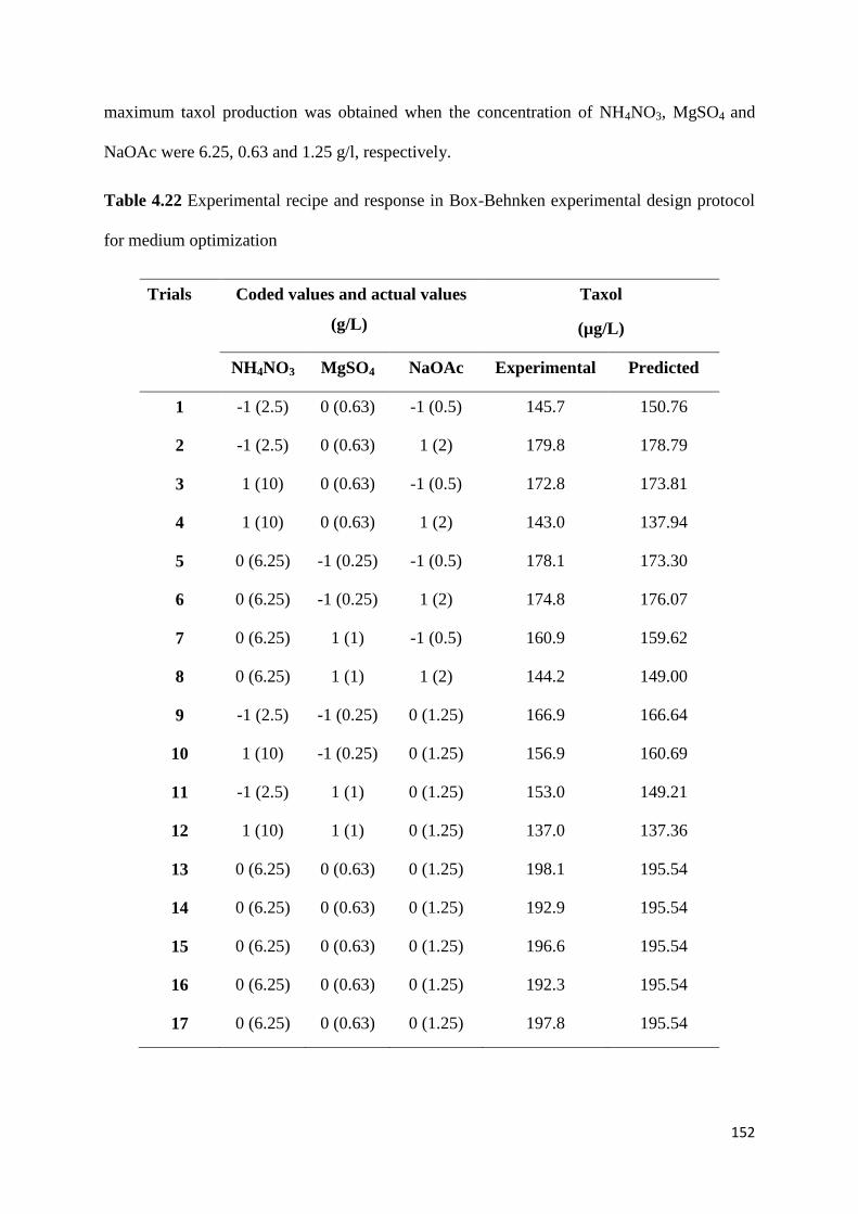
152
maximum taxol production was obtained when the concentration of NH4NO3, MgSO4 and
NaOAc were 6.25, 0.63 and 1.25 g/l, respectively.
Table 4.22 Experimental recipe and response in Box-Behnken experimental design protocol
for medium optimization
Trials Coded values and actual values
(g/L)
Taxol
(µg/L)
NH4NO3 MgSO4 NaOAc Experimental Predicted
1 -1 (2.5) 0 (0.63) -1 (0.5) 145.7 150.76
2 -1 (2.5) 0 (0.63) 1 (2) 179.8 178.79
3 1 (10) 0 (0.63) -1 (0.5) 172.8 173.81
4 1 (10) 0 (0.63) 1 (2) 143.0 137.94
5 0 (6.25) -1 (0.25) -1 (0.5) 178.1 173.30
6 0 (6.25) -1 (0.25) 1 (2) 174.8 176.07
7 0 (6.25) 1 (1) -1 (0.5) 160.9 159.62
8 0 (6.25) 1 (1) 1 (2) 144.2 149.00
9 -1 (2.5) -1 (0.25) 0 (1.25) 166.9 166.64
10 1 (10) -1 (0.25) 0 (1.25) 156.9 160.69
11 -1 (2.5) 1 (1) 0 (1.25) 153.0 149.21
12 1 (10) 1 (1) 0 (1.25) 137.0 137.36
13 0 (6.25) 0 (0.63) 0 (1.25) 198.1 195.54
14 0 (6.25) 0 (0.63) 0 (1.25) 192.9 195.54
15 0 (6.25) 0 (0.63) 0 (1.25) 196.6 195.54
16 0 (6.25) 0 (0.63) 0 (1.25) 192.3 195.54
17 0 (6.25) 0 (0.63) 0 (1.25) 197.8 195.54

153
Fig. 4.34 a, b Fungal growth (Fusarium redolens) as per seventeen experiments formulated
using Box-Behnken design
(a)
(b)

154
Table 4.23 Comparison of mycelial growth (Fresh weight) and taxol production of Fusarium
redolens as per experimental recipe in Box-Behnken experimental design protocol for
medium optimization
Trials Fresh weight
(mycelial growth in gm: 50 mL medium in 250 mL
flask as per Box-Behnken experimental design)
Taxol
(µg/L)
1 16.20 150.76
2 21.00 178.79
3 20.77 173.81
4 14.03 137.94
5 20.12 173.30
6 20.89 176.07
7 17.50 159.62
8 15.72 149.00
9 18.94 166.64
10 17.87 160.69
11 15.90 149.21
12 14.20 137.36
13 25.12 195.54
14 24.89 195.54
15 24.76 195.54
16 24.83 195.54
17 24.90 195.54

155
Table 4.24 Regression coefficients and their significance for response surface model
Source Sum of squares df Mean square F-value Prob ˃ F
Model 6943.04 9 771.45 33.39 <0.0001
(significant)
A 158.42 1 158.42 6.86 0.0345
B 830.28 1 830.28 35.94 0.0005
C 30.81 1 30.81 1.33 0.2861
A2 8.70 1 8.70 0.38 <0.0001
B2 1020.80 1 1020.80 44.19 <0.0001
C2 44.89 1 44.89 1.94 0.0013
AB 2250.67 1 2250.67 97.42 0.5588
AC 1511.21 1 1511.21 65.41 0.0003
BC 615.95 1 615.95 26.66 0.2060
Lack of fit 131.50 3 43.82 5.79 0.0614
A-NH4NO3; B-MgSO4; C-NaOAc
The model F-value of 33.39 implies the model is significant
Values of “probability ˃ F” less than 0.0500 indicate model terms are significant
In this case A, B, AC, A2, B
2, C
2 are significant model terms
Values greater than 0.1000 indicate the model terms are not significant

156
(A)
Design-Expert® SoftwareFactor Coding: ActualTaxol
Design points above predicted valueDesign points below predicted value198.1
137.1
X1 = B: MgSO4X2 = A: NH4NO3
Actual FactorC: NaOAC = 0.00
-1.00
-0.50
0.00
0.50
1.00
-1.00
-0.50
0.00
0.50
1.00
120
140
160
180
200
220
T
ax
ol
B: MgSO4 A: NH4NO3
195.54195.54
Design-Expert® SoftwareFactor Coding: ActualTaxol
Design Points198.1
137.1
X1 = B: MgSO4X2 = A: NH4NO3
Actual FactorC: NaOAC = 0.00
-1.00 -0.50 0.00 0.50 1.00
-1.00
-0.50
0.00
0.50
1.00Taxol
B: MgSO4
A:
NH
4N
O3
150
160
160
170
170
180
190
195.768
193.876
5
Prediction 195.54
Actual Factor
C: NaOAc = 1.25
A: NH4NO3
B: MgSO4
A: NH4NO3
B: MgSO4

157
B) Design-Expert® SoftwareFactor Coding: ActualTaxol
Design points above predicted valueDesign points below predicted value198.1
137.1
X1 = C: NaOACX2 = A: NH4NO3
Actual FactorB: MgSO4 = 0.00
-1.00
-0.50
0.00
0.50
1.00
-1.00
-0.50
0.00
0.50
1.00
120
140
160
180
200
220 T
ax
ol
C: NaOAC
A: NH4NO3
195.54195.54
Design-Expert® SoftwareFactor Coding: ActualTaxol
Design Points198.1
137.1
X1 = C: NaOACX2 = A: NH4NO3
Actual FactorB: MgSO4 = 0.00
-1.00 -0.50 0.00 0.50 1.00
-1.00
-0.50
0.00
0.50
1.00Taxol
C: NaOAC
A:
NH
4N
O3
150
160
160
170
170
180
180
180
190
5
Prediction 195.54
Actual Factor
B: MgSo4 = 0.63
(B)
A: NH4NO3
C: NaOAc
C: NaOAc
A: NH4NO3

158
(C)
Design-Expert® SoftwareFactor Coding: ActualTaxol
Design points above predicted valueDesign points below predicted value198.1
137.1
Taxol = 178.1Std # 5 Run # 4X1 = C: NaOAC = -1.00X2 = B: MgSO4 = -1.00
Actual FactorA: NH4NO3 = 0.00
-1.00
-0.50
0.00
0.50
1.00
-1.00
-0.50
0.00
0.50
1.00
120
140
160
180
200
220
T
ax
ol
C: NaOAC B: MgSO4
195.54195.54
Design-Expert® SoftwareFactor Coding: ActualTaxol
Design Points198.1
137.1
X1 = C: NaOACX2 = B: MgSO4
Actual FactorA: NH4NO3 = 0.00
-1.00 -0.50 0.00 0.50 1.00
-1.00
-0.50
0.00
0.50
1.00Taxol
C: NaOAC
B:
Mg
SO
4
160170
180 180
180
190
5
Prediction 195.54
Fig. 4.35 3D surface and contour plots showing the effect of different variables on taxol
production. (A) effect of NH4NO3 and MgSO4 on taxol production; (B) effect of NH4NO3
and NaOAc on taxol production; (C) effect of MgSO4 and NaOAc on taxol production
B: MgSO4
C: NaOAc
C: NaOAc
B: MgSO4
Actual Factor
A: NH4NO3 = 6.25
(C)

159
Fig. 4.36 a, b Growth of Fusarium redolens in optimized S7 medium
In order to verify the RSM predicted results, an experiment was performed under the
optimized nutrients levels (Fig. 4.34). The values obtained (experimental) were compared
with the predicted values. The predicted response in Box-Behnken experimental design for
taxol production gave a value of 195 µg/L, while the actual experimental value was 198
µg/L, suggesting that experimental and predicted values were in good agreement. Further
relation of taxol production with biomass was also noticed in this case also. Highest amount
of taxol was found in the case of higher biomass favouring positive correlation between the
two (Table 4.23).
(a)
(b)

160
Conclusions and salient findings
The current study has culminated in production of three times higher taxol production by
application of statistical tool; Response surface methodology. The physical and chemical
factors which affect the production of taxol in Fusarium redolens were firstly varied one at a
time and after that, Plackett-Burman design was implied to screen multiple variables which
affect the production of taxol. NH4NO3, MgSO4 and NaOAc were found to be the most
significant factors that affected the taxol yield. Based on the results of Box-Behnken design,
the optimized media and conditions for maximum taxol production comprised sucrose,
NH4NO3, MgSO4, NaOAc incubated at 25oC for 7 days with the initial pH of 6.5 which
resulted in 3 folds increase in the production of taxol in the optimized media. Based on the
promising results of this study, enhancement of taxol yield by application of statistical tools
including RSM seems to be a promising solution for production of these highly demanding
commercial drugs.

161
5. Discussion
5.1 Isolation and characterization of taxol producing endophytic fungi
Taxol is the best known and most studied affiliate of the taxane diterpenoids, or taxoids and
has been utilized in chemotherapy for many types of cancers since 1970s. Traditional
practices of extracting taxol from the bark of Taxus species are inefficient and
environmentally costly. For example, while 1 kg of taxol treats just five hundred patients; the
production of 1 Kg of taxol demands 10 tons of bark or 300 trees. For this reason, seeking
new ways of obtaining taxol is the key to protect this limited, endangered resource and
minimizing the cost of drug therapy. In this regard, scientists all over the world have focused
on endophytic fungi, which are extensively found in almost all kinds of plants including
Taxus species, that can produce physiologically active compounds, such as taxanes, which
are the same or analogous with those obtained from their hosts (Lu et al. 2000, Strobel et al.
2004). This constitutes a new approach to resolve resource limitation and an alternative taxol
source. Indeed, it represents a sumptuous opportunity to find new and interesting
microorganisms among Taxus species in different settings and ecosystems.
In the present study, Northern Himalayan yew was selected as the source for isolating
endophytic fungi as to date no endophytes having taxol-generating capability have been
reported from T. baccata subsp. wallichiana growing in this region. This yew species ranges
widely in the temperate Himalayan region and in the hills of Meghalaya, Nagaland and
Manipur and most of its growth takes place in moist and damp environments. Endophytes
associated with T. baccata (Himalayan yew) may have certain biological links with its host
for attaining the capability of producing taxol. Being endosymbiotic in nature, several
endophytic fungi may also inherit taxol-producing capability and this may be considered as a
reason for the transfer of genes involved in taxol biosynthesis to endophytes harbouring in

162
different parts of yew trees. Taxol, a known wonder cancer drug holds many properties such
as stability, less side-effects, and higher effectiveness and has always been in surplus demand
in the market owing to its broad spectrum of antitumor action (Wani et al. 1971). In order to
fulfil the increasing demand of pure drug, scientists have always been searching for efficient
endophytic strains from Taxus sp. and species other than Taxus that have the capability of
generating a good amount of taxol. Current work is one more step towards this quest.
Number of (around 60) endophytic fungi harbouring in the bark of Taxus baccata L.
subsp. wallichiana (Zucc.) tree were isolated in this study. In contrast to the biochemical
screening (traditional) method, the molecular marker-based screening was also used as it is a
rapid, economical and efficient alternative for the screening of taxol-producing endophytic
fungi (Zhang et al. 2008; Mirjalili et al. 2012). The main advantage of this method is that it is
not dependent on the production of taxol; rather, it indicates the existance of some key genes
required for taxol biosynthesis in the fungal genome. The taxol biosynthetic pathway in yew
trees involves 19 enzymatic steps (Croteau et al. 2006), and we chose the two main end step
genes dbat (involved in the formation of Baccatin III) and bapt (involved in phenylpropanoid
side chain formation at C13) involved in taxol biosynthesis, for primary screening of taxol-
producing endophytes by molecular screening. The use of gene-specific PCR amplification
for screening the isolated endophytes made it feasible to screen all the isolates, which would
otherwise have been very laborious and practically time consuming by biochemical screening
method. Eight out of the 60 fungi had positive hits of dbat gene and had approximately 200
bp amplified fragments of this gene. Presence of DBAT gene is essential for taxol
biosynthesis but cannot be relied completely because some fungi having DBAT gene may
produce baccatin III, but not taxol. So, eight fungi harbouring DBAT gene were screened for
the presence of the BAPT gene. Five fungal isolates (TBPJ-B, TBPJ-A, TBPJ-13, B-7 and C-
1) showed amplification of 530 bp fragment of BAPT gene, suggesting that all of them may

163
produce taxol. Currently, DBAT and BAPT genes have been mostly used as molecular probes
for the primary screening of taxol producing microorganisms (Zhou et al. 2007; Zhang et al.
2008), thus designing appropriate degenerate primers for amplification of more target genes,
e.g. the final acylation step in taxol biosynthesis, taxoid C13-side-chain N-benzoyltransferase
(DBTNBT), may be a better choice for screening. When the amplified DNA fragments of
BAPT gene from all 5 endophytic isolates were analyzed and aligned with the protein
sequences of BAPT gene of various Taxus species, the characterization gave a new
understanding to the controversial hypothesis of horizontal gene transfer (HGT). It showed
that evolutionary trajectory of taxol gene clusters amidst microbial and plant origin might be
coexisting. Although HGT in fungi are predominantly reported (Fitzpatrick 2012), the
ultimate plausibility of microbial taxol gene cluster by HGT hypothesis should be revisited
and further investigated because approximately 20 genes ramified in the taxol biosynthesis
make HGT rather improbable. Moreover, taxol-producing endophytic fungi have been
isolated from plants which themselves are incapable of producing taxol (Li et al. 1996;
Kumaran et al. 2009), implying that taxol biosynthesis in fungi may not be attained from
HGT. In nature, gibberellin biosynthetic pathways in fungi and higher plants have evolved
independently and not by HGT (Tudzynski 2005; Hedden et al. 2001). We thus presumed that
taxol biosynthetic cluster might be repeatedly originated during evolution.
The polymerase chain reaction (PCR) provides a rapid and highly sensitive method for
the primer-mediated enzymatic amplification of specific target sequences resulting in the
exponential increase of target DNA copies. Molecular identification has increasingly been
used as supplementary tool for the traditional systematic classification. Genomic DNA from
all the 5 selected endophytic fungi was subjected to amplification of ITS region using ITS1
and ITS4 universal primers. Resultant PCR products were sequenced and sequence analysis
revealed that fungi belong to Fusarium, Gibberella, Microdiplodia and Paraconiothyrium

164
species. Out of the two most widely used methods for molecular phylogenetic analyses
[Maximum parsimony (MP) and Maximum Likelihood (ML)] MP was used to check the
clades of these endophytes based on sequence analysis. The phylogenetic tree was
reconstructed using ITS sequences of TBPJ-B, TBPJ-A, TBPJ-13, B-7 and C-1 wherein
strain TBPJ-B clustered with Fusarium redolens (KC924920), B-7 clustered with Fusarium
tricinctum (KF010839), C-1 with Gibberella avenacea (KF010838), TBPJ-A clustered with
Microdiplodia sp. G16A (KF010841) and TBPJ-13 with Paraconiothyrium brasiliense
(KF010840). All the isolates were also studied based on their morphology and microscopic
characters. Phylogenetic analysis of the ITS region showed a reasonable degree of correlation
with the morphological classification schemes of species within the genus.
In the past two decades, several endophytic microorganisms isolated from different
geographical settings have been reported to produce taxol through biochemical or molecular
marker screening (Flores-Bustamante et al. 2010), the majority of which belongs to
Alternaria sp., Alternaria taxi, Alternaria alternata, Aspergillus sp., Cladosporium sp.,
Fusarium laterititum, Monochaetia sp., Ozonium sp., Pestalotiopsis microspora,
Pestalotiopsis guepinii, Pithomyces sp., Taxomyces andreanae, Trichoderma sp.,
Tubercularia sp., Phomopsis sp., Penicillium, Botrytis taxi etc. (Stierle et al. 1993;
Gangadevi and Muthumary 2008; Zhao et al. 2008, 2009; Zhou et al. 2010). Endophytic
fungi capable of taxol production obtained from the bark of Taxus baccata subsp. wallichiana
in the northern Himalayan regions of India represented a phylogenetically diverse array of
fungal taxa, including some frequent and some rare genera, confirming that a few species are
frequent colonizers and a majority of groups are rare inhabitants in woody plants of temperate
to sub-tropical regions (Tejesvi et al. 2005).

165
5.2 Biochemical screening of taxol production by endophytic fungi
In order to screen the presence of taxol by different endophytic fungi, extracts of all the five
representative species Fusarium redolens (TBPJ-B), Microdiplodia sp. (TBPJ-A),
Paraconiothyrium brasiliense (TBPJ-13), Fusarium verticillioides (B-7) and Phomopsis sp. (C-1)
with positive results in the primary screening were put through chromatographic analysis by
TLC, HPLC and LC-MS for the detection of fungal taxol. The first insight into the presence
of taxol in the extracts of the 5 fungal endophytes (TBPJ-13, B-7, TBPJ-A, TBPJ-B and C-1)
was done by thin layer chromatography. Authentic taxol was used as positive control. The
active principle on development of the thin layer plate appeared as a dark band under UV
illuminator at 254 nm. In order to further verify these results, HPLC analysis of the fungal
extract and authentic taxol was studied. The results gave a single peak under the same
conditions when eluted from a reverse phase C18 column. The HPLC peak positions and
peak shapes of taxol from fungal extracts had same retention time as that of standard taxol.
Mycelium from 21 day old culture in S-7 semi synthetic medium was subjected to HPLC to
quantify the amount of taxol produced by the fungus. Among the 5 taxol-producing fungi,
TBPJ-B had the highest taxol yield of 66.25 µg/L, in comparison with those of TBPJ-A
which produced 27.40 µg/L, B-7 produced 23.47 µg/L, TBPJ-13 produced 19.60 µg/L and C-
1 produced 11.03 µg/L of taxol in S-7 semi synthetic liquid medium. Quantitative HPLC
analysis of the fungal taxol showed that TBPJ-B (Fusarium redolens) produced the maximum
amount of taxol compared to other isolates. These results are comparable with previously
reported taxol-producing fungi isolated from different geographical settings (Stierle et al.
1993; Gangadevi and Muthumary 2008; Zhao et al. 2008, 2010). There are a number of
reports in the literature about Fusarium sp. capable of taxol production in range of 0.13-286
µg/L with strain improvement procedures; F. mairei (20 µg/L, Dai and Tao 2008), F. solani
(Taxus celebica, 1.6 µg/L, Chakravarthi et al. 2008), F. solani Tax-3 (Taxus chinensis,

166
163.35 µg/L, Deng et al. 2009). Fusarium and some Aspergillus species have also been
reported earlier to be capable of producing taxol in vitro (Caruso et al. 2000; Zhao et al.
2008; Deng et al. 2009), but F. redolens and F. verticillioides are the first ever reports of
endophytic fungi capable of taxol production obtained from T. baccata subsp. wallichiana.
Paraconiothyrium brasiliense, Microdiplodia sp. and Phomopsis sp. reported from other
Taxus plants have not been acquired from the Himalayan yew until now, implying that yews
growing in different geographical settings can harbour novel, highly diverse taxol-producing
endophytic strains and that certain taxol-producing fungi seem to be host specific. These
endophytes that possess the capability to produce such important secondary metabolites may
succeed in occupying a niche within the plant tissue or even contribute to host defence
against the invading pathogens (Liu et al. 2009; Chandra 2012).
Convincing evidence for the identification of the fungal taxol was also obtained by high
resolution mass spectroscopy (MS). Characteristically, the authentic taxol yielded an (M+H)+
peak at m/z 854.7 and an (M+Na)+ peak at m/z 876.8. By comparison, the fungal taxol also
yielded a peak MH+ at m/z 854.3 and MNa
+ at m/z 876.3 with characteristic fragments at m/z
569, 551, 509, 286 and 268. Major fragment ions observed in the mass spectrum of taxol can
be placed into three categories which represents major portions of the molecule (McClure et
al. 1992). The peaks of fungal taxol gave m/z ratios similar to the molecular ions of standard
taxol, verifying that the 5 endophytic strains can produce taxol in vitro.
In this study, based on morphological and molecular data as well as phytochemical
analysis, Fusarium redolens, Microdiplodia sp., Paraconiothyrium brasiliense, Fusarium
verticillioides and Phomopsis sp. were determined as novel and promising strains of taxol
producing endophytic fungi. As mentioned above, quantitative HPLC analysis showed that
the taxol content of all these taxol producing endophytes was in the range of previously
reported fungi, but was found to be relatively lower than that of Taxus species, the high

167
growth rate and short generation time make these endophytes advantageous to continue
investigation. Notably, the extraction yield from taxol producing fungi has been recently
enhanced by the use of new biotechnological techniques, such as fungal strain improvement
(Xu et al. 2006), recombinant techniques (Wang et al. 2007; Zhou et al. 2008; Wei et al.
2009) and microbial fermentation engineering (Xu et al. 2006; Gogoi et al. 2008; Zhao et al.
2011). Thus, in order to fulfill the commercial demand for taxol, further work was focussed
on ameliorating taxol yield in fungi by optimization of culture conditions. In addition, the
paucity of a complete taxol biosynthetic cluster (5 unknown enzymatic steps) is at present a
bottleneck for basic and applied research. Genome sequencing and analysis of taxol-
producing microorganisms thus could significantly expand the number of known taxol
biosynthetic genes to illuminate the whole pathway and provide the basis for heterologous
production. From an evolutionary adaptation point of view, endophytic microorganism
harbouring discrete metabolic functions could achieve extraordinary success in occupying a
alcove within plant tissues or even contribute to host defense against various invading
pathogens (Liu et al. 2009). Hence, analysis of genes involved in taxol synthesis from the
diverse fungi will significantly intensify our understanding of the co-evolutionary mechanism
of the endophyte host (Mirjalili et al. 2012).
5.3 Antitumorous activity of fungal taxol
Other than chromatographic methods for the determination of taxol in biological matrices, an
immunoassay to study the antitumor effect of fungal extracts was employed. The
antimitogenic activity of fungal taxol was assessed by potato disc tumor induction assay. This
assay is known for its simplicity and reliability by many researchers (Coker et al. 2003) and
has been used in screening of antitumor agents irrespective of their mode of action. Authentic
taxol served as positive control and inhibited tumor production at all the tested
concentrations. Fungal taxol from 5 endophytes (TBPJ-B, TBPJ-A, B-7, TBPJ-13 and C-1)

168
also inhibited tumor formation the same way the authentic taxol did. It was justified as starch
in the potato tissue took up the stain and appeared dark brown in color, but tumors produced
by A. tumefaciens did not take up the stain and appeared creamy to orange (Mc Laughlin &
Rogers 1998). As both fungal taxol and authentic taxol inhibited tumor formation in potato
discs, it was authenticated that the fungal taxol has antitumorigenic activity. Bacterial
viability test showed that the standard drug and the drug in the fungal extracts did not affect
the viability and growth of the bacteria i.e., drug didn’t hinder bacterial tumor causing ability.
So, it was depicted that action of drugs tested is on the formation of tumors and not on the
bacterial viability.
5.4 Process optimization for taxol production from endophytic fungus Fusarium
redolens
As it was envisaged that fungal taxol is novel and promising alternative for producing large
amounts of the drug in very efficient and economical way in short periods of time, an attempt
was made to enhance the production of taxol from efficient taxol producing endophyte
Fusarium redolens by optimizing the media components through One factor at a time
(OFAT) and Response Surface Methodology analysis. Response surface methodology (RSM)
allows a rapid screening of most significant factors affecting the production of any
metabolite/ enzyme or other by products.
After initial screening of different variables by OFAT, Plackett-Burman design was
employed to screen significant factors affecting Taxol production. This test showed that
NaOAc, MgSO4 and ZnSO4 were most important components which gave main contribution
on taxol production. Although the PB design could be successfully used for equitable
prediction of the significant level of different variables (micronutrients) affecting the
response (taxol production), some of the significant interactive effects of the chosen variables
(two-factor interaction) were staggered in its complex structure. Because of this reason, the

169
actual main effects of these variables may have been influenced. Hence, the significant
components NaOAc and MgSO4 whose interactive effects were demonstrated by PB design
were further modelled more precisely by RSM. From the study of response surface contour
plots, maximum taxol production was obtained when the concentration of NH4NO3, MgSO4
and NaOAc were 6.25, 0.63 and 1.25 g/l, respectively. The verification of RSM predicted
results was done by performing experiment under the optimized nutrients levels for taxol
production. The predicted response in Box-Behnken experimental design for taxol production
gave a value of 195 µg/L, while the actual experimental value was 198 µg/L, suggesting that
experimental and predicted values were in good agreement. From these results, the basic
parameters optimized for biomass and taxol production by F. redolens were observed when
the culture was grown at 25oC for 7 days with the initial pH of 6.5. The growth and taxol
production remained almost static after 20 days of incubation. Huang et al. 2001 also reported
antitumor and antifungal activity of endophytic fungi when the cultures were incubated at
25ºC for 7 days. Taxol is a secondary metabolin whose synthesis is regulated by carbon and
nitrogen sources, phosphate, trace elements and precursors (Parra et al. 2005). In the present
study, sucrose served as best carbon source for taxol production compared to other carbon
sources tested. Contrary, Xu et al. 2006 reported glucose as best carbon source for taxol
production by Fusarium maire. Ammonium nitrate served as good source of nitrogen in this
study. Strobel et al. 1996 reported ammonium nitrate and peptone as good nitrogen sources
for taxol production by Pestalotiopsis microspora. NH4NO3 was also found to have
significant effect on taxol production in case of F. maire (Xu et al. 2006). Nutrition plays a
crucial role in the onset and intensity of secondary metabolism. To achieve high product
yield, it is prerequisite to design appropriate production medium as there is relationship
between media composition and biosynthesis of secondary metabolites (Wang et al. 2013).
NaOAc, MgSO4 and ZnSO4 were shown as important components for taxol production in this

170
study. Sodium acetate as an activator was also reported with Taxomyces andreanae (Stierle et
al. 1993). Similar results were also reported by Xu et al. (2006) who found enhancement of
taxol yield from 20 µg/L to 225 µg/L in mutant strain of F. maire. Zhao et al. (2011) found
enhancement of taxol production from 397 to 456 µg/ L while Xu et al. (2006) reported 31%
increase in taxol production upon media optimization. Luo and He (2004) also found 2 times
higher production of Paclitaxel upon optimizing the concentration of elicitors and precursors.
The present study led to enhancement of fungal taxol up to three folds which are encouraging
in terms of product yield for scale up studies and commercial exploitation of fungal taxol.
Statistical analysis of coefficients in Plackett-Burman design experiments demonstrated that
MgSO4 and NaOAc are the major factors influencing the production of taxol. With the
application of RSM, production of fungal taxol increased 3 folds compared to unoptimized
medium. Our work has proved the effectiveness of statistical tools in bioprocess optimization
for exploring scale up feasibility, augmenting the economic viability of the process. Further
improvement in taxol production can be achieved by improvement of the fungal strains
through genetic manipulations and/ or mutagenesis or by augmentation of elicitor and
precursor molecules to this fungal strain.
In conclusion, the present study led to isolation of several novel endophytic fungi
capable of producing the anticancerous drug taxol which is highly demandable and
pharmaceutically important medicine. DBAT and BAPT genes were found to be reliable
molecular markers for initial screening of taxol biosynthetic pathway in endophytic fungi.
Taxol producing endophytes were found to belong to Fusarium redolens, Microdiplodia sp.,
Paraconiothyrium brasiliense, Fusarium tricinctum and Gibberella avenacea. Several of
these fungal species have been found to be the first reports of taxol producing endophytes.
Along with molecular screening, biochemical screening of fungal taxol by HPLC and LC MS
further confirmed that all the isolates were highly efficient in producing taxol. Potato disc

171
tumour assay verified the efficacy of fungal taxol comparable to that of commercial
paclitaxel drug which paved way for fulfilling the demand of this wonder drug by these
alternative sources other than plant source. Statistical tool RSM further led to three times
enhanced production of taxol in endophytic fungus Fusarium redolens by optimization of
various media components, highlighting its usage for commercial applications. This study is a
step forward in proving the potential of endophytes in production of highly demandable
pharmaceutical drugs and paves way for replacement of conventional sources of these drugs
by serving as effective alternatives.

172
6. Summary
Endophytes, the organisms which inhabit living tissues of plants have long been recognized
as important and novel resources of bioactive products. Number of medicinal plants has been
reported to be source of fungal endophytes with capability to produce several novel
pharmaceutically important compounds. Paclitaxel, the world’s first billion dollar anticancer
blockbuster, has long been isolated from yew plants which are conventionally largest sources
for this drug. But due to various shortcomings of harvesting huge quantities of these plants
for retrieval of small amounts of taxol and to fulfill the large demand of this drug, alternative
sources need to be investigated. The primary focus of the current work is to investigate the
potential of endophytic fungi for production of this wonder drug.
Till now, not much work has been reported on taxol producing capability of fungal
endophytes from Taxus baccata L. subsp. wallichiana (Zucc.) Pilger growing in northern
Himalayan region of India. The present work was aimed to study the diversity of taxol-
producing endophytic fungi associated with Taxus baccata subsp. wallichiana, screening
endophytic fungi for taxol production using molecular markers, evaluating anticancerous
activity of fungal taxol and optimization of media components and conditions for enhancing
the taxol production.
In this work, endophytic fungi from bark of yew trees of Indian Himalayan Region
(IHR) were isolated and screened for production of anticancerous drug, taxol. Bark samples
of T. baccata subsp. wallichiana were collected from different locations of Northern Indian
Himalayan Region (IHR); Bhadrewah (district Doda, Jammu and Kashmir), Shimla
(Himachal Pradesh) and Almora (Uttrakhand). The bark pieces were surface disinfected,
small pieces of inner bark were excised and placed on the surface of potato dextrose agar
(PDA) medium. Petri plates were incubated at 28ºC for 10 days to allow the growth of

173
endophytic fungi. Total of 60 fungal morphotypes were isolated and the isolates were coded
and stored in sterile distilled water as agar plugs.
Taxol producing endophytic fungi were firstly screened on the basis of essential genes
involved in taxol biosynthetic pathway i.e. DBAT (10-deacetylbaccatin III-10-O-acetyl
transferase) and BAPT (C-13 phenylpropanoid side chain-CoA acyltransferase). Both the
genes served as reliable molecular markers for initial screening of taxol producing capability
in endophytes. The analysis of individual endophytic fungal genome with regard to presence
of potential genes involved in taxol synthesis revealed that 8 out of 60 isolated endophytes
were harbouring DBAT gene and 5 endophytic isolates out of these 8 gave positive hits for
BAPT gene. Thus, five efficient endophytic fungi were isolated from Himalayan yew bark
harbouring dbat and bapt genes of taxol biosynthetic pathway. These cultures were
designated as TBPJ-B, TBPJ-A, TBPJ-13, B-7 and C-1. Five fungal isolates showed
amplification of a 530 bp fragment of the bapt gene, suggesting that all of them may produce
taxol. Sequences of bapt gene of endophytic fungi were analyzed using BLASTx and aligned
with the protein sequences of the bapt gene of various Taxus species. The partial protein
sequences of the bapt gene of all 5 fungi showed high homology with protein sequences of
the bapt gene of various yew species, confirming the presence of a taxol biosynthetic
pathway in all the endophytic fungal strains.
The fungal isolates which showed positive results in molecular screening were
characterized based on morphological characters. Morphological characteristics of the fungi
like mycelia, conidiophores and conidia were microscopically examined. To authenticate the
classical identification of the five fungal internal transcribed spacer (ITS) regions of the
rDNA from genomic DNA was amplified using the universal primers, ITS1 and ITS4.
BLAST search was performed to find the possible homologous sequences of newly
sequenced taxa from the GenBank database. The phylogenetic tree was reconstructed using

174
the maximum parsimony method and the Kimura two parameter distance calculation by the
MEGA5 software. Based on classical and molecular profiling, the isolates were identified as
Fusarium redolens (TBPJ-B), Fusarium tricinctum (B-7), Gibberella avenacea (C-1),
Microdiplodia sp. G16A (TBPJ-A) and Paraconiothyrium brasiliense (TBPJ-13).
Biochemical screening of fungal taxol was done by TLC, HPLC and LC-MS from
extracts of fungal cultures in accordance to the literature which verified the efficacy of all
isolates in production of taxol. The 5 fungal endophytes were grown in S7 medium for 3
weeks to detect production of taxol. The fungal mycelia of all the isolates were extracted 3
times in methanol individually, while the fermentation broths were extracted with three equal
volumes of dichloromethane (DCM). The extracts of each fungal isolate were purified by
column chromatography and quantified using HPLC. Among the 5 taxol producing fungi,
Fusarium redolens produced significantly higher taxol yield of 66.25 µg/L, compared to
other fungi. Microdiplodia species produced 27.40 µg/L, Fusarium tricinctum 23.47 µg/L,
Paraconiothyrium brasiliense 19.60 µg/L and Gibberella avenacea produced 11.03 µg/L of
taxol in S7 liquid medium. Characteristically, the authentic taxol yielded an (M+H)+ peak at
m/z 854.7 and an (M+Na)+ peak at m/z 876.8. By comparison, the fungal taxol also yielded a
peak MH+ at m/z 854.3 and MNa
+ at m/z 876.3 with characteristic fragments at m/z 569, 551,
509, 286 and 268 by LC-MS analysis. The peaks of fungal taxol gave m/z ratios similar to the
molecular ions of standard taxol, verifying that the 5 endophytic strains are able to produce
taxol in vitro.
Anticancerous activity of the 5 endophytic fungal isolates of the present study was
investigated by potato disc tumour assay in comparison to authentic paclitaxel using
Agrobacterium tumefaciens as the tumor causing agent along with bacterial viability test. It
was noticed that fungal taxol was equally effective in inhibiting tumour formation as that of
the standard paclitaxel. This further paved way for use of endophytic fungus as an effective

175
and novel source for production of anticancer drug, taxol. On the other hand the bacterial
viability tests showed that the standard drug and the drug in the fungal extracts did not affect
the viability of the bacteria, i.e., the drug did not hinder bacterial tumor causing ability.
In order to meet the ever increasing demand of this wonder drug, there is need for
enhancing the production of this anticancer agent. In this regard, optimization of different
physical and nutritional parameters for enhanced production of taxol from Fusarium redolens
was done by one-factor-at-a-time (OFAT) and response surface methodology (RSM)
approach. F. redolens was selected for process optimization as it was capable of producing
maximum amount of taxol compared to other isolates. Highest taxol production was found to
be at incubation period of 20 days, medium to flask volume (Vm/Vf) ratio of 0.20, pH 6.5 and
incubation temperature of 25ºC. The statistical optimization led to enhancement in taxol
production from 66 µg/L to 198 µg/L when optimized concentrations of factors: NH4NO3
(6.25 g/L), MgSO4 (0.63 g/L) and NaOAc (1.25g/L) were added to S-7 liquid medium along
with 8% (w/v) sucrose as carbon source and 0.8% (w/v) ammonium nitrate as nitrogen
source. Upon optimization, there was three fold increase in the production of taxol by this
endophyte. RSM was found to be effective tool for ameliorating the production of fungal
taxol in the present work.
In summary, the research presented in the current study investigated the potential of
yet untapped endophytes as prolific source of pharmaceutically relevant natural products,
with focus on paclitaxel. As the highly desirable search for sustainable and economically
feasible sources of this excellent antitumour agent has tempted various authors to draw
premature conclusions proclaiming endophytes to be independent taxane bio-factories, we
contend that the answer to the issue of paclitaxel supply which might lie within the interplay
between the plant hosts and their microbial inhabitants, under evolutionary and
environmental control. While one has to be mindful that the problem we set out to address is

176
several orders of magnitude larger than those with which we are familiar, no one can deny the
opportunities that present themselves in the era of modern functional genomics and systems
biology. There is need for further research at the molecular level for a better understanding of
the host endophyte interaction in order to explore the reasons for host-endophyte gene
transfer.

177
References
Achal V, Kumari D, Xiangliang P (2011). Bioremediation of chromium contaminated soil by
a Brown-rot fungus, Gloeophyllum sepiarium. Res J Microbiol 6: 166-171.
Agrios GN (1997). Plant diseases caused by prokaryotes: bacteria and mollicutes. Plant
Pathology. Academic Press, San Diego. pp: 407-470.
Agusta A, Maehara S, Ohashi K, Simanjuntak P, Shibuya H (2005). Stereoselective oxidation
at C-4 of flavans by the endophytic fungus Diaporthe sp. isolated from a tea plant.
Chem Pharm Bull 53: 1565-1569.
Ahmed MS, Fong HH, Soejarto DD, Dobberstein RH, Waller DP, Moreno-Azorero RJ
(1981). High-performance liquid chromatographic separation and quantitation of
maytansinoids in Maytenus ilicifolia. J Chroma 213: 340-344.
Ajikumar PK, Xiao WH, Tyo KE, Wang Y, Simeon F, Leonard E, Mucha O, Phon TH,
Pfeifer B, Stephanopoulos G (2010). Isoprenoid pathway optimization for Taxol
precursor overproduction in Escherichia coli. Science 330: 70-74.
Ally AH, Edrada-Ebe R, Indriani ID, Wray V, Muller WEG, Frank T, Zirrgiebel U,
Schachetele C, Kubbutat MHG, Lin WH, Proksch P, Ebel R (2008). Cytotoxic
metabolites from the fungal endophyte Alternaria sp. and their subsequent detection in
its host plant Polygonum senegalense. J Nat prod 71: 972-980.
Altschul SF, Madden TL, Schaffer AA, Zhang J, Zhang Z, Miller W, Lipman DJ (1997).
Gapped BLAST and PSI-BLAST: a new generation of protein database search
programs. Nucleic Acids Res 25: 3389-3402.
Alva P, McKenzie EHC, Pointing SB, Pena-Muralla R, Hyde KD (2002). Do sea grasses
harbour endophytes? Fungal Diversity Res Ser 7: 167-178.

178
Aly AH, Debbab A, Kjer J, Proksch P (2010). Fungal endophytes from higher plants: a
prolific source of phytochemicals and other bioactive natural products. Fungal div 41:
1-16.
Aly AH, Debbab A, Proksch P (2011). Fungal endophytes: unique plant inhabitants with
great promises. Appl Microbiol Biotechnol 90: 1829-1845.
Amna T, Puri SC, Verma V, Sharma JP, Khajuria RK, Musarrat J, Spiteller M, Qazi GN
(2006). Bioreactor studies on the endophytic fungus Entrophospora infrequens for the
production of an anticancer alkaloid camptothecin. Can J Microbiol 52: 189-196.
Anterola A, Shanle E, Perroud PF, Quatrano R (2009). Production of taxa-4(5),11(12)-diene
by transgenic Physcomitrella patens. Trans Res 18: 655-660.
Arbuck SG, Christian MC, Fisherman JS, Cazenave LA, Sarosy G, Suffness M, Adams J,
Canetta R, Cole KE, Friedman MA (1993). Clinical development of taxol. J Natl
Cancer Inst Monogr 15: 11-24.
Arnold AE (2007). Understanding the diversity of foliar endophytic fungi, progress,
challenges and frontiers. Fungal Biol Rev 21: 51-66.
Arnold AE, Maynard Z, Gilbert GS (2001). Fungal endophytes in dicotyledonous neotropical
trees, patterns of abundance and diversity. Mycol Res 105: 1502-1507.
Arnold AE, Mejia LC, Kyllo D, Rojas EI, Maynard Z, Robbins N, Herre EA (2003). Fungal
endophytes limit pathogen damage in a tropical tree. Proc Natl Acad Sci USA 100:
15649-15654.
Asai M, Mizuta E, Izawa M, Haibara K, Kishi T (1978). Isolation, chemical characterization
and structure of ansamitocin, a new antitumour antibiotic. Tetrahedron 35: 1079-1085.

179
Baadhe RR, Mekala NK, Parcha SR, Devi YP (2014). Optimization of amorphadiene
production in engineered yeast by response surface methodology. 3 Biotech 4: 317-324.
Bacon CW, White JF (2000). Microbial endophytes. Marcel Dekker Inc., New York.
Bajaj V, Tilay A, Annapure U (2012). Enhanced production of bioactive Sophorolipids by
Starmerella bombicola NRRL Y-17069 by Design of Experiment Approach with
successive purification and characterization. J Oleo Sci 61: 377-386.
Banerjee S, Upadhyay N, Kukreja AK, Ahuja PS, Kumar S, Saha GC, Sharma RP,
Chattopadhyay SK (1996). Taxanes from in vitro cultures of the Himalayan Yew Taxus
wallichiana. Planta Med 62: 333-335.
Banik RM, Pandey DK (2008). Optimizing conditions for oleanolic acid extraction from
Lantana camara roots using response surface methodology. Ind Cr pro 27: 241-248.
Barnett CJ, Cullinan GJ, Gerzon K, Hoying RC, Jones WE, Newlon WM, Poore GA,
Robison RL, Sweeney MJ, Todd GC, Dyke RW, Nelson RL (1978). Structure-activity
relationships of dimeric Catharanthus alkaloids 1. Deacetyl vinblastine amide
(vindesine) sulfate. J Med Chem 21: 88.
Barnett HL, Hunter BB (1998). Illustrated genera of imperfect fungi (4th edn). APS Press, St.
Paul, Minnesota, USA.
Becker FF (1975). Cancer: A Comprehensive Treatise. Volume 2. Etiology: Viral
Carcinogenesis. Plenum Press, NY.
Bedi YS, Ogra RK, Koul K, Kaul BL, Kapil RS (1996). Yew (Taxus spp.); A new look on
utilization, cultivation and conservation. In: Handa SS, Kaul MK, editors. Supplement
to cultivation and utilization of medicinal plants. Jammu-Tawi: Regional Research
Laboratory; pp: 443-456.

180
Bellis M (2012). The History of Penicillin; Alexander Fleming, John Sheehan and Andrew J
Moyer; http://inventors.about.com/od/pstartinventions/a/Penicillin.htm.
Besumbes O, Sauret-Güeto S, Phillips MA, Imperial S, Rodriguez-Concepcion M, Boronat A
(2004). Metabolic engineering of isoprenoid biosynthesis in Arabidopsis for the
production of taxadiene, the first committed precursor of taxol. Biotechnol Bioeng 88:
168-175.
Bettucci L, Simeto S, Alonso R, Lupo S (2004). Endophytic fungi of twigs and leaves of
three native species of Myrtaceae in Uruguay. Sydowia 56: 8-23.
Bi J, Ji Y, Pan J, Yu Y, Chen H, Zhu X (2011). A new taxol-producing fungus (Pestalotiopsis
malicola) and evidence for taxol as a transient product in the culture. Afr J Biotechnol
10: 6647-6654.
Bills GF, Gonzalez-Menendez V, Martin J, Platas G, Fournier J, Persoh D, Stadler M (2012).
Hypoxylon pulicicidum sp. nov. (Ascomycota, Xylariales), a Pantropical Insecticide-
Producing Endophyte. PLoS ONE 7: e46687.
Borges KB, Borges WDS, Pupo MT, Bonato PS (2008). Stereoselective analysis of
thioridazine-2-sulfoxide and thioridazine-5-sulfoxide: an investigation of rac-
thioridazine biotransformation by some endophytic fungi. J Pharm Biomed 46: 945-
952.
Box GEP, Meyer RD (1985). Some new ideas in the analysis of screening designs. J Res Nat
Bur Stand 90: 495-502.
Braun AC (1972). The relevance of plant tumor systems to an understanding of the basic
cellular mechanisms underlying tumorigenesis. Prog Exp Tumor Res 15: 165-187.

181
Bridge PD, Spooner BM, Roberts PJ (2005). The impact of molecular data in fungal
systematics. Adv Bot Res 42: 33-67.
Brown T, Tangen C, Flemming T, Macdonald J (1993). A phase II trial of taxol and
granulocyte colony stimulating factor (G-CSF) in patients with adenocarcinoma of
pancreas. Proc Am Soc Clin Onco 12 (abstracts) 200.
Buss T, Hayes MA (2000). In Biodiversity: New Leads for Pharmaceutical and Agrochemical
Industries; Wrigley SK, Hayes MA, Thomas R, Chrystal EJT, Nicholson N, Eds.; The
Royal Society of Chemistry: Cambridge, UK, pp: 75-85.
Butler MS (2005). Natural products to drugs: natural product derived compounds in clinical
trials. Nat Prod Rep 22: 162-195.
Cai M, Zhou X, Sun X, Tao K, Zhang Y (2009). Statistical optimization of medium
composition for aspergiolide A production by marine-derived fungus Aspergillus
glaucus. J Ind Microbiol Biotechnol 36: 381-389.
Cai YZ, Luo Q, Sun M, Corke H (2004). Antioxidant activity and phenolic compounds of
112 traditional Chinese medicinal plants associated with anticancer. Life Sci 74: 2157-
2184.
Cannon PF, Simmons CM (2002). Diversity and host preference of leaf endophytic fungi in
the Iwokrama Forest Reserve, Guyana. Mycol 94: 210-220.
Cao L, Huang J, Li J (2007). Fermentation conditions of Sinopodophyllum hexandrum
endophytic fungus on production of podophyllotoxin. Food Fermen Ind 33: 28-32.
Cardellina JH (1991). HPLC separation of taxol and cephalomannine. J Liquid Chrom 14:
659-665.

182
Cardinale BJ, Palmer MA, Collins SL (2002). Species diversity enhances ecosystem
functioning through interspecific facilitation. Nature 415: 426-429.
Carrnichael JW, Kendrick BW, Conner IL, Lynne S (1980). Genera of Hyphomycetes. The
University of Alberta Press, Edmonton.
Carroll G (1988). Fungal endophytes in stems and leaves: from latent pathogen to mutualistic
symbiont. Ecology 69: 2-9.
Caruso M, Colombo AL, Fedeli L, Pavesi A, Quaroni S, Saracchi M, Ventrella G (2000).
Isolation of endophytic fungi and actinomycetes taxane producers. Ann Microbiol 50:
3-13.
Cassady JM, Chan KK, Floss HG, Leistner E (2004). Recent developments in the
maytansinoids antitumour agents. Chem Pharm Bull 52: 1-26.
Chakravarthi BV, Das P, Surendranath K, Karande AA, Jayabaskaran C (2008). Production
of paclitaxel by Fusarium solani isolated from Taxus celebica. J Biosci 33: 259-267.
Chandra S (2012). Endophytic fungi: novel sources of anticancer lead molecules. Appl
Microbiol Biotechnol 95: 47-59.
Chang MY, Tsai GJ, Houng JY (2006). Optimization of the medium composition for the
submerged culture of Ganoderma lucidum by Taguchi array design and steepest ascent
method. Enz Microb Tech 38: 407-414.
Chattopadhyay S, Srivastava AK, Bisaria VS (2002). Optimization of culture parameters for
production of podophyllotoxin in suspension culture of Podophyllum hexandrum. Appl
Biochem Biotechnol 102-103: 381-393.

183
Chauhan K, Trivedi U, Patel KC (2006). Application of Response Surface Methodology for
Optimization of Lactic Acid Production Using Date Juice. J Microbiol Biotechnol 16:
1410-1415.
Chen JH, Liu JJ, Zang GG, Li YJ, Zhao LN (2004). Screening of taxol-producing endophytic
fungi and regulation of fermentation conditions. J Central South Univ 35: 65-69. (In
Chinese)
Cheng L, Ma QM, Tao GJ, Tao WY, Wang RM, Yang J, Guo XL (2007). Systemic
identification of a paclitaxel-producing endofungus. Industrial Microbiol 4: 23-30. (In
Chinese)
Chuang S, Puryear S, Cairney J (1993). A simple and efficient method for isolating RNA
from pine trees. Plant molecular biology reporter 11: 113-116.
Cingeetham V, Bandaru S, Dhar S, Gudimella R, Nallapeta S, Medicherla KM (2014).
Evolutionary Perspective of Fungal Pathogenic Genes. Int J Comp Biol 3: 39-42.
Clay K, Leuchtmann A (1989). Infection of woodland grasses by fungal endophytes.
Mycologia 81: 805-811.
Coker PS, Radecke J, Guy C, Camper ND (2003). Potato disc tumour induction assay: a
multiple mode of drug action assay. J Phytomed 10: 133-138.
Cragg G, Suffness M (1988). Metabolism of plant-derived anti-cancer agents. Pharmacol
Ther 37: 425.
Cragg GM, Boyd MR, Cardellina JH II, Grever MR, Schepartz S, Snader KM, Suffness M
(1993). The search for new pharmaceutical crops. In: Janick J, Simon JE (eds) Drug
discovery and development at the national cancer institute: new crops. Wiley, New
York, pp: 61-167.

184
Cragg GM, Newman DJ (2006). Plants as a source of anticancer agents. In: Elisabetsky E,
Etkin NL (eds) Ethnopharmacology. Encyclopedia of Life Support Systems (EOLSS),
developed under the Auspices of the UNESCO, Oxford, UK. EOLSS Publishers,
Oxford.
Croom EM (1995). Taxus for taxol and taxoids. In: Suffness M (ed) Taxol: Sci. Appl., CRC
Press, Boca Raton, pp: 37-70.
Croteau R, Ketchum RB, Long RM, Kaspera R, Wildung MR (2006). Taxol biosynthesis and
molecular genetics. Phytochem Rev 5: 75-97.
Cusido RM, Palazon J, Bonfill M, Exposito O, Moyano E, Pinol MT (2007). Source of
isopentenyl diphosphate for taxol and baccatin III biosynthesis in cell cultures of Taxus
baccata. Biochem Eng J 33: 159-67.
Daghino S, Turci F, Tomatis M, Girlanda M, Fubini B, Perotto S (2009). Weathering of
chrysotile asbestos by the serpentine rock-inhabiting fungus Verticillium leptobactrum.
FEMS Microbiol Ecol 69: 132-141.
Dai WL, Tao WY (2008). Preliminary study on fermentation conditions of taxol-producing
endophytic fungus. Chem Ind Eng Prog 27: 883-886, 891.
Daija KK, Florke V, Gehle D, Aust HJ, drarger S, Schulz B, Rheinheimer J (2005). Eur J org
chem 5100-5105.
Daisy BH, Strobel GA, Castillo U, Ezra D, Sears J, Weaver D, Runyon JB (2002).
Naphthalene, an insect repellent, is produced by Muscodor vitigenus, a novel
endophytic fungus. Microbiol 148: 3737-3741.
Damon C, Lehembre F, Desfeux CO, Luis P, Ranger J, Tachet LF, Marmeisse R (2012).
Metatranscriptomics Reveals the Diversity of Genes Expressed by Eukaryotes in Forest
Soils. PLoS ONE 7: 1-12.

185
Danishefsky SJ, Masters JJ, Young WB, Link JT, Snyder LB, Magee TV, Jung DK, Isaacs
RCA, Bornmann WG, Alaimo CA, Coburn CA, Di Grandi MJ (1996). Total Synthesis
of Baccatin III and Taxol. J Am Chem Soc 118: 2843-2859.
Davis KE, Joseph SJ, Janssen PH (2005). Effects of growth medium, inoculum size, and
incubation time on culturability and isolation of soil bacteria. Appl Environ Microbiol
71: 826-834.
de Bary A (1866). Morphologie und Physiologie der Pilze, Flechten, und Myxomyceten. Vol.
II. Hofmeister’s Handbook of Physiological Botany. Leipzig, Germany.
Debbab A, Aly AH, Edrada-Ebel RA, Muller Werner EG, Mosaddak M, Hakiki A, Rainer
Ebel R, Proksch P (2009). Bioactive secondary metabolites from the endophytic fungus
Chaetomium sp. isolated from Salvia officinalis growing in Morocco. Biotechnol Agron
Soc Environ 13: 229-234.
DeJong JHM, Liu Y, Bollon AP, Long RM, Jennewein S, Williams DC, Croteau RB (2006).
Genetic engineering of taxol biosynthetic genes in Saccharomyces cerevisiae.
Biotechnol Bioeng 93: 212-224.
Demain AL (2000). In Biodiversity: New Leads for Pharmaceutical and Agrochemical
Industries; Wrigley SK, Hayes MA, Thomas R, Chrystal EJT, Nicholson N, Eds.; The
Royal Society of Chemistry: Cambridge, UK, pp: 3-16.
Deng BW, Liu KH, Chen WQ, Ding XW, Xie XC (2009). Fusarium solani Tax-3, a new
endophytic taxol-producing fungus from Taxus chinensis. World J Microbiol
Biotechnol 25: 139-143.

186
Deshmukh SK, Mishra PD, Almeida AK, Verekar SA, Sahoo MR, Periyasamy G,
Goswami H, Khanna A, Balakrishnan A, Vishwakarma R (2009). Anti-inflammatory
and anticancer activity of ergoflavin isolated from an endophytic fungus. Chem and
Biodiv 6: 784-789.
Deshmukh SK, Verekar SA (2008). Fungal Endophytes: A Potential Source of Anticancer
Compounds. In book: Novel Therapeutic Agents from Plants: Progress and Future
Perspectives, Science Publishers, Inc Enfield USA, Editors: Carpinella MC, Rai MK,
pp: 175-206.
Deshmukh SK, Verekar SA (2012). Fungal endophytes: a potential source of antifungal
compounds. Frontiers in bioscience (Elite edition) 4: 2045-2070.
Deus-Neumann B, Zenk MH (1984). Instability of indole alkaloid production in
Catharanthus roseus cell suspension cultures. Plant Med 50: 427-431.
Donadio S, Monciardini P, Alduina R, Mazza P, Chiocchini C, Cavaletti L, Sosio M, Puglia
AM (2002). Microbial technologies for the discovery of novel bioactive metabolites. J
Biotechnol 99: 187-198.
Doolittle WF (1998). You are what you eat: a gene transfer ratchet could account for bacterial
genes in eukaryotic nuclear genomes. Trends Genet 14: 307-311.
Doolittle WF (1999). Phylogenetic classification and the universal tree. Science 284: 2124-
2129.
Dreyfuss MM, Chapela IH (1994). Potential of fungi in the discovery of novel, low-
molecular weight pharmaceuticals. In, Gullao VP ed., The Discovery of Natural
Products with Therapeutic Potential Butterworth-Heinemann, London, UK. pp: 49-80.

187
Dymowski W, Furmanowa M (1990). Investigating cytostatic substances in tissue of plants
Maytenus molina in in vitro cultures. Chromatographic test of extracts from callus of
Maytenus wallichiana. Acta Pol Pharm 47: 51-54.
Einzig AI, Wiernik PH, Schwartz EL (1991). Taxol: a new agent active in melanoma and
ovarian cancer. Cancer Treat Res 58: 89-10.
Eisenreich W, Mewnhard B, Hylands PJ, Zenk MH, Bacher A (1996). Studies on the
biosynthesis of taxol: the taxane carbon skeleton is not of mevalonoid origin. Proc Natl
Acad Sci USA 93: 6431-6436.
Elavarasi A, Rathna GS, Kalaiselvam M (2012). Taxol producing mangrove endophytic fungi
Fusarium oxysporum from Rhizophora annamalayana. Asian Pac J Trop Biomed
S1081-1085.
Embong Z, Hitam WHW, Yean CY, Rashid NHA, Kamarudin B, Abidin SKZ, Osman S,
Zainuddin ZF, Ravichandran M (2008). Specific detection of fungal pathogens by 18S
rRNA gene PCR in microbial keratitis. BMC Ophth 8: 7.
Engels B, Dahm P, Jennewein S (2008). Metabolic engineering of taxadiene biosynthesis in
yeast as a first step towards Taxol (Paclitaxel) production. Metab Eng 10: 201-206.
Ettinger DS (1992). Taxol in the treatment of lung cancer. In: Abstracts of Second National
Cancer Institute Workshop on Taxol and Taxus. Alexandria, Virginia, pp: 23-24.
Exposito O, Bonfill M, Onrubia M, Jane A, Moyano E, Cusido RM, Palazon J, Pinol MT
(2009). Effect of taxol feeding on taxol and related taxane production in Taxus baccata
suspension cultures. New Biotechnol 25: 252-9.
Eyberger AL, Dondapati R, Porter JR (2006). Endophyte fungal isolates from Podophyllum
peltatum produce podophyllotoxin. J Nat Prod 69: 1121-1124.

188
Ferigni NR, Putnam JE, Anderson B, Jacobsen LB, Nichols DE, Moore DS, McLaughlin JL,
Powell PG, Smith CR Jr (1982). Modification and evaluation of the potato disc assay
and antitumor screening of Euphorbiacae seeds. J Nat Prod 45: 679-686.
Fett-Neto AG, DiCosmo F, Reynolds WF, Sakata K (1992). Cell culture of Taxus as a source
of the antineoplastic drug taxol and related taxanes. Biotechnol 10: 1572-1575.
Fitzpatrick DA (2012). Horizontal gene transfer in fungi. FEMS Microbiol Lett 329: 1-8.
Flores HE, Sgrignoli PJ (1991). In vitro culture and precocious germination of Taxus
embryos. In Vitro Cell Dev Biol 27: 139-142.
Flores-Bustamante ZR, Rivera-Orduna FN, Martizen-Cardenas A, Flores-Cotera LB (2010).
Microbial Paclitaxel: advances and perspectives. J Antibiot 63: 460-467.
Flores-Bustamante ZR, Rivera-Orduna FN, Martizen-Cardenas A, Flores-Cotera LB (2010).
Microbial Paclitaxel: advances and perspectives. J Antibiot 63: 460-467.
Floss HG, Mocek U (1995). Biosynthesis of taxol. In: Suffness M, editor. Taxol: science and
applications. Boca Raton, FL: CRC Press; pp: 191-208.
Forastiere AA, Neuberg D, Taylor SG, DeConti R, Adams G (1993). Phase II evaluation of
taxol in advanced head and neck cancer: an Eastern Cooperative Oncology Group Trial.
J Natl Cancer Inst Monogr 15: 181-184.
Freeman EM (1904). The seed fungus of Lolium temulentum L. Phil Trans R Soc Lond (Biol)
196: 1-27.
Frohlich J, Hyde KD, Petrini O (2000). Endophytic fungi associated with palms. Mycol Res
104: 1202-1212.

189
Furmanowa M, Syklowska-Baranek K (2000). Hairy root cultures of Taxus x media var.
Hicksii Rehd. as a new source of paclitaxel and 10-deacetylbaccatin III. Biotechnol Lett
22: 683-686.
Galsky AG, Kozimer R, Piotrowski D, Powell RG (1981). The crown-gall potato disc
bioassay as a preliminary screen for compounds with antitumor activity. J Natl Can Inst
67: 689-692.
Galsky AG, Wilsey JP, Powell RG (1980). Crown gall tumor disc bioassay: a possible aid in
the detection of compounds with antitumor activity. Plant Physiol 65: 184-185.
Gangadevi V, Murugan M, Muthumary J (2008). Taxol determination from Pestalotiopsis
pauciseta, a fungal endophyte of a medicinal plant. Chin J Biotech 24: 1433-1438.
Gangadevi V, Muthumary J (2008). Isolation of Colletotrichum gloeosporioides: a novel
endophytic taxol-producing fungus from the leaves of a medicinal plant, Justicia
gendarussa. Mycol Balc 5: 1-4.
Gangadevi V, Muthumary J (2008). Taxol, an anticancer drug produced by an endophytic
fungus Bartalinia robillardoides, Tassi, isolated from a medicinal plant, Aegle
marmelos Correa ex Roxb. World J Microbiol Biotechnol 24: 717-724.
Gardes M, Bruns TD (1996). Community structure of ectomycorrhizal fungi in a Pinus
muricata forest: above-and below-ground views. Canadian J Bot 74: 1572-1583.
Georg GI, Boge TC, Cheruvallath ZS, Clowers SJ, Harriman GCB, Hepperle M, Park H
(1995). The medical chemistry of taxol. In: Suffness M (ed.) Taxol science and
applications. CRC Press, Boca Raton, pp: 317-375.
Gibson DM, Ketchum REB, Vance NV, Christen AA (1993). Initiation and growth of cell
lines of Taxus brevifolia (Pacific yew). Plant Cell Rep 12: 479-482.

190
Giridharan P, Verekar SA, Khanna A, Mishra PD, Deshmukh SK (2012). Anticancer
activity of sclerotiorin, isolated from an endophytic fungus Cephalotheca faveolata
Yaguchi, Nishim. & Udagawa. Ind J Exp Biol 50: 464-468.
Girlanda M, Favero-Longo SE, Lazzari A, Segreto R, Perotto S, Siniscalco C (2009).
Indigenous microfungi and plants reduce soil nonylphenol contamination and stimulate
resident microfungal communities. Appl Microbiol Biotechnol 82: 359-370.
Gogi DK, Boruah HPD, Saikia R, Bora TC (2008). Optimization of process parameters for
improved production of bioactive metabolite by a novel endophytic fungus Fusarium
sp. DF2 isolated from Taxus wallichiana of north east India. World J Microbiol
Biotechnol 24: 79-87.
Gonthier P, Gennaro M, Nicolotti G (2006). Effects of water stress on the endophytic mycota
of Quercus robur. Fun Div 21: 69-80.
Gordaliza M, Castro MA, Garcia-Gravalos MD, Ruiz P, Miguel del Corral JM, San Felicano
A (1994). Antineoplastic and antiviral activities of podophyllotoxin related lignans.
Arch Pharm 327: 175-179.
Govindachari TR, Viswanathan N (1972). Alkaloids of Mappia foetida. Phytochem 11: 3529-
3531.
Grothaus PG, Raybould TJ, Bignami GS, Lazo CB, Byrnes JB (1993). An enzyme
immunoassay for the determination of taxol and taxanes in Taxus sp. tissues and human
plasma. J Immunol Methods 158: 5-15.
Guadet J, Julien J, Lafay JF, Brygoo Y (1989). Phylogeny of some Fusarium species, as
determined by large-subunit rRNA sequence comparison. Mol Biol Evol 6: 227-242.

191
Gunatilaka AAL (2006). Natural products from plant-associated microorganisms:
distribution, structural diversity, bioactivity, and implications of their occurance. J Nat
Prod 69: 509-526.
Guo B, Dai J, Ng S, Huang Y, Leong C, Ong W, Carte BK (2000). Cytonic acids A and B,
novel tridepside inhibitors of hCMV protease from the endophytic fungus Cytonaema
species. J Nat Prod 63: 602-604.
Guo BH, Wang YC, Zhou XW, Hu K, Tan F, Miao ZQ, Tang KX (2006). An endophytic
taxol-producing fungus BT2 isolated from Taxus chinensis var. mairei. Afr J Biotechnol
5: 875-877.
Guo X, Zou X, Sun M (2010). Optimization of extraction process by response surface
methodology and preliminary characterization of polysaccharides from Phellinus
igniarius. Carbohyd Poly 80: 344-349.
Hall RD, Yeoman MM (1987). Intercellular and intercultural heterogeneity in secondary
metabolite accumulation in cultures of Catharanthus roseus following cell line
selection. J Exp Bot 38: 1391-1398.
Hall TA (1999). Bio Edit: a user-friendly biological sequence alignment editor and analysis
program for Windows 95/98/NT. Nuc Ac Symp Ser 41: 95-98.
Han Y, Li ZY, Miao XL, Zhang FL (2008). Statistical optimization of medium components
to improve the chitinase activity of Streptomyces sp. da11 associated with the South
China Sea sponge Craniella australiensis. Pro Biochem 43: 1088-1093.
Harper JK, Ford EJ, Strobel GA, Arif A, Grant DM, Porco J, Tomer DP, Oneill K (2003).
Pestacin: a 1,3-dihydro isobenzofuran from Pestalotiopsis microspora possessing
antioxidant and antimycotic activities. Tetrahedron 59: 2471-2476.
Hartzell H (1991). The yew tree, a thousand whispers. Hulogosi, Eugene, Oregon.

192
Hedden P, Phillips AL, Rojas MC, Carrera E, Tudzynski B (2001). Gibberellin Biosynthesis
in Plants and Fungi: A Case of Convergent Evolution? J Plant Growth Regul 20: 319-
331.
Hefner J, Rubenstein SM, Ketchum REB, Gibson DM, Williams RM, Croteau R (1996).
Cytochrome p450-catalyzed hydroxylation of taxa-4(5),11(12)-dien to taxa-
4(20),11(12)-dien-α-ol. The first oxygenation step in taxol biosynthesis. Chem Biol 3:
479-489.
Hegde S, Bhadri G, Narsapur K, Koppal S, Oswal P, Turmuri N, Jumnal V, Hungund B
(2013). Statistical optimization of medium components by Response Surface
Methodology for enhanced production of bacterial cellulose by Gluconacetobacter
persimmonis. J Biopro Biotech 4: 142.
Hezari M, Lewis NG, Croteau R (1995). Purification and characterization of taxa-4(5),11(12)
diene synthase from pacific yew (Taxus brevifolia) that catalyzes the first committed
step of taxol biosynthesis. Arch Biochem Biophys 322: 437-444.
Higashide E, Asai M, Ootsu K, Tanida S, Kozay Y, Hasegawa T, Kishi T, Sugino Y, Yoneda
M (1977). Ansamitocins, a group of novel maytansinoid antibiotics with anti-tumour
properties from Nocardia. Nature 270: 721-722.
Hirsch G.U. Braun U (1992). Communities of parasitic microfungi. In, Winterhoff W, ed.,
Handbook of Vegetation Science, Fungi in vegetation science, Vol. 19, Dordrecht,
Netherlands, Kluwer Academic. pp: 225-250.
Holmes FA, Walters RS, Theriault RL, Forman AD, Newton LK, Raber MN, Buzdar AU,
Frye DK, Hortabagyi GN (1991). Phase II trial of taxol, an active drug in the treatment
of metastatic breast cancer. J Natl Cancer Inst 83: 1797-1805.

193
Holton RA, Biediger RJ, Boatman PD (1995). Semisynthesis of taxol and taxotere. In:
Suffness M (ed) Taxol: Sci. Appl., CRC Press, Boca Raton, pp: 97-121.
Holton RA, Kim HB, Somoza C, Liang F, Biediger RJ, Boatman PD, Shindo M, Smith CC,
Kim S, Nadizadeh H, Suzuki Y, Tao C, Vu P, Tang S, Zhang P, Murthi KK, Gentile
LN, Liu JH (1994b). First total synthesis of taxol. 2. Completion of the C and D rings. J
Am Chem Soc 116: 1599-1600.
Holton RA, Somoza C, Kim HB, Liang F, Biediger RJ, Boatman PD, Shindo MS, Mith CC,
Kim SC (1994a). First total synthesis of taxol. 1. Functionalization of the B ring. J Am
Chem Soc 116: 1597-1598.
Horowitz SB, Lothsteia L, Manfredi JJ, MelladoW, Parness J, Roy SN, Schiff PB, Sorbara L,
Zeheb R (1986). Taxol: mechanism of action and resistance. Ann NY Acad Sci 466:
733-743.
Horton TR, Bruns TD (2001). The molecular revolution in ectomycorrhizal ecology: peeking
into the black-box. Mol Ecol 10: 1855-1871.
Huang Q, Roessner CA, Croteau R, Scott AI (2001). Engineering Escherichia coli for the
synthesis of taxadiene, a key intermediate in the biosynthesis of Taxol. Bioorg Med
Chem 9: 2237-2242.
Huang WY, Cai YZ, Hyde KD, Corke H, Sun M (2008). Biodiversity of endophytic fungi
associated with 29 traditional Chinese medicinal plants. Fun Div 33: 61-75.
Huang YJ, Wang JF, Li GL, Zheng Z, Su WJ (2001). Antitumor and antifungal activities in
endophytic fungi isolated from pharmaceutical plants Taxus mairei, Cephalataxus
fortunei and Torreya grandis. FEMS Immunol Med Microbiol 1: 163-167.

194
Huang Z, Cai X, Shao C, She Z, Xia X, Chen Y, Yang J, Zhou S, Lin Y (2008). Chemistry
and weak antimicrobial activities of phomopsins produced by mangrove endophytic
fungus Phomopsis sp. ZSU-H76. Phytochem 69: 1604-1608.
Hughes EH, Shanks JV (2002). Metabolic engineering of plants for alkaloid production.
Metab Eng 4: 41-48.
Hymavathi M, Sathish T, Brahmaiah P, Prakasham RS (2010). Impact of carbon and nitrogen
sources on L-asparaginase production by isolated Bacillus circulans (MTCC 8574):
Application of saturated Plackett–Burman design. Chem Biochem Eng Q 24: 473-480.
Immaculate Nancy Rebecca A, Mukesh Kumar DJ, Srimathi S, Muthumary J, Kalaichelvan
PT (2011). Isolation of phoma species from Aloe vera: an endophyte and screening the
fungus for taxol production. World J Sci Technol 1: 23-31.
Jackson DV Jr, Rosenbaum DL, Carlisle LJ, Long TR, Wells HB, Spurr CL (1984). Glutamic
acid modification of vincristine toxicity. Cancer Biochem Biophys 7: 245-252.
Jagadish LK, Venkata krishnan V, Shenbhagaraman R, Kaviyarasan V (2009). Comparitive
study on the antioxidant, anticancer and antimicrobial property of Agaricus bisporus (J.
E. Lange) Imbach before and after boiling. Afr J Biotechnol 8: 654-661.
Jennewein S, Croteau R (2001). Taxol: biosynthesis, molecular genetics, and
biotechnological applications. Appl Microbiol Biotechnol 57: 13-19.
Jennewein S, Long RM, Williams RM, Croteau R (2004). Cytochrome p450 taxadiene 5α-
hydroxylase, a mechanistically unusual monooxygenase catalyzing the first
oxygenation step of taxol biosynthesis. Chem Biol 11: 379-387.
Jennewein S, Park H, DeJong JM, Long RM, Bollon AP and Croteau RB (2005).
Coexpression in yeast of Taxus cytochrome P450 reductase with cytochrome P450
oxygenases involved in Taxol biosynthesis. Biotechnol Bioeng 89: 588-598.

195
Jennewein S, Rithner CD, Williams RM, Croteau RB (2003). Taxol metabolism: taxoid 14β-
hydroxylase is a cytochrome P450-dependent monooxygenase. Arch Biochem Biophys
413: 262-270.
Jennewein S, Rithner CD, WilliamsRM, Croteau RB (2001). Taxol biosynthesis: taxane 13α-
hydroxylase is a Cytochrome P450-dependent monooxygenase. Proc Natl Acad Sci
USA 98: 13595-13600.
Jin KS, Lee HK, Yim JH (2008). Statistical optimization of medium components for the
production of prodigiosin by Hahella chejuensis KCTC 2396. J Microbiol Biotechnol
18: 1903-1907.
Kahl G, Schell JS (1982). Molecular Biology of Plant Tumors. Academic Press. NY.
Kamalraj S, Muthumary J (2012). Screening and identification of potential coelomycetous
strain(s) for the production of extracellular antimicrobial metabolites. Asian J Pharm
Clin Res 5: 137-145.
Karpas A (1982). Viruses and leukemias. Am Sci 70: 277-285.
Kathiravan G, Raman VS (2010). In vitro taxol production, by Pestalotiopsis breviseta – a
first report. Fitoterapia 81: 557-564.
Kaul S, Gupta S, Ahmed M, Dhar MK (2012). Endophytic fungi from medicinal plants: a
treasure hunt for bioactive metabolites. Phytochem Rev 11: 487-505.
Keller NP, Turner G, Bennett JW (2005). Fungal secondary metabolism - from biochemistry
to genomics. Nat Rev Microbiol 3: 937-947.

196
Ketchum RE, Croteau R (1998). Recent progress toward an understanding of taxol
biosynthesis in plant cell cultures. In: Ageta H, Aimi N, Ebizuka Y, Fujita T, Honda G,
editors. Towards natural medicine research in the 21st century. Proceeding of the
International Symposium on Natural Medicines. Elsevier Science, Amsterdam, the
Netherlands, pp: 339-348.
Ketchum REB, Croteau RB (2006). The Taxus metabolome and the elucidation of the Taxol
biosynthetic pathway in cell suspension cultures. Biotechnol Agric For 57: 291-309.
Ketchum REB, Gibson DM (1996). Paclitaxel production in suspension cell cultures of
Taxus. Plant Cell Tiss Org Cult 46: 9-16.
Kharwar RN, Mishra A, Gond SK, Stierle A, Stierle D (2011). Anticancer compounds
derived from fungal endophytes: their importance and future challenges. Nat Prod Rep
28: 1208-1228.
Kharwar RN, Verma VC, Strobel G, Ezra D (2008). The endophytic fungal complex of
Catharanthus roseus (L.) G. Don. Curr Sci 95: 228-233.
Khosroushahi AY, Valizadeh M, Ghasempour A, Khosrowshahli M, Naghdibadi H, Dadpour
MR, Omidi Y (2006). Improved taxol production by combination of inducing factors in
suspension cell culture of Taxus baccata. Cell Biol Internat 30: 262-269.
Kim SU, Strobel GA, Ford E (1999). Screening of taxol-producing endophytic fungi from
Ginkgo biloba and Taxus cuspidata in Korea. Agric Chem Biotechnol 42: 97-99.
Kiran RRS, Konduri R, Rao GH, Madhu GM (2010). Statistical optimization of endo
polygalacturonase production by overproducing mutants of Aspergillus niger in solid-
state fermentation. J Biochem Tech 2: 154-157.

197
Koepp AE, Hezari M, Zajicek J, Vogel BS, LaFever RE, Lewis NG, Croteau R (1995).
Cyclization of geranylgeranyl diphosphate to taxa-4(5),11(12)-diene is the committed
step of taxol biosynthesis in Pacific yew. J Biol Chem 270: 8686-8690.
Kogel KH, Franken P, Huckelhoven R (2006). Endophyte or parasite - what decides? Curr
Opin Plant Biol 9: 358-363.
Konstantinova P, Yli-mattila T (2004). IGS-RFLP analysis and development of molecular
markers for identification of Fusarium poae, Fusarium langsethiae, Fusarium sporot
richioides and Fusarium kyushuense. Int J Food Microbiol 95: 321-331.
Kour A, Shawl AS, Rehman S, Sultan P, Qazi PH, Suden P, Khajuria RK, Verma V (2008).
Isolation and identification of an endophytic strain of Fusarium oxysporum producing
podophyllotoxin from Juniperus recurva. World J Microbiol Biotechnol 24: 1115-1121.
Krings M, Taylor TN, Hass H, Kerp H, Dotzler N, Hermsen EJ (2007). Fungal endophytes in
a 400-million-yr-old land plant, infection pathways, spatial distribution, and host
responses. New Phytologist 174: 648-657.
Kumar DSS, Hyde KD (2004). Biodiversity and tissue recurrence of endophytic fungi from
Tripterygium wilfordii. Fungal Diversity 17: 69-90.
Kumar SS, Gupta R (2008). An extracellular lipase from Trichosporon asahii MSR 54:
medium optimization and enantioselective deacetylation of phenyl ethyl acetate.
Process Biochem 43: 1054-1060.
Kumaran RS, Jung H, Kim HJ (2011). In vitro screening of taxol, an anticancer drug
produced by the fungus, Colletotrichum capsici. Eng Life Sci 3: 264-271.
Kumaran RS, Kim HJ, Hur BK (2010). Taxol promising fungal endophyte, Pestalotiopsis
species isolated from Taxus cuspidate. J Biosci Bioeng 110: 541-546.

198
Kumaran RS, Muthumary J, Hur BK (2009). Isolation and identification of an anticancer
drug, taxol from Phyllosticta tabernaemontanae, a leaf spot fungus of an angiosperm,
Wrightia tinctoria. J Microbiol 47: 40-49.
Kumaran RS, Muthumary J, Kim EK, Hur BK (2009). Production of taxol from Phyllosticta
dioscoreae, a leaf spot fungus isolated from Hibiscus rosa-sinensis. Biotechnol Biopr
Eng14: 76-83.
Kunamneni A, Kumar KS, Singh S (2005). Response surface methodological approach to
optimize the nutritional parameters for enhanced production of amylase in solid state
fermentation by Thermomyces lanuginosus. Afric J Biotechnol 4: 708-716.
Kupchan SM, Komoda Y, Court WA, Thomas GJ, Smith RM, Karim A, Gilmore CJ,
Haltiwanger RC, Bryan RF (1972). Maytansine, a novel antileukaemic ansa macrolide
from Maytenus ovatus. J Am Chem Soc 94: 1355-1356.
Kurland CG, Canback B, Berg OG (2003). Horizontal gene transfer: a critical view. P Natl
Acad Sci USA 100: 9658-9662.
Kusari S, Hertweck C, Spiteller M (2012). Chemical Ecology of Endophytic Fungi: Origins
of Secondary Metabolites. Cell Press, Chem and Biol 19: 792-798.
Kusari S, Lamshoft M, Spiteller M (2009). Aspergillus fumigates Fresenius, an endophytic
fungus from Juniperus communis L. Horstmann as a novel source of the anticancer pro-
drug deoxypodophyllotoxin. J App Microbiol 107: 1019-1030.
Kusari S, Pandey SP, Spiteller M (2013). Untapped mutualistic paradigms linking host plant
and endophytic fungal production of similar bioactive secondary metabolites.
Phytochem 91: 81-87
Kusari S, Singh S, Jayabaskaran C (2014). Rethinking production of Taxol (paclitaxel) using
endophyte biotechnology. Tre Biotechnol 32: 304-311.

199
Kusari S, Zuhlke S, Spiteller M (2009). An endophytic fungus from Camptotheca acuminate
that produces camptothecin and analogues. J Nat Prod 72: 2-7.
Kutney JP, Beale MH, Salisbury PJ, Stuart KL, Worth BR, Townsley PM, Chalmers WT,
Nilson K, Jacoli GG (1981). Isolation and characterization of natural products from
plant tissue cultures of Maytenus buchananii. Phytochem 4: 653-657.
Lam KS (2007). New aspects of natural products in drug discovery. Trends Microbiol 15:
279-289.
Lansing A, Haertel M, Gordon M, Floss HG (1991). Biosynthetic studies on taxol. Planta
Med 57: 83-4.
Larkin MA, Blackshields G, Brown NP, Chenna R, McGettigan PA, McWilliam H, Valentin
F, Wallace IM, Wilm A, Lopez R, Thompson JD, Gibson TJ, Higgins DG (2007).
Clustal W and Clustal X version 2.0. Bioinform 23: 2947-2948.
Larran S, Monaco C, Alippi HE (2001). Endophytic fungi in leaves of Lycopersicon
esculentum Mill. World J Microbiol Biotechnol 17: 181-184.
Lee J, Lobkovsky E, Pliam NB, Strobel GA, Clardy J (1995). Subglutinols A & B:
immunosuppressive compounds from the endophytic fungus Fusarium subglutinans. J
Org Chem 60: 7076-707.
Leuchtman A (2003). In, White JF, Bacon J, CW, Hywel-Jones NL, Spatafora JW, eds, New
York, NY, USA, Marcel-Dekker 341-175.
Levine JM, D’Antonio CM (1999). Elton revisited a review of evidence linking diversity and
invasibility. Oikos 87: 15-26.
Lewis WH, Elvin-Lewis MPF (1977). Medical Botany: Plants Affecting Man’s Health. John
Wiley and Sons, NY.

200
Li J, Hu Y, Chen W, Lin Z (2006). Identification and pilot study of Taxus endophytic fungi’s
taxol-producing correlation gene BAPT. Biotechnol Bull S1: 356-371.
Li JL (2006), Studies on the taxol-producing of Taxus endophytic fungus. Dissertation,
Northern Agriculture University, Harbin, China.
Li JY, Sidhu RS, Ford EJ, Long DM, Hess WM, Strobel GA (1998). The induction of taxol
production in the endophytic fungus (Periconia sp.) from Torreya grandifolia. J Ind
Microbiol Biotechnol 20: 259-264.
Li JY, Strobel GA, Sidhu R, Hess WM, Ford EJ (1996). Endophytic taxol producing fungi
from bald cypress Taxodium distichum. Microbiol 142: 2223-2226.
Li Y, Strobel GA (2001). Jesterone and hydroxy-jesterone antioomycete cyclohexenenone
epoxides from the endophytic fungus-Pestalotiopsis jesteri. Phytochem 57: 261-265.
Li YC, Tao WY, Cheng L (2009). Paclitaxel production using co-culture of Taxus suspension
cells and paclitaxel-producing endophytic fungi in a co-bioreactor. Appl Microbiol
Biotechnol 83: 233-239.
Lin FC, Liu XH, Wang HK, Zhang CL (2003). Recent research and prospect on taxol and its
producing fungi. Acta Microbiol Sin 43: 534-538. (In Chinese)
Lippincott JA, Lippincott BB (1975). The genus Agrobacterium and plant tumorigenesis. Ann
Rev Microbiol 29: 377-405.
Liu K, Ding X, Deng B, Chen W (2009). Isolation and characterization of endophytic taxol-
producing fungi from Taxus chinensis. J Ind Microbiol Biotechnol 36: 1171-1177.
Lu H, Zou WX, Meng JC, Hu J, Tan RX (2000). New bioactive metabolites produced by
Colletotrichum sp., an endophytic fungus in Artemisia annua. Plant Sci 151: 67-73.

201
Lu W, Li J, Yu P (2009). Optimization of critical medium components for higher
phycocyanin holo-α subunit production in Escherichia coli using statistical approach.
African J Biotechnol 8: 4251-4259.
Luo J, He GY (2004). Optimization of elicitors and precursors for Paclitaxel production in
cell suspension culture of Taxus chinesis in the presence of nutrient feeding. Proc
Biochem 39: 1073-1079.
Lv GY, Wang P, He JY, Li XN (2008). Medium Optimization for L-Cysteine Production by
Pseudomonas sp. Food Technol Biotechnol 46: 395-401.
Madhusudhan DN, Mazhari BBZ, Dastager SG, Agsar D (2014). Production and cytotoxicity
of extracellular insoluble and droplets of soluble Melanin by Streptomyces lusitanus
DMZ-3. BioMed Res Int. DOI: doi.org/10.1155/2014/306895
Majumdar A, Singh A, Goyal A (2009). Application of response surface methodology for
glucan production from Leuconostoc dextranicum and its structural characterization.
Carbohyd Poly 75: 150-156.
Majumder A, Jha S (2009). Biotechnological approaches for the production of potential
anticancer leads podophyllotoxin and paclitaxel: an overview. J Bio Sci 1: 46-69.
Manoharachary C, Sridhar K, Singh R, Adholeya A, Suryanarayanan TS, Rawat S, Johri BN
(2005). Fungal biodiversity: Distribution, conservation and prospecting of fungi from
India. Cur Sci 89: 58-71.
Markman M (1991). Taxol: an important new drug in the management of epithelial ovarian
cancer. Yale J Biol Med 64: 583-590.
McClure TD, Schram KN, Reimer MLJ (1992). The mass spectroscopy of taxol. Am Soc
Mass Spectrom 3: 672-679.

202
McGuire WP, Rowinsky EK, Rosenshein NB, Grumbine FC, Ettinger DS, Armstrong DK,
Donehower RC (1989). Taxol: a unique antineoplastic agent with significant activity in
advanced ovarian epithelial neoplasms. Ann Int Med 11: 273-279.
McLaughlin JL (1991). Crown gall tumors on potato discs and bine shrimp lethality: Two
single bioassays for plant screening and fractionation. In Methods in Plant
Biochemistry (ed Hostettmann K). Academic Press, London. Volume 6, pp: 1-31.
McLaughlin JL, Rogers LL (1998). The use of biological assays to evaluate botanicals. Drug
Inform J 32: 513-524.
Mirjalili MH, Farzaneh M, Bonfill M, Rezadoost H, Ghassempour A (2012). Isolation and
characterization of Stemphylium sedicola SBU-16 as a new endophytic taxol-producing
fungus from Taxus baccata growing in Iran. FEMS Microbiol Lett 328: 122-129.
Mishra PD, Verekar SA, Almeida AK, Roy SK, Jain S, Balakrishnan A, Vishwakarma R,
Deshmukh SK (2013). Anti-inflammatory and anti-diabetic naphthoquinones from an
endophytic fungus Dendryphion nanum (Nees) S. Hughes. Ind J Chem 52: 565-567.
Mitchell AM, Strobel GA, Hess WM, Vargas PN, Ezra D (2008). Muscodor crispans, a novel
endophyte from Ananas ananassoides in the Bolivian Amazon. Fungal Diversity 31:
37- 43.
Miu CD, Hong K (2007). Research progress on technology of fungi classification. J Anhui
Agric Sci 35: 6695-6697, 6700. (In Chinese)
Moricca S, Ragazzi A (2008). Fungal Endophytes in Mediterranean Oak Forests: A Lesson
from Discula quercina. Phytopath 98: 380-386.
Morris P, Rudge K, Cresswell R, Fowler MW (1989). Regulation of product synthesis in cell
cultures of Catharanthus roseus. V. Longterm maintenance of cells on a production
medium. Pla Cel Tis Org Cult 17: 79-90.

203
Mou Y, Luo H, Mao Z, Shan T, Sun W, Zhou K, Zhou L (2013). Enhancement of
Palmarumycins C12 and C13 production in liquid culture of endophytic fungus
Berkleasmium sp. Dzf12 after treatments with metal ions. Int J Mol Sci 14: 979-998.
Nadeem M, Mauji R, Pravej A, Ahmad MM, Mohammad A, Qurainy FA, Khan S, Abdin MZ
(2012). Fusarium solani, P1, a new endophytic podophyllotoxin-producing fungus
from roots of Podophyllum hexandrum. Afr J Microbiol Res 6: 2493-2499.
Nadeem M, Rikhari HC, Kumar Anil, Palni LMS, Nandi SK (2002) Taxol content in the bark
of Himalayan Yew in relation to tree age and sex. Phytochemistry 60: 627-631.
Naeem S (2002). Ecosystem consequences of biodiversity loss, the evolution of a Paradigm.
Ecol 83: 1537-1552.
Naill MC, Roberts SC (2005). Cell cycle analysis of Taxus suspension cultures at the single
cell level as an indicator of culture heterogeneity. Biotech Bioeng 90: 491-500.
Nalawade SM, Sagare AP, Lee CY, Kao CL, Tsay HS (2003). Studies on tissue culture of
Chinese medicinal plant resources in Taiwan and their sustainable utilization. Bot Acad
Sin 44: 79-98.
Naveena BJ, Altaf M, Bhadriah K, Reddy G (2005). Selection of medium components by
Plackett-Burman design for production of L (+) lactic acid by Lactobacillus
amylophilus GV6 in SSF using wheat bran. Biores Technol 96: 485-490.
Nicolau KC, Yang Z, Liu JJ (1994). Total synthesis of taxol. Nat Biotechnol 367: 630-634.
Niladevi KN, Sukumaran RK, Jacob N, Anisha GS, Prema P (2009). Optimization of laccase
production from a novel strain-Streptomyces psammoticus using response surface
methodology. Microbiol Res 164: 105-113.

204
Oehme M, Berger U, Brombacher S, Kuhn F, Kolliker S (2002). Trace analysis by HPLC-
MS: contamination problems and systematic errors. Trends Anal Chem 21: 322-331.
Oses R, Valenzuela S, Freer J, Sanfuentes E, Rodriguez J (2008). Fungal endophytes in
xylem of healthy Chilean trees and their possible role in early wood decay. Fun Div 33:
77-86.
Owen NL, Hundley N (2004). Endophytes - the chemical synthesizers inside plants. Sci Prog
87: 79-99.
Padhi L, Mohanta YK, Panda SK (2013). Endophytic fungi with great promises: A Review. J
Adv Pharm Edu and Res 3: 152-170.
Pandey AK, Reddy MS, Suryanarayanan TS (2003). ITS-RFLP and ITS sequence analysis of
a foliar endophytic Phyllosticta from different tropical trees. Mycol Res 107: 439-444.
Pandi M, Kumaran RS, Choi YK, Kim HJ, Muthumary J (2011). Isolation and detection of
taxol, an anticancer drug produced from Lasiodiplodia theobromae, an endophytic
fungus of the medicinal plant Morinda citrifolia. Afr J Biotechnol 10: 1428-1435.
Pandi M, Manikandan R, Muthumary J (2010). Anticancer activity of fungal taxol derived
from Botryodiplodia theobromae Pat., an endophytic fungus against 7,12 dimethyl
benz(a)anthracene (DMBA)-induced mammary gland carcinogenesis in Sprague
Dawley rats. Biomed Pharmacother 64: 48-53.
Parr AJ, Payne J, Eagles J, Chapman BT, Robins RJ, Rhodes MJC (1990). Variation in
tropane alkaloid accumulation within the Solanaceae and strategies for its exploitation.
Phytochem 29: 2545-2550.
Parra R, Aldred D, Magan N (2005). Medium optimization for the production of the
secondary metabolite squalestatin S1 by a Phoma sp. combining orthogonal design and
response surface methodology. Enz Microb Technol 37: 704-711.

205
Patel B, Das S, Prakash R, Yasir M (2010). Natural bioactive compound with anticancer
potential. Int J Adv Pharm Sci 1: 32-41.
Perotto S, Girlanda M, Martino E (2002). Ericoid mycorrhizal fungi: some new
perspectives on old acquaintances. Plant and Soil 244: 41-53.
Penalva MA, Rowlands RT, Turner G (1998). The optimization of penicillin biosynthesis in
fungi. Trends Biotechnol 16: 483.
Petrini O (1986). Taxonomy of endophytic fungi of aerial plant tissues. In: Fokkema NJ, Van
den Heuvel (eds) Microbiology of the phyllopshere. Cambridge University Press,
Cambridge, pp: 175-187.
Petrini O (1991). Fungal endophytes in tree leaves. In: Microbial Ecology of Leaves.
Andrews JH, Hirano SS, eds., Springer, New York. pp: 179-197.
Petrini O (1996). Ecological and physiological aspects of host specificity in endophytic fungi.
In, Redlin SC, Carris LM, eds., Endophytic Fungi in Grasses and Woody Plants,
systematics, ecology and evolution, APS Press, St. Paul, Minnesota. pp: 87-100.
Photita W, Lumyong S, Lumyong P, Hyde KD (2001). Endophytic fungi of wild banana
(Musa acuminata) at Doi Suthep Pui National Park, Thailand. Mycol Res 105: 1508-
1513.
Plackett RL, Burman JP (1946). The design of optimum multifactorial experiments.
Biometrika 33: 305-325.
Poncharoen W, Rukachaisirikul V, Phongpaichit S, Kuhn T, Pelzing M, Sakayaroj M, Taylor
WC (2008). Metabolites from the endophytic fungus Xylaria sp. PSU-D14. Phytochem
69: 1900-1902.

206
Porras-Alfaro A, Bayman P (2011). Hidden fungi, emergent properties: endophytes and
microbiomes. Annu Rev Phytopathol 49: 291-315.
Powel RG, Weisleder D, Smith CR, Kozlowski J, Rohwedder WK (1982). Treflorine,
trenudine, and N-methyltrenudone: novel maytansinoids tumour inhibitors containing
two fused macrocyclic rings. J Am Chem Soc 104:4929-4934
Prakash G, Srivastava AK (2005). Statistical media optimization for cell growth and
azadirachtin production in Azadirachta indica (A. Juss) suspension cultures. Pro
Biochem 40: 3795-3800.
Preeti V, Ramesha BT, Singh S, Ravikanth G, Ganeshaiah KN, Suryanarayanan TS,
Shaanker RU (2009). How promising are endophytic fungi as alternative sources of
plant secondary metabolites? Curr Sci 97: 477-478.
Pullen CB, Schmitz P, Hoffmann D, Meurer K, Boettcher T, von Bamberg D, Pereira AM, de
Castro França S, Hauser M, Geertsema H, van Wyk A, Mahmud T, Floss HG, Leistner
E (2003). Occurence and nondetectability of maytansinoids in individual plants of the
genera Maytenus and Putterlickia. Phytochem 62: 377-387.
Puri NSC, Chawla AR (2006). The endophytic fungus Trametes hirsuta as a novel alternative
source of podophyllotoxin and related aryl tetralin lignans. J Biotechnol 122: 494-510.
Puri SC, Verma V, Amna T, Qazi GN, Spiteller M (2005). An endophytic fungus from
Nothapodytes foetida that produces camptothecin. J Nat Prod 68: 1717-1719.
Qiu D, Huang M, Fang X, Zhe C (1994). Isolation of an endophytic fungus associated with
Taxus yunnanensis. Acta Mycol Sin 13:314-316.
Rajendran A, Palanisamy A, Thangavelu V (2008). Evaluation of medium components by
Plackett–Burman statistical design for lipase production by Candida rugosaand kinetic
modeling. Chinese J Biotechnol 24: 436-444.

207
Ramesh G, Podila GK, Gay G, Marmeisse R, Reddy MS (2009). Different Patterns of
Regulation for the Copper and Cadmium Metallothioneins of the Ectomycorrhizal
Fungus Hebeloma cylindrosporum. Appl Environ Microbiol 75: 2266-2274.
Rathi P, Goswami VK, Sahai V, Gupta R (2002). Response surface methodology for
improving production of hyperthermostable lipase from Burkholderia cepacia. J Appl
Microbiol 93: 930-936.
Riley MR (ed) (1999). Facts and Comparisons. St. Louis, MO.
Roberts K, Hyams JS (1979). Microtubules. Academic press, New York, Chapter 2, p: 65.
Rodriguez RJ, White JF, Arnold AE, Redman RS (2009). Fungal endophytes: diversity and
functional roles. New Phytologist 182: 314-330.
Rosewich UL, Kistler HC (2000). Role of horizontal gene transfer in the evolution of fungi.
Annu Rev Phytopathol 38: 325-363.
Roth BJ, Yep BY, Wilding G, Kasimes B, McLeod D, Loehrer PJ (1993). Taxol in advanced
hormone refractory carcinoma of the prostrate: a phase II trial of the Eastern
Cooperative Oncology Group. Cancer 72: 2457.
Rowinsky EK, Cazenave LA, Donehower RC (1990). Taxol: a novel investigational
antimicrotubule agent. J Natl Cancer Inst 17: 283-304.
Ruiz-Sanchez J, Flores-Bustamante ZR, Dendooven L, Favela-Torres E, Soca-Chafre G,
Galindez-Mayer J, Flores-Cotera LB (2010). A comparative study of taxol production
in liquid and solidstate fermentation with Nigrospora sp., a fungus isolated from Taxus
globosa. J Appl Microbiol 109: 2144-2150.
Rukachaisirikul V, Sommart U, Phongpaichit S, Sakayaroj J, Kirtikara K (2008). Metabolites
from the endophytic fungus Phomopsis sp. PSU-D15. Phytochem 69: 783-787.

208
Rungjindamai N, Pinruan U, Choeyklin R, Hattori T, Jones EBG (2008). Molecular
characterization of basidiomycetous endophytes isolated from leaves, rachis and
petioles of the oil palm, Elaeis guineensis, in Thailand. Fu Div 33: 139-161.
Saikkonen K, Faeth SH, Helander M, Sullivan TJ (1998). Fungal endophytes, A continuum
of interactions with host plants. Ann Rev Ecol Syst 29: 319-343.
Sakai K, Ichikawa T, Yamada K, Yamashita M, Tanimoto M, Hikita A, Ijuin Y, Kondo K
(1988). Antitumour principles in mosses: the first isolation and identification of
maytansinoids, including a novel 15-methoxyansamitocin P-3. J Nat Prod 51: 845-850.
Samuelsson G (1999). Drugs of natural origin, 4th edn. Swedish Pharmaceutical Press,
Stockholm, pp: 487.
Sanger F, Nicklen S, Coulson AR (1977). DNA sequencing with chain-terminating
inhibitors. Proc of the Nat Acad of Sci USA 74: 5463-5467.
Sappapan R, Sommit D, Ngamrojanavanchi N, Pengpreecha S, Wiyakrutta S, Sriubolamas
N, Pudhom K (2008). 11-Hydroxymonocerin from the plant endophytic fungus
Exserohilum rostratum. J of Nat prod 71: 1657-1659.
Saravanakumar K, Kaviyarasan V (2010). Response Surface methodological approach to
optimize the Nutritional parameters for extracellular Laccase production by Lentinus
sp. J Biosci Res 1: 40-51.
Saunders M, Kohn LM (2009). Evidence for alteration of fungal endophyte community
assembly by host defense compounds. New Phytol 182: 229-238.
Sayyad SA, Panda BP, Javed S, Ali M (2007). Screening of nutrient parameters for lovastatin
production by Monascus purpureus MTCC 369 under submerged fermentation using
Plackett–Burman design. Res J Microbiol 2: 601-605.

209
Schiff PB, Fant J, Auster LA, Horowitz SB (1978). Effects of taxol on cell growth and in
vitro microtubule assembly. J Supramol Structm Suppl 8: 328.
Schiff PB, Horowitz SB (1980). Taxol stabilizes microtubules in mouse fibroblast cells. Proc
Natl Acad Sci 77: 1561-1565.
Schippmann U (2001). Medicinal plants significant trade study (CITES Project S-109).
Germany: German Federal Agency for Nature Conservation Bonn, pp: 97.
Schoendorf A, Rithner CD, Williams R, Croteau RB (2001). Molecular cloning of a
cytochrome P450 taxane 10β-hydroxylase cDNA from Taxus and functional expression
in yeast. Proc Natl Acad Sci USA 98: 1501-1506.
Schripsema J, Verpoorte R (1992). Search for factors related to the indole alkaloid production
in cell suspension cultures of Tabernaemontana divaricata. Planta Med 58: 245-249.
Schulz B, Boyle C (2005). The endophytic continuum. Mycol Res 109: 661-686.
Schulz B, Boyle C, Draeger S, Rommert AK (2002). Endophytic fungi, a source of novel
biologically active secondary metabolites. Mycol Res 106: 996-1004.
Seifert KA, Lvesque CA (2004). Phylogeny and molecular diagnosis of mycotoxigenic fungi.
Eur J Plant Pathol 110: 449-471.
Shih IL, Tsai KL, Hsieh C (2007). Effects of culture conditions on the mycelial growth and
bioactive metabolite production in submerged culture of Cordyceps militaris. Biochem
Eng J 33: 193-201.
Si H, Xu WJ, Xu HX, Shi HD (2008). The 18S rRNA sequencing and analysis of pathogenic
yeast, a causative agent of emulsification disease in Portunus trituberculatus. Marine
Fisheries Res 29: 34-38. (In Chinese)

210
Singh A, Bishnoi NR (2012). Enzymatic hydrolysis optimization of microwave alkali pre-
treated wheat straw and ethanol production by yeast. Biores Technol 108: 94-101.
Singh K, Richa K, Bose H, Karthik L, Kumar G, Rao KVB (2013). Statistical media
optimization and cellulase production from marine Bacillus VITRKHB. 3 Biotech DOI
10.1007/s13205-013-0173-x
Singh MP, Janso JE, Luckman SW, Brady SF, Clardy J, Greenstein M, Maiese WM (2000).
Biological activity of guanacastepene, a novel diterpenoid antibiotic produced by an
unidentified fungus CR115. J Antibiot 53: 256-261.
Singh SB, Ondeyka JG, Tsipouras N, Ruby C, Sardana V, Schulman M, Sanchez M, Pelaez
F, Stahlhut MW, Munshi S, Olsen DB, Lingham RB (2004). Hinnuliquinone, a C2-
symmetric dimeric non-peptide fungal metabolite inhibitor of HIV-1 protease. Biochem
Biophys Res Commun 324: 108-113.
Smith RF, Cameron SI (2002). Domesticating ground hemlock (Taxus canadensis) for
producing taxanes: a case study. In: Proceedings of 29th annual meeting of the plant
growth regulation society of America; pp: 40-45.
Smith SE, Read DJ, editors (1997). Mycorrhizal symbiosis. 2nd ed. Academic Press, San
Diego, USA.
Sohn H, Okos MR (1998). Paclitaxel (taxol): from Nutt to drug. J Microbiol Biotechnol 8:
427-440.
Sreekanth D, Sushim GK, Syed A, Khan BM, Ahmed A (2011). Molecular and
morphological characterization of a taxol producing endophytic fungus (Gliocladium
sp.) from Taxus baccata. Mycobiol 3: 151-157.
Sridhar KR, Raviraja NS (1995). Endophytes-a crucial issue. Current Sc 69: 570-574.

211
Srinivasan V, Ciddi V, Bringi V, Shuler ML (1996). Metabolic inhibitors, elicitors, and
precursors as tools for probing yield limitation in taxane production by Taxus chinensis
cell cultures. Biotechnol Progr 12: 457-65.
Srinivasan V, Pestchanker L, Moser S, Hirasuma TJ, Taticek RA, Shuler ML (1995). Taxol
production in bioreactors: kinetics of biomass accumulation, nutrient uptake, and taxol
production by cell suspension of Taxus baccata. Biotechnol Bioeng 47: 666-676.
Srivastava S, Srivastava AK (2005). Studies on phosphate uptake by Acinetobacter
calcoaceticus under aerobic conditions. Enzyme Microb Tech 36: 362-368.
Srivastava S, Srivastava AK (2012). Statistical medium optimization for enhanced
azadirachitin production in hairy root culture of Azadirachta indica. In Vitro Cell Dev
Biol Plant 48: 73-84.
Srivastava V, Negi AS, Kumar KJ, Gupta MM, Khanuja SPS (2005). Plant-based anticancer
molecules: a chemical and biological profile of some important leads. Bioorg Med
Chem 13: 5892-5908.
Staniek A, Woerdenbag HJ, Kayser O (2008). Endophytes: exploiting biodiversity for the
improvement of natural product-based drug discovery. J Plant Interact 3: 75-93.
Staniek A, Woerdenbag HJ, Kayser O (2009). Taxomyces andreanae: a presumed paclitaxel
producer demystified? Planta Med 75: 1561-1566.
Staniek A, Woerdenbag HJ, Kayser O (2010).Screening the endophytic flora of Wollemia
nobilis for alternative paclitaxel sources. J Plant Interact 5: 189-195.
Stierle A, Strobel G, Stierle D (1993). Taxol and taxane production by Taxomyces
andreanae, an endophytic fungus of Pacific yew. Science 260: 214-216.

212
Stierle A, Strobel G, Stierle D, Grothaus P, Bignami G (1995). The search for a taxol-
producing microorganism among the endophytic fungi of the Pacific yew, Taxus
brevifolia. J Nat Prod 58:1315-1324.
Stone JK, Bacon CW, White JF (2000). An overview of endophytic microbes: endophytism
defined. In: Bacon CW, White JF, editors. Microbial endophytes. Marcel Dekker Inc,
New York, USA, 1: 3-29.
Strobel G, Daisy B, Castillo U, Harper J (2004). Natural products from endophytic
microorganisms. J Nat Prod 67: 257-268.
Strobel G, Stierle A, Stierle D (1994). Taxol production by Taxomyces andreanae. US
5322779.
Strobel GA (2003). Endophytes as sources of bioactive products. Micr Inf 5: 535-544.
Strobel GA (2006). Harnessing endophytes for industrial microbiology. Curr Opin Microbiol
9: 240-244.
Strobel GA, Daisy B (2003). Bioprospecting for microbial endophytes and their natural
products. Microbiol Mol Biol Rev 67: 491-502.
Strobel GA, Ford E, Li JY, Sears J, Sidhu RS, Hess WM (1999). Seimatoantlerium tepuiense
gen. nov., a unique epiphytic fungus producing taxol from the Venezuelan Guyana. Syst
Appl Microbiol 22: 426-433.
Strobel GA, Ford E, Worapong J, Harper JK, Arif AM, Grant DM, Fung PCW, Chan K
(2002). Isopestacin, an isobenzofuranone from Pestalotiopsis microspora, possessing
antifungal and antioxidant activities. Phytochem 60: 179-183.
Strobel GA, Hess WM, Ford E, Sidhu RS, Yang X (1996). Taxol from fungal endophytes and
the issue of biodiversity. J Ind Microbiol 17: 417-423.

213
Strobel GA, Hess WM, Li JY, Ford E, Sears J, Sidhu RS, Summerell B (1997). Pestalotiopsis
guepinii, a taxol producing endophyte of the Wollemi Pine, Wollemia nobilis. Aust J
Bot 45: 1073-1082.
Strobel GA, Yang X, Sears J, Kramer R, Sidhu R, Hess WM (1996). Taxol from
Pestalotiopsis microspora, an endophytic fungus of Taxus wallichiana. Microbiol 142:
435-440.
Sturza AV, Nowak J (2000). Endophytic communities of rhizobacteria and the strategies
required to create yield enhancing associations with crops. App Soil Eco 15: 183-190.
Suffness M, Douros J (1979). In: Methods in Cancer Research (eds DeVita VT Jr, Bush H).
Academic Press, NY 16: 73-126.
Suffness M, Wall ME (1995). Discovery and development of Taxol In: Suffness M (ed)
Taxol-Science and Applications. CRC Press, Boca Raton, FL, USA, pp: 3-25.
Sun DF, Ran XQ, Wang JF (2008). Isolation and identification of a taxol producing
endophytic fungus from Podocarpus. Acta Microbiol Sin 48: 589-595.
Suryanarayanan TS, Govindarajulu MB, Thirumalai E, Reddy MS, Money NP (2011). Agni’s
fungi: heat-resistant spores from the Western Ghats, southern India. Fungal biol 115:
833-838.
Suryanarayanan TS, Thirunavukkarasu N, Govindarajulu MB, Gopalan V (2012). Fungal
endophytes: an untapped source of biocatalysts. Fungal Diversity 54: 19-30.
Suryanarayanan TS, Thirunavukkarasu N, Govindarajulu MB, Sasse F, Jansen R, Murli TS
(2009). Fungal endophytes and bioprospecting. Fungal biol rev 23: 9-19.

214
Suwanborirux K, Chang CJ, Spjut RW, Cassady JM (1990). Ansamitocin P-3, a
maytansinoid from Claopodium crispifolium and Anomodon attenuates or associated
actinomycetes. Experientia 46: 117-120.
Tamura K, Peterson D, Peterson N, Stecher G, Nei M, Kumar S (2011). MEGA5: Molecular
Evolutionary Genetics Analysis Using Maximum Likelihood, Evolutionary Distance,
and Maximum Parsimony Methods. Mol Biol Evol 28: 2731-273.
Tan RX, Zou WX (2001). Endophytes, a rich source of functional metabolites. Nat Prod Rep
18: 448-459.
Taylor JE, Hyde KD, Jones EBG (1999). Endophytic fungi associated with the temperate
palm Trachycarpus fortunei within and outside its natural geographic Range. New
Phytologist 142: 335-346.
Tejesvi MV, Kini KR, Prakash HS, Subbiah V, Shetty HS (2007). Genetic diversity and
antifungal activity of species of Pestalotiopsis isolated as endophytes from medicinal
plants. Fun Div 24: 37-54.
Tejesvi MV, Nahesh B, Nalini MS, Prakash HS, Kini KR, Subbiah V, Shetty HS (2005).
Endophytic fungal assemblages from inner bark and twig of Terminalia arjuna W & A
(Combretaceae). World J Microbiol Biotechnol 21: 1535-1540.
Tejesvi MV, Nalini MS, Mahesh B, Prakash HS, Kini KR, Shetty HS, Ven S (2007). New
hopes from endophytic fungal secondary metabolites. Bol Soc Quím Mex 1: 19-26.
Theodore K, Panda T (1995). Application of response surface methodology to evaluate the
influence of temperature and initial pH on the production of β-1,3-glucanase and
carboxymethylcellulase from Trichoderma harzianum. Enzyme Microb Tech 17: 1043-
1049.

215
Tian R, Yang Q, Zhou G, Tan J, Zhang L, Fang C (2006). Taxonomic study on a taxol
producing fungus isolated from bark of Taxus chinensis var. mairei. 24: 541-545.
Tilman D, Lehman CL, Thomson KT (1997). Plant diversity and ecosystem productivity,
theoretical considerations. Proc Nat Acad Sc 1997a; 94: 1857-1861.
Tudzynski B (2005). Gibberellin biosynthesis in fungi: genes, enzymes, evolution, and
impact on biotechnology. Appl Microbiol Biotechnol 66: 597-611.
Tyssedal J, Samset O (1997). Analysis of the 12 run Plackett–Burman design. Technical
Report 8, Department of Mathematical Sciences. The Norwegian University of Science
and Technology, Trondheim, Norway.
Varshney AK, Medicherla KM, Vidyarthi AS, Nigam VK, Ghosh P (2013). Statistical
optimization of medium components to increase the manganese peroxidase productivity
by Phanerochaete chrysosporium NCIM 1197. Biotech Biopro Eng 18: 1176-1184.
Venkatachalam R, Subban K, Paul MJ (2008). Taxol from Botryodiplodia theobromae (BT
115) - an endophytic fungus of Taxus baccata. J Biotechnol 136: S189-S190.
Verekar SA, Mishra PD, Sreekumar ES, Deshmukh SK, Fiebig HH, Kelter G, Maier A
(2014). Anticancer activity of new depsipeptide compound isolated from an endophytic
fungus. J Antibiot DOI:10.1038/ja.2014.58
Verma VC, Gond SK, Kumara A, Kharwar RN, Strobel GA (2007). Endophytic mycoflora
from leaf, bark, and stem of Azadirachta indica A. Juss. from Varanasi, India.
Microbial Ecology 54: 119-125.
Verma VC, Kharwar RN, Strobel GA (2009). Chemical and Functional Diversity of Natural
Products from Plant Associated Endophytic Fungi. Nat Prod Commun 4: 1511-1532.

216
Verpoorte R, Van der Heijden R, Moreno PRH (1997). Biosynthesis of terpenoid indole
alkaloids in Catharanthus roseus cells. In: Cordell GA (ed) The alkaloids, vol 49.
Academic, San Diego, pp: 221.
Verza M, Arakawa NS, Lope NP, Kato MJ, Pupo MT, Said S, Carvalho I (2009).
Biotransformation of a tetrahydrofuran lignin by the endophytic fungus Phomopsis Sp.
J Braz Chem Soc 20: 195-200.
Vijayaraghavan P, Vincent SGP (2014). Statistical optimization of fibrinolytic enzyme
production by Pseudoalteromonas sp. IND11 using cow dung substrate by response
surface methodology. SpringerPlus 3: 60.
Visalakchi S, Muthumary J (2010). Taxol (Anticancer Drug) producing endophytic fungi: An
overview. Int J Phar Bio Sci 1: 1-9.
Vohra A, Satyanarayana T (2002). Statistical optimization of the medium components by
response surface methodology to enhance phytase production by Pichia anomala. Proc
Biochem 37: 999-1004.
Vongpaseuth K, Roberts SC (2007). Advancements in the understanding of paclitaxel
metabolism in tissue culture. Curr Pharm Biotechnol 8: 219-236.
Walker K, Croteau R (2000). Molecular cloning of 10-deacetylbaccatin III-10-O-acetly
transferase cDNA from Taxus and functional expression in Escherichia coli. Proc Natl
Acad Sci USA 97: 583-587.
Walker K, Croteau R (2001). Taxol biosynthetic genes. Phytochem 58: 1-7.
Walker K, Fujisaki S, Long R, Croteau R (2002). Molecular cloning and heterologous
expression of the C-13 phenyl propanoid side chain-CoA acyltransferase that functions
in Taxol biosynthesis. Proc Natl Acad Sci USA 99: 12715-12720.

217
Walker K, Klettke K, Akiyama T, Croteau RB (2004). Cloning, heterologous expression, and
characterization of a phenylalanine aminomutase involved in taxol biosynthesis. J Biol
Chem 279: 53947-53954.
Walker K, Long R, Croteau R (2002). The final acylation step in Taxol biosynthesis: cloning
of the taxoid C13-side-chain N-benzoyltransferase from Taxus. Proc Natl Acad Sci
USA 99: 9166-9171.
Walker KD, Schoendorf A, Croteau R (2000). Molecular cloning of taxa-4(20),11(12)-dien-
5α-ol-O-acetyl-transferase cDNA from Taxus and functional expression in Escherichia
coli. Arch Biochem Biophys 374: 371-380.
Walker KY, Croteau R (1999). Taxol biosynthesis: a review of some determinant steps.
Recent Adv Phytochem 33: 31-50.
Wall ME, Wani MC (1977). Antineoplastic agents from plants. Ann Rev Pharmacol Toxicol
17: 117-132.
Wall ME, Wani MC, Cook CE, Palmer KH, McPhail AT, Sim GA (1966). Plant antitumour
agents I. The isolation and structure of camptothecin, a novel alkaloidal leukemia and
tumour inhibitor from Camptotheca acuminata. J Am Chem Soc 88: 3888-3890.
Wan B, Li A, Wang X (2001). Separation of a fungus producing taxol. Science in China Vol.
44 No. 2 pp: 156-160.
Wang C, Wu J, Mei X (2001). Enhancement of taxol production and excretion in Taxus
chinensis cell culture by fungal elicitation and medium renewal. Appl Microbiol
Biotechnol 55: 404-410.
Wang J, Li G, Lu H, Zheng Z, Huang Y, Su W (2000). Taxol from Tubercularia sp. strain
TF5, an endophytic fungus of Taxus mairei. FEMS Microbiol Lett 193: 249-253.

218
Wang J, Yao L, Lu Y (2013). Ceriporia lacerata DMC1106, a New Endophytic Fungus:
Isolation, Identification, and Optimal Medium for 2',4'-dihydroxy-6'-methoxy-3',5'-
dimethylchalcone Production. Biotechnol Bioprocess Engg 18: 669-678.
Wang JW, Xia ZH, Tan RX (2002). Elicitation on artemisinin biosynthesis in Artemisia
annua hairy roots by the oligosaccharide extract from the endophytic Colletotrichum
sp. B501. Acta Bot Sin 44: 1233-1238.
Wang JW, Zhang Z, Tan RX (2001). Stimulation of artemisinin production in Artemisia
annua hairy roots by the elicitor from the endophytic Colletotrichum sp. Biotechnol
Lett 23: 85-860.
Wang JW, Zheng LP, Tan RX (2007). Involvement of nitric oxide in cerebroside-induced
defense responses and taxol production in Taxus yunnanensis suspension cells. Appl
Microbiol Biotechnol 75: 1183-1190.
Wang SW, Ma X, Ping WX, Zhou DP (2007). Research advances on taxol production by
microbe fermentation. Microbiology 34: 561-565. (In Chinese)
Wang Y, Dai CC (2011). Endophytes: a potential resource for biosynthesis,
biotransformation, and biodegradation. Ann Microbiol 61: 207-215.
Wang Y, Tang KX (2011). A new endophytic taxol and baccatin III producing fungus
isolated from Taxus chinensis var. mairei. Afr J Biotechnol 10: 16379-16386.
Wang YC, Guo BH, Miao ZQ, Tang KX (2007). Transformation of taxol producing
endophytic fungi by restriction enzyme-mediated integration (REMI). FEMS Microbiol
Lett 273: 253-259.
Wang YJ, Yuan M, Lu JC, Wu, Jiang JL (2003). Inhibitor studies of isopentenyl
pyrophosphate biosynthesis in suspension cultures of the yew Taxus chinensis var.
mairei. Biotechnol Appl Bioc 37: 39-43.

219
Wang YT, Hui-Shan L, Wang PH (2008). Endophytic fungi from Taxus mairei in Taiwan:
first report of Colletotrichum gloeosporioides as an endophyte of Taxus mairei. Bot
Stud 49: 39-43.
Wang ZY, Zhong JJ (2002). Repeated elicitation enhances taxane production in suspension
cultures of Taxus chinensis in bioreactors. Biotechnol Lett 24: 445-448.
Wani MC, Ronman PE, Lindley JT, Wall ME (1980). Plant antitumor agents. 18. Synthesis
and biological activity of camptothecin analogues. J Med Chem 23: 554-560.
Wani MC, Taylor HL, Wall ME (1973). Plant antitumour agents: colubrinol acetate and
colubrinol, antileukaemic ansa macrolides from Colubrina texensis. J Chem Soc Chem
Commun 390.
Wani MC, Taylor HL, Wall ME, Coggon P, McPhail AT (1971). Plant antitumor agents VI:
the isolation and structure of taxol, a novel antileukemic and antitumor agent from
Taxus brevifolia. J of the Am Chem Soc 93: 2325-2327.
Wedge DE, Camper ND (1999). Connections between agrochemicals and pharmaceuticals.
In: Biologically Active Natural products: Pharmaceuticals, Volume 2. Natural Products
(ed Cutler SJ, Cutler HG). CRC Press LLC, Boca Raton, FL. pp: 1-15.
Wei JC (1979). Handbook for identification of fungi. Shanghai Science and Technology
Press, Shanghai, pp: 405-649.
Wei Y, Zhou X, Liu L, Lu J, Wang Z, Yu G, Hu L, Lin J, Sun X, Tang K (2010). An efficient
transformation system of taxol producing endophytic fungus EFY-21 (Ozonium sp.).
Afr J Biotechnol 9: 1726-1733.
Wei YM (2008). Isolation and genetic transformation of taxol producing endophytic fungi
from Taxus. Dissertation, Fudan University, Shanghai, China.

220
Wheeler AL, Long RM, Ketchum RE, Rithner CD, Williams RM, Croteau R (2001). Taxol
biosynthesis: differential transformations of taxadien-α-ol and its acetate ester by
cytochrome P450 hydroxylases from Taxus suspension cells. Arch Biochem Biophys
390: 265-278.
Wheeler NC, Jech K, Masters S (1992). Effects of genetic, epigenetic and environmental
factors on taxol content in Taxus brevifolia and related species. J Nat Prod 55: 432-
440.
White TJ, Bruns T, Lee S, Taylor J (1990). Amplification and direct sequencing of fungal
ribosomal RNA genes for phylogenetics. In: Innis MA, Gelfand DH, Sninsky JJ, White
TJ (eds) PCR protocols: a guide to methods and application. Academic, San Diego, pp:
315-322.
Wildung MR, Croteau RA (1996). cDNA clone for taxadiene synthase, the diterpene cyclase
that catalyzes the committed step of taxol biosynthesis. J Biol Chem 271: 9201-9204.
Wilkie N, Wingrove PB, Bilsland JG, Young L, Harper SJ, Hefti F, Ellis S, Pollack SJ
(2001). The non-peptidyl fungal metabolite L-783,281 activates TRK neurotrophin
receptors. J Neurochem 78: 1135-1145.
Wilson AD (1995). Endophyte - the evolution of the term, a clarification of its use and
definition. Oikos 73: 274-276.
Wu J, Lin L (2003). Enhancement of taxol production and release in Taxus chinensis cell
cultures by ultrasound, methyl jasmonate and in situ solvent extraction. Appl Microbiol
Biotechnol 62: 151-155.
Wu Q, Song YC, Xu H, Guo Y, Li J, Tan RX (2011). Medium optimization for enhanced co-
production of two bioactive metabolites in the same fermentation by a statistical
approach. J Asi Nat Prod Res 13: 1110-1121.

221
Xiang Y, Cui JX, Lu A (2002). Isolation and screen of Taxus cuspidata Sieb. et Zucc.
endophytic fungi. J Northeast Forestry Univ 30: 30-34. (In Chinese)
Xiao Z, Itokawa H, Lee KH (2003). Total synthesis of taxoids. In: Itokawa H, Lee KH (eds),
Taxus - The Genus Taxus, Taylor & Francis, London, pp: 245-297.
Xiong YH, Liu JZ, Song HY, Ji LN (2004). Enhanced production of extracellular
ribonuclease by optimization of culture conditions using response surface methodology.
Biochem Eng J 21: 27-32.
Xiong ZQ, Yang YY, Zhao N, Wang Y (2013). Diversity of endophytic fungi and screening
of fungal paclitaxel producer from Anglojap yew, Taxus x media. BMC Microbiology
13: 71.
Xu F, Tao W, Cheng L, Guo L (2006). Strain improvement and optimization of the media of
taxol-producing fungus Fusarium maire. Biochem Eng J 31: 67-73.
Yang SS, Zhou JC (2004). Biology of microorganisms. Science Press, Beijing.
Yin G, Dang Y (2008). Optimization of extraction technology of the Lycium barbarum
polysaccharides by Box-Behnken statistical design. Carbohyd Poly 74: 603-610.
Yin Y, Gao Q, Zhang F, Li Z (2012). Medium optimization for the high yield production of
single (+)-terrein by Aspergillus terreus strain PF26 derived from marine sponge
Phakellia fusca. Process Biochem 47: 887-891.
Yli-Mattila T, Mach RL, Alekhina IA, Bulat SA, Koskinen S, Kullnig-Gradinger CM,
Kubicek CP, Klemsdal SS (2004). Phylogenetic relationship of Fusarium langsethiae
to Fusarium poae and Fusarium sporot richioides as inferred by IGS, ITS, β-tubulin
sequence and UP-PCR hybridization analysis. Int J Food Microbiol 95: 267-285.

222
Yu TW, Bai L, Clade D, Hoffmann D, Toelzer S, Trinh KQ, Xu J, Moss SJ, Leistner E, Floss
HG (2002). The biosynthetic gene cluster of the maytansinoid antitumour agent
ansamitocin from Actinosynnema pretiosum. PNAS 99: 7968-7973.
Yuan JH, Zhang RP, Zhang RG, Guo LX, Wang XW, Luo D, Xie Y, Xie H (2000). Growth
inhibiting effects of taxol on human liver cancer in vitro and in nude mice. World J
Gastroenterol 6: 210-215.
Yukimune Y, Hara Y, Nomura E, Seto H, Yoshida S (2000). The configuration of methyl
jasmonate affects paclitaxel and baccatin III production in Taxus cells. Phytochem 54:
13-17.
Yukimune Y, Tabata H, Higashi Y, Hara Y (1996). Methyl jasmonate induced
overproduction of paclitaxel and baccatin III in Taxus cell suspension cultures. Nat
Biotechnol 14: 1129-1132.
Zamir LO, Nedea M, Belair S, Sauriol F, Mamer O, Jacqmain E, Jean FI, Garneau FX
(1992). Rearrangement of the major taxane from Taxus canadensis. Tetrahedron Lett
33: 5235-5236.
Zhang B, Maiti A, Shively S, Lakhani F, McDonald-Jones G, Bruce J, Lee EB, Xie SX,
Joyce S, Li C, Toleikis PM, Lee VM, Trojanowski JQ (2005). Microtubule-binding
drugs offset tau sequestration by stabilizing microtubules and reversing fast axonal
transport deficits in a tauopathy model. Proc Natl Acad Sci USA 102: 227-231.
Zhang B, Salituro G, Szalkowski D, Li Z, Zhang Y, Royo I, Vilella D, Dez M, Pelaez F,
Ruby C, Kendall RL, Mao X, Griffin P, Calaycay J, Zierath JR, Heck JV, Smith RG,
Moller DE (1999). Discovery of small molecule insulin mimetic with antidiabetic
activity in mice. Science 284: 974-98.

223
Zhang CH, Ma YJ, Yang FX, Liu W, Zhang YD (2009). Optimization of medium
composition for butyric acid production by Clostridium thermobutyricum using
response surface methodology. Biores Tech 100: 4284-4288.
Zhang CL, Zheng BQ, Lao JP, Mao LJ, Chen SY, Kubicek CP, Lin FC (2008). Clavatol and
Patulin formation as the antagonistic principle of Aspergillus clavatonanicus, an
endophytic fungus of Taxus mairei. App microb biotech 78: 833-840.
Zhang D, Yang Y, Castlebury LA, Cerniglia CE (1996). A method for the large scale
isolation of high transformation efficiency fungal genomic DNA. FEMS Microbiol Lett
145: 261-265.
Zhang HC, Liu JM, Lu HY, Gao SL (2009). Enhanced flavonoid production in hairy root
cultures of Glycyrrhiza uralensis Fisch by combining the over-expression of chalcone
isomerase gene with the elicitation treatment. Plant Cell Rep 28: 1205-1213.
Zhang HW, Song YC, Tan RX (2006). Biology and chemistry of endophytes. Nat Prod
Reports 23: 753-771.
Zhang P, Zhou P, Chen J, Yu H, Yu L (2008). Screening of taxol-producing fungi based on
PCR amplification from Taxus. Biotechnol Lett 30: 2119-2123.
Zhang P, Zhou PP, Yu LJ (2009). An endophytic taxol-producing fungus from Taxus media,
Cladosporium cladosporioides MD2. Curr Microbiol 59: 227-232.
Zhang Y, Han T, Ming Q, Wu L, Rahman K, Qin L (2012). Alkaloids produced by
endophytic fungi: a review. Nat Prod Commun 7: 963-968.
Zhao J, Zhou L, Wang J, Shan T, Zhong L, Liu X, Gao X (2010). Endophytic fungi for
producing bioactive compounds originally from their host plants. Current Research,
Technology and Education Topics in: Applied Microbiology and Microbial
Biotechnology, A. Mendez-Vilas (Ed.).

224
Zhao K, Li Z, Ge N, Li X, Wang X, Zhou D (2011). Investigation of fermentation conditions
and optimization of medium for taxol production from taxol-producing fungi. J Med
Plant Res 5: 6528-6535.
Zhao K, Ping W, Li Q, Hao S, Zhao L, Gao T, Zhou D (2009). Aspergillus niger var. taxi, a
new species variant of taxol producing fungus isolated from Taxus cuspidate in China.
J Appl Microbiol 107: 1202-1207.
Zhao K, Ping WX, Zhou DP (2008). Recent advance and prospect on taxol production by
endophytic fungus fermentation-a review. Acta Microbiol Sin 48: 403-407. (In Chinese)
Zhao K, Zhao LF, Wei JYY, HX PWX, Zhou DP (2008). Isolation of a taxol-producing
endophytic fungus and inhibiting effect of the fungus metabolites on HeLa cell.
Mycosystema 27: 735-744.
Zhin-Lin Y, Chuan-chao D, Lian-qing C (2007). Regulation and accumulation of secondary
metabolites in plant-fungus symbiotic system. Afr J Biotechnol 6: 1266-1271.
Zhou DP, Ping WX, Sun JQ, Zhou XH, Liu XL, Yang DZ, Zhang JP, Zheng XQ (2001).
Isolation of taxol producing fungi. J Microbiol 21: 18-19, 32. (In Chinese)
Zhou L, Wu J (2006). Development and application of medicinal plant tissue cultures for
production of drugs and herbal medicinals in China. Natural Product Reports 23: 789-
810.
Zhou X, Zhu H, Liu L, Lin J, Tang K (2010). A review: recent advances and future prospects
of taxol-producing endophytic fungi. Appl Microbiol Biotechnol 86: 1707-1717.
Zhou XW, Wang ZN, Jiang KJ, Wei YM, Lin J, Sun XF, Tang KX (2007). Screening of
taxol-producing endophytic fungi from Taxus chinensis var. mairei. Appl Biochem
Microbiol 43: 439-443.

225
Zhou XW, Wei YM, Zhu HF, Wang ZN, Lin J, Liu L, Tang KX (2008). Protoplast formation,
regeneration and transformation from the taxol-producing fungus Ozonium sp. Afr J
Biotechnol 7: 2017-2024.
Zhou XW, Zheng WL, Zhu HF, Tang KX (2009). Identification of a taxol-producing
endophytic fungus EYF-36. Afr J Biotechnol 8: 2623-2625.
Zhu JX, Li YC, Meng L (2008). Comparative study on different parts of taxol-producing
endophytic fungi from T. chinesis in Taihang Mountain. Biotechnol Bull 4: 191-194. (In
Chinese)
Zikmundova M, Drandarov K, Bigler L, Hesse M, Werner C (2002). Biotransformation of 2-
benzoxazolinone and 2-hydroxy-1,4-benzoxazin-3-one by endophytic fungi isolated
from Aphelandra tetragona. Appl Environ Microbiol 10: 4863-4870.

226
Appendix I
1. Chemicals and consumables
All chemicals used had at least pro analysis quality. Solvents for extraction were for synthesis
grade, whereas solvents for chromatography were gradient grade or HPLC/LC-MS grade.
Chemicals, media components, enzymes and consumables were purchased from the
following companies: Hi-Media, Merck, Agilent Technologies, Applied Biosystems, Waters,
BioRad, Yamato, Thermo scientific/Fermentas, Eppendorf, Invitrogen, Nikon, Millipore,
New England Biolabs, Qiagen, Sigma, Whatman and Tarsons.
2. Vector
The vector(s) used for cloning was pTZ 57R/T (Thermo scientific, USA).
3. Microorganisms
For amplification of plasmid DNA or for expression of proteins Escherichia coli (E.
coli) strain E. coli DH5α™ (Invitrogen) was used. Genotype DH5α™: F-
80lacZΔM15Δ (lacZYA-argF), U169, recA1, endA1, hsdR17 (rk-, mk+), phoA,
supE44λ-thi-1, gyrA96, relA1.
Agrobacterium tumefaciens (MTCC No. 431) was used as the tumor-causing agent in
Potato disc tumor induction assay to check the antitumorigenic activity of fungal
taxol.
4. Media and antibiotics
All media listed were prepared with double distilled water and sterilized for 15 min at 121ºC
prior to usage. For production of solid media 1.5-2 % agar was added before sterilization.

227
(a)
Yeast Extract-Mannitol (YEM) medium
Yeast extract 1 g/L
Mannitol 10 g/L
K2HPO4 0.5 g/L
Magnesium sulphate 0.2 g/L
Sodium chloride 0.1 g/L
pH 7.0 ± 0.2
(b)
Luria-Bertani (LB) medium
NaCl 10 gm/L
Tryptone 10 gm/L
Yeast extract 5 gm/L
pH 7.0 ± 0.2
(c)
Nutrient broth (NB)
Peptone 5 g/L
Beef extract 1.5 g/L
Yeast extract 1.5 g/L
Sodium chloride 5 g/L
7.4 ± 0.2

228
(d)
S7 medium
Glucose 1 gm/L
Fructose 3 gm/L
Sucrose 6 gm/L
Sodium acetate 1 gm/L
Peptone 1 gm/L
Thiamine 1 mg/L
Biotin 1 mg/L
Pyridoxal 1 mg/L
Calcium pantothenate 1 mg/L
MgSO4 3.6 mg/L
CaNO3 6.5 mg/L
Cu(NO3)2 1 mg/L
ZnSO4 2.5 mg/L
MnCl2 5 mg/L
FeCl3 2 mg/L
Phenylalanine 5 mg/L
Sodium benzoate 100 mg/L
Potassium
dihydrogenphosphate
(KH2PO4; 1M, pH 6.8)
1 mL
Antibiotic used
Ampicillin was used as antibiotic with stock concentration of 100 mg/mL and 100 μg/mL as
the working concentration in the media. Dependent on the experiment, antibiotic(s) was
added after sterilization of medium and cooling to room temperature. Antibiotic solution was
filter sterilized before use.

229
(e) LB/amp+ agar plates
Prepared LB broth as mentioned above, added agar (15 gm/L), autoclaved, and cooled to
50°C, added ampicillin 50 μg/ml, Poured plates and stored at 4°C.
(f) IPTG stock solution (0.1M)
1.2 gm IPTG, added water to 50 ml final volume, filtered and stored at 4°C.
(g) X-Gal (2ml)
100 mg 5-bromo,4-chloro,3-indolyl, D galactoside dissolved in 2ml N,N-dimethylformamide,
covered with aluminum foil and stored at 20°C.
(h) LB plates with Ampicillin/IPTG/X-Gal
Prepared LB plates with ampicillin as above; 100 μl of 100 mM IPTG and 20 μl of 50 mg/ml
X-Gal spread over the surface of LB ampicillin plate and allowed to absorb for 30 minutes at
37°C prior to use.
(5) Reagents
(a)
1 M Tris HCl pH 8.0
Tris 121 gm
Dissolve in about 700 mL of H2O
Bring pH down to 8.0 by adding concentrated HCl (approx. 50 mL).
Bring total volume to 1 L with dd H2O

230
(b)
0.5 M EDTA
EDTA 186.12 gm
Add about 700 ml H2O
NaOH pellets 16-18 gm
Adjust pH to 8.0 with a few more pellets,
EDTA won’t dissolve until the pH is near 8.0
Bring total volume to 1 L with dd H2O.
(c)
TE buffer
1M Tris HCl pH 8.0 10 ml
0.5M EDTA 2 ml
Bring total volume to 1 L with dd H2O
(d)
CTAB (cetyltrimethyl ammonium bromide) buffer
(for DNA extraction)
2% CTAB 20 gm CTAB
20 mM EDTA 40 mL 0.5 M EDTA
100 mM Tris-HCl pH 8.0 100 mL 1M Tris-HCl
1.4M NaCl 280 mL 5 M NaCl
Make up to 1 L with dd H2O and autoclave

231
(e)
CTAB (cetyltrimethyl ammonium bromide) buffer
(for RNA extraction)
CTAB 2 gm
2% PVP
(Polyvinylpyrolidone K30)
2 gm
25 mM EDTA 5 mL 0.5M disodium EDTA
1M Tris-HCl pH 8.0 10 mL
2M NaCl 11.68 gm
Make up to 100 mL 0.1% DEPC water and autoclave
(f) Plasmid extraction solutions
Plasmid Extraction Solution I
Glucose 50 mM
Tris-HCl 25 mM (pH 8.0)
Na2EDTA 10 mM (pH 8.0)
Filter sterilizes small batches of 100 mL and store at 4ºC. Glucose
will caramelize if autoclaved.
Plasmid Extraction Solution II
(Prepare fresh)
5M NaOH 0.4 mL
20% SDS 0.5 mL
Sterile distilled water 9.1 mL

232
(g)
Agarose Gel Loading Dye (6X)
Bromophenol blue 0.25%
Xylene cyanol FF 0.25%
Glycerol in water 30.0%
(h)
TBE Buffer (10x)
Tris-HCl 0.09 M (pH 8)
Boric acid 0.9 M
EDTA 0.02 M (pH 8)
Plasmid Extraction Solution III
5M K*Acetate
(Final concentration 3M)
60 mL
Glacial acetic acid
(Final concentration 5M)
11.5 mL
Distilled water 28.5 mL

233
(6) Kit systems
Qiagen QIAquick® Gel Extraction Kit
QIAquick® PCR purification kit
Thermo
scientific
InsTAclone PCR Cloning Kit
RevertAid First Strand cDNA Synthesis Kit

234
Appendix II
Internal transcribed spacer (ITS) sequences of five fungal isolates
1. Fusarium redolens TBPJ-B 18S ribosomal RNA gene, partial sequence; internal
transcribed spacer 1, 5.8S ribosomal RNA gene, and internal transcribed spacer 2,
complete sequence; and 28S ribosomal RNA gene, partial sequence GenBank: KC924920.1 FASTA Graphics Go to: LOCUS KC924920 567 bp DNA linear PLN 12-AUG-2013
DEFINITION Fusarium redolens 18S ribosomal RNA gene, partial sequence;
internal transcribed spacer 1, 5.8S ribosomal RNA gene, and
internal transcribed spacer 2, complete sequence; and 28S ribosomal
RNA gene, partial sequence.
ACCESSION KC924920
VERSION KC924920.1 GI:529280073
KEYWORDS .
SOURCE Fusarium redolens
ORGANISM Fusarium redolens
Eukaryota; Fungi; Dikarya; Ascomycota; Pezizomycotina;
Sordariomycetes; Hypocreomycetidae; Hypocreales; Nectriaceae;
Fusarium.
REFERENCE 1 (bases 1 to 567)
AUTHORS Garyali,S., Reddy,M.S. and Kumar,A.
TITLE Fusarium redolens, a new taxol producing endophytic fungus isolated
from Taxus baccata subsp. wallichiana (Zucc.)
JOURNAL Unpublished
REFERENCE 2 (bases 1 to 567)
AUTHORS Garyali,S., Reddy,M.S. and Kumar,A.
TITLE Direct Submission
JOURNAL Submitted (22-APR-2013) Department of Biotechnology, Thapar
University, Bhadson Road, Patiala, Punjab 147004, India
COMMENT ##Assembly-Data-START##
Sequencing Technology :: Sanger dideoxy sequencing
##Assembly-Data-END##
FEATURES Location/Qualifiers
source 1..567
/organism="Fusarium redolens"
/mol_type="genomic DNA"
/isolation_source="bark"
/host="Taxus baccata subsp. wallichiana"
/db_xref="taxon:48865"
/tissue_type="mycelium"
/country="India"
/PCR_primers="fwd_seq: tccgtaggtgaacctgcgg, rev_seq:
tcctccgcttattgatatgc"
misc_RNA <1..>567
/note="contains 18S ribosomal RNA, internal transcribed
spacer 1, 5.8S ribosomal RNA, internal transcribed spacer
2, and 28S ribosomal RNA"
ORIGIN
1 tgatcttccg taggtgaacc tgcggaggga tcattaccga gtttacaact cccaaacccc
61 tgtgaacata ccttactgtt gcctcggcgg atcagcccgc tcccggtaaa acgggacggc
121 ccgccagagg acccctaaac tctgtttcta tatgtaactt ctgagtaaaa ccataaataa
181 atcaaaactt tcaacaacgg atctcttggt tctggcatcg atgaagaacg cagcaaaatg
241 cgataagtaa tgtgaattgc agaattcagt gaatcatcga atctttgaac gcacattgcg
301 cccgccagta ttctggcggg catgcctgtt cgagcgtcat ttcaaccctc aagccctcgg
361 gtttggtgtt ggggatcggc gagcctttct ggcaagccgg ccccgaaatc tagtggcggt
421 ctcgctgcag cctccattgc gtagtagtaa aaccctcgca actggaacgc ggcgcggcca
481 agccgttaaa cccccaactt ctgaatgttg acctcggatc aggtaggaat acccgctgaa
541 cttaagcata tcaataagcg gagggag
//

235
2. Microdiplodia sp. TBPJ-A 18S ribosomal RNA gene, partial sequence; internal
transcribed spacer 1, 5.8S ribosomal RNA gene, and internal transcribed spacer 2,
complete sequence; and 28S ribosomal RNA gene, partial sequence
GenBank: KF010841.1
FASTA Graphics Go to:
LOCUS KF010841 752 bp DNA linear PLN 22-SEP-2013
DEFINITION Microdiplodia sp. TBPJ-A 18S ribosomal RNA gene, partial sequence;
internal transcribed spacer 1, 5.8S ribosomal RNA gene, and
internal transcribed spacer 2, complete sequence; and 28S ribosomal
RNA gene, partial sequence.
ACCESSION KF010841
VERSION KF010841.1 GI:544169412
KEYWORDS .
SOURCE Microdiplodia sp. TBPJ-A
ORGANISM Microdiplodia sp. TBPJ-A
Eukaryota; Fungi; Dikarya; Ascomycota; Pezizomycotina;
Dothideomycetes; Dothideomycetes incertae sedis; Botryosphaeriales;
Botryosphaeriaceae; Microdiplodia.
REFERENCE 1 (bases 1 to 752)
AUTHORS Garyali,S., Reddy,M.S. and Kumar,A.
TITLE Diversity of taxol producing endophytic fungi from the bark of
Himalayan Yew and study of antimitotic activity of fungal taxol
JOURNAL Unpublished
REFERENCE 2 (bases 1 to 752)
AUTHORS Garyali,S., Reddy,M.S. and Kumar,A.
TITLE Direct Submission
JOURNAL Submitted (03-MAY-2013) Biotechnology, Thapar University, Patiala,
Punjab 147004, India
COMMENT ##Assembly-Data-START##
Sequencing Technology :: Sanger dideoxy sequencing
##Assembly-Data-END##
FEATURES Location/Qualifiers
source 1..752
/organism="Microdiplodia sp. TBPJ-A"
/mol_type="genomic DNA"
/strain="TBPJ-A"
/isolation_source="bark"
/host="Taxus baccata L. subsp. wallichiana (Zucc.) Pilger
(Himalayan Yew)"
/db_xref="taxon:1394173"
/country="India"
/collected_by="Sanjog Garyali"
/PCR_primers="fwd_name: ITS1, fwd_seq:
tccgtaggtgaacctgcgg, rev_name: ITS4, rev_seq:
tcctccgcttattgatatgc"
misc_RNA <1..>752
/note="contains 18S ribosomal RNA, internal transcribed
spacer 1, 5.8S ribosomal RNA, internal transcribed spacer
2, and 28S ribosomal RNA"
ORIGIN
1 taacaaggtt tccgtaggtg aacctgcgga aggatcatta tctattccat gaggtgcggt
61 cgcggccctc ggcgggagca acagctaccg tcgggcggta gaggtaacac tttcacgcgc
121 cgcatgtctg aatccttttt ttacgagcac ctttcgttct ccttcggcgg ggcaacctgc
181 cgttggaacc tatcaaaacc tttttttgca tctagcatta cctgttctga tacaaacaat
241 cgttacaact ttcaacaatg gatctcttgg ctctggcatc gatgaagaac gcagcgaaat
301 gcgataagta gtgtgaattg cagaattcag tgaatcatcg aatctttgaa cgcacattgc
361 gccccttggt attccatggg gcatgcctgt tcgagcgtca tctacaccct caagctctgc
421 ttggtgttgg gcgtctgtcc cgcctctgcg cgcggactcg ccccaaattc attggcagcg
481 gtccttgcct cctctcgcgc agcacattgc gcttctcgag gtgcgcggcc cgcgtccacg
541 aagcaacatt accgtctttg acctcggatc aggtagggat acccgctgaa cttaagcata
601 tcaataagcg gaggaaaaga aaccaacagg gattgcccta gtaacggcga gtgaagcggc
661 aacagctcaa atttgaaatc tggctctctt tgggggtccg agttgtaatt tgcagaggat
721 gctttggcat tggcggcggt ctaagttcct tg
//

236
3. Paraconiothyrium brasiliense strain TBPJ-13 18S ribosomal RNA gene, partial sequence; internal transcribed spacer 1, 5.8S ribosomal RNA gene, and internal transcribed spacer 2, complete sequence; and 28S ribosomal RNA gene, partial sequence
GenBank: KF010840.1
FASTA Graphics Go to:
LOCUS KF010840 643 bp DNA linear PLN 22-SEP-2013
DEFINITION Paraconiothyrium brasiliense strain TBPJ-13 18S ribosomal RNA gene,
partial sequence; internal transcribed spacer 1, 5.8S ribosomal RNA
gene, and internal transcribed spacer 2, complete sequence; and 28S
ribosomal RNA gene, partial sequence.
ACCESSION KF010840
VERSION KF010840.1 GI:544169411
KEYWORDS .
SOURCE Paraconiothyrium brasiliense
ORGANISM Paraconiothyrium brasiliense
Eukaryota; Fungi; Dikarya; Ascomycota; Pezizomycotina;
Dothideomycetes; Pleosporomycetidae; Pleosporales; Massarineae;
Montagnulaceae; Paraconiothyrium.
REFERENCE 1 (bases 1 to 643)
AUTHORS Garyali,S., Reddy,M.S. and Kumar,A.
TITLE Diversity of taxol producing endophytic fungi from the bark of
Himalayan Yew and study of antimitotic activity of fungal taxol
JOURNAL Unpublished
REFERENCE 2 (bases 1 to 643)
AUTHORS Garyali,S., Reddy,M.S. and Kumar,A.
TITLE Direct Submission
JOURNAL Submitted (03-MAY-2013) Biotechnology, Thapar University, Patiala,
Punjab 147004, India
COMMENT ##Assembly-Data-START##
Sequencing Technology :: Sanger dideoxy sequencing
##Assembly-Data-END##
FEATURES Location/Qualifiers
source 1..643
/organism="Paraconiothyrium brasiliense"
/mol_type="genomic DNA"
/strain="TBPJ-13"
/isolation_source="bark"
/host="Taxus baccata L. subsp. wallichiana (Zucc.) Pilger
(Himalayan Yew)"
/db_xref="taxon:300254"
/country="India"
/collected_by="Sanjog Garyali"
/PCR_primers="fwd_name: ITS1, fwd_seq:
tccgtaggtgaacctgcgg, rev_name: ITS4, rev_seq:
tcctccgcttattgatatgc"
misc_RNA <1..>643
/note="contains 18S ribosomal RNA, internal transcribed
spacer 1, 5.8S ribosomal RNA, internal transcribed spacer
2, and 28S ribosomal RNA"
ORIGIN
1 cttggtcatt tagaggaagt aaaagtcgta acaaggtttc cgtaggtgaa cctgcggaag
61 gatcattatc tattccatga ggtgcggtcg cggccctcgg cgggagcaac agctaccgtc
121 gggcggtaga ggtaacactt tcacgcgccg catgtctgaa tccttttttt acgagcacct
181 ttcgttctcc ttcggcgggg caacctgccg ttggaaccta tcaaaacctt tttttgcatc
241 tagcattacc tgttctgata caaacaatcg ttacaacttt caacaatgga tctcttggct
301 ctggcatcga tgaagaacgc agcgaaatgc gataagtagt gtgaattgca gaattcagtg
361 aatcatcgaa tctttgaacg cacattgcgc cccttggtat tccatggggc atgcctgttc
421 gagcgtcatc tacaccctca agctctgctt ggtgttgggc gtctgtcccg cctctgcgcg
481 cggactcgcc ccaaattcat tggcagcggt ccttgcctcc tctcgcgcag cacattgcgc
541 ttctcgaggt gcgcggcccg cgtccacgaa gcaacattac cgtctttgac ctcggatcag
601 gtagggatac ccgctgaact taagcatatc aataagcgga gga
//

237
4. Fusarium tricinctum strain B-7 internal transcribed spacer 1, partial sequence; 5.8S ribosomal RNA gene and internal transcribed spacer 2, complete sequence; and 28S ribosomal RNA gene, partial sequence
GenBank: KF010839.1
FASTA Graphics Go to:
LOCUS KF010839 517 bp DNA linear PLN 22-SEP-2013
DEFINITION Fusarium tricinctum strain B-7 internal transcribed spacer 1,
partial sequence; 5.8S ribosomal RNA gene and internal transcribed
spacer 2, complete sequence; and 28S ribosomal RNA gene, partial
sequence.
ACCESSION KF010839
VERSION KF010839.1 GI:544169410
KEYWORDS .
SOURCE Fusarium tricinctum
ORGANISM Fusarium tricinctum
Eukaryota; Fungi; Dikarya; Ascomycota; Pezizomycotina;
Sordariomycetes; Hypocreomycetidae; Hypocreales; Nectriaceae;
Fusarium; Fusarium tricinctum species complex.
REFERENCE 1 (bases 1 to 517)
AUTHORS Garyali,S., Reddy,M.S. and Kumar,A.
TITLE Diversity of taxol producing endophytic fungi from the bark of
Himalayan Yew and study of antimitotic activity of fungal taxol
JOURNAL Unpublished
REFERENCE 2 (bases 1 to 517)
AUTHORS Garyali,S., Reddy,M.S. and Kumar,A.
TITLE Direct Submission
JOURNAL Submitted (03-MAY-2013) Biotechnology, Thapar University, Patiala,
Punjab 147004, India
COMMENT ##Assembly-Data-START##
Sequencing Technology :: Sanger dideoxy sequencing
##Assembly-Data-END##
FEATURES Location/Qualifiers
source 1..517
/organism="Fusarium tricinctum"
/mol_type="genomic DNA"
/strain="B-7"
/isolation_source="bark"
/host="Taxus baccata L. subsp. wallichiana (Zucc.) Pilger
(Himalayan Yew)"
/db_xref="taxon:61284"
/country="India"
/collected_by="Sanjog Garyali"
/PCR_primers="fwd_name: ITS1, fwd_seq:
tccgtaggtgaacctgcgg, rev_name: ITS4, rev_seq:
tcctccgcttattgatatgc"
misc_RNA <1..>517
/note="contains internal transcribed spacer 1, 5.8S
ribosomal RNA, internal transcribed spacer 2, and 28S
ribosomal RNA"
ORIGIN
1 caactcccaa cccctgtgac ataccttaat gttgcctcgg cggatcagcc cgcgcccggt
61 aaaacgggac ggcccgccag aggatccaaa ctcttgctgt tattgtaact tctgagtaaa
121 acaaacaaat aaatcaaaac tttcaacaac ggatctcttg gttctggcat cgatgaagaa
181 cgcagcaaaa tgcgataagt aatgtgaatt gcagaattca gtgaatcatc gaatctttga
241 acgcacattg cgcccgccag tattctggcg ggcatgcctg ttcgagcgtc atttcaaccc
301 tcaagccccc gggtttggtg ttggggatcg gcaagccttc tggcgagccg ccccctaaat
361 ctagtggcgg tctcactgca gcctccattg cgtagtagct aacacctcgc aactggaacg
421 cggtgcggcc atgccgttaa acccccaact tctgaatgtt gacctcggat caggtaggaa
481 tacccgctga acttaagcat atcaataagc ggaggaa
//

238
5. Fusarium avenaceum strain C1 18S ribosomal RNA gene, partial sequence; internal transcribed spacer 1, 5.8S ribosomal RNA gene, and internal transcribed spacer 2, complete sequence; and 28S ribosomal RNA gene, partial sequence
GenBank: KF010838.1
FASTA Graphics Go to:
LOCUS KF010838 590 bp DNA linear PLN 22-SEP-2013
DEFINITION Fusarium avenaceum strain C1 18S ribosomal RNA gene, partial
sequence; internal transcribed spacer 1, 5.8S ribosomal RNA gene,
and internal transcribed spacer 2, complete sequence; and 28S
ribosomal RNA gene, partial sequence.
ACCESSION KF010838
VERSION KF010838.1 GI:544169409
KEYWORDS .
SOURCE Fusarium avenaceum
ORGANISM Fusarium avenaceum
Eukaryota; Fungi; Dikarya; Ascomycota; Pezizomycotina;
Sordariomycetes; Hypocreomycetidae; Hypocreales; Nectriaceae;
Fusarium; Fusarium tricinctum species complex.
REFERENCE 1 (bases 1 to 590)
AUTHORS Garyali,S., Reddy,M.S. and Kumar,A.
TITLE Diversity of taxol producing endophytic fungi from the bark of
Himalayan Yew and study of antimitotic activity of fungal taxol
JOURNAL Unpublished
REFERENCE 2 (bases 1 to 590)
AUTHORS Garyali,S., Reddy,M.S. and Kumar,A.
TITLE Direct Submission
JOURNAL Submitted (03-MAY-2013) Biotechnology, Thapar University, Patiala,
Punjab 147004, India
COMMENT ##Assembly-Data-START##
Sequencing Technology :: Sanger dideoxy sequencing
##Assembly-Data-END##
FEATURES Location/Qualifiers
source 1..590
/organism="Fusarium avenaceum"
/mol_type="genomic DNA"
/strain="C1"
/isolation_source="bark"
/host="Taxus baccata L. subsp. wallichiana (Zucc.) Pilger
(Himalayan Yew)"
/db_xref="taxon:40199"
/country="India"
/collected_by="Sanjog Garyali"
/PCR_primers="fwd_name: ITS1, fwd_seq:
tccgtaggtgaacctgcgg, rev_name: ITS4, rev_seq:
tcctccgcttattgatatgc"
/note="teleomorph: Gibberella avenacea"
misc_RNA <1..>590
/note="contains 18S ribosomal RNA, internal transcribed
spacer 1, 5.8S ribosomal RNA, internal transcribed spacer
2, and 28S ribosomal RNA"
ORIGIN
1 tgttaggggg gcctgtgagt tagttctcgt tggtgaccag cggagggatc attaccgagt
61 ttacaactcc caaacccctg tgaacatacc ttaatgttgc ctcggcggat cagcccgcgc
121 cccgtaaaac gggacggccc gccagaggac ccaaactcta atgtttctta ttgtaacttc
181 tgagtaaaac aaacaaataa atcaaaactt tcaacaacgg atctcttggt tctggcatcg
241 atgaagaacg cagcaaaatg cgataagtaa tgtgaattgc agaattcagt gaatcatcga
301 atctttgaac gcacattgcg cccgctggta ttccggcggg catgcctgtt cgagcgtcat
361 ttcaaccctc aagcccccgg gtttggtgtt ggggatcggc tctgccttct ggcggtgccg
421 cccccgaaat acattggcgg tctcgctgca gcctccattg cgtagtagct aacacctcgc
481 aactggaacg cggcgcggcc atgccgtaaa accccaactt ctgaatgttg acctcggatc
541 aggtaggaat acccgctgaa cttaagcata tcaataggcc gggagggaag
//

239
Partial DNA sequences encoding BAPT (baccatin III-aminophenylpropanoyl-13-O-
transferase) of five fungal isolates
1. Fusarium redolens TBPJ-B baccatin III-aminophenylpropanoyl-13-O-transferase (BAPT) gene, partial cds GenBank: KC924919.1
FASTA Graphics Go to:
LOCUS KC924919 570 bp DNA linear PLN 12-AUG-2013
DEFINITION Fusarium redolens C-13 phenylpropanoid side chain-CoA
acyltransferase gene, partial cds.
ACCESSION KC924919
VERSION KC924919.1 GI:529280071
KEYWORDS .
SOURCE Fusarium redolens
ORGANISM Fusarium redolens
Eukaryota; Fungi; Dikarya; Ascomycota; Pezizomycotina;
Sordariomycetes; Hypocreomycetidae; Hypocreales; Nectriaceae;
Fusarium.
REFERENCE 1 (bases 1 to 570)
AUTHORS Garyali,S., Reddy,M.S. and Kumar,A.
TITLE Fusarium redolens, a new taxol producing endophytic fungus isolated
from Taxus baccata subsp. wallichiana (Zucc.)
JOURNAL Unpublished
REFERENCE 2 (bases 1 to 570)
AUTHORS Garyali,S. and Kumar,A.
TITLE Direct Submission
JOURNAL Submitted (17-APR-2013) Department of Biotechnology, Thapar
University, Bhadson Road, Patiala, Punjab 147004, India
COMMENT ##Assembly-Data-START##
Sequencing Technology :: Sanger dideoxy sequencing
##Assembly-Data-END##
FEATURES Location/Qualifiers
source 1..570
/organism="Fusarium redolens"
/mol_type="genomic DNA"
/isolation_source="bark"
/host="Taxus baccata subsp. wallichiana"
/db_xref="taxon:48865"
/PCR_primers="fwd_seq: cctctctccgccattgacaa, rev_seq:
tcgccatctctgccatactt"
mRNA join(<1..342,462..>570)
/product="C-13 phenylpropanoid side chain-CoA
acyltransferase"
CDS join(<1..342,462..>570)
/function="catalyses the selective 13-O-acylation of
baccatin III with beta-phenylalanoyl-CoA as the acyl donor
to form N-debenzoyl-2'-deoxytaxol"
/note="BAPT"
/codon_start=1
/product="C-13 phenylpropanoid side chain-CoA
acyltransferase"
/protein_id="AGS80342.1"
/db_xref="GI:529280072"
/translation="PLSAIDNIARAFSNILLVYSASHRVSADPAKVIREALSKVLVYY
YPFAGRLRKKENGELEVECTGQGVLFVEAMADSDLSVLTDLDDYNPSFQQLLFSVPQD
ADIEDLHLFIVQVTRFTCGGFVVGANVYGSVCDGKGFGQFLQSMAEMA"
ORIGIN
1 cctctctccg ccattgacaa catagcaaga gctttttcta acatattgtt ggtctacagt
61 gcctctcaca gggtctctgc agatcctgca aaagtgattc gagaggctct ctccaaggtg
121 ctggtttatt attacccttt tgctgggcgg ctcagaaaaa aagaaaatgg ggaacttgaa
181 gtggagtgca cagggcaggg tgttctgttt gtggaagcca tggctgacag cgacctttca
241 gtcttaacag atctggatga ctacaatcca tcatttcagc agttgctttt ttctgtacca
301 caggatgcag atattgagga cctccatctc ttcattgttc aggtgattat atggctggtt
361 tgatatttgg gtaacttgaa atgatgtctc tattaatggt tacatagatg tttctattga
421 cagcatgctt cggttctctt tgttgaatgt ctttcatgca ggtaactcgt tttacatgtg
481 ggggttttgt tgtgggagcg aatgtgtatg gtagtgtatg tgatggaaaa ggatttggcc
541 agtttcttca aagtatggca gagatggcga
//

240
2. Microdiplodia sp. TBPJ-A baccatin III-aminophenylpropanoyl-13-O-transferase (BAPT) gene, partial cds GenBank: KF010845.1
FASTA Graphics Go to:
LOCUS KF010845 569 bp DNA linear PLN 22-SEP-2013
DEFINITION Microdiplodia sp. TBPJ-A baccatin
III-aminophenylpropanoyl-13-O-transferase (BAPT) gene, partial cds.
ACCESSION KF010845
VERSION KF010845.1 GI:544169419
KEYWORDS .
SOURCE Microdiplodia sp. TBPJ-A
ORGANISM Microdiplodia sp. TBPJ-A
Eukaryota; Fungi; Dikarya; Ascomycota; Pezizomycotina;
Dothideomycetes; Dothideomycetes incertae sedis; Botryosphaeriales;
Botryosphaeriaceae; Microdiplodia.
REFERENCE 1 (bases 1 to 569)
AUTHORS Garyali,S., Reddy,M.S. and Kumar,A.
TITLE Diversity of taxol producing endophytic fungi from the bark of
Himalayan Yew and study of antimitotic activity of fungal taxol
JOURNAL Unpublished
REFERENCE 2 (bases 1 to 569)
AUTHORS Garyali,S., Reddy,M.S. and Kumar,A.
TITLE Direct Submission
JOURNAL Submitted (03-MAY-2013) Biotechnology, Thapar University, Patiala,
Punjab 147004, India
COMMENT ##Assembly-Data-START##
Sequencing Technology :: Sanger dideoxy sequencing
##Assembly-Data-END##
FEATURES Location/Qualifiers
source 1..569
/organism="Microdiplodia sp. TBPJ-A"
/mol_type="genomic DNA"
/strain="TBPJ-A"
/isolation_source="bark"
/host="Taxus baccata L. subsp. wallichiana (Zucc.) Pilger
(Himalayan Yew)"
/db_xref="taxon:1394173"
/country="India"
/PCR_primers="fwd_name: bapt F, fwd_seq:
cctctctccgccattgacaa, rev_name: bapt R, rev_seq:
tcgccatctctgccatactt"
gene <1..>569
/gene="BAPT"
mRNA join(<1..342,462..>569)
/gene="BAPT"
/product="baccatin
III-aminophenylpropanoyl-13-O-transferase"
CDS join(<1..342,462..>569)
/gene="BAPT"
/function="catalyses the selective 13-O-acylation of
baccatin III with beta-phenylalanoyl-CoA as the acyl donor
to form N-debenzoyl-2'-deoxytaxol"
/note="involved in taxol biosynthetic pathway"
/codon_start=1
/product="baccatin
III-aminophenylpropanoyl-13-O-transferase"
/protein_id="AGW01020.1"
/db_xref="GI:544169420"
/translation="PLSAIDNIARAFSNILLVYSASHRVSADPAKVIREALSKVLVYY
YPFAGRLRKKENGELEVECTGQGVLFVEAMADSDLSVLTDLDDYNPSFQQLLFSVPQD
ADIEDLHLFIVQVTRFTCGGFVVGANVYGSVCDGKGFGQFLQSMAEMA"
ORIGIN
1 cctctctccg ccattgacaa catagcaaga gctttttcta acatattgtt ggtctacagt
61 gcctctcaca gggtctctgc agatcctgca aaagtgattc gagaggctct ctccaaggtg
121 ctggtttatt attacccttt tgctgggcgg ctcagaaaaa aagaaaatgg ggaacttgaa
181 gtggagtgca cagggcaggg tgttctgttt gtggaagcca tggctgacag cgacctttca
241 gtcttaacag atctggatga ctacaatcca tcatttcagc agttgctttt ttctgtacca
301 caggatgcag atattgagga cctccatctc ttcattgttc aggtgattat atggctggtt
361 tgatatttgg gtaacttgaa atgatgtctc tattaatggt tacatagatg tttctattga
421 cagcatgctt cggttctctt tgttgaatgt ctttcatgca ggtaactcgt tttacatgtg
481 ggggttttgt tgtgggagcg aatgtgtatg gtagtgtatg tgatggaaaa ggatttggcc
541 agtttcttca aagtatggca gagatggcg
//

241
3. Paraconiothyrium brasiliense strain TBPJ-13 baccatin III-aminophenylpropanoyl-13-O-transferase (BAPT) gene, partial cds GenBank: KF010844.1
FASTA Graphics Go to:
LOCUS KF010844 569 bp DNA linear PLN 22-SEP-2013
DEFINITION Paraconiothyrium brasiliense strain TBPJ-13 baccatin
III-aminophenylpropanoyl-13-O-transferase (BAPT) gene, partial cds.
ACCESSION KF010844
VERSION KF010844.1 GI:544169417
KEYWORDS .
SOURCE Paraconiothyrium brasiliense
ORGANISM Paraconiothyrium brasiliense
Eukaryota; Fungi; Dikarya; Ascomycota; Pezizomycotina;
Dothideomycetes; Pleosporomycetidae; Pleosporales; Massarineae;
Montagnulaceae; Paraconiothyrium.
REFERENCE 1 (bases 1 to 569)
AUTHORS Garyali,S., Reddy,M.S. and Kumar,A.
TITLE Diversity of taxol producing endophytic fungi from the bark of
Himalayan Yew and study of antimitotic activity of fungal taxol
JOURNAL Unpublished
REFERENCE 2 (bases 1 to 569)
AUTHORS Garyali,S., Reddy,M.S. and Kumar,A.
TITLE Direct Submission
JOURNAL Submitted (03-MAY-2013) Biotechnology, Thapar University, Patiala,
Punjab 147004, India
COMMENT ##Assembly-Data-START##
Sequencing Technology :: Sanger dideoxy sequencing
##Assembly-Data-END##
FEATURES Location/Qualifiers
source 1..569
/organism="Paraconiothyrium brasiliense"
/mol_type="genomic DNA"
/strain="TBPJ-13"
/isolation_source="bark"
/host="Taxus baccata L. subsp. wallichiana (Zucc.) Pilger
(Himalayan Yew)"
/db_xref="taxon:300254"
/country="India"
/PCR_primers="fwd_name: bapt F, fwd_seq:
cctctctccgccattgacaa, rev_name: bapt R, rev_seq:
tcgccatctctgccatactt"
gene <1..>569
/gene="BAPT"
mRNA join(<1..342,462..>569)
/gene="BAPT"
/product="baccatin
III-aminophenylpropanoyl-13-O-transferase"
CDS join(<1..342,462..>569)
/gene="BAPT"
/function="catalyses the selective 13-O-acylation of
baccatin III with beta-phenylalanoyl-CoA as the acyl donor
to form N-debenzoyl-2'-deoxytaxol"
/note="involved in taxol biosynthetic pathway"
/codon_start=1
/product="baccatin
III-aminophenylpropanoyl-13-O-transferase"
/protein_id="AGW01019.1"
/db_xref="GI:544169418"
/translation="PLSAIDNIARAFSNILLVYSASHRVSADPAKVIREALSKVLVYY
YPFAGRLRKKENGELEVECTGQGVLFVEAMADSDLSVLTDLDDYNPSFQQLLFSVPQD
ADIEDLHLFIVQVTRFTCGGFVVGANVYGSVCDGKGFGQFLQSMAEMA"
ORIGIN
1 cctctctccg ccattgacaa catagcaaga gctttttcta acatattgtt ggtctacagt
61 gcctctcaca gggtctctgc agatcctgca aaagtgattc gagaggctct ctccaaggtg
121 ctggtttatt attacccttt tgctgggcgg ctcagaaaaa aagaaaatgg ggaacttgaa
181 gtggagtgca cagggcaggg tgttctgttt gtggaagcca tggctgacag cgacctttca
241 gtcttaacag atctggatga ctacaatcca tcatttcagc agttgctttt ttctgtacca
301 caggatgcag atattgagga cctccatctc ttcattgttc aggtgattat atggctggtt
361 tgatatttgg gtaacttgaa atgatgtctc tattaatggt tacatagatg tttctattga
421 cagcatgctt cggttctctt tgttgaatgt ctttcatgca ggtaactcgt tttacatgtg
481 ggggttttgt tgtgggagcg aatgtgtatg gtagtgtatg tgatggaaaa ggatttggcc
541 agtttcttca aagtatggca gagatggcg
//

242
4. Fusarium tricinctum strain B-7 baccatin III-aminophenylpropanoyl-13-O-transferase (BAPT) gene, partial cds GenBank: KF010842.1
FASTA Graphics Go to:
LOCUS KF010842 569 bp DNA linear PLN 22-SEP-2013
DEFINITION Fusarium tricinctum strain B-7 baccatin
III-aminophenylpropanoyl-13-O-transferase (BAPT) gene, partial cds.
ACCESSION KF010842
VERSION KF010842.1 GI:544169413
KEYWORDS .
SOURCE Fusarium tricinctum
ORGANISM Fusarium tricinctum
Eukaryota; Fungi; Dikarya; Ascomycota; Pezizomycotina;
Sordariomycetes; Hypocreomycetidae; Hypocreales; Nectriaceae;
Fusarium; Fusarium tricinctum species complex.
REFERENCE 1 (bases 1 to 569)
AUTHORS Garyali,S., Reddy,M.S. and Kumar,A.
TITLE Diversity of taxol producing endophytic fungi from the bark of
Himalayan Yew and study of antimitotic activity of fungal taxol
JOURNAL Unpublished
REFERENCE 2 (bases 1 to 569)
AUTHORS Garyali,S., Reddy,M.S. and Kumar,A.
TITLE Direct Submission
JOURNAL Submitted (03-MAY-2013) Biotechnology, Thapar University, Patiala,
Punjab 147004, India
COMMENT ##Assembly-Data-START##
Sequencing Technology :: Sanger dideoxy sequencing
##Assembly-Data-END##
FEATURES Location/Qualifiers
source 1..569
/organism="Fusarium tricinctum"
/mol_type="genomic DNA"
/strain="B-7"
/isolation_source="bark"
/host="Taxus baccata L. subsp. wallichiana (Zucc.) Pilger
(Himalayan Yew)"
/db_xref="taxon:61284"
/country="India"
/PCR_primers="fwd_name: bapt F, fwd_seq:
cctctctccgccattgacaa, rev_name: bapt R, rev_seq:
tcgccatctctgccatactt"
gene <1..>569
/gene="BAPT"
mRNA join(<1..342,462..>569)
/gene="BAPT"
/product="baccatin
III-aminophenylpropanoyl-13-O-transferase"
CDS join(<1..342,462..>569)
/gene="BAPT"
/function="catalyses the selective 13-O-acylation of
baccatin III with beta-phenylalanoyl-CoA as the acyl donor
to form N-debenzoyl-2'-deoxytaxol"
/note="involved in taxol biosynthetic pathway"
/codon_start=1
/product="baccatin
III-aminophenylpropanoyl-13-O-transferase"
/protein_id="AGW01017.1"
/db_xref="GI:544169414"
/translation="PLSAIDNIARAFSNILLVYSASHRVSADPAKVIREALSKVLVYY
YPFAGRLRKKENGELEVECTGQGVLFVEAMADSDLSVLTDLDDYNPSFQQLLFSVPQD
ADIEDLHLFIVQVTRFTCGGFVVGANVYGSVCDGKGFGQFLQSMAEMA"
ORIGIN
1 cctctctccg ccattgacaa catagcaaga gctttttcta acatattgtt ggtctacagt
61 gcctctcaca gggtctctgc agatcctgca aaagtgattc gagaggctct ctccaaggtg
121 ctggtttatt attacccttt tgctgggcgg ctcagaaaaa aagaaaatgg ggaacttgaa
181 gtggagtgca cagggcaggg tgttctgttt gtggaagcca tggctgacag cgacctttca
241 gtcttaacag atctggatga ctacaatcca tcatttcagc agttgctttt ttctgtacca
301 caggatgcag atattgagga cctccatctc ttcattgttc aggtgattat atggctggtt
361 tgatatttgg gtaacttgaa atgatgtctc tattaatggt tacatagatg tttctattga
421 cagcatgctt cggttctctt tgttgaatgt ctttcatgca ggtaactcgt tttacatgtg
481 ggggttttgt tgtgggagcg aatgtgtatg gtagtgtatg tgatggaaaa ggatttggcc
541 agtttcttca aagtatggca gagatggcg
//

243
5. Fusarium avenaceum strain C1 baccatin III-aminophenylpropanoyl-13-O-transferase (BAPT) gene, partial cds GenBank: KF010843.1 FASTA Graphics Go to: LOCUS KF010843 569 bp DNA linear PLN 22-SEP-2013
DEFINITION Fusarium avenaceum strain C1 baccatin
III-aminophenylpropanoyl-13-O-transferase (BAPT) gene, partial cds.
ACCESSION KF010843
VERSION KF010843.1 GI:544169415
KEYWORDS .
SOURCE Fusarium avenaceum
ORGANISM Fusarium avenaceum
Eukaryota; Fungi; Dikarya; Ascomycota; Pezizomycotina;
Sordariomycetes; Hypocreomycetidae; Hypocreales; Nectriaceae;
Fusarium; Fusarium tricinctum species complex.
REFERENCE 1 (bases 1 to 569)
AUTHORS Garyali,S., Reddy,M.S. and Kumar,A.
TITLE Diversity of taxol producing endophytic fungi from the bark of
Himalayan Yew and study of antimitotic activity of fungal taxol
JOURNAL Unpublished
REFERENCE 2 (bases 1 to 569)
AUTHORS Garyali,S., Reddy,M.S. and Kumar,A.
TITLE Direct Submission
JOURNAL Submitted (03-MAY-2013) Biotechnology, Thapar University, Patiala,
Punjab, India
COMMENT ##Assembly-Data-START##
Sequencing Technology :: Sanger dideoxy sequencing
##Assembly-Data-END##
FEATURES Location/Qualifiers
source 1..569
/organism="Fusarium avenaceum"
/mol_type="genomic DNA"
/strain="C1"
/isolation_source="bark"
/host="Taxus baccata L. subsp. wallichiana (Zucc.) Pilger
(Himalayan Yew)"
/db_xref="taxon:40199"
/country="India"
/PCR_primers="fwd_name: bapt F, fwd_seq:
cctctctccgccattgacaa, rev_name: bapt R, rev_seq:
tcgccatctctgccatactt"
/note="teleomorph: Gibberella avenacea"
gene <1..>569
/gene="BAPT"
mRNA join(<1..342,462..>569)
/gene="BAPT"
/product="baccatin
III-aminophenylpropanoyl-13-O-transferase"
CDS join(<1..342,462..>569)
/gene="BAPT"
/function="catalyses the selective 13-O-acylation of
baccatin III with beta-phenylalanoyl-CoA as the acyl donor
to form N-debenzoyl-2'-deoxytaxol"
/note="involved in taxol biosynthetic pathway"
/codon_start=1
/product="baccatin
III-aminophenylpropanoyl-13-O-transferase"
/protein_id="AGW01018.1"
/db_xref="GI:544169416"
/translation="PLSAIDNIARAFSNILLVYSASHRVSADPAKVIREALSKVLVYY
YPFAGRLRKKENGELEVECTGQGVLFVEAMADSDLSVLTDLDDYNPSFQQLLFSVPQD
ADIEDLHLFIVQVTRFTCGGFVVGANVYGSVCDGKGFGQFLQSMAEMA"
ORIGIN
1 cctctctccg ccattgacaa catagcaaga gctttttcta acatattgtt ggtctacagt
61 gcctctcaca gggtctctgc agatcctgca aaagtgattc gagaggctct ctccaaggtg
121 ctggtttatt attacccttt tgctgggcgg ctcagaaaaa aagaaaatgg ggaacttgaa
181 gtggagtgca cagggcaggg tgttctgttt gtggaagcca tggctgacag cgacctttca
241 gtcttaacag atctggatga ctacaatcca tcatttcagc agttgctttt ttctgtacca
301 caggatgcag atattgagga cctccatctc ttcattgttc aggtgattat atggctggtt
361 tgatatttgg gtaacttgaa atgatgtctc tattaatggt tacatagatg tttctattga
421 cagcatgctt cggttctctt tgttgaatgt ctttcatgca ggtaactcgt tttacatgtg
481 ggggttttgt tgtgggagcg aatgtgtatg gtagtgtatg tgatggaaaa ggatttggcc
541 agtttcttca aagtatggca gagatggcg
//