diversity of epithelial morphogenesis during eggshell ... · diversity of epithelial morphogenesis...
TRANSCRIPT

RESEARCH REPORT
Diversity of epithelial morphogenesis during eggshell formation indrosophilidsMiriam Osterfield1,*, Trudi Schupbach2, Eric Wieschaus2 and Stanislav Y. Shvartsman1,3
ABSTRACTThe eggshells of drosophilid species provide a powerful modelfor studying the origins of morphological diversity. The dorsalappendages, or respiratory filaments, of these eggshells display aremarkable interspecies variation in number and shape, and theepithelial patterning underlying the formation of these structures is anarea of active research. To extend the analysis of dorsal appendageformation to include morphogenesis, we developed an improved 3Dimage reconstruction approach. This approach revealed considerableinterspecies variation in the cell shape changes and neighborexchanges underlying appendage formation. Specifically, althoughthe appendage floor in Drosophila melanogaster is formed throughspatially ordered neighbor exchanges, the same structure inScaptodrosophila pattersoni is formed through extreme changes incell shape, whereas Drosophila funebris appears to display acombination of both cellular mechanisms. Furthermore, localizationpatterns of Par3/Bazooka suggest a self-organized, cell polarity-based origin for the variability of appendage number in S. pattersoni.Our results suggest that species deploy different combinations ofapically and basally driven mechanisms to convert a two-dimensionalprimordium into a three-dimensional structure, and provide newdirections for exploring the molecular origins of interspeciesmorphological variation.
KEY WORDS: Epithelial morphogenesis, Tube formation, Evolution,Drosophila, Scaptodrosophila
INTRODUCTIONThe extent of morphological diversity across the animal kingdom isextremely large, but our current understanding of how this diversityarises is limited (Mallarino and Abzhanov, 2012). One promisingsystem for studying evolution and morphogenesis is the drosophilideggshell. In all species of the Drosophilidae family, the anteriorregion of each eggshell contains several specialized structures, themost prominent of which are the dorsal appendages, which are alsocalled the respiratory filaments. Eggshell components are producedby the follicle cells, which cover the developing oocyte (Fig. 1A).The follicle cells secrete these components into the space betweentheir apical surfaces and the oocyte. Therefore, the shape of theeggshell reflects the final morphology of the follicular epithelium.The dorsal appendages are therefore the products of epithelial tubes,formed by cell rearrangements, cell shape changes and cellmigration (Dorman et al., 2004).
The dorsal appendages display striking interspecies variation innumber and shape (Hinton, 1981); so far, attempts to explain thisvariation have focused on patterning of the follicular epithelium.The control of dorsal appendage formation in D. melanogastereggshells has been extensively studied, with a major focus onpatterning by morphogen gradients and the downstream cascades ofsignaling and transcription (Berg, 2005; Cheung et al., 2011; Wuet al., 2008). Several studies have also examined patterning of thefollicular epithelium in other drosophilid species (James andBerg, 2003; Kagesawa et al., 2008; Marmion et al., 2013;Nakamura et al., 2007; Nakamura and Matsuno, 2003; Niepielkoet al., 2011, 2012, 2014; Niepielko and Yakoby, 2014; Peri et al.,1999; Vreede et al., 2013; Zartman et al., 2008). However, themechanisms responsible for the observed differences in shapes andnumber of respiratory filaments are still unclear.
InD.melanogaster, where eggshell patterning andmorphogenesishave been studied most extensively, the first phase of tubemorphogenesis appears to be driven by apical patterns of forcesthat cause cell neighbor rearrangements and tissue buckling(Osterfield et al., 2013). Subsequent tube elongation, by contrast,appears to be driven basally; specifically, the basal sides of theappendage-forming cells crawl forward on the surroundingextracellular matrix, pulling the apical surface into an elongatedshape (Boyle andBerg, 2009; French et al., 2003; Peters et al., 2013).This raises the question: which, if any, of these morphogeneticmechanisms are conserved in other species?Here, we use a 3D imagereconstruction approach to address this question in species with cleardifferences in the number and shape of dorsal appendages.
RESULTS AND DISCUSSIONIn this study,we compare similarities and difference inmorphogenesisamong D. melanogaster, Drosophila funebris and Scaptodrosophilapattersoni, the eggshells of which are shown in Fig. 1B. While theD. melanogaster egg invariably has two appendages with largerpaddle structures at the end, D. funebris eggs have four appendagesand S. pattersoni eggs have a variable number of long thinappendages.
As described in previous work (Osterfield et al., 2013), webelieve that appendage formation in D. melanogaster can beunderstood to a significant degree by considering just the apicalsurface of the follicular epithelium. With this in mind, we set out toreconstruct apical surfaces of the follicular epithelium in additionalspecies. The apical surface can be described essentially as a two-dimensional (2D) network of vertices and cell-cell edges embeddedin 3D space. To visualize this network, we took confocal z-stackimages in which an adherens junction marker, either E-cadherin orβ-catenin, was fluorescently labeled. We then adapted algorithmsdeveloped to trace neurites and other filamentous structures (Haas-Koffler et al., 2012; Meijering et al., 2004) to reconstruct thenetwork of apical edges in our data, using the existing image-tracingsoftware as a computer-guided ‘3D camera lucida’.Received 3 November 2014; Accepted 30 March 2015
1Lewis-Sigler Institute for Integrative Genomics, Princeton University, Princeton, NJ08544, USA. 2Department of Molecular Biology, Princeton University, Princeton, NJ08544, USA. 3Department of Chemical and Biological Engineering, PrincetonUniversity, Princeton, NJ 08544, USA.
*Author for correspondence ([email protected])
1971
© 2015. Published by The Company of Biologists Ltd | Development (2015) 142, 1971-1977 doi:10.1242/dev.119404
DEVELO
PM
ENT

D. melanogaster: tube formation through ordered floor cellintercalationAs a first test of this approach, we performed 3D reconstructions ofseveral time points from a previously characterized time-lapse movie(Osterfield et al., 2013). This movie captures the early phase of dorsalappendage formation in aD.melanagaster egg chamber that expressesE-cadherin:GFP. To aid visualization, we have color-coded the edgesin our reconstructions according to presumptive cell type, using cellmorphology as a guide. The tissue shape changes and cell neighborrearrangements involved in dorsal appendage tube formation areclearly visible in these 3D image reconstructions (Fig. 1C;supplementary material Movie 1). Of particular note are the roof(blue) and floor (red) cell types that form the roof and floor,respectively, of the final appendage structure (Dorman et al., 2004).The tissue shape changes and cell neighbor rearrangements involved indorsal appendage tube formation are clearly visible in these 3D imagereconstructions (Fig. 1C).Most informative are the changes in the floorcell domain, shown in red. Before the onset of appendage formation,the floor domain is a one-cell wide row of cells separating the roofcells (blue) from the so-called ‘midline’ or operculum-forming cells(yellow) within a relatively flat sheet. As the appendage forms, thisdomain converts into a two-cellwide rowof cells that forms the floorof
a highly curved tube. This transformation occurs, at least in part, as aresult of floor cells exchanging contacts with operculum-forming cellsfor new contacts with other floor cells, through a spatially orderedsequence of cell rearrangements (Osterfield et al., 2013).
S. pattersoni: tube formation with major floor cell shapechangesHaving validated our image reconstruction approach inD.melanogaster,we went on to analyze eggshell morphogenesis in S. pattersoni, amember of the Scaptodrosophila genus, in which eggs generallyhave a large and variable number of filaments. Using β-cateninimmunostaining to visualize apical edges, we reconstructed 3Dnetworks from a series of fixed time points (Fig. 2A-D). Strikingly,this analysis revealed that all of the filaments in an egg chamberdevelop from a single primordium.
During S. pattersoni appendage formation, the first cell types tobecome visually distinct are the midline and floor cell types, whichexhibit increased levels of β-catenin (Fig. 2A,A′). The roof domain thenbecomes recognizable by a smoothened posterior border and asignificant reduction in apical cell area (Fig. 2B). As the tubes form,the roof cells rearrange contacts among themselves to transition from adomain with a single concave border on its anterior side, to a domain
Fig. 1. Drosophilid eggs provide a useful system for studying changes in morphogenesis across related species. (A) Schematic of a developing eggchamber shortly before the onset of dorsal appendage formation. The oocyte (light gray) is surrounded byamonolayer of follicle cells, the apical surfaces (purple) ofwhich face inwards toward the oocyte and the basal surfaces (blue) of which face outwards towards the basement membrane or extracellular matrix (ECM; green).The nurse cells (dark gray), whichmake up the rest of the egg chamber, are not specifically examined in this study. (B) Scanning electronmicroscope (SEM) imagesof eggshells from the species considered in this paper. These eggs differ in both number and shape of dorsal appendages. (C) 3D reconstructions of dorsalappendage formation in D. melanogaster. The filament-tracing module of Imaris was used to trace the apical outlines of follicle cells, visualized with E-cadherin:GFP. The primary data, a time-lapse confocal z series, has been described in a previous publication (Osterfield et al., 2013). Times from the beginning of themovie,rounded to the nearest minute, are indicated in each box. Putative cell types were assigned according to cell morphologyand color-coded as follows: blue, roof; red,floor; yellow, ‘midline’ or operculum-forming cells; gray, main body cells. Dorsal views are shown in the left of each box. Views chosen to optimize visualization of thefloor cells (roughly, from the dorsal-anterior) are shown on the right, including either floor cells only (above) or roof+floor+midline (below).
1972
RESEARCH REPORT Development (2015) 142, 1971-1977 doi:10.1242/dev.119404
DEVELO
PM
ENT
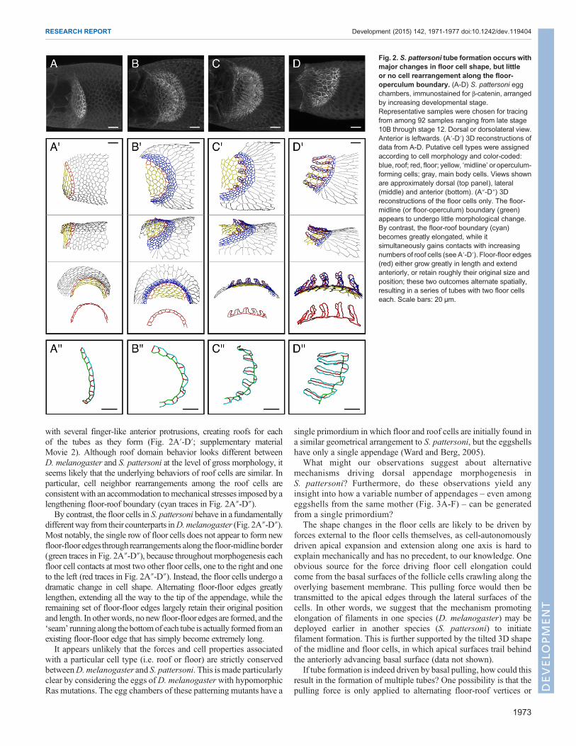
with several finger-like anterior protrusions, creating roofs for eachof the tubes as they form (Fig. 2A′-D′; supplementary materialMovie 2). Although roof domain behavior looks different betweenD. melanogaster and S. pattersoni at the level of gross morphology, itseems likely that the underlying behaviors of roof cells are similar. Inparticular, cell neighbor rearrangements among the roof cells areconsistent with an accommodation tomechanical stresses imposed byalengthening floor-roof boundary (cyan traces in Fig. 2A″-D″).By contrast, the floor cells in S. pattersoni behave in a fundamentally
differentway from their counterparts inD.melanogaster (Fig. 2A″-D″).Most notably, the single row of floor cells does not appear to form newfloor-flooredges through rearrangements along the floor-midline border(green traces in Fig. 2A″-D″), because throughout morphogenesis eachfloor cell contacts at most two other floor cells, one to the right and oneto the left (red traces in Fig. 2A″-D″). Instead, the floor cells undergo adramatic change in cell shape. Alternating floor-floor edges greatlylengthen, extending all the way to the tip of the appendage, while theremaining set of floor-floor edges largely retain their original positionand length. In otherwords, no new floor-floor edges are formed, and the‘seam’ running along the bottomof each tube is actually formed fromanexisting floor-floor edge that has simply become extremely long.It appears unlikely that the forces and cell properties associated
with a particular cell type (i.e. roof or floor) are strictly conservedbetweenD.melanogaster and S. pattersoni. This is made particularlyclear by considering the eggs ofD. melanogasterwith hypomorphicRas mutations. The egg chambers of these patterning mutants have a
single primordium in which floor and roof cells are initially found ina similar geometrical arrangement to S. pattersoni, but the eggshellshave only a single appendage (Ward and Berg, 2005).
What might our observations suggest about alternativemechanisms driving dorsal appendage morphogenesis inS. pattersoni? Furthermore, do these observations yield anyinsight into how a variable number of appendages – even amongeggshells from the same mother (Fig. 3A-F) – can be generatedfrom a single primordium?
The shape changes in the floor cells are likely to be driven byforces external to the floor cells themselves, as cell-autonomouslydriven apical expansion and extension along one axis is hard toexplain mechanically and has no precedent, to our knowledge. Oneobvious source for the force driving floor cell elongation couldcome from the basal surfaces of the follicle cells crawling along theoverlying basement membrane. This pulling force would then betransmitted to the apical edges through the lateral surfaces of thecells. In other words, we suggest that the mechanism promotingelongation of filaments in one species (D. melanogaster) may bedeployed earlier in another species (S. pattersoni) to initiatefilament formation. This is further supported by the tilted 3D shapeof the midline and floor cells, in which apical surfaces trail behindthe anteriorly advancing basal surface (data not shown).
If tube formation is indeed driven by basal pulling, how could thisresult in the formation of multiple tubes? One possibility is that thepulling force is only applied to alternating floor-roof vertices or
Fig. 2. S. pattersoni tube formation occurs withmajor changes in floor cell shape, but littleor no cell rearrangement along the floor-operculum boundary. (A-D) S. pattersoni eggchambers, immunostained for β-catenin, arrangedby increasing developmental stage.Representative samples were chosen for tracingfrom among 92 samples ranging from late stage10B through stage 12. Dorsal or dorsolateral view.Anterior is leftwards. (A′-D′) 3D reconstructions ofdata from A-D. Putative cell types were assignedaccording to cell morphology and color-coded:blue, roof; red, floor; yellow, ‘midline’ or operculum-forming cells; gray, main body cells. Views shownare approximately dorsal (top panel), lateral(middle) and anterior (bottom). (A″-D″) 3Dreconstructions of the floor cells only. The floor-midline (or floor-operculum) boundary (green)appears to undergo little morphological change.By contrast, the floor-roof boundary (cyan)becomes greatly elongated, while itsimultaneously gains contacts with increasingnumbers of roof cells (see A′-D′). Floor-floor edges(red) either grow greatly in length and extendanteriorly, or retain roughly their original size andposition; these two outcomes alternate spatially,resulting in a series of tubes with two floor cellseach. Scale bars: 20 μm.
1973
RESEARCH REPORT Development (2015) 142, 1971-1977 doi:10.1242/dev.119404
DEVELO
PM
ENT
floor-floor edges. Alternatively, these may experience equal forces,while the relative deformability of the floor-floor edges alternates.To explore these possibilities, we examined the localization ofmyosin and the polarity protein Par3/Bazooka, as spatial patterningof these molecules within the apical surface of follicle cells waspreviously proposed to drive appendage morphogenesis inD. melanogaster (Osterfield et al., 2013).In S. pattersoni, we found that Bazooka localizes specifically to
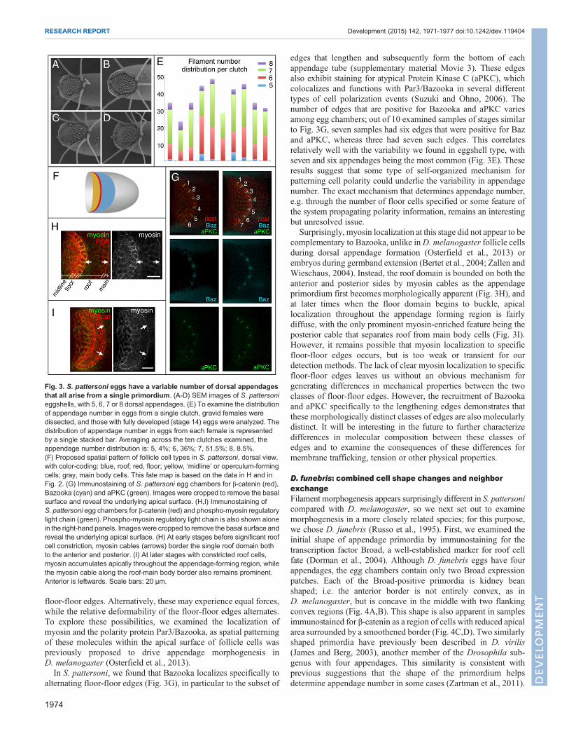
alternating floor-floor edges (Fig. 3G), in particular to the subset of
edges that lengthen and subsequently form the bottom of eachappendage tube (supplementary material Movie 3). These edgesalso exhibit staining for atypical Protein Kinase C (aPKC), whichcolocalizes and functions with Par3/Bazooka in several differenttypes of cell polarization events (Suzuki and Ohno, 2006). Thenumber of edges that are positive for Bazooka and aPKC variesamong egg chambers; out of 10 examined samples of stages similarto Fig. 3G, seven samples had six edges that were positive for Bazand aPKC, whereas three had seven such edges. This correlatesrelatively well with the variability we found in eggshell type, withseven and six appendages being the most common (Fig. 3E). Theseresults suggest that some type of self-organized mechanism forpatterning cell polarity could underlie the variability in appendagenumber. The exact mechanism that determines appendage number,e.g. through the number of floor cells specified or some feature ofthe system propagating polarity information, remains an interestingbut unresolved issue.
Surprisingly, myosin localization at this stage did not appear to becomplementary to Bazooka, unlike inD. melanogaster follicle cellsduring dorsal appendage formation (Osterfield et al., 2013) orembryos during germband extension (Bertet et al., 2004; Zallen andWieschaus, 2004). Instead, the roof domain is bounded on both theanterior and posterior sides by myosin cables as the appendageprimordium first becomes morphologically apparent (Fig. 3H), andat later times when the floor domain begins to buckle, apicallocalization throughout the appendage forming region is fairlydiffuse, with the only prominent myosin-enriched feature being theposterior cable that separates roof from main body cells (Fig. 3I).However, it remains possible that myosin localization to specificfloor-floor edges occurs, but is too weak or transient for ourdetection methods. The lack of clear myosin localization to specificfloor-floor edges leaves us without an obvious mechanism forgenerating differences in mechanical properties between the twoclasses of floor-floor edges. However, the recruitment of Bazookaand aPKC specifically to the lengthening edges demonstrates thatthese morphologically distinct classes of edges are also molecularlydistinct. It will be interesting in the future to further characterizedifferences in molecular composition between these classes ofedges and to examine the consequences of these differences formembrane trafficking, tension or other physical properties.
D. funebris: combined cell shape changes and neighborexchangeFilament morphogenesis appears surprisingly different in S. pattersonicompared with D. melanogaster, so we next set out to examinemorphogenesis in a more closely related species; for this purpose,we chose D. funebris (Russo et al., 1995). First, we examined theinitial shape of appendage primordia by immunostaining for thetranscription factor Broad, a well-established marker for roof cellfate (Dorman et al., 2004). Although D. funebris eggs have fourappendages, the egg chambers contain only two Broad expressionpatches. Each of the Broad-positive primordia is kidney beanshaped; i.e. the anterior border is not entirely convex, as inD. melanogaster, but is concave in the middle with two flankingconvex regions (Fig. 4A,B). This shape is also apparent in samplesimmunostained for β-catenin as a region of cells with reduced apicalarea surrounded by a smoothened border (Fig. 4C,D). Two similarlyshaped primordia have previously been described in D. virilis(James and Berg, 2003), another member of the Drosophila sub-genus with four appendages. This similarity is consistent withprevious suggestions that the shape of the primordium helpsdetermine appendage number in some cases (Zartman et al., 2011).
Fig. 3. S. pattersoni eggs have a variable number of dorsal appendagesthat all arise from a single primordium. (A-D) SEM images of S. pattersonieggshells, with 5, 6, 7 or 8 dorsal appendages. (E) To examine the distributionof appendage number in eggs from a single clutch, gravid females weredissected, and those with fully developed (stage 14) eggs were analyzed. Thedistribution of appendage number in eggs from each female is representedby a single stacked bar. Averaging across the ten clutches examined, theappendage number distribution is: 5, 4%; 6, 36%; 7, 51.5%; 8, 8.5%.(F) Proposed spatial pattern of follicle cell types in S. pattersoni, dorsal view,with color-coding: blue, roof; red, floor; yellow, ‘midline’ or operculum-formingcells; gray, main body cells. This fate map is based on the data in H and inFig. 2. (G) Immunostaining of S. pattersoni egg chambers for β-catenin (red),Bazooka (cyan) and aPKC (green). Images were cropped to remove the basalsurface and reveal the underlying apical surface. (H,I) Immunostaining ofS. pattersoni egg chambers for β-catenin (red) and phospho-myosin regulatorylight chain (green). Phospho-myosin regulatory light chain is also shown alonein the right-hand panels. Imageswere cropped to remove the basal surface andreveal the underlying apical surface. (H) At early stages before significant roofcell constriction, myosin cables (arrows) border the single roof domain bothto the anterior and posterior. (I) At later stages with constricted roof cells,myosin accumulates apically throughout the appendage-forming region, whilethe myosin cable along the roof-main body border also remains prominent.Anterior is leftwards. Scale bars: 20 μm.
1974
RESEARCH REPORT Development (2015) 142, 1971-1977 doi:10.1242/dev.119404
DEVELO
PM
ENT
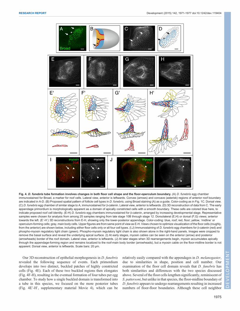
Our 3D reconstruction of epithelial morphogenesis in D. funebrisrevealed the following sequence of events. Each primordiumdevelops into two distinct, buckled patches of highly constrictedcells (Fig. 4E). Each of these two buckled regions then elongates(Fig. 4F-H), resulting in the eventual formation of four tubes per eggchamber. To study how a single buckled domain is transformed intoa tube in this species, we focused on the more posterior tubes(Fig. 4E′-H′, supplementary material Movie 4), which can be
relatively easily compared with the appendages in D. melanogaster,due to similarities in shape, position and cell number. Ourexamination of the floor cell domain reveals that D. funebris hasboth similarities and differences with the two species discussedabove. Several of the floor cells lengthen significantly, reminiscent ofS. pattersoni, but unlike in that species, the floor-midline boundary ofD. funebris appears to undergo rearrangements resulting in increasednumbers of floor-floor boundaries. Although these cell neighbor
Fig. 4. D. funebris tube formation involves changes in both floor cell shape and the floor-operculum boundary. (A) D. funebris egg chamberimmunostained for Broad, a marker for roof cells. Lateral view, anterior is leftwards. Convex (arrows) and concave (asterisk) regions of anterior roof boundaryare indicated in A-D. (B) Proposed spatial pattern of follicle cell types in D. funebris, using Broad staining (A) as a guide. Color-coding as in Fig. 1C. Dorsal view.(C) D. funebris egg chamber of similar stage to A, immunostained for β-catenin. Lateral view, anterior is leftwards. (D) 3D reconstruction of data from C. The earlyappendage primordium is morphologically apparent as a domain of apically constricted cells with a smooth boundary. These cells are colored blue here, toindicate proposed roof cell identity. (E-H) D. funebris egg chambers immunostained for β-catenin, arranged by increasing developmental stage. Representativesamples were chosen for analysis from among 25 samples ranging from late stage 10B through stage 12. Dorsolateral (E,H) or dorsal (F,G) views; anteriortowards the left. (E′-H′) 3D reconstructions from E-H, showing only the lower-posterior appendage. Color-coding: blue, roof; red, floor; yellow, ‘midline’ oroperculum-forming cells; gray, main body cells. Upper figures are from same point of viewas E-H. Views chosen to optimize visualization of the floor cells (roughly,from the anterior) are shown below, including either floor cells only or all four cell types. (I,J) Immunostaining of D. funebris egg chambers for β-catenin (red) andphospho-myosin regulatory light chain (green). Phospho-myosin regulatory light chain is also shown alone in the right-hand panels. Images were cropped toremove the basal surface and reveal the underlying apical surface. (I) At early stages, myosin cables can be seen on the anterior (arrow) and posterior(arrowheads) border of the roof domain. Lateral view, anterior is leftwards. (J) At later stages when 3D rearrangements begin, myosin accumulates apicallythrough the appendage-forming region and remains localized to the roof-main body border (arrowheads), but a myosin cable on the floor-midline border is notapparent. Dorsal view, anterior is leftwards. Scale bars: 20 μm.
1975
RESEARCH REPORT Development (2015) 142, 1971-1977 doi:10.1242/dev.119404
DEVELO
PM
ENT

rearrangements clearly resemble those seen inD. melanogaster, theyappear to be less ordered spatially: they initiate off-center (Fig. 4F′)and form a final floor domain with no clear seam or spatial structure(Fig. 4G′-H′). Furthermore, although myosin appears to be eitherrecruited or activated apically throughout much of the appendageprimordium, we do not observe a myosin cable on the floor-midlineborder (Fig. 4I,J). By contrast, our previous work suggested that amyosin cable along the floor-midline border is crucial for drivingspatially ordered cell neighbor exchange in D. melanogaster(Osterfield et al., 2013). We therefore suggest that D. funebrisappendage formation is not actively driven by ordered floor cellrearrangement. Instead, we hypothesize that pulling forces from thebasal side provide the main driving force, with the floor domainpassively yielding to this stress through a combination of cell shapechanges and neighbor exchanges.
D. melanogaster: redundant morphogenetic mechanisms?The results described above suggest that we should reconsiderproposed mechanisms of dorsal appendage formation. In particular,a disordered appendage floor with no clear seam can be found notonly in D. funebris (Fig. 4H′), but also in D. melanogasterpatterning mutants, such as fs(1)K10 (Ward and Berg, 2005) andCY2>Mae (supplementary material Fig. S1). Myosin does appearto form a cable along the floor-midline border in CY2>Mae(supplementary material Fig. S1E), but it is currently unclearwhether this is sufficient to drive the pattern of cell rearrangementsthat occurs in this genetic background. Additionally, experimentsfrom Celeste Berg and colleagues on the effect of ectopicallyexpressing rhomboid in D. melanogaster follicle cells indicates thatectopic appendage tubes can form with as few as two cells,specifically one roof plus one floor (Ward and Berg, 2005). Cellneighbor rearrangements increasing the number of floor-flooredges are impossible in this case, raising the possibility thatappendage formation in this genetic background may occurthrough a pattersoni-like mechanism involving changes in floorcell shape. Together, these observations highlight the need todissect more carefully the relative contribution of myosin cabledriven intercalation, basal pulling, roof cell constriction (Dormanet al., 2004) or other mechanisms as potential forces drivingappendage formation in mutant and wild-type D. melanogasteregg chambers.
ConclusionAt the outset of this work, we expected that dorsal appendageformation in different species would occur through largely similarcellular mechanisms. In particular, we expected that appendageformation would be driven by ordered floor cell intercalations thatwere themselves driven by highly patterned apical tensions, as wasobserved in D. melanogaster. However, our studies of S. pattersoniprovide a clear counter-example to this mechanism, revealing thattubes can form though changes in cell shape, rather than throughneighbor exchanges within the floor domain. The inter-speciesvariation in eggshell morphogenesis emphasizes a general point:the details of morphogenesis may be extremely important inunderstanding how changes in gene expression pattern lead tochanges in morphology among evolutionarily homologousstructures (Mallarino et al., 2012).In spite of clear differences in cellular behavior during eggshell
morphogenesis among the species we studied, it appears that differentcombinations of just a few physical mechanisms may be at work;testing these mechanisms in detail promises to be an interesting futuredirection. This study also helps lay the groundwork for future studies
on the relationship between expression profiles and cellular propertiesin epithelial cells, and for studies exploring the degree to whichmechanisms of epithelial morphogenesis are conserved acrossspecies.
MATERIALS AND METHODSFly stocksD. melanogaster and D. funebris flies (Drosophila Species Stock Center,#15120-1911.01) were maintained on standard cornmeal media. Toenrich for stage 11 egg chambers, yeast was added to the media 1(D. melanogaster) or 2 (D. funebris) days before dissection. S. pattersoniflies (Drosophila Species Stock Center, #11010-0031.00) were generallymaintained on standard cornmeal media supplemented with a small quantity(0.2-0.6 ml per vial) of banana purée (Gerber). To enrich for stage 11 eggchambers, S. pattersoni females and males were moved to cornmeal mediawithin 1-2 days after eclosion, kept on cornmeal media for several days, thenplaced together on banana-supplemented media 1 day before dissection.
D. melanogaster genotypes used included E-cadherin:GFP transgenicflies for Fig. 1 (Huang et al., 2009), CY2-GAL4, UAS-Mae/CyO forsupplementary material Fig. S1C,D and CY2-GAL4, UAS-Mae/ed[CPTI000616] for supplementary material Fig. S1A,B. The echinoid-YFPtrap line ed[CPTI000616] was acquired from the DGRC in Kyoto, Japan.The CY2-GAL4, UAS-Mae/CyO line was a gift from J. Duffy (WPI,Worcester, MA, USA); the effect of this genotype on eggshell patterning hasbeen described previously (Yamada et al., 2003).
ImmunohistochemistryImmunostaining was generally carried out as described previously(Osterfield et al., 2013; Ward and Berg, 2005). However, the protocol wasmodified for samples stained for phospho-myosin: after primary antibodyincubation, samples were washed in 4°C with ice-cold PBSTwn for3×10 min, post-fixed for 20 min at room temperature with 4%paraformaldehyde in PBSTwn, washed for 3×10 min and re-blocked in1% bovine serum albumin (Sigma-Aldrich) in PBSTwn at least 1 hour beforeproceeding to the secondary antibody incubation step. For most samples, thefinal dilution series of glycerol/PBS was omitted. Instead, the samples werewashed into RapiClear 1.47 (SunJin Lab, Taiwan) then mounted for imagingbetween two glass coverslips, which were separated by a 0.2 mm spacer(SunJin Lab, #IS007) and sealed with nail polish. Primary antibodies usedincluded anti-β-catenin (anti-armadillo N2 7A1, mouse, DevelopmentalStudies Hybridoma Bank, 1:20), anti-Broad-core (mouse, DevelopmentalStudies Hybridoma Bank, 1:50), anti-Fasciclin III (7G10, mouse,Developmental Studies Hybridoma Bank, 1:50), anti-phospho-myosinlight chain 2 (Ser 19) (rabbit, Cell Signaling Technology, 1:20), anti-Bazooka [guinea pig (Blankenship et al., 2006), 1:250-1:500] and anti-aPKC(rabbit, 1:1000, Santa Cruz Biotechnology). Atto 488-conjugated GFP-booster (1:200, ChromoTek) was used during the secondary antibodyincubation step for enhancing echinoid-YFP signal. Alexa-Fluor-conjugatedsecondary antibodies (Invitrogen) were generally used at 1:500.
Microscopy and image processingAll fluorescent images were acquired using a Leica SP5 confocalmicroscope, with a 63× (NA 1.3) glycerol immersion objective. Imageswere adjusted for brightness/contrast and displayed as a maximum projectionusing either ImageJ/FIJI or the surpass mode of Imaris software (Bitplane).In several cases (Figs 3G-I and 4I,J; supplementary material Fig. S1E),images were cropped in 3D to remove the basal surface, which wouldotherwise block or overwhelm signal from the apical surface. Tracing ofapical edges was carried out with the filament-tracing module of Imarissoftware, primarily using the AutoPath function, which allows for supervisedautomatic tracing (Meijering et al., 2004). The apical markers we used fortracing (E-cadherin or β-catenin) also localize to some extent to lateralsurfaces, particularly in midline type cells, so direct proximity to the oocyteor appendage tube lumen was also considered in determining whetherfluorescent signal indicated the presence of an apical edge. As all eggshellsstudied here appear to exhibit complete left-right symmetry, some imageswere flipped, either to preserve anterior=left and dorsal=up orientation, or (as
1976
RESEARCH REPORT Development (2015) 142, 1971-1977 doi:10.1242/dev.119404
DEVELO
PM
ENT
in Fig. 4) to allow easier comparison between structures that originallydiffered in left- versus right-handedness.
AcknowledgementsWe thankMatthewGastinger (Bitplane) for extensive guidance in using Imaris for 3Dreconstructions; Nir Yakoby, Matt Niepielko, Rob Marmion, Anna DiPaola andTherese Markow for advice regarding fly species; Joe Goodhouse and GaryLaevsky for imaging advice; Jennifer Zallen for the generous gift of several anti-Bazooka antibodies; and Celeste Berg, Olivier Devergne and members of theWieschaus, Schupbach and Shvartsman labs for discussions. We also thank ourthree anonymous reviewers for helpful feedback and suggestions. Several stockswere obtained from the Drosophila Species Stock Center at the University ofCalifornia, San Diego. Monoclonal antibodies originally developed by GregoryGuild, Eric Wieschaus and Corey Goodman were obtained from the DevelopmentalStudies Hybridoma Bank, created by the NICHD of the NIH and maintained at TheUniversity of Iowa, Department of Biology, Iowa City, IA 52242.
Competing interestsThe authors declare no competing or financial interests.
Author contributionsM.O. performed experiments and data analysis. M.O., T.S., E.W. and S.Y.S.developed the approach and prepared or edited the manuscript.
FundingThis research was funded by the National Institute of General Medical Sciences[1R01GM107103- 01A1]. E.W. is a HHMI Investigator. Deposited in PMC for releaseafter 6 months.
Supplementary materialSupplementary material available online athttp://dev.biologists.org/lookup/suppl/doi:10.1242/dev.119404/-/DC1
ReferencesBerg, C. A. (2005). The Drosophila shell game: patterning genes andmorphologicalchange. Trends Genet. 21, 346-355.
Bertet, C., Sulak, L. and Lecuit, T. (2004). Myosin-dependent junction remodellingcontrols planar cell intercalation and axis elongation. Nature 429, 667-671.
Blankenship, J. T., Backovic, S. T., Sanny, J. S. P., Weitz, O. and Zallen, J. A.(2006). Multicellular rosette formation links planar cell polarity to tissuemorphogenesis. Dev. Cell 11, 459-470.
Boyle, M. J. and Berg, C. A. (2009). Control in time and space: Tramtrack69cooperates with Notch and Ecdysone to repress ectopic fate and shape changesduring Drosophila egg chamber maturation. Development 136, 4187-4197.
Cheung, L. S., Schupbach, T. and Shvartsman, S. Y. (2011). Pattern formation byreceptor tyrosine kinases: analysis of the Gurken gradient in Drosophilaoogenesis. Curr. Opin. Genet. Dev. 21, 719-725.
Dorman, J. B., James, K. E., Fraser, S. E., Kiehart, D. P. and Berg, C. A. (2004).bullwinkle is required for epithelial morphogenesis during Drosophila oogenesis.Dev. Biol. 267, 320-341.
French, R. L., Cosand, K. A. and Berg, C. A. (2003). The Drosophila female sterilemutation twin peaks is a novel allele of tramtrack and reveals a requirement forTTK69 in epithelial morphogenesis. Dev. Biol. 253, 18-35.
Haas-Koffler, C. L., Naeemuddin, M. and Bartlett, S. E. (2012). An analytical toolthat quantifies cellular morphology changes from three-dimensional fluorescenceimages. J. Vis. Exp. e4233.
Hinton, H. E. (1981). Biology of Insect Eggs. Oxford; New York: Pergamon Press.Huang, J., Zhou, W., Dong, W., Watson, A. M. and Hong, Y. (2009). Directed,efficient, and versatile modifications of the Drosophila genome by genomicengineering. Proc. Natl. Acad. Sci. USA 106, 8284-8289.
James, K. E. and Berg, C. A. (2003). Temporal comparison of Broad-Complexexpression during eggshell-appendage patterning and morphogenesis in twoDrosophila species with different eggshell-appendage numbers. Gene Expr.Patterns 3, 629-634.
Kagesawa, T., Nakamura, Y., Nishikawa, M., Akiyama, Y., Kajiwara, M. andMatsuno, K. (2008). Distinct activation patterns of EGF receptor signaling in thehomoplastic evolution of eggshell morphology in genus Drosophila. Mech. Dev.125, 1020-1032.
Mallarino, R. and Abzhanov, A. (2012). Paths less traveled: evo-devo approachesto investigating animal morphological evolution. Annu. Rev. Cell Dev. Biol. 28,743-763.
Mallarino, R., Campas, O., Fritz, J. A., Burns, K. J., Weeks, O. G., Brenner, M. P.and Abzhanov, A. (2012). Closely related bird species demonstrate flexibilitybetween beak morphology and underlying developmental programs. Proc. Natl.Acad. Sci. USA 109, 16222-16227.
Marmion, R. A., Jevtic, M., Springhorn, A., Pyrowolakis, G. and Yakoby, N.(2013). The Drosophila BMPRII, wishful thinking, is required for eggshellpatterning. Dev. Biol. 375, 45-53.
Meijering, E., Jacob, M., Sarria, J.-C. F., Steiner, P., Hirling, H. and Unser, M.(2004). Design and validation of a tool for neurite tracing and analysis influorescence microscopy images. Cytometry 58A, 167-176.
Nakamura, Y. andMatsuno, K. (2003). Species-specific activation of EGF receptorsignaling underlies evolutionary diversity in the dorsal appendage number of thegenus Drosophila eggshells. Mech. Dev. 120, 897-907.
Nakamura, Y., Kagesawa, T., Nishikawa, M., Hayashi, Y., Kobayashi, S., Niimi,T. and Matsuno, K. (2007). Soma-dependent modulations contribute todivergence of rhomboid expression during evolution of Drosophila eggshellmorphology. Development 134, 1529-1537.
Niepielko, M. G. and Yakoby, N. (2014). Evolutionary changes in TGFalphadistribution underlie morphological diversity in eggshells from Drosophila species.Development 141, 4710-4715.
Niepielko, M. G., Hernaiz-Hernandez, Y. and Yakoby, N. (2011). BMP signalingdynamics in the follicle cells of multiple Drosophila species. Dev. Biol. 354,151-159.
Niepielko, M. G., Ip, K., Kanodia, J. S., Lun, D. S. andYakoby, N. (2012). Evolutionof BMP signaling in Drosophila oogenesis: a receptor-basedmechanism.Biophys.J. 102, 1722-1730.
Niepielko, M. G., Marmion, R. A., Kim, K., Luor, D., Ray, C. and Yakoby, N.(2014). Chorion patterning: awindow into gene regulation andDrosophila species’relatedness. Mol. Biol. Evol. 31, 154-164.
Osterfield, M., Du, X., Schupbach, T., Wieschaus, E. and Shvartsman, S. Y.(2013). Three-dimensional epithelial morphogenesis in the developing Drosophilaegg. Dev. Cell 24, 400-410.
Peri, F., Bokel, C. and Roth, S. (1999). Local Gurken signaling and dynamic MAPKactivation during Drosophila oogenesis. Mech. Dev. 81, 75-88.
Peters, N. C., Thayer, N. H., Kerr, S. A., Tompa, M. and Berg, C. A. (2013).Following the ‘tracks’: Tramtrack69 regulates epithelial tube expansion in theDrosophila ovary through Paxillin, Dynamin, and the homeobox protein Mirror.Dev. Biol. 378, 154-169.
Russo, C. A., Takezaki, N. and Nei, M. (1995). Molecular phylogeny anddivergence times of drosophilid species. Mol. Biol. Evol. 12, 391-404.
Suzuki, A. and Ohno, S. (2006). The PAR-aPKC system: lessons in polarity. J. CellSci. 119, 979-987.
Vreede, B. M. I., Lynch, J. A., Roth, S. and Sucena, E. (2013). Co-option of acoordinate system defined by the EGFr and Dpp pathways in the evolution of amorphological novelty. Evodevo 4, 7.
Ward, E. J. and Berg, C. A. (2005). Juxtaposition between two cell types isnecessary for dorsal appendage tube formation. Mech. Dev. 122, 241-255.
Wu, X., Tanwar, P. S. and Raftery, L. A. (2008). Drosophila follicle cells:morphogenesis in an eggshell. Semin. Cell Dev. Biol. 19, 271-282.
Yamada, T., Okabe, M. and Hiromi, Y. (2003). EDL/MAE regulates EGF-mediatedinduction by antagonizing Ets transcription factor Pointed. Development 130,4085-4096.
Zallen, J. A. and Wieschaus, E. (2004). Patterned gene expression directs bipolarplanar polarity in Drosophila. Dev. Cell 6, 343-355.
Zartman, J. J., Yakoby, N., Bristow, C. A., Zhou, X., Schlichting, K., Dahmann,C. and Shvartsman, S. Y. (2008). Cad74A is regulated by BR and is required forrobust dorsal appendage formation in Drosophila oogenesis. Dev. Biol. 322,289-301.
Zartman, J. J., Cheung, L. S., Niepielko, M. G., Bonini, C., Haley, B., Yakoby, N.and Shvartsman, S. Y. (2011). Pattern formation by amovingmorphogen source.Phys. Biol. 8, 045003.
1977
RESEARCH REPORT Development (2015) 142, 1971-1977 doi:10.1242/dev.119404
DEVELO
PM
ENT