diversity of cnidarian muscles: function, anatomy ... found in cnidaria. ... (all animals except...
TRANSCRIPT
REVIEWpublished: 23 January 2017
doi: 10.3389/fcell.2016.00157
Frontiers in Cell and Developmental Biology | www.frontiersin.org 1 January 2017 | Volume 4 | Article 157
Edited by:
Stefano Biressi,
University of Trento, Italy
Reviewed by:
Maja Adamska,
Australian National University, Australia
Patrick Steinmetz,
University of Bergen, Norway
*Correspondence:
Lucas Leclère
Eric Röttinger
Specialty section:
This article was submitted to
Stem Cell Research,
a section of the journal
Frontiers in Cell and Developmental
Biology
Received: 21 November 2016
Accepted: 30 December 2016
Published: 23 January 2017
Citation:
Leclère L and Röttinger E (2017)
Diversity of Cnidarian Muscles:
Function, Anatomy, Development and
Regeneration.
Front. Cell Dev. Biol. 4:157.
doi: 10.3389/fcell.2016.00157
Diversity of Cnidarian Muscles:Function, Anatomy, Development andRegeneration
Lucas Leclère 1* and Eric Röttinger 2*
1 Sorbonne Universités, UPMC Univ Paris 06, CNRS, Laboratoire de Biologie du Développement de Villefranche-sur-mer
(LBDV), Villefranche-sur-mer, France, 2Université Côte d’Azur, CNRS, INSERM, Institute for Research on Cancer and Aging
(IRCAN), Nice, France
The ability to perform muscle contractions is one of the most important and distinctive
features of eumetazoans. As the sister group to bilaterians, cnidarians (sea anemones,
corals, jellyfish, and hydroids) hold an informative phylogenetic position for understanding
muscle evolution. Here, we review current knowledge on muscle function, diversity,
development, regeneration and evolution in cnidarians. Cnidarian muscles are involved in
various activities, such as feeding, escape, locomotion and defense, in close association
with the nervous system. This variety is reflected in the large diversity of muscle
organizations found in Cnidaria. Smooth epithelial muscle is thought to be the most
common type, and is inferred to be the ancestral muscle type for Cnidaria, while
striated muscle fibers and non-epithelial myocytes would have been convergently
acquired within Cnidaria. Current knowledge of cnidarian muscle development and its
regeneration is limited. While orthologs of myogenic regulatory factors such as MyoD
have yet to be found in cnidarian genomes, striated muscle formation potentially involves
well-conserved myogenic genes, such as twist and mef2. Although satellite cells have
yet to be identified in cnidarians, muscle plasticity (e.g., de- and re-differentiation, fiber
repolarization) in a regenerative context and its potential role during regeneration has
started to be addressed in a few cnidarian systems. The development of novel tools
to study those organisms has created new opportunities to investigate in depth the
development and regeneration of cnidarian muscle cells and how they contribute to the
regenerative process.
Keywords: cnidaria, muscle, myoepithelial cells, development, regeneration, evolution, epitheliomuscular cells
INTRODUCTION
Muscles, tissues specialized for contraction, are an essential component of the eumetazoan (allanimals except sponges and placozoans) body. They are involved in various functions of the bodyand are well characterized in various vertebrate and main non-vertebrate models (reviewed inSchmidt-Rhaesa, 2007; Bryson-Richardson and Currie, 2008; Bentzinger et al., 2012; Andrikouand Arnone, 2015; Almada and Wagers, 2016). In bilaterians, muscles are rich in myofilaments(organized arrays composed principally of actin and myosin II) and present two basic types of cells:true muscle cells (myocytes) and myoepithelial cells. Myocytes are individual muscle cells, usually
Leclère and Röttinger Cnidarian Muscle Diversity
not anchored to the extracellular matrix (ECM), which duringembryogenesis derive mainly (but not exclusively) from themesoderm layer. In contrast, myoepithelial cells, which havea variety of embryological origins, are anchored to the ECMand are fully integrated into an epithelial tissue layer. Bothof these muscle cell-types can be further defined as eitherstriated or smooth, depending on the internal organization of themyofilaments. Visible striations represent repeating functionalunits of the muscle (the sarcomeres), which result from alignedrows of alternating antiparallel actin and myosin myofilaments,spaced by their supporting Z-discs. Conversely, in smoothmuscles, the myofilaments are organized irregularly.
The diversity of muscle organizations is best characterizedin mammals. There are four muscular organizations: two arestriated, named skeletal and cardiac muscles; the other two arethe smooth and myoepithelial muscles (Alberts et al., 2015).In skeletal muscles, myocytes fuse to form multinucleatedsyncytia called muscle fibers or myotubes. In contrast, cardiacand smooth muscles are composed of mononucleated musclecells for which mechanical, chemical, and electrical couplingis possible via complex junctions (adherens and gap), formingthe typical “intercalated disc” structures of cardiac muscles.Myoepithelial cells in mammals are generally found in glandularepithelia such as the mammary or salivary glands and display
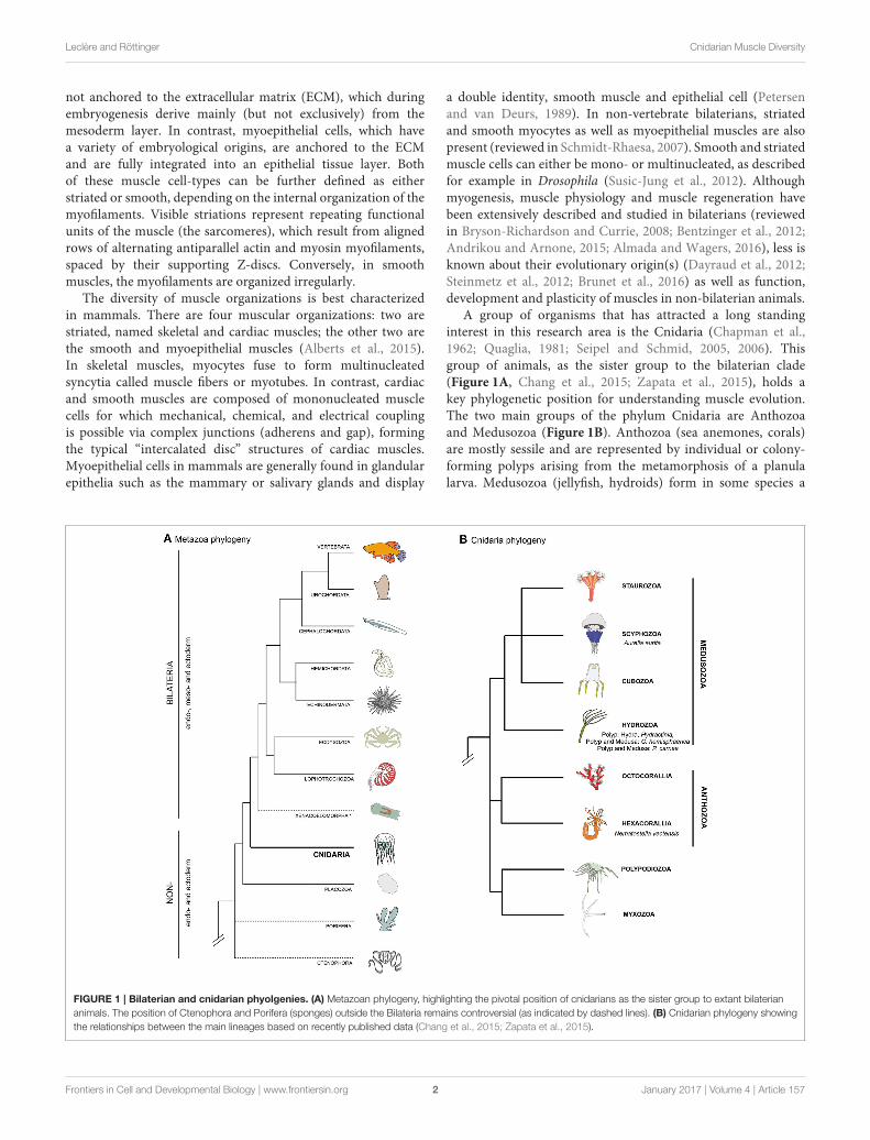
FIGURE 1 | Bilaterian and cnidarian phyolgenies. (A) Metazoan phylogeny, highlighting the pivotal position of cnidarians as the sister group to extant bilaterian
animals. The position of Ctenophora and Porifera (sponges) outside the Bilateria remains controversial (as indicated by dashed lines). (B) Cnidarian phylogeny showing
the relationships between the main lineages based on recently published data (Chang et al., 2015; Zapata et al., 2015).
a double identity, smooth muscle and epithelial cell (Petersenand van Deurs, 1989). In non-vertebrate bilaterians, striatedand smooth myocytes as well as myoepithelial muscles are alsopresent (reviewed in Schmidt-Rhaesa, 2007). Smooth and striatedmuscle cells can either be mono- or multinucleated, as describedfor example in Drosophila (Susic-Jung et al., 2012). Althoughmyogenesis, muscle physiology and muscle regeneration havebeen extensively described and studied in bilaterians (reviewedin Bryson-Richardson and Currie, 2008; Bentzinger et al., 2012;Andrikou and Arnone, 2015; Almada and Wagers, 2016), less isknown about their evolutionary origin(s) (Dayraud et al., 2012;Steinmetz et al., 2012; Brunet et al., 2016) as well as function,development and plasticity of muscles in non-bilaterian animals.
A group of organisms that has attracted a long standinginterest in this research area is the Cnidaria (Chapman et al.,1962; Quaglia, 1981; Seipel and Schmid, 2005, 2006). Thisgroup of animals, as the sister group to the bilaterian clade(Figure 1A, Chang et al., 2015; Zapata et al., 2015), holds akey phylogenetic position for understanding muscle evolution.The two main groups of the phylum Cnidaria are Anthozoaand Medusozoa (Figure 1B). Anthozoa (sea anemones, corals)are mostly sessile and are represented by individual or colony-forming polyps arising from the metamorphosis of a planulalarva. Medusozoa (jellyfish, hydroids) form in some species a
Frontiers in Cell and Developmental Biology | www.frontiersin.org 2 January 2017 | Volume 4 | Article 157

Leclère and Röttinger Cnidarian Muscle Diversity
free-swimming medusa (jellyfish), in addition to the polyp andplanula stages. Beside anthozoans and medusozoans, a group ofparasites, myxozoans, have recently been formally identified ascnidarians on the basis of molecular phylogenies (Figure 1B)(Chang et al., 2015) and presence of cnidarian specific genes(Holland et al., 2011; Shpirer et al., 2014). They have beenproposed to be the sister group to another cnidarian parasiticspecies, Polypodium hydriforme (Chang et al., 2015), forming theclade Endocnidozoa (Zrzavý and Hypša, 2003).
A handful of cnidarians has emerged in the past decadesas experimental models in molecular, cell and developmentalbiology, providing insights into the evolution of developmentalprograms, including regeneration, stem cell biology and theevolution of key bilaterian traits (Kraus et al., 2007, 2016;Momose and Houliston, 2007; Amiel et al., 2009; Chera et al.,2009; Boehm et al., 2012; Layden et al., 2012; Röttingeret al., 2012; Sinigaglia et al., 2013; Leclère and Rentzsch,2014; Abrams et al., 2015; Bradshaw et al., 2015; Helmet al., 2015; reviewed in Technau and Steele, 2011; Laydenet al., 2016; Leclère et al., 2016; Rentzsch and Technau,2016). The main, but not exclusive, cnidarian models are themedusozoan hydrozoans Hydra, Hydractinia, Podocoryna andClytia (reviewed in Houliston et al., 2010; Galliot, 2012; Plickertet al., 2012; Gahan et al., 2016; Leclère et al., 2016) as wellas the anthozoans Nematostella vectensis (reviewed in Laydenet al., 2016; Rentzsch and Technau, 2016) and the coral Acropora
(Shinzato et al., 2011; Hayward et al., 2015; Okubo et al.,2016).
Cnidarians display a broad variety of muscle organizationsperforming various functions. Unlike bilaterians, the mainmuscle cell type of cnidarians is the epitheliomuscular cell,a specialized epithelial cell containing smooth myofilaments,and which constitutes the principal building block of thetwo body layers (ectodermal and endodermal epithelia, alsoreferred as epidermis and gastrodermis for both polyps andmedusae, e.g., Brusca and Brusca, 2003; Schmidt-Rhaesa, 2007).The terms “epitheliomuscular cell” and “myoepithelial cell” areoften used interchangeably (e.g., Brusca and Brusca, 2003).Some authors, however, apply morphology-based definitions:“epitheliomuscular cells” are exposed to both sides of theepithelium, while “myoepithelial cells” have reduced apicalends and are not exposed to the apical surface (e.g., Ruppertet al., 2004). Following most of the literature, here we simplydefine those terms taxonomically, using “epitheliomuscular cells”and “myoepithelial cells” when referring to the myofilaments-containing epithelial cells of, respectively, cnidarians andbilaterians. Other muscle types are also found in Cnidaria, suchas the striated muscle of the medusa required for swimming.The complex life cycles and high regenerative capabilitiesfound in Cnidaria involve a remarkable plasticity of musclesystems, which can take on different configurations during thelife cycle of a given species (Figure 2). Cnidaria display both
FIGURE 2 | Cnidarian life cycles. The life cycles of (A) the solitary fresh water polyp Hydra, (B) the marine jellyfish Clytia (both hydrozoans) and (C) the anthozoan
polyp Nematostella. At the lower part of the panels are indicated their asexual reproductive potentials (budding, physal pinching) that give rise to new (A) Hydra or
(C) Nematostella polyps, or (B) juvenile Clytia medusae, respectively. Under harsh environmental conditions, gonads develop and sexual reproduction in (A) Hydra can
occur. Depending on the species, Hydra can be gonochoric or hermaphroditic (represented here). After fertilization, embryonic development occurs within a solid
capsule that, after hatching, frees a juvenile Hydra. (B) Clytia and (C) Nematostella are gonochoric and oocytes and sperm are released into the water column. After
fertilization, embryonic development leads to the formation of swimming planula larvae that after metamorphosis develop into (B) a polyp colony for Clytia or (C) a
solitary juvenile polyp for Nematostella.
Frontiers in Cell and Developmental Biology | www.frontiersin.org 3 January 2017 | Volume 4 | Article 157

Leclère and Röttinger Cnidarian Muscle Diversity
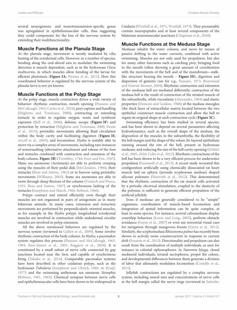
FIGURE 3 | Cnidarian muscle functions. (A) Planula larva crawling, (B), Hydra polyp somersaulting, (C) jellyfish pulsation, (D) guided tentacle retraction of the
jellyfish to bring the food toward the mouth, (E) digestive peristaltic movements of the polyp (red rings along the body column indicate circumferential muscle
contractions), (F) protective retraction of the polyp in response to predation pressure.
similarities and differences to its sister group, the bilaterians,with respect to muscle organization and cellular constituents.Additional data from cnidarian muscles can therefore provideimportant insights into their ontogeny, function and plasticity,in particular within an evolutionary framework. In thisreview, we discuss muscle diversity, function, developmentand regeneration in cnidarians. We conclude by proposingthat cnidarians, in addition to increasing our understandingof metazoan muscle evolution, may also provide new insightsinto the development/regeneration and (re-) patterning ofepitheliomuscular/myoepithelial cells, as well as into the role thatmuscle fibers play in the regeneration process.
CNIDARIAN MUSCLE FUNCTIONS
Cnidarian muscles play crucial roles in locomotion, defensefrom predators (e.g., contracting and burying in crevices/sand),feeding and digestion through continuous peristaltic movements(Shimizu et al., 2004, Figure 3). In the following section we brieflyreview the described functions of muscles at each stage of thecnidarian life cycle and the known connections to the nervoussystem.
Most of cnidarian muscle cells are epitheliomuscular and
one distinctive feature of those cells compared to muscles cells
of other animal groups is their multifunctionality. In Hydrafor instance, endodermal epitheliomuscular cells participatein nutrient absorption during the digestion process (Buzgariuet al., 2015). Epitheliomuscular cells in the ectoderm ofthe foot produce vesicles containing an adhesive substanceresponsible for attachment to the substrate, while specializedepitheliomuscular ectodermal cells, the “battery cells,” functionas supporting cells for the nematocytes (Hufnagel et al., 1985;Campbell, 1987). In many anthozoans, epitheliomuscularcells of the endodermal body wall host a large populationof dinoflagellate symbionts, and take part in the digestiveprocess, mixing the content of the gastrovascular cavityvia beating of apical cilia and performing intracellulardigestion (Hyman, 1940). Multifunctionality is thought tobe an ancestral characteristic of epitheliomuscular/myoepithelialcells (Arendt, 2008). The inherent multifunctional potentialof epitheliomuscular cells has been recently demonstrated inHydra, whose epitheliomuscular cells displayed a remarkablefunctional plasticity (Wenger et al., 2016). The authorsshowed that in strains lacking nerve cells, the expression of
Frontiers in Cell and Developmental Biology | www.frontiersin.org 4 January 2017 | Volume 4 | Article 157
Leclère and Röttinger Cnidarian Muscle Diversity
several neurogenesis- and neurotransmission-specific geneswas upregulated in epitheliomuscular cells, thus suggestingthey could compensate for the loss of the nervous system byextending their multifunctionality.
Muscle Functions at the Planula StageAt the planula stage, movement is mostly mediated by ciliabeating of the ectodermal cells. However, in a number of species,bending along the oral-aboral axis to modulate the swimmingdirection is muscle dependent, such as in the hydrozoan Clavamulticornis, in which muscles allow bending of the larvae forefficient phototaxis (Figure 3A; Piraino et al., 2011). How thiscoordinated behavior is regulated by the nervous system of theplanula larva is not yet known.
Muscle Functions at the Polyp StageAt the polyp stage, muscle contraction drives a wide variety ofbehavior: rhythmic contraction, mouth opening (Passano andMcCullough, 1963; Carter et al., 2016), prey capture and handling(Miglietta and Tommasa, 2000), contracting or extendingtentacle in order to regulate oxygen, waste and symbiontexposure (Bell et al., 2006), defense, escape (Figure 3F) andprotection by retraction (Miglietta and Tommasa, 2000; Swainet al., 2015), peristaltic movements allowing fluid circulationwithin the body cavity and facilitating digestion (Figure 3E;Anctil et al., 2005), and locomotion. Hydra is notably able tomove via a complex array of movements, including rare instancesof somersaulting (alternative attachment and release of the footand tentacles combined with contraction and extension of thebody column, Figure 3B) (Trembley, 1744; Ewer and Fox, 1947).Many sea anemones (Actiniaria) are able to perform creepingusing the muscles of their pedal disk (McClendon, 1906), theirtentacles (Ross and Sutton, 1961) or to burrow using peristalticmovements (Williams, 2003). Some sea anemones are able toswim through sharp flexions of the column (Yentsch and Pierce,1955; Ross and Sutton, 1967) or synchronous lashing of thetentacles (Josephson and March, 1966; Robson, 1966).
Polyps contract and extend efficiently even though theirmuscles are not organized in pairs of antagonists as in manybilaterian animals. In many cases, extension and retractionmovements are performed by perpendicularly oriented muscles,as for example in the Hydra polyps: longitudinal ectodermalmuscles are involved in contraction while endodermal circularmuscles are involved in polyp extension.
All the above mentioned behaviors are regulated by thenervous system (reviewed in Galliot et al., 2009). Some involverhythmic contraction of the body column. InHydra, a pacemakersystem regulates this process (Passano and McCullough, 1963,1964; Kass-Simon et al., 2003; Ruggieri et al., 2004). It isconstituted by a small subset of nerve cells connected by gapjunctions located near the foot, and capable of synchronousfiring (Takaku et al., 2014). Comparable pacemaker systemshave been described in other cnidarian polyps, such as thehydrozoan Tubularia (Josephson and Uhrich, 1969; de Kruijf,1977) and the swimming anthozoan sea anemone Stomphia(Robson, 1961, 1963). Chemical synapses between nerve cellsand epitheliomuscular cells have been shown to be widespread in
Cnidaria (Westfall et al., 1971; Westfall, 1973). They presumablycontain neuropeptides and at least several components of thebilaterian neuromuscular junctions (Chapman et al., 2010).
Muscle Functions at the Medusa StageMedusae inhabit the water column, and move by means ofpassive drifting in the water currents, combined with activeswimming. Muscles are not only used for propulsion, but alsofor many other functions such as catching prey, bringing foodto the mouth (often showing a great amount of coordinationwith the movements of the bell and of the manubrium—stalk-like structure bearing the mouth – Figure 3D), digestion anddispersion of gametes (see for e.g., Passano, 1973; Bourmaudand Gravier-Bonnet, 2004). Rhythmic contraction and extensionof the medusae bell are mediated differently; contraction of themedusa bell is the result of contraction of the striated muscle ofthe subumbrella, while viscoelastic (Alexander, 1964) and elasticproperties (Demont and Gosline, 1988) of the medusa mesoglea(the thick layer of extracellular matrix located between the twoepithelia) counteract muscle contraction and allow the bell toregain its original shape at each contraction cycle (Figure 3C).
Swimming efficiency has been studied in several species,and has been shown to depend on several parameters affectinghydrodynamics, such as the overall shape of the medusa, thedisposition of the muscles in the subumbrella, the flexibility ofthe bell margin and the shape of the velum (bi-layered epitheliumrunning around the rim of the bell, present in hydrozoanmedusae, and reducing the size of the bell cavity opening) (Dabiriet al., 2005, 2010; Colin et al., 2012). Rhythmic contractions of thebell has been shown to be a very efficient process for underwaterpropulsion (Gemmell et al., 2013). A recent study recreated thisconfiguration artificially using chemically dissociated rat heartmuscle laid on ephyra (juvenile scyphozoan medusa) shapedsilicone polymers (Nawroth et al., 2012). This demonstratedthat the rhythmic contraction of the rat muscle cells activatedby a periodic electrical stimulation, coupled to the elasticity ofthe polymer, is sufficient to generate efficient propulsion of theartificial jellyfish.
Even if medusae are generally considered to be “simple”organisms, coordination of muscle-based locomotion andintegration of spatial information can be quite complex, atleast in some species. For instance, several cubomedusae displaycourtship behaviors (Lewis and Long, 2005), perform obstacleavoidance (Garm et al., 2007) or even use terrestrial visual cuesfor navigation through mangroves forests (Garm et al., 2011).Similarly, the scyphomedusa Rhizostoma pulmo has recently beenshown to actively swim countercurrent in response to currentdrift (Fossette et al., 2015). Directionality and propulsion can alsoresult from the coordination of multiple individuals, as seen forinstance in colonial siphonophores. In Nanomia bijuga, clonalmedusoid individuals, termed nectophores, propel the colony,and developmental differences between them generate a divisionof labor that ultimately modulates locomotion (Costello et al.,2015).
Jellyfish contractions are regulated by a complex nervoussystem, including neural nets and concentrations of nerve cellsat the bell margin called the nerve rings (reviewed in Satterlie,
Frontiers in Cell and Developmental Biology | www.frontiersin.org 5 January 2017 | Volume 4 | Article 157

Leclère and Röttinger Cnidarian Muscle Diversity
2011). Pacemaker neurons regulating bell margin contractionshave been described in cubozoan, scyphozoan and hydrozoanjellyfish (reviewed in Satterlie and Nolen, 2001; Mackie, 2004;Katsuki and Greenspan, 2013). How the photoreceptor systemscontrol the swim pacemaker has started to be addressed incubomedusae (Garm and Mori, 2009; Stöckl et al., 2011; Bieleckiet al., 2013). In hydrozoan medusae, the contraction of striatedmuscle in the subumbrella is notably regulated by gap junctions,which electrically couple the muscle cells (Satterlie, 2008). Thisprocess has yet not been reported in other cnidarian groups.Finally, anatomical specialization of the nerve nets can allowfor a fine tuning of movements: for example, in Aglantha, acomplex and well-characterized neuromuscular system allowsthe jellyfish to swim either slowly or fast, thanks to differentneural circuitries, modulating the escape response (reviewed inMackie, 2004).
CNIDARIAN MUSCLE TYPES
The main, and in many species exclusive, muscle cell typein cnidarians is the epitheliomuscular cell. These cells have atypical polarized epithelial morphology, including apical cilia,with the specificity that myofibrils project from the basal side,aligning within the extracellular matrix of the tissue to provideits contractile property. In a few cnidarians, smooth muscles arefound totally embedded in the mesoglea, having lost contact withthe epithelia (see below for more details). Interestingly though,in most of the free-swimming medusae, muscles are composed ofstriated epitheliomusclar cells. Despite the few muscle cell typesfound in cnidarians, there is a wide diversity of muscle systemsin this phylum. In this section we describe briefly the diversity ofmuscle organization and muscle cell types described in the majorgroups of cnidarians.
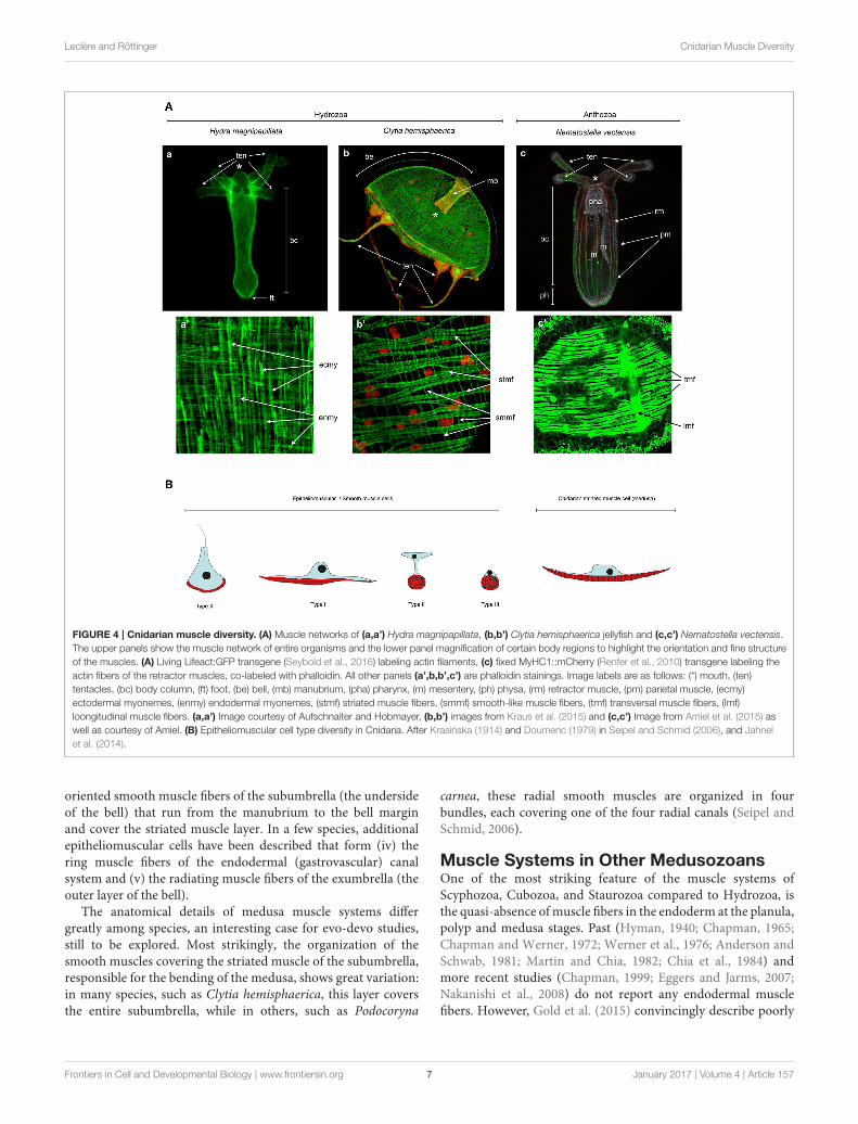
Muscle Systems in HydrozoansMost ectodermal and endodermal epithelial cells in hydrozoanplanulae, polyps, and medusae are epitheliomuscular (West,1978). Much of the available information about hydrozoanepitheliomuscular cells comes from anatomical and physiologicalstudies on Hydra polyps. Hydra ectodermal and endodermalepitheliomuscular cells display, respectively, longitudinally andcircularly oriented processes, called myonemes (Figures 4Aa,a’;Mueller, 1950). Ectodermal epitheliomuscular cells of Hydra arelarge columnar or cuboidal cells bearing two long myonemesoriented along the oral-aboral axis (David, 1973). These twomyonemes of roughly cylindrical shape, composed of irregularlyarranged myofilaments, are found in each ectodermal cell, asvisualized by electron microscopy (West, 1978) or recently byLifeAct-GFP transgenic polyps (Seybold et al., 2016). Instead,endodermal epitheliomuscular cells are tall and columnar, haveshort muscle processes at the basal end and several flagella atthe apical end. Their myonemes are oriented perpendicularlyand have a structure similar to those found in the ectoderm,though more numerous (David, 1973; Seybold et al., 2016).Epitheliomuscular cells of the body column (both in theendoderm and ectoderm) divide continuously, thus displacing
cells toward the oral (mouth) and aboral (foot) extremities wherethey are ultimately eliminated (Campbell, 1967).
Each epitheliomuscular cell process of a Hydra polyp isin contact with the basal processes of several adjacent cells,thus forming a continuous muscle fiber network spanning theentire body (Mueller, 1950). It should be noted here that theterm “muscle fiber” is generally associated to the multinucleatedsyncytia of skeletal muscles; following most of the literature oncnidarian muscle, we will use henceforth this term to indicatecondensed actin filaments constituting the contractile elementsof cnidarian muscles. Adjacent epitheliomuscular cells in Hydraare connected by septate and gap junctions; additionally, wherethe myoneme-containing regions of two adjacent cells come intocontact, they form a characteristic and unique type of junction,which structurally resembles the intercalated discs found invertebrate cardiac muscles: on the inner surface of each cellmembrane is an irregular band of dense material through whichthe filaments of the myoneme itself pass (Haynes et al., 1968).
As a general rule, the muscle fibers of hydrozoan planulalarvae and polyps are circularly arranged in the endoderm andlongitudinally in the ectoderm (Hyman, 1940; Bouillon, 1993).Common parts of the polyp colony also harbor epitheliomuscularcells, such as the endodermal epitheliomuscular cells of thestolon in Podocoryna carnea (Buss et al., 2013). However, notall hydrozoan epithelial cells are epitheliomuscular. For instance,endodermal epithelial cells of the tentacles of many hydrozoanspecies are arranged in only one row of turgescent cells and donot contain myofilaments (Bouillon, 1993).
The main muscle of the hydrozoan medusae is the circularstriated muscle found in the subumbrella (the inner layer of thebell—Figures 4Ab,b’), responsible for the rhythmic contractionof the bell, and composed of epitheliomuscular cells. As forsmooth epithelial muscles, basally located striated myofilamentsare connected between neighboring cells, forming a continuouscircular muscle. Each epitheliomuscular cell contains about30–50 sarcomeres, as for instance in the hydrozoan medusaeAglantha digitale (Singla, 1978). Sarcomeres in a relaxed state areapproximately 1 µm long. As described in various hydrozoanspecies (e.g., Keough and Summers, 1976; Boelsterli, 1977; Singla,1978), they are of very similar structure compared to thoseof vertebrate striated muscles, being separated by Z-discs andcomposed of ordered arrays of thick and thin filament areasforming denser A-bands and rarer I-bands. As in vertebrates,A-bands contain a central H-band and a M-line. An interestingdeviation can be observed in Obelia medusae (Chapman, 1968),probably linked to their unusually flat shape. In these species,the striated myofilaments of the subumbrella are not orientedcircularly but distributed in two perpendicularly oriented sets,generating a grid-like pattern. In addition to the subumbrella,in most hydrozoan medusae striated epitheliomuscular cells alsoconstitute a contractile ring on the inner layer of the velum.
In hydrozoan medusae, while swimming is generallyperformed by the circular striated muscles, other behaviors aremostly mediated by the smooth muscles. Hydrozoan medusaeare therefore rich in smooth epitheliomuscular cells (Bouillon,1993) such as (i) the longitudinal muscle fibers of the tentacleectoderm, (ii) the outer layer of the velum, and (iii) the radially
Frontiers in Cell and Developmental Biology | www.frontiersin.org 6 January 2017 | Volume 4 | Article 157
Leclère and Röttinger Cnidarian Muscle Diversity
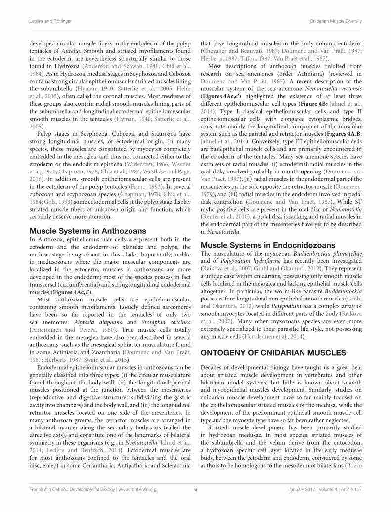
FIGURE 4 | Cnidarian muscle diversity. (A) Muscle networks of (a,a’) Hydra magnipapillata, (b,b’) Clytia hemisphaerica jellyfish and (c,c’) Nematostella vectensis.
The upper panels show the muscle network of entire organisms and the lower panel magnification of certain body regions to highlight the orientation and fine structure
of the muscles. (A) Living Lifeact:GFP transgene (Seybold et al., 2016) labeling actin filaments, (c) fixed MyHC1::mCherry (Renfer et al., 2010) transgene labeling the
actin fibers of the retractor muscles, co-labeled with phalloidin. All other panels (a’,b,b’,c’) are phalloidin stainings. Image labels are as follows: (*) mouth, (ten)
tentacles, (bc) body column, (ft) foot, (be) bell, (mb) manubrium, (pha) pharynx, (m) mesentery, (ph) physa, (rm) retractor muscle, (pm) parietal muscle, (ecmy)
ectodermal myonemes, (enmy) endodermal myonemes, (stmf) striated muscle fibers, (smmf) smooth-like muscle fibers, (tmf) transversal muscle fibers, (lmf)
loongitudinal muscle fibers. (a,a’) Image courtesy of Aufschnaiter and Hobmayer, (b,b’) images from Kraus et al. (2015) and (c,c’) Image from Amiel et al. (2015) as
well as courtesy of Amiel. (B) Epitheliomuscular cell type diversity in Cnidaria. After Krasinska (1914) and Doumenc (1979) in Seipel and Schmid (2006), and Jahnel
et al. (2014).
oriented smooth muscle fibers of the subumbrella (the undersideof the bell) that run from the manubrium to the bell marginand cover the striated muscle layer. In a few species, additionalepitheliomuscular cells have been described that form (iv) thering muscle fibers of the endodermal (gastrovascular) canalsystem and (v) the radiating muscle fibers of the exumbrella (theouter layer of the bell).
The anatomical details of medusa muscle systems differgreatly among species, an interesting case for evo-devo studies,still to be explored. Most strikingly, the organization of thesmooth muscles covering the striated muscle of the subumbrella,responsible for the bending of the medusa, shows great variation:in many species, such as Clytia hemisphaerica, this layer coversthe entire subumbrella, while in others, such as Podocoryna
carnea, these radial smooth muscles are organized in fourbundles, each covering one of the four radial canals (Seipel andSchmid, 2006).
Muscle Systems in Other MedusozoansOne of the most striking feature of the muscle systems ofScyphozoa, Cubozoa, and Staurozoa compared to Hydrozoa, isthe quasi-absence ofmuscle fibers in the endoderm at the planula,polyp and medusa stages. Past (Hyman, 1940; Chapman, 1965;Chapman and Werner, 1972; Werner et al., 1976; Anderson andSchwab, 1981; Martin and Chia, 1982; Chia et al., 1984) andmore recent studies (Chapman, 1999; Eggers and Jarms, 2007;Nakanishi et al., 2008) do not report any endodermal musclefibers. However, Gold et al. (2015) convincingly describe poorly
Frontiers in Cell and Developmental Biology | www.frontiersin.org 7 January 2017 | Volume 4 | Article 157
Leclère and Röttinger Cnidarian Muscle Diversity
developed circular muscle fibers in the endoderm of the polyptentacles of Aurelia. Smooth and striated myofilaments foundin the ectoderm, are nevertheless structurally similar to thosefound in Hydrozoa (Anderson and Schwab, 1981; Chia et al.,1984). As inHydrozoa, medusa stages in Scyphozoa and Cubozoacontains strong circular epitheliomuscular striatedmuscles liningthe subumbrella (Hyman, 1940; Satterlie et al., 2005; Helmet al., 2015), often called the coronal muscles. Most medusae ofthese groups also contain radial smooth muscles lining parts ofthe subumbrella and longitudinal ectodermal epitheliomuscularsmooth muscles in the tentacles (Hyman, 1940; Satterlie et al.,2005).
Polyp stages in Scyphozoa, Cubozoa, and Staurozoa havestrong longitudinal muscles, of ectodermal origin. In manyspecies, these muscles are constituted by myocytes completelyembedded in the mesoglea, and thus not connected either to theectoderm or the endoderm epithelia (Widersten, 1966; Werneret al., 1976; Chapman, 1978; Chia et al., 1984; Westlake and Page,2016). In addition, smooth epitheliomuscular cells are presentin the ectoderm of the polyp tentacles (Franc, 1993). In severalcubozoan and scyphozoan species (Chapman, 1978; Chia et al.,1984; Golz, 1993) some ectodermal cells at the polyp stage displaystriated muscle fibers of unknown origin and function, whichcertainly deserve more attention.
Muscle Systems in AnthozoansIn Anthozoa, epitheliomuscular cells are present both in theectoderm and the endoderm of planulae and polyps, themedusa stage being absent in this clade. Importantly, unlikein medusozoans where the major muscular components arelocalized in the ectoderm, muscles in anthozoans are moredeveloped in the endoderm; most of the species possess in facttransversal (circumferential) and strong longitudinal endodermalmuscles (Figures 4Ac,c’).
Most anthozoan muscle cells are epitheliomuscular,containing smooth myofilaments. Loosely defined sarcomereshave been so far reported in the tentacles of only twosea anemones: Aiptasia diaphana and Stomphia coccinea(Amerongen and Peteya, 1980). True muscle cells totallyembedded in the mesoglea have also been described in severalanthozoans, such as the mesogleal sphincter musculature foundin some Actiniaria and Zoantharia (Doumenc and Van Praët,1987; Herberts, 1987; Swain et al., 2015).
Endodermal epitheliomuscular muscles in anthozoans can begenerally classified into three types: (i) the circular musculaturefound throughout the body wall, (ii) the longitudinal parietalmuscles positioned at the junction between the mesenteries(reproductive and digestive structures subdividing the gastriccavity into chambers) and the body wall, and (iii) the longitudinalretractor muscles located on one side of the mesenteries. Inmany anthozoan groups, the retractor muscles are arranged ina bilateral manner along the secondary body axis (called thedirective axis), and constitute one of the landmarks of bilateralsymmetry in these organisms (e.g., in Nematostella: Jahnel et al.,2014; Leclère and Rentzsch, 2014). Ectodermal muscles arefor most anthozoans confined to the tentacles and the oraldisc, except in some Ceriantharia, Antipatharia and Scleractinia
that have longitudinal muscles in the body column ectoderm(Chevalier and Beauvais, 1987; Doumenc and Van Praët, 1987;Herberts, 1987; Tiffon, 1987; Van Praët et al., 1987).
Most descriptions of anthozoan muscles resulted fromresearch on sea anemones (order Actiniaria) (reviewed inDoumenc and Van Praët, 1987). A recent description of themuscular system of the sea anemone Nematostella vectensis(Figures 4Ac,c’) highlighted the existence of at least threedifferent epitheliomuscular cell types (Figure 4B; Jahnel et al.,2014). Type I classical epitheliomuscular cells and type IIepitheliomuscular cells, with elongated cytoplasmic bridges,constitute mainly the longitudinal component of the muscularsystem such as the parietal and retractor muscles (Figures 4A,B;Jahnel et al., 2014). Conversely, type III epitheliomuscular cellsare basiepithelial muscle cells and are primarily encountered inthe ectoderm of the tentacles. Many sea anemone species haveextra sets of radial muscles: (i) ectodermal radial muscles in theoral disk, involved probably in mouth opening (Doumenc andVan Praët, 1987), (ii) radial muscles in the endodermal part of themesenteries on the side opposite the retractor muscle (Doumenc,1979), and (iii) radial muscles in the endoderm involved in pedaldisk contraction (Doumenc and Van Praët, 1987). While STmyhc-positive cells are present in the oral disc of Nematostella(Renfer et al., 2010), a pedal disk is lacking and radial muscles inthe endodermal part of the mesenteries have yet to be describedin Nematostella.
Muscle Systems in EndocnidozoansThe musculature of the myxozoan Buddenbrockia plumatellaeand of Polypodium hydriforme has recently been investigated(Raikova et al., 2007; Gruhl and Okamura, 2012). They representa unique case within cnidarians, possessing only smooth musclecells localized in the mesoglea and lacking epithelial muscle cellsaltogether. In particular, the worm-like parasite Buddenbrockiapossesses four longitudinal non epithelial smoothmuscles (Gruhland Okamura, 2012) while Polypodium has a complex array ofsmooth myocytes located in different parts of the body (Raikovaet al., 2007). Many other myxozoans species are even moreextremely specialized to their parasitic life style, not possessingany muscle cells (Hartikainen et al., 2014).
ONTOGENY OF CNIDARIAN MUSCLES
Decades of developmental biology have taught us a great dealabout striated muscle development in vertebrates and otherbilaterian model systems, but little is known about smoothand myoepithelial muscles development. Similarly, studies oncnidarian muscle development have so far mainly focused onthe epitheliomuscular striated muscles of the medusa, while thedevelopment of the predominant epithelial smooth muscle celltype and the myocyte type have so far been rather neglected.
Striated muscle development has been primarily studiedin hydrozoan medusae. In most species, striated muscles ofthe subumbrella and the velum derive from the entocodon,a hydrozoan specific cell layer located in the early medusaebuds, between the ectoderm and endoderm, considered by someauthors to be homologous to the mesoderm of bilaterians (Boero
Frontiers in Cell and Developmental Biology | www.frontiersin.org 8 January 2017 | Volume 4 | Article 157
Leclère and Röttinger Cnidarian Muscle Diversity
et al., 1998; Seipel and Schmid, 2006), but see (Martindaleet al., 2004; Burton, 2008) for an alternative opinion. In mosthydrozoan species, this territory derives from the ectoderm(Boelsterli, 1977; Bouillon, 1993; Seipel and Schmid, 2006;Kraus et al., 2015). The work of Schmid and colleagueson the hydrozoan medusae Podocoryna carnea providedvaluable data about cnidarian striated muscle differentiation andtransdifferentiation (see below). The other medusozoan groupslack an entocodon and their striatedmuscles instead, differentiatefrom the ectoderm of the subumbrella. In fact, a recent studyshowed that the striatedmuscles of the scyphozoanChrysaora areproduced anew during ephyra formation (Helm et al., 2015).
Cnidarian epitheliomuscular cells reside in the ectodermaland/or endodermal epithelia. Their fate is probably specifiedduring germ layer formation, but data are scarce, and it is stillunclear what drives epithelial cells toward a epitheliomuscularfate in a given cnidarian, germ layer or body region. In a fewspecies, epitheliomuscular cells derive from non-epithelial stem-cells, such as the interstitial stem cells (i-cells) of Hydractiniaechinata (Müller et al., 2004; Künzel et al., 2010). I-cells arehydrozoan-specific stem-cells, capable of giving rise to multiplecell types, such as neurons, gametes, gland cells and nematocytes.It is worth noting that in Hydra, epitheliomuscular cells donot differentiate from i-cells, but solely from fate-restrictedectodermal and endodermal epithelial stem cells (Hobmayeret al., 2012).
MOLECULAR CHARACTERIZATION OFMUSCLES IN CNIDARIANS
Myogenic GenesThe development of vertebrate skeletal muscles is fairly wellcharacterized at the molecular level, while a few factors involvedin vertebrate smoothmuscle formation have been identified, suchas Myocardin, SRF and Capsulin (a paralog of MyoR; Kumarand Owens, 2003; Wang et al., 2003). In contrast, little is knownabout the cellular and molecular characteristics of myoepithelialcell precursors as well as the mechanisms controlling the double“myo” and “epithelial” phenotype (Tamgadge et al., 2013).
A set of bHLH (basic helix-loop-helix) domain containingtranscription factors, the Myogenic Regulatory Factors (MRFs),play key roles in vertebrate skeletal myoblast specification anddifferentiation. MRFs are notably able, when overexpressed, totransform fibroblasts into myoblasts (Davis et al., 1987). They arealso present in non-vertebrate bilaterians where they similarlyregulate specification and differentiation of striated muscles(reviewed in Andrikou and Arnone, 2015). The four vertebrateMRF paralogs—Myf5, MyoD, Mrf4, and Myogenin—resultedfrom vertebrate specific duplications; therefore, only one MRFortholog, usually called MyoD, is found in most non-vertebratebilaterian groups.
MRFs are part of a conserved myogenesis gene regulatorynetwork, which includes the transcription factors Dach(Dachshund), Pax3, Pax7, Six1, Six4, as well as their co-factorsEya1 and Eya2 (Grifone et al., 2005; Christensen et al., 2008).MRFs are also able to induce differential transcription of
specific mef2 splice variants, a MADS family transcription factor(Potthoff and Olson, 2007; Potthoff et al., 2007). While Mef2governs expression of a set of downstream factors includingMyocardin, a protein required for muscle development (Wanget al., 2001), Mef2 per se does not have myogenic activity,but cooperates transcriptionally to potentiate the effects ofMRFs (Molkentin et al., 1995). Two other bHLH factors,MyoR (Myogenic Repressor) and Twist negatively regulateskeletal muscle differentiation by repressing MyoD activity(Spicer et al., 1996; Hebrok et al., 1997; Lu et al., 1999). Anon-exhaustive list of the major bilaterian myogenic factors isshown in Figure 5 (reviewed in Bentzinger et al., 2012; Andrikouand Arnone, 2015). The set of Pax, bHLH, Six, Eya, Dach, andMADS transcription factors involved in myogenesis is conservedthroughout Bilateria. However, the hierarchy of gene interactionshas been reshuffled in some bilaterian groups, and some keymyogenic factors were lost in some lineages during evolution,such as Pax3/7 in sea urchins (Andrikou et al., 2015). The generalconsensus is that MRFs play a crucial role in bilaterian musclespecification and differentiation (reviewed in Bentzinger et al.,2012; Andrikou and Arnone, 2015).
No MRFs have been identified in the published cnidariangenomes (Putnam et al., 2007; Chapman et al., 2010; Shinzatoet al., 2011), while several orthologs to other bilateriantranscription factors and signaling components related tomyogenesis were found (Figure 5). Thorough phylogeneticanalyses showed that a previously reported MyoD putativeortholog from Podocoryna named JellyD1 (Müller et al., 2003),is indeed not related to the MyoD family of bHLH factors(Simionato et al., 2007). The absence of MRF orthologs incnidarians raises the pivotal question of the developmentalmechanisms underlying muscle formation in these organisms.An unbiased systematic analysis of genes regulating muscleformation would be particularly helpful.
The first extensive search for myogenic genes in cnidarianswas carried out in the hydrozoan medusae Podocoryna. VolkerSchmid and collaborators identified and characterized the bHLHtranscription factor Twist, the MADS factor mef2, as well as thehomeobox transcription factor msx, and showed that all threegenes are expressed in the entocodon of the medusa bud and itsderivatives, from which the smooth-like and striated muscles ofthe bell originate (Spring et al., 2000, 2002; Galle et al., 2005).While msx expression is downregulated in bilaterian striatedmuscles, striated muscles of the medusa maintain elevatedlevels of msx expression (Galle et al., 2005). The transcriptionfactors twist and mef2 are also expressed in non-muscle tissues,thus suggesting they could play additional roles during jellyfishdevelopment (Spring et al., 2000, 2002).
In the sea anemone Nematostella, orthologs for nearly allof the main “myogenic genes” [Figure 5, with the exceptionof foxF (Santagata et al., 2012), myoR and myoD] havebeen identified. Among those genes potentially involved inmuscle formation, only mef2 has been studied functionally(Genikhovich and Technau, 2011). Genikhovich and colleaguesdescribed several differentially expressed splice variants, andin particular one responsible for proper endoderm formation.Through a combination of TEM analysis and transgenic
Frontiers in Cell and Developmental Biology | www.frontiersin.org 9 January 2017 | Volume 4 | Article 157
Leclère and Röttinger Cnidarian Muscle Diversity
FIGURE 5 | Cnidarian “muscle” gene repertoire. Overview of the cnidarian “muscle” gene repertoire in regard to known bilaterian myogenic factors. Cnidarians
are represented by Hydra, Clytia, Podocoryna, and Nematostella. The potential role in myogenesis of a given gene in the indicated species has been assessed by
functional studies if available or by published gene expression patterns, (n/a) no information available. References cited in this figure: (1) Chapman et al., 2010; (2)
Hoffmann and Kroiher, 2001; (3) Jager et al., 2011; (4) Steinmetz et al., 2012; (5) Chiori et al., 2009; (6) Kraus et al., 2015; (7) Stierwald et al., 2004; (8) Spring et al.,
2002; (9) Galle et al., 2005; (10) Spring et al., 2000; (11) Ryan et al., 2006; (12) Matus et al., 2007; (13) Saina and Technau, 2009; (14) Putnam et al., 2007; (15) Magie
et al., 2005; (16) Nakanishi et al., 2015; (17) Genikhovich and Technau, 2011; (18) Martindale et al., 2004; (19) Ryan et al., 2007; (20) Ryan et al., 2006.
approaches, using a Myosin Heavy Chain promoter-drivenmCherry [MyHC1::mCherry (Renfer et al., 2010) also calledST myhc::mCherry (Steinmetz et al., 2012)], the authorsshowed that longitudinal muscle formation is impaired in someNvMef2 splice-specific morphants (Genikhovich and Technau,2011). However, given that the defects in endoderm formationappeared prior to the condensation of actin filaments thatwill form the retractor muscles, and also that direct bindingof NvMef2 to the ST myhc promoter is not required forthe expression of the myosin reporter, the direct role ofNvMef2 is still unclear (Genikhovich and Technau, 2011).Therefore, the function of all potential myogenic factors duringmuscle specification and formation in cnidarians remains to bedetermined.
Structural Muscle GenesThe essential contractile machinery—alternation of actin thinfilaments and Myosin II thick filaments—is conserved betweencnidarians and bilaterians. However, contrary to bilaterians,actin paralogs specific for muscle and cytoplasm have not beenreported from cnidarians (Fisher and Bode, 1989). ST myhc(“striated muscle” type II Myosin Heavy Chain) is present in thethick filaments of both smooth (Renfer et al., 2010; Steinmetz
et al., 2012) and striated muscles (Schuchert et al., 1993; Aerneet al., 1996; Steinmetz et al., 2012) in several cnidarians, while NMmyhc (“non-muscle” type II Myosin Heavy Chain) is expressedin either smooth-muscle and non-muscle cells in Clytia andNematostella (Steinmetz et al., 2012). This situation resemblesthe arrangement found in most bilaterians for which ST myhcis used in fast contracting muscles, while NM myhc functions inslow contracting muscles (Brunet et al., 2016) and constitutes animportant component of the cytoskeleton (Vicente-Manzanareset al., 2009).
Several actin or myosin regulators and binding partnerscharacterizing bilaterian muscles (reviewed in Hooper andThuma, 2005) were also found in cnidarians. Myosin Essentialand Regulatory Light Chains, Myosin Light Chain-Kinase andPhosphatase, as well as the smooth muscle ATPase regulatorCalponin are present in cnidarians genomes (Steinmetz et al.,2012) but have not been functionally characterized yet. SeveralTropomyosin paralogs have also been described in cnidarians(Baader et al., 1993; López de Haro et al., 1994; Gröger et al.,1999, 2000; Fujinoki et al., 2002; Steinmetz et al., 2012), includingone specific to the striated muscle cells of Podocoryna (Grögeret al., 1999, 2000). However, Troponins, important componentsof the striated muscle thin filaments, have to date not been
Frontiers in Cell and Developmental Biology | www.frontiersin.org 10 January 2017 | Volume 4 | Article 157

Leclère and Röttinger Cnidarian Muscle Diversity
found in any cnidarian genomes (Steinmetz et al., 2012). Finally,all major components of the Dystroglycan complex, a proteincomplex involved in anchoring muscle fibers to the extracellularmatrix in many bilaterians, have been identified in cnidariangenomes (Adams and Brancaccio, 2015) and await functionalcharacterization.
Sarcomeres consist of a succession of thin and thick filamentsorganized in arrays by proteins complexes located at the Z-disks and M-lines. Recent work investigated the evolution ofthe most conserved Z-disk proteins (Steinmetz et al., 2012).The authors could show that most conserved proteins presentin both vertebrate and Drosophila Z-disks, such as α-Actinin,Muscle-LIM and ZASP/LDB3, were present in cnidarians.However, in Clytia medusae, in situ hybridization signal wasnot detected in striated muscles for orthologs of the Z-diskproteins (Muscle-LIM and ZASP/LDB3), or showed ubiquitousexpression (α-Actinin). Conversely, clear orthologs of Titin,the large protein which links Z-disk to thick filaments inbilaterians, could not be found. Most of the proteins regulatingthe organization of the M-line have yet to be investigatedin cnidarians. Orthologs of Obscurin/UNC-89, giant proteinsinvolved in M-line alignment in diverse bilaterians (Benian et al.,1996; Katzemich et al., 2012), have been identified in Hydra,Clytia and Nematostella (Steinmetz et al., 2012) and appearto be broadly expressed in striated, smooth, and non-muscle-cells.
ORIGIN AND EVOLUTION OF CNIDARIANMUSCLES
It is generally accepted that smooth epitheliomuscular cells ofcnidarians are homologous to bilaterian smooth muscles andmyoepithelial cells (Steinmetz et al., 2012). Epitheliomuscularcells are found in all cnidarian species, except for some highlyderived parasitic groups (see Section Cnidarian Muscle Types),and most of the molecular components of smooth musclemyofilaments are conserved between Cnidaria and Bilateria(Steinmetz et al., 2012). The current lack of functional data,however, does not allow discriminating whether the sameregulatory cascade in Cnidaria and Bilateria controls smoothmuscle development.
A recent study concluded that the striated muscles foundin hydrozoan medusae originated independently from thosefound in bilaterians (Steinmetz et al., 2012). As described inthe previous section, available cnidarian genomes lack keystriated muscle proteins, such as the Troponins and the Z-disks component Titin while others, such as muscle-LIM andLDB3, were found to be excluded from striated muscle tissue inClytia medusae. The structural convergence between hydrozoanand bilaterian sarcomeres represents an interesting and well-supported hypothesis, nevertheless awaiting confirmation fromother cnidarian species. A stimulating possibility would bethat striated muscles appeared during cnidarian evolution inconcomitance with the acquisition of the medusa stage, andthus with the functional requirement for a fast-contractingswimming muscle. More work is therefore needed to understand
the evolutionary tinkering that produced so similar phenotypeswith different sets of proteins.
Smooth myocytes, muscles cells that lost connection to theepithelia, and are therefore totally embedded in the mesoglea,likely originated several times within Cnidaria. They have onlybeen described in a few disparate instances, such as the sphinctermuscle of some Anthozoa (in Actiniaria and Zoantharia), thelongitudinal ectodermal muscles of scyphozoan and cubozoanpolyps and staurozoans, and they represent the sole muscletype described in the parasitic groups Myxozoa and Polypodium(see Section Cnidarian Muscle Types). The most parsimoniousinterpretation for this pattern is that they represent clade-specificadaptations. Indeed, phylogenetic reconstructions of Zoantharia(Swain et al., 2015) and Actiniaria (Rodriguez et al., 2014)support several convergent acquisitions of myocytes within thesegroups. Furthermore, acquisition of true myocytes and loss ofepitheliomuscular cells in the myxozoan Buddenbrockia and inPolypodium are likely a direct consequence of the adoption of aparasitic life style.
Several losses of either striated or smooth muscle cell typeswere inferred in Cnidaria, often in relation to the evolution oftheir complex life cycles. For instance, the multiple evolutionarylosses of the medusa stage in Hydrozoa led to likewise losses ofstriated muscles (Leclère et al., 2009). As a consequence, Hydradoes not develop striated muscle at any stage of its simplifiedlife cycle (Nawrocki et al., 2012). Similarly, many myxozoanspecies completely lost muscle cells following extreme adaptationto the parasitic life style (Hartikainen et al., 2014). Genomicdata analyses are still scarce (Chapman et al., 2010; Chang et al.,2015), though, and it remains to be determined how these lossesimpacted the structural and regulatory muscle genes.
MUSCLE PLASTICITY ANDREGENERATION IN CNIDARIANS
While regeneration phenomena are widespread amongmetazoans, the regenerative capacity varies considerably withina given phylum and at the organ/tissue levels within an organism(Bely and Nyberg, 2010; Tiozzo and Copley, 2015). Althoughstill quite variable within the phylum, cnidarians in generalexhibit tremendous tissue plasticity and regeneration abilities(Figure 6). Our understanding about (i) muscle plasticity/muscleregeneration itself (at the tissue, cellular, and/or molecularlevels), and (ii) the role that muscles play in the regenerativeprocess of lost tissues or body parts is still sparse. In bilaterians,muscle regeneration is fueled by specific stem cells called satellitecells, however no such cells have yet been identified in cnidarians.The PaxD transcription factor Pax3/7, crucial for satellite cellactivation and muscle regeneration/renewal in bilaterians(Konstantinides and Averof, 2014; reviewed in Dumont et al.,2015) has been retrieved from anthozoan genomes and furthercharacterized in Nematostella (Figure 5). However, given itsgene expression pattern in restricted regions of the ectoderm, itdoes not seem to be associated with a potential muscle renewalprocess (Matus et al., 2007). This rather limited set of evidencesuggests that the cnidarian muscle regeneration process may
Frontiers in Cell and Developmental Biology | www.frontiersin.org 11 January 2017 | Volume 4 | Article 157

Leclère and Röttinger Cnidarian Muscle Diversity
FIGURE 6 | Cnidarian regeneration potential. Regenerative capacities of (A) Hydra, (B) Clytia medusa and (D) Nematostella as cnidarian representatives.
(C) Illustrates the re-symmetrization process of juvenile medusa that is not regeneration per se, but allows a quick regain of the medusa functionality. (E) Illustrates the
transdifferentiation and regeneration potential of striated muscle cells isolated from jellyfish and cultured in vitro. See text for further details.
differ from the one described in bilaterians. In this section wereview current knowledge about muscle regeneration/plasticityin cnidarians and the potential role played by muscle cells duringinjury-response and remodeling processes.
Epithelial Muscle Plasticity in Hydra PolypsA recent study on Hydra analyzed the repolarization of epithelialcells during the regenerative process (Seybold et al., 2016), taking
advantage of the fact that dissociated Hydra cells are able ofaggregating and regenerating a polyp. Hydra is indeed a classicalmodel organism to study whole body regeneration. It can reformfully functional polyps when bisected (Trembley, 1744; Wittliebet al., 2006), from small tissue pieces (Shimizu et al., 1993),isolated germ layers (Normandin, 1960; Kishimoto et al., 1996)and even from dissociated cell aggregates (Gierer et al., 1972;Technau et al., 2000; Seybold et al., 2016) (Figure 6A). The
Frontiers in Cell and Developmental Biology | www.frontiersin.org 12 January 2017 | Volume 4 | Article 157

Leclère and Röttinger Cnidarian Muscle Diversity
regenerative capacity of Hydra and the role of stem cells in thisprocess have been extensively studied and reviewed elsewhere(Galliot and Schmid, 2002; Holstein et al., 2003; Bosch, 2007;Bosch et al., 2010; Galliot and Chera, 2010; Galliot and Ghila,2010; Hobmayer et al., 2012).
Thanks to the development of a transgenic lifeact::GFPline (staining actin filaments in vivo, Figures 4Aa), Seyboldet al. (2016) observed that polarized actin structures appearedprogressively, in a cell autonomous manner, between 6 and 24h post dissociation (hpd). Interestingly, the orientation of thereforming myonemes (in number of 2–3 per cell) appears tobe polarized within a single cell, but randomly aligned to themyonemes in surrounding cells (Seybold et al., 2016). Onlyat about 48 hpd the myonemes of each cell align to form acoordinated muscle system. The polarization process takes placein a very comparable manner within the ectodermal and theendodermal epithelia, though the two muscle networks willultimately be orthogonally oriented (Seybold et al., 2016). Furtherwork is required to characterize the molecular mechanismsunderlying not only the cellular autonomous repolarization ofthe myonemes in Hydra, but also how the individual cellscommunicate in order to form the polarized and coordinatedmuscle networks.
Striated Muscle Transdifferentiation inPodocorynaCellular plasticity plays a crucial role in most regenerativeprocesses. The most extensive work aimed at understandingmuscle plasticity in cnidarians, was carried out by Schmidand colleagues in the hydrozoan jellyfish Podocoryna carnea.In a seminal series of papers they detailed the remarkabletransdifferentiation process able to convert isolated striatedmuscle cells to neuronal or smooth muscle fates (Figure 6E) andto ultimately regenerate a fully functional manubrium (reviewedin Schmid, 1988; Schmid et al., 1988; Brockes, 1994; Reber-Mülleret al., 1995).
From the subumbrella of the medusa Podocoryna carnea,endodermal and striated muscle layers can be isolated andcultivated for weeks. Following collagenase treatment todisrupt muscle and ECM interaction, striated muscle cellstransdifferentiate into smooth-like muscle cells (Schmid, 1978,1992). They lose their striated myofibrils, develop a ciliumand adopt a morphology that is similar to smooth musclecells. This process is transcription and translation but notproliferation dependent (Schmid, 1975;Weber et al., 1987). Oncetransdifferentiated, those cells behave in a stem cell fashion, asthey self-renew and give rise to a differentiated cell, followinga strict pattern (Figure 6E). In fact, each subsequent divisionresults in a nerve cell expressing the neurotransmitter FMRF-amide and a cycling smooth muscle cell (Alder and Schmid,1987).
Combined with isolated endodermal cells of the umbrella,isolated striated muscle cells can regenerate a functionalmanubrium containing at least seven new cell types, includinggametes (Schmid, 1974, 1976; Schmid et al., 1982). Furtherrefinement of the cell separation protocol allowed Schmid and
colleagues to obtain a fully regenerated manubrium from apure population of destabilized (collagenase treated) striatedmuscle cells (Schmid and Alder, 1984). These transdifferentiationexperiments were also successfully performed using striatedmuscles of other hydrozoan medusae (Schmid, 1978) albeit withlower efficiency than in Podocoryna carnea.
A number of genes expressed in Podocoryna striated musclecells and whose expression is altered during transdifferentiation,have been characterized. While twist is likely not involved in thisprocess (Spring et al., 2000), msx expression is downregulated inresponse to cellular dissociation and strongly reactivated duringsmooth-like muscle differentiation (Galle et al., 2005). bmp2/4expression is initiated immediately after excision and bmp5/8in the initial phase of the transdifferentiation process (Reber-Müller et al., 2006). Interestingly, expression of the PodocorynaPiwi homolog, cniwi, is upregulated during transdifferentiation(Seipel et al., 2004) and potentially involved in the potency(Van Wolfswinkel, 2014) of the muscle cells to become neurons.Although these data suggest an implication of the transcriptionfactor Msx, the RNA-binding protein Cniwi and BMP signalingin the transformation potential of striatedmuscles in Podocoryna,no functional data is available.
Muscle Plasticity during JellyfishSelf-RepairFollowing up on this in vitro work on Podocoryna, Lin andcolleagues analyzed wound healing and remodeling of thestriated muscle cells in toto, in the umbrella of the jellyfishPolyorchis penicillatus (Lin et al., 2000). After wounding, thestriated muscles cells can lose their condensed actin fibersand dedifferentiate, enabling them to migrate toward thewound. During the migration process, which is dependent onintracellular calcium resources, the cells lose also their contractileability while surrounding intact epitheliomuscular cells remainable to contract in response to a chemical stimulus (Lin et al.,2000). The dedifferentiation-migration response to woundingtakes about 8–10 h and does not seem to involve cell proliferation.Once the dedifferentiated cells have filled up the wound site, theystop migrating and begin to re-differentiate and re-polarize, forfinally becoming fully functional muscle cells within 24–48 h postinjury (Lin et al., 2000).
In culture, the migrating striated muscle cells in Podocorynainduce a change in gene expression that is rapidly communicatedto the non-migrating cells, thus allowing a coordinatedtissue reorganization (Yanze et al., 1999). In addition to thetransdifferentiation potential of striated muscle cells in vitro,these observations show the de- and re-differentiation capacityof the same muscle cell type in vivo.
Another hydrozoan jellyfish that has been used to understandmedusa self-repair mechanism is Clytia hemisphaerica(previously named Phialidium hemisphaericum or Campanulariajohnstoni). Differently sized fragments of the jellyfish umbrellaare able to rapidly restore the bell shape by a “morphodynamicprocess” and subsequently reform, at various degrees, missingstructures such as the canals, tentacles and gonads (Figure 6B;Schmid and Tardent, 1971; Schmid et al., 1976). The mechanisms
Frontiers in Cell and Developmental Biology | www.frontiersin.org 13 January 2017 | Volume 4 | Article 157

Leclère and Röttinger Cnidarian Muscle Diversity
by which Clytia medusae reform the missing structures andrestore the bell shape are still unknown.
More recently, the scyphozoan Aurelia aurita was used togain insights into a particular mechanism that enables injuredephrya (juvenile jellyfish) to rapidly regain a functional shape andpursue its development into adulthood (Abrams et al., 2015). Inthis specific case, the “healing” process does not involve cellularproliferation or apoptosis but a so-called symmetrization (termintroduced by Abrams et al., 2015). This process reshapes theanimal by reorganizing the existing parts, without reformationof the missing parts (Figure 6C). This self-repairing eventis crucial to allow subsequent development of the damagedjuvenile jellyfish into a radially symmetrical adult. Unlike duringregeneration in other cnidarians (Chera et al., 2009; Passamaneckand Martindale, 2012; Amiel et al., 2015), inhibition of cellularproliferation or apoptosis does not affect the symmetrizationprocess in Aurelia aurita (Abrams et al., 2015). Interestingly,inhibiting the muscle reconnection following injury using lowdoses of cytochalasin D (to avoid nonspecific actin dependenteffects and to still allow contraction of the existing muscles) doesnot affect symmetrization (Abrams et al., 2015). However, furtheranalyses using muscle relaxants, such as magnesium chloride ormenthol causing the decrease of the pulsation frequency and theinhibition of symmetrization, revealed that contraction forcesthat are generated by the musculature network of the juvenilejellyfish are likely important for this process (Abrams et al., 2015).If this contraction dependent symmetrization process is specificto Aurelia aurita ephrya, or represents a general strategy formaintaining the swimming capacity in injured adult jellyfish, iscurrently unknown.
Muscle and Regeneration in NematostellaNematostella vectensis is emerging as a new regeneration model(Reitzel et al., 2007; Trevino et al., 2011; Passamaneck andMartindale, 2012; Bossert et al., 2013; DuBuc et al., 2014; Amielet al., 2015), particularly well-suited to compare developmentand regeneration within the same organism (Burton andFinnerty, 2009; Layden et al., 2016). Recent studies haveanalyzed its basic regeneration capacity (Figure 6D; Reitzelet al., 2007; Amiel et al., 2015), establishing precise stagingsystems to analyze the regeneration process under physiologicaland perturbation conditions (Bossert et al., 2013; Amiel et al.,2015), and developing new in vivo tools to asses woundhealing, pharynx formation and tissue tracing (Amiel et al.,2015). These studies have shown that cellular proliferation isinduced at the amputation site and required for the regenerationprocess (Passamaneck and Martindale, 2012; Amiel et al., 2015).Additional work in Nematostella is required to identify stem andprogenitor cells.
The muscle regeneration process in Nematostella was initiallystudied with a MyHC1::mCherry transgenic line labeling theretractor muscles (Renfer et al., 2010). Immediately afteramputation, these muscles retracted from the wound site. Laterin the process, numerous cells accumulated at the regeneratingsite expressing the MyHC1::mCherry transgene (in a non-polarized manner) suggesting active cellular differentiation andreorganization events in this region (Renfer et al., 2010).
However, nothing is known about the cellular origin of the newlyformed retractor muscle fibers, nor the cellular and molecularmechanisms underlying this process.
A recent study suggested that muscle contraction could play arole in regeneration of missing body parts (Bossert et al., 2013).In fact, contraction of the circumferential muscle fibers may beinvolved in reducing the size of the wound in isolated adult physa(the most aboral part of the polyp in burrowing actiniaria) andthus, potentially promoting the wound healing process and thereformation of oral structures. A detailed characterization of theoral regeneration process in juveniles shows a very stereotypedand dynamic behavior of the tissues during the regenerationprogram (Amiel et al., 2015), suggesting that muscle contractionsmay play a role also during later steps of regeneration. Additionalanalyses are required to understand the process of musclefiber regeneration and repolarization as well as the role thatmuscles and muscle contractions play during wound healing andregeneration in Nematostella.
CONCLUSION AND PERSPECTIVES
In this overview, we have introduced the cnidarians (Figure 1),a group of animals with diverse life cycles (Figure 2) andholding a key phylogenetic position as the sister group tobilaterians. Cnidarian muscles are composed of a set ofepitheliomuscular cell types and, in some species, includeadditional, independently–evolved, striated muscles (Figure 4)and myocytes. The epitheliomuscular cells play a role in preycapture, locomotion or defense from predators (Figure 3).Intriguingly, cnidarians possess genes that are generallyassociated with muscle formation in bilaterians (e.g., Mef2),but lack classical myogenic regulatory factors such as MyoD(Figure 5) as well as terminal differentiation proteins typicalof bilaterian striated muscles such as the Troponins and Titin.Cnidarians possess quite extraordinary regenerative capacities asthey can regrowmissing body parts from isolated fragments or insome species even from dissociated cells aggregations (Figure 6).Although the regenerative capacity has intrigued scientists forover 300 years, currently little is known about their capacity toreform/regenerate injured muscles. In order to better understandthe similarities and differences of muscle plasticity in cnidarians,an emphasis has to be put on carrying out functional studies inexisting as well as new models. Recent technological advanceswill be greatly beneficial for both aspects.
Open Questions in Cnidarian MuscleDevelopment and RegenerationAs described above, muscle plasticity has only been studied ina handful of cnidarians and is to date rather descriptive. Workcarried out in jellyfish suggest that de- and re-differentiationas well as cell migration might be involved in wound healingand reformation of striated muscle fiber network. The fact thatcellular proliferation is not detected during this process raisesthe question about how cellular homeostasis is maintained.Are there undifferentiated precursors involved in the woundhealing process as well? Which are the molecular signals
Frontiers in Cell and Developmental Biology | www.frontiersin.org 14 January 2017 | Volume 4 | Article 157

Leclère and Röttinger Cnidarian Muscle Diversity
inducing dedifferentiation process, cellular migration and itsguidance, as well as the re-differentiation into striated muscles?While latter questions are rather medusa specific, there arealso questions that are relevant to all cnidarians. How arethe condensation and the polarization of the actin fiberscontrolled to reform a perfectly organized and integratedmuscle network? What are the molecular cues responsiblefor myoneme polarization during re-aggregation/regenerationexperiments in Hydra? What controls the suggested de-and re-differentiation of retractor muscle cells during oralregeneration in Nematostella? Does this anthozoan possessepithelial stem cells similar to the ones described in hydrozoansor do they possess multi-potent stem cells? What causesthe condensation and orientation of the thick longitudinalretractor muscle fibers, compared to the thin circumferentialmuscle fibers, both residing in the endodermal epithelium?Are those dependent on signaling molecules released bythe mesenteries, mechanical forces induced by mesenterialinfoldings, or a combination of the two? Do “smooth-muscle-like” epitheliomuscular cells possess transdifferentiationpotential similar to the ones described from striated jellyfishmuscle cells? Answering this non-exhaustive catalog of openquestions is not only important to provide new insights intocnidarian muscle plasticity, but will also help providing a betterunderstanding of the mechanisms underlying initial cnidarianmuscle development.
Understanding Muscle Polarization inCnidariansEpitheliomuscular cells associate to form condensed musclefibers (e.g., muscle net in Hydra Figures 4Aa,a’ or longitudinalmuscle fibers in Nematostella, Figures 4Cc,c’) in diverseorientations (Figures 4A–C). While recent work has nicelydescribed the repolarization process in Hydra (Seybold et al.,2016), the molecular and/or mechanical signals that controlthe condensation and orientation/polarization of cnidarianmuscle fibers during development are unknown. A studyusing transgenic Nematostella MyHC1::mCherry (Renfer et al.,2010) has shown that condensed muscle fibers of the retractormuscles appear progressively during the late planula-primarypolyp transition (Jahnel et al., 2014). One intriguing aspectof muscle development in Nematostella is that the polarity ofepitheliomuscular cells within the same endodermal epitheliumvaries according to their spatial coordinates. The myonemeslocalized in the portions of the body column in-between themesenteries form the circumferential ring musculature, whilethose included at and in the mesenteries (parietal and retractormuscles) are oriented longitudinally (Figures 4Cc,c’; Jahnel et al.,2014). As the infolding of the endodermal epithelium is likelycontributing to the formation of the mesenteries (Jahnel et al.,2014; Leclère and Rentzsch, 2014), it would be interesting toinvestigate the mechanical aspects of this process, by looking atthe role that mechanical forces play on the orientation of themyonemes, or conversely, the role that longitudinal muscle fibershave on the guidance/formation of mesenteries.
Cnidarians as New Models to StudyMyoepithelial Development/PlasticityStudies on cnidarians (Figure 1) could help gaining insights intothe evolution of themesodermal germ layer (absent in cnidarians,but present in bilaterians) and thus about those tissues thatin bilaterians are mesodermal derivatives, such as muscles(reviewed by Seipel and Schmid, 2006; Burton, 2008; Technauand Steele, 2011; Layden et al., 2016). The developmentalprogram of cnidarian muscles is currently largely unknown andrequires intense functional work. Thus, addressing this questionhas been initiated by studying the expression (Spring et al., 2000,2002; Fritzenwanker et al., 2004; Martindale et al., 2004) andfunction (Genikhovich and Technau, 2011) of “mesodermal”genes (e.g., brachyury, mef2) or the gene regulatory networkscontrolling endomesoderm development (Röttinger et al., 2012).However, these studies have an undeniable bias, the implicitassumption being that cnidarian muscle cells are essentiallysimilar to bilaterian muscles. If on one hand the hypothesis of anindependent origin for cnidarian and bilaterian striated muscleshas been taken into account (Steinmetz et al., 2012), on the otherhand all other cnidarian muscle cell types have generically beenconsidered as “smooth muscle cells” (Seipel and Schmid, 2005;Burton, 2008; Steinmetz et al., 2012). The latter statement issupported by the fact that they are mononucleated and express“smooth muscle” proteins such as Myosin Heavy Chain (Renferet al., 2010).
It is however important to keep in mind that theembryological origin of cnidarian muscle cells is not themesodermal germ layer, but either the endodermal or theectodermal layer and, importantly, that cnidarian muscle cellsare mostly epithelial. As for cnidarian epitheliomuscular cells,bilaterians myoepithelial cells originate from tissues of variousdevelopmental origins (Petersen and van Deurs, 1989; Schmidt-Rhaesa, 2007; Tamgadge et al., 2013). Furthermore, myoepithelialcells in mammalians are receiving increasing interest, because oftheir importance in processes such as gland development, growthand differentiation, in pathologies such as breast cancer (Silvaet al., 2015) as well as their capacity to control tumorigenesis(Gudjonsson et al., 2005; reviewed in Deugnier et al., 2002;Sopel, 2010). Thus, it could be particularly fruitful to comparebilaterian myoepithelial and cnidarian epitheliomuscular cells,their developmental origin, the molecular or mechanical signalsthat control their polarization, condensation and organizationinto muscle nets, rings or fibers and how they regenerate afterinjury. The easy access to biological material offered by cnidarianmodels, combined with various modern approaches that arenow routinely performed on these organisms make them veryinteresting models to investigate myoepithelium formation andregeneration.
Potential Roles of Muscles in CnidarianRegenerationRecent work suggests that muscle contraction could play aprimary role during the regenerative process, by promotingwound healing in Nematostella (Bossert et al., 2013) andallowing juvenile scyphozoan jellyfish to reshape rapidly into a
Frontiers in Cell and Developmental Biology | www.frontiersin.org 15 January 2017 | Volume 4 | Article 157

Leclère and Röttinger Cnidarian Muscle Diversity
functional body, in a process recently called “symmetrization”(Abrams et al., 2015). Interestingly, mammary myoepithelialcells, in addition to their contractile function, preserve also theregenerative potential of the tissue and are able to modulate inan integrin mediated process the proliferation and differentiationof surrounding cells (Deugnier et al., 2006; Shackleton et al.,2006; Stingl et al., 2006; Sleeman et al., 2007; reviewedin Moumen et al., 2011). In planarians, which similarly tocnidarians display impressive regenerative capacities, severallines of evidence show that the vast majority of “position controlgenes,” (Reddien, 2011) such as wnt1 or notum (Adell et al.,2009; Petersen and Reddien, 2011), are not only responsible forthe polarity and patterning events during regeneration, but arealso expressed in the muscles cells (Witchley et al., 2013). Theseobservations led to the hypothesis that planarian muscle cellsprovide positional information to the surrounding stem cells,thus promoting regional differentiation and body polarization(Witchley et al., 2013; reviewed in Cebrià, 2016). Along theselines, it would be particularly interesting to address whethercnidarian epitheliomuscular cells and/or muscle networks playa role during wound healing and subsequent regenerativeprocesses by providing biochemical or biomechanical cues.
The development of new molecular tools in a handfulof hydrozoan and anthozoan species has already providednew insights into several long-standing evolutionary anddevelopmental questions (Houliston et al., 2010; Technau andSteele, 2011; Galliot, 2012; Nebel and Bosch, 2012; Plickert et al.,2012; Sinigaglia et al., 2013; Layden et al., 2016; Rentzsch andTechnau, 2016). Those tools, in combination with “omics” and
functional genomics approaches (Momose and Houliston, 2007;Rentzsch et al., 2008; Amiel et al., 2009; Chera et al., 2009;Genikhovich and Technau, 2009; Boehm et al., 2012; Röttingeret al., 2012; Layden et al., 2013; Lapébie et al., 2014; Bradshawet al., 2015) as well as with the recently developed techniquesfor genome editing (Ikmi et al., 2014) are now openingnew opportunities to functionally and thoroughly address thedevelopmental and regenerative program of cnidarian musclessystems, but also the role(s) that epitheliomuscular cells, musclefibers and muscle contraction can play on the regenerationprocess.
AUTHOR CONTRIBUTIONS
All authors listed, have made substantial, direct and intellectualcontribution to the work, and approved it for publication.
ACKNOWLEDGMENTS
The authors thank Bert Hobmayer, Johanna Kraus, and AldineAmiel for sharing unpublished and published images that wereused in Figure 4. We are also grateful to Evelyn Houliston,Aldine Amiel, and Chiara Sinigaglia for comments on themanuscript and to the reviewers for their comments thathelped improving the final version of the manuscript. LL issupported by the ANR (MEDUSEVO-ANR-13-PDOC-0016). ERis supported by an ATIP-Avenir award (Plan Cancer), a Marie-Curie Career Integration Grant (CIG-FP7 - 631665) as well as bythe “Fondation ARC pour la Recherche sur le Cancer”.
REFERENCES
Abrams, M. J., Basinger, T., Yuan, W., Guo, C.-L., and Goentoro, L. (2015). Self-
repairing symmetry in jellyfish through mechanically driven reorganization.
Proc. Natl. Acad. Sci. U.S.A. 112, E3365–E3373. doi: 10.1073/pnas.1502
497112
Adams, J. C., and Brancaccio, A. (2015). The evolution of the dystroglycan
complex, a major mediator of muscle integrity. Biol. Open 4, 1163–1179.
doi: 10.1242/bio.012468
Adell, T., Saló, E., Boutros, M., and Bartscherer, K. (2009). Smed-Evi/Wntless
is required for beta-catenin-dependent and -independent processes during
planarian regeneration. Development 136, 905–910. doi: 10.1242/dev.033761
Aerne, B., Gröger, H., Schuchert, P., Spring, J., and Schmid, V. (1996). The polyp
and its medusa: a molecular approach. Sci. Mar. 60, 7–16.
Alberts, B., Johnson, A., Lewis, J., Morgan, D., Raff, M., Roberts, K., et al. (2015).
Molecular Biology of the Cell, 6th Edn. New York, NY: Garland Science.
Alder, H., and Schmid, V. (1987). Cell cycles and in vitro transdifferentiation and
regeneration of isolated, striated muscle of jellyfish. Dev. Biol. 124, 358–369.
doi: 10.1016/0012-1606(87)90488-X
Alexander, R. M. (1964). Visco-elastic properties of the mesogloea of jellyfish. J.
Exp. Biol. 41, 363–369.
Almada, A. E., and Wagers, A. J. (2016). Molecular circuitry of stem cell fate in
skeletal muscle regeneration, ageing and disease. Nat. Rev. Mol. Cell. Biol. 17,
267–279. doi: 10.1038/nrm.2016.7
Amerongen, H. M., and Peteya, D. J. (1980). Ultrastructural study of two kinds of
muscle in sea anemones: the existence of fast and slowmuscles. J. Morphol. 166,
145–154. doi: 10.1002/jmor.1051660203
Amiel, A., Leclère, L., Robert, L., Chevalier, S., andHouliston, E. (2009). Conserved
functions for Mos in eumetazoan oocyte maturation revealed by studies in a
cnidarian. Curr. Biol. 19, 305–311. doi: 10.1016/j.cub.2008.12.054
Amiel, A. R., Johnston, H. T., Nedoncelle, K., Warner, J. F., Ferreira, S.,
and Röttinger, E. (2015). Characterization of morphological and cellular
events underlying oral regeneration in the sea anemone, Nematostella
vectensis. Int. J. Mol. Sci. 16, 28449–28471. doi: 10.3390/ijms1612
26100
Anctil, M., Poulain, I., and Pelletier, C. (2005). Nitric oxide modulates peristaltic
muscle activity associated with fluid circulation in the sea pansy Renilla
koellikeri. J. Exp. Biol. 208, 2005–2017. doi: 10.1242/jeb.01607
Anderson, P. A. V., and Schwab, W. E. (1981). The organization and structure of
nerve and muscle in the jellyfish Cyanea capillata (coelenterata; scyphozoa). J.
Morphol. 170, 383–399. doi: 10.1002/jmor.1051700309
Andrikou, C., and Arnone, M. I. (2015). Too many ways to make a
muscle: evolution of GRNs governing myogenesis. Zool. Anz. 256, 2–13.
doi: 10.1016/j.jcz.2015.03.005
Andrikou, C., Pai, C.-Y., Su, Y.-H., and Arnone, M. I. (2015). Logics and properties
of a genetic regulatory program that drives embryonic muscle development in
an echinoderm. eLife 4:e07343. doi: 10.7554/eLife.07343
Arendt, D. (2008). The evolution of cell types in animals: emerging principles from
molecular studies. Nat. Rev. Genet. 9, 868–882. doi: 10.1038/nrg2416
Baader, C. D., Schmid, V., and Schuchert, P. (1993). Characterization of a
tropomyosin cDNA from the hydrozoan Podocoryne carnea. FEBS Lett. 328,
63–66. doi: 10.1016/0014-5793(93)80966-X
Bell, J. J., Shaw, C., and Turner, J. R. (2006). Factors controlling the tentacle and
polyp expansion behaviour of selected temperate Anthozoa. J. Mar. Biol. Assoc.
86, 977–992. doi: 10.1017/S0025315406013956
Bely, A. E., and Nyberg, K. G. (2010). Evolution of animal
regeneration: re-emergence of a field. Trends Ecol. Evol. 25, 161–170.
doi: 10.1016/j.tree.2009.08.005
Benian, G. M., Tinley, T. L., Tang, X., and Borodovsky, M. (1996). The
Caenorhabditis elegans gene unc-89, required for muscle M-line assembly,
Frontiers in Cell and Developmental Biology | www.frontiersin.org 16 January 2017 | Volume 4 | Article 157

Leclère and Röttinger Cnidarian Muscle Diversity
encodes a giant modular protein composed of Ig and signal transduction
domains. J. Cell Biol. 132, 835–848. doi: 10.1083/jcb.132.5.835
Bentzinger, C. F., Wang, Y. X., and Rudnicki, M. A. (2012). Building muscle:
molecular regulation of myogenesis. Cold Spring Harb. Perspect. Biol.
4:a008342. doi: 10.1101/cshperspect.a008342
Bielecki, J., Nachman, G., and Garm, A. (2013). Swim pacemaker response
to bath applied neurotransmitters in the cubozoan Tripedalia cystophora.
J. Comp. Physiol. A Neuroethol. Sens. Neural. Behav. Physiol. 199, 785–797.
doi: 10.1007/s00359-013-0839-1
Boehm, A.-M., Khalturin, K., Anton-Erxleben, F., Hemmrich, G., Klostermeier,
U. C., Lopez-Quintero, J. A., et al. (2012). FoxO is a critical regulator of
stem cell maintenance in immortal Hydra. Proc. Natl. Acad. Sci. U.S.A. 109,
19697–19702. doi: 10.1073/pnas.1209714109
Boelsterli, U. (1977). An electron microscopic study of early developmental stages,
myogenesis, oogenesis and cnidogenesis in the anthomedusa, Podocoryne
carneaM. Sars. J. Morphol. 154, 259–289. doi: 10.1002/jmor.1051540206
Boero, F., Gravili, C., Pagliara, P., Piraino, S., Bouillon, J., and Schmid, V. (1998).
The cnidarian premises of metazoan evolution: from triploblasty, to coelom
formation, to metamery. Ital. J. Zool. 65, 5–9. doi: 10.1080/11250009809386722
Bosch, T. C. G. (2007). Why polyps regenerate and we don’t: towards a cellular
and molecular framework for Hydra regeneration. Dev. Biol. 303, 421–433.
doi: 10.1016/j.ydbio.2006.12.012
Bosch, T. C. G., Anton-Erxleben, F., Hemmrich, G., and Khalturin, K. (2010). The
Hydra polyp: nothing but an active stem cell community. Dev. Growth. Differ.
52, 15–25. doi: 10.1111/j.1440-169X.2009.01143.x
Bossert, P. E., Dunn, M. P., and Thomsen, G. H. (2013). A staging system
for the regeneration of a polyp from the aboral physa of the anthozoan
cnidarian Nematostella vectensis. Dev. Dyn. 242, 1320–1331. doi: 10.1002/dvdy.
24021
Bouillon, J. (1993). “Classe des hydrozoaires,” in Traité de Zoologie: Cnidaires,
Ctenaires, Vol. III Fascicule 2. ed P. P. Grassé (Paris: Masson), 29–416.
Bourmaud, C., and Gravier-Bonnet, N. (2004). Medusoid release and
spawning in the life cycle of Macrorynchia philippina Kirchenpauer
1872 (Cnidaria, Hydrozoa, Aglaopheniidae). Hydrobiologia 530, 365–372.
doi: 10.1007/s10750-004-2665-5
Bradshaw, B., Thompson, K., and Frank, U. (2015). Distinct mechanisms underlie
oral versus aboral regeneration in the cnidarian Hydractinia echinata. eLife
4:e05506. doi: 10.7554/eLife.05506
Brockes, J. (1994). Cell differentiation: muscle escapes from a jelly mould. Curr.
Biol. 4, 1030–1032. doi: 10.1016/S0960-9822(00)00235-9
Brunet, T., Fischer, A. H., Steinmetz, P. R., Lauri, A., Bertucci, P., and Arendt,
D. (2016). The evolutionary origin of bilaterian smooth and striated myocytes.
eLife 5:e19607. doi: 10.1101/064881
Brusca, R. C., and Brusca, G. J. (2003). Invertebrates 2nd Edn. Sunderland, MA:
Sinauer Associates.
Bryson-Richardson, R. J., and Currie, P. D. (2008). The genetics of vertebrate
myogenesis. Nat. Rev. Genet. 9, 632–646. doi: 10.1038/nrg2369
Burton, P. M. (2008). Insights from diploblasts; the evolution of mesoderm and
muscle. J. Exp. Zool. 310, 5–14. doi: 10.1002/jez.b.21150
Burton, P. M., and Finnerty, J. R. (2009). Conserved and novel gene expression
between regeneration and asexual fission in Nematostella vectensis. Dev. Genes
Evol. 219, 79–87. doi: 10.1007/s00427-009-0271-2
Buss, L. W., Anderson, C., and Bolton, E. W. (2013). Muscular
anatomy of the Podocoryna carnea hydrorhiza. PLoS ONE 8:e72221.
doi: 10.1371/journal.pone.0072221
Buzgariu, W., Haddad, Al. S., Tomczyk, S., Wenger, Y., and Galliot, B. (2015).
Multi-functionality and plasticity characterize epithelial cells in Hydra. Tissue
Barriers 3:e1068908. doi: 10.1080/21688370.2015.1068908
Campbell, R. D. (1967). Tissue dynamics of steady state growth in Hydra
littoralis. II. Patterns of tissue movement. J. Morphol. 121, 19–28.
doi: 10.1002/jmor.1051210103
Campbell, R. D. (1987). Organization of the nematocyst battery in the tentacle of
hydra: arrangement of the complex anchoring junctions between nematocytes,
epithelial cells, and basement membrane. Cell. Tissue Res. 249, 647–655.
doi: 10.1007/BF00217337
Carter, J. A., Hyland, C., Steele, R. E., and Collins, E.-M. S. (2016).
Dynamics of mouth opening in Hydra. Biophys. J. 110, 1191–1201.
doi: 10.1016/j.bpj.2016.01.008
Cebrià, F. (2016). Planarian body-wall muscle: regeneration and function beyond a
simple skeletal support. Front. Cell Dev. Biol. 4:8. doi: 10.3389/fcell.2016.00008
Chang, E. S., Neuhof, M., Rubinstein, N. D., Diamant, A., Philippe, H.,
Huchon, D., et al. (2015). Genomic insights into the evolutionary origin of
Myxozoa within Cnidaria. Proc. Natl. Acad. Sci. U.S.A. 112, 14912–14917.
doi: 10.1073/pnas.1511468112
Chapman, D. M. (1965). Co-ordination in a scyphistoma. Integr. Comp. Biol. 5,
455–464. doi: 10.1093/icb/5.3.455
Chapman, D. M. (1968). A new type of muscle cell from the subumbrella ofObelia.
J. Mar. Biol. Assoc. UK 48, 667–688. doi: 10.1017/S0025315400019226
Chapman, D. M. (1978). Microanatomy of the cubopolyp,Tripedalia cystophora.
Helgol. Wiss. Meeresunters 31, 128–168. doi: 10.1007/BF02296994
Chapman, D. M. (1999). Microanatomy of the bell rim of Aurelia aurita (Cnidaria:
Scyphozoa). Can. J. Zool. 77, 34–46. doi: 10.1139/z98-193
Chapman, D. M., Pantin, C. F., and Robson, E. A. (1962). Muscle in coelenterates.
Rev. Can. Biol. 21, 267–278.
Chapman, D. M., and Werner, B. (1972). Structure of a solitary and
a colonial species of Stephanoscyphus (Scyphozoa, Coronatae) with
observations on periderm repair. Helgol. Wiss. Meeresunters 23, 393–421.
doi: 10.1007/BF01625293
Chapman, J. A., Kirkness, E. F., Simakov, O., Hampson, S. E., Mitros, T.,
Weinmaier, T., et al. (2010). The dynamic genome of Hydra. Nature 464,
592–596. doi: 10.1038/nature08830
Chera, S., Ghila, L., Dobretz, K., Wenger, Y., Bauer, C., Buzgariu, W., et al. (2009).
Apoptotic cells provide an unexpected source of Wnt3 signaling to driveHydra
head regeneration. Dev. Cell. 17, 279–289. doi: 10.1016/j.devcel.2009.07.014
Chevalier, J. P., and Beauvais, L. (1987). “Ordre des scléractiniaires” in Traité
de Zoologie: Cnidaires, Ctenaires, Vol. III Fascicule 3, ed P. P. Grassé (Paris:
Masson), 403–764.
Chia, F.-S., Amerongen, H. M., and Peteya, D. J. (1984). Ultrastructure of the
neuromuscular system of the polyp of Aurelia aurita L., 1758 (Cnidaria,
scyphozoa). J. Morphol. 180, 69–79. doi: 10.1002/jmor.1051800108
Chiori, R., Jager, M., Denker, E., Wincker, P., Da Silva, C., Le Guyader, H.,
et al. (2009). Are Hox genes ancestrally involved in axial patterning? Evidence
from the hydrozoan Clytia hemisphaerica (Cnidaria). PLoS ONE 4:e4231.
doi: 10.1371/journal.pone.0004231
Christensen, K. L., Patrick, A. N., and McCoy, E. L. (2008). The six family of
homeobox genes in development and cancer. Adv. Cancer Res. 101, 93–126.
doi: 10.1016/S0065-230X(08)00405-3
Colin, S. P., Costello, J. H., Dabiri, J. O., Villanueva, A., Blottman, J. B.,
Gemmell, B. J., et al. (2012). Biomimetic and live medusae reveal the
mechanistic advantages of a flexible bell margin. PLoS ONE 7:e48909.
doi: 10.1371/journal.pone.0048909
Costello, J. H., Colin, S. P., Gemmell, B. J., Dabiri, J. O., and Sutherland, K. R.
(2015). Multi-jet propulsion organized by clonal development in a colonial
siphonophore. Nat. Comm. 6:8158. doi: 10.1038/ncomms9158
Dabiri, J. O., Colin, S. P., Costello, J. H., and Gharib, M. (2005). Flow patterns
generated by oblate medusan jellyfish: field measurements and laboratory
analyses. J. Exp. Biol. 208, 1257–1265. doi: 10.1242/jeb.01519
Dabiri, J. O., Colin, S. P., Katija, K., and Costello, J. H. (2010). A wake-
based correlate of swimming performance and foraging behavior in seven co-
occurring jellyfish species. J. Exp. Biol. 213, 1217–1225. doi: 10.1242/jeb.034660
David, C. N. (1973). A quantitative method for maceration of Hydra tissue.
Wilhelm Roux Arch. Entwickl. Mech. 171, 259–268. doi: 10.1007/BF00577724
Davis, R. L., Weintraub, H., and Lassar, A. B. (1987). Expression of a single
transfected cDNA converts fibroblasts to myoblasts. Cell 51, 987–1000.
doi: 10.1016/0092-8674(87)90585-X
Dayraud, C., Alié, A., Jager,M., Chang, P., Le Guyader, H.,Manuel,M., et al. (2012).
Independent specialisation of myosin II paralogues in muscle vs. non-muscle
functions during early animal evolution: a ctenophore perspective. BMC Evol.
Biol. 12:107. doi: 10.1186/1471-2148-12-107
de Kruijf, H. A. (1977). Bursting pacemaker activity in the solitary hydroid
Tubularia solitaria. J. Exp. Biol. 68, 19–34.
Demont, M. E., and Gosline, J. M. (1988). Mechanics of jet propulsion in the
hydromedusan jellyfish, Polyorchis penicillatus: II. Energetics of the jet cycle.
J. Exp. Biol. 134, 333–345.
Deugnier, M.-A., Faraldo, M. M., Teulière, J., Thiery, J. P., Medina, D., and
Glukhova, M. A. (2006). Isolation of mouse mammary epithelial progenitor
Frontiers in Cell and Developmental Biology | www.frontiersin.org 17 January 2017 | Volume 4 | Article 157
Leclère and Röttinger Cnidarian Muscle Diversity
cells with basal characteristics from the Comma-Dβ cell line. Dev. Biol. 293,
414–425. doi: 10.1016/j.ydbio.2006.02.007
Deugnier, M.-A., Teulière, J., Faraldo, M. M., Thiery, J. P., and Glukhova, M. A.
(2002). The importance of being a myoepithelial cell. Breast Cancer Res. 4,
224–230. doi: 10.1186/bcr459
Doumenc, D. A. (1979). Structure et origine des systèmes squelettiques et
neuromusculaires au cours de l’organogenèse des stades postlarvaires de
l’actinie Cereus pedunculatus. Arch. Zool. Exp. Gen. 120, 431–476.
Doumenc, D. A., and Van Praët, M. (1987). “Ordre des actiniaires, ordre
des ptychodactiniaires, ordre des corallimorphaires” in Traité de Zoologie:
Cnidaires, Ctenaires, Vol. III Fascicule 3, ed P. P. Grassé (Paris: Masson),
257–401.
DuBuc, T. Q., Traylor-Knowles, N., and Martindale, M. Q. (2014). Initiating
a regenerative response; cellular and molecular features of wound
healing in the cnidarian Nematostella vectensis. BMC Biol. 12:24.
doi: 10.1186/1741-7007-12-24
Dumont, N. A., Bentzinger, C. F., Sincennes, M.-C., and Rudnicki, M. A. (2015).
Satellite cells and skeletal muscle regeneration. Compr. Physiol. 5, 1027–1059.
doi: 10.1002/cphy.c140068
Eggers, N., and Jarms, G. (2007). The morphogenesis of ephyra
in Coronatae (Cnidaria, Scyphozoa). Mar. Biol. 152, 495–502.
doi: 10.1007/s00227-007-0719-8
Ewer, R. F., and Fox, H. M. (1947). On the functions and mode
of action of the nematocysts of Hydra. J. Zool. 117, 365–376.
doi: 10.1111/j.1096-3642.1947.tb00524.x
Fisher, D. A., and Bode, H. R. (1989). Nucleotide sequence of an actin-encoding
gene from Hydra attenuata: structural characteristics and evolutionary
implications. Gene 84, 55–64. doi: 10.1016/0378-1119(89)90139-X
Fossette, S., Gleiss, A. C., Chalumeau, J., Bastian, T., Armstrong, C.
D., Vandenabeele, S., et al. (2015). Current-oriented swimming by
jellyfish and its role in bloom maintenance. Curr. Biol. 25, 342–347.
doi: 10.1016/j.cub.2014.11.050
Franc, A. (1993). “Classe des scyphozoaires” in Traité de Zoologie: Cnidaires,
Ctenaires, Vol. III Fascicule 2, ed. P. P. Grassé (Paris: Masson), 597–884.
Fritzenwanker, J. H. J., Saina, M. M., and Technau, U. U. (2004). Analysis
of forkhead and snail expression reveals epithelial-mesenchymal transitions
during embryonic and larval development of Nematostella vectensis. Dev. Biol.
275, 389–402. doi: 10.1016/j.ydbio.2004.08.014
Fujinoki, M., Tomiyama, T., and Ishimoda-Takagi, T. (2002). Tropomyosin
isoforms present in the sea anemone, Anthopleura japonica (Anthozoa,
Cnidaria). J. Exp. Zool. 293, 649–663. doi: 10.1002/jez.10180
Gahan, J. M., Bradshaw, B., Flici, H., and Frank, U. (2016). The interstitial stem
cells in Hydractinia and their role in regeneration. Curr. Opin. Genet. Dev. 40,
65–73. doi: 10.1016/j.gde.2016.06.006
Galle, S., Yanze, N., and Seipel, K. (2005). The homeobox geneMsx in development
and transdifferentiation of jellyfish striated muscle. Int. J. Dev. Biol. 49,
961–967. doi: 10.1387/ijdb.052009sg
Galliot, B. (2012).Hydra, a fruitful model system for 270 years. Int. J. Dev. Biol. 56,
411–423. doi: 10.1387/ijdb.120086bg
Galliot, B., and Chera, S. (2010). The Hydra model: disclosing an apoptosis-
driven generator of Wnt-based regeneration. Trends Cell Biol. 20, 514–523.
doi: 10.1016/j.tcb.2010.05.006
Galliot, B., and Ghila, L. (2010). Cell plasticity in homeostasis and regeneration.
Mol. Reprod. Dev. 77, 837–855. doi: 10.1002/mrd.21206
Galliot, B., Quiquand, M., Ghila, L., de Rosa, R., Miljkovic-Licina, M., and Chera,
S. (2009). Origins of neurogenesis, a cnidarian view. Dev. Biol. 332, 2–24.
doi: 10.1016/j.ydbio.2009.05.563
Galliot, B., and Schmid, V. (2002). Cnidarians as a model system for understanding
evolution and regeneration. Int. J. Dev. Biol. 46, 39–48.
Garm, A., and Mori, S. (2009). Multiple photoreceptor systems control the
swim pacemaker activity in box jellyfish. J. Exp. Biol. 212, 3951–3960.
doi: 10.1242/jeb.031559
Garm, A., O’Connor, M., Parkefelt, L., and Nilsson, D.-E. (2007). Visually guided
obstacle avoidance in the box jellyfish Tripedalia cystophora and Chiropsella
bronzie. J. Exp. Biol. 210, 3616–3623. doi: 10.1242/jeb.004044
Garm, A., Oskarsson, M., and Nilsson, D.-E. (2011). Box jellyfish
use terrestrial visual cues for navigation. Curr. Biol. 21, 798–803.
doi: 10.1016/j.cub.2011.03.054
Gemmell, B. J., Costello, J. H., Colin, S. P., Stewart, C. J., Dabiri, J. O.,
Tafti, D., et al. (2013). Passive energy recapture in jellyfish contributes to
propulsive advantage over other metazoans. Proc. Natl. Acad. Sci. U.S.A. 110,
17904–17909. doi: 10.1073/pnas.1306983110
Genikhovich, G., and Technau, U. (2009). The starlet sea anemone Nematostella
vectensis: an anthozoan model organism for studies in comparative genomics
and functional evolutionary developmental biology. Cold Spring Harb. Protoc.
2009, pdb.emo129–pdb.emo129. doi: 10.1101/pdb.emo129
Genikhovich, G., and Technau, U. (2011). Complex functions of Mef2
splice variants in the differentiation of endoderm and of a neuronal cell
type in a sea anemone. Development 138, 4911–4919. doi: 10.1242/dev.
068122
Gierer, A., Berking, S., Bode, H., David, C. N., Flick, K., Hansmann, G., et al. (1972).
Regeneration of hydra from reaggregated cells. Nat. New Biol. 239, 98–101.
doi: 10.1038/newbio239098a0
Gold, D. A., Nakanishi, N., Hensley, N. M., Cozzolino, K., Tabatabaee,
M., Martin, M., et al. (2015). Structural and developmental disparity in
the tentacles of the moon jellyfish Aurelia sp.1. PLoS ONE 10:e0134741.
doi: 10.1371/journal.pone.0134741
Golz, R. (1993). Anchorage and retraction of nematocytes in the tentacles of the
cubopolyp Carybdea marsupialis are mediated by a species-specific mesogleal
support. Cell Tissue Res. 274, 173–180. doi: 10.1007/BF00327998
Grifone, R., Demignon, J., Houbron, C., Souil, E., Niro, C., Seller, M. J., et al.
(2005). Six1 and Six4 homeoproteins are required for Pax3 and Mrf expression
during myogenesis in the mouse embryo. Development 132, 2235–2249.
doi: 10.1242/dev.01773
Gröger, H., Callaerts, P., Gehring, W. J., and Schmid, V. (1999). Gene duplication
and recruitment of a specific tropomyosin into striated muscle cells in the
jellyfish Podocoryne carnea. J. Exp. Zool. 285, 378–386.
Gröger, H., Callaerts, P., Gehring, W. J., and Schmid, V. (2000).
Characterization and expression analysis of an ancestor-type Pax gene
in the hydrozoan jellyfish Podocoryne carnea. Mech. Dev. 94, 157–169.
doi: 10.1016/S0925-4773(00)00286-0
Gruhl, A., and Okamura, B. (2012). Development and myogenesis of the
vermiform Buddenbrockia (Myxozoa) and implications for cnidarian body-
plan evolution. EvoDevo 3:10. doi: 10.1186/2041-9139-3-10
Gudjonsson, T., Adriance, M. C., Sternlicht, M. D., Petersen, O. W., and
Bissell, M. J. (2005). Myoepithelial cells: their origin and function in breast
morphogenesis and neoplasia. J. Mammary Gland Biol. Neoplasia 10, 261–272.
doi: 10.1007/s10911-005-9586-4
Hartikainen, H., Gruhl, A., and Okamura, B. (2014). Diversification
and repeated morphological transitions in endoparasitic cnidarians
(Myxozoa: Malacosporea). Mol. Phylogenet. Evol. 76, 261–269.
doi: 10.1016/j.ympev.2014.03.010
Haynes, J. F., Burnett, A. L., andDavis, L. E. (1968). Histological and ultrastructural
study of the muscular and nervous systems in Hydra. I. The muscular
system and the mesoglea. J. Exp. Zool. 167, 283–293. doi: 10.1002/jez.14016
70304
Hayward, D. C., Grasso, L. C., Saint, R., Miller, D. J., and Ball, E. E. (2015). The
organizer in evolution-gastrulation and organizer gene expression highlight the
importance of brachyury during development of the coral, Acropora millepora.
Dev. Biol. 399, 337–347. doi: 10.1016/j.ydbio.2015.01.006
Hebrok, M., Füchtbauer, A., and Füchtbauer, E. M. (1997). Repression of muscle-
specific gene activation by the murine Twist protein. Exp. Cell Res. 232,
295–303. doi: 10.1006/excr.1997.3541
Helm, R. R., Tiozzo, S., Lilley, M. K. S., Lombard, F., and Dunn, C.
W. (2015). Comparative muscle development of scyphozoan jellyfish with
simple and complex life cycles. EvoDevo 6:11. doi: 10.1186/s13227-015-
0005-7
Herberts, C. (1987). “Ordre des Zoanthaires” in Traité de Zoologie:
Cnidaires, Ctenaires, Vol. III Fascicule 3. ed P. P. Grassé (Paris: Masson),
783–810.
Hobmayer, B., Jenewein, M., Eder, D., Eder, M.-K., Glasauer, S., Gufler, S., et al.
(2012). Stemness inHydra - a current perspective. Int. J. Dev. Biol. 56, 509–517.
doi: 10.1387/ijdb.113426bh
Hoffmann, U., and Kroiher,M. (2001). A possible role for the cnidarian homologue
of serum response factor in decision making by undifferentiated cells.Dev. Biol.
236, 304–315. doi: 10.1006/dbio.2001.0335
Frontiers in Cell and Developmental Biology | www.frontiersin.org 18 January 2017 | Volume 4 | Article 157

Leclère and Röttinger Cnidarian Muscle Diversity
Holland, J. W., Okamura, B., Hartikainen, H., and Secombes, C. J. (2011). A novel
minicollagen gene links cnidarians andmyxozoans. Proc. R. Soc. Lond. Biol. Sci.
278, 546–553. doi: 10.1098/rspb.2010.1301
Holstein, T. W., Hobmayer, E., and Technau, U. (2003). Cnidarians: an
evolutionarily conserved model system for regeneration? Dev. Dyn. 226,
257–267. doi: 10.1002/dvdy.10227
Hooper, S. L., and Thuma, J. B. (2005). Invertebrate muscles: muscle specific genes
and proteins. Physiol. Rev. 85, 1001–1060. doi: 10.1152/physrev.00019.2004
Houliston, E., Momose, T., and Manuel, M. (2010). Clytia hemisphaerica:
a jellyfish cousin joins the laboratory. Trends Genet. 26, 159–167.
doi: 10.1016/j.tig.2010.01.008
Hufnagel, L. A., Kass-Simon, G., and Lyon, M. K. (1985). Functional organization
of battery cell complexes in tentacles of Hydra attenuata. J. Morphol. 184,
323–341. doi: 10.1002/jmor.1051840307
Hyman, L. (1940). The Invertebrates: Protozoa through Ctenophora. New York, NY:
McGraw–Hill.
Ikmi, A., McKinney, S. A., Delventhal, K. M., and Gibson, M. C. (2014). TALEN
and CRISPR/Cas9-mediated genome editing in the early-branching metazoan
Nematostella vectensis. Nat. Comm. 5:5486. doi: 10.1038/ncomms6486
Jager, M., Quéinnec, E., Le Guyader, H., and Manuel, M. (2011). Multiple Sox
genes are expressed in stem cells or in differentiating neuro-sensory cells in the
hydrozoan Clytia hemisphaerica. EvoDevo 2:12. doi: 10.1186/2041-9139-2-12
Jahnel, S. M., Walzl, M., and Technau, U. (2014). Development and epithelial
organisation of muscle cells in the sea anemone Nematostella vectensis. Front.
Zool. 11:44. doi: 10.1186/1742-9994-11-44
Josephson, R. K., and March, S. C. (1966). The swimming performance of the
sea-anemone Boloceroides. J. Exp. Biol. 44, 493–506.
Josephson, R. K., and Uhrich, J. (1969). Inhibition of pacemaker systems in the
hydroid Tubularia. J. Exp. Biol. 50, 1–14.
Kass-Simon, G., Pannaccione, A., and Pierobon, P. (2003). GABA and
glutamate receptors are involved in modulating pacemaker activity in
Hydra. Comp. Biochem. Physiol. A Mol. Integr. Physiol. 136, 329–342.
doi: 10.1016/S1095-6433(03)00168-5
Katsuki, T., and Greenspan, R. J. (2013). Jellyfish nervous systems. Curr. Biol. 23,
R592–R594. doi: 10.1016/j.cub.2013.03.057
Katzemich, A., Kreisköther, N., Alexandrovich, A., Elliott, C., Schöck, F., Leonard,
K., et al. (2012). The function of the M-line protein obscurin in controlling the
symmetry of the sarcomere in the flight muscle of Drosophila. J. Cell Sci. 125,
3367–3379. doi: 10.1242/jcs.097345
Keough, E. M., and Summers, R. G. (1976). An ultrastructural investigation of the
striated subumbrellar musculature of the anthomedusan, Pennaria tiarella. J.
Morphol. 149, 507–525. doi: 10.1002/jmor.1051490405
Kishimoto, Y. Y., Murate, M. M., and Sugiyama, T. T. (1996). Hydra regeneration
from recombined ectodermal and endodermal tissue. I. Epibolic ectodermal
spreading is driven by cell intercalation. J. Cell Sci. 109, 763–772.
Konstantinides, N., and Averof, M. (2014). A common cellular basis for
muscle regeneration in arthropods and vertebrates. Science 343, 788–791.
doi: 10.1126/science.1243529
Krasinska, V. S. (1914). Beiträge zur Histologie der Medusen. Inaugural-
Dissertation, Universität Zürich, Wilhelm Engelmann (Leipzig; Berlin).
Kraus, J. E. M., Fredman, D., Wang, W., Khalturin, K., and Technau, U. (2015).
Adoption of conserved developmental genes in development and origin of the
medusa body plan. EvoDevo 6, 67–15. doi: 10.1186/s13227-015-0017-3
Kraus, Y., Aman, A., Technau, U., and Genikhovich, G. (2016). Pre-
bilaterian origin of the blastoporal axial organizer. Nat. Comm. 7:11694.
doi: 10.1038/ncomms11694
Kraus, Y., Fritzenwanker, J. H., Genikhovich, G., and Technau, U. (2007).
The blastoporal organiser of a sea anemone. Curr. Biol. 17, R874–R876.
doi: 10.1016/j.cub.2007.08.017
Kumar, M. S., and Owens, G. K. (2003). combinatorial control of smooth
muscle–specific gene expression. Arterioscler. Thromb. Vasc. Biol. 23, 737–747.
doi: 10.1161/01.ATV.0000065197.07635.BA
Künzel, T., Heiermann, R., Frank, U., Müller, W., Tilmann, W., Bause, M., et al.
(2010). Migration and differentiation potential of stem cells in the cnidarian
Hydractinia analysed in eGFP-transgenic animals and chimeras. Dev. Biol. 348,
120–129. doi: 10.1016/j.ydbio.2010.08.017
Lapébie, P., Ruggiero, A., Barreau, C., Chevalier, S., Chang, P., Dru, P.,
et al. (2014). Differential responses to Wnt and PCP disruption predict
expression and developmental function of conserved and novel genes
in a cnidarian. PLoS Genet. 10:e1004590. doi: 10.1371/journal.pgen.10
04590
Layden, M. J., Boekhout, M., and Martindale, M. Q. (2012). Nematostella vectensis
achaete-scute homolog NvashA regulates embryonic ectodermal neurogenesis
and represents an ancient component of the metazoan neural specification
pathway. Development 139, 1013–1022. doi: 10.1242/dev.073221
Layden, M. J., Rentzsch, F., and Röttinger, E. (2016). The rise of the starlet
sea anemone Nematostella vectensis as a model system to investigate
development and regeneration. Wiley Interdiscip. Rev. Dev. Biol. 5, 408–428.
doi: 10.1002/wdev.222
Layden, M. J., Röttinger, E., Wolenski, F. S., Gilmore, T. D., and Martindale, M. Q.
(2013). Microinjection of mRNA or morpholinos for reverse genetic analysis
in the starlet sea anemone, Nematostella vectensis. Nat. Protoc. 8, 924–934.
doi: 10.1038/nprot.2013.009
Leclère, L., Copley, R. R., Momose, T., and Houliston, E. (2016). Hydrozoan
insights in animal development and evolution. Curr. Opin. Genet. Dev. 39,
157–167. doi: 10.1016/j.gde.2016.07.006
Leclère, L., and Rentzsch, F. (2014). RGM regulates BMP-mediated secondary axis
formation in the sea anemone Nematostella vectensis. Cell Rep. 9, 1921–1930.
doi: 10.1016/j.celrep.2014.11.009
Leclère, L., Schuchert, P., Cruaud, C., Couloux, A., and Manuel, M. (2009).
Molecular phylogenetics of Thecata (Hydrozoa, Cnidaria) reveals long-term
maintenance of life history traits despite high frequency of recent character
changes. Syst. Biol. 58, 509–526. doi: 10.1093/sysbio/syp044
Lewis, C., and Long, T. A. F. (2005). Courtship and reproduction in
Carybdea sivickisi (Cnidaria: Cubozoa). Mar. Biol. 147, 477–483.
doi: 10.1007/s00227-005-1602-0
Lin, Y. C., Grigoriev, N. G., and Spencer, A. N. (2000). Wound healing in jellyfish
striated muscle involves rapid switching between two modes of cell motility
and a change in the source of regulatory calcium. Dev. Biol. 225, 87–100.
doi: 10.1006/dbio.2000.9807
López de Haro, M. S., Salgado, L. M., David, C. N., and Bosch, T. C. (1994). Hydra
tropomyosin TROP1 is expressed in head-specific epithelial cells and is a major
component of the cytoskeletal structure that anchors nematocytes. J. Cell. Sci.
107, 1403–1411.
Lu, J., Webb, R., Richardson, J. A., and Olson, E. N. (1999). MyoR:
a muscle-restricted basic helix-loop-helix transcription factor that
antagonizes the actions of MyoD. Proc. Natl. Acad. Sci. U.S.A. 96, 552–557.
doi: 10.1073/pnas.96.2.552
Mackie, G. O. (2004). Central neural circuitry in the jellyfish Aglantha: a model
“simple nervous system”. Neurosignals 13, 5–19. doi: 10.1159/000076155
Magie, C. R., Pang, K., and Martindale, M. Q. (2005). Genomic inventory and
expression of Sox and Fox genes in the cnidarian Nematostella vectensis. Dev.
Genes Evol. 215, 618–630. doi: 10.1007/s00427-005-0022-y
Martin, V. J., and Chia, F.-S. (1982). Fine structure of a scyphozoan planula,
Cassiopeia xamnchana. Biol. Bull. 163, 320–328. doi: 10.2307/1541269
Martindale, M. Q., Pang, K., and Finnerty, J. R. (2004). Investigating the origins
of triploblasty: “mesodermal” gene expression in a diploblastic animal, the
sea anemone Nematostella vectensis (phylum, Cnidaria; class, Anthozoa).
Development 131, 2463–2474. doi: 10.1242/dev.01119
Matus, D. Q., Pang, K., Daly, M., and Martindale, M. Q. (2007). Expression of Pax
gene family members in the anthozoan cnidarian, Nematostella vectensis. Evol.
Dev. 9, 25–38. doi: 10.1111/j.1525-142X.2006.00135.x
McClendon, J. F. (1906). On the locomotion of a sea anemone (Metridium
marginatum). Biol. Bull. 10, 66–67. doi: 10.2307/1535667
Miglietta, M. P., and Tommasa, D. L. (2000). Approaches to the ethology
of hydroids and medusae (Cnidaria, Hydrozoa). Sci. Mar. 64, 63–71.
doi: 10.3989/scimar.2000.64s163
Molkentin, J. D., Black, B. L., Martin, J. F., and Olson, E. N. (1995). Cooperative
activation of muscle gene expression by MEF2 and myogenic bHLH proteins.
Cell 83, 1125–1136. doi: 10.1016/0092-8674(95)90139-6
Momose, T., and Houliston, E. (2007). Two oppositely localised frizzled
RNAs as axis determinants in a cnidarian embryo. PLoS Biol. 5:e70.
doi: 10.1371/journal.pbio.0050070
Moumen, M., Chiche, A., Cagnet, S., Petit, V., Raymond, K., Faraldo, M. M.,
et al. (2011). The mammary myoepithelial cell. Int. J. Dev. Biol. 55, 763–771.
doi: 10.1387/ijdb.113385mm
Frontiers in Cell and Developmental Biology | www.frontiersin.org 19 January 2017 | Volume 4 | Article 157
Leclère and Röttinger Cnidarian Muscle Diversity
Mueller, J. F. (1950). Some observations on the structure of Hydra, with
particular reference to the muscular system. Trans. Am. Microsc. Soc. 69, 133.
doi: 10.2307/3223402
Müller, P., Seipel, K., Yanze, N., Reber-Muller, S., Streitwolf-Engel, R., Stierwald,
M., et al. (2003). Evolutionary aspects of developmentally regulated helix-loop-
helix transcription factors in striatedmuscle of jellyfish.Dev. Biol. 255, 216–229.
doi: 10.1016/S0012-1606(02)00091-X
Müller, W. A., Teo, R., and Frank, U. (2004). Totipotent migratory stem cells in a
hydroid. Dev. Biol. 275, 215–224. doi: 10.1016/j.ydbio.2004.08.006
Nakanishi, N., Camara, A. C., Yuan, D. C., Gold, D. A., and Jacobs, D.
K. (2015). Gene expression data from the moon jelly, Aurelia, provide
insights into the evolution of the combinatorial code controlling animal
sense organ development. PLoS ONE 10:e0132544. doi: 10.1371/journal.pone.
0132544
Nakanishi, N., Yuan, D., and Jacobs, D. K. (2008). Early development, pattern, and
reorganization of the planula nervous system in Aurelia (Cnidaria, Scyphozoa).
Dev. Genes Evol. 218, 511–524. doi: 10.1007/s00427-008-0239-7
Nawrocki, A. M., Schuchert, P., and Cartwright, P. (2012). A novel mode of colony
formation in a hydrozoan through fusion of sexually generated individuals.
Curr. Biol. 22, 825–829. doi: 10.1016/j.cub.2012.03.026
Nawroth, J. C., Lee, H., Feinberg, A. W., Ripplinger, C. M., McCain, M. L.,
Grosberg, A., et al. (2012). A tissue-engineered jellyfish with biomimetic
propulsion. Nat. Biotechnol. 30, 792–797. doi: 10.1038/nbt.2269
Nebel, A., and Bosch, T. C. G. (2012). Evolution of human longevity: lessons from
Hydra. Aging 4, 730–731. doi: 10.18632/aging.100510
Normandin, D. (1960). Regeneration of Hydra from the endoderm. Science 132,
678. doi: 10.1126/science.132.3428.678
Okubo, N., Hayward, D. C., Forêt, S., and Ball, E. E. (2016). A comparative
view of early development in the corals Favia lizardensis, Ctenactis echinata,
and Acropora millepora - morphology, transcriptome, and developmental gene
expression. BMC Evol. Biol. 16:48. doi: 10.1186/s12862-016-0615-2
Passamaneck, Y. J., and Martindale, M. Q. (2012). Cell proliferation is necessary
for the regeneration of oral structures in the anthozoan cnidarian Nematostella
vectensis. BMC Dev. Biol. 12:34. doi: 10.1186/1471-213X-12-34
Passano, L. M. (1973). Behavioral control systems in medusae: a comparison
between hydro- and scyphomedusae. Pub. Seto Mar. Biol. Lab. 20, 615–645.
Passano, L. M., and McCullough, C. B. (1963). Pacemaker hierarchies controlling
the behaviour of hydras. Nature 1999, 1174–1175. doi: 10.1038/1991174a0
Passano, L. M., and McCullough, C. B. (1964). Co-ordinating systems and
behaviour in Hydra: I. Pacemaker system of the periodic contractions. J. Exp.
Biol. 41, 643–664.
Petersen, C. P., and Reddien, P. W. (2011). Polarized notum activation at wounds
inhibits Wnt function to promote planarian head regeneration. Science 332,
852–855. doi: 10.1126/science.1202143
Petersen, O. W., and van Deurs, B. (1989). Distinction between vascular smooth
muscle cells and myoepithelial cells in primary monolayer cultures of human
breast tissue. In Vitro Cell. Dev. Biol. 25, 259–266. doi: 10.1007/BF026
28464
Piraino, S., Zega, G., Di Benedetto, C., Leone, A., Dell’Anna, A., Pennati,
R., et al. (2011). Complex neural architecture in the diploblastic larva of
Clava multicornis (Hydrozoa, Cnidaria). J. Comp. Neurol. 519, 1931–1951.
doi: 10.1002/cne.22614
Plickert, G., Frank, U., and Müller, W. A. (2012). Hydractinia, a pioneering model
for stem cell biology and reprogramming somatic cells to pluripotency. Int. J.
Dev. Biol. 56, 519–534. doi: 10.1387/ijdb.123502gp
Potthoff, M. J., Arnold, M. A., McAnally, J., Richardson, J. A., Bassel-Duby, R.,
and Olson, E. N. (2007). Regulation of skeletal muscle sarcomere integrity
and postnatal muscle function by Mef2c. Mol. Cell Biol. 27, 8143–8151.
doi: 10.1128/MCB.01187-07
Potthoff, M. J., and Olson, E. N. (2007). MEF2: a central regulator
of diverse developmental programs. Development 134, 4131–4140.
doi: 10.1242/dev.008367
Putnam, N. H., Srivastava, M., Hellsten, U., Dirks, B., Chapman, J.,
Salamov, A., et al. (2007). Sea anemone genome reveals ancestral
eumetazoan gene repertoire and genomic organization. Science 317, 86–94.
doi: 10.1126/science.1139158
Quaglia, A. (1981). The muscular system of coelenterates. Ital. J. Zool. 48, 51–56.
doi: 10.1080/11250008109438715
Raikova, E. V., Ibragimov, A. Y., and Raikova, O. I. (2007). Muscular system of a
peculiar parasitic cnidarian Polypodium hydriforme: a phalloidin fluorescence
study. Tissue Cell 39, 79–87. doi: 10.1016/j.tice.2007.01.003
Reber-Müller, S., Spissinger, T., Schuchert, P., Spring, J., and Schmid, V. (1995).
An extracellular matrix protein of jellyfish homologous to mammalian fibrillins
forms different fibrils depending on the life stage of the animal. Dev. Biol. 169,
662–672. doi: 10.1006/dbio.1995.1177
Reber-Müller, S., Streitwolf-Engel, R., Yanze, N., Schmid, V., Stierwald, M.,
Erb, M., et al. (2006). BMP2/4 and BMP5-8 in jellyfish development and
transdifferentiation. Int. J. Dev. Biol. 50, 377–384. doi: 10.1387/ijdb.052085sr
Reddien, P. W. (2011). Constitutive gene expression and the specification
of tissue identity in adult planarian biology. Trends Genet. 27, 277–285.
doi: 10.1016/j.tig.2011.04.004
Reitzel, A., Burton, P., Krone, C., and Finnerty, J. (2007). Comparison of
developmental trajectories in the starlet sea anemone Nematostella vectensis:
embryogenesis, regeneration, and two forms of asexual fission. Invert. Biol. 126,
99–112. doi: 10.1111/j.1744-7410.2007.00081.x
Renfer, E., Amon-Hassenzahl, A., Steinmetz, P. R. H., and Technau, U.
(2010). A muscle-specific transgenic reporter line of the sea anemone,
Nematostella vectensis. Proc. Natl. Acad. Sci. U.S.A. 107, 104–108.
doi: 10.1073/pnas.0909148107
Rentzsch, F., Fritzenwanker, J. H., Scholz, C. B., and Technau, U. (2008). FGF
signalling controls formation of the apical sensory organ in the cnidarian
Nematostella vectensis. Development 135, 1761–1769. doi: 10.1242/dev.
020784
Rentzsch, F., and Technau, U. (2016). Genomics and development of Nematostella
vectensis and other anthozoans. Curr. Opin. Genet. Dev. 39, 63–70.
doi: 10.1016/j.gde.2016.05.024
Robson, E. A. (1961). The swimming response and its pacemaker system in the
anemone Stomphia coccinea. J. Exp. Biol. 38, 685–694.
Robson, E. A. (1963). The nerve-net of a swimming anemone, Stomphia coccinea.
J. Cell Sci. s3-104, 535–549.
Robson, E. A. (1966). “Swimming in Actiniaria,” in The Cnidaria and their
evolution, ed W. J. Reese (New York, NY: Academic Press), 333–360.
Rodriguez, E., Barbeitos, M. S., Brugler, M. R., Crowley, L. M., Grajales, A.,
Gusmão, L., et al. (2014). Hidden among sea anemones: the first comprehensive
phylogenetic reconstruction of the order Actiniaria (Cnidaria, Anthozoa,
Hexacorallia) reveals a novel group of hexacorals. PLoS ONE 9:e96998.
doi: 10.1371/journal.pone.0096998
Ross, D. M., and Sutton, L. (1961). The response of the sea anemone Calliactis
parasitica to shells of the hermit crab Pagurus bernhardus. Proc. R. Soc. Lond. B.
Biol. Sci. 155, 266–281. doi: 10.1098/rspb.1961.0070
Ross, D. M., and Sutton, L. (1967). Swimming sea anemones of
Puget Sound: swimming of Actinostola new species in response to
Stomphia coccinea. Science 155, 1419–1421. doi: 10.1126/science.155.
3768.1419
Röttinger, E., Dahlin, P., and Martindale, M. Q. (2012). A Framework for the
Establishment of a cnidarian gene regulatory network for “endomesoderm”
specification: the inputs of ß-Catenin/TCF signaling. PLoS Genet. 8:e1003164.
doi: 10.1371/journal.pgen.1003164
Ruggieri, R. D., Pierobon, P., and Kass-Simon, G. (2004). Pacemaker activity
in hydra is modulated by glycine receptor ligands. Comp. Biochem.
Physiol. A Mol. Integr. Physiol. 138, 193–202. doi: 10.1016/j.cbpb.2004.
03.015
Ruppert, E. E., Fox, R. S. and Barnes, R. D. (2004). Invertebrate Zoology:
A Functional Evolutionary Approach, 7th Edn. Belmont, CA: Thomson
Brooks/Cole.
Ryan, J. F., Burton, P. M., Mazza, M. E., Kwong, G. K., Mullikin, J. C., and
Finnerty, J. R. (2006). The cnidarian-bilaterian ancestor possessed at least 56
homeoboxes: evidence from the starlet sea anemone, Nematostella vectensis.
Genome Biol. 7:R64. doi: 10.1186/gb-2006-7-7-r64
Ryan, J. F., Mazza, M. E., Pang, K., Matus, D. Q., Baxevanis, A. D., Martindale,
M. Q., et al. (2007). Pre-bilaterian origins of the Hox cluster and the Hox
code: evidence from the sea anemone,Nematostella vectensis. PLoS ONE 2:e153.
doi: 10.1371/journal.pone.0000153
Saina, M., and Technau, U. (2009). Characterization of myostatin/gdf8/11 in
the starlet sea anemone Nematostella vectensis. J. Exp. Zool. 312, 780–788.
doi: 10.1002/jez.b.21304
Frontiers in Cell and Developmental Biology | www.frontiersin.org 20 January 2017 | Volume 4 | Article 157
Leclère and Röttinger Cnidarian Muscle Diversity
Santagata, S., Resh, C., Hejnol, A., Martindale, M. Q., and Passamaneck,
Y. J. (2012). Development of the larval anterior neurogenic domains of
Terebratalia transversa (Brachiopoda) provides insights into the diversification
of larval apical organs and the spiralian nervous system. EvoDevo 3:3.
doi: 10.1186/2041-9139-3-3
Satterlie, R. A. (2008). Control of swimming in the hydrozoan jellyfish Aequorea
victoria: subumbrellar organization and local inhibition. J. Exp. Biol. 211,
3467–3477. doi: 10.1242/jeb.018952
Satterlie, R. A. (2011). Do jellyfish have central nervous systems? J. Exp. Biol. 214,
1215–1223. doi: 10.1242/jeb.043687
Satterlie, R. A., andNolen, T. G. (2001).Why do cubomedusae have only four swim
pacemakers? J. Exp. Biol. 204, 1413–1419.
Satterlie, R. A., Thomas, K. S., and Gray, G. C. (2005). Muscle organization of the
cubozoan jellyfish Tripedalia cystophora Conant 1897. Biol. Bull. 209, 154–163.
doi: 10.2307/3593133
Schmid, V. (1974). Structural alterations in cultivated striated muscle cells from
anthomedusae (Hydrozoa). A metaplasiaic event. Exp. Cell Res. 86, 193–198.
doi: 10.1016/0014-4827(74)90672-7
Schmid, V. (1975). Cell transformation in isolated striated muscle of
hydromedusae independent of DNA synthesis. Exp. Cell Res. 94, 401–408.
doi: 10.1016/0014-4827(75)90506-6
Schmid, V. (1976). The transformational potential of striated muscle in
hydromedusae. Dev. Biol. 49, 508–517. doi: 10.1016/0012-1606(76)90192-5
Schmid, V. (1978). Striated muscle: influence of an acellular layer on the
maintenance of muscle differentiation in anthomedusa. Dev. Biol. 64, 48–59.
doi: 10.1016/0012-1606(78)90059-3
Schmid, V. (1988). The potential for transdifferentiation and regeneration
of isolated striated muscle of medusae in vitro. Cell Differ. 22, 173–182.
doi: 10.1016/0045-6039(88)90009-7
Schmid, V. (1992). Transdifferentiation in Medusae. Int. Rev. Cytol. 142, 213–261.
doi: 10.1016/S0074-7696(08)62077-X
Schmid, V., and Alder, H. (1984). Isolated, mononucleated, striated muscle can
undergo pluripotent transdifferentiation and form a complex regenerate. Cell
38, 801–809. doi: 10.1016/0092-8674(84)90275-7
Schmid, V., Alder, H., Plickert, G., and Weber, C. (1988). Transdifferentiation
from striated muscle of medusae in vitro. Cell Differ. 25, 137–146.
doi: 10.1016/0922-3371(88)90110-4
Schmid, V., Schmid, B., Schneider, B., Stidwill, R., and Baker, G. (1976). Factors
effecting manubrium-regeneration in hydromedusae (Coelenterata). Wilhelm
Roux Arch. Entwickl. Mech. Org. 179, 41–56. doi: 10.1007/BF00857639
Schmid, V., and Tardent, P. (1971). The reconstitutional performances
of the Leptomedusa Campanularia jonstoni. Mar. Biol. 8, 99–104.
doi: 10.1007/BF00350924
Schmid, V., Wydler, M., and Alder, H. (1982). Transdifferentiation and
regeneration in vitro. Dev. Biol. 92, 476–488. doi: 10.1016/0012-1606(82)
90193-2
Schmidt-Rhaesa, A. (2007). The Evolution of Organ Systems. Oxford University
Press.
Schuchert, P., Müller, S. R., and Schmid, V. (1993). Life stage specific expression of
a myosin heavy chain in the hydrozoan Podocoryne carnea. Differentiation 54,
11–18. doi: 10.1111/j.1432-0436.1993.tb00654.x
Seipel, K., and Schmid, V. (2005). Evolution of striated muscle: jellyfish and
the origin of triploblasty. Dev. Biol. 282, 14–26. doi: 10.1016/j.ydbio.2005.
03.032
Seipel, K., and Schmid, V. (2006). Mesodermal anatomies in cnidarian polyps and
medusae. Int. J. Dev. Biol. 50, 589–599. doi: 10.1387/ijdb.062150ks
Seipel, K., Yanze, N., and Schmid, V. (2004). The germ line and somatic stem
cell gene Cniwi in the jellyfish Podocoryne carnea. Int. J. Dev. Biol. 48, 1–7.
doi: 10.1387/ijdb.15005568
Seybold, A., Salvenmoser, W., and Hobmayer, B. (2016). Sequential development
of apical-basal and planar polarities in aggregating epitheliomuscular cells of
Hydra. Dev. Biol. 412, 148–159. doi: 10.1016/j.ydbio.2016.02.022
Shackleton, M., Vaillant, F., Simpson, K. J., Stingl, J., Smyth, G. K., Asselin-Labat,
M.-L., et al. (2006). Generation of a functional mammary gland from a single
stem cell. Nature 439, 84–88. doi: 10.1038/nature04372
Shimizu, H., Koizumi, O., and Fujisawa, T. (2004). Three digestive movements
in Hydra regulated by the diffuse nerve net in the body column. J.
Comp. Physiol. A Neuroethol. Sens. Neural. Behav. Physiol. 190, 623–630.
doi: 10.1007/s00359-004-0518-3
Shimizu, H., Sawada, Y., and Sugiyama, T. (1993). Minimum tissue size required
for Hydra regeneration. Dev. Biol. 155, 287–296. doi: 10.1006/dbio.1993.1028
Shinzato, C., Shoguchi, E., Kawashima, T., Hamada, M., Hisata, K.,
Tanaka, M., et al. (2011). Using the Acropora digitifera genome to
understand coral responses to environmental change. Nature 476, 320–323.
doi: 10.1038/nature10249
Shpirer, E., Chang, E. S., Diamant, A., Rubinstein, N., Cartwright, P., and
Huchon, D. (2014). Diversity and evolution of myxozoan minicollagens and
nematogalectins. BMC Evol. Biol. 14:205. doi: 10.1186/s12862-014-0205-0
Silva, C. A. B., Martinez, E. F., Demasi, A. P. D., Altemani, A., da Silveira
Bossonaro, J. P., Araújo, N. S., et al. (2015). Cellular senescence and autophagy
of myoepithelial cells are involved in the progression of in situ areas of
carcinoma ex-pleomorphic adenoma to invasive carcinoma. An in vitromodel.
J. Cell Commun. Signal. 9, 255–265. doi: 10.1007/s12079-015-0291-9
Simionato, E., Ledent, V., Richards, G., Thomas-Chollier, M., Kerner, P.,
Coornaert, D., et al. (2007). Origin and diversification of the basic helix-loop-
helix gene family in metazoans: insights from comparative genomics. BMC
Evol. Biol. 7:33. doi: 10.1186/1471-2148-7-33
Singla, C. L. (1978). Locomotion and neuromuscular system of Aglantha digitale.
Cell Tissue Res. 188, 317–327. doi: 10.1007/BF00222640
Sinigaglia, C., Busengdal, H., Leclère, L., Technau, U., and Rentzsch, F. (2013). The
bilaterian head patterning gene six3/6 controls aboral domain development in
a cnidarian. PLoS Biol. 11:e1001488. doi: 10.1371/journal.pbio.1001488
Sleeman, K. E., Kendrick, H., Robertson, D., Isacke, C. M., Ashworth, A., and
Smalley, M. J. (2007). Dissociation of estrogen receptor expression and in
vivo stem cell activity in the mammary gland. J. Cell Biol. 176, 19–26.
doi: 10.1083/jcb.200604065
Sopel, M. (2010). The myoepithelial cell: its role in normal mammary glands and
breast cancer. Folia Morphol. 69, 1–14.
Spicer, D. B., Rhee, J., Cheung, W. L., and Lassar, A. B. (1996). Inhibition of
myogenic bHLH and MEF2 transcription factors by the bHLH protein Twist.
Science 272, 1476–1480. doi: 10.1126/science.272.5267.1476
Spring, J., Yanze, N., Jösch, C., Middel, A. M., Winninger, B., and Schmid, V.
(2002). Conservation of Brachyury, Mef2, and Snail in the myogenic lineage
of jellyfish: a connection to the mesoderm of Bilateria. Dev. Biol. 244, 372–384.
doi: 10.1006/dbio.2002.0616
Spring, J., Yanze, N., Middel, A. M., Stierwald, M., Gröger, H., and Schmid, V.
(2000). Themesoderm specification factor twist in the life cycle of jellyfish.Dev.
Biol. 228, 363–375. doi: 10.1006/dbio.2000.9956
Steinmetz, P. R. H., Kraus, J. E. M., Larroux, C., Hammel, J. U., Amon-Hassenzahl,
A., Houliston, E., et al. (2012). Independent evolution of striated muscles in
cnidarians and bilaterians. Nature 487, 231–234. doi: 10.1038/nature11180
Stierwald, M., Yanze, N., Bamert, R. P., Kammermeier, L., and Schmid, V.
(2004). The Sine oculis/Six class family of homeobox genes in jellyfish with
and without eyes: development and eye regeneration. Dev. Biol. 274, 70–81.
doi: 10.1016/j.ydbio.2004.06.018
Stingl, J., Eirew, P., Ricketson, I., Shackleton, M., Vaillant, F., Choi, D., et al. (2006).
Purification and unique properties of mammary epithelial stem cells. Nature
439, 993–997. doi: 10.1038/nature04496
Stöckl, A. L., Petie, R., and Nilsson, D.-E. (2011). Setting the pace: new insights into
central pattern generator interactions in box jellyfish swimming. PLoS ONE
6:e27201. doi: 10.1371/journal.pone.0027201
Susic-Jung, L., Hornbruch-Freitag, C., Kuckwa, J., Rexer, K.-H., Lammel,
U., and Renkawitz-Pohl, R. (2012). Multinucleated smooth muscles and
mononucleated as well as multinucleated striated muscles develop during
establishment of the male reproductive organs ofDrosophila melanogaster.Dev.
Biol. 370, 86–97. doi: 10.1016/j.ydbio.2012.07.022
Swain, T. D., Schellinger, J. L., Strimaitis, A. M., and Reuter, K. E. (2015).
Evolution of anthozoan polyp retraction mechanisms: convergent functional
morphology and evolutionary allometry of the marginal musculature in
order Zoanthidea (Cnidaria: Anthozoa: Hexacorallia). BMC Evol. Biol. 15:123.
doi: 10.1186/s12862-015-0406-1
Takaku, Y., Hwang, J. S., Wolf, A., Böttger, A., Shimizu, H., David, C. N., et al.
(2014). Innexin gap junctions in nerve cells coordinate spontaneous contractile
behavior in Hydra polyps. Sci. Rep. 4:3573. doi: 10.1038/srep03573
Frontiers in Cell and Developmental Biology | www.frontiersin.org 21 January 2017 | Volume 4 | Article 157

Leclère and Röttinger Cnidarian Muscle Diversity
Tamgadge, S., Tamgadge, A., and Satheesan, E. (2013). Myoepithelial cell–A
morphologic diversity–A review. Res. Rev. J. Dent. 4, 5–13.
Technau, U., Laue, von, C. C., Rentzsch, F., Luft, S., Hobmayer, B., Bode, H. R.,
et al. (2000). Parameters of self-organization in Hydra aggregates. Proc. Natl.
Acad. Sci. U.S.A. 97, 12127–12131. doi: 10.1073/pnas.97.22.12127
Technau, U., and Steele, R. E. (2011). Evolutionary crossroads in developmental
biology: cnidaria. Development 138, 1447–1458. doi: 10.1242/dev.048959
Tiffon, J. (1987). “Ordre des Cérianthaires” in Traité de Zoologie: Cnidaires,
Ctenaires, Vol. III Fascicule 3, ed P. P. Grassé (Paris: Masson), 211–256.
Tiozzo, S., and Copley, R. R. (2015). Reconsidering regeneration in metazoans: an
evo-devo approach. Front. Ecol. Evol. 3:67. doi: 10.3389/fevo.2015.00067
Trembley, A. (1744). Mémoires pour servir à l’histoire d‘un genre de polypes d’eau
douce, à bras en forme de cornes. Leiden: Chen Jean and Herman Verbeek.
Trevino, M., Stefanik, D. J., Rodriguez, R., Harmon, S., and Burton, P. M. (2011).
Induction of canonical Wnt signaling by alsterpaullone is sufficient for oral
tissue fate during regeneration and embryogenesis in Nematostella vectensis.
Dev. Dyn. 240, 2673–2679. doi: 10.1002/dvdy.22774
Van Praët, M., Doumenc, D., and Pax, F. (1987). “Ordre des Antipathaires” in
Traité de Zoologie: Cnidaires, Ctenaires, vol. III Fascicule 3, ed P. P. Grassé
(Paris: Masson), 189–210.
Van Wolfswinkel, J. C. (2014). Piwi and potency: PIWI proteins in animal stem
cells and regeneration. Integr. Comp. Biol. 54, 700–713. doi: 10.1093/icb/icu084
Vicente-Manzanares, M., Ma, X., Adelstein, R. S., and Horwitz, A. R. (2009). Non-
muscle myosin II takes centre stage in cell adhesion and migration. Nat. Rev.
Mol. Cell. Biol. 10, 778–790. doi: 10.1038/nrm2786
Wang, D., Chang, P. S., Wang, Z., Sutherland, L., Richardson, J. A., Small,
E., et al. (2001). Activation of cardiac gene expression by myocardin,
a transcriptional cofactor for serum response factor. Cell 105, 851–862.
doi: 10.1016/S0092-8674(01)00404-4
Wang, Z., Wang, D.-Z., Pipes, G. C. T., and Olson, E. N. (2003). Myocardin is a
master regulator of smoothmuscle gene expression. Proc. Natl. Acad. Sci. U.S.A.
100, 7129–7134. doi: 10.1073/pnas.1232341100
Weber, C., Alder, H., and Schmid, V. (1987). In vitro transdifferentiation of
striated muscle to smooth muscle cells of a medusa. Cell Differ. 20, 103–115.
doi: 10.1016/0045-6039(87)90424-6
Wenger, Y., Buzgariu, W., and Galliot, B. (2016). Loss of neurogenesis in Hydra
leads to compensatory regulation of neurogenic and neurotransmission genes
in epithelial cells. Philos. Trans. R. Soc. Lond. B Biol. Sci. 371:20150040.
doi: 10.1098/rstb.2015.0040
Werner, B., Chapman, D. M., and Cutress, C. E. (1976). Muscular and
nervous systems of the cubopolyp (Cnidaria). Experientia 32, 1047–1049.
doi: 10.1007/BF01933964
West, D. L. (1978). The epitheliomuscular cell of Hydra: Its fine structure,
three-dimensional architecture and relation to morphogenesis. Tissue Cell 10,
629–646. doi: 10.1016/0040-8166(78)90051-4
Westfall, J. A. (1973). Ultrastructural evidence for neuromuscular systems
in coelenterates. Integr. Comp. Biol. 13, 237–246. doi: 10.1093/icb/13.
2.237
Westfall, J. A., Yamataka, S., and Enos, P. D. (1971). Ultrastructural evidence of
polarized synapses in the nerve net of Hydra. J. Cell Biol. 51, 318–323.
Westlake, H. E., and Page, L. R. (2016). Muscle and nerve net organization
in stalked jellyfish (Medusozoa: Staurozoa). J. Morphol. 278, 29–49.
doi: 10.1002/jmor.20617
Widersten, B. (1966). On the development of septal muscles, supporting fibrils
and periderm in the scyphistoma of semaeostome Scyphozoa. Arkiv. Zool. 18,
567–575.
Williams, R. B. (2003). Locomotory behaviour and functional morphology of
Nemalostella vectensis (Anthozoa: Actiniaria: Edwardsiidae): a contribution to
a comparative study of burrowing behaviour in athenarian sea anemones. Zool.
Verhandel. 345, 437–484.
Witchley, J. N., Mayer, M., Wagner, D. E., Owen, J. H., and Reddien, P. W.
(2013). Muscle cells provide instructions for planarian regeneration. Cell Rep.
4, 633–641. doi: 10.1016/j.celrep.2013.07.022
Wittlieb, J., Khalturin, K., Lohmann, J. U., Anton-Erxleben, F., and Bosch, T.
C. G. (2006). Transgenic Hydra allow in vivo tracking of individual stem
cells during morphogenesis. Proc. Natl. Acad. Sci. U.S.A. 103, 6208–6211.
doi: 10.1073/pnas.0510163103
Yanze, N., Gröger, H., Müller, P., and Schmid, V. (1999). Reversible inactivation of
cell-type-specific regulatory and structural genes in migrating isolated striated
muscle cells of jellyfish. Dev. Biol. 213, 194–201. doi: 10.1006/dbio.1999.9347
Yentsch, C. S., and Pierce, D. C. (1955). “Swimming” anemone from Puget Sound.
Science 122, 1231–1233. doi: 10.1126/science.122.3182.1231
Zapata, F., Goetz, F. E., Smith, S. A., Howison, M., Siebert, S., Church, S. H.,
et al. (2015). Phylogenomic analyses support traditional relationships within
cnidaria. PLoS ONE 10:e0139068. doi: 10.1371/journal.pone.0139068
Zrzavý, J., and Hypša, V. (2003). Myxozoa, Polypodium, and the origin
of the Bilateria: the phylogenetic position of “Endocnidozoa” in
light of the rediscovery of Buddenbrockia. Cladistics 198, 164–169.
doi: 10.1016/S0748-3007(03)00007-0
Conflict of Interest Statement: The authors declare that the research was
conducted in the absence of any commercial or financial relationships that could
be construed as a potential conflict of interest.
Copyright © 2017 Leclère and Röttinger. This is an open-access article distributed
under the terms of the Creative Commons Attribution License (CC BY). The use,
distribution or reproduction in other forums is permitted, provided the original
author(s) or licensor are credited and that the original publication in this journal
is cited, in accordance with accepted academic practice. No use, distribution or
reproduction is permitted which does not comply with these terms.
Frontiers in Cell and Developmental Biology | www.frontiersin.org 22 January 2017 | Volume 4 | Article 157