diversity and biological control of sclerotium rolfsii
TRANSCRIPT

Diversity and biological control of Sclerotium rolfsii,
causal agent of stem rot of groundnut
Cuong N. Le

Thesis committee
Thesis supervisor
Prof. dr. ir. F.P.M. Govers
Personal Chair at the Laboratory of Phytopathology
Wageningen University
Thesis co-supervisor
Dr. J.M. Raaijmakers
Associate Professor, Laboratory of Phytopathology
Wageningen University
Other members
Dr. P.A.H.M. Bakker, Utrecht University
Prof. dr. T.W. Kuyper, Wageningen University
Dr. M.H. Nicolaisen, University of Copenhagen, Denmark
Dr. Ir. A.J. Termorshuizen, BLGG AgroXpertus, Wageningen
This research was conducted under the auspices of
the Graduate School of Experimental Plant Sciences

Diversity and biological control of Sclerotium rolfsii,
causal agent of stem rot of groundnut
Cuong N. Le
Thesis
submitted in fulfillment of the requirements for the degree of doctor
at Wageningen University
by the authority of the Rector Magnificus
Prof. dr. M.J. Kropff,
in the presence of the
Thesis Committee appointed by the Academic Board
to be defended in public
on Friday 16 December 2011
at 1:30 p.m. in the Aula

Cuong N. Le
Diversity and biological control of Sclerotium rolfsii,
causal agent of stem rot of groundnut
PhD Thesis, Wageningen University, Wageningen, The Netherlands (2011)
With summaries in English and Dutch
ISBN 978-94-6173-107-4

Contents
Chapter 1. Introduction and outline of the thesis 7
Chapter 2. Genetic and phenotypic diversity of Sclerotium rolfsii Sacc. in
groundnut fields in central Vietnam
29
Chapter 3. Involvement of phenazine antibiotics and lipopeptide surfactants
in suppression of stem rot disease of groundnut by Pseudomonas
species
49
Chapter 4. Diversity and antifungal activity of groundnut-associated bacteria 71
Chapter 5.
Biological control of groundnut diseases and plant growth
promotion by beneficial Pseudomonas and Bacillus species
93
Chapter 6. Diversity of endophytic bacteria of groundnut nodules and their
effects on plant growth and groundnut diseases
113
Chapter 7. General discussion 131
Summary 143
Samenvatting 145
Acknowledgements 147
Curriculum vitae 150
Education certificate of Graduate School EPS 151


Chapter 1. Introduction and outline of the thesis
Chapter 1
Introduction and outline of the thesis
Parts of this chapter are integrated in the review: "Stem rot of groundnut caused by the soil-
borne pathogen Sclerotium rolfsii” (to be submitted)

Chapter 1. Introduction and outline of the thesis
8

Chapter 1. Introduction and outline of the thesis
9
Introduction and outline of the thesis
Introduction
The central theme in this thesis is biological control of stem rot disease of groundnut in
Vietnam. Groundnut (Arachis hypogaea L.) is an annual leguminous plant that is cultivated in
many countries around the world. In Vietnam, it is the most important oil seed crop with a total
area of 249,200 ha and a production of 0.53 million ton in 2009 (FAO 2011). Groundnut
cultivation is hampered by a wide range of pests and diseases. One of the most important soil-
borne fungal diseases of groundnut is stem rot caused by Sclerotium rolfsii. Control of stem rot
disease mostly relies on cultural practices and fungicide treatment. However, cultural practices
are not always effective due to the wide host range of the pathogen, and fungicides are often too
expensive for local groundnut farmers in Vietnam. Biological control has been proposed as a
sustainable, affordable and supplementary measure to control S. rolfsii, but has not been
explored and exploited in detail.
The overall aim of the research described in this thesis was to study the feasibility of
biological control of stem rot disease on groundnut. The first part of this introduction describes
several features of groundnut, in particular its biology, distribution, its symbiosis with nitrogen-
fixing bacteria, and the agronomic and economic importance of this crop. Subsequently, the
major yield-limiting factors in groundnut cultivation will be presented, followed by a detailed
description of stem rot disease on groundnut, the life cycle and characteristics of the pathogen S.
rolfsii. Then the current state-of-the-art of biological control of stem rot is summarized. The last
section of this chapter describes the outline of this thesis.
Groundnut
Groundnut (Arachis hypogaea L.), also known as peanut, earthnut, monkey nut, or
goobers, is an annual leguminous plant believed to originate in South America in a region
encompassing Bolivia, Paraguay, Peru
and parts of Western Brazil and Northern
Argentina (Hoammons 1994). Groundnut
production is important in several
countries which are highly populated and
where groundnut plays an important role
as a food crop (Florkowski 1994). Today,
groundnut is widely distributed and is
cultivated in more than 80 countries in
tropical and sub-tropical regions of the
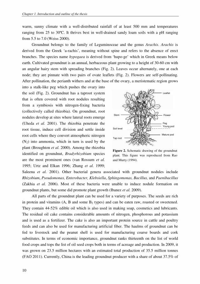
world (Fig. 1). Groundnut requires a
Figure 1. Groundnut growing regions in the world. Solid
line: center of origin; dotted line: area of cultivation;
black: areas of intensive cultivation. This figure is
reproduced from Weiss (2000).
Chapter 1. Introduction and outline of the thesis
10
warm, sunny climate with a well-distributed rainfall of at least 500 mm and temperatures
ranging from 25 to 30ºC. It thrives best in well-drained sandy loam soils with a pH ranging
from 5.5 to 7.0 (Weiss 2000).
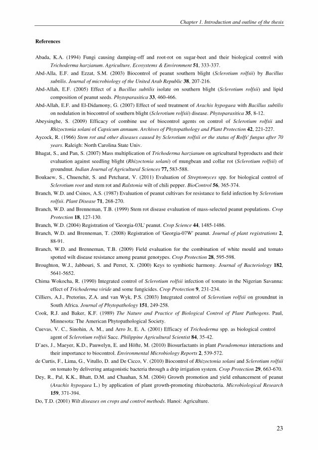
Groundnut belongs to the family of Leguminoceae and the genus Arachis. Arachis is
derived from the Greek ‘a-rachis’, meaning without spine and refers to the absence of erect
branches. The species name hypogaea is derived from ‘hupo-ge’ which in Greek means below
earth. Cultivated groundnut is an annual, herbaceous plant growing to a height of 30-60 cm with
an angular hairy stem with spreading branches (Fig. 2). Leaves occur alternately, one at each
node; they are pinnate with two pairs of ovate leaflets (Fig. 2). Flowers are self-pollinating.
After pollination, the perianth withers and at the base of the ovary, a meristematic region grows
into a stalk-like peg which pushes the ovary into
the soil (Fig. 2). Groundnut has a taproot system
that is often covered with root nodules resulting
from a symbiosis with nitrogen-fixing bacteria
(collectively called rhizobia). On groundnut, root
nodules develop at sites where lateral roots emerge
(Uheda et al. 2001). The rhizobia penetrate the
root tissue, induce cell division and settle inside
root cells where they convert atmospheric nitrogen
(N2) into ammonia, which in turn is used by the
plant (Broughton et al. 2000). Among the rhizobia
identified on groundnut, Bradyrhizobium species
are the most prominent ones (van Rossum et al.
1995; Urtz and Elkan 1996; Zhang et al. 1999;
Saleena et al. 2001). Other bacterial genera associated with groundnut nodules include
Rhizobium, Pseudomonas, Enterobacter, Klebsiella, Sphingomonas, Bacillus, and Paenibacillus
(Zakhia et al. 2006). Most of these bacteria were unable to induce nodule formation on
groundnut plants, but some did promote plant growth (Ibanez et al. 2009).
All parts of the groundnut plant can be used for a variety of purposes. The seeds are rich
in protein and vitamins (A, B and some B2 types) and can be eaten raw, roasted or sweetened.
They contain 44-52% edible oil which is also used in making soap, cosmetics and lubricants.
The residual oil cake contains considerable amounts of nitrogen, phosphorous and potassium
and is used as a fertilizer. The cake is also an important protein source in cattle and poultry
feeds and can also be used for manufacturing artificial fiber. The haulms of groundnut can be
fed to livestock and the peanut shell is used for manufacturing coarse boards and cork
substitutes. In terms of economic importance, groundnut ranks thirteenth on the list of world
food crops and tops the list of oil seed crops both in terms of acreage and production. In 2009, it
was grown on 23.5 million hectares with an estimated total production of 35.5 million tonnes
(FAO 2011). Currently, China is the leading groundnut producer with a share of about 37.5% of
Leaflet
Flower
Peg
Young pod
Mature pod
Tap root
Soil level
Stem
Leaflet
Flower
Peg
Young pod
Mature pod
Tap root
Soil level
Stem
Figure 2. Schematic drawing of the groundnut
plant. This figure was reproduced from Rao
and Murty (1994).

Chapter 1. Introduction and outline of the thesis
11
the overall world production, followed by India, the United States of America (USA), Argentina
and Vietnam (Fabra et al. 2010). India has the largest groundnut cultivation area, but has a low
average yield of approximately 1.0 ton ha-1
. The USA have the highest average yield of 3.8 ton
ha-1
(FAO 2011).
Yield limiting factors in groundnut cultivation
Several abiotic and biotic factors affect the growth and development of groundnut leading
to qualitative and quantitative yield losses. Temperature, relative humidity, and soil
characteristics are the major abiotic factors that can directly or indirectly influence productivity.
In addition to the direct impact on growth, these abiotic factors can also have an impact on the
proliferation and damage caused by pests and diseases. The major insect pests that attack
groundnut are termites, white grubs, thrips, aphids, leaf miners, and caterpillars. The pathogens
parasitizing on groundnut include viruses, mycoplasmas, bacteria, oomycetes, fungi, nematodes,
and parasitic plants (Middleton et al. 1994; Wightman and Ranga Rao 1994). The diseases that
are most damaging and cause the largest economic losses are stem rot, damping-off, black collar
rot, early and late leaf spot, rust and bacterial wilt (Middleton et al. 1994; Podile and Kishore
2002).
Stem rot caused by the fungus S. rolfsii will be discussed in detail in the next section. The
fungus Rhizoctonia solani causes seed decay, pre- and post-emergence damping-off of
seedlings, hypocotyl and root necrosis, root rot of young seedlings, peg and pod rot, and foliar
blight of mature plants (Porter et al. 1984). The fungus Aspergillus niger causes black collar rot,
an important disease found in all major groundnut-growing areas of the world. Seedlings and
young plants are particularly susceptible and infection of juvenile tissue usually results in high
mortality rates. Seed rot and pre-emergence damping-off are common symptoms of this disease,
but the most obvious symptom is sudden wilting of young plants. Early and late leaf spots
caused by the fungi Cercospora arachidicola and Cercosporidium personatum, respectively,
damage the plant by reducing the available photosynthetic area via lesion formation and leaflet
abscission. Yield losses can range from 10% to over 50% (McDonald et al. 1985). The fungus
Puccinia arachidis is the causal agent of groundnut rust which, in conjunction with leaf spots, is
devastating in many South and Central American countries (Porter et al. 1984). Ralstonia
solanacearum, the causal agent of bacterial wilt, is distributed worldwide and is an important
yield-limiting factor in groundnut cultivation in Africa and Asia (Porter et al. 1984). In
Vietnam, stem rot, damping-off, black collar rot, bacterial wilt and leaf spots are the most
devastating diseases in many regions (Le 1977; Mehan and Hong 1994; Nguyen et al. 1998; Do
2001; Le 2004; Nguyen et al. 2004).
Stem rot disease of groundnut caused by Sclerotium rolfsii
The first report of stem rot dates back to 1892 with Peter Henry Rolfs’ discovery of this
fungus in association with tomato blight in Florida (Aycock 1966). The wide host range of S.

Chapter 1. Introduction and outline of the thesis
12
rolfsii, its prolific growth and ability to produce persistent sclerotia all contribute to the large
economic losses associated with this pathogen (Chima Wokocha 1990; Cilliers et al. 2003;
Singh et al. 2003). On groundnut, the disease caused by S. rolfsii is reported as stem rot, white
mould, or southern blight, but stem rot is most commonly used. The disease occurs in most
groundnut production areas in the world and appears to be more serious when the plants also
suffer from tomato spotted wilt virus (Branch and Brenneman 2009). Pod yield losses range
from 10-25% and sometimes up to 80% (Mehan et al. 1994). In North Carolina, groundnut
crops sustained higher losses than any other agricultural crop (Aycock 1966). In 1959, the
United States Department of Agriculture estimated losses of $10-20 million associated with S.
rolfsii in the southern groundnut-growing regions, with yield depletions up to 60% in the coastal
plains of North Carolina (Garren 1959). In Georgia (USA), economic losses due to stem rot
disease and disease management costs were estimated to be approximately $38 million from
2004 to 2007 (Kemerait 2005; 2006; 2007; 2008).
Infection is usually restricted to plant parts that are in direct contact with the
soil. On groundnut, S. rolfsii attacks stems, roots, leaves, pegs and pods. Initial disease
symptoms comprise small, water-soaked lesions on the lower stem or near the soil surface,
followed by yellowing and wilting of the lateral branches, main stem, and eventually the entire
plant (Fig. 3). Diagnostic signs of the fungus include characteristic white mycelial fans and
brown sclerotia extending from infected tissues (Fig. 3). The fungus infects pegs and pods and
causes rot (Fig. 3).
Figure 3. Disease symptoms on groundnut caused by Sclerotium rolfsii on groundnut A- yellow leaves and
wilting; B- mycelium and sclerotia on infected tissue; C- stem rot symptoms; D- peg and pod rot.
A B
D C

Chapter 1. Introduction and outline of the thesis
13
The disease cycle of stem rot on groundnut
is shown in Figure 4. It is modeled based on the
disease cycle of stem rot on apple (Mullen 2001)
and hosta (Upchurch 2000). Sclerotia are the
principal overwintering structures and the primary
inoculum source for the disease. Under favorable
conditions, sclerotia germinate and fungal hyphae
grow towards and attack the lower part of the
stem base. On diseased tissues, a hyphal mat and
sclerotia are produced and, sometimes, also
basidiospores are produced. The role of
basidiospores in the disease cycle under field
conditions has not been investigated in detail.
Infected groundnut shows yellow leaves, wilting
Hyphal mat and sclerotia
produced on diseased tissue
Fungus produces oxalic acid and other compounds to destroy cell walls
Mycelium is the second source of inoculum in the field
Under favorable conditions, sclerotia germinate
Sclerotia survive in soil Healthy
groundnut
Figure 4. Disease cycle of stem rot on groundnut
caused by Sclerotium rolfsii.
The soil-borne pathogen Sclerotium rolfsii
Sclerotium rolfsii was named by Saccardo in 1911 in recognition of Rolf’s pioneer work
in Florida referred to in Tu and Kimbrough (1978). Initially, Sclerotium was a genus assigned to
the artificial class of Fungi Imperfecti that included many diverse species. These species were
grouped in one genus because of their shared phenotypic characteristics. They generally form
small, tan to dark-brown/black, spherical sclerotia, that function as survival structures. As more
became known about these fungi and their sexual states, some were reclassified into other
genera, either in the Basidiomycota or the Ascomycota, and sometimes renamed according to
their teleomorph. In 1931, Cruzi first described the perfect stage of S. rolfsii and Corticium
rolfsii was the first teleomorph name (referred by Tu and Kimbrough (1978)). In 1978, Tu and
Kimbrough proposed to classify the pathogen in the genus Athelia and the binominal name,
Athelia rolfsii (Cruzi) Tu and Kimbrough, has been used since. Some Sclerotium species still
have no known sexual state, but by exploring molecular identification methods each species can
now be assigned to the right genus. For example, based on sequence analysis of the rDNA large
subunit (LSU) and internal transcribed spacer (ITS) regions, some Sclerotium species were re-
named and moved from Ascomycota to Basidiomycota, whereas others, including Sclerotium
denigrans and Sclerotium perniciosum, were moved from the Basidiomycota to the Ascomycota
(Xu et al. 2010).
S. rolfsii has a wide host range with more than 500 plant species (Aycock 1966). They
consist of mono- and di-cotyledons (Farr et al. 1989). Until now, no worldwide compilation of
host genera has been published, however, more than 270 host genera have been reported in the
USA. These include agricultural crops such as sweet potato (Ipomoea batatas), pumpkin
(Cucurbita pepo), corn (Zea mays), wheat (Triticum vulgare), groundnut (Arachis hypogeae),
and some horticultural crops such as Narcissus (Narcissus spp.), Iris (Iris spp.), Lilium (Lilium

Chapter 1. Introduction and outline of the thesis
14
spp.), Zinnia (Zinnia spp.), and Chrysanthemum (Chrysanthemum spp.) (Farr et al. 1989). In
Vietnam, many crops are infected by S. rolfsii including groundnut (Arachis hypogaea),
mungbean (Vigna radiata), soybean (Glycine max), tomato (Lycopersicon esculentum), potato
(Solanum tuberosum), eggplant (Solanum melongena), pepper (Capsicum annuum), cabbage
(Brassica oleracea), cucumber (Cucumis sativus) and taro (Colocasia esculenta) (Le 1977; Do
2001).
Colonies of S. rolfsii can be readily distinguished on infected plant material or artificial
media by gross morphological characteristics (Fig. 5). Rapidly growing, silky-white hyphae
tend to aggregate into rhizomorphic cords (Aycock 1966; Harlton et al. 1995). In culture, agar
media are rapidly (2-3 days) covered with mycelium, including aerial hyphae. The optimum
temperature for hyphal growth and sclerotial formation is 27-30ºC (Aycock 1966; Mathur and
Sinha 1970; Punja 1985; Punja and Rahe 1993). Xu et al. (2008) showed that the difference in
temperature in the southern and northern parts of the United States of America affects survival
of sclerotia. As a result, the severity of S. rolfsii in the southern part was higher than that in the
northern part. Sclerotia (0.3-3.0 mm diameter) begin to develop after 4-7 days of growth (Punja
and Rahe 1993) when hyphae cluster together as a compact mass. After an initial white
appearance, the sclerotia quickly become dark brown (Aycock 1966). Sclerotia contain viable
hyphae and serve as the primary inoculum source in the disease cycle. Oxalic acid plays an
important role in the virulence of S. rolfsii (Kritzman et al. 1977; Punja 1985). By producing
oxalic acid as well as pectinolytic and cellulolytic enzymes, S. rolfsii kills and disintegrates host
tissues before it penetrates (Prasad and Naik 2008).
Figure 5. Mycelium and sclerotia of Sclerotium rolfsii on diseased tissue (A) and on Potato Dextrose Agar plates
(B, C).
The genetic diversity of S. rolfsii has been studied by a variety of techniques, including
mycelial compatibility (Fig. 6), restriction fragment length polymorphism (RFLP) analysis of
ITS-rDNA, and by ITS-rDNA or LSU sequencing (Harlton et al. 1995; Okabe et al. 2000;
Okabe et al. 2001; Punja and Sun 2001; Okabe and Matsumoto 2003; Xu et al. 2010). Harlton
et al. (1995) found 49 mycelial compatibility groups (MCGs) and 12 RFLP-ITS groups in a
A B C

Chapter 1. Introduction and outline of the thesis
15
worldwide collection of isolates, but could not establish correlations between MCGs and
pathogenicity. Some RFLP-ITS grouping patterns were correlated with MCGs, but isolates
belonging to one MCG sometimes showed
different RFLP-ITS patterns and certain
patterns were dispersed among different
MCGs. Recently, Xu et al. (2010) reported a
close relationship between S. rolfsii, S. rolfsii
var. delphinii and S. coffeicola by LSU
sequence analysis. The phylogeny of S. rolfsii,
S. delphinii and S. coffeicola based on ITS-
rDNA shows two clades with most S. rolfsii
strains. One clade contains most of the S.
delphinii strains, and one clade contains strains
of both S. rolfsii and S. delphinii, suggesting a
close relationship between the latter two
species.
Figure 6. Example of mycelial compatibility
analysis with three isolates of Sclerotium rolfsii.
Hyphae of isolate I and II intermingle and are
considered compatible. The barrage zone between
isolate III and isolates I and II indicates mycelial
incompatibility.
Control methods of stem rot disease
Because of its wide host range, fast growth rate, and the production of large numbers of
persistent sclerotia, stem rot disease caused by S. rolfsii is very difficult to control (Punja 1985;
Lakpale et al. 2007). Current control measures comprise the use of resistant cultivars and
various physical, chemical, and biological control strategies (Le 1977; Redy and McDonald
1983; Punja 1985; Punja and Rahe 1993; Mehan and Hong 1994; Mehan et al. 1995; Le 2004;
Nguyen et al. 2004; dos Santos et al. 2005; Vargas Gil et al. 2008).
Physical and cultural control
Cultural and physical methods to control soil-borne S. rolfsii are deep ploughing, solar
heating, chemical fertilizer and crop rotation. Deep ploughing to bury sclerotia or disease
tissues under 6-20 centimetres, was reported to reduce the viability of sclerotia or to kill hyphae
of the pathogen in the fields (Elad et al. 1980; Porter and Merriman 1983; Mihail and Alcorn
1984). For solar heating, soils mulched with transparent polyethylene for 6 weeks in July-
August and then sown with groundnuts the following spring, showed significant decreases in
the percentage of diseased plants and rotten pods (Grinstein et al. 1979). Ammonium
compounds were shown to inhibit germination of sclerotia and promoted colonization of
sclerotia by soil microorganisms (Prasad and Naik 2008). However, for control of S. rolfsii on
groundnut, supplementation with ammonium compounds is not recommended because this
reduces the N2-fixiation in root nodules. Rotation with non-host crops not only improves the
I II
III

Chapter 1. Introduction and outline of the thesis
16
soil nutritional status, but also may adversely affect pathogen inoculum densities. For S. rolfsii,
however, this strategy is not effective due to its broad host range. Nevertheless, Taylor and
Rodriguez Kabana (1999) found that stem rot of groundnut can be suppressed by rotation with
cotton. Also paddy rice was recommended as a rotation crop with groundnut in order to reduce
stem rot in Vietnam (Le 1977).
Resistant cultivars
Because it is not easy to breed cultivars highly resistant to S. rolfsii, disease-tolerant
cultivars may be used as a component of an integrated control effort (Punja 1985). In the mid-
90s, two cultivars, i.e. Toalson and Southern Runner, were developed which are less
susceptible/partially resistant to S. rolfsii (Branch and Csinos 1987; Mehan et al. 1995; Branch
and Brenneman 1999) and more recently, additional partially stem rot resistant groundnut
cultivars have been released including C-99R (Gorbet and Shokes 2002a), Florida MDR 98
(Gorbet and Shokes 2002b), Georgia-03L (Branch 2004), Georgia-07W (Branch and
Brenneman 2008), and Florida-07 (Gorbet and Tillman 2009). The use of these cultivars is still
limited, possibly due to the relatively low level of resistance.
Chemical control
In many cases, fungicides such as tebuconazole, pentachloronitrobenzene (PCNB) and
flutolanil are used to control S. rolfsii. However, tolerance to tebuconazole and PCNB has been
reported for S. rolfsii populations in USA (Wadsworth and Melouk 1984; Franke et al. 1998;
Shim et al. 1998). Other fungicides that are used to control stem rot disease include
difenoconazole, carbendazim, flusilazole and chlorothalonil (Cilliers et al. 2003). When
difenoconazole was tested in combination with Trichoderma harzianum, a biocontrol agent of S.
rolfsii, no reduction of the effect of T. hazianum was observed (Cilliers et al. 2003). Although
fungicides can protect groundnut plants from infection by S. rolfsii, chemical control should be
gradually minimized because of its potential harmful effects to the environment. Therefore
integration of several different control measures is proposed to provide sustainable management
of S. rolfsii and other diseases of groundnut. In this context, biological control can be an
alternative or supplement to current management practices for S. rolfsii (Singh et al. 2003; Dey
et al. 2004; Tonelli et al. 2010).
Biological control
Application of beneficial microorganisms to soil, seeds or planting materials has been
proposed as a sustainable and supplementary approach to control plant diseases (Cook and
Baker 1989). The most widely studied microorganisms with antagonistic activity against plant
pathogens and with beneficial effects on plant growth, belong to the bacterial genera Bacillus,
Pseudomonas, Rhizobium, or the fungal genus Trichoderma (Ongena and Jacques 2008;
Raaijmakers et al. 2009; Lorito et al. 2010).

Chapter 1. Introduction and outline of the thesis
17
Fungal biocontrol agents may directly or indirectly kill sclerotia or mycelium of S. rolfsii.
Lectins produced by S. rolfsii were proposed to serve as recognition factors for fungal
biocontrol agents (Prasad and Naik 2008). Among the fungal biocontrol agents, Trichoderma
species are the most widely studied (Table 1). In a direct interaction, hyphae of Trichoderma
penetrate the rind and the cortex of sclerotia and lyse the medullar tissue. Degraded sclerotia
become dark, soft and disintegrate under slight pressure (Prasad and Naik 2008) and it was
shown that chitinase and β-1,3-glucanase play a role in the interaction between Trichoderma
harzianum and S. rolfsii (Prasad and Naik 2008). Next to Trichoderma, several other fungal
genera have been tested for their ability to control diseases caused by S. rolfsii on bean, carrot,
chilli, ginger, wheat, lentil, sesame, soybean, sugar beet, sunflower, tomato, or groundnut.
These antagonistic fungi include Gliocladium virens, Gliocladium roseum, Glomus fascicatum,
Penicillium pinophilum, Gigaspora margarita and also Sclerotium rolfsii (Table 1).
For biocontrol of S. rolfsii, several bacterial genera and species have been studied. Most
of them belong to the genera Pseudomonas and Bacillus (Table 2). Pseudomonas strains can
restrict in vitro hyphal growth or reduce germination of sclerotia of S. rolfsii (Ganesan and
Gnanamanickam 1987; Kishore et al. 2005; Ganesan et al. 2007; de Curtis et al. 2010; Pastor et
al. 2010; Tonelli et al. 2010). Although pseudomonads are well-known for the production of a
diverse array of antimicrobial compounds, including 2,4-diacetylphloroglucinol, pyrrolnitrin,
pyoluteorin, rhizoxins, phenazines and lipopeptides (Raaijmakers et al. 2002; Haas and Defago
2005; Raaijmakers et al. 2009; D’aes et al. 2010; Raaijmakers et al. 2010) the role of these or
other bioactive compounds in biocontrol of S. rolfsii has not been studied in detail (Table 2).
Also for most of the tested Pseudomonas strains, there is a lack of knowledge on the genes
involved in their activity against S. rolfsii. Next to Pseudomonas, several Bacillus species and
strains have been studied for their efficacy to control stem rot disease of groundnut. Pre-
treatment of groundnut seeds with Bacillus subtilis protected groundnut seeds against S. rolfsii
and significantly increased the number of pods (Abd-Allah 2005). Also for the Bacillus strains
and other bacterial genera tested to date (Table 2), little or no knowledge is available on the
fundamental mechanisms involved in their activity against S. rolfsii. Moreover, most of these
studies were conducted under controlled conditions and only few studies were performed under
field conditions.

Chapter 1. Introduction and outline of the thesis
18
Tab
le 1
. F
un
gal
gen
era
and s
pec
ies
test
ed f
or
bio
contr
ol
of
Scle
roti
um
ro
lfsi
i o
n g
roun
dnut
and
som
e o
ther
cro
ps.
Par
ts o
f th
ese d
ata
wer
e re
pro
duce
d f
rom
Pra
sad (
2008
).
Fu
ngal
bio
contr
ol
agen
ts
Cro
p
Pro
pose
d
mec
han
ism
s*
Ref
eren
ces
Gli
ocl
adiu
m r
ose
um
Gli
ocl
adiu
m v
iren
s
Gli
ocl
adiu
m s
p.
carr
ot,
gra
m, gro
un
dn
ut,
sunfl
ow
er,
tom
ato, m
ung b
ean
ISR
an
d p
aras
itis
m
Pra
sad a
nd N
aik 2
00
8;
Yaq
ub a
nd
Shah
zad 2
00
8
Glo
mus
cale
doniu
m,
Glo
mus
fasc
icula
tum
,
gro
un
dn
ut
par
asit
ism
O
zgonen e
t al.
20
10
; P
rasa
d a
nd N
aik 2
008
Gig
asp
ora
marg
ari
ta
gro
un
dn
ut
par
asit
ism
O
zgonen e
t al.
20
10
Penic
illi
um
pin
ophil
um
se
sam
e u
nknow
n
Pra
sad a
nd N
aik 2
00
8
Scl
ero
tiu
m r
olf
sii
gro
un
dn
ut
ISR
N
andin
i et
al.
20
10
Tri
chod
erm
a a
ure
ovi
ride
tom
ato, so
yb
ean
unknow
n
Pra
sad a
nd N
aik 2
00
8
Tri
chod
erm
a h
arz
ianum
ch
illi
, gro
un
dn
ut,
gra
m, le
nti
l, m
un
g
bea
n,
sesa
me,
so
yb
ean, su
gar
bee
t,
sunfl
ow
er, te
a, t
om
ato,
whea
t
par
asit
ism
IS
R
Abada
1994;
Bhag
at a
nd P
an 2
007;
Bo
ukaew
et
al.
20
11;
Ela
d e
t al.
198
2;
Gan
esan
et
al.
2007;
Pra
sad
and
Nai
k 2
00
8;
Shai
gan e
t a
l. 2
008
; Y
aqu
b a
nd S
hahza
d
20
08
Tri
chod
erm
a k
onin
gii
le
nti
l, s
oyb
ean, to
mato
p
aras
itis
m
Pra
sad a
nd N
aik 2
00
8;
Tsa
ho
uri
do
u a
nd
Than
asso
ulo
po
ulo
s 200
2
Tri
chod
erm
a l
ongib
rach
iatu
m
gro
un
dn
ut,
tea
p
aras
itis
m
Pra
sad a
nd N
aik 2
00
8;
Shai
gan
et
al.
20
08
Tri
chod
erm
a p
seu
dokonin
gii
C
hin
ese
cab
bag
e, gin
ger
, su
nfl
ow
er
ISR
an
d p
aras
itis
m
Cuev
as e
t al.
20
01
; P
rasa
d a
nd N
aik 2
008;
Yaq
ub a
nd
Shah
zad 2
00
8
Tri
chod
erm
a v
irid
e gro
un
dn
ut,
gra
m, le
nti
l, s
oy b
ean
, su
gar
bee
t, t
ea, to
mato
par
asit
ism
C
him
a W
oko
cha
199
0;
Pra
sad
and
Nai
k 2
00
8;
Shai
gan
et
al.
20
08
Tri
chod
erm
a p
oly
sporu
m,
sunfl
ow
er
ISR
an
d p
aras
itis
m
Yaq
ub a
nd S
hah
zad 2
008
Tri
chod
erm
a h
am
atu
m,
tea
par
asit
ism
S
hai
gan
et
al.
20
08
Tri
chod
erm
a p
arc
eram
osu
m
Ch
ines
e cab
bag
e, te
a p
aras
itis
m
Cuev
as e
t al.
20
01
; S
hai
gan
et
al.
200
8
Tri
chod
erm
a s
p.
bea
n,
gro
undnu
t, su
nfl
ow
er, st
ring
bea
n, to
mato
par
asit
ism
L
iu e
t a
l. 2
00
8;
Pra
sad
an
d N
aik 2
008;
Sai
et
al.
20
10
*IS
R =
ind
uce
d s
yst
em
ic r
esis
tan
ce

Chapter 1. Introduction and outline of the thesis
19
Tab
le 2
. B
acte
rial
gener
a an
d s
pec
ies
test
ed f
or
bio
logic
al c
ontr
ol
of
Scl
eroti
um
rolf
sii
on g
rou
ndn
ut
and s
om
e o
ther
cro
ps.
Bac
teri
al g
ener
a/sp
ecie
s
Ori
gin
C
rop
Tes
t m
etho
d
Pro
po
sed
mech
anis
ms*
Ref
eren
ces
in
vit
ro
gro
wth
cham
ber
net
-
ho
use
fiel
d
Pse
ud
om
on
as
P. fl
uore
scen
s unkno
wn
gro
und
nut
x
x
un
kn
ow
n
Gan
esan
and G
nan
aman
ickam
198
7
P. fl
uore
scen
s
chic
kpea
rhiz
osp
her
e
bet
el v
ine
x
x
un
kn
ow
n
Sin
gh e
t al.
20
03
P. fl
uore
scen
s
pep
per
rhiz
osp
her
e pep
per
x
x
un
kn
ow
n
Abey
singh
e 2
009
P. fl
uore
scen
s cu
lture
coll
ecti
on
gro
und
nut
x
x
x
ISR
S
enth
ilra
ja e
t al.
201
0
P. aeru
gin
osa
gro
undn
ut
seed
endo
ph
yte
gro
und
nut
x
x
inte
rfer
ence
wit
h C
WD
E
Kis
hore
et
al.
200
5
P. aeru
gin
osa
oil
pal
m r
oots
ch
ili
pep
per
x
x
IS
R
Sid
diq
ui
and M
eon 2
009
P. pu
tid
a
pep
per
rhiz
osp
her
e pep
per
x
x
un
kn
ow
n
Abey
singh
e 2
009
P. m
on
teil
ii
unkno
wn
gro
und
nut
x
x
anti
bio
sis
R
akh
et
al.
201
1
Pse
ud
om
on
as
sp.
tom
ato r
oots
to
mat
o
x
x
ISR
and
anti
bio
sis
Pas
tor
et a
l. 2
01
0
Pse
ud
om
on
as
sp.
com
post
s ch
ickpea
x
x
an
tib
iosi
s H
amee
da
et
al.
2010
Pse
ud
om
on
as
sp.
gro
undn
ut
gro
und
nut
x
IS
R
To
nel
li e
t al.
201
1
Baci
llus
B. su
bti
lis
to
mat
o r
hiz
osp
her
e gro
und
nut
x
x
un
kn
ow
n
Abd-A
lla
2003
B. su
bti
lis
tom
ato r
hiz
osp
her
e
gro
und
nut
x
IS
R
Abd-a
lla
200
5;
Ab
d-A
llah
and
El-
Did
am
on
y 2
00
7
B. su
bti
lis
org
anic
am
end
men
ts
tom
ato
x
x
x
un
kn
ow
n
De
Curt
is e
t al.
20
10
B. su
bti
lis
pep
per
rhiz
osp
her
e pep
per
x
x
an
tib
iosi
s
Abey
singh
e 2
00
9

Chapter 1. Introduction and outline of the thesis
20
‘Ta
ble
2 c
on
tin
ued
’
Bac
teri
al g
ener
a/sp
ecie
s
Ori
gin
C
rop
T
est
met
hod
P
rop
ose
d
mec
han
ism
s*
Ref
erence
s
in
vit
ro
gro
wth
cham
ber
net-
ho
use
fiel
d
Baci
llus
spp.
com
po
sts
chic
kp
ea
x
x
anti
bio
sis
Ham
eed
a et
al.
201
0
Baci
llus
sp.
gro
und
nut
gro
un
dnu
t
x
ISR
T
onel
li e
t al.
2011
Oth
er
Burk
hold
eri
a c
epaci
a
org
anic
am
endm
ents
to
mat
o
x
x
x
unkno
wn
De
Curt
is e
t al.
2010
Burk
hold
eri
a c
epaci
a
oil
palm
roots
ch
ili
pep
per
x
x
ISR
S
iddiq
ui
and
Meo
n 2
00
9
Ste
pto
myc
es m
ycaro
faci
ens
pep
per
ch
ili
pep
per
x
x
x
anti
bio
sis
Bou
kaew
et
al.
201
1
Ste
pto
myc
es p
hil
anth
i
pep
per
ch
ili
pep
per
x
x
x
anti
bio
sis
Bou
kaew
et
al.
201
1
Str
epto
myc
es s
pp.
sugar
beet
soil
su
gar
bee
t x
x
anti
bio
sis
Err
akh
i et
al.
2007
Agro
bact
eri
um
un
kno
wn
tom
ato
x
x
com
pet
itio
n
and a
nti
bio
sis
Pel
zer
et a
l. 2
011
Klu
yver
a
un
kno
wn
tom
ato
x
x
com
pet
itio
n
and a
nti
bio
sis
Pel
zer
et a
l. 2
011
Ser
rati
a m
arc
escen
s un
kno
wn
S. ro
lfsi
i x
an
tibio
sis
Ord
entl
ich e
t al.
19
88
Meth
ylob
act
eriu
m s
p.
gro
und
nut
gro
un
dnu
t x
x
ISR
M
adh
aiyan
et
al.
200
6
Rhiz
obiu
m s
p.
un
kno
wn
gro
un
dnu
t x
x
ISR
M
adh
aiyan
et
al.
200
6
Rhiz
obiu
m s
p.
gro
und
nut
nod
ule
s gro
un
dn
ut
x
x
anti
bio
sis
Gan
esan
et
al.
20
07
Rhiz
obiu
m s
pp.
tom
ato
rhiz
osp
her
e
gro
un
dnu
t
x
unkno
wn
Ab
d-A
llah a
nd E
l-D
idam
on
y
2007
* I
SR
= i
nd
uce
d s
yst
emic
res
ista
nce
, C
WD
E =
Cel
l W
all
Deg
rad
ing E
nzy
me

Chapter 1. Introduction and outline of the thesis
21
Outline of this thesis
Groundnut is an important oil seed crop in many countries including Vietnam. Cultivation
of groundnut is adversely affected by a wide range of pests and diseases. Stem rot caused by S.
rolfsii is among the most damaging soil-borne fungal diseases and causes significant economic
losses. The overall aim of the research described in this thesis was to study the efficacy of
biological control of stem rot disease of groundnut in Vietnam. When we started this research
project, there was little knowledge on the genetic diversity of S. rolfsii populations on
groundnut in Vietnam. Moreover, there was hardly any information on the occurrence and
distribution of beneficial soil bacteria in groundnut farmer fields and the potential of specific
bacterial genera to control stem rot disease under field conditions.
As a first step (chapter 2), we made an inventory of the incidence of stem rot disease on
groundnut in farmer fields in central Vietnam. Approximately 200 isolates of S. rolfsii were
successfully isolated from more than 400 diseased samples collected at eight locations in four
provinces. We subsequently analyzed the phenotypic diversity of S. rolfsii populations based on
hyphal growth rate and different sclerotial characteristics. The genetic diversity of S. rolfsii
populations was assessed by mycelial compatibility assays and ITS-rDNA sequencing. In
addition, we tested the pathogenicity of S. rolfsii isolates and their sensitivity to tebuconazole, a
fungicide commonly used to control stem rot disease.
To date, several bacterial strains have been studied in biocontrol of stem rot of groundnut.
However, in most of these studies biocontrol was studied under controlled conditions and the
mechanisms involved in the biocontrol activity were not identified. In this thesis (chapter 3),
the biocontrol efficacy of several well-characterized Pseudomonas species and strains was
evaluated. These included phenazine-producing strain Pseudomonas chlororaphis Phz24 and
lipopeptide-producing strains Pseudomonas sp. SH-C52, Pseudomonas fluorescens SS101, P.
fluorescens SBW25 and Pseudomonas putida 267. To determine the role of phenazines and
lipopeptides in inhibition of S. rolfsii, mutants deficient in the production of phenazines or
lipopeptides were generated and included in the bioassays. Subsequently, strains P.
chlororaphis Phz24, Pseudomonas sp. SH-C52 and P. fluorescens SS101 were tested for
biocontrol of stem rot of groundnut under nethouse and field conditions in Vietnam. Among
these bacterial strains, Pseudomonas sp. SH-C52 and P. fluorescens SS101 showed promising
results in biological control and growth promotion in the field experiments conducted in 2010.
Their consistency to control stem rot disease and/or to increase pod yield were again evaluated
in field experiments conducted in 2011 (chapter 5).
Besides the well-characterized Pseudomonads, we isolated and characterized indigenous
bacterial populations from the stem base and roots of groundnut plants grown in farmer fields in
central Vietnam (chapter 4). Four bacterial isolates, identified by 16S-rDNA sequencing as
Pseudomonas, Bacillus and Chryseobacterium species, were tested for growth promotion and
biocontrol of stem rot of groundnut. Among the tested bacterial strains, Pseudomonas sp. R4D2

Chapter 1. Introduction and outline of the thesis
22
was effective in biocontrol of stem rot, whereas Bacillus sp. strains S18F11 and S20D12
significantly increased pod yield. All three strains were again tested for biocontrol of stem rot
and plant growth promotion in 2011. In these field trials, we also evaluated the effects of these
bacterial strains on various other groundnut diseases, including damping-off, black collar rot,
bacterial wilt and leaf spots (chapter 5).
In chapter 6, the diversity of bacteria associated with groundnut nodules and their effects
on plant growth were investigated. Two isolates, identified by 16S-rDNA sequencing as
Sphingomonas and Rhizobium species, were tested in field trials for their ability to control stem
rot and other groundnut diseases.
The major results obtained in this thesis are summarized and discussed in chapter 7. In
this final chapter, we also discuss the consistency and future perspectives of biological control
of stem rot disease of groundnut.
Chapter 1. Introduction and outline of the thesis
23
References
Abada, K.A. (1994) Fungi causing damping-off and root-rot on sugar-beet and their biological control with
Trichoderma harzianum. Agriculture, Ecosystems & Environment 51, 333-337.
Abd-Alla, E.F. and Ezzat, S.M. (2003) Biocontrol of peanut southern blight (Sclerotium rolfsii) by Bacillus
subtilis. Journal of microbiology of the United Arab Republic 38, 207-216.
Abd-Allah, E.F. (2005) Effect of a Bacillus subtilis isolate on southern blight (Sclerotium rolfsii) and lipid
composition of peanut seeds. Phytoparasitica 33, 460-466.
Abd-Allah, E.F. and El-Didamony, G. (2007) Effect of seed treatment of Arachis hypogaea with Bacillus subtilis
on nodulation in biocontrol of southern blight (Sclerotium rolfsii) disease. Phytoparasitica 35, 8-12.
Abeysinghe, S. (2009) Efficacy of combine use of biocontrol agents on control of Sclerotium rolfsii and
Rhizoctonia solani of Capsicum annuum. Archives of Phytopathology and Plant Protection 42, 221-227.
Aycock, R. (1966) Stem rot and other diseases caused by Sclerotium rolfsii or the status of Rolfs' fungus after 70
years. Raleigh: North Carolina State Univ.
Bhagat, S., and Pan, S. (2007) Mass multiplication of Trichoderma harzianum on agricultural byproducts and their
evaluation against seedling blight (Rhizoctonia solani) of mungbean and collar rot (Sclerotium rolfsii) of
groundnut. Indian Journal of Agricultural Sciences 77, 583-588.
Boukaew, S., Chuenchit, S. and Petcharat, V. (2011) Evaluation of Streptomyces spp. for biological control of
Sclerotium root and stem rot and Ralstonia wilt of chili pepper. BioControl 56, 365-374.
Branch, W.D. and Csinos, A.S. (1987) Evaluation of peanut cultivars for resistance to field infection by Sclerotium
rolfsii. Plant Disease 71, 268-270.
Branch, W.D. and Brenneman, T.B. (1999) Stem rot disease evaluation of mass-selected peanut populations. Crop
Protection 18, 127-130.
Branch, W.D. (2004) Registration of 'Georgia-03L' peanut. Crop Science 44, 1485-1486.
Branch, W.D. and Brenneman, T. (2008) Registration of 'Georgia-07W' peanut. Journal of plant registrations 2,
88-91.
Branch, W.D. and Brenneman, T.B. (2009) Field evaluation for the combination of white mould and tomato
spotted wilt disease resistance among peanut genotypes. Crop Protection 28, 595-598.
Broughton, W.J., Jabbouri, S. and Perret, X. (2000) Keys to symbiotic harmony. Journal of Bacteriology 182,
5641-5652.
Chima Wokocha, R. (1990) Integrated control of Sclerotium rolfsii infection of tomato in the Nigerian Savanna:
effect of Trichoderma viride and some fungicides. Crop Protection 9, 231-234.
Cilliers, A.J., Pretorius, Z.A. and van Wyk, P.S. (2003) Integrated control of Sclerotium rolfsii on groundnut in
South Africa. Journal of Phytopathology 151, 249-258.
Cook, R.J. and Baker, K.F. (1989) The Nature and Practice of Biological Control of Plant Pathogens. Paul,
Minnesota: The American Phytopathological Society.
Cuevas, V. C., Sinohin, A. M., and Arro Jr, E. A. (2001) Efficacy of Trichoderma spp. as biological control
agent of Sclerotium rolfsii Sacc. Philippine Agricultural Scientist 84, 35-42.
D’aes, J., Maeyer, K.D., Pauwelyn, E. and Höfte, M. (2010) Biosurfactants in plant Pseudomonas interactions and
their importance to biocontrol. Environmental Microbiology Reports 2, 539-572.
de Curtis, F., Lima, G., Vitullo, D. and De Cicco, V. (2010) Biocontrol of Rhizoctonia solani and Sclerotium rolfsii
on tomato by delivering antagonistic bacteria through a drip irrigation system. Crop Protection 29, 663-670.
Dey, R., Pal, K.K., Bhatt, D.M. and Chauhan, S.M. (2004) Growth promotion and yield enhancement of peanut
(Arachis hypogaea L.) by application of plant growth-promoting rhizobacteria. Microbiological Research
159, 371-394.
Do, T.D. (2001) Wilt diseases on crops and control methods. Hanoi: Agriculture.

Chapter 1. Introduction and outline of the thesis
24
Do, T.D. (2006) Survey on stem rot disease (Sclerotium rolfsii Sacc.) in Hanoi region in 2005-2006.
dos Santos, J.B., Ferreira, E.A., Kasuya, M.C.M., da Silva, A.A. and Procopio, S.d.O. (2005) Tolerance of
Bradyrhizobium strains to glyphosate formulations. Crop Protection 24, 543-547.
Elad, Y., Katan, J. and Chet, I. (1980) Physical, biological, and chemical control integrated for soilborne diseases
in potatoes. Phytopathology 70, 418-422.
Elad, Y., Hadar, Y., Chet, I., and Henis, Y. (1982) Prevention with Trichoderma harzianum Rifai aggr., of
reinfestation by Sclerotium rolfsii Sacc. and Rhizoctonia solani Kühn of soil fumigated with methyl
bromide, and improvement of disease control in tomatoes and peanuts. Crop Protection 1, 199-211
Errakhi, R., Bouteau, F., Lebrihi, A. and Barakate, M. (2007) Evidences of biological control capacities of
Streptomyces spp. against Sclerotium rolfsii responsible for damping-off disease in sugar beet (Beta vulgaris
L.). World Journal of Microbiology & Biotechnology 23, 1503-1509.
Fabra, A., Castro, S., Taurian, T., Angelini, J., Ibanez, F., Dardanelli, M., Tonelli, M., Bianucci, E. and Valetti, L.
(2010) Interaction among Arachis hypogaea L. (peanut) and beneficial soil microorganisms: how much is it
known? Crit Rev Microbiol 36, 179-194.
Farr, D.F., Bills, G.F., Chamuris, G.P. and Rossman, A.Y. (1989) Fungi on plants and plant products in the United
States. American Phytopathology Socciety, 1252.
Florkowski, W.J. (1994) Groundnut production and trade. In The groundnut crop: a scientific basis for
improvement ed. Smartt, J. pp.1-22. London: Chapman & Hall.
Franke, M.D., Brenneman, T.B., Stevenson, K.L. and Padgett, G.B. (1998) Sensitivity of isolates of Sclerotium
rolfsii from peanut in Georgia to selected fungicides. Plant Disease 82, 578-583.
Ganesan, P. and Gnanamanickam, S.S. (1987) Biological control of Sclerotium rolfsii Sacc. in peanut by
inoculation with Pseudomonas fluorescens. Soil Biology and Biochemistry 19, 35-38.
Ganesan, S., Kuppusamy, R. and Sekar, R. (2007) Integrated management of stem rot disease (Sclerotium rolfsii)
of groundnut (Arachis hypogaea L.) using Rhizobium and Trichoderma harzianum (ITCC-4572). Turkish
journal of agriculture & forestry 31, 103-108.
Garren, K.H. (1959) The stem rot of peanuts and its control. Virginia Agr, 144.
Gorbet, D.W. and Shokes, F.M. (2002a) Registration of 'C-99R' peanut. Crop Science 42, 2207.
Gorbet, D.W. and Shokes, F.M. (2002b) Registration of ‘Florida MDR 98’ Peanut. Crop Science 42, 2207-2208.
Gorbet, D.W. and Tillman, B.L. (2009) Registration of 'Florida-07' peanut. Journal of Plant Registrations 3, 14-18.
Grinstein, A., Elad, Y., Katan, J. and Chet, I. (1979) Control of Sclerotium rolfsii by means of a herbicide and
Trichoderma harzianum. Plant Disease Reporter 63, 823-826.
Haas, D. and Defago, G. (2005) Biological control of soil-borne pathogens by fluorescent pseudomonads. Nature
Reviews Microbiology 3, 307-319.
Hameeda, B., Harini, G., Rupela, O.P., Rao, J.V.D.K.K. and Reddy, G. (2010) Biological control of chickpea collar
rot by co-inoculation of antagonistic bacteria and compatible Rhizobia. Indian Journal of Microbiology 50,
419-424.
Harlton, C.E., Levesque, C.A. and Punja, Z.K. (1995) Genetic diversity in Sclerotium (Athelia) rolfsii and related
species. Phytopathology 85, 1269-1281.
Hoammons, R.O. (1994) The origin and history of groudnut. In The Groundnut crop: A scientific basis for
improvement ed. Smartt, J. pp.24-39. London: Chapman & Hall.
Ibanez, F., Angelini, J., Taurian, T., Tonelli, M.L. and Fabra, A. (2009) Endophytic occupation of peanut root
nodules by opportunistic Gammaproteobacteria. Systematic and Applied Microbiology 32, 49-55.
Kemerait, R. (2005) Peanut. In 2004 Georgia Plant Disease Loss Estimates ed. Pearce, M.J. p.11. Georgia:
University of Georgia, Cooperative Extension Service.
Kemerait, R. (2006) Peanut. In 2005 Georgia Plant Disease Loss Estimates ed. Martinez, A. p.11. Georgia:
University of Georgia, Cooperative Extension Service.

Chapter 1. Introduction and outline of the thesis
25
Kemerait, R. (2007) Peanut. In 2006 Georgia Plant Disease Loss Estimates ed. Martinez, A. p.11. Georgia:
University of Georgia, Cooperative Extension Service.
Kemerait, R. (2008) Peanut. In 2007 Georgia Plant Disease Loss Estimates ed. Martinez, A. p.15. Georgia:
University of Georgia, Cooperative Extension Service.
Kishore, G.K., Pande, S., Rao, J.N. and Podile, A.R. (2005) Pseudomonas aeruginosa inhibits the plant cell wall
degrading enzymes of Sclerotium rolfsii and reduces the severity of groundnut stem rot. European Journal
of Plant Pathology 113, 315-320.
Kritzman, G., Chet, I. and Henis, Y. (1977) The role of oxalic acid in the pathogenic behavior of Sclerotium rolfsii
Sacc. Experimental Mycology 1, 280-285.
Lakpale, N., Khare, N. and Thrimurty, V.S. (2007) Suppression of Sclerotium rolfsii Sacc.: An intergrated
Approach. Soils and Crops 17, 241-245.
Le, C.N. (2004) Study wilt diseases on groundnut and some methods to control them in Thua Thien Hue province.
Vietnam National Journal of Plant Protection 1, 9-15.
Le, L.T. ed. (1977) Bệnh cây nông nghiệp. Hà nội: Nông nghiệp.
Liu, B., Glenn, D., and Buckley, K. (2008) Trichoderma communities in soils from organic, sustainable, and
conventional farms, and their relation with Southern blight of tomato. Soil Biology and Biochemistry
40,1124-1136.
Lorito, M., Woo, S.L., Harman, G.E. and Monte, E. (2010) Translational research on Trichoderma: From 'Omics to
the field. In Annual Review of Phytopathology eds. VanAlfen, N.K., Bruening, G. and Leach, J.E. pp.395-
417. Palo Alto: Annual Reviews.
Madhaiyan, M., Reddy, B.V.S., Anandham, R., Senthilkumar, M., Poonguzhali, S., Sundaram, S.P. and Sa, T.M.
(2006) Plant growth-promoting Methylobacterium induces defense responses in groundnut (Arachis
hypogaea L.) compared with rot pathogens. Current Microbiology 53, 270-276.
Mathur, S.B. and Sinha, S. (1970) Role of manuring in control of root-rot of guar (Cyamopsis psoralioides Dc.)
and wilt of gram (Cicer arietinum L.) caused by Sclerotium rolfsii Sacc. Mycopathologia et Mycologia
Applicata 40, 155-159.
McDonald, D., Subrahmanya, P., Gibbons, R.W. and Smith, D.H. (1985) Early and late peaf spots of groundnut.
Patancheru, A.P. 502 324 India: International Crops Research Institute for the Semi-Arid Tropics.
Mehan, V.K. and Hong, N.X. (1994) Disease constraints to groundnut production in Vietnam - research and
management strategies. In Newsletter 14. pp.8-11. Andhra Pradesh, India: International Crops Research
Institute for the Semi-Arid Tropics.
Mehan, V.K., Mayee, C.D. and McDonald, D. (1994) Management of Sclerotium rolfsii caused stem and pod rots
of groundnut - A critical review. International journal of pest management 40, 313-320.
Mehan, V.K., Mayee, C.D., Brenneman, T.B. and McDonald, D. (1995) Stem and pod rots of groundnut In
ICRISAT Information Bulletin no 44. pp.1-19. Andhra Pradesh, India.
Middleton, K.J., Pande, S., Sharma, S.B. and Smith, D.H. (1994) Diseases. In The groundnut crop: a scientific
basis for improvement ed. Smartt, J. pp.336-394. London: Chapman & Hall.
Mihail, J.D. and Alcorn, S.M. (1984) Effects of soil solarization on Macrophomina phaseolina and Sclerotium
rolfsii. Plant Disease 68, 156-159.
Mullen, J. (2001) Southern blight, southern stem blight, white mold. In The Plant Health Instructor. APSnet
Nandini, D., Mohan, J.S.S. and Singh, G. (2010) Induction of systemic acquired resistance in Arachis hypogaea L.
by Sclerotium rolfsii derived elicitors. Journal of Phytopathology 158, 594-600.
Nguyen, D.T., Le, K.V., Le, L.T. and Vu, T.M. (1998) Crop diseases. Hanoi: Agricutural public house.
Nguyen, T.N., Tran, V.M., Nguyen, T.T. and Le, C.N. (2004) Research on groundnut diseases in Quang Binh
provice. National Agriculture and Rural Development 17, 337-342.

Chapter 1. Introduction and outline of the thesis
26
Okabe, I., Norikawa, C. and Matsumoto, N. (2000) Variation in southern blight fungus in Japan detected by ITS-
RFLP analysis.
Okabe, I., Arakawa, M. and Matsumoto, N. (2001) ITS polymorphism within a single strain of Sclerotium rolfsii.
Mycoscience 42, 107-113.
Okabe, I. and Matsumoto, N. (2003) Phylogenetic relationship of Sclerotium rolfsii (teleomorph Athelia rolfsii) and
S. delphinii based on ITS sequences. Mycological Research 107, 164-168.
Ongena, M. and Jacques, P. (2008) Bacillus lipopeptides: versatile weapons for plant disease biocontrol. Trends
Microbiol 16, 115-125.
Ordentlich, A., Elad, Y. and Chet, I. (1988) The role of chitinase of Serratia marcescens in biocontrol of
Sclerotium rolfsii. Phytopathology 78, 84-88.
Ozgonen, H., Akgul, D.S. and Erkilic, A. (2010) The effects of arbuscular mycorrhizal fungi on yield and stem rot
caused by Sclerotium rolfsii Sacc. in peanut. Afr J Agric Res 5, 128-132.
Pastor, N.A., Reynoso, M.M., Tonelli, M.L., Masciarelli, O., Rosas, S.B. and Rovera, M. (2010) Potential
biological control Pseudomonas sp. PCI2 against damping-off of tomato caused by Sclerotium rolfsii.
Journal of Plant Pathology 92, 737-745.
Pelzer, G.Q., Halfeld-Vieira, B.A., Nechet, K.D., de Souza, G.R., Zilli, J.E. and Perin, L. (2011) Control
mechanisms of southern blight and growth promotion on tomato mediated by rhizobacteria. Trop Plant
Pathol 36, 95-103.
Podile, A.R. and Kishore, G.K. (2002) Biological control of peanut diseases. In Biological control of major crop
plant diseases ed. Gnanamanickam. pp.131-160. New York: Marcel Dekker.
Porter, D.M., Smith, D.H. and Rodriguez - Kabana, R. (1984) Compendium of peanut diseases. St. Paul: American
Phytopathological Society.
Porter, I.J. and Merriman, P.R. (1983) Effects of solarization of soil on nematode and fungal pathogens at 2 sites in
Victoria. Soil Biology & Biochemistry 15, 39-44.
Prasad, R.D. and Naik, M.K. (2008) Advances in plant diseases caused by Sclerotium rolfsii and their management.
In Advances in soil orne plant diseases eds. Naik and Rani, D. pp.89-127. New Delhi: New India Publishing
Agency.
Punja, Z.K. (1985) The biology, ecology, and control of Sclerotium rolfsii. Annual Review of Phytopathology 23,
97-127.
Punja, Z.K. and Rahe, J.E. (1993) Sclerotium. In Methods for research on soilborne phytopathologenic fungi eds.
Singleton, L.L., Mihail, J.D. and Rush, C.M. St. Paul, Minnesota: The American Phytopathological Society.
Punja, Z.K. and Sun, L.J. (2001) Genetic diversity among mycelial compatability groups of Sclerotium rolfsii
(teleomorph Athelia rolfsii) and S. delphinii. Mycological Research 105, 537-546
Raaijmakers, J.M., Vlami, M. and de Souza, J.T. (2002) Antibiotic production by bacterial biocontrol agents.
Antonie Van Leeuwenhoek International Journal of General and Molecular Microbiology 81, 537-547.
Raaijmakers, J., Paulitz, T., Steinberg, C., Alabouvette, C. and Moënne-Loccoz, Y. (2009) The rhizosphere: a
playground and battlefield for soilborne pathogens and beneficial microorganisms. Plant and Soil 321, 341-
361.
Raaijmakers, J.M., de Bruijn, I., Nybroe, O. and Ongena, M. (2010) Natural functions of lipopeptides from
Bacillus and Pseudomonas: more than surfactants and antibiotics. Fems Microbiology Reviews 34, 1037-
1062.
Rakh, R.R., Raut, L.S., Dalvi, S.M. and Manwar, A.V. ( 2011) Biological control of Sclerotium rolfsii, causing
stem rot of groundnut by Pseudomonas cf. monteilii 9. Recent Research in Science and Technology 3, 26-
34.
Rao, V.R. and Murty, U.R. (1994) Botany - morphology and anatomy. In The Groundnut crop: A scientific basis
for improvement ed. Smartt, J. pp.43-89. London: Chapman & Hall.

Chapter 1. Introduction and outline of the thesis
27
Redy, D.V. and McDonald, D. (1983) Management of groundnut diseases. In Proceedings of the national seminar
on management of diseases of oilseed crops ed. Shanmugam, N. pp.1-9. Madurai, India: Tamilnadu
Agricultural University Press.
Sai, L. V., Anuradha, P., Vijayalakshmi, K., and Reddy, N. P. E. (2010) Biocontrol of stem rot of groundnut incited
by Sclerotium rolfsii and in vitro compatibility of potential native antagonists with fungicides. Journal of
Pure and Applied Microbiology 4, 565-570.
Saleena, L.M., Loganathan, P., Rangarajan, S. and Nair, S. (2001) Genetic diversity of Bradyrhizobium strains
isolated from Arachis hypogaea. Canadian journal of microbiology 47, 118-122.
Senthilraja, G., Anand, T., Durairaj, C., Raguchander, T. and Samiyappan, R. (2010) Chitin-based bioformulation
of Beauveria bassiana and Pseudomonas fluorescens for improved control of leafminer and collar rot in
groundnut. Crop Protection 29, 1003-1010.
Shaigan, S., Seraji, A., and Moghaddam, S. A. M. (2008) Identification and investigation on antagonistic effect of
Trichoderma spp. on tea seedlings white foot and root rot (Sclerotium rolfsii Sacc.) in vitro condition.
Pakistan Journal of Biological Sciences 11, 2346-2350.
Shim, M.Y., Starr, J.L., Keller, N.P., Woodard, K.E. and Lee, T.A. (1998) Distribution of isolates of Sclerotium
rolfsii tolerant to pentachloronitrobenzene in Texas peanut fields. Plant Disease 82, 103-106.
Siddiqui, Y. and Meon, S. (2009) Effect of seed bacterization on plant growth response and induction of disease
resistance in chilli. Agric Sci China 8, 963-971.
Singh, A., Mehta, S., Singh, H.B. and Nautiyal, C.S. (2003) Biocontrol of collar rot disease of betelvine (Piper
betle L.) caused by Sclerotium rolfsii by using rhizosphere-competent Pseudomonas fluorescens NBRI-N6
and P-fluorescens NBRI-N. Current Microbiology 47, 153-158.
Taylor, C.R. and Rodriguez-Kabana, R. (1999) Optimal rotation of peanuts and cotton to manage soil-borne
organisms. Agricultural Systems 61, 57-68.
Tonelli, M.L., Taurian, T., Ibanez, F., Angelini, J. and Fabra, A. (2010) Selection and in vitro characterization of
biocontrol agents with potential to protect peanut plants against fungal pathogens. Journal of Plant
Pathology 92, 73-82.
Tonelli, M.L., Furlan, A., Taurian, T., Castro, S. and Fabra, A. (2011) Peanut priming induced by biocontrol
agents. Physiological and Molecular Plant Pathology 75, 100-105.
Tsahouridou, P. C., and Thanassoulopoulos, C. C. (2002) Proliferation of Trichoderma koningii in the tomato
rhizosphere and the suppression of damping-off by Sclerotium rolfsii. Soil Biology and Biochemistry 34,
767-776.
Tu, C.C. and Kimbrough, J.W. (1978) Systematics and phylogeny of fungi in the Rhizoctonia complex. Botanical
Gazette 139, 454-466.
Uheda, E., Daimon, H. and Yoshizako, F. (2001) Colonization and invasion of peanut (Arachis hypogaea L.) roots
by gusA-marked Bradyrhizobium sp. Canadian Journal of Botany 79, 733-738.
Upchurch, N. (2000) Disease cycle of crown rot on hosta In Crown rot A Serious Disease of Hosta and Other
Ornamentals ed. Edwards, E. Iowa State: Inowa State University.
Urtz, B.E. and Elkan, G.H. (1996) Genetic diversity among Bradyrhizobium isolates that effectively nodulate
peanut (Arachis hypogaea). Canadian journal of microbiology 42, 1121-1130.
van Rossum, D., Schuurmans, F.P., Gillis, M., Muyotcha, A., Vanverseveld, H.W., Stouthamer, A.H. and Boogerd,
F.C. (1995) Genetic and phenetic analyses of Bradyrhizobium strains nodulating peanut (Arachis hypogaea
L.) roots. Applied and Environmental Microbiology 61, 1599-1609.
Vargas Gil, S., Haro, R., Oddino, C., Kearney, M., Zuza, M., Marinelli, A. and March, G.J. (2008) Crop
management practices in the control of peanut diseases caused by soilborne fungi. Crop Protection 27, 1-9.
Wadsworth, D.F. and Melouk, H.A. (1984) Tolerance of Sclerotium rolfsii to pentachloronitrobenzene.
Phytopathology 74, 634-634.

Chapter 1. Introduction and outline of the thesis
28
Weiss, E.A. (2000) Oilseed crops. Victoria Australia: Blackwell Scinece Ltd.
Wightman, J.A. and Ranga Rao, G.V. (1994) Groundnut pests. In The groundnut crop: a scientific basis for
improvement ed. Smartt, J. pp.395-479. London: Chapman & Hall.
Wolcan, S.M. and Grego, P.J. (2009) Sclerotium rolfsii causing collar rot on Chloraea membranacea
(Orchidaceae) in Argentina. Journal of Plant Pathology 91, 236-236.
Xu, Z., Gleason, M.L., Mueller, D.S., Esker, P.D., Bradley, C.A., Buck, J.W., Benson, D.M., Dixon, P.M. and
Monteiro, J.E.B.A. (2008) Overwintering of Sclerotium rolfsii and S. rolfsii var. delphinii in different
latitudes of the United States. Plant Disease 92, 719-724.
Xu, Z.H., Harrington, T.C., Gleason, M.L. and Batzer, J.C. (2010) Phylogenetic placement of plant pathogenic
Sclerotium species among teleomorph genera. Mycologia 102, 337-346.
Yaqub, F., and Shahzad, S. (2008) Effect of seed pelleting with Trichoderma spp., and Gliocladium virens on
growth and colonization of roots of sunflower and mung bean by Sclerotium rolfsii. Pakistan Journal of
Botany 40, 947-953.
Zakhia, F., Jeder, H., Willems, A., Gillis, M., Dreyfus, B. and de Lajudie, P. (2006) Diverse bacteria associated
with root nodules of spontaneous legumes in Tunisia and first report for nifH-like gene within the genera
Microbacterium and Starkeya. Microb Ecol 51, 375-393.
Zhang, X., Nick, G., Kaijalainen, S., Terefework, Z., Paulin, L., Tighe, S.W., Graham, P.H. and Lindström, K.
(1999) Phylogeny and diversity of Bradyrhizobium strains isolated from the root nodules of peanut (Arachis
hypogaea) in Sichuan, China. Systematic and Applied Microbiology 22, 378-386.

Chapter 2. Diversity of Sclerotium rolfsii
Chapter 2
Genetic and phenotypic diversity of Sclerotium rolfsii
Sacc. in groundnut fields in central Vietnam
Le, C.N., Mendes, R., Kruijt, M. and Raaijmakers, J.M.
Plant Disease (In Press. DOI: 10.1094/PDIS-06-11-0468)

Chapter 2. Diversity of Sclerotium rolfsii
30
Chapter 2. Diversity of Sclerotium rolfsii
31
Genetic and phenotypic diversity of Sclerotium rolfsii Sacc.
in groundnut fields in central Vietnam
Le, C.N.1, 2
, Mendes, R.1,3
, Kruijt, M.1,4
and Raaijmakers, J.M.1
1 Laboratory of Phytopathology, Wageningen University, the Netherlands.
2 Department of Plant Protection, Hue University of Agriculture and Forestry, Vietnam.
3 Present address: Laboratory of Environmental Microbiology, Embrapa, Jaguariuna, Brazil.
4 Present address: Monsanto Holland, Bergschenhoek, the Netherlands.
Abstract
Groundnut (Arachis hypogaea L.) is an economically important legume crop in
Vietnam and many other countries worldwide. Stem and pod rot caused by the
soil-borne fungus Sclerotium rolfsii Sacc. is a major yield limiting factor in
groundnut cultivation. To develop sustainable measures to control this disease,
fundamental knowledge of the epidemiology and diversity of S. rolfsii
populations is essential. In this study, disease incidence was monitored in eight
groundnut areas in central Vietnam with a total of 240 plots. The results
showed that 5-25% of the field-grown groundnut plants were infected by S.
rolfsii. Based on ITS-rDNA sequence analyses, three distinct groups were
identified among a total of 103 randomly selected S. rolfsii field isolates, with
the majority of the isolates (n=90) in one ITS group. S. rolfsii isolates
originating from groundnut, tomato and taro were all pathogenic on groundnut
and relatively sensitive to the fungicide tebuconazole, but displayed substantial
diversity of various genetic and phenotypic traits including mycelial
compatibility, growth rate, and sclerotial characteristics.
Keywords: Athelia rolfsii, ITS-rDNA, MCG, sclerotia
Introduction
Groundnut (Arachis hypogaea L.) is an annual legume crop cultivated in more than 80
countries in the tropics, subtropics, and warm temperate zones (Hammons 1994). It is a major
source of edible oil, vitamins, amino acids and used extensively for feed and food (Savage and

Chapter 2. Diversity of Sclerotium rolfsii
32
Keenan 1994). In terms of economic importance, it ranks thirteenth among the world food crops
and tops the list of oil seed crops both in terms of acreage and production. In 2009, groundnut
was grown on 23.5 million hectares world wide with an estimated total production of 35.5
million ton (FAO 2010). In Vietnam, groundnut is the most important oil crop with a total area
of 256,000 ha and a production of 534,000 ton in 2008 (FAO 2010).
Groundnut cultivation is hampered by a wide range of pests and diseases, including
subterranean pests and foliage feeders (Brown 2009), leaf spot, rust, stem rot, seedling diseases,
limb and pod rot, nematode and viral diseases (Shew and Waliyar 2005). In Vietnam, black
collar root rot caused by Aspergillus niger Van Tiegh., damping-off caused by Rhizoctonia
solani Kühn, and stem and pod rot caused by Sclerotium rolfsii Sacc. (teleomorph: Athelia
rolfsii (Curzi) Tu & Kimbrough) are the most important soil-borne fungal diseases of groundnut
(Le 2004; Nguyen et al. 2004). The basidiomycete S. rolfsii overwinters as mycelium or
sclerotia in infected plant tissues and soil. Under favourable conditions, hyphae or germinating
sclerotia infect the stembase of the plant and subsequently colonize and invade the root and
stem tissue with the typical silky-white mycelium (Brewster 2001). Infected plants become
yellow and then wilt, the collar root turns brown and rots; in addition, S. rolfsii infects the
groundnut pegs and pods leading to yield losses.
S. rolfsii is difficult to control due to its wide host range of over 500 plant species
(Aycock 1966; Punja 1985) and persistent sclerotia (Lakpale et al. 2007; Punja 1985).
Currently, there are only a few resistant cultivars commercially available (Branch and
Brenneman 1999; 2009; Woodward et al. 2008). In Vietnam, methods to control S. rolfsii
include rotation with non-host crops or deep coverage of infected crop debris with soil during
land preparation. However, these methods are laborious and not effective due to the broad-host
range and persistence of S. rolfsii. Fungicides currently used to control S. rolfsii include
pentachloronitrobenzene (PCNB), flutolanil (Scinos 1989) and tebuconazole (Besler et al. 2006;
Branch and Brenneman 1996; Brenneman and Murphy 1991). All three fungicides are effective
in many cases although tolerance to these fungicides was reported for S. rolfsii populations from
groundnut fields in USA (Franke et al. 1998; Shim et al. 1998; Wadsworth and Melouk 1984).
In Vietnam, these fungicides are not yet used on a regular basis and large scale due to their
relatively high costs for subsistence farmers.
To successfully implement management practices (e.g. chemical, biological) to control S.
rolfsii, knowledge of the distribution and diversity of the pathogen is essential. The diversity of
S. rolfsii has been assessed for field populations in Georgia (USA) and Ibaraki (Japan) (Franke
et al. 1998; Okabe and Matsumoto 2000), but for most other groundnut-producing countries,
including Vietnam, the information on the distribution, severity and diversity is scarce or not
available. Here, we monitored the incidence of Sclerotium stem rot of groundnut in fields in
central Vietnam and characterized S. rolfsii populations genetically and phenotypically. The
implications of our findings for developing sustainable and appropriate strategies to control
stem and pod rot in Vietnam are discussed.
Chapter 2. Diversity of Sclerotium rolfsii
33
Results and Discussion
Disease incidence
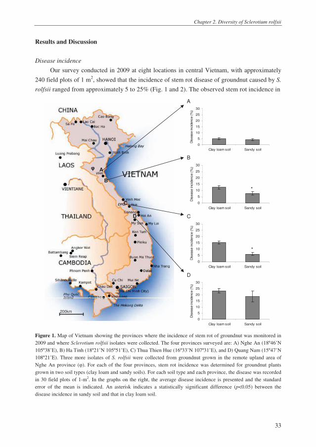
Our survey conducted in 2009 at eight locations in central Vietnam, with approximately
240 field plots of 1 m2, showed that the incidence of stem rot disease of groundnut caused by S.
rolfsii ranged from approximately 5 to 25% (Fig. 1 and 2). The observed stem rot incidence in
A
0
5
10
15
20
25
30
Clay loam soil Sandy soil
Dis
ease in
cid
ence (
%)
B
0
5
10
15
20
25
30
Clay loam soil Sandy soil
Dis
ease in
cid
ence (
%)
C
0
5
10
15
20
25
30
Clay loam soil Sandy soil
Dis
ease in
cid
ence (
%)
D
0
5
10
15
20
25
30
Clay loam soil Sandy soil
Dis
ease in
cid
ence (
%)
Figure 1. Map of Vietnam showing the provinces where the incidence of stem rot of groundnut was monitored in
2009 and where Sclerotium rolfsii isolates were collected. The four provinces surveyed are: A) Nghe An (18º46’N
105º38’E), B) Ha Tinh (18º21’N 105º51’E), C) Thua Thien Hue (16º33’N 107º31’E), and D) Quang Nam (15º47’N
108º21’E). Three more isolates of S. rolfsii were collected from groundnut grown in the remote upland area of
Nghe An province (φ). For each of the four provinces, stem rot incidence was determined for groundnut plants
grown in two soil types (clay loam and sandy soils). For each soil type and each province, the disease was recorded
in 30 field plots of 1-m2. In the graphs on the right, the average disease incidence is presented and the standard
error of the mean is indicated. An asterisk indicates a statistically significant difference (p<0.05) between the
disease incidence in sandy soil and that in clay loam soil.
*
200km200km
A
B
C
D
φ
*

Chapter 2. Diversity of Sclerotium rolfsii
34
central Vietnam is similar to that reported in Georgia (USA) from 1983 to 1985, i.e.
approximately 8.5% (Branch and Csinos 1987), but much higher than the disease incidence
reported in Texas (USA) in 1992 and 1993 (less than 1%) (Shim et al. 1998). On the other hand,
it was lower than in Ibaraki (Japan) where disease incidence ranged from 10 to 40% (Okabe and
Matsumoto 2000).
Sclerotia Sclerotia Sclerotia
Figure 2. A) Overview of the sampling location in Quang Nam province; B) effects of Sclerotium rolfsii on
emergence and growth of groundnut seedlings in the field, symptoms of stem rot of groundnut (C), tomato (D) and
taro (E). For groundnut, tomato and taro, the sclerotia on the infected plant tissues are indicated by an arrow.
Our field survey further indicated a gradient in stem rot disease incidence across central
Vietnam with an increase in disease incidence from the northern to the southern field sites (Fig.
1). This might be related to the effect of temperature on disease development (Punja 1985)
because along with the lower latitude, the average daily temperature increases from the North to
the South of Vietnam. Important to note also is that the field sites surveyed in the two southern
provinces (locations C and D, Fig. 1) were sites where tomato and taro were also cultivated.
Both crops are hosts of S. rolfsii (Fig. 2D and E) and may have contributed to the build-up of
pathogen inoculum. For two of the four provinces, i.e. locations B and C, the disease incidence
of groundnut plants cultivated in clay loam soil was significantly higher than in sandy soil (Fig.
1). This difference may be related in part to the higher organic content of the clay loam soil, a
characteristic that is known to support germination of sclerotia and subsequent hyphal growth
toward the host plant (Punja 1985).
A B
C D E

Chapter 2. Diversity of Sclerotium rolfsii
35
Genetic diversity of S. rolfsii
A total of 198 purified S. rolfsii isolates, obtained from more than 400 randomly collected
samples from different host plants (groundnut, tomato and taro) in the four provinces and in the
remote upland region, were subjected to ITS-rDNA sequence analysis. For 103 S. rolfsii
isolates, high quality forward and reverse sequences were obtained, whereas for the other 95
isolates discrepancies were observed in the assembly of the forward and reverse sequences. Re-
sequencing of the ITS-fragments of twelve selected isolates did not resolve this inconsistency,
which may suggest polymorphisms in different ITS copies in each of these isolates. ITS
polymorphism within a single isolate has been found for several fungal genera and was also
reported earlier for S. rolfsii by Okabe et al. (2001) and Okabe and Matsumoto (2003).
Phylogenetic analysis of the ITS-rDNA sequences of the reference isolates of S. rolfsii,
i.e. isolates for which ITS-rDNA sequences are present in the NCBI database (Table 1),
revealed three main groups designated ITS-groups 1-3 (Fig. 3). Most isolates (n=90) collected
from central Vietnam belong to ITS-group 1 and were identical to reference isolates of S. rolfsii
that were previously collected from groundnut, tobacco and sweet pepper (Fig. 3, Table 1). The
three isolates in ITS-group 2 were collected from the remote upland region of Nghe An
province (Fig. 3) and cluster with an S. rolfsii isolate collected from Ascocenda orchids in
Florida, USA (Cating et al. 2009). Out of the ten isolates in ITS-group 3, eight were
collected in Quang Nam province (Fig. 3). Collectively, these results suggest that, based on
ITS-rDNA sequences, the S. rolfsii population in groundnut fields in central Vietnam
appears relatively uniform. However, that ITS-rDNA sequencing does not give detailed insight
into the intraspecific diversity and that other molecular markers or traits should be considered.
Table 1. ITS-rDNA sequences of reference strains of Sclerotium rolfsii, S. delphinii and S. coffeicola used in the
phylogenetic analyses.
Accession
number
Host Origin Year of
isolation
Name Reference
GQ358518 Ascocentrum spp.,
Ascocenda spp.
South Florida, USA 2008 S. rolfsii Cating et al. 2009
DQ484060 unknown unknown unknown S. rolfsii Matheny et al.
2006
DQ484061 unknown unknown unknown S. rolfsii Matheny et al.
2006
DQ484062 unknown unknown unknown S. rolfsii Matheny et al.
2006
AB075307 Arachis hypogaea Georgia,USA 1991 S. rolfsii Okabe and
Matsumoto 2003
GU080230 Capsicum annuum Spain 2009 S. rolfsii Remesal et al. 2010
DQ059578 Nicotiana tabacum North Carolina, USA 2007 S. rolfsii Ristaino et al. 2007
AB075318 unknown Washington, USA 1991 S. delphinii Okabe and
Matsumoto 2003
AB075314 unknown Japan 1995 S. delphinii Okabe and
Matsumoto 2003
AB075312 unknown Japan 1992 S. delphinii Okabe and
Matsumoto 2003
AB075319 unknown Surinam 1919 S. coffeicola Okabe and
Matsumoto 2003

Chapter 2. Diversity of Sclerotium rolfsii
36
NAGC4T2
NAGC7T1
GQ358518 (S. rolfsii)
NAGC4T1
DQ484062 (S. rolfsii)
QNGS86T1
QNGC003T1
QNGC9T1
QNTS13T1
HTGS9T1
QNGS13T1
QNGC28T1
QNGC17T1
HUGS16-1T1
QNGC002-2T1
DQ484061 (S. rolfsii)
AB075318 (S. delphinii)
AB075312 (S. delphinii)
AB075314 (S. delphinii)
AB075319 (S. coffeicola)
99
95
99
80
93
93
99
0200400600800
Figure 3. Phylogeny of ITS-rDNA sequences of Sclerotium rolfsii isolates from groundnut fields in central
Vietnam and of reference strains of S. rolfsii, S. delphinii and S. coffeicola (underlined, see Table 1) using the
Unweighted pair-group method (UPGMA). Bootstrap values with 1,000 replications are indicated at the nodes of
the branches. The bar represents the evolutionary distances computed using the Maximum Composite Likelihood
method. The sequence codes of the isolates collected in this study refer to the province (first two letters), host crop
(third letter), soil type (fourth letter), and the isolate number (digits): NA- Nghe An province, HT- Ha Tinh
province, HU- Thua Thien Hue province, QN- Quang Nam province; G- groundnut, T- tomato, M- taro; C- clay
loam soil, S- sandy soil (see appendix).
For S. rolfsii, several other techniques and molecular markers have been used to assess
intraspecific diversity, especially random amplified polymorphic DNA (RAPD) analysis (Punja
and Sun 2001). Multiple of these tests aimed at characterizing the intraspecific diversity of the
isolates were performed here with an initial seven S. rolfsii isolates from the collection, i.e. 3
isolates from Vietnam and 4 reference strains from the culture collection. However, RAPD
analysis as well as rep-PCR and ERIC-PCR, performed according to methods previously
described for other fungi (McDonald et al. 2000), gave inconsistent/irreproducible results and
were not considered useful. Therefore, a range of other traits, including pathogenicity, growth
rate, sclerotial characteristics, mycelial compatibility and tebuconazole sensitivity, were
90 isolates (this study)
AB075307 (S. rolfsii)
DQ484060 (S. rolfsii)
DQ059578 (S. rolfsii)
GU080230 (S. rolfsii)
ITS-group 1
ITS-group 2
ITS-group 3

Chapter 2. Diversity of Sclerotium rolfsii
37
analysed to get more insight into the intraspecific diversity of the collection of S. rolfsii isolates
from central Vietnam.
Pathogenicity
Pathogenicity assays showed that S. rolfsii isolates obtained from groundnut (n=8),
tomato (n=5) and taro (n=5) from central Vietnam and representing ITS-groups 1-3 were all
pathogenic on groundnut (Table 2). The time for the first symptoms to appear ranged from 2-5
days and no statistically significant differences in disease incidence and severity were found
between the isolates (Table 2). These results confirmed pathogenicity of i) a subset of S. rolfsii
isolates collected from groundnut, ii) all S. rolfsii isolates collected from other crops (i.e. tomato
and taro), and iii) the three isolates collected from groundnut cultivated in the remote upland
region in Nghe An province (ITS-group 2).
Table 2. Pathogenicity of Sclerotium rolfsii isolates on groundnut. The isolates used for these assays are indicated
in the appendix with an asterisk.
Host crop ITS-group1
Incubation
period2 (day)
Disease
incidence3 (%)
Disease severity ±
SE4 (%)
Groundnut (N=5) 1, 3 2-3 100 92.7 ± 2.21
Tomato (N=5) 1, 3 4-5 100 86.3 ± 4.26
Taro (N=5) 1 3-4 100 95.3 ± 1.11
Groundnut (N=3) 2 3-4 100 75.0 ± 2.89
1 ITS-group as indicated in Fig. 3; 2 Days to first symptom appearance after fungal inoculation; 3Disease incidence
(%) = (diseased plants / total number of plants) × 100%; 4Disease severity: diseased plants were ranked on a 0-4
scale based on the symptoms with 0: no disease symptoms, 1: infection without visible outgrowth of the fungus,
2: infection with visible outgrowth, 3: partial wilting of the plant, and 4: complete wilting and plant death.
Disease severity (%) = [(1 × number of plants classified in scale 1) + (2 × number of plants classified in scale 2) +
(3 × number of plants classified in scale 3) + (4 × number of plants classified in scale 4)] × 100% / (4 × total
number of plants)].
Growth rate and sclerotial characteristics
S. rolfsii is notorious for its high growth rate in vitro (Akram et al. 2008; Punja 1985;
Punja and Damiani 1996). Indeed, the 103 Vietnamese isolates tested here exhibited a high
growth rate ranging from 0.28 to 0.79 mm h-1
(Table 3A, appendix). On average, isolates from
ITS-groups 1, 2 and 3 showed no significant differences in growth rate (Table 3B). On PDA
medium, the S. rolfsii isolates produced the typical silky-white mycelium and the brown or dark
brown sclerotia (Fig. 4) (Harlton et al. 1995). For the 103 isolates tested, considerable variation
was observed in the time to form sclerotia, their maturation time, and their number and size
(Table 3B, appendix). For example, the number of sclerotia formed per plate after 21 days of
incubation ranged from 79 to 1,080 and their size from 0.88 to 2.24 mm (Table 3A). These
results confirm and extend the data obtained in previous studies on S. rolfsii isolates from other
crops and geographic locations (Kokub et al. 2007; Prasad and Naik 2008; Punja and Damiani
1996; Xu et al. 2010). When comparing the sclerotial traits, the three isolates in ITS-group 2

Chapter 2. Diversity of Sclerotium rolfsii
38
were different from the isolates in ITS-groups 1 and 3 (Table 3B). ITS-group 2 isolates
produced substantially less sclerotia per plate and also the average maturation time and
sclerotial diameter were approximately two times higher than for isolates of ITS-groups 1 and 3.
These characteristics combined with the light brown colour of the sclerotia of these three ITS-
group 2 isolates were also reported for isolates classified morphologically or by LSU-
sequencing as S. rolfsii var. delphinii (Aycock 1966; Punja and Damiani 1996; Xu et al. 2010).
Table 3. Growth rate and sclerotial characteristics of Sclerotium rolfsii isolates obtained from groundnut fields in
central Vietnam. In panel A, averages, minimum (Min), and maximum (Max) values of each of these phenotypic
characteristics are given for 103 isolates; in panel B, averages ± the standard deviation (SD) of these phenotypic
characteristics are given for each of the three identified ITS-rDNA groups of S. rolfsii.
Sclerotia production and size A Hyphal growth rate
during 48 h
(mm h-1
) Days to form
Days to mature Number per plate Diameter
(mm)
Average 0.40 4.8 8.0 589 1.1
Min 0.28 4.0 7.0 79 0.9
Max 0.79 11.0 16.0 1080 2.2
SD 0.06 1.1 1.4 204 0.2
B
ITS-group
1 (N=90) 0.41 ± 0.06 4.7 ± 0.6 7.8 ± 0.7 595 ± 181 1.1 ± 0.1
2 (N=3) 0.36 ± 0.04 10.0 ± 1.7 14.3 ± 2.9 98 ± 16 2.0 ± 0.2
3 (N=10) 0.41 ± 0.04 4.2 ± 0.4 7.8 ± 1.2 689 ± 236 1.1 ± 0.2
Average 0.40 ± 0.06 4.8 ± 1.1 8.0 ± 1.4 589 ± 204 1.1 ± 0.2
Figure 4. Phenotypic characteristics of mycelium and sclerotia of three Sclerotium rolfsii isolates belonging to
ITS-rDNA groups 1 (panel A), 2 (panel B) and 3 (panel C). A 5-mm-diameter mycelial agar plug was inoculated in
the centre of a 9-cm-diameter PDA agar plate and incubated at 25°C. All isolates have the typical white-silky
mycelium with round or irregular, light- to dark-brown sclerotia.
A B C

Chapter 2. Diversity of Sclerotium rolfsii
39
Mycelial Compatibility
For several fungi, mycelial compatibility is used to get insight into the genetic relatedness
of isolates or to identify intraspecific variation within field populations of plant pathogens
(Kohn et al. 1991). When confronted with one another, a demarcation or barrage zone between
the fungal colonies places these isolates in a different mycelial compatibility group (MCG).
Isolates in the same MCG can be clonal (Kohn et al. 1991) or generally exhibit a higher degree
of genetic relatedness than isolates from different MCGs (Rayner 1991; Remesal et al. 2010). In
a study by Punja and Sun (2001) on S. rolfsii isolates from 13 countries and 36 different host
species, a total of 71 MCGs were found and there was no clear relationship between the original
host plant and MCG, except for S. rolfsii isolates from turfgrass. Within each of the three ITS-
groups identified in our study, a high variation in mycelial compatibility was observed (Table
4). For isolates of ITS-group 1, a total of 91 combinations were tested and only 6.6% of these
combinations showed a compatible interaction pointing to at least 9 MCGs. For ITS-group 2
and for ITS-group 3, we found two and four MCGs, respectively (Table 4). In contrast, studies
by Adandonon et al. (2005) on S. rolfsii from cowpea fields in Benin revealed four MCGs
among a total of 66 isolates. Similarly, Okabe and Matsumoto (2000) found four MCGs among
a total of 132 isolates from groundnut fields in Ibaraki (Japan) and concluded, based on RAPD
analysis, that many isolates were clonal. Based on MCG analysis, our results suggest that S.
rolfsii populations from groundnut fields in central Vietnam are more diverse. This may be
partly due to the fact that the fields sampled in our study are far apart, i.e. approximately 400
km from the northern fields sites (location A, Fig. 1) to the southern field sites (location D, Fig.
1) as compared to only 2.5 km in the study by Okabe and Matsumoto (2000).
Table 4. Mycelial compatibility between Sclerotium rolfsii isolates for each of the three identified ITS groups.
ITS group Number of
isolates
Number of
combinations
Compatible
combinations
%
compatibility
Number of
MCG*
1 14 91 6 6.6 9
2 3 3 1 33.3 2
3 9 36 4 11.1 6
* mycelial compatibility group.
Tebuconazole sensitivity
The results showed that the variation in sensitivity of S. rolfsii isolates from groundnut
fields in central Vietnam to tebuconazole is substantially less than reported earlier by Franke et al.
(1998) for the S. rolfsii population from Georgia, USA (Table 5, Fig. 5). The three isolates from
ITS-group 2, collected from the remote upland region, were significantly more sensitive to
tebuconazole than those from ITS-group 1 (Table 5). Although this may be due in part to a sample
size difference, it may also be linked to the fact that farmers in this remote area do not use
tebuconazole. In the other farmer fields surveyed, however, no records are kept on fungicide use,
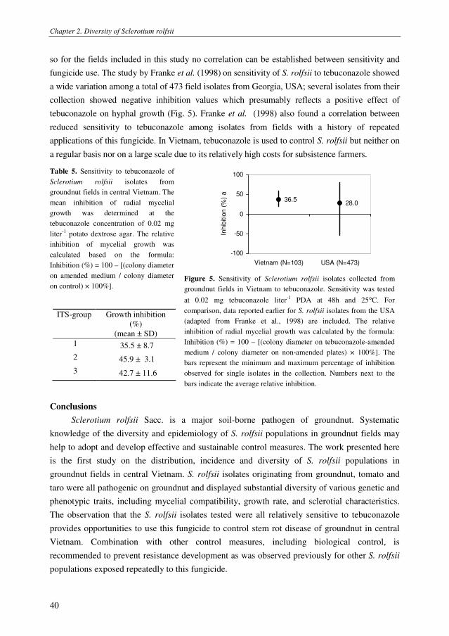
Chapter 2. Diversity of Sclerotium rolfsii
40
so for the fields included in this study no correlation can be established between sensitivity and
fungicide use. The study by Franke et al. (1998) on sensitivity of S. rolfsii to tebuconazole showed
a wide variation among a total of 473 field isolates from Georgia, USA; several isolates from their
collection showed negative inhibition values which presumably reflects a positive effect of
tebuconazole on hyphal growth (Fig. 5). Franke et al. (1998) also found a correlation between
reduced sensitivity to tebuconazole among isolates from fields with a history of repeated
applications of this fungicide. In Vietnam, tebuconazole is used to control S. rolfsii but neither on
a regular basis nor on a large scale due to its relatively high costs for subsistence farmers.
36.528.0
-100
-50
0
50
100
Vietnam (N=103) USA (N=473)
Inhib
itio
n (
%)
a
Table 5. Sensitivity to tebuconazole of
Sclerotium rolfsii isolates from
groundnut fields in central Vietnam. The
mean inhibition of radial mycelial
growth was determined at the
tebuconazole concentration of 0.02 mg
liter-1
potato dextrose agar. The relative
inhibition of mycelial growth was
calculated based on the formula:
Inhibition (%) = 100 – [(colony diameter
on amended medium / colony diameter
on control) × 100%].
ITS-group Growth inhibition
(%)
(mean ± SD)
1 35.5 ± 8.7
2 45.9 ± 3.1
3 42.7 ± 11.6
Figure 5. Sensitivity of Sclerotium rolfsii isolates collected from
groundnut fields in Vietnam to tebuconazole. Sensitivity was tested
at 0.02 mg tebuconazole liter-1
PDA at 48h and 25°C. For
comparison, data reported earlier for S. rolfsii isolates from the USA
(adapted from Franke et al., 1998) are included. The relative
inhibition of radial mycelial growth was calculated by the formula:
Inhibition (%) = 100 – [(colony diameter on tebuconazole-amended
medium / colony diameter on non-amended plates) × 100%]. The
bars represent the minimum and maximum percentage of inhibition
observed for single isolates in the collection. Numbers next to the
bars indicate the average relative inhibition.
Conclusions
Sclerotium rolfsii Sacc. is a major soil-borne pathogen of groundnut. Systematic
knowledge of the diversity and epidemiology of S. rolfsii populations in groundnut fields may
help to adopt and develop effective and sustainable control measures. The work presented here
is the first study on the distribution, incidence and diversity of S. rolfsii populations in
groundnut fields in central Vietnam. S. rolfsii isolates originating from groundnut, tomato and
taro were all pathogenic on groundnut and displayed substantial diversity of various genetic and
phenotypic traits, including mycelial compatibility, growth rate, and sclerotial characteristics.
The observation that the S. rolfsii isolates tested were all relatively sensitive to tebuconazole
provides opportunities to use this fungicide to control stem rot disease of groundnut in central
Vietnam. Combination with other control measures, including biological control, is
recommended to prevent resistance development as was observed previously for other S. rolfsii
populations exposed repeatedly to this fungicide.
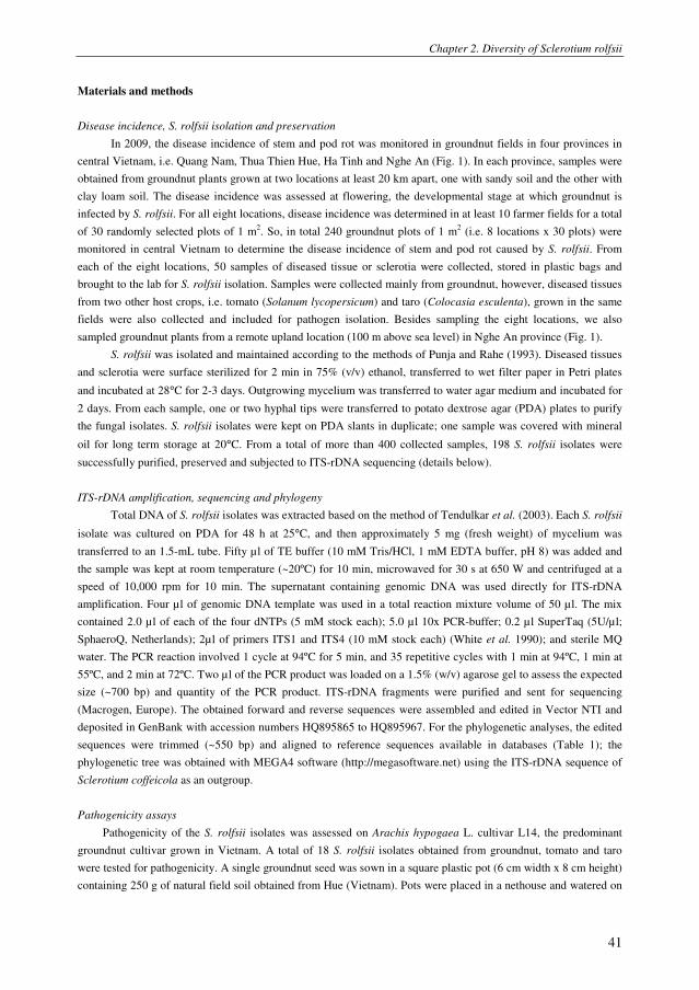
Chapter 2. Diversity of Sclerotium rolfsii
41
Materials and methods
Disease incidence, S. rolfsii isolation and preservation
In 2009, the disease incidence of stem and pod rot was monitored in groundnut fields in four provinces in
central Vietnam, i.e. Quang Nam, Thua Thien Hue, Ha Tinh and Nghe An (Fig. 1). In each province, samples were
obtained from groundnut plants grown at two locations at least 20 km apart, one with sandy soil and the other with
clay loam soil. The disease incidence was assessed at flowering, the developmental stage at which groundnut is
infected by S. rolfsii. For all eight locations, disease incidence was determined in at least 10 farmer fields for a total
of 30 randomly selected plots of 1 m2. So, in total 240 groundnut plots of 1 m
2 (i.e. 8 locations x 30 plots) were
monitored in central Vietnam to determine the disease incidence of stem and pod rot caused by S. rolfsii. From
each of the eight locations, 50 samples of diseased tissue or sclerotia were collected, stored in plastic bags and
brought to the lab for S. rolfsii isolation. Samples were collected mainly from groundnut, however, diseased tissues
from two other host crops, i.e. tomato (Solanum lycopersicum) and taro (Colocasia esculenta), grown in the same
fields were also collected and included for pathogen isolation. Besides sampling the eight locations, we also
sampled groundnut plants from a remote upland location (100 m above sea level) in Nghe An province (Fig. 1).
S. rolfsii was isolated and maintained according to the methods of Punja and Rahe (1993). Diseased tissues
and sclerotia were surface sterilized for 2 min in 75% (v/v) ethanol, transferred to wet filter paper in Petri plates
and incubated at 28°C for 2-3 days. Outgrowing mycelium was transferred to water agar medium and incubated for
2 days. From each sample, one or two hyphal tips were transferred to potato dextrose agar (PDA) plates to purify
the fungal isolates. S. rolfsii isolates were kept on PDA slants in duplicate; one sample was covered with mineral
oil for long term storage at 20°C. From a total of more than 400 collected samples, 198 S. rolfsii isolates were
successfully purified, preserved and subjected to ITS-rDNA sequencing (details below).
ITS-rDNA amplification, sequencing and phylogeny
Total DNA of S. rolfsii isolates was extracted based on the method of Tendulkar et al. (2003). Each S. rolfsii
isolate was cultured on PDA for 48 h at 25°C, and then approximately 5 mg (fresh weight) of mycelium was
transferred to an 1.5-mL tube. Fifty µl of TE buffer (10 mM Tris/HCl, 1 mM EDTA buffer, pH 8) was added and
the sample was kept at room temperature (~20ºC) for 10 min, microwaved for 30 s at 650 W and centrifuged at a
speed of 10,000 rpm for 10 min. The supernatant containing genomic DNA was used directly for ITS-rDNA
amplification. Four µl of genomic DNA template was used in a total reaction mixture volume of 50 µl. The mix
contained 2.0 µl of each of the four dNTPs (5 mM stock each); 5.0 µl 10x PCR-buffer; 0.2 µl SuperTaq (5U/µl;
SphaeroQ, Netherlands); 2µl of primers ITS1 and ITS4 (10 mM stock each) (White et al. 1990); and sterile MQ
water. The PCR reaction involved 1 cycle at 94ºC for 5 min, and 35 repetitive cycles with 1 min at 94ºC, 1 min at
55ºC, and 2 min at 72ºC. Two µl of the PCR product was loaded on a 1.5% (w/v) agarose gel to assess the expected
size (~700 bp) and quantity of the PCR product. ITS-rDNA fragments were purified and sent for sequencing
(Macrogen, Europe). The obtained forward and reverse sequences were assembled and edited in Vector NTI and
deposited in GenBank with accession numbers HQ895865 to HQ895967. For the phylogenetic analyses, the edited
sequences were trimmed (~550 bp) and aligned to reference sequences available in databases (Table 1); the
phylogenetic tree was obtained with MEGA4 software (http://megasoftware.net) using the ITS-rDNA sequence of
Sclerotium coffeicola as an outgroup.
Pathogenicity assays
Pathogenicity of the S. rolfsii isolates was assessed on Arachis hypogaea L. cultivar L14, the predominant
groundnut cultivar grown in Vietnam. A total of 18 S. rolfsii isolates obtained from groundnut, tomato and taro
were tested for pathogenicity. A single groundnut seed was sown in a square plastic pot (6 cm width x 8 cm height)
containing 250 g of natural field soil obtained from Hue (Vietnam). Pots were placed in a nethouse and watered on

Chapter 2. Diversity of Sclerotium rolfsii
42
a need-to-need basis. One week after sowing, a mycelial plug (1-cm-diameter) of a 3-day-old S. rolfsii PDA plate
culture was placed at the stembase. Disease incidence and severity were determined 2 weeks after fungal
inoculation. Disease incidence refers to the percentage of infected plants and disease severity was rated on a scale
from 0 – 4 (Fig. 6), with 0: no disease symptoms, 1: infection without visible fungal outgrowth, 2: infection with
visible fungal outgrowth, 3: partial wilting of the plant, and 4: complete wilting and plant death. For each S. rolfsii
isolate, three trays of five pots each were used.
0 1 2 3 4
Figure 6. Disease scales of groundnut infected by Sclerotium rolfsii; 0: no disease symptoms, 1: infection without
visible outgrowth of the fungus, 2: infection with visible outgrowth, 3: partial wilting of the plant, and 4: complete
wilting and plant death.
Phenotypic characterization
S. rolfsii isolates were cultured on PDA plates at 25°C. Per isolate, two 25-mL PDA agar plate (9-cm-
diameter) were inoculated in the centre with a mycelial plug (5-mm-diameter) and radial mycelial growth of the
fungal colony was determined 48h after incubation. For each plate, the number of sclerotia was determined 21 days
after incubation and the sclerotial diameter was determined for 30 randomly selected sclerotia.
A total of 26 S. rolfsii isolates were tested for mycelial compatibility. Multiple combinations of two isolates
of S. rolfsii were inoculated on PDA plates and incubated at 25°C for 2 weeks. Mycelial compatibility was assessed
by the method of Punja and Grogan (1983). If there was a distinct barrage zone at the contact area between two
isolates, these isolates were considered to belong to a different mycelial compatibility group (MCG).
The ergosterol biosynthesis inhibitor tebuconazole is a fungicide widely used to control S. rolfsii. At a
concentration of 0.02 mg liter-1
of PDA, tebuconazole reduced hyphal growth rate of S. rolfsii by approximately
28% (Franke et al. 1998). In our study, S. rolfsii isolates were cultured in duplicate on PDA amended with
tebuconazole (Sigma-Aldrich) to final concentrations of 0, 0.02 and 0.2 mg liter-1
. Hyphal growth was assessed 48
h after incubation at 25°C. Sensitivity of S. rolfsii to tebuconazole was calculated with the formula used by Franke
et al. (1998): Inhibition (%) = 100 – [(colony diameter on amended medium ÷ colony diameter on control medium)
× 100].
Acknowledgements
We would like to thank Le Thi Ngoc Thuy and Le Thi Lam Giang for their help during the field studies in
Vietnam. Many thanks also to all of farmers, who made their fields available for this survey. We also would like to
thank Francine Govers for critically reading this manuscript and valuable suggestions.

Chapter 2. Diversity of Sclerotium rolfsii
43
References
Adandonon, A., Aveling, T. A. S., van der Merwe, N. A., and Sanders, G. 2005. Genetic variation among
Sclerotium isolates from Benin and South Africa, determined using mycelial compatibility and ITS rDNA
sequence data. Australasian Plant Pathology 34, 19-25.
Akram, A., Iqbal, S. H. M., Qureshi, R. A., and Rauf, C. A. 2008. Variability among isolates of Sclerotium rolfsii
associated with collar rot disease of chickpea in Pakistan. Pakistan Journal of Botany 40, 453-460.
Aycock, R. 1966. Stem rot and other diseases caused by Sclerotium rolfsii or the status of Rolfs' fungus after 70
years, vol. North Carolina State Univ., Raleigh.
Besler, B. A., Grichar, W. J., Starr, J. A., Senseman, S. A., Lemon, R. G., and Jaks, A.J. 2006. Effects of peanut
row pattern, cultivar, and fungicides on control of Southern stem rot, early leaf spot, and rust. Peanut
Science 33, 1-6.
Branch, W. D., and Brenneman, T. B. 1996. Pod yield and stem rot evaluation of peanut cultivars treated with
tebuconazole. Agronomy Journal 88, 933-936.
Branch, W. D., and Brenneman, T. B. 1999. Stem rot disease evaluation of mass-selected peanut populations. Crop
Protection 18, 127-130.
Branch, W. D., and Brenneman, T. B. 2009. Field evaluation for the combination of white mould and tomato
spotted wilt disease resistance among peanut genotypes. Crop Protection 28, 595-598.
Branch, W. D., and A. S. Csinos. 1987. Evaluation of peanut cultivars for resistance to field infection by
Sclerotium rolfsii. Plant Disease 71, 268-270.
Brenneman, T. B., and Murphy, A. P. 1991. Activity of tebuconazole on Cercosporidium personatum, a foliar
pathogen of peanut. Plant Disease 75, 699-703.
Brewster, V. 2001. Southern blight, Southern stem blight, White mold. APSnet. [Online.]
Brown, S. L. 2009. Peanut Insects: Ecology and Control. Encyclopedia of Pest Management:1 - 10.
Cating, R. A., Palmateer, A. J., and McMillan, J. R. T. 2009. First report of Sclerotium rolfsii on Ascocentrum and
Ascocenda Orchids in Florida. Plant Disease 93, 963.
Franke, M. D., Brenneman, T. B., Stevenson, K. L., and Padgett, G. B. 1998. Sensitivity of isolates of Sclerotium
rolfsii from peanut in Georgia to selected fungicides. Plant Disease 82, 578-583.
Hammons, R. O. 1994. The origin and history of the groundnut, p. 24-42. In: Smartt, J. (ed.), The Groundnut crop:
A scientific basis for improvement Chapman & Hall, London.
Harlton, C. E., Levesque, C. A., and Punja, Z. K.. 1995. Genetic diversity in Sclerotium (Athelia) rolfsii and related
species. Phytopathology 85, 1269-1281.
Kohn, L. M., Stasovski, E., Carbone, I., Royer, J., and Anderson, J. B. 1991. Mycelial incompatibility and
molecular markers identify genetic variability in field populations of Sclerotinia sclerotiorum.
Phytopathology 81, 480-485.
Kokub, D., Azam, F., Hassan, A., Ansar, M., Asad, M. J., Khanumand, A. 2007. Comparative growth,
morphological and molecular characterization of indigenous Sclerotium rolfsii strains isolated from different
locations of Pakistan. Pakistan Journal of Botany 39, 1849-1866.
Lakpale, N., Khare, N., and Thrimurty, V. S. 2007. Suppression of Sclerotium rolfsii Sacc.: an intergrated
approach. Soils and Crops 17, 241-245.
Le, C. N. 2004. Study wilt diseases on groundnut and some methods to control them in Thua Thien Hue province.
National Journal of Plant Protection 1, 9-15.
Matheny, P. B., Curtis, J. M., Hofstetter, V., Aime, M. C., Moncalvo, J. M., Ge, Z. W., Yang, Z. L., Slot, J. C.,
Ammirati, J. F., Baroni, T. J., Bougher, N. L., Hughes, K. W., Lodge, D. J., Kerrigan, R. W., Seidl, M. T.,
Aanen, D. K., DeNitis, M., Daniele, G. M., Desjardin, D. E., Kropp, B. R., Norvell, L. L., Parker, A.,

Chapter 2. Diversity of Sclerotium rolfsii
44
Vellinga, E. C., Vilgalys, R., and Hibbett, D. S. 2006. Major clades of Agaricales: a multilocus
phylogenetic overview. Mycologia 98, 982-995.
McDonald, J. G., Wong, E., and White, G. P. 2000. Differentiation of Tilletia species by rep-PCR genomic
fingerprinting. Plant Disease 84, 1121-1125.
Nguyen, T. N., Tran, V. M., Nguyen, T. T., and Le, C. N. 2004. Research on groundnut diseases in Quang Binh
province. National Agriculture and Rural Development 17, 337-342.
Okabe, I., Arakawa, M., and Matsumoto, N. 2001. ITS polymorphism within a single strain of Sclerotium rolfsii.
Mycoscience 42, 107-113.
Okabe, I., and Matsumoto, N. 2000. Population structure of Sclerotium rolfsii in peanut fields. Mycoscience 41,
145-148.
Okabe, I., and Matsumoto, N. 2003. Phylogenetic relationship of Sclerotium rolfsii (teleomorph Athelia rolfsii) and
S. delphinii based on ITS sequences. Mycological Research 107, 164-168.
Prasad, R. D., and Naik, M. K. 2008. Advances in plant diseases caused by Sclerotium rolfsii and their
management, p. 89-127. In: Naik and Rani, D. (eds.). Advances in Soil Borne Plant Diseases. New India
Publishing Agency, New Delhi.
Punja, Z. K. 1985. The biology, ecology, and control of Sclerotium rolfsii. Annual Review of Phytopathology 23,
97-127.
Punja, Z. K., and Damiani, A. 1996. Comparative growth, morphology, and physiology of three Sclerotium species.
Mycologia 88, 694-706.
Punja, Z. K., and Grogan, R. G. 1983. Hyphal interactions and antagonism among field isolates and single-
basidiospore strains of Athelia (Sclerotium) rolfsii. Phytopathology 73, 1279-1284.
Punja, Z. K., and Rahe, J. E. 1993. Sclerotium. In: Singleton, L. L., Mihail, J. D., and Rush, C. M. (eds.), Methods
for research on soilborne phytopathologenic fungi. The American Phytopathological Society, St. Paul,
Minnesota.
Punja, Z. K., and Sun, L. J. 2001. Genetic diversity among mycelial compatability groups of Sclerotium rolfsii
(teleomorph Athelia rolfsii) and S. delphinii. Mycological Research 105, 537-546.
Rayner, A. D. M. 1991. The challenge of the individualistic mycelium. Mycologia 83, 48-71.
Remesal, E., Lucena, C., Azpilicueta, A., Landa, B. B., and Navas-Cortes, J. A. 2010. First report of Southern
blight of pepper caused by Sclerotium rolfsii in Southern Spain. Plant Disease 94, 280-280.
Ristaino, J. B., Johnson, A., Blanco-Meneses, M., and Liu, B. 2007. Identification of the tobacco blue mold
pathogen, Peronospora tabacina, by polymerase chain reaction. Plant Disease 91, 685-691.
Savage, G. P., and Keenan, J. I. 1994. The composition and nutritive value of groudnut kernels, p. 173-213. In:
Smartt, J. (ed.). The Groundnut crop: A scientific basis for improvement Chapman & Hall, London.
Scinos, A. S. 1989. Targeting fungicides for control of Southern stem rot on peanut. Plant Disease 73, 723-726.
Shew, B. B., and Waliyar, F. 2005. Peanut diseases: ecology and control. Page 1450. In: Encyclopedia of Pest
Management, Vol. 33. Taylor & Francis, London, UK.
Shim, M. Y., Starr, J. L., Keller, N. P., Woodard, K. E., and Lee, T. A. 1998. Distribution of isolates of Sclerotium
rolfsii tolerant to pentachloronitrobenzene in Texas peanut fields. Plant Disease 82:103-106.
Tendulkar, S. R., Gupta, A., and Chattoo, B. B. 2003. A simple protocol for isolation of fungal DNA.
Biotechnology Letters 25, 1941-1944.
Wadsworth, D. F., and Melouk, H. A. 1984. Tolerance of Sclerotium rolfsii to pentachloronitrobenzene.
Phytopathology 74, 634-634.
White, T. J., Bruns, T., Lee, S. and Taylor, J. 1990. Amplification and direct sequencing of fungal ribosomal RNA
genes for phylogenetics. In: Innis, M. A., Gelfand, D. H., Sminsky, J. J., and White, T. J., (eds.), PCR
Protocols: A guide to methods and applications. Academic press, Inc., New York.

Chapter 2. Diversity of Sclerotium rolfsii
45
Woodward, J. E., Brenneman, T. B., Kemerait, R. C., Smith, N. B., Culbreath, A. K., and Stevenson, K. L. 2008.
Use of resistant cultivars and reduced fungicide programs to manage peanut diseases in irrigated and
nonirrigated fields. Plant disease 92, 896-902.
Worrall, J. J. 1997. Somatic incompatibility in basidiomycetes. Mycologia 89, 24-36.
Xu, Z. H., Harrington, T. C., Gleason, M. L., and Batzer, J. C. 2010. Phylogenetic placement of plant pathogenic
Sclerotium species among teleomorph genera. Mycologia 102, 337-346.
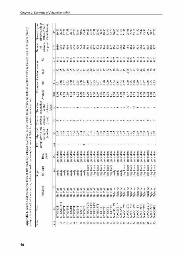
Chapter 2. Diversity of Sclerotium rolfsii
46
Ap
pen
dix
1.
Gen
eti
c an
d p
henoty
pic
tra
its
of
103
ran
do
mly
sel
ecte
d S
cler
oti
um
rolf
sii
isola
tes
fro
m g
round
nut
fiel
ds
in c
entr
al V
ietn
am
. Is
ola
tes
use
d i
n t
he p
athogenic
ity
assa
ys
are
ind
icat
ed w
ith a
n a
steri
sk;
iso
late
s fr
om
the
rem
ote
up
land a
rea
of
Nghe
An p
rovin
ce a
re u
nder
lined
.
Ori
gin
D
iam
eter
of
scle
roti
a (m
m)
Ord
er
Cod
e
Pro
vin
ce
Soil
type
Host
pla
nt
ITS
gro
up
Myce
lial
gro
wth
rat
e
duri
ng 4
8 h
(mm
/h)
Tim
e to
pro
duce
scle
roti
a
(day
s)
Tim
e fo
r
mat
ura
tio
n
of
the
scle
roti
a
(day
s)
Aver
age
min
m
ax
SD
Num
ber
of
scle
roti
a
per
pla
te
Sen
siti
vit
y t
o
tebuco
nazo
le a
t
0.0
2m
g/l
iter
-1
(%in
hib
itio
n)
(1)
(2)
(3)
(4)
(5)
(6)
(7)
(8)
(9)
(10
) (1
1)
(12)
(13
) (1
5)
(15
)
1
HT
GS
12T
1
Ha T
inh
sand
y
gro
und
nu
t
1
0.4
3
5
8
1.0
6
0.7
1
1.3
2
0.1
6
108
0
19
.80
2
HT
GS
13
-2T
1
Ha T
inh
sand
y
gro
und
nu
t
1
0.3
6
5
8
0.8
9
0.5
8
1.3
5
0.1
9
618
26
.26
3
HT
GS
17T
1
Ha T
inh
sand
y
gro
und
nu
t
1
0.3
2
6
9
1.2
9
1.0
2
1.5
7
0.1
6
278
22
.77
4
HT
GS
18T
1
Ha T
inh
sand
y
gro
und
nu
t
1
0.4
2
4
7
0.9
9
0.7
2
1.2
6
0.1
3
616
37
.58
5
HT
GS
20T
1
Ha T
inh
sand
y
gro
und
nu
t
1
0.3
7
5
8
1.1
4
0.8
0
1.6
5
0.2
0
508
28
.19
6
HT
GS
23T
1
Ha T
inh
sand
y
gro
und
nu
t
1
0.3
6
4
8
0.9
3
0.6
7
1.1
7
0.1
2
658
47
.70
7
HT
GS
24T
1
Ha T
inh
sand
y
gro
und
nu
t
1
0.3
9
5
8
1.2
2
0.7
8
1.4
3
0.1
6
326
38
.04
8
HT
GS
6T
1
Ha T
inh
sand
y
gro
und
nu
t
1
0.3
4
5
8
1.0
0
0.6
8
1.3
7
0.1
4
724
50
.07
9
HT
GS
9T
1
Ha T
inh
sand
y
gro
und
nu
t
3
0.3
5
5
8
1.0
2
0.7
2
1.3
7
0.1
9
584
45
.40
10
H
TG
C10-2
T1
Ha T
inh
clay l
oam
gro
und
nu
t
1
0.4
1
4
7
1.2
0
0.8
6
1.5
3
0.1
8
568
36
.29
11
H
TG
C10-1
T1
Ha T
inh
clay l
oam
gro
und
nu
t
1
0.3
6
5
9
1.0
7
0.7
7
1.4
0
0.1
9
516
24
.25
12
H
TG
C13-1
T1
Ha T
inh
clay l
oam
gro
und
nu
t
1
0.4
1
5
8
1.1
0
0.8
5
1.4
7
0.1
6
452
42
.31
13
H
TG
C13T
1
Ha T
inh
clay l
oam
gro
und
nu
t
1
0.3
3
5
8
1.0
9
0.6
7
1.4
3
0.1
9
472
28
.09
14
H
TG
C3T
1
Ha T
inh
clay l
oam
gro
und
nu
t
1
0.4
0
4
7
1.0
6
0.8
1
1.2
9
0.1
2
704
38
.34
15
H
TG
C5T
1
Ha T
inh
clay l
oam
gro
und
nu
t
1
0.3
8
5
8
1.2
9
0.9
0
1.5
7
0.1
7
346
19
.85
16
H
TG
C9T
1
Ha T
inh
clay l
oam
gro
und
nu
t
1
0.4
5
4
7
1.2
4
0.9
0
1.7
2
0.1
8
356
53
.90
17
H
TG
C9T
2
Ha T
inh
clay l
oam
gro
und
nu
t
1
0.4
4
4
7
1.2
1
0.9
9
1.5
5
0.1
2
422
42
.70
18
N
AG
S2-1
T1
Ng
he
An
sand
y
gro
und
nu
t
1
0.4
2
5
8
1.0
9
0.6
4
1.5
0
0.2
5
576
30
.06
19
N
AG
S1T
1 *
N
ghe
An
sand
y
gro
und
nu
t
1
0.4
0
4
8
1.0
4
0.8
4
1.2
9
0.1
3
472
31
.97
20
N
AG
S5T
1
Ng
he
An
sand
y
gro
und
nu
t
1
0.4
2
4
7
1.0
9
0.8
4
1.7
7
0.1
8
688
41
.64
21
N
AG
C10
T1
Ng
he
An
clay l
oam
gro
und
nu
t
1
0.4
2
5
8
0.9
8
0.6
7
1.3
5
0.1
9
574
51
.92
22
N
AG
C12
T1
Ng
he
An
clay l
oam
gro
und
nu
t
1
0.3
5
6
10
1.4
7
1.0
0
2.0
5
0.2
7
184
25
.61
23
N
AG
C15
T1
Ng
he
An
clay l
oam
gro
und
nu
t
1
0.4
1
5
8
1.1
4
0.7
5
1.6
9
0.2
2
464
35
.90
24
N
AG
C18
T1
Ng
he
An
clay l
oam
gro
und
nu
t
1
0.3
5
6
10
1.2
3
0.9
9
1.6
1
0.1
5
282
25
.13
25
N
AG
C1T
1
Ng
he
An
clay l
oam
gro
und
nu
t
1
0.3
9
5
8
1.1
2
0.7
9
1.4
0
0.1
6
538
34
.52
26
N
AG
C21
T1
Ng
he
An
clay l
oam
gro
und
nu
t
1
0.3
8
4
7
1.0
9
0.8
7
1.4
4
0.1
4
564
39
.42
27
N
AG
C22
T1
Ng
he
An
clay l
oam
gro
und
nu
t
1
0.3
5
5
8
1.0
8
0.8
7
1.4
0
0.1
2
542
43
.80
28
N
AG
C2
-2T
1
Ng
he
An
clay l
oam
gro
und
nu
t
1
0.3
5
5
7
1.0
2
0.7
6
1.4
5
0.1
3
605
29
.17
29
N
AG
C27
T1
Ng
he
An
clay l
oam
gro
und
nu
t
1
0.4
1
5
8
1.0
0
0.7
5
1.2
0
0.1
3
880
38
.23
30
N
AG
C9T
1
Ng
he
An
clay l
oam
gro
und
nu
t
1
0.3
7
6
9
1.2
3
0.7
5
1.5
9
0.2
0
337
27
.75
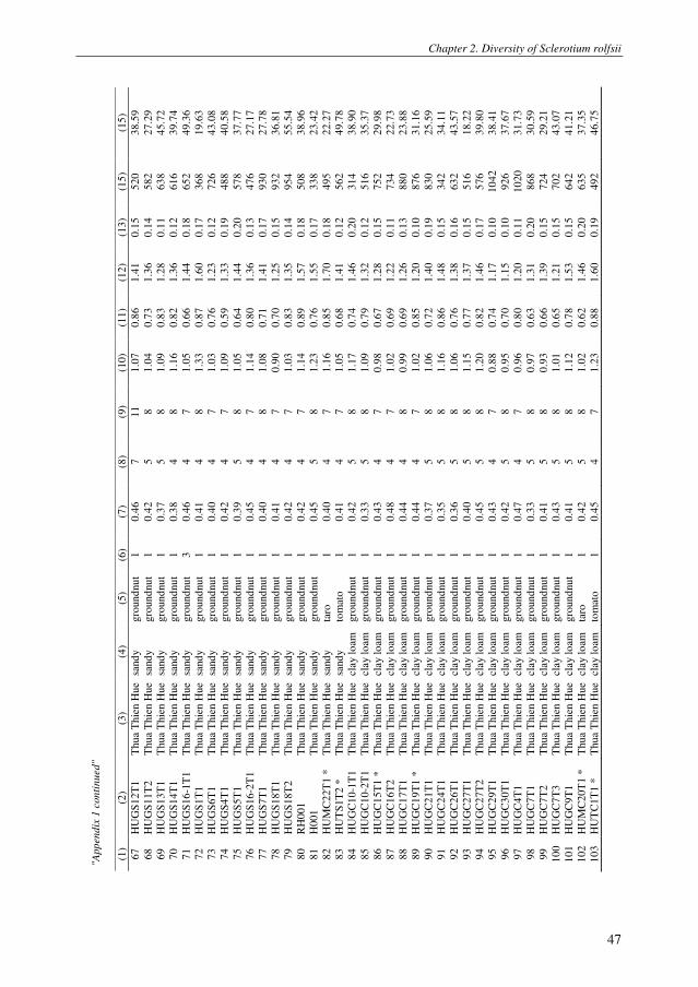
Chapter 2. Diversity of Sclerotium rolfsii
47
"App
en
dix
1 c
on
tin
ued"
(1)
(2)
(3)
(4)
(5)
(6)
(7)
(8)
(9)
(10
) (1
1)
(12
) (1
3)
(15
) (1
5)
67
H
UG
S1
2T
1
Thu
a T
hie
n H
ue
sand
y
gro
un
dnu
t 1
0
.46
7
1
1
1.0
7
0.8
6
1.4
1
0.1
5
52
0
38
.59
68
H
UG
S1
1T
2
Thu
a T
hie
n H
ue
sand
y
gro
un
dnu
t
1
0.4
2
5
8
1.0
4
0.7
3
1.3
6
0.1
4
58
2
27
.29
69
H
UG
S1
3T
1
Thu
a T
hie
n H
ue
sand
y
gro
un
dnu
t
1
0.3
7
5
8
1.0
9
0.8
3
1.2
8
0.1
1
63
8
45
.72
70
H
UG
S1
4T
1
Thu
a T
hie
n H
ue
sand
y
gro
un
dnu
t
1
0.3
8
4
8
1.1
6
0.8
2
1.3
6
0.1
2
61
6
39
.74
71
H
UG
S1
6-1
T1
T
hu
a T
hie
n H
ue
sand
y
gro
un
dnu
t
3
0.4
6
4
7
1.0
5
0.6
6
1.4
4
0.1
8
65
2
49
.36
72
H
UG
S1
T1
T
hu
a T
hie
n H
ue
sand
y
gro
un
dnu
t
1
0.4
1
4
8
1.3
3
0.8
7
1.6
0
0.1
7
36
8
19
.63
73
H
UG
S6
T1
T
hu
a T
hie
n H
ue
sand
y
gro
un
dnu
t
1
0.4
0
4
7
1.0
3
0.7
6
1.2
3
0.1
2
72
6
43
.08
74
H
UG
S4
T1
T
hu
a T
hie
n H
ue
sand
y
gro
un
dnu
t
1
0.4
2
4
7
1.0
9
0.5
9
1.3
3
0.1
9
48
8
40
.58
75
H
UG
S5
T1
T
hu
a T
hie
n H
ue
sand
y
gro
un
dnu
t
1
0.3
9
5
8
1.0
5
0.6
4
1.4
4
0.2
0
57
8
37
.77
76
H
UG
S1
6-2
T1
T
hu
a T
hie
n H
ue
sand
y
gro
un
dnu
t
1
0.4
5
4
7
1.1
4
0.8
0
1.3
6
0.1
3
47
6
27
.17
77
H
UG
S7
T1
T
hu
a T
hie
n H
ue
sand
y
gro
un
dnu
t
1
0.4
0
4
8
1.0
8
0.7
1
1.4
1
0.1
7
93
0
27
.78
78
H
UG
S1
8T
1
Thu
a T
hie
n H
ue
sand
y
gro
un
dnu
t
1
0.4
1
4
7
0.9
0
0.7
0
1.2
5
0.1
5
93
2
36
.81
79
H
UG
S1
8T
2
Thu
a T
hie
n H
ue
sand
y
gro
un
dnu
t
1
0.4
2
4
7
1.0
3
0.8
3
1.3
5
0.1
4
95
4
55
.54
80
R
H0
01
T
hu
a T
hie
n H
ue
sand
y
gro
un
dnu
t
1
0.4
2
4
7
1.1
4
0.8
9
1.5
7
0.1
8
50
8
38
.96
81
H
00
1
Thu
a T
hie
n H
ue
sand
y
gro
un
dnu
t
1
0.4
5
5
8
1.2
3
0.7
6
1.5
5
0.1
7
33
8
23
.42
82
H
UM
C2
2T
1 *
T
hu
a T
hie
n H
ue
sand
y
taro
1
0
.40
4
7
1
.16
0
.85
1
.70
0
.18
4
95
2
2.2
7
83
H
UT
S1
T2
*
Thu
a T
hie
n H
ue
sand
y
tom
ato
1
0
.41
4
7
1
.05
0
.68
1
.41
0
.12
5
62
4
9.7
8
84
H
UG
C1
0-1
T1
T
hu
a T
hie
n H
ue
clay l
oam
gro
un
dnu
t
1
0.4
2
5
8
1.1
7
0.7
4
1.4
6
0.2
0
31
4
38
.90
85
H
UG
C1
0-2
T1
T
hu
a T
hie
n H
ue
clay l
oam
gro
un
dnu
t
1
0.3
3
5
8
1.0
9
0.7
9
1.3
2
0.1
2
51
6
35
.37
86
H
UG
C1
5T
1 *
T
hu
a T
hie
n H
ue
clay l
oam
gro
un
dnu
t
1
0.4
3
4
7
0.9
8
0.6
7
1.2
8
0.1
5
75
2
29
.98
87
H
UG
C1
6T
2
Thu
a T
hie
n H
ue
clay l
oam
gro
un
dnu
t
1
0.4
8
4
7
1.0
2
0.6
9
1.2
2
0.1
1
73
4
22
.73
88
H
UG
C1
7T
1
Thu
a T
hie
n H
ue
clay l
oam
gro
un
dnu
t
1
0.4
4
4
8
0.9
9
0.6
9
1.2
6
0.1
3
88
0
23
.88
89
H
UG
C1
9T
1 *
T
hu
a T
hie
n H
ue
clay l
oam
gro
un
dnu
t
1
0.4
4
4
7
1.0
2
0.8
5
1.2
0
0.1
0
87
6
31
.16
90
H
UG
C2
1T
1
Thu
a T
hie
n H
ue
clay l
oam
gro
un
dnu
t
1
0.3
7
5
8
1.0
6
0.7
2
1.4
0
0.1
9
83
0
25
.59
91
H
UG
C2
4T
1
Thu
a T
hie
n H
ue
clay l
oam
gro
un
dnu
t
1
0.3
5
5
8
1.1
6
0.8
6
1.4
8
0.1
5
34
2
34
.11
92
H
UG
C2
6T
1
Thu
a T
hie
n H
ue
clay l
oam
gro
un
dnu
t
1
0.3
6
5
8
1.0
6
0.7
6
1.3
8
0.1
6
63
2
43
.57
93
H
UG
C2
7T
1
Thu
a T
hie
n H
ue
clay l
oam
gro
un
dnu
t
1
0.4
0
5
8
1.1
5
0.7
7
1.3
7
0.1
5
51
6
18
.22
94
H
UG
C2
7T
2
Thu
a T
hie
n H
ue
clay l
oam
gro
un
dnu
t
1
0.4
5
5
8
1.2
0
0.8
2
1.4
6
0.1
7
57
6
39
.80
95
H
UG
C2
9T
1
Thu
a T
hie
n H
ue
clay l
oam
gro
un
dnu
t
1
0.4
3
4
7
0.8
8
0.7
4
1.1
7
0.1
0
10
42
3
8.4
1
96
H
UG
C3
0T
1
Thu
a T
hie
n H
ue
clay l
oam
gro
un
dnu
t
1
0.4
2
5
8
0.9
5
0.7
0
1.1
5
0.1
0
92
6
37
.67
97
H
UG
C4
T1
T
hu
a T
hie
n H
ue
clay l
oam
gro
un
dnu
t
1
0.4
7
4
7
0.9
6
0.8
0
1.2
0
0.1
1
10
20
3
1.7
3
98
H
UG
C7
T1
T
hu
a T
hie
n H
ue
clay l
oam
gro
un
dnu
t
1
0.3
3
5
8
0.9
7
0.6
3
1.3
1
0.2
0
86
8
30
.59
99
H
UG
C7
T2
T
hu
a T
hie
n H
ue
clay l
oam
gro
un
dnu
t
1
0.4
1
5
8
0.9
3
0.6
6
1.3
9
0.1
5
72
4
29
.21
10
0
HU
GC
7T
3
Thu
a T
hie
n H
ue
clay l
oam
gro
un
dnu
t
1
0.4
3
5
8
1.0
1
0.6
5
1.2
1
0.1
5
70
2
43
.07
10
1
HU
GC
9T
1
Thu
a T
hie
n H
ue
clay l
oam
gro
un
dnu
t
1
0.4
1
5
8
1.1
2
0.7
8
1.5
3
0.1
5
64
2
41
.21
10
2
HU
MC
20
T1
*
Thu
a T
hie
n H
ue
clay l
oam
ta
ro
1
0.4
2
5
8
1.0
2
0.6
2
1.4
6
0.2
0
63
5
37
.35
10
3
HU
TC
1T
1 *
T
hu
a T
hie
n H
ue
clay l
oam
to
mat
o
1
0.4
5
4
7
1.2
3
0.8
8
1.6
0
0.1
9
49
2
46
.75

Chapter 2. Diversity of Sclerotium rolfsii
48
"App
en
dix
1 c
on
tin
ued"
(1)
(2)
(3)
(4)
(5)
(6)
(7)
(8)
(9)
(10
) (1
1)
(12
) (1
3)
(15
) (1
5)
67
H
UG
S1
2T
1
Thu
a T
hie
n H
ue
sand
y
gro
un
dnu
t 1
0
.46
7
1
1
1.0
7
0.8
6
1.4
1
0.1
5
52
0
38
.59
68
H
UG
S1
1T
2
Thu
a T
hie
n H
ue
sand
y
gro
un
dnu
t
1
0.4
2
5
8
1.0
4
0.7
3
1.3
6
0.1
4
58
2
27
.29
69
H
UG
S1
3T
1
Thu
a T
hie
n H
ue
sand
y
gro
un
dnu
t
1
0.3
7
5
8
1.0
9
0.8
3
1.2
8
0.1
1
63
8
45
.72
70
H
UG
S1
4T
1
Thu
a T
hie
n H
ue
sand
y
gro
un
dnu
t
1
0.3
8
4
8
1.1
6
0.8
2
1.3
6
0.1
2
61
6
39
.74
71
H
UG
S1
6-1
T1
T
hu
a T
hie
n H
ue
sand
y
gro
un
dnu
t
3
0.4
6
4
7
1.0
5
0.6
6
1.4
4
0.1
8
65
2
49
.36
72
H
UG
S1
T1
T
hu
a T
hie
n H
ue
sand
y
gro
un
dnu
t
1
0.4
1
4
8
1.3
3
0.8
7
1.6
0
0.1
7
36
8
19
.63
73
H
UG
S6
T1
T
hu
a T
hie
n H
ue
sand
y
gro
un
dnu
t
1
0.4
0
4
7
1.0
3
0.7
6
1.2
3
0.1
2
72
6
43
.08
74
H
UG
S4
T1
T
hu
a T
hie
n H
ue
sand
y
gro
un
dnu
t
1
0.4
2
4
7
1.0
9
0.5
9
1.3
3
0.1
9
48
8
40
.58
75
H
UG
S5
T1
T
hu
a T
hie
n H
ue
sand
y
gro
un
dnu
t
1
0.3
9
5
8
1.0
5
0.6
4
1.4
4
0.2
0
57
8
37
.77
76
H
UG
S1
6-2
T1
T
hu
a T
hie
n H
ue
sand
y
gro
un
dnu
t
1
0.4
5
4
7
1.1
4
0.8
0
1.3
6
0.1
3
47
6
27
.17
77
H
UG
S7
T1
T
hu
a T
hie
n H
ue
sand
y
gro
un
dnu
t
1
0.4
0
4
8
1.0
8
0.7
1
1.4
1
0.1
7
93
0
27
.78
78
H
UG
S1
8T
1
Thu
a T
hie
n H
ue
sand
y
gro
un
dnu
t
1
0.4
1
4
7
0.9
0
0.7
0
1.2
5
0.1
5
93
2
36
.81
79
H
UG
S1
8T
2
Thu
a T
hie
n H
ue
sand
y
gro
un
dnu
t
1
0.4
2
4
7
1.0
3
0.8
3
1.3
5
0.1
4
95
4
55
.54
80
R
H0
01
T
hu
a T
hie
n H
ue
sand
y
gro
un
dnu
t
1
0.4
2
4
7
1.1
4
0.8
9
1.5
7
0.1
8
50
8
38
.96
81
H
00
1
Thu
a T
hie
n H
ue
sand
y
gro
un
dnu
t
1
0.4
5
5
8
1.2
3
0.7
6
1.5
5
0.1
7
33
8
23
.42
82
H
UM
C2
2T
1 *
T
hu
a T
hie
n H
ue
sand
y
taro
1
0
.40
4
7
1
.16
0
.85
1
.70
0
.18
4
95
2
2.2
7
83
H
UT
S1
T2
*
Thu
a T
hie
n H
ue
sand
y
tom
ato
1
0
.41
4
7
1
.05
0
.68
1
.41
0
.12
5
62
4
9.7
8
84
H
UG
C1
0-1
T1
T
hu
a T
hie
n H
ue
clay l
oam
gro
un
dnu
t
1
0.4
2
5
8
1.1
7
0.7
4
1.4
6
0.2
0
31
4
38
.90
85
H
UG
C1
0-2
T1
T
hu
a T
hie
n H
ue
clay l
oam
gro
un
dnu
t
1
0.3
3
5
8
1.0
9
0.7
9
1.3
2
0.1
2
51
6
35
.37
86
H
UG
C1
5T
1 *
T
hu
a T
hie
n H
ue
clay l
oam
gro
un
dnu
t
1
0.4
3
4
7
0.9
8
0.6
7
1.2
8
0.1
5
75
2
29
.98
87
H
UG
C1
6T
2
Thu
a T
hie
n H
ue
clay l
oam
gro
un
dnu
t
1
0.4
8
4
7
1.0
2
0.6
9
1.2
2
0.1
1
73
4
22
.73
88
H
UG
C1
7T
1
Thu
a T
hie
n H
ue
clay l
oam
gro
un
dnu
t
1
0.4
4
4
8
0.9
9
0.6
9
1.2
6
0.1
3
88
0
23
.88
89
H
UG
C1
9T
1 *
T
hu
a T
hie
n H
ue
clay l
oam
gro
un
dnu
t
1
0.4
4
4
7
1.0
2
0.8
5
1.2
0
0.1
0
87
6
31
.16
90
H
UG
C2
1T
1
Thu
a T
hie
n H
ue
clay l
oam
gro
un
dnu
t
1
0.3
7
5
8
1.0
6
0.7
2
1.4
0
0.1
9
83
0
25
.59
91
H
UG
C2
4T
1
Thu
a T
hie
n H
ue
clay l
oam
gro
un
dnu
t
1
0.3
5
5
8
1.1
6
0.8
6
1.4
8
0.1
5
34
2
34
.11
92
H
UG
C2
6T
1
Thu
a T
hie
n H
ue
clay l
oam
gro
un
dnu
t
1
0.3
6
5
8
1.0
6
0.7
6
1.3
8
0.1
6
63
2
43
.57
93
H
UG
C2
7T
1
Thu
a T
hie
n H
ue
clay l
oam
gro
un
dnu
t
1
0.4
0
5
8
1.1
5
0.7
7
1.3
7
0.1
5
51
6
18
.22
94
H
UG
C2
7T
2
Thu
a T
hie
n H
ue
clay l
oam
gro
un
dnu
t
1
0.4
5
5
8
1.2
0
0.8
2
1.4
6
0.1
7
57
6
39
.80
95
H
UG
C2
9T
1
Thu
a T
hie
n H
ue
clay l
oam
gro
un
dnu
t
1
0.4
3
4
7
0.8
8
0.7
4
1.1
7
0.1
0
10
42
3
8.4
1
96
H
UG
C3
0T
1
Thu
a T
hie
n H
ue
clay l
oam
gro
un
dnu
t
1
0.4
2
5
8
0.9
5
0.7
0
1.1
5
0.1
0
92
6
37
.67
97
H
UG
C4
T1
T
hu
a T
hie
n H
ue
clay l
oam
gro
un
dnu
t
1
0.4
7
4
7
0.9
6
0.8
0
1.2
0
0.1
1
10
20
3
1.7
3
98
H
UG
C7
T1
T
hu
a T
hie
n H
ue
clay l
oam
gro
un
dnu
t
1
0.3
3
5
8
0.9
7
0.6
3
1.3
1
0.2
0
86
8
30
.59
99
H
UG
C7
T2
T
hu
a T
hie
n H
ue
clay l
oam
gro
un
dnu
t
1
0.4
1
5
8
0.9
3
0.6
6
1.3
9
0.1
5
72
4
29
.21
10
0
HU
GC
7T
3
Thu
a T
hie
n H
ue
clay l
oam
gro
un
dnu
t
1
0.4
3
5
8
1.0
1
0.6
5
1.2
1
0.1
5
70
2
43
.07
10
1
HU
GC
9T
1
Thu
a T
hie
n H
ue
clay l
oam
gro
un
dnu
t
1
0.4
1
5
8
1.1
2
0.7
8
1.5
3
0.1
5
64
2
41
.21
10
2
HU
MC
20
T1
*
Thu
a T
hie
n H
ue
clay l
oam
ta
ro
1
0.4
2
5
8
1.0
2
0.6
2
1.4
6
0.2
0
63
5
37
.35
10
3
HU
TC
1T
1 *
T
hu
a T
hie
n H
ue
clay l
oam
to
mat
o
1
0.4
5
4
7
1.2
3
0.8
8
1.6
0
0.1
9
49
2
46
.75
Chapter 3. Involvement of phenazines and lipopeptides in suppression of stem rot
Chapter 3
Involvement of phenazine antibiotics and lipopeptide
surfactants in suppression of stem rot disease of
groundnut by Pseudomonas species
Le, C.N., Kruijt, M. and Raaijmakers, J.M.
Journal of Applied Microbiology (accepted with minor revisions)

Chapter 3. Involvement of phenazines and lipopeptides in suppression of stem rot
50

Chapter 3. Involvement of phenazines and lipopeptides in suppression of stem rot
51
Involvement of phenazine antibiotics and lipopeptide surfactants in
suppression of stem rot disease of groundnut by Pseudomonas species
Le, C.N.1, 2
, Kruijt, M.1,3
and Raaijmakers, J.M.1
1 Laboratory of Phytopathology, Wageningen University, the Netherlands.
2 Department of Plant Protection, Hue University of Agriculture and Forestry, Vietnam.
3 Present address: Monsanto Holland, Bergschenhoek, the Netherlands.
Abstract
Aims: To determine the efficacy and role of phenazine antibiotics and
lipopeptide surfactants produced by different Pseudomonas strains in
biological control of stem rot disease of groundnut, caused by the fungal
pathogen Sclerotium rolfsii.
Methods and Results: Pseudomonas strains that produce phenazines (PHZ) or
lipopeptides (LPs) were tested for antagonism toward S. rolfsii. In vitro dual
culture assays showed that PHZ-producing Pseudomonas chlororaphis strain
Phz24 did not affect germination of sclerotia but significantly inhibited hyphal
growth of S. rolfsii. Biosynthetic and regulatory mutants of strain Phz24
deficient in PHZ biosynthesis did not inhibit hyphal growth of S. rolfsii.
Pseudomonas fluorescens strains SS101 and SBW25, producing viscosin-type
LPs, and Pseudomonas putida strain 267, producing putisolvin-type LPs, only
marginally inhibited hyphal growth and did not adversely affect sclerotial
germination. In contrast, Pseudomonas sp. strain SH-C52, producing the
chlorinated LP thanamycin, significantly inhibited hyphal growth of S. rolfsii
whereas its LP-biosynthesis mutant was not effective. Growth chamber
bioassays showed that LP-producing strains SS101, SBW25 and 267 did not
suppress stem rot disease of groundnut. PHZ-producing strain Phz24 and LP-
producing strain SH-C52 significantly reduced stem rot disease of groundnut,
whereas their respective PHZ- or LP-biosynthetic mutants were not effective in
disease control. Also in nethouse and field experiments conducted in central
Vietnam, strains Phz24 and SH-C52 significantly suppressed stem rot disease
of groundnut. LP-producing strain SS101 did not suppress disease but
significantly increased groundnut yield.
Conclusion: Phenazine (PHZ) antibiotics and lipopeptides (LPs) play an
important role in suppression of stem rot disease of groundnut by specific root-
colonizing Pseudomonas strains. The results further suggest variation in the

Chapter 3. Involvement of phenazines and lipopeptides in suppression of stem rot
52
activity of structurally different LPs against the stem rot pathogen S. rolfsii,
with thanamycin having significant inhibitory effects on hyphal growth and
plant infection.
Significance and Impact of Study: PHZ-producing P. chlororaphis strain
Phz24 and LP-producing Pseudomonas strain SH-C52 showed significant
control of stem rot disease of groundnut in growth chamber, nethouse and field
experiments in Vietnam. Treatment of seeds and/or field soil with these
bacterial strains provides a promising supplementary or alternative strategy to
control stem rot disease of groundnut.
Keywords: Biological control, Sclerotium rolfsii, Pseudomonas, antibiotics.
Introduction
Groundnut (Arachis hypogaea L.) is one of the most important oil seed crops worldwide
(Florkowski 1994), but its production is hampered by diverse pests and diseases (Brown 2007;
Shew and Waliyar 2007). Sclerotium rolfsii is among the most important soil-borne fungal
pathogens of groundnut and is widely distributed in groundnut-producing countries, including
the USA and Vietnam (Branch and Brenneman 2009; Le et al. 2011). On groundnut, S. rolfsii
causes stem, peg and pod rot with yield reductions of up to 80% (Mehan et al. 1994). For
example, estimated yield losses in Georgia (USA) amounted to approximately 38 mUSD in the
period from 2004 to 2007 (Kemerait 2005; 2006; 2007; 2008). Field surveys conducted in
central Vietnam in 2009 revealed that 5-25% of the groundnut plants were infected by S. rolfsii
(Le et al. 2011).
Sclerotia are the primary inoculum source for infection and extremely difficult to
eradicate (Coleysmi and Cooke 1971; Punja 1985). Current measures to control S. rolfsii
include crop rotation and, to some extent, the use of disease-tolerant cultivars. In many cases,
fungicides such as tebuconazole, pentachloronitrobenzene (PCNB) and flutolanil are used to
control S. rolfsii. However, none of these control methods alone is effective enough. For
example, tolerance to tebuconazole and PCNB has been reported for S. rolfsii populations in
Georgia and Texas, USA (Wadsworth and Melouk 1984; Franke et al. 1998; Shim et al. 1998).
In Vietnam, these fungicides are not used on a large scale due to their relatively high costs for
subsistence farmers. Therefore, integration of several different control measures is proposed to
provide sustainable management of S. rolfsii and other soil-borne diseases of groundnut. In this
context, biological control can be an alternative or supplement to current management practices
for S. rolfsii (Dey et al. 2004; Tonelli et al. 2010).
Biocontrol of S. rolfsii has not been investigated extensively, but some studies have
shown promising results. For example, Ganesan et al. (2007) showed that Rhizobium in
combination with Trichoderma improved plant growth and reduced the damage caused by S.

Chapter 3. Involvement of phenazines and lipopeptides in suppression of stem rot
53
rolfsii. Among the beneficial rhizosphere bacteria, Bacillus and Pseudomonas species are
widely studied for their abilities to control soil-borne pathogens. Some Pseudomonas species
and strains have been tested for their ability to control stem rot disease of groundnut caused by
S. rolfsii (Ganesan and Gnanamanickam 1987; Kishore et al. 2005; Tonelli et al. 2011), but the
mechanisms and bioactive compounds underlying disease suppression were not identified in
these studies. Pseudomonads are well-known for the production of a diverse array of antifungal
compounds, including 2,4-diacetylphloroglucinol, pyrrolnitrin, pyoluteorin, rhizoxins, PHZ and
LPs (Raaijmakers et al. 2002; Haas and Defago 2005; Raaijmakers et al. 2009; Gross and
Loper, 2009; D’aes et al. 2010; Raaijmakers et al. 2010). Several of these bioactive compounds,
in particular the LPs, exhibit substantial structural diversity which may determine the spectrum
of their antimicrobial activity (Mavrodi et al. 2006; Raaijmakers et al. 2010).
Here, we tested the activity of different Pseudomonas species and strains against S.
rolfsii and determined the role of PHZ antibiotics and four structurally different LPs, i.e.
massetolide A, viscosin, putisolvin and thanamycin, in disease control. Both in vitro and in vivo
bioassays were conducted to determine the efficacy of these Pseudomonads to: 1) suppress
hyphal growth and germination of sclerotia of S. rolfsii, 2) colonize the roots and stem base of
groundnut plants in the field, and 3) reduce plant infection by S. rolfsii under growth chamber,
nethouse and field conditions. To determine the role of PHZ and each of the four LPs in the
activity of these Pseudomonads against S. rolfsii, biosynthetic and regulatory mutants deficient
in the production of these bioactive compounds were generated and their activities compared
with their parental strains.
Results
In vitro antagonism of phenazine-producing Pseudomonas toward S. rolfsii
PHZ-producing Ps. chlororaphis strain Phz24 did not affect sclerotial germination but
significantly inhibited hyphal growth of S. rolfsii (Table 2). To determine the role of PHZ
production in hyphal growth inhibition, random plasposon mutagenesis of strain Phz24 was
performed to obtain PHZ-mutants. Out of an initial screening of approximately 2500 random
mutants, two mutants, designated M1 and M15, had lost the orange appearance of wild type
strain Phz24 (Fig. 1A), a phenotype that is typically associated with the production of 2-
hydroxy-phenazine-1-carboxylic acid (Mavrodi et al. 2006). Subsequent Y-linker based cloning
and sequencing of the regions flanking the plasposon insertion revealed that the plasposon in
mutant M1 was integrated in a gene with 98% identity to phzC from Ps. chlororaphis strain
PGS12 (Table 3). The phzC gene encodes 3-deoxy-D-arabino-heptulosonate-7-phosphate
(DAHP) synthase, an enzyme that catalyzes the first step of the shikimate pathway to re-direct
intermediates from the primary metabolism toward phenazine biosynthesis (Mavrodi et al.
2006). In mutant M15, the plasposon was integrated in a gene with 95% identity to phzI, the

Chapter 3. Involvement of phenazines and lipopeptides in suppression of stem rot
54
autoinducer synthase gene involved in quorum-sensing-based regulation of PHZ biosynthesis
(Mavrodi et al. 2006). The loss of PHZ production in mutants M1 and M15 was confirmed by
RP-HPLC: in cell-free culture extracts of mutants M1 and M15, peaks 1-3 were missing that
were present in cell-free extract of wildtype strain Phz24 (Fig. 1B). Peak 3 has the same
retention time (RT) (25.3 min) as pure phenazine-1-carboxylic acid (PCA) under the described
RP-HPLC conditions and exhibits very similar spectral characteristics as pure PCA (Fig. 1C).
Peak 1 has a different RT than PCA and exhibits similar spectral characteristics as 2-
hydroxyphenazine (Delaney et al. 2001; D. Mavrodi, pers. comm.). Peak 2 with a RT of
approximately 22 min was also lacking in mutants M1 and M15 and exhibits spectral
characteristics (Fig. 1C) that are somewhat similar to that of 2-OH-PCA (D. Mavrodi, pers.
comm.). Subsequent fraction followed by LC/MS-MS will be required to confirm the identity of
peak 2.
Table 1. Pseudomonas strains and mutants used in this study.
Pseudomonas
species, strains
and mutants
Origin Bioactive
compound
Target
pathogens
Source/ Reference
Ps. chlororaphis
strain Phz24
tomato
rhizosphere
Phenazines Botrytis
cinerea,
Fusarium
oxysporum
Schoonbeek et al. (2002);
Mazurier et al. (2009)
mutant M1 Phenazine
deficient
This study
mutant M15 Phenazine
deficient
This study
Pseudomonas sp.
strain SH-C52
sugar beet
rhizosphere
Thanamycin
Rhizoctonia
solani
Mendes et al. (2011)
mutant O33 Thanamycin
deficient
Mendes et al. (2011)
Ps. fluorescens
strain SS101
wheat
rhizosphere
Massetolide A Pythium spp.
Phytophthora
infestans
Phytophthora
capsici
de Souza et al. (2003)
Tran et al. (2007)
de Bruijn et al. (2008)
mutant MassA Massetolide A
deficient
de Bruijn et al. (2008)
Ps. fluorescens
strain SBW25
sugar beet
phyllosphere
Viscosin Phytophthora
infestans
de Bruijn et al. (2007)
mutant ViscA Viscosin
deficient
de Bruijn et al. (2007)
Ps. putida strain
267
black pepper Putisolvin Phytophthora
capsici
Tran et al. (2008)
mutant EP1 Putisolvin
deficient
Kruijt et al. (2009)
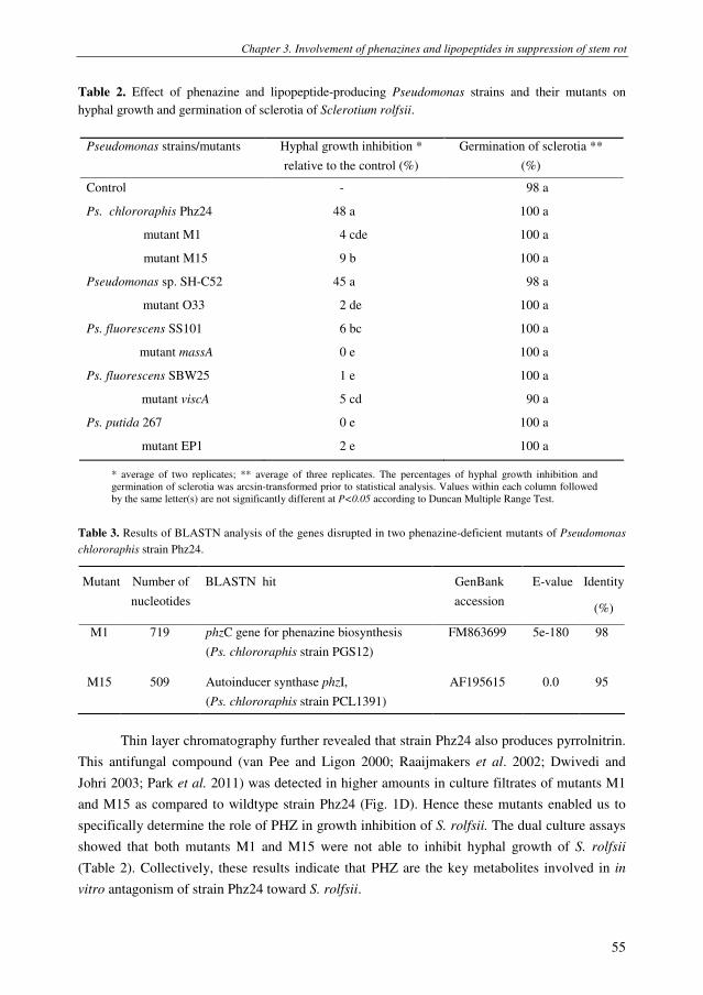
Chapter 3. Involvement of phenazines and lipopeptides in suppression of stem rot
55
Table 2. Effect of phenazine and lipopeptide-producing Pseudomonas strains and their mutants on
hyphal growth and germination of sclerotia of Sclerotium rolfsii.
* average of two replicates; ** average of three replicates. The percentages of hyphal growth inhibition and
germination of sclerotia was arcsin-transformed prior to statistical analysis. Values within each column followed
by the same letter(s) are not significantly different at P<0.05 according to Duncan Multiple Range Test.
Table 3. Results of BLASTN analysis of the genes disrupted in two phenazine-deficient mutants of Pseudomonas
chlororaphis strain Phz24.
Mutant Number of
nucleotides
BLASTN hit
GenBank
accession
E-value Identity
(%)
M1 719 phzC gene for phenazine biosynthesis
(Ps. chlororaphis strain PGS12)
FM863699 5e-180 98
M15 509 Autoinducer synthase phzI,
(Ps. chlororaphis strain PCL1391)
AF195615 0.0 95
Thin layer chromatography further revealed that strain Phz24 also produces pyrrolnitrin.
This antifungal compound (van Pee and Ligon 2000; Raaijmakers et al. 2002; Dwivedi and
Johri 2003; Park et al. 2011) was detected in higher amounts in culture filtrates of mutants M1
and M15 as compared to wildtype strain Phz24 (Fig. 1D). Hence these mutants enabled us to
specifically determine the role of PHZ in growth inhibition of S. rolfsii. The dual culture assays
showed that both mutants M1 and M15 were not able to inhibit hyphal growth of S. rolfsii
(Table 2). Collectively, these results indicate that PHZ are the key metabolites involved in in
vitro antagonism of strain Phz24 toward S. rolfsii.
Pseudomonas strains/mutants Hyphal growth inhibition *
relative to the control (%)
Germination of sclerotia **
(%)
Control - 98 a
Ps. chlororaphis Phz24 48 a 100 a
mutant M1 4 cde 100 a
mutant M15 9 b 100 a
Pseudomonas sp. SH-C52 45 a 98 a
mutant O33 2 de 100 a
Ps. fluorescens SS101 6 bc 100 a
mutant massA 0 e 100 a
Ps. fluorescens SBW25 1 e 100 a
mutant viscA 5 cd 90 a
Ps. putida 267 0 e 100 a
mutant EP1 2 e 100 a

Chapter 3. Involvement of phenazines and lipopeptides in suppression of stem rot
56
Fig
ure
1.
Ph
eno
typ
ic a
nd
bio
chem
ical
an
aly
sis
of
Pse
ud
om
on
as
ch
loro
rap
his
str
ain
Ph
z24
an
d m
uta
nts
M1
5 a
nd
M1
. (A
) ty
pic
al p
hen
oty
pe
of
bac
teri
al c
olo
nie
s o
n
PS
A
agar
p
late
s fo
r w
ild
typ
e st
rain
P
hz2
4
(to
p)
and
fo
r m
uta
nt
M1
5
(bo
tto
m);
th
e p
hen
oty
pe
of
mu
tan
t M
1
is
iden
tica
l to
th
at
of
M1
5;
(B)
RP
-HP
LC
chro
mat
og
ram
s (2
47
nm
) o
f ce
ll-f
ree
cult
ure
ex
trac
ts o
f w
ild
typ
e st
rain
Ph
z24 (
top)
and o
f m
uta
nt
M1
5 (
bo
ttom
); t
he
chro
mat
og
ram
fo
r m
uta
nt
M1
is
iden
tica
l to
th
at
of
M1
5;
(C):
Sp
ectr
al c
har
acte
rist
ics
of
pea
ks
1,
2,
and
3 s
ho
wn
in
pan
el B
fo
r w
ild
ty
pe
stra
in P
hz2
4 a
t re
ten
tion
tim
es (
RT
) o
f 2
0.2
, 22
.2,
and
25
.4 m
in,
resp
ecti
vel
y.
As
refe
ren
ce,
spec
tral
ch
arac
teri
stic
s o
f p
ure
ph
enaz
ine-
1-c
arb
oxy
lic
acid
(P
CA
; R
T=
25
.3 m
in)
are
show
n.
(D)
Th
in l
ayer
ch
rom
atog
rap
hy
(T
LC
)
anal
ysi
s o
f ce
ll-f
ree
cult
ure
extr
acts
of
wil
dty
pe
stra
in P
hz2
4 (
PcP
hz2
4)
and
mu
tan
ts M
1 a
nd
M1
5;
pu
re p
yrr
oln
itri
n (
PR
N;
Rf=
0.6
5)
is i
nd
icat
ed b
y a
n a
rro
w.
AU
0.0
0
1.0
0
2.0
0
3.0
0
Min
ute
s
0.0
010.0
020
.00
30
.00
40.0
0
AU
0.0
0
1.0
0
2.0
0
3.0
0
Min
ute
s
0.0
010.0
020.0
030
.00
40.0
0
AU
0.0
0
0.2
0
0.4
0
0.6
0
0.8
0
1.0
0
1.2
0
1.4
0
nm
200.0
0250.0
0300.0
0350.0
0400.0
0
214.9
257.3
366.4
386.8
AU
0.0
5
0.1
0
0.1
5
0.2
0
0.2
5
0.3
0
0.3
5
nm
200.0
0250.0
0300.0
0350.0
0400.0
0
213.72
42.0
263.2
400.1
AU
0.0
0
0.2
0
0.4
0
0.6
0
0.8
0
1.0
0
1.2
0
nm
20
0.0
025
0.0
03
00
.00
35
0.0
040
0.0
0
203.2
247.9
367.6
AU
0.0
0
0.1
0
0.2
0
0.3
0
0.4
0
0.5
0
0.6
0
0.7
0
nm
20
0.0
02
50.0
03
00
.00
35
0.0
04
00.0
0
246.7
365.2
1
2
3P
ea
k 1
Pea
k 2
Pe
ak 3
Pu
re P
CA
AB
CD
AU
0.0
0
1.0
0
2.0
0
3.0
0
Min
ute
s
0.0
010.0
020
.00
30
.00
40.0
0
AU
0.0
0
1.0
0
2.0
0
3.0
0
Min
ute
s
0.0
010.0
020.0
030
.00
40.0
0
AU
0.0
0
0.2
0
0.4
0
0.6
0
0.8
0
1.0
0
1.2
0
1.4
0
nm
20
0.0
02
50.0
03
00
.00
35
0.0
040
0.0
0
214.9
257.3
366.4
386.8
AU
0.0
5
0.1
0
0.1
5
0.2
0
0.2
5
0.3
0
0.3
5
nm
200.0
0250.0
0300.0
0350.0
0400.0
0
213.72
42.0
263.2
400.1
AU
0.0
0
0.2
0
0.4
0
0.6
0
0.8
0
1.0
0
1.2
0
nm
20
0.0
025
0.0
03
00
.00
35
0.0
040
0.0
0
203.2
247.9
367.6
AU
0.0
0
0.1
0
0.2
0
0.3
0
0.4
0
0.5
0
0.6
0
0.7
0
nm
20
0.0
02
50.0
03
00
.00
35
0.0
04
00.0
0
246.7
365.2
1
2
3P
ea
k 1
Pea
k 2
Pe
ak 3
Pu
re P
CA
AB
CD

Chapter 3. Involvement of phenazines and lipopeptides in suppression of stem rot
57
In vitro antagonism of lipopeptide-producing Pseudomonas toward S. rolfsii
Four LP-producing Pseudomonas strains and their mutants (Table 1) were tested for
their ability to inhibit germination of sclerotia and hyphal growth of S. rolfsii. None of the
bacterial strains and mutants tested inhibited germination of sclerotia of S. rolfsii (Table 2).
Dual culture assays showed that hyphal growth of S. rolfsii was not significantly inhibited
by Pseudomonas strains 267, SS101 and SBW25 producing putisolvin or viscosin-type LPs.
In contrast, Pseudomonas sp. strain SH-C52 significantly inhibited hyphal growth of S.
rolfsii, whereas mutant O33, deficient in the production of the chlorinated LP thanamycin,
was not effective. Collectively, these results suggest that inhibition of hyphal growth of S.
rolfsii is dependent on the type of LP produced by the introduced Pseudomonas strain.
Control of stem rot disease of groundnut under growth chamber conditions
Ps. chlororaphis strain Phz24 significantly reduced DI and MR of groundnut plants
(Fig. 2). PHZ-mutants M1 and M15 colonized the roots and stem base of groundnut plants
to the same extent as wild type strain Phz24, but did not provide significant disease control
(Fig. 2). These results suggest that PHZ determines the ability of strain Phz24 to control
stem rot disease. The LP-producing strains SS101, SBW25, 267 and their respective LP-
biosynthesis mutants did not significantly reduce disease incidence or mortality (Fig. 2).
Also strain SH-C52 did not reduce disease incidence but significantly lowered mortality of
groundnut plants (Fig. 2). Thanamycin-deficient mutant O33 established a density on the
roots of groundnut similar to that of wild type strain SH-C52 (Fig. 2), but was not able to
significantly reduce mortality (Fig. 2). Strains SS101, SBW25 and 267 colonized the roots
of groundnut significantly less than strains Phz24 and SH-C52: the rhizosphere population
densities of strains SBW25, SS101 and 267 were more than 10 to 100-fold (1 to 2 log units)
lower than those of Phz24 and SH-C52 (Fig. 2), suggesting a correlation between biocontrol
activity and root colonization. However, with respect to the population density of strains
Phz24 an SH-C52 on the stem base of groundnut, the correlation with the level of disease
control was not evident. For example, strains SS101 and SBW25, which lack biocontrol
activity, established similar to higher densities on the stem base than Phz24 and SH-C52,
the two strains that did suppress disease (Fig. 2).
Control of stem rot disease under nethouse and field conditions
Under nethouse conditions, only strain SH-C52 significantly reduced disease
incidence, but did not reduce disease severity and mortality significantly (Fig. 3). After two
weeks of plant growth, strains SS101 and Phz24 established similar densities on roots of
groundnut, but neither of the two strains provided disease control under nethouse conditions
(Fig. 3). None of the three introduced bacterial strains were detectable on the stem base of
groundnut plants after 2 weeks days of plant growth.

Chapter 3. Involvement of phenazines and lipopeptides in suppression of stem rot
58
In the field experiment in Quang Nam province, disease incidence in the control
treatment was relatively low with approximately 10% of the plants naturally infected with S.
rolfsii at the pod set stage (Fig. 4). Disease incidence increased during the growth season
more rapidly in the untreated control plots as compared to field plots with the bacterial
strains. At pod set stage, both strains Phz24 and SH-C52 significantly suppressed stem rot
disease, whereas strain SS101 did not significantly reduce DI relative to the control
treatment. None of the three bacterial strains adversely affected nodulation (Table 4) nor did
they adversely affect or increase plant height or the length of the first branch (data not
shown). Strains SH-C52 and Phz24 did not increase pod yield, whereas strain SS101
significantly increased yield by on average 12% compared to the control treatment (Table
4).
Table 4. Effects of different Pseudomonas strains on nodulation and yield of groundnut plants grown under
field conditions in Quang Nam province, Vietnam. Population densities of the introduced Pseudomonas
strains on the stem base and roots of groundnut plants were determined at two developmental stages
(flowering, pod set) of the groundnut plants. Averages of three replicates are given. For each column,
different letters indicate a statistically significant difference between the treatments (P< 0.05, Duncan
Multiple Range Test).
Population density introduced Pseudomonas strain
(log cfu g-1
)
Flowering Pod set
Treatment
Stem base Roots Stem base Roots
Nodules
per plant
Pod yield
(kg ha-1
)
Control 133 a 1220 a
Ps. chlororaphis Phz24 4.9 a 4.6 a 5.1 a 5.6 a 165 a 1260 ab
Pseudomonas sp. SH-C52 5.1 a 4.0 ab 5.9 a 6.0 a 135 a 1190 a
Ps. fluorescens SS101 5.2 a 3.5 b 5.7 a 5.5 a 134 a 1370 b
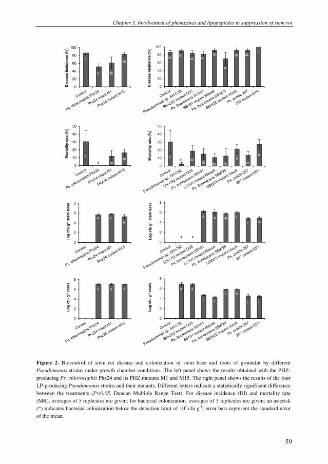
Chapter 3. Involvement of phenazines and lipopeptides in suppression of stem rot
59
0
20
40
60
80
100
Dis
ease i
ncid
en
ce (
%)
0
20
40
60
80
100
Dis
ea
se
in
cid
en
ce
(%
)
0
10
20
30
40
50
Mo
rta
lity
ra
te (
%)
0
10
20
30
40
50
Mo
rtality
rate
(%
)
0
2
4
6
8
Lo
g c
fu g
-1ste
m b
ase
0
2
4
6
8
Lo
g c
fu g
-1ste
m b
ase
0
2
4
6
8
Lo
g c
fu g
-1ro
ots
0
2
4
6
8
Lo
g c
fu g
-1ro
ots
b
a ab
ab
ab ab
ab ab
ab ab ab
a
b
a a
b
ab
ab b
b
b ab ab ab ab ab
a a
a
a a
a ab
c bc
a a a a a
c c c c
b b
* *
Figure 2. Biocontrol of stem rot disease and colonization of stem base and roots of groundut by different
Pseudomonas strains under growth chamber conditions. The left panel shows the results obtained with the PHZ-
producing Ps. chlororaphis Phz24 and its PHZ mutants M1 and M15. The right panel shows the results of the four
LP-producing Pseudomonas strains and their mutants. Different letters indicate a statistically significant difference
between the treatments (P<0.05, Duncan Multiple Range Test). For disease incidence (DI) and mortality rate
(MR), averages of 5 replicates are given; for bacterial colonization, averages of 3 replicates are given; an asterisk
(*) indicates bacterial colonization below the detection limit of 104
cfu g-1
; error bars represent the standard error
of the mean.

Chapter 3. Involvement of phenazines and lipopeptides in suppression of stem rot
60
0
20
40
60
80
100
Dis
ease in
cid
en
ce (%
)
b
ab
a
ab
0
10
20
30
40
50
Mo
rtality
rate
(%
)
a
a
a
a
0
2
4
6
8
Lo
g c
fu g
-1ro
ots
a
a a
Figure 3. Biocontrol of stem rot disease of groundnut by Pseudomonas chlororaphis strain Phz24, Pseudomonas
sp. strain SH-C52, and Ps. fluorescens strain SS101 under nethouse conditions in Vietnam. Averages of 3
replicates are given; error bars represent the standard error of the mean. Different letters indicate a statistically
significant difference between the treatments (P<0.05, Duncan Multiple Range Test). The lower panel shows the
population densities of the applied bacterial strains on roots of groundnut; for bacterial colonization, averages of 3
replicates are given. Error bars represent the standard error of the mean.
Figure 4. Biocontrol of stem rot of groundnut by Pseudomonas chlororaphis strain Phz24, Pseudomonas sp. strain
SH-C52, and Ps. fluorescens strain SS101 under field conditions in Quang Nam province, Vietnam. Disease
incidence was monitored at the seedling stage when plants had 3-5 true leaves (20 days after sowing), and
subsequently every 15 days. AUDPC represents the Area Under the Disease Progress Curve and was calculated
based on the method used by Landa et al. (2002) and Kruijt et al. (2009). For each developmental stage, averages
of 3 replicates are given. Different letters indicate a statistically significant difference between the treatments
(P<0.05, Duncan Multiple Range Test).
0
20
40
60
Dis
ease s
iveri
ty (
%)
ab
ab
a
b
Dis
ease s
ev
eri
ty (
%)
0
20
40
60
Dis
ease s
iveri
ty (
%)
ab
ab
a
b
Dis
ease s
ev
eri
ty (
%)

Chapter 3. Involvement of phenazines and lipopeptides in suppression of stem rot
61
Discussion
Sclerotium rolfsii causes serious diseases on many crops including groundnut. Current
methods to control this pathogen are limited or not effective. Hence, development and
integration of different control strategies is necessary for sustainable control of this soil-borne
pathogen. In a series of experiments conducted in the lab, growth chamber, nethouse and the
field, we showed that specific Pseudomonas strains, producing either PHZ or LPs, suppressed
hyphal growth of S. rolfsii and significantly reduced stem rot disease of groundnut. In other
studies (Ganesan and Gnanamanickam 1987; Dey et al. 2004; Kishore et al. 2005; Tonelli et al.
2010; Tonelli et al. 2011), Pseudomonads were tested mostly under controlled conditions for
their efficacy to suppress stem rot disease of groundnut and the mechanisms underlying disease
control were not addressed in these studies. By comparing the activities of wildtype
Pseudomonas strains with that of mutants disrupted in specific biosynthesis and regulatory
genes, we demonstrated that PHZ and specific LPs are key bioactive metabolites in biological
control of stem rot disease of groundnut.
In vitro and growth chamber bioassays with Ps. chlororaphis strain Phz24 showed that
PHZ are key metabolites in suppression of hyphal growth of S. rolfsii and of stem rot disease.
These results confirm and extend results of Rane et al. (2007) who showed that S. rolfsii could
not grow on agar plates amended with phenazine-1-carboxylic acid (PCA) from Pseudomonas
aeruginosa. Based on RP-HPLC analysis, we showed that strain Phz24 produces at least three
PHZ derivatives, i.e. PCA, 2-OH-PHZ and possibly 2-OH-PCA, and that disruption of the phzC
or phzI genes eliminates production of all three PHZ derivatives and concomitantly pathogen
inhibition. It is not yet known which of these three PHZ contributes mostly to the activity of
strain Phz24 against S. rolfsii, but work by Delaney et al. (2001) and Smirnov and Kiprianova
(1990; reviewed in Delaney et al. (2001)) indicated that the hydroxylated PHZ exhibit stronger
fungistatic activities. TLC analysis indicated that, next to the phenazines, strain Phz24 also
produces pyrrolnitrin, a potent antifungal compound produced by multiple Pseudomonas strains
and various other bacterial genera (van Pee and Ligon 2000). The observation that pyrrolnitrin
production was enhanced in PHZ mutants M1 and M15 suggests that this compound does not
play a role in growth inhibition and biocontrol of S. rolfsii by strain Phz24.
Sclerotia are the primary inoculum source for infection by S. rolfsii and various other soil-
borne pathogens, but extremely difficult to eradicate (Coleysmi and Cooke 1971; Punja 1985).
Ganesan and Gnanamanickam (1987) reported that some Ps. fluorescens strains can reduce
germination of sclerotia of S. rolfsii by 50-60%. Also culture filtrates of Actinomycetes were
shown to inhibit germination of sclerotia of S. rolfsii (Errakhi et al. 2009). Work by Debode et
al. (2007) with microsclerotia of Verticillium dahliae showed suppressive effects of different
Pseudomonas strains on germination. By using well-defined mutants, they further demonstrated
that PHZ and LPs play an essential role in this suppressive activity. In this study, however, none
of the tested phenazine and LP-producing Pseudomonas strains significantly reduced

Chapter 3. Involvement of phenazines and lipopeptides in suppression of stem rot
62
germination of S. rolfsii sclerotia. Possible explanations for this discrepancy may be the shorter
exposure time of the sclerotia to the bacteria, the potential poor survival of these bacterial
strains on the sclerotia, or differences in the sensitivity of S. rolfsii to the bacteria and/or
bioactive compounds.
In natural habitats, PHZ and LPs can confer a competitive advantage in survival and root
colonization (Mazzola et al. 1992; Nielsen et al. 2005; Tran et al. 2007; D'aes et al. 2011). For
the phenazines, the competitive advantage may relate to their broad-spectrum antimicrobial
activities (Mazzola et al. 1992; Mavrodi et al. 2006) and their role in biofilm formation
(Dietrich et al. 2008; Maddula et al. 2008). Also LPs play a role in biofilm formation and are
key metabolites in surface motility of the producing strains and in defense against protozoan
predation (Mazzola et al. 2009; Raaijmakers et al. 2010). The results of our growth chamber
experiments, however, showed that the introduced phenazine and LP-producing Pseudomonas
strains and their biosynthesis or regulatory mutants established similar population densities on
the roots and stem base of groundnut. Probably the time point used to determine the population
densities, i.e. two weeks after sowing, and the fact that the spatial colonization patterns were not
considered in our analysis may account for the discrepancy with results obtained in previous
studies.
Among the LPs produced by Pseudomonas species, there is considerable structural
diversity due to differences in the length and composition of their lipid and peptide moieties
(Nybroe and Sorensen 2004; Raaijmakers et al. 2006). Based on these structural characteristics,
the LPs produced by Pseudomonas were initially classified into four major groups, i.e. the
viscosin, amphisin, tolaasin and syringomycin groups (Nybroe and Sorensen 2004). In addition,
a number of new LPs have been identified for Pseudomonas in recent years, including
arthrofactin (Morikawa et al. 1993; Roongsawang et al. 2003), putisolvin (Kuiper et al. 2004),
orfamide (Paulsen et al. 2005; Gross et al. 2007), pseudodesmins (Sinnaeve et al. 2009), and
thanamycin (Mendes et al. 2011). In this study, we compared the antifungal activity of
Pseudomonas strains producing one of four structurally different LPs, i.e. massetolide A by
strain SS101, viscosin by strain SBW25, putisolvin by strain 267 and thanamycin by strain SH-
C52. Due to the lack of sufficient amounts of pure forms of the four LPs, we were not able to
conduct dose-response assays with S. rolfsii to determine EC50 and minimal inhibitory
concentrations (MIC) of these LPs. Instead, we compared the antagonistic activities of the
wildtype strains with those of well-defined mutants disrupted in the respective LP-biosynthesis
genes.
The results of the in vitro dual culture assays showed that strains SS101 and SBW25,
producing the viscosin-type LPs, and strain 267, producing putisolvins, did not or only
marginally inhibit hyphal growth of S. rolfsii. Lack of inhibiting activity of LPs was also
observed previously for viscosinamide and putisolvins in hyphal growth assays with oomycete
pathogens (Thrane et al. 1999; Gross et al. 2007; Kruijt et al. 2009). In contrast, thanamycin-
producing strain SH-C52 significantly inhibited hyphal growth of S. rolfsii, whereas its

Chapter 3. Involvement of phenazines and lipopeptides in suppression of stem rot
63
thanamycin-deficient mutant was not effective. Growth chamber bioassays further showed that
strain SH-C52 significantly reduced stem rot disease of groundnut, whereas its thanamycin-
deficient mutant and also strains SS101, SBW25 and 267 were not effective in disease control.
Collectively, these results suggest differential activity of structurally different LPs against the
stem rot pathogen S. rolfsii, with thanamycin having significant inhibitory effects on hyphal
growth and plant infection. Based on sequence similarities with the syringomycin biosynthesis
pathway and bioinformatic analyses, thanamycin is predicted to be a 9-amino acid LP with a C-
terminal chlorinated threonine residue (Mendes et al. 2011). Due to difficulties to isolate
sufficient amounts of pure thanamycin, the predicted structure has not been confirmed by LC-
MS/MS and NMR yet. The activity of thanamycin against S. rolfsii is most likely related to the
C-terminal chlorinated threonine, the residue that is also responsible for most of the antifungal
activity of the structurally related LP syringomycin produced by P. syringae pv. syringae
(Grgurina et al. 1994). It should be emphasized, however, that the presumed differential activity
of the four LPs is based on the assumption that each of the bacterial strains produces similar
amounts of the LPs on agar plates and in the groundnut rhizosphere. The culture conditions of
the in vitro assays are known to induce LP production (X. Cheng, M. van der Voort, J.M.
Raaijmakers, unpubl. data), but at this point we have no data on the concentrations of each of
the LPs produced on roots of groundnut. To that end, reporter-based studies (Keel et al. 1992)
or advanced analytical chemical analyses (Ongena et al. 2007) should be conducted to confirm
that each of the four structurally different LPs is indeed produced in situ and to determine the
amounts produced.
The proposed mode of action of LPs is pore formation in membranes, leading to an
inbalance in transmembrane ion fluxes and cell death (Abada 1994; Bender et al. 1999; Baltz
2009). In addition, LPs may induce systemic resistance in plants. For example, massetolide A
produced by Ps. fluorescens strain SS101 enhanced resistance of tomato against Phytophthora
infestans (Tran et al. 2007). Similarly, fengycins and surfactin produced by Bacillus induced
systemic protection in bean and tomato leaves against Botrytis cinerea (Ongena et al. 2007).
Whether thanamycin produced by strain SH-C52 directly impacts hyphal growth of S. rolfsii in
the rhizosphere or on the stem base of groundnut, or limits plant infection through induced
systemic resistance is not known. To investigate this, bioassays need to be conducted where the
pathogen and the inducers (strain SH-C52, mutant O33, pure thanamycin) are physically
separated.
In conclusion, phenazine-producing strain Phz24 and thanamycin-producing strain SH-
C52 are promising candidates for biological control of stem rot disease of groundnut caused by
S. rolfsii. Based on the observed additive and synergistic activity of phenazines and
biosurfactant compounds in control of Pythium species (Perneel et al. 2008) and the fungal
pathogen Rhizctonia solani (D'aes et al. 2011), combination of strains Phz24 and SH-C52 may
provide even better or more consistent control of S. rolfsii. Alternatively, strain SH-C52 may be
transformed with genes for phenazine biosynthesis leading to enhanced antifungal activity, an

Chapter 3. Involvement of phenazines and lipopeptides in suppression of stem rot
64
approach that was successfully used previously with other Pseudomonas strains (Huang et al.
2004). Also combining strain SH-C52 with Ps. fluorescens strain SS101 should be pursued as
the latter strain significantly improved yield in the field experiment. The mechanisms
underlying this increase in pod yield by strain SS101 is not yet known and is subject of ongoing
investigations.
Materials and methods
Bacterial and fungal strains
The Pseudomonas strains used in this study are listed in Table 1. Bacterial strains were grown at 25°C on
Pseudomonas Agar (PSA; Difco, France). For all strains, spontaneous rifampicin resistant derivatives were
generated in order to monitor their population densities on roots and stem base of groundnut plants. In the in vitro
and in vivo bioassays, S. rolfsii strain H001 was used. This strain was collected from groundnut plants in Vietnam
in 2008 (Le et al. 2011). Sclerotia of strain H001 were obtained as follows: 50 g of dried (110°C overnight)
groundnut plants were put in an Erlenmeyer flask, wetted with 100 ml sterile demineralized water, sterilized at
120°C for 30 min and inoculated with three mycelial agar plugs (5-mm-diameter) of S. rolfsii. After 3 weeks of
incubation at 25ºC, the sclerotia were collected, air-dried and stored at room temperature.
In vitro assays
Inhibition of hyphal growth of S. rolfsii by the Pseudomonas strains was tested in dual culture assays
according to the conditions described previously (Kruijt et al. 2009) and determined after 3 days of incubation at
25°C. Inhibition of sclerotial germination by the Pseudomonas strains was determined as follows: sclerotia were
surface-sterilized with 3% (v/v) sodium hypochlorite for 5 min and rinsed three times with ample sterile distilled
water. Pseudomonas strains were grown on PSA plates for 24 h at 25ºC, washed three times with sterile distilled
water and adjusted to a final density of 109
cells ml-1
(OD600=1.0). Sclerotia were soaked in sterile distilled water
(control) or in bacterial suspension for 30 min and placed on sterile wet filter paper in a Petri dish. For each
treatment, three replicate plates were used with fifteen sclerotia per plate. After 48 hours of incubation at 25ºC,
germination of sclerotia was examined with a binocular.
Selection and identification of phenazine mutants
Phenazine-producing Pseudomonas chlororaphis strain Phz24 was subjected to random transposon
mutagenesis using the plasposon TnModOTc (Dennis and Zylstra 1998). From approximately 2500 random
mutants, two mutants were selected that had lost the typical orange pigment of wildtype strain Phz24 (Fig. 2A)
which is linked to the production of 2-hydroxyphenazine-1-carboxylic acid. The loss of PHZ production in these
two mutants, designated M1 and M15, was analyzed by RP-HPLC analyses followed by photodiode array
spectroscopy according to the methods described by De Souza et al. (2003). To identify the genes disrupted in
mutants M1 and M15, the Y-linker method described by Kwon and Ricke (2000) was used. Genomic DNA was
isolated from each of the mutants, digested with SphI and linked to Y-linkers 1 and 2. The sequence of Y-linker 1
is TTTCTGCTCGAATTCAAGCTTCTAACGATGTACGGGGACACATG, and that of Y-linker 2 is
TGTCCCCGTACATCGTTAGAACTACTCGTACCATCCACAT. To amplify the genetic region flanking the
transposon insertion, Y-linker primer CTGCTCGAATTCAAGCTTCT and Tn5 primer
GGCCAGATCTGATCAAGAGA were used in the polymerase chain reaction (PCR). DNA amplicons obtained
from the two mutants were purified and sent for sequencing (Macrogen Corp. Europe, Amsterdam, The
Netherlands). Quality assessment and analysis of the obtained sequences were performed with Vector NTI
(Invitrogen, version 8.0). For BLAST searches, databases at the National Center for Biotechnology Information

Chapter 3. Involvement of phenazines and lipopeptides in suppression of stem rot
65
(http://www.ncbi.nlm.nih.gov) and Computational & Genome Biology Initiative Bioinformatics (CGBI:
http://cgbi.cgrb.oregonstate.edu/) were used.
Growth chamber bioassays
The efficacy of the Pseudomonas strains and mutants to control stem rot disease of groundnut was
investigated in growth chamber assays at Wageningen University (Wageningen, Netherlands). For these bioassays,
groundnut cultivar L14 was used, which is the most widely grown cultivar in Vietnam. Growth chamber conditions
comprised a continuous temperature of 25ºC during day (12h) and night (12h), a relative air humidity of 80%, and
a light intensity of 250 µmol m
–2 s
–1.
Groundnut seeds were surface sterilized in 3% (v/v) sodium hypochlorite solution for 15 min, rinsed with
70% ethanol and rinsed three times with ample sterile distilled water. Sterilized seeds were soaked in sterile water
for 4 h at 25°C and then incubated overnight in Petri dishes at 25°C. The pre-germinated seeds were subsequently
soaked for 30 min in a bacterial suspension of 107 cells ml
-1. For the control treatment, germinated seeds were
soaked in sterile water for 30 min. Treated seeds were sown in pots containing 250 g of a mix (1:3 (w/w)) of
commercial potting soil and river sand. Each treatment consisted of five boxes with eight pots per box. In the
growth chamber, the treatments were randomized. Plants with three true leaves (~ two weeks after sowing), were
inoculated with the fungal pathogen by placing a mycelial agar plug (5-mm-diameter) of a 3-day-old PDA plate
culture of S. rolfsii approximately 1 cm below the soil surface close to the base of the stem. The plugs were
covered with soil. Two weeks after pathogen inoculation, disease incidence (DI), disease severity (DS) and
mortality (MR) were recorded. DI is defined as the number of infected plants divided by the total number of plants
tested. For determining the DS, disease symptoms were rated on a scale from 0-4 (Fig. 5) with 0: no disease
symptoms, 1: disease symptoms without visible outgrowth of the fungus, 2: disease symptoms with visible
outgrowth of the fungus, 3: partial wilting of the plant, and 4: complete wilting and plant death (Le et al. 2011). DS
was calculated based on the formula: DS= [(1× number of plants rated as scale 1) + (2 × number of plants rated as
scale 2) + (3 × number of plants rated as scale 3) + (4 × number of plants rated as scale 4)] × 100 ÷ (4 × total
number of plants). MR is the number of wilted and dead plants (scale 4) divided by the total number of plants.
0 1 2 3 4
Figure 5. Disease severity index scales of groundnut infected by Sclerotium rolfsii. 0: no disease symptoms, 1:
disease symptoms without visible outgrowth of the fungus, 2: disease symptoms with visible outgrowth, 3: partial
wilting of the plant, and 4: complete wilting and plant death.
Nethouse experiments
For the nethouse experiments conducted at Hue University of Agriculture and Forestry (Hue, Vietnam), the
set-up was similar to that of the growth chamber bioassays except that field soil, collected from a groundnut field in
Thua Thien Hue province (clay loam soil), was used instead of the potting soil: river sand mixture. Moreover, due
to a lack of greenhouses at Hue University for contained used of genetically modified microorganisms, the mutants
of the Pseudomonas strains could not be tested. Hence, in the nethouse experiments only wildtype strains were
tested.

Chapter 3. Involvement of phenazines and lipopeptides in suppression of stem rot
66
Field experiment
The field experiment for biocontrol of stem rot disease caused by S. rolfsii was conducted in 2010 in Quang
Nam province (Vietnam), where approximately 20% of the groundnut plants in the field were naturally infected by
S. rolfsii (Le et al. 2011). The experiment consisted of four treatments, i.e. one control treatment and three bacterial
treatments. The field experiment was set-up based on a randomized complete block design (RCBD) with three
blocks as three replicates and a plot size of 15 m2 (3 X 5 m). The distance between rows was 30 cm and between
plants within a row 10 cm. The bacterial strains tested were Pseudomonas Phz24, SH-C52, and SS101. Bacterial
strains were grown on PSA plates at 25°C for 48 h, harvested and washed with sterile water. Prior to sowing
groundnut seeds, bacterial suspensions were applied to the furrows at a final density of 106 cells per cm
2 soil.
Bacterial suspensions were applied again at the same density at the flowering stage of the groundnut plants.
The field soil was fertilized with nitrogen (N) at 40 kg/ha, phosphorus (P2O5) at 60 kg/ha, potassium (K2O)
at 60 kg/ha, and calcium (Ca(OH)2) at 300 kg/ha. Total phosphorus and calcium was applied at the time of soil
preparation. Seeds were sown at a depth of 3-5 cm and covered with soil. When plants had three true leaves
(seedling stage), 70% of the nitrogen and 50% of the potassium was applied. The remainder of the N and K
fertilization was applied at flowering. Weeds were manually removed at four developmental stages of the
groundnut plants, i.e. seedling, flowering, peg and pod set. At these four developmental stages, DI was monitored
in 1 m2
of each plot, i.e. approximately 33 plants per plot. Also other characteristics including plant height,
nodulation, and yield were monitored. The number of nodules per plant root system was determined at the pod
stage. To that end, three randomly selected plants per plot were harvested; their root systems were washed to
remove adhering soil and the number of nodules was counted.
Bacterial colonization of groundnut roots and stem base
The spontaneous rifampicin-resistant derivatives of the Pseudomonas strains were re-isolated from roots and
stem base of groundnut according to methods described by Tran et al. (2008). Briefly, one gram (fresh weight) of
groundnut roots or stem base was transferred to a 15-ml tube with 5 ml 10 mM MgSO4-7H2O, followed by
vigorous vortexing for 1 min, sonication (Elma D-78224, Singen/HTW, Germany) for 1 min and vortexing again
for 15 sec. The suspensions were serially diluted in 10-fold steps and 50 µl of the 100X and 1000X diluted samples
was plated onto PSA plates supplemented with chloroamphenicol (12.5 µg ml-1
), ampicillin (40 µg ml-1
),
rifampicin (100 µg ml-1
) and delvocid (100 µg ml-1
). The number of bacterial colonies was counted after 2- 4 days
of incubation at 25°C.
Statistical analysis
All experiments, except the field experiment, were performed at least twice and representative results are
shown. DI, DS and MR are expressed in percentages and were arcsin-transformed prior to statistical analysis.
Statistical differences (P < 0.05) between treatments were analyzed by ANOVA followed by the Dunnet test or the
Duncan Multiple Range Test (SPSS Statistics, USA). Normal distribution of the data and homogeneity of variances
was tested prior to ANOVA.
Acknowledgements
This work was financially supported by the Vietnamese Government through the 322 project and the
Laboratory of Phytopathology, Wageningen University, the Netherlands. We would like to thank Francine Govers
for critically reading this manuscript and valuable suggestions. We thank Daniel Lopisso for his work on phenazine
mutants of Phz24, we also thank the Laboratory of Plant Protection, Hue University of Agriculture and Forestry
(Vietnam) and Le My Linh and Vo Thi La, for preparation of materials for the nethouse experiment.

Chapter 3. Involvement of phenazines and lipopeptides in suppression of stem rot
67
References
Abada, K.A. (1994) Fungi causing damping-off and root-rot on sugar-beet and their biological control with
Trichoderma harzianum. Agr Ecosyst Environ 51, 333-337.
Baltz, R.H. (2009) Daptomycin: mechanisms of action and resistance, and biosynthetic engineering. Curr Opin
Chem Biol 13, 144-151.
Bender, C.L., Alarcon-Chaidez, F. and Gross, D.C. (1999) Pseudomonas syringae phytotoxins: mode of action,
regulation, and biosynthesis by peptide and polyketide synthetases. Microbiol Mol Biol Rev 63, 266-292.
Branch, W.D. and Brenneman, T.B. (2009) Field evaluation for the combination of white mould and tomato
spotted wilt disease resistance among peanut genotypes. Crop Prot 28, 595-598.
Brown, S.L. (2007) Peanut insects: ecology and control. In Encyclopedia of Pest Management ed. Pimentel, D.
pp.1 - 10: Taylor & Francis
Coleysmi, J.R. and Cooke, R.C. (1971) Survival and germination of fungal sclerotia. Annu Rev Phytopathol 9, 65-
92.
D’aes, J., Maeyer, K.D., Pauwelyn, E. and Höfte, M. (2010) Biosurfactants in plant Pseudomonas interactions and
their importance to biocontrol. Environ Microbiol Reports 2, 539-572.
D'aes, J., Gia, K.H.H., De Maeyer, K., Pannecoucque, J., Forrez, I., Ongena, M., Dietrich, L.E.P., Thomashow,
L.S., Mavrodi, D.V. and Hofte, M. (2011) Biological control of Rhizoctonia root rot on bean by phenazine-
and cyclic lipopeptide-producing Pseudomonas CMR12a. Phytopathology 101, 996-1004.
de Bruijn, I., de Kock, M.J.D., Yang, M., de Waard, P., van Beek, T.A. and Raaijmakers, J.M. (2007) Genome-
based discovery, structure prediction and functional analysis of cyclic lipopeptide antibiotics in
Pseudomonas species. Mol Microbiol 63, 417-428.
de Bruijn, I., de Kock, M.J.D., de Waard, P., van Beek, T.A. and Raaijmakers, J.M. (2008) Massetolide A
biosynthesis in Pseudomonas fluorescens. J Bacteriol 190, 2777-2789.
de Souza, J.T., de Boer, M., de Waard, P., van Beek, T.A. and Raaijmakers, J.M. (2003) Biochemical, genetic, and
zoosporicidal properties of cyclic lipopeptide surfactants produced by Pseudomonas fluorescens. Appl
Environ Microbiol 69, 7161-7172.
Debode, J., De Maeyer, K., Perneel, M., Pannecoucque, J., De Backer, G. and Hofte, M. (2007) Biosurfactants are
involved in the biological control of Verticillium microsclerotia by Pseudomonas spp. J Appl Microbiol 103,
1184-1196.
Delaney, S.M., Mavrodi, D.V., Bonsall, R.F. and Thomashow, L.S. (2001) phzO, a gene for biosynthesis of 2-
hydrolyated phenazine compounds in Pseudomonas aureofaciens 30-84. J Bacteriol 183, 318-327.
Dennis, J.J. and Zylstra, G.J. (1998) Plasposons: modular self-cloning minitransposon derivatives for rapid genetic
analysis of gram-negative bacterial genomes. Appl Environ Microbiol 64, 2710-2715.
Dey, R., Pal, K.K., Bhatt, D.M. and Chauhan, S.M. (2004) Growth promotion and yield enhancement of peanut
(Arachis hypogaea L.) by application of plant growth-promoting rhizobacteria. Mycol Res 159, 371-394.
Dietrich, L.E.P., Teal, T.K., Price-Whelan, A. and Newman, D.K. (2008) Redox-active antibiotics control gene
expression and community behavior in divergent bacteria. Science 321, 1203-1206.
Dwivedi, D. and Johri, B.N. (2003) Antifungals from fluorescent pseudomonads: biosynthesis and regulation. Curr
Sci 85, 1693-1703.
Errakhi, R., Lebrihi, A. and Barakate, M. (2009) In vitro and in vivo antagonism of actinomycetes isolated from
Moroccan rhizospherical soils against Sclerotium rolfsii: a causal agent of root rot on sugar beet (Beta
vulgaris L.). J Appl Microbiol 107, 672-681.
Florkowski, W.J. (1994) Groundnut production and trade. In The groundnut crop: a scientific basis for
improvement ed. Smartt, J. pp.1-22. London: Chapman & Hall.
Chapter 3. Involvement of phenazines and lipopeptides in suppression of stem rot
68
Franke, M.D., Brenneman, T.B., Stevenson, K.L. and Padgett, G.B. (1998) Sensitivity of isolates of Sclerotium
rolfsii from peanut in Georgia to selected fungicides. Plant Dis 82, 578-583.
Ganesan, P. and Gnanamanickam, S.S. (1987) Biological control of Sclerotium rolfsii Sacc. in peanut by
inoculation with Pseudomonas fluorescens. Soil Biol Biochem 19, 35-38.
Ganesan, S., Kuppusamy, R. and Sekar, R. (2007) Integrated management of stem rot disease (Sclerotium rolfsii)
of groundnut (Arachis hypogaea L.) using Rhizobium and Trichoderma harzianum (ITCC-4572). Turk J
Agric For 31, 103-108.
Grgurina, I., Barca, A., Cervigni, S., Gallo, M., Scaloni, A. and Pucci, P. (1994) Relevance of chlorine-substituent
for the antifungal activity of syringomycin and syringotoxin, metabolites of the phytopathogenic bacterium
Pseudomonas syringae pv. syringae. Experientia 50, 130-133.
Gross, H., Stockwell, V.O., Henkels, M.D., Nowak-Thompson, B., Loper, J.E. and Gerwick, W.H. (2007) The
genomisotopic approach: a systematic method to isolate products of orphan biosynthetic gene clusters. Chem
Biol 14, 53-63.
Gross, H. and Loper, J.E. (2009) Genomics of secondary metabolite production by Pseudomonas spp. Nat Prod
Rep 26, 1408-1446.
Haas, D. and Defago, G. (2005) Biological control of soil-borne pathogens by fluorescent pseudomonads. Nat Rev
Microbiol 3, 307-319.
Huang, Z.Y., Bonsall, R.F., Mavrodi, D.V., Weller, D.M. and Thomashow, L.S. (2004) Transformation of
Pseudomonas fluorescens with genes for biosynthesis of phenazine-1-carboxylic acid improves biocontrol
of rhizoctonia root rot and in situ antibiotic production. FEMS Microbiol Ecol 49, 243-251.
Keel, C., Schnider, U., Maurhofer, M., Voisard, C., Laville, J., Burger, U., Wirthner, P., Haas, D. and Défago, G.
(1992) Suppression of root diseases by Pseudomonas fluorescens CHA0: importance of the bacterial
secondary metabolite 2, 4-diacetylphloroglucinol. Mol Plant Microbe Interact 5, 4-13.
Kemerait, R. (2005) Peanut. In 2004 Georgia Plant Disease Loss Estimates ed. Pearce, M.J. p.11. Georgia:
University of Georgia, Cooperative Extension Service.
Kemerait, R. (2006) Peanut. In 2005 Georgia Plant Disease Loss Estimates ed. Martinez, A. p.11. Georgia:
University of Georgia, Cooperative Extension Service.
Kemerait, R. (2007) Peanut. In 2006 Georgia Plant Disease Loss Estimates ed. Martinez, A. p.11. Georgia:
University of Georgia, Cooperative Extension Service.
Kemerait, R. (2008) Peanut. In 2007 Georgia Plant Disease Loss Estimates ed. Martinez, A. p.15. Georgia:
University of Georgia, Cooperative Extension Service.
Kishore, G.K., Pande, S., Rao, J.N. and Podile, A.R. (2005) Pseudomonas aeruginosa inhibits the plant cell wall
degrading enzymes of Sclerotium rolfsii and reduces the severity of groundnut stem rot. Eur J Plant Pathol
113, 315-320.
Kruijt, M., Tran, H. and Raaijmakers, J.M. (2009) Functional, genetic and chemical characterization of
biosurfactants produced by plant growth-promoting Pseudomonas putida 267. J Appl Microbiol 107, 546-
556.
Kuiper, I., Lagendijk, E.L., Pickford, R., Derrick, J.P., Lamers, G.E., Thomas-Oates, J.E., Lugtenberg, B.J. and
Bloemberg, G.V. (2004) Characterization of two Pseudomonas putida lipopeptide biosurfactants, putisolvin
I and II, which inhibit biofilm formation and break down existing biofilms. Mol Microbiol 51, 97-113.
Kwon, Y.M. and Ricke, S.C. (2000) Efficient amplification of multiple transposon-flanking sequences. J Microbiol
Meth 41, 195-199.
Landa, B.B., Mavrodi, O.V., Raaijmakers, J.M., Gardener, B.B.M., Thomashow, L.S. and Weller, D.M. (2002)
Differential ability of genotypes of 2,4-diacetylphloroglucinol-producing Pseudomonas fluorescens strains
to colonize the roots of pea plants. Appl Environ Microbiol 68, 3226-3237.

Chapter 3. Involvement of phenazines and lipopeptides in suppression of stem rot
69
Le, C.N., Mendes, R., Kruijt, M. and Raaijmakers, J. (2011) Genetic and phenotypic diversity of Sclerotium rolfsii
Sacc. in groundnut fields in central Vietnam. Plant Dis (accepted with minor changes).
Maddula, V., Pierson, E.A. and Pierson, L.S. (2008) Altering the ratio of phenazines in Pseudomonas chlororaphis
(aureofaciens) strain 30-84: Effects on biofilm formation and pathogen inhibition. J Bacteriol 190, 2759-
2766.
Mavrodi, D.V., Blankenfeldt, W. and Thomashow, L.S. (2006) Phenazine compounds in fluorescent Pseudomonas
spp. biosynthesis and regulation. Annu Rev Phytopathol 44, 417-445.
Mazurier, S., Corberand, T., Lemanceau, P. and Raaijmakers, J.M. (2009) Phenazine antibiotics produced by
fluorescent pseudomonads contribute to natural soil suppressiveness to Fusarium wilt. Isme J 3, 977-991.
Mazzola, M., Cook, R.J., Thomashow, L.S., Weller, D.M. and Pierson, L.S. (1992) Contribution of phenazine
antibiotic biosynthesis to the ecological competence of fluorescent pseudomonads in soil habitats. Appl
Environ Microbiol 58, 2616-2624.
Mazzola, M., de Bruijn, I., Cohen, M.F. and Raaijmakers, J.M. (2009) Protozoan-induced regulation of cyclic
lipopeptide biosynthesis is an effective predation defense mechanism for Pseudomonas fluorescens. Appl
Environ Microbiol 75, 6804-6811.
Mehan, V.K., Mayee, C.D. and McDonald, D. (1994) Management of Sclerotium rolfsii caused stem and pod rots
of groundnut - A critical review. Int J Pest Manage 40, 313-320.
Mendes, R., Kruijt, M., de Bruijn, I., Dekkers, E., van der Voort, M., Schneider, J.H.M., Piceno, Y.M., DeSantis,
T.Z., Andersen, G.L., Bakker, P. and Raaijmakers, J.M. (2011) Deciphering the rhizosphere microbiome for
disease-suppressive bacteria. Science 332, 1097-1100.
Morikawa, M., Daido, H., Takao, T., Murata, S., Shimonishi, Y. and Imanaka, T. (1993) A new lipopeptide
biosurfactant produced by Arthrobacter sp. strain MIS38. J Bacteriol 175, 6459-6466.
Nielsen, T.H., Nybroe, O., Koch, B., Hansen, M. and Sorensen, J. (2005) Genes involved in cyclic lipopeptide
production are important for seed and straw colonization by Pseudomonas sp. strain DSS73. Appl Environ
Microbiol 71, 4112-4116.
Nybroe, O. and Sorensen, J. (2004) Production of cyclic lipopeptides by fluorescent pseudomonads. New York:
Pages 147-172 in: Pseudomonas volume 3, Biosynthesis of Macromolecules and Molecular Metabolism,
Ramos, J-L., ed., Kluwer Academic/Plenum Publishers.
Ongena, M., Jourdan, E., Adam, A., Paquot, M., Brans, A., Joris, B., Arpigny, J.L. and Thonart, P. (2007) Surfactin
and fengycin lipopeptides of Bacillus subtilis as elicitors of induced systemic resistance in plants. Environ
Microbiol 9, 1084-1090.
Park, J.Y.P.J.Y., Oh, S.A., Anderson, A.J., Neiswender, J., Kim, J.C. and Kim, Y.C. (2011) Production of the
antifungal compounds phenazine and pyrrolnitrin from Pseudomonas chlororaphis O6 is differentially
regulated by glucose. Lett Appl Microbiol 52, 532-537.
Paulsen, I.T., Press, C.M., Ravel, J., Kobayashi, D.Y., Myers, G.S.A., Mavrodi, D.V., DeBoy, R.T., Seshadri, R.,
Ren, Q.H., Madupu, R., Dodson, R.J., Durkin, A.S., Brinkac, L.M., Daugherty, S.C., Sullivan, S.A.,
Rosovitz, M.J., Gwinn, M.L., Zhou, L.W., Schneider, D.J., Cartinhour, S.W., Nelson, W.C., Weidman, J.,
Watkins, K., Tran, K., Khouri, H., Pierson, E.A., Pierson, L.S., Thomashow, L.S. and Loper, J.E. (2005)
Complete genome sequence of the plant commensal Pseudomonas fluorescens Pf-5. Nature Biotechnol 23,
873-878.
Perneel, M., D'Hondt, L., De Maeyer, K., Adiobo, A., Rabaey, K. and Hofte, M. (2008) Phenazines and
biosurfactants interact in the biological control of soil-borne diseases caused by Pythium spp. Environ
Microbiol 10, 778-788.
Punja, Z.K. (1985) The biology, ecology, and control of Sclerotium rolfsii. Annu Rev Phytopathol 23, 97-127.
Raaijmakers, J.M., Vlami, M. and de Souza, J.T. (2002) Antibiotic production by bacterial biocontrol agents.
Antonie Van Leeuwenhoek Int J Gen Mol Microbiol 81, 537-547.

Chapter 3. Involvement of phenazines and lipopeptides in suppression of stem rot
70
Raaijmakers, J.M., de Bruijn, I. and de Kock, M.J. (2006) Cyclic lipopeptide production by plant-associated
Pseudomonas spp.: diversity, activity, biosynthesis, and regulation. Mol Plant Microbe Interact 19, 699-710.
Raaijmakers, J.M, Paulitz, T., Steinberg, C., Alabouvette, C. and Moënne-Loccoz, Y. (2009) The rhizosphere: a
playground and battlefield for soilborne pathogens and beneficial microorganisms. Plant Soil 321, 341-361.
Raaijmakers, J.M., de Bruijn, I., Nybroe, O. and Ongena, M. (2010) Natural functions of lipopeptides from
Bacillus and Pseudomonas: more than surfactants and antibiotics. FEMS Microbiol Rev 34, 1037-1062.
Rane, M.R., Sarode, P.D., Chaudhari, B.L. and Chincholkar, S.B. (2007) Detection, isolation and identification of
phenazine-1-carboxylic acid produced by biocontrol strains of Pseudomonas aeruginosa. J Sci Ind Res 66,
627-631.
Roongsawang, N., Hase, K., Haruki, M., Imanaka, T., Morikawa, M. and Kanaya, S. (2003) Cloning and
characterization of the gene cluster encoding arthrofactin synthetase from Pseudomonas sp. MIS38. Chem
Biol 10, 869-880.
Schoonbeek, H., Raaijmakers, J.M. and De Waard, M.A. (2002) Fungal ABC transporters and microbial
interactions in natural environments. Mol Plant Microbe Interact 15, 1165-1172.
Shew, B.B. and Waliyar, F. (2007) Peanut diseases: ecology and control. In Encyclopedia of Pest Management ed.
Pimentel, D. pp.469–473: Taylor & Francis.
Shim, M.Y., Starr, J.L., Keller, N.P., Woodard, K.E. and Lee, T.A. (1998) Distribution of isolates of Sclerotium
rolfsii tolerant to pentachloronitrobenzene in Texas peanut fields. Plant Dis 82, 103-106.
Sinnaeve, D., Michaux, C., Van Hemel, J., Vandenkerckhove, J., Peys, E., Borremans, F.A.M., Sas, B., Wouters, J.
and Martins, J.C. (2009) Structure and X-ray conformation of pseudodesmins A and B, two new cyclic
lipodepsipeptides from Pseudomonas bacteria. Tetrahedron 65, 4173-4181.
Thrane, C., Olsson, S., Nielsen, T.H. and Sorensen, J. (1999) Vital fluorescent stains for detection of stress in
Pythium ultimum and Rhizoctonia solani challenged with viscosinamide from Pseudomonas fluorescens
DR54. FEMS Microbiol Ecol 30, 11-23.
Tonelli, M.L., Taurian, T., Ibanez, F., Angelini, J. and Fabra, A. (2010) Selection and in vitro characterization of
biocontrol agents with potential to protect peanut plants against fungal pathogens. J. Plant Pathol 92, 73-82.
Tonelli, M.L., Furlan, A., Taurian, T., Castro, S. and Fabra, A. (2011) Peanut priming induced by biocontrol
agents. Physiol Mol Plant Path 75, 100-105.
Tran, H., Ficke, A., Asiimwe, T., Höfte, M. and Raaijmakers, J.M. (2007) Role of the cyclic lipopeptide
massetolide A in biological control of Phytophthora infestans and in colonization of tomato plants by
Pseudomonas fluorescens. New Phytol 175, 731-742.
Tran, H., Kruijt, M. and Raaijmakers, J.M. (2008) Diversity and activity of biosurfactant-producing Pseudomonas
in the rhizosphere of black pepper in Vietnam. J Appl Microbiol 104, 839-851.
van Pee, K.H. and Ligon, J.M. (2000) Biosynthesis of pyrrolnitrin and other phenylpyrrole derivatives by bacteria.
Nat Prod Rep 17, 157-164.
Wadsworth, D.F. and Melouk, H.A. (1984) Tolerance of Sclerotium rolfsii to pentachloronitrobenzene.
Phytopathology 74, 634-634.

Chapter 4. Diversity and antifungal activity of groundnut-associated bacteria
Chapter 4
Diversity and antifungal activity of groundnut-associated
bacteria
Le, C.N., Lopisso, D. and Raaijmakers, J.M.
To be submitted

Chapter 4. Diversity and antifungal activity of groundnut-associated bacteria
72

Chapter 4. Diversity and antifungal activity of groundnut-associated bacteria
73
Diversity and antifungal activity of groundnut-associated bacteria
Le, C.N.1,2
, Lopisso, D.1 and Raaijmakers, J.M.
1
1 Laboratory of Phytopathology, Wageningen University, the Netherlands
2 Department of Plant Protection, Hue University of Agriculture and Forestry, Vietnam
Abstract
Groundnut (Arachis hypogaea L.) is an important oil seed crop worldwide and
used extensively for feed and food. In Vietnam, groundnut cultivation is
hampered by several soil-borne fungal pathogens, in particular Sclerotium
rolfsii. To develop sustainable measures to control stem rot disease caused by
S. rolfsii, bacteria were isolated from the stem base and roots of groundnut
plants grown in farmer fields in central Vietnam and tested for activity against
S. rolfsii. Among a total of 3,360 randomly selected bacterial isolates, only
thirteen (0.4%) inhibited hyphal growth of S. rolfsii. BOX-PCR and 16S-rDNA
sequence analyses revealed that these bacterial isolates were genetically
diverse and belonged to the γ-Proteobacteria (Pseudomonas), Firmicutes
(Bacillus) and Bacteroidetes (Chryseobacterium). Subsequent nethouse and
field experiments conducted in central Vietnam showed that treatment of
groundnut seeds or soil with strains of each of these three bacterial taxa
significantly reduced the incidence of stem rot disease and led to significant
yield increases of up to 21%. The level of disease protection provided by the
bacterial strains was similar to that achieved by the fungicide tebuconazole. For
Pseudomonas sp. strain R4D2, two mutants were obtained that did not inhibit
hyphal growth of S. rolfsii and were not effective in controlling stem rot
disease of groundnut under controlled growth chamber conditions.
Characterization of these two mutants suggested that Pseudomonas sp. strain
R4D2 produces (a) biosurfactant compound(s) that might be responsible for
biocontrol of S. rolfsii.
Keywords: Groundnut, antagonistic bacteria, Bacillus, Pseudomonas,
Chryseobacterium, Sclerotium rolfsii.

Chapter 4. Diversity and antifungal activity of groundnut-associated bacteria
74
Introduction
In Vietnam, groundnut (Arachis hypogaea L.) is the second most important annual crop
after paddy rice with an area of 249,200 ha and an annual production of approximately 0.53
million tons in 2009 (FAO 2011). Groundnut production can be improved considerably by
controlling a number of pests and diseases (Brown 2007; Shew and Waliyar 2007). Among the
soil-borne fungal diseases, stem rot caused by Sclerotium (Athelia) rolfsii Sacc. is a destructive
disease (Mehan et al. 1994). Recent surveys conducted in central Vietnam showed that 5-25%
of the groundnut plants in agricultural fields were infected by S. rolfsii (Le et al. 2011; Chapter
2). This pathogen has a broad-host range and can survive in soil and plant debris for
considerable time periods by means of persistent sclerotia (Coleysmi and Cooke 1971; Punja
1985). Sustainable control of this pathogen requires a combination of different strategies
including chemical, cultural and biological measures.
To date, studies on biological control of S. rolfsii by beneficial microorganisms have
shown that bacteria from the genus Pseudomonas can restrict in vitro hyphal growth of S. rolfsii
(Ganesan and Gnanamanickam 1987; Kishore et al. 2005a; Ganesan et al. 2007; de Curtis et al.
2010; Pastor et al. 2010; Tonelli et al. 2010). Germination of sclerotia was reduced by 10-20%
and 50-60% after immersion in a bacterial cell suspensions for 1 h and 1 week, respectively
(Ganesan and Gnanamanickam 1987). Kishore et al. (2005b) further showed that cell-free
culture filtrates of P. aeruginosa strains GSE18 and GSE19 inhibited the in vitro activity of the
cell wall degrading enzymes polygalacturonase and cellulase produced by S. rolfsii. Strains
GSE18 and GSE19 also suppressed the growth of S. rolfsii and reduced the incidence of stem
rot of groundnut (Kishore et al. 2005b). Recent studies conducted in our lab indicated that
phenazine-producing Pseudomonas chlororaphis strain Phz24 and lipopeptide-producing
Pseudomonas sp. strain SH-C52 control stem rot disease of groundnut under controlled
conditions and in the field in central Vietnam (Chapter 3). Next to pseudomonads, also Bacillus
species are studied extensively for biocontrol of plant diseases including stem rot disease of
groundnut. Pre-treatment of groundnut seeds with Bacillus subtilis protected groundnut seeds
against S. rolfsii and significantly increased the number of pods (Abd-Allah 2005). Other
microorganisms tested for control of stem rot disease include Rhizobium and Trichoderma
(Ganesan et al. 2007). Collectively, these limited studies indicate that application of
antagonistic microorganisms to seeds may provide a promising alternative or supplementary
strategy to control stem rot disease of groundnut.
To further develop biocontrol as an integral part of disease management practices to
control S. rolfsii and other pathogens of groundnut, the biocontrol efficacy of selected beneficial
microorganisms needs to be evaluated under field conditions. Most of the microorganisms
tested to date for biocontrol of S. rolfsii, however, have not been tested under field conditions.
Furthermore, most of these microorganisms do not originate from groundnut and may be less
adapted to the microenvironment of the groundnut plant and to the (a)biotic conditions

Chapter 4. Diversity and antifungal activity of groundnut-associated bacteria
75
prevailing in groundnut fields. The overall aims of this study were to 1) isolate and characterize
bacteria from the stem base and roots of groundnut plants grown in agricultural fields in central
Vietnam, 2) test selected bacterial strains under field conditions in Vietnam for their efficacy to
control stem rot disease of groundnut and to improve yield, and 3) elucidate the mechanism
underlying the biocontrol capacity of one of the selected bacterial strain. The overall set-up and
procedures adopted to achieve these goals are included in figure 1.
Figure 1. Schematic presentation of approaches and techniques used to isolate, identify, and characterize
antagonistic bacteria towards Sclerotium rolfsii on stem base and roots of groundnut grown in center on Vietnam.
1
2
Isolation and monitoring of population density of groundnut-associated
bacteria on stem base (1) and roots (2) of groundnut (left), and screening for in vitro antagonism against Sclerotium rolfsii (right)
Identification of antagonistic bacterial
isolates by BOX-PCR grouping (left) and 16S sequencing (right)
0 . 0 5
Biocontrol of S. rolfsiiby four representative antagonistic bacterial
strains under nethouse conditions (left) and field conditions (right)
AU
-0.02
0.00
0.02
0.04
0.06
0.08
0.10
0.12
0.14
Minutes
10.00 20.00 30.00 40.00 50.00 60.00
Genetic and biochemical analysis of bacterial strain
R4D2.
Comparison of biocontrol of stem rot by R4D2 and mutants
1
2
1
2
Isolation and monitoring of population density of groundnut-associated
bacteria on stem base (1) and roots (2) of groundnut (left), and screening for in vitro antagonism against Sclerotium rolfsii (right)
Identification of antagonistic bacterial
isolates by BOX-PCR grouping (left) and 16S sequencing (right)
0 . 0 50 . 0 5
Biocontrol of S. rolfsiiby four representative antagonistic bacterial
strains under nethouse conditions (left) and field conditions (right)
AU
-0.02
0.00
0.02
0.04
0.06
0.08
0.10
0.12
0.14
Minutes
10.00 20.00 30.00 40.00 50.00 60.00
AU
-0.02
0.00
0.02
0.04
0.06
0.08
0.10
0.12
0.14
Minutes
10.00 20.00 30.00 40.00 50.00 60.00
Genetic and biochemical analysis of bacterial strain
R4D2.
Comparison of biocontrol of stem rot by R4D2 and mutants

Chapter 4. Diversity and antifungal activity of groundnut-associated bacteria
76
Results and Discussion
Frequency of antagonistic bacteria on groundnut plants
The number of bacteria isolated on PSA agar plates from the stem base and roots of
groundnut plants grown in two farmer fields in central Vietnam, represented densities of
approximately 3 X 10⁶ CFU per gram (Table 1). Out of a total of 3,360 randomly selected
isolates, thirteen (0.4%) inhibited hyphal growth of S. rolfsii in vitro, i.e., six isolates from the
stem base and seven from roots of groundnut (Table 1). Also Tonelli et al. (2010) reported a
rather low percentage (1.5%) isolates with inhibitory activity against S. rolfsii (only three out of
a total of 193 from groundnut plants grown in Córdoba, Argentina). Kishore et al. (2005a), who
collected bacterial isolates from the rhizosphere of groundnut plants grown in Andhra Pradesh
(India), reported that approximately 9% of the isolates (34 out of a total of 393) significantly
inhibited hyphal growth of S. rolfsii. These differences in frequency of indigenous, groundnut-
associated bacteria with in vitro activity against S. rolfsii can be due to a multitude of factors,
including soil type, groundnut cultivar and the developmental stage of the groundnut plants at
the time of bacterial isolation. Also the culture conditions, and the S. rolfsii isolate used in the
inhibition assays may play a role. In hyphal growth inhibition we compared the inhibitory
activity of the different bacterial isolates. As shown in Fig. 2, the six bacterial isolates obtained
from the stem base of groundnut were substantially more active in inhibition of hyphal growth
of S. rolfsii than the seven isolates obtained from the roots of groundnut.
Table 1. Frequency and genotypic diversity of antagonistic bacteria isolated from the stem base and roots of
groundnut plants grown in agricultural fields in two provinces in central Vietnam.
Antagonism toward Sclerotium rolfsii**
Province Plant part Bacteria*
Tested Inhibitory (%) BOX-PCR
Group3
Quang Nam Stem base 3.4 x 10⁶ ± 0.5 x 10
6 960 2 0.2 1, 3
Roots 3.5 x 10⁶ ± 0.5 x 10
6 960 7 0.7 27, 37
Thua Thien Hue Stem base 3.0 x 10⁶ ± 1.1 x 10
6 720 4 0.6 2, 4
Roots 3.3 x 10⁶ ± 0.8 x 10
6 720 0 0.0
*Population density of bacteria expressed as CFU g-1 stem base or root fresh weight; ± refers to the standard error
of the mean. **Number of bacterial isolates tested in vitro for hyphal growth inhibition of Sclerotium rolfsii. The
thirteen antagonistic bacterial isolates and 48 non-antagonistic isolates were subjected to BOX-PCR analysis and
grouped in a total of 42 BOX-PCR groups (see also appendix 1).

Chapter 4. Diversity and antifungal activity of groundnut-associated bacteria
77
Figure 2. Hyphal growth inhibition (HGI) of Sclerotium rolfsii on 1/5th
PDA by different bacteria isolated from
stem base and roots of groundnut plants in Vietnam. The first letter of the bacterial isolates’ code refers to the
origin, i.e. stem base (S) or roots (R). The percentage of hyphal growth inhibition (HGI) was arcsin-transformed
prior to statistical analysis. The bars show averages of three replicates and error bars represent the standard error of
the mean. The pictures at the bottom show examples of the variation in hyphal growth inhibition of S. rolfsii among
three bacterial isolates on 1/5th
PDA plates after 48 h of incubation at 25ºC. The control spot (no bacteria) is
indicated by ©
Diversity and classification of the antagonistic bacteria
The genotypic diversity of the 13 antagonistic isolates and of the non-antagonistic isolates
obtained from groundnut plants was analyzed by BOX-PCR analysis. The 13 antagonistic
isolates were grouped in six BOX groups and were genotypically different from the thirty-six
BOX groups found for the non-antagonistic isolates (Table 1, Appendix 1). BOX-groups 37 and
2 harboured most of the antagonistic isolates with six isolates from the roots and three from the
stem base of groundnut, respectively (Appendix 1). BOX-groups 1, 3 and 4 harboured one
antagonistic isolate each (Appendix 1). The relatively high genotypic diversity of groundnut-
associated bacteria that we observed here was also reported by Tonelli et al. (2010) for bacterial
populations from groundnut plants in Argentina. They showed 20 different genotypic groups for
24 Gram-positive isolates and 8 groups for the 9 Gram-positive bacteria.
0
10
20
30
40
50
60
70
S1A1 S1F3 S13E2 S13E3 S18F11 S20D12 R4B3 R4C3 R4D2 R4F4 R4G3 R4H1 R4H5
Hyp
hal g
row
th in
hib
itio
n (%
)
b
c
a
bb bb
d
e
cdcdcdcd
0
10
20
30
40
50
60
70
S1A1 S1F3 S13E2 S13E3 S18F11 S20D12 R4B3 R4C3 R4D2 R4F4 R4G3 R4H1 R4H5
Hyp
hal g
row
th in
hib
itio
n (%
)
b
c
a
bb bb
d
e
cdcdcdcd
© © ©
S18F11 R4B3 R4D2
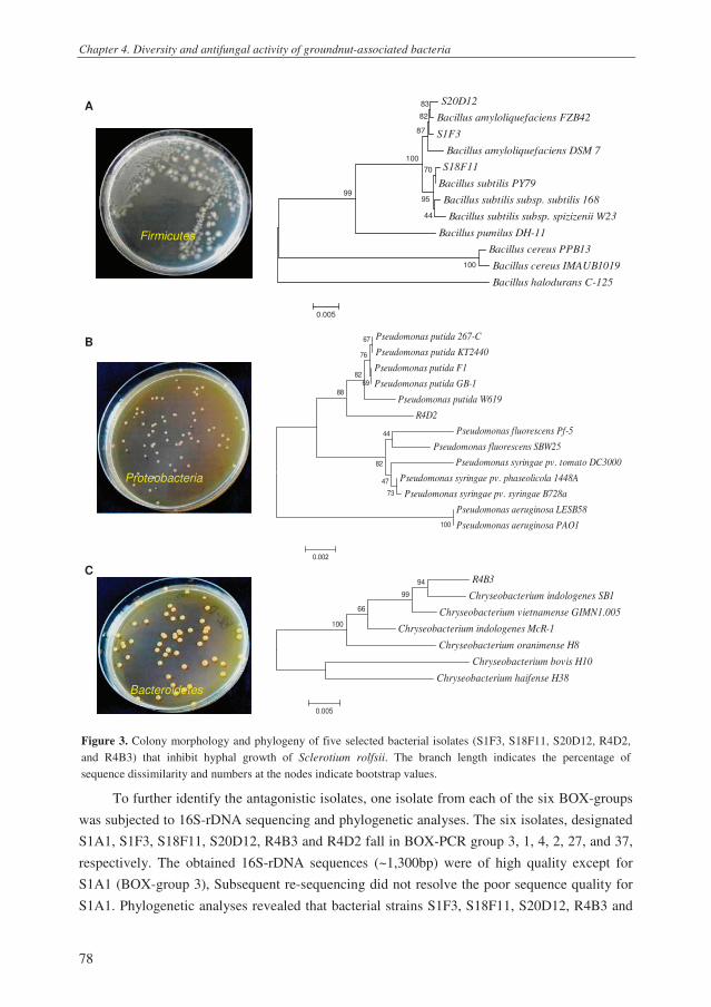
Chapter 4. Diversity and antifungal activity of groundnut-associated bacteria
78
A S20D12
Bacillus amyloliquefaciens FZB42
S1F3
Bacillus amyloliquefaciens DSM 7
S18F11
Bacillus subtilis PY79
Bacillus subtilis subsp. subtilis 168
Bacillus subtilis subsp. spizizenii W23
Bacillus pumilus DH-11
Bacillus cereus PPB13
Bacillus cereus IMAUB1019
Bacillus halodurans C-125
100
99
70
44
95
100
87
83
82
0.005 B
Pseudomonas putida 267-C
Pseudomonas putida KT2440
Pseudomonas putida F1
Pseudomonas putida GB-1
Pseudomonas putida W619
R4D2
Pseudomonas fluorescens Pf-5
Pseudomonas fluorescens SBW25
Pseudomonas syringae pv. tomato DC3000
Pseudomonas syringae pv. phaseolicola 1448A
Pseudomonas syringae pv. syringae B728a
Pseudomonas aeruginosa LESB58
Pseudomonas aeruginosa PAO1100
73
44
47
82
88
82
67
69
76
0.002 C R4B3
Chryseobacterium indologenes SB1
Chryseobacterium vietnamense GIMN1.005
Chryseobacterium indologenes McR-1
Chryseobacterium oranimense H8
Chryseobacterium bovis H10
Chryseobacterium haifense H38
100
66
94
99
0.005
Figure 3. Colony morphology and phylogeny of five selected bacterial isolates (S1F3, S18F11, S20D12, R4D2,
and R4B3) that inhibit hyphal growth of Sclerotium rolfsii. The branch length indicates the percentage of
sequence dissimilarity and numbers at the nodes indicate bootstrap values.
To further identify the antagonistic isolates, one isolate from each of the six BOX-groups
was subjected to 16S-rDNA sequencing and phylogenetic analyses. The six isolates, designated
S1A1, S1F3, S18F11, S20D12, R4B3 and R4D2 fall in BOX-PCR group 3, 1, 4, 2, 27, and 37,
respectively. The obtained 16S-rDNA sequences (~1,300bp) were of high quality except for
S1A1 (BOX-group 3), Subsequent re-sequencing did not resolve the poor sequence quality for
S1A1. Phylogenetic analyses revealed that bacterial strains S1F3, S18F11, S20D12, R4B3 and
Firmicutes
Proteobacteria
Bacteroidetes

Chapter 4. Diversity and antifungal activity of groundnut-associated bacteria
79
R4D2 belong to three bacterial taxa, i.e. the Firmicutes (Bacillus), Proteobacteria
(Pseudomonas), and Bacteroidetes (Chryseobacterium) (Fig. 3). Although the medium used for
isolation of these bacterial strains is semi-selective for Pseudomonas species, these results
indicate that also other bacterial genera can grow on this medium. This is consistent with earlier
observations of De Souza et al. (2003b) who showed that only 35.8% to 73.5% of the bacteria
isolated from wheat roots and plated on this semi-selective medium are Pseudomonas species;
the other bacteria represented different genera.
The three antagonistic strains classified as Bacillus (i.e. S1F3, S18F11, and S20D12)
originated from the stem base. At the species level, strains S1F3, and S20D12 were in the same
cluster as Bacillus amyloliquefaciens reference strains, whereas strain S18F11 clustered with
several Bacillus subtilis strains (Fig. 3). For the two antagonistic strain originated from the roots
of groundnut, strain R4D2 clustered close to the Pseudomonas putida group, and strain R4B3 to
Chryseobacterium species, including Chryseobacterium vietnamense strain GIMN1.005
isolated from forest soil in Vietnam (Li and Zhu 2011). Many Bacillus and Pseudomonas
species are well known for their antagonistic activities against plant pathogenic fungi and
oomycetes (Ongena and Jacques 2008; Raaijmakers et al. 2009; Raaijmakers et al. 2010).
Several Bacillus and Pseudomonas strains have been isolated from groundnut and studied as
biocontrol agents of S. rolfsii on groundnut (Pleban et al. 1995; Abd-Alla and Ezzat 2003; Abd-
Allah 2005; Abd-Allah and El-Didamony 2007; de Curtis et al. 2010; Hameeda et al. 2010;
Tonelli et al. 2010; Tonelli et al. 2011). For the genus Chryseobacterium, formerly known as
Flavobacterium (Vandamme et al. 1994), relatively little is known about their effects on plant
pathogens and plant growth. Chryseobacterium was reported to control Fusarium and
Rhizoctonia on tomato and pepper (Domenech et al. 2006) and Phytophthora capsici on pepper
(Kim et al. 2008). Recently, Chryseobacterium was reported as a potential biocontrol agent of
Pyricularia oryzae causing rice blast (Gandhi et al. 2009; Lucas et al. 2009). Interestingly,
Chryseobacterium was also reported to remove aflatoxin B1 from groundnut milk (Hao and
Brackett 1988). Since Chryseobacterium is commonly found in the geocarposphere, i.e. the soil
surrounding groundnut pods (Kloepper et al. 1992), representatives of this bacterial genus may
be useful to reduce contamination of groundnut pods with aflatoxin produced by Aspergillus
species.
Biocontrol of stem rot of groundnut under nethouse and field conditions
Based on the results of the phylogenetic analysis, four antagonistic strains were chosen
for the biocontrol assays, i.e. Bacillus sp. strains S18F11 and S20D12, Pseudomonas sp. strain
R4D2 and Chryseobacterium sp. strain R4B3. Under nethouse conditions, Pseudomonas strain
R4D2 significantly reduced stem rot disease incidence and severity (Fig. 4). The other three
bacterial isolates did not significantly suppress the disease relative to the control despite the fact
that they established similar population densities as strain R4D2 on the stem base and roots of
groundnut after two weeks of plant growth (Fig. 4). In the field experiment conducted in Quang

Chapter 4. Diversity and antifungal activity of groundnut-associated bacteria
80
Nam province (Vietnam), all four bacterial strains provided significant disease control at pod set
stage to a level similar to that of the fungicide Folicur (Fig. 5). When the disease progress
monitored from seedling stage until pod set was taken into account, only strain R4B3 reduced
DI significantly. The four applied bacterial strains established population densities on the stem
base and roots of groundnut, at flowering and pod set stages, ranging from 4.2-5.5 log cfu g-1
.
Bacillus sp. strain S20D12 established the lowest densities at pod set stage (Table 2). Next to
stem rot, damping-off disease, caused by Rhizoctonia solani, and black collar rot, caused by
Aspergillus niger, were monitored. For both diseases, the incidence in the control treatment was
not very high (i.e. ~3.7% and 8.3%, respectively) and no significant effects of the bacterial
treatments or the chemical Folicur on disease development were observed (data not shown).
Figure 4. Biocontrol of stem rot of groundnut and colonization of stem base and roots by different bacterial strains
under nethouse condition. Averages of three replicates are given. Different letters indicate a statistically significant
difference between the treatments (P=0.05, Duncan Multiple Range Test). Error bars represent the standard error
of the mean.
The bacterial treatments and the chemical Folicur had no effect on plant height, branch
length (data not shown), and the number of nodules per plant (Table 2). Only Bacillus strain
S20D12 significantly increased pod yield by 21% relative to the untreated control, whereas the
yield increases observed for most of the other bacterial and chemical treatments were
0
1
2
3
4
5
6
Lo
g c
fu g
-1ro
ots
a
a
a a
0
1
2
3
4
5
6
Lo
g c
fu g
-1ro
ots
a
a
a a
0
1
2
3
4
5
6
Lo
g c
fu g
-1ste
m b
ase
a a
a a
0
1
2
3
4
5
6
Lo
g c
fu g
-1ste
m b
ase
a a
a a
0
20
40
60
80
Dis
ease s
iveri
ty (
%)
Dis
ea
se
se
ve
rity
(%
)
ab
abab
a
b
0
20
40
60
80
Dis
ease s
iveri
ty (
%)
Dis
ea
se
se
ve
rity
(%
)
ab
abab
a
b
ab
abab
a
b
0
20
40
60
80
100
Dis
ease in
cid
en
ce (%
)
b ab
ab
a
b
0
20
40
60
80
100
Dis
ease in
cid
en
ce (%
)
b ab
ab
a
b

Chapter 4. Diversity and antifungal activity of groundnut-associated bacteria
81
intermediate between the control and S20D12 treatments (Table 2). Collectively these results
suggest that there is no apparent correlation between suppression of stem rot disease, root
colonization and yield increase. Indeed, correlation analysis showed that there was no
correlation (correlation coefficient = -0.04) between stem rot disease at pod set and pod yield of
groundnut.
Figure 5. Biocontrol of stem rot of groundnut by chemical fungicide Folicur, and groundnut-associated bacteria
Chryseobacterium sp. strain R4B3, Pseudomonas sp. strain R4D2, Bacillus sp. strains S18F11 and S20D12 under
field conditions in Quang Nam province, Vietnam. A) Disease incidence was monitored at seedling stage when
plants had 3-5 true leaves (20 days after sowing), and subsequently every 15 days. B) AUDPC represents the Area
Under the Disease Progress Curve, and was calculated based on the method used by Landa et al. (2002) and Kruijt
et al. (2009). For each developmental stage, averages of three replicates are given. Different letters indicate a
statistical significant difference between the treatments (P=0.05, Duncan Multiple Range Test).
Table 2. Effects of four different bacterial strains and the chemical fungicide Folicur on nodulation and pod yield
of groundnut plants grown under field conditions in Quang Nam province, Vietnam. Population densities of the
introduced bacterial strains on the stem base and roots of groundnut plants were determined at two developmental
stages (flowering, pod set). Averages of three replications are given. For each column, different letters indicate a
statistically significant difference between the treatments (P= 0.05, Duncan Multiple Range Test).
Population density introduced bacterial strain
(log cfu g-1
)
Flowering Pod set
Treatment
Stem base Roots Stem base Roots
Nodules
per plant
Dry pod
yield
(kg ha-1
)
Control 133 a 1220 b
Folicur 126 a 1300 ab
Chryseobacterium sp. R4B3 5.4 a 5.1 a 5.0 ab 5.4 ab 121 a 1190 b
Pseudomonas sp. R4D2 5.3 a 5.1 a 5.2 a 5.5 a 126 a 1320 ab
Bacillus sp. S18F11 5.4 a 5.4 a 5.0 ab 5.0 ab 116 a 1320 ab
Bacillus sp. S20D12 5.5 a 5.2 a 4.4 b 4.2 b 134 a 1480 a
0
2
4
6
8
10
AU
DP
C
b
ab
a
ab
ab
ab
0
2
4
6
8
10
Seedling Flowering Peg Pod set
Dis
ea
se
inc
ide
nc
e (
%)
Control
Folicur
Chryseobacterium sp. R4B3
Pseudomonas sp. R4D2
Bacillus sp. S18F11
Bacillus sp. S20D12
b
a
a a a a a
Developmental stages
0
2
4
6
8
10
Seedling Flowering Peg Pod set
Dis
ea
se
inc
ide
nc
e (
%)
Control
Folicur
Chryseobacterium sp. R4B3
Pseudomonas sp. R4D2
Bacillus sp. S18F11
Bacillus sp. S20D12
b
a
a a a a a
Developmental stages

Chapter 4. Diversity and antifungal activity of groundnut-associated bacteria
82
Mechanisms involved in antagonism of Pseudomonas sp. strain R4D2 toward S. rolfsii
To study the mechanisms underlying disease suppression of the tested bacterial strains,
we chose Pseudomonas sp. strain R4D2 for an in-depth analysis. This strain represents the
largest group of antagonistic bacteria isolated from groundnut based on BOX-PCR analysis
(Appendix 1) and gave good consistent results in the biocontrol experiments conducted in the
nethouse and field. Furthermore, initial attempts to perform random mutagenesis on the two
Bacillus and the Chryseobacterium strains were not successful.
Pseudomonas species are well-known for the production of a range of antibiotics (Haas
and Defago 2005; Gross and Loper 2009). Phenazines, pyrrolnitrin, pyoluteorin and 2,4-
diacetylphloroglucinol have been shown to play a role in the control of numerous fungal
pathogens including S. rolfsii (Raaijmakers et al. 2002; Haas and Defago 2005; D'aes et al.
2011; Chapter 3). Since RP-HPLC and TLC analyses showed that strain R4D2 does not produce
any of these four antibiotics in culture (data not shown) it is very unlikely that these compounds
contribute to the biocontrol activity of this strain. To identify genes and compounds, random
mutagenesis was performed. Among a relatively low number of transformants (154), two
mutants, designated MT38 and MT138, were identified that had completely lossed the ability to
inhibit hyphal growth of S. rolfsii in vitro (Fig. 6A).
Subsequently, a series of phenotypic analyses were performed to begin to identify the
nature of the mutations in MT38 and MT138. The drop collapse assay showed that wildtype
strain R4D2 produces a surfactant compound which is lacking in mutants MT38 and MT138
(Fig. 6B). To further support the hypothesis that R4D2 produces a surfactant compound,
swarming and biofilm assays were conducted. Previous studies with Pseudomonas and Bacillus
strains have shown that surfactants, in particular lipopeptide surfactants, are key metabolites in
swarming and biofilm formation (reviewed in Raaijmakers et al. 2010). The results showed that
mutants MT38 and MT138 formed less biofilm on the walls of plastic multi-well plates (Fig.
6C) and lost the ability to swarm on soft agar plates (Fig. 6D). For many Pseudomonas strains,
production of lipopeptide surfactants is governed by nonribosomal peptide synthetases
(Raaijmakers et al. 2010). These enzymes are encoded by large gene clusters of more than 30
kb (Raaijmakers et al. 2010), which may explain the high frequency of mutants (i.e. 2 out of
154) in the random mutagenesis experiment described above. To identify the genes disrupted in
these two mutants, the Y-linker method was used. Unfortunately, this method yielded no
amplicons (for mutant MT38) or amplicons with very poor sequence quality (for mutant
MT138). As an alternative, IPCR was used to amplify the genetic regions flanking the
transposon insertion, but also this method was so far not successful. Additional experiments are
required to support the hypotheses that strain R4D2 produces lipopeptide surfactants and that
the transposon insertions in mutants MT38 and MT138 are located in nonribosomal peptide
synthetase genes.
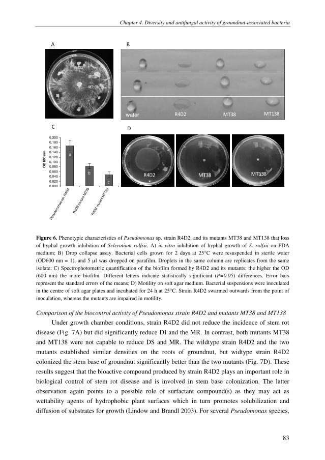
Chapter 4. Diversity and antifungal activity of groundnut-associated bacteria
83
MT38
R4D2
MT138
0.000
0.020
0.040
0.060
0.080
0.100
0.120
0.140
0.160
0.180
0.200
Pseud
om
ona
s sp. R
4D
2
R4D
2 m
uta
nt M
T38
R4D
2 m
uta
nt M
T138
OD
600 n
mA B
R4D2 MT38 MT138
water R4D2 MT38 MT138
C D
a
b
c
Figure 6. Phenotypic characteristics of Pseudomonas sp. strain R4D2, and its mutants MT38 and MT138 that loss
of hyphal growth inhibition of Sclerotium rolfsii. A) in vitro inhibition of hyphal growth of S. rolfsii on PDA
medium; B) Drop collapse assay. Bacterial cells grown for 2 days at 25°C were resuspended in sterile water
(OD600 nm = 1), and 5 µl was dropped on parafilm. Droplets in the same column are replicates from the same
isolate; C) Spectrophotometric quantification of the biofilm formed by R4D2 and its mutants; the higher the OD
(600 nm) the more biofilm. Different letters indicate statistically significant (P=0.05) differences. Error bars
represent the standard errors of the means; D) Motility on soft agar medium. Bacterial suspensions were inoculated
in the centre of soft agar plates and incubated for 24 h at 25°C. Strain R4D2 swarmed outwards from the point of
inoculation, whereas the mutants are impaired in motility.
Comparison of the biocontrol activity of Pseudomonas strain R4D2 and mutants MT38 and MT138
Under growth chamber conditions, strain R4D2 did not reduce the incidence of stem rot
disease (Fig. 7A) but did significantly reduce DI and the MR. In contrast, both mutants MT38
and MT138 were not capable to reduce DS and MR. The wildtype strain R4D2 and the two
mutants established similar densities on the roots of groundnut, but widtype strain R4D2
colonized the stem base of groundnut significantly better than the two mutants (Fig. 7D). These
results suggest that the bioactive compound produced by strain R4D2 plays an important role in
biological control of stem rot disease and is involved in stem base colonization. The latter
observation again points to a possible role of surfactant compound(s) as they may act as
wettability agents of hydrophobic plant surfaces which in turn promotes solubilization and
diffusion of substrates for growth (Lindow and Brandl 2003). For several Pseudomonas species,

Chapter 4. Diversity and antifungal activity of groundnut-associated bacteria
84
the lipopeptide surfactants have indeed been implicated in plant colonization (Hildebrand et al.
1998; Nielsen et al. 2005; Tran et al. 2007).
Figure 7. Biocontrol of stem rot of groundnut and colonization of stem base and roots by Pseudomonas sp. strain
R4D2 and its mutants MT38 and MT138 under growth chamber conditions. Averages of 3 replicates are given.
Different letters indicate a statistically significant difference between the treatments (P=0.05, Duncan Multiple
Range Test). Error bars represent the standard error of the mean.
0
20
40
60
80
Mo
rta
lity
ra
te (
%)
ba
ab b
0
20
40
60
80
Mo
rta
lity
ra
te (
%)
ba
ab bba
ab b
0
2
4
6L
og
cfu
g-1
roo
ts
aa a
0
2
4
6L
og
cfu
g-1
roo
ts
aa a
0
20
40
60
80
100
Dis
ea
se
siv
eri
ty (
%)
Dis
ease s
everi
ty (
%)
0
20
40
60
80
100
Dis
ea
se
siv
eri
ty (
%)
Dis
ease s
everi
ty (
%)
b
a
abb
0
20
40
60
80
100
Dis
ea
se
siv
eri
ty (
%)
Dis
ease s
everi
ty (
%)
0
20
40
60
80
100
Dis
ea
se
siv
eri
ty (
%)
Dis
ease s
everi
ty (
%)
b
a
abb
0
2
4
6
Lo
g c
fu g
-1s
tem
ba
se
a
b b
0
2
4
6
Lo
g c
fu g
-1s
tem
ba
se
a
b b
0
20
40
60
80
100
Dis
ea
se
inc
ide
nc
e (
%) a a a a
0
20
40
60
80
100
Dis
ea
se
inc
ide
nc
e (
%) a a a a

Chapter 4. Diversity and antifungal activity of groundnut-associated bacteria
85
Material and methods
Bacterial isolation and growth conditions
Healthy groundnut plants were collected from farmer fields in Quang Nam and Thua Thien Hue provinces
in Vietnam in 2009. Quang Nam and Thua Thien Hue are located in central Vietnam where groundnut is
commonly grown and where stem rot disease caused by S. rolfsii is widespread (Le et al. 2011). In Quang Nam and
Thua Thien Hue, a total of 40 and 30 groundnut plants at flowering stage, respectively, were randomly collected.
For each groundnut plant, roots and stem base were separated and kept in plastic bags on ice in an insulated box.
Bacterial isolations were performed in the laboratory the next day according to the method of Tran et al. (2008).
Briefly, approximately 1 gram (fresh weight) of roots with tightly adhering soil or 1 gram of stem base (i.e. 1 cm
up and down from the lowest branching point) was transferred to a 15 ml glass tube with 5 ml sterile 10 mM
MgSO4.7H2O, followed by vigorous vortexing for 1 min, sonication (Elma D-78224, Singen/HTW, Germany) for 1
min and vortexing again for 15 sec. The suspensions were then serially diluted in 10-fold steps in sterile 10 mM
MgSO4.7H2O, and 50 µl of the 100X and 1000X diluted samples were plated onto Pseudomonas agar (PSA) plates
supplemented with delvocid (100 µg ml-1
), ampicilin (40 µg ml-1
), and chloroamphenicol (12.5 µg ml-1
), and
incubated at 25°C for 48-72 h. This medium is semi-selective for fluorescent Pseudomonads (Simon and Ridge
1974), but also allows growth of several other bacterial genera (de Souza et al. 2003b). From each replicate sample,
forty-eight bacterial colonies were randomly picked and purified on PSA. Those isolates that were inhibitory to the
growth of S. rolfsii in dual culture inhibition assays (see below) were stored in glycerol (40%, v/v) at -20ºC and -
80°C. For comparison, a set of bacterial isolates that did not show any in vitro activity against S. rolfsii was also
purified and stored.
Hyphal growth inhibition assays
Inhibition of hyphal growth of S. rolfsii by bacterial isolates obtained from the stem base and roots of
groundnut was tested in dual culture assays according to the method of Kruijt et al. (2009). Briefly, bacterial
isolates were spot-inoculated at the edge of a 1/5th
-strength potato dextrose agar plate (1/5th
PDA, pH 6.5). After
incubation for 48 h at 25ºC, a 5-mm-diameter agar plug of a 3-day-old culture of S. rolfsii strain H001 (Le et al.
2011) was placed in the centre of the 1/5th
PDA plate and incubated at 25ºC. Inhibition of mycelial growth of S.
rolfsii by the bacterial isolates was recorded 3-4 days after fungal inoculation. In the initial screen, a total of 3,360
randomly selected bacterial isolates was tested. Isolates that showed in vitro inhibition of S. rolfsii were tested
again and their inhibition of hyphal growth of S. rolfsii was quantified. For each bacterial isolate, three plates
(replicates) were used. Hyphal growth (in mm) of S. rolfsii toward the bacterial colony and the control (no bacterial
colony) was measured after 48 h of incubation at 25ºC. Based on these two parameters, hyphal growth inhibition
(HGI) of the bacterial isolate relative to the control was calculated.
Bacterial identification
The genotypic diversity of the bacterial isolates with antifungal activity was investigated by BOX-PCR
analysis according to methods described by Tran et al. (2008). Amplicons ranging from 200 to 5000 bp were
scored visually for presence or absence. Bacterial isolates with identical BOX-PCR profiles were considered to be
genotypically identical. Representative isolates of several BOX-PCR groups were sent for 16S-rDNA sequencing
to Macrogen Inc. (Seoul, South Korea). The obtained forward and reverse sequences were assembled and edited in
Vector NTI (Invitrogen, version 8.0) and deposited in GenBank with accession numbers from JN572706 to
572710. For the phylogenetic analyses, the edited sequences were aligned to reference sequences available in
databases (http://www.ncbi.nlm.nih.gov/Genomes/ and http://www.pseudomonas.com/overview.jsp). Sequences
were trimmed to the same size (~1300 bp) and a phylogenetic tree was obtained with MEGA4 software
(http://megasoftware.net).

Chapter 4. Diversity and antifungal activity of groundnut-associated bacteria
86
Biochemical characterization of antagonistic bacterial isolates
After identification by 16S-rDNA sequencing, several of the bacterial strains representative of different
BOX-PCR groups and different bacterial genera were tested for the production of well-known antifungal
compounds, i.e. phenazines (PHZ), 2,4-diacetylphloroglucinol (DAPG), pyoluteorin (PLT) and pyrrolnitrin (PRN).
Thin layer chromatography (TLC) and reverse-phase high performance liquid chromatography (RP-HPLC) were
performed according to the methods described by de Souza et al. (2003a).
Nethouse experiments
Four selected bacterial strains, designated Chryseobacterium sp. R4B3, Pseudomonas sp. R4D2, Bacillus
sp. S18F11, and Bacillus sp. S20D12, were tested for biocontrol of stem rot disease of groundnut under nethouse
conditions at the Department of Plant Protection, Hue University of Agriculture and Forestry, Vietnam. Groundnut
seeds were soaked in water for 4 h at 25°C and then incubated overnight in Petri dishes at 25°C. Bacterial strains
were cultured on PSA plates for 48 h at 25ºC, harvested and washed three times with sterile water. The pre-
germinated seeds were subsequently soaked for 30 min in bacterial suspension with a density of 107 cells ml
-1. For
the control treatment, germinated seeds were soaked in sterile water for 30 min. One treated seed was sown in a
plastic bag containing 250 g of clay loam soil collected from a groundnut field in Thua Thien Hue province. Each
treatment consisted of three trays (three replicates) with 12 bags per tray. The trays were randomized. Two weeks
after pathogen inoculation at the base of the stem, disease incidence (DI), disease severity (DS) and mortality rate
(MR) were recorded. DS was rated on a scale from 0-4 with 0: no disease symptoms, 1: disease symptoms without
visible outgrowth of the fungus, 2: disease symptoms with visible outgrowth of the fungus, 3: partial wilting of the
plant, and 4: complete wilting and plant death (Le et al. 2011). DS is calculated based on the formula: DS= [(1×
number of plants rated as scale 1) + (2 × number of plants rated as scale 2) + (3 × number of plants rated as scale 3)
+ (4 × number of plants rated as scale 4)] × 100 ÷ (4 × total number of plants). MR is the number of dead plants
(scale 4) divided by the total number of plants.
Field experiment
The field experiment was conducted in 2010 in Quang Nam province, Vietnam, where approximately 20%
of the groundnut plants in the field were naturally infected by S. rolfsii (Le et al. 2011). The experiment consisted
of five treatments, with i) chemical fungicide Folicur, ii) Chryseobacterium strain R4B3, iii) Pseudomonas strain
R4D2, iv) Bacillus strain S18F11, and v) Bacillus strain S20D12, and one control (no treatment). The field
experiment was set-up based on a randomized complete block design (RCBD) with three blocks as three
replications and a plot size of 15 m2 (3 X 5 m). The distance was 30 cm between rows and 10 cm between plants
within a row. Bacterial strains were grown on PSA plates at 25°C for 48 h, harvested and washed with sterile
water. Prior to sowing groundnut seeds, bacterial suspensions were applied to the furrows at a final density of 106
cells per cm2 soil. Bacterial suspensions were applied again to the soil at the same density at the flowering stage of
the groundnut by watering bacterial suspension to the soil.
The field soil was fertilized with nitrogen (N) at 40 kg/ha, phosphorus (P2O5) at 60 kg/ha, potassium (K2O)
at 60 kg/ha, and calcium (Ca(OH)2) at 300 kg/ha. Phosphorus and calcium were applied at the time of soil
preparation. Seeds were sown at a depth of 3-5 cm and covered with soil. When plants had three true leaves
(seedling stage), 70% of the nitrogen and 50% of the potassium was applied. The remainder of the N and K
fertilization was applied at flowering. Weeds were manually removed at four developmental stages of the
groundnut plants, i.e. seedling, flowering, peg and pod set. At these four developmental stages, DI was monitored
in 1 m2
of each plot, i.e. approximately 33 plants per plot. Also other characteristics such as plant height,
nodulation, and pod yield were monitored. The number of nodules per plant root system was determined at the pod
stage. To that end, three plants per plot were harvested; their root systems were washed to remove adhering soil
and the number of nodules was counted.

Chapter 4. Diversity and antifungal activity of groundnut-associated bacteria
87
Identification of bacterial genes involved in antifungal activity
To identify genes involved in activity of Pseudomonas sp. strain R4D2 against S. rolfsii, we conducted
random transposon mutagenesis by bi-parental mating of a spontaneous rifampicin-resistant derivative of R4D2
with E. coli S17-1 (λpir) containing plasmid pUT LuxCDABE TcR (Winson et al. 1998). Mutants were selected on
PSA plates supplemented with tetracyclin (100 µg ml-1
) and rifampicin (100 µg ml-1
) and transferred to microtitre
plates containing 100 µl King’s medium B per well. After 2 days of growth at 25°C, transformants were tested for
in vitro activity against S. rolfsii as described above. For mutants that had lost the ability to inhibit hyphal growth
of S. rolfsii, the regions flanking the transposon insertion were amplified using the Y-linker method (Kwon and
Ricke 2000). Briefly, genomic DNA was digested with a restriction enzyme (NlaIII or SphI) and ligated with Y-
linkers 1 and 2. The sequence of Y-linker 1 is 5’-TTTCTGCTCGAATTCAAGCTTCTAACGATGTACGGGGACACATG-3’,
and that of Y-linker 2 is 5’-TGTCCCCGTACATCGTTAGAACTACTCGTACCATCCACAT-3’. To amplify the genetic region
flanking the transposon insertion, Y-linker primer 5’-CTGCTCGAATTCAAGCTTCT-3’ and Tn5 primer 5’-
GGCCAGATCTGATCAAGAGA-3’ were used in PCR. Amplicons were purified and sent for sequencing (Macrogen
Corp. Europe, Amsterdam, The Netherlands). Quality assessment and analysis of the obtained sequences were
performed with Vector NTI (Invitrogen, version 8.0). For BLAST searches, databases at the National Center for
Biotechnology Information (http://www.ncbi.nlm.nih.gov) and Computational & Genome Biology Initiative
Bioinformatics (CGBI: http://cgbi.cgrb.oregonstate.edu/) were used. As a second approach to identify the regions
flanking the transposon insertion, we used the inverse PCR method (Lewenza et al. 2005). For this, genomic DNA
was digested with NarI or SstII. Linear genomic fragments were circularized by ligation with T4 DNA Ligase. The
ligation product was used as template for an inverse PCR (IPCR) reaction using outward facing transposon-specific
primers. Primer 1 (Tn5-NarI) and primer 2 (Tn5-out2) for DNA digested by NarI; primer 1 (Tn5-SstII), and primer
2 (Tn5-out) for DNA digested by SstII.
Mutants of strain R4D2 that showed no or reduced activity against S. rolfsii were also characterized
phenotypically by analyzing their growth rate, swarming motility, and their capacities to form biofilms and to
produce biosurfactant compounds. The methods used for these phenotypic traits were described previously by De
Bruijn et al. (2007; 2008).
Growth chamber bioassay
The efficacy of Pseudomonas strain R4D2 wildtype and its mutants to control S. rolfsii infection of
groundnut was investigated in growth chamber bioassays at Wageningen University (Wageningen, Netherlands).
Growth chamber conditions comprised a continuous temperature of 25ºC during day (12h) and night (12h), a
relative air humidity of 80%, and a light intensity of 315 µmol light m
–2 s
–1. Groundnut seeds were surface
sterilized in 3% (v/v) sodium hypochlorite solution for 15 min, rinsed with ethanol 70% (v/v) and rinsed three
times with ample sterile distilled water. Sterilized seeds were soaked in sterile water for 4 h at 25°C and then
incubated in Petri dishes at 25°C overnight. The pre-germinated seeds were subsequently soaked for 30 min in
bacterial suspension with a density of 107 cells ml
-1. For the control treatment, germinated seeds were soaked in
sterile water for 30 min. Treated seeds were sown in pots containing 250 g of a mix 1:3 (w/w) of commercial
potting soil and river sand. Each treatment consisted of three replications with five plants per replication. In the
growth chamber, the treatments were randomized. Infection of seedlings with S. rolfsii and disease assessment was
monitored as described above for the nethouse experiment.
Chapter 4. Diversity and antifungal activity of groundnut-associated bacteria
88
References
Abd-Alla, E.F. and Ezzat (2003) Biocontrol of peanut southern blight (Sclerotium rolfsii) by Bacillus subtilis.
Journal of microbiology of the United Arab Republic 38, 207-216.
Abd-Allah, E.F. (2005) Effect of a Bacillus subtilis isolate on southern blight (Sclerotium rolfsii) and lipid
composition of peanut seeds. Phytoparasitica 33, 460-466.
Abd-Allah, E.F. and El-Didamony, G. (2007) Effect of seed treatment of Arachis hypogaea with Bacillus subtilis
on nodulation in biocontrol of southern blight (Sclerotium rolfsii) disease. Phytoparasitica 35, 8-12.
Brown, S.L. (2007) Peanut insects: ecology and control. In Encyclopedia of Pest Management ed. Pimentel, D.
pp.1 - 10: Taylor & Francis
Coleysmi, J.R. and Cooke, R.C. (1971) Survival and germination of fungal sclerotia. Annual Review of
Phytopathology 9, 65-92.
D'aes, J., Gia, K.H.H., De Maeyer, K., Pannecoucque, J., Forrez, I., Ongena, M., Dietrich, L.E.P., Thomashow,
L.S., Mavrodi, D.V. and Hofte, M. (2011) Biological control of Rhizoctonia root rot on bean by phenazine-
and cyclic lipopeptide-producing Pseudomonas CMR12a. Phytopathology 101, 996-1004.
de Bruijn, I., de Kock, M.J.D., Yang, M., de Waard, P., van Beek, T.A. and Raaijmakers, J.M. (2007) Genome-
based discovery, structure prediction and functional analysis of cyclic lipopeptide antibiotics in
Pseudomonas species. Molecular Microbiology 63, 417-428.
de Bruijn, I., de Kock, M.J.D., de Waard, P., van Beek, T.A. and Raaijmakers, J.M. (2008) Massetolide A
biosynthesis in Pseudomonas fluorescens. Journal of Bacteriology 190, 2777-2789.
de Curtis, F., Lima, G., Vitullo, D. and De Cicco, V. (2010) Biocontrol of Rhizoctonia solani and Sclerotium rolfsii
on tomato by delivering antagonistic bacteria through a drip irrigation system. Crop Protection 29, 663-670.
de Souza, J.T., de Boer, M., de Waard, P., van Beek, T.A. and Raaijmakers, J.M. (2003a) Biochemical, genetic,
and zoosporicidal properties of cyclic lipopeptide surfactants produced by Pseudomonas fluorescens.
Applied and Environmental Microbiology 69, 7161-7172.
de Souza, J.T., Mazzola, M. and Raaijmakers, J.M. (2003b) Conservation of the response regulator gene gacA in
Pseudomonas species. Environmental Microbiology 5, 1328-1340.
Domenech, J., Reddy, M.S., Kloepper, J.W., Ramos, B. and Gutierrez-Manero, J. (2006) Combined application of
the biological product LS213 with Bacillus, Pseudomonas or Chryseobacterium for growth promotion and
biological control of soil-borne diseases in pepper and tomato. BioControl (Dordrecht) 51, 245-258.
Gandhi, P.M., Narayanan, K.B., Naik, P.R. and Sakthivel, N. (2009) Characterization of Chryseobacterium
aquaticum strain PUPC1 producing a novel antifungal protease from rice rhizosphere soil. Journal of
Microbiology & Biotechnology 19, 99-107.
Ganesan, P. and Gnanamanickam, S.S. (1987) Biological control of Sclerotium rolfsii Sacc. in peanut by
inoculation with Pseudomonas fluorescens. Soil Biology and Biochemistry 19, 35-38.
Ganesan, S., Kuppusamy, R. and Sekar, R. (2007) Integrated management of stem rot disease (Sclerotium rolfsii)
of groundnut (Arachis hypogaea L.) using Rhizobium and Trichoderma harzianum (ITCC-4572). Turkish
journal of agriculture & forestry 31, 103-108.
Gross, H. and Loper, J.E. (2009) Genomics of secondary metabolite production by Pseudomonas spp. Nat Prod
Rep 26, 1408-1446.
Haas, D. and Defago, G. (2005) Biological control of soil-borne pathogens by fluorescent pseudomonads. Nature
Reviews Microbiology 3, 307-319.
Hameeda, B., Harini, G., Rupela, O.P., Rao, J.V.D.K.K. and Reddy, G. (2010) Biological control of chickpea collar
rot by co-inoculation of antagonistic bacteria and compatible Rhizobia. Indian Journal of Microbiology 50,
419-424.
Hao, Y.Y. and Brackett, R.E. (1988) Removal of aflatoxin B1 from peanut milk inoculated with Flavobacterium
aurantiacum. Journal of Food Science 53, 1384-1386.

Chapter 4. Diversity and antifungal activity of groundnut-associated bacteria
89
Hildebrand, P.D., Braun, P.G., McRae, K.B. and Lu, X. (1998) Role of the biosurfactant viscosin in broccoli head
rot caused by a pectolytic strain of Pseudomonas fluorescens. Can J Plant Pathol-Rev Can Phytopathol 20,
296-303.
Kim, Y.S., Jang, B., Chung, I.-M., Sang, M.K., Ku, H.-M., Kim, K.D. and Chun, S.-C. (2008) Enhancement of
biocontrol activity of antagonistic Chryseobacterium strain KJ1R5 by adding carbon sources against
Phytophthora capsici. Plant Pathology Journal 24, 164-170.
Kishore, G.K., Pande, S. and Podile, A.R. (2005a) Biological control of collar rot disease with broad-spectrum
antifungal bacteria associated with groundnut. Canadian Journal of Microbiology 51, 123-132.
Kishore, G.K., Pande, S., Rao, J.N. and Podile, A.R. (2005b) Pseudomonas aeruginosa inhibits the plant cell wall
degrading enzymes of Sclerotium rolfsii and reduces the severity of groundnut stem rot. European Journal
of Plant Pathology 113, 315-320.
Kloepper, J.W., McInroy, J.A. and Bowen, K.L. (1992) Comparative identification by fatty-acid analysis of soil,
rhizosphere, and geocarposphere bacteria of peanut (Arachis hypogaea L.). Plant and Soil 139, 85-90.
Kruijt, M., Tran, H. and Raaijmakers, J.M. (2009) Functional, genetic and chemical characterization of
biosurfactants produced by plant growth-promoting Pseudomonas putida 267. Journal of Applied
Microbiology 107, 546-556.
Kwon, Y.M. and Ricke, S.C. (2000) Efficient amplification of multiple transposon-flanking sequences. Journal of
Microbiological Methods 41, 195-199.
Landa, B.B., Mavrodi, O.V., Raaijmakers, J.M., Gardener, B.B.M., Thomashow, L.S. and Weller, D.M. (2002)
Differential ability of genotypes of 2,4-diacetylphloroglucinol-producing Pseudomonas fluorescens strains
to colonize the roots of pea plants. Applied and Environmental Microbiology 68, 3226-3237
Lewenza, S., Falsafi, R.K., Winsor, G., Gooderham, W.J., McPhee, J.B., Brinkman, F.S.L. and Hancock, R.E.W.
(2005) Construction of a mini-Tn5-luxCDABE mutant library in Pseudomonas aeruginosa PAO1: A tool
for identifying differentially regulated genes. Genome Research 15, 583-589.
Li, Z. and Zhu, H. (2011) Chryseobacterium vietnamense sp. nov., isolated from forest soil. International Journal
of Systematic and Evolutionary Microbiology.
Lindow, S.E. and Brandl, M.T. (2003) Microbiology of the phyllosphere. Applied and Environmental
Microbiology 69, 1875-1883.
Lucas, J.A., Ramos Solano, B., Montes, F., Ojeda, J., Megias, M. and Gutierrez Manero, F.J. (2009) Use of two
PGPR strains in the integrated management of blast disease in rice (Oryza sativa) in Southern Spain. Field
Crops Research 114, 404-410.
Mehan, V.K., Mayee, C.D. and McDonald, D. (1994) Management of Sclerotium rolfsii caused stem and pod rots
of groundnut - A critical review. International journal of pest management 40, 313-320.
Nielsen, T.H., Nybroe, O., Koch, B., Hansen, M. and Sorensen, J. (2005) Genes involved in cyclic lipopeptide
production are important for seed and straw colonization by Pseudomonas sp. strain DSS73. Applied and
Environmental Microbiology 71, 4112-4116.
Ongena, M. and Jacques, P. (2008) Bacillus lipopeptides: versatile weapons for plant disease biocontrol. Trends
Microbiol 16, 115-125.
Pastor, N.A., Reynoso, M.M., Tonelli, M.L., Masciarelli, O., Rosas, S.B. and Rovera, M. (2010) Potential
biological control Pseudomonas sp. PCI2 against damping-off of tomato caused by Sclerotium rolfsii.
Journal of Plant Pathology 92, 737-745.
Pleban, S., I, N.F. and Chet, I. (1995) Control of Rhizoctonia solani and Sclerotium rolfsii in the greenhouse using
endophytic Bacillus spp. European Journal of Plant Pathology 101, 665-672.
Punja, Z.K. (1985) The biology, ecology, and control of Sclerotium rolfsii. Annual Review of Phytopathology 23,
97-127.

Chapter 4. Diversity and antifungal activity of groundnut-associated bacteria
90
Raaijmakers, J.M., Vlami, M. and de Souza, J.T. (2002) Antibiotic production by bacterial biocontrol agents.
Antonie Van Leeuwenhoek International Journal of General and Molecular Microbiology 81, 537-547.
Raaijmakers, J.M., Paulitz, T.C., Steinberg, C., Alabouvette, C. and Moonne-Loccoz, Y. (2009) The rhizophere: a
playground and battlefield for soiborne pathogens and beneficial microorganisms. plant & soil. 321, 341-
361
Raaijmakers, J.M., de Bruijn, I., Nybroe, O. and Ongena, M. (2010) Natural functions of lipopeptides from
Bacillus and Pseudomonas: more than surfactants and antibiotics. Fems Microbiology Reviews 34, 1037-
1062.
Shew, B.B. and Waliyar, F. (2007) Peanut diseases: ecology and control. In Encyclopedia of Pest Management ed.
Pimentel, D. pp.469–473: Taylor & Francis.
Simon, A. and Ridge, E.H. (1974) Ue of ampicillin in a simplified selective medium for isolation of fluorescent
pseudomonads. Journal of Applied Bacteriology 37, 459-460.
Tonelli, M.L., Furlan, A., Taurian, T., Castro, S. and Fabra, A. (2011) Peanut priming induced by biocontrol
agents. Physiological and Molecular Plant Pathology 75, 100-105.
Tonelli, M.L., Taurian, T., Ibanez, F., Angelini, J. and Fabra, A. (2010) Selection and in vitro characterization of
biocontrol agents with potential to protect peanut plants against fungal pathogens. Journal of Plant
Pathology 92, 73-82.
Tran, H., Ficke, A., Asiimwe, T., Höfte, M. and Raaijmakers, J.M. (2007) Role of the cyclic lipopeptide
massetolide A in biological control of Phytophthora infestans and in colonization of tomato plants by
Pseudomonas fluorescens. New Phytologist 175, 731-742.
Tran, H., Kruijt, M. and Raaijmakers, J.M. (2008) Diversity and activity of biosurfactant-producing Pseudomonas
in the rhizosphere of black pepper in Vietnam. Journal of Applied Microbiology 104, 839-851.
Vandamme, P., Bernardet, J.-F., Segers, P., Kersters, K. and Holmes, B. (1994) Notes: New perspectives in the
classification of the Flavobacteria: Description of Chryseobacterium gen. nov., Bergeyella gen. nov., and
Empedobacter nom. rev. International Journal of Systematic Bacteriology 44, 827-831.
Winson, M.K., Swift, S., Hill, P.J., Sims, C.M., Griesmayr, G., Bycroft, B.W., Williams, P. and Stewart, G. (1998)
Engineering the luxCDABE genes from Photorhabdus luminescens to provide a bioluminescent reporter for
constitutive and promoter probe plasmids and mini-Tn5 constructs. FEMS Microbiology Letters 163, 193-
202.

Chapter 4. Diversity and antifungal activity of groundnut-associated bacteria
91
Appendix 1. BOX-PCR grouping of bacterial isolates from stem base and roots of groundnut plants grown in
farmer fields in 2010 in central Vietnam. In vitro antagonism refers to the inhibition of hyphal growth of the fungal
pathogen Sclerotium rolfsii.
Stem base Roots
BOX-PCR
Group
No. of
isolates
In vitro
antagonism
BOX-PCR
Group
No. of
isolates
In vitro
antagonism
1 1 + 20 4 -
2 3 + 21 1 -
3 1 + 22 2 -
4 1 + 23 3 -
5 1 - 24 1 -
6 1 - 25 1 -
7 1 - 26 1 -
8 1 - 27 1 +
9 1 - 28 2 -
10 1 - 29 4 -
11 1 - 30 1 -
12 1 - 31 1 -
13 1 - 32 1 -
14 1 - 33 2 -
15 1 - 34 1 -
16 1 - 35 1 -
17 1 - 36 2 -
18 1 - 37 6 +
19 1 - 38 1 -
39 1 -
40 1 -
41 1 -
42 1 -

Chapter 4. Diversity and antifungal activity of groundnut-associated bacteria
92

Chapter 5. Biocontrol of groundnut diseases and plant growth promotion
Chapter 5
Biological control of groundnut diseases and plant growth
promotion by beneficial Pseudomonas and Bacillus species
Le, C.N., Thai, T.H. and Raaijmakers, J.M.
To be submitted

Chapter 5. Biocontrol of groundnut diseases and plant growth promotion
94

Chapter 5. Biocontrol of groundnut diseases and plant growth promotion
95
Biological control of groundnut diseases and plant growth promotion
by beneficial Pseudomonas and Bacillus species
Le, C.N.1,2
, Thai, T.H.3 and Raaijmakers, J.M.
1
1 Laboratory of Phytopathology, Wageningen University, the Netherlands
2 Department of Plant Protection, Hue University of Agriculture and Forestry, Vietnam
3 Department of Soil Sciences and Environment, Hue University of Agriculture and Forestry, Vietnam
Abstract
In Vietnam, groundnut yield is adversely affected by several diseases,
including stem rot disease caused by Sclerotium rolfsii. In this study, various
Pseudomonas and Bacillus strains were tested in field experiments in Thua
Thien Hue province, Vietnam for their abilities to (i) control stem rot and other
groundnut diseases, and (ii) promote plant growth and enhance pod yield.
Pseudomonas sp. strains SH-C52 and R4D2 significantly reduced stem rot
disease of groundnut to the same level as the azole fungicide Folicur. Both
bacterial strains were not effective against Rhizoctonia damping-off disease,
black collar rot caused by Aspergillus niger, and early and late leaf spot caused
by Cercospora arachidicola and Cercosporidium personatum, respectively.
Both strains, however, improved pod yield up to 14% relative to the untreated
control. Also Pseudomonas fluorescens strain SS101 and Bacillus sp. strains
S18F11 and S20D12 led to significant increases in pod yield (up to 30%).
These three strains were unable to control stem rot, damping-off, black collar
rot, and bacterial wilt disease but significantly reduced early and late leaf spot
diseases. Collectively, these field experiments in central Vietnam indicate that
specific Pseudomonas and Bacillus strains are promising candidates for
integrated management of various groundnut diseases and for improving pod
yield.
Keywords: Bacillus, Pseudomonas, Sclerotium rolfsii

Chapter 5. Biocontrol of groundnut diseases and plant growth promotion
96
Introduction
Groundnut (Arachis hypogaea L.) is a major source of edible oil, vitamins, and amino
acids, and used extensively for food and feed (Woodroof 1983; Florkowski 1994; Savage and
Keenan 1994). In terms of economic importance, groundnut ranks thirteenth among the world
food crops and tops the list of oil seed crops both in terms of acreage and production (Sharma
2002). It is cultivated in more than 80 countries in the tropics and subtropics (Ramantha Rao
and Murty 1994). In 2009, groundnut was grown on 24 million hectares worldwide with an
estimated total production of 36.5 million ton (FAO, 2011). In most developing countries,
however, groundnut yield is low. For example, yield in India was only around 1,000 kg ha-1
in
2009 compared to 3,350 kg ha-1
in China and 3,800 kg ha-1
in the USA. The average groundnut
yield in the world is around 1,500 kg ha-1
(FAO, 2011).
In Vietnam, groundnut is the second annual crop after paddy rice with a total area of
249,200 ha and an average yield of 2,100 kg ha-1
in 2009 (FAO, 2011). Vietnam ranks fifth
among the groundnut-producing countries after China, India, USA and Argentina (Fabra et al.
2010). Groundnut cultivation in Vietnam and other countries is hampered by a wide range of
pests and diseases (Middleton et al. 1994; Wightman and Ranga Rao 1994; Mehan and Hong
1994; Pham 1997; Le 2004; Nguyen et al. 2004; Brown 2007; Shew and Waliyar 2007). The
most widespread fungal and bacterial diseases of groundnut are stem and pod rot caused by
Sclerotium rolfsii, black collar rot by Aspergillus niger, damping-off by Rhizoctonia solani,
bacterial wilt by Ralstonia solanacearum, rust by Puccinia arachidis, early leaf spot by
Cercospora arachidicola, and late leaf spot by Cercosporidium personatum (Porter et al. 1984;
Shew and Waliyar 2007).
Breeding for new groundnut cultivars, improving fertilization and applying chemical
pesticides are measures to improve groundnut growth and yield. However, several of these
measures are costly for subsistence farmers in Vietnam and other developing countries. In this
context, biological control has been proposed as an environmentally friendly, affordable and
sustainable means to control pests and diseases (Fravel 2005; Höfte and Altier 2010). Biological
control may also offer a solution for controlling pests and diseases for which no other effective
measures are available or when agricultural products must be certified as ‘organic’ without
pesticide residues (Fravel 2005). Biological control based on treating soil, seeds or other
planting materials with beneficial microorganisms may not only limit the proliferation of pests
and diseases, but may also directly promote plant growth or result in the degradation of toxic
soil pollutants (van Loon 2007; Lugtenberg and Kamilova 2009).
In the past decades, various strains of Pseudomonas and Bacillus species have been
screened for their abilities to control specific groundnut diseases (Ganesan and Gnanamanickam
1987; Savithiry and Gnanamanickam 1987; Kokalis-Burelle et al. 1992; Shanmugam et al.
2002; Abd-Alla and Ezzat 2003; Dey et al. 2004; Abd-Allah 2005; Kishore et al. 2005a;
Kishore et al. 2005b; Murugalakshmi et al. 2009; Senthilraja et al. 2010a; Senthilraja et al.

Chapter 5. Biocontrol of groundnut diseases and plant growth promotion
97
2010b; Taurian et al. 2010; Tonelli et al. 2010; Tonelli et al. 2011). In nethouse and field
studies conducted in 2010 in Vietnam, we identified several Pseudomonas and Bacillus strains
that significantly suppressed stem rot disease caused by S. rolfsii (Chapters 3 & 4). For two of
these strains, i.e. Pseudomonas sp. strains SH-C52 and R4D2, the ability to suppress stem rot
disease appeared to be linked to the production of biosurfactant compounds (Chapters 3 & 4).
Another set of strains, comprising P. fluorescens strain SS101 and Bacillus sp. strains S18F11
and S20D12, did not significantly or consistently suppress stem rot disease in nethouse and field
experiments in 2010, but substantially enhanced pod yield up to 21% relative to the untreated
control (Chapters 3 & 4). For successful implementation of one or more of these bacterial
strains in groundnut cultivation, their consistency to suppress diseases and/or to improve yield
under field conditions should be evaluated as well as their efficacy to control other root and leaf
diseases of groundnut. In this study, field experiments were conducted in central Vietnam in
2011 to determine the efficacy of Pseudomonas strains SH-C52, R4D2 and SS101, and of
Bacillus strains S18F11 and S20D12 to (i) suppress stem rot disease, damping-off, black collar
rot, leaf spots and bacterial wilt, and (ii) promote plant growth and enhance pod yield.
Results and Discussion
Biocontrol of groundnut diseases by Pseudomonas
As reported previously (Chapters 3 & 4), Pseudomonas sp. strains SH-C52 and R4D2
significantly suppressed stem rot disease caused by S. rolfsii in nethouse and field experiments
conducted in 2010 in Vietnam. Also in the 2011 field experiment, application of Pseudomonas
sp. strains SH-C52 and R4D2 resulted in a significant suppression of stem rot disease caused by
S. rolfsii (Fig. 1A, B). At pod set stage, i.e. 10 weeks after sowing, disease incidence was
significantly lower in the two bacterial treatments than in the control treatment (Fig. 1A). Both
strains were as effective as Folicur (Fig. 1A, B), the azole fungicide commonly used to control
S. rolfsii (Brenneman and Murphy, 1991). Strains SH-C52 and R4D2 established similar
densities on the stem base and roots of groundnut ranging from 4.5- 5 log cfu g-1
after 8 weeks
of plant growth (Fig. 1C). For strain SH-C25, the chlorinated lipopeptide thanamycin plays a
key role in suppression of S. rolfsii (Chapter 3). Also for strain R4D2, a biosurfactant appears to
be an important factor in growth inhibition and biocontrol of S. rolfsii (Chapter 4), but the
chemical nature of this biosurfactant and the corresponding biosynthesis genes have not yet
been identified.
Given that biosurfactant compounds, and in particular lipopeptides, have a broad-
spectrum antimicrobial activity (Raaijmakers 2006; D’aes et al. 2010; Raaijmakers et al. 2010;
Mendes et al. 2011), and are known to induce systemic resistance in plants against oomycete
and fungal pathogens (Ongena et al. 2007; Tran et al. 2007), we also monitored the effects of
strains SH-C52 and R4D2 on various other diseases of groundnut. The results showed that the
incidence of Rhizoctonia damping-off disease was low to insignificant and that the bacterial

Chapter 5. Biocontrol of groundnut diseases and plant growth promotion
98
strains and Folicur did not affect this disease (Table 1). The incidence of black collar rot, caused
by Aspergillus niger, was higher, but also for this disease no significant effects of the two
bacterial treatments and Folicur were observed (Table 1). Pseudomonas strain R4D2
significantly reduced bacterial wilt caused by R. solanacearum at pod set stage (Table 1).
Folicur was not effective and strain SH-C52 reduced wilt disease to a level intermediate
between the control and R4D2 treatment (Table 1).
A B
0
2
4
6
8
10
12
14
Seedling Flowering Peg Pod set
Dis
ea
se
in
cid
en
ce
(%
)
Developmental stages
Control
Pseudomonas sp. SH-C52
Pseudomonas sp. R4D2
Folicur
0
5
10
15
20
25
AU
DP
C
C
Pseu
dom
on
as s
p. S
H-C
52
Pseu
dom
on
as s
p. S
H-C
52
Pseu
dom
ona
s s
p. R
4D
2
Pseu
dom
ona
s s
p. R
4D
2
0
1
2
3
4
5
6
Stem base Roots
Lo
g c
fu g
-1
Figure 1. Effects of Pseudomonas sp. strain
SH-C52, Pseudomonas sp. strain R4D2 and the
fungicide Folicur on stem rot disease incidence
of groundnut under field conditions in Thua
Thien Hue, Vietnam, in 2011. A) incidence of
stem rot disease, caused by Sclerotium rolfsii, at
four developmental stages of groundnut, B) area
under the disease progress curve for stem rot
disease (AUDPC), and C) population densities
of the applied bacterial strains on the stem base
and roots of groundnut at the peg stage, i.e.
eight weeks after sowing.
The incidence and severity of leaf spot diseases caused by C. arachidicola and C.
personatum were not reduced by the two bacterial treatments (Table 1). Also Folicur treatment
did not lead to a reduction in leaf spot diseases (Table 1), which is in contrast to results of
earlier studies conducted in Georgia and in Texas, USA (Brenneman and Murphy 1991; Besler
et al. 2006). This may be due to the limited number of fungicide applications in our experiment
(i.e. 3 times at a rate of 250 g a.i. ha-1
) as compared to other studies, with seven applications in
a
a a
b
a a
b
a
a a a
a

Chapter 5. Biocontrol of groundnut diseases and plant growth promotion
99
experiments in Georgia (USA) at a rate of 188 g a.i. ha-1
(Brenneman and Murphy 1991), and
four applications in Texas (USA) at a rate of 230 g a.i. ha-1
(Besler et al. 2006). Furthermore,
since S. rolfsii was the primary target pathogen in our experiment, the fungicide was applied
mostly to the stem base of groundnut. As a result, many leaves of groundnut were not exposed
to the fungicide.
Table 1. Effects of Pseudomonas sp. strain SH-C52, Pseudomonas sp. strain R4D2 and the fungicide Folicur on
the incidence and severity of different groundnut diseases under field conditions. The experiment was conducted in
2011 in Thua Thien Hue province, Vietnam. For each of the parameters, average values of four replications are
given. For each row, different letters indicate a statistically significant difference between the treatments (P=0.05,
Duncan Multi Range Test).
Disease Treatment
Control Pseudomonas
sp. SH-C52
Pseudomonas
sp. R4D2
Folicur
Damping off (Rhizoctonia solani) (%)
1.58 a 1.18 a 0.80 a 1.30 a
Black collar rot (Aspergillus niger) (AUDPC)*
8.75 a 6.25 a 7.92 a 6.25 a
Bacterial wilt (Ralstonia solanacearum) (%)
2.79 b 1.74 ab 0.94 a 2.04 b
Leaf spot (Cercospora arachidicola, Cercosporidium personatum) (%)**
DI-F 66.5 a 66.5 a 69.0 a 66.5 a
DS-F 26.0 a 25.3 a 26.0 a 24.9 a
DI-Peg 42.0 a 36.0 a 39.5 a 44.0 a
DS-Peg 15.6 a 12.7 a 16.6 a 15.9 a
*AUDPC- area under disease progress curve. **Fifty leaves of each plot were investigated for leaf spot diseases.
DI-F: disease incidence at flowering stage, DS-F: disease severity at flowering stage, DI-Peg: DI at peg stage,
DS-Peg: DS at peg stage.
Bacterial strains R4D2 and SH-C52 did not promote or adversely affect plant biomass and
nodulation (Table 2), and did not significantly alter the nitrogen and carbon contents of
groundnut (Appendix 1A). However, both strains did lead to significant pod yield increases of
groundnut up to 13.8% for strain R4D2 (Table 2). The pod yield in the R4D2 treatment was also
significantly higher than that in the fungicide treatment (Table 2). Given that Folicur reduced
stem rot disease to a similar level as the bacterial treatments, there seems to be no apparent
correlation between a reduction of stem rot disease incidence and an increase in pod yield.

Chapter 5. Biocontrol of groundnut diseases and plant growth promotion
100
Table 2. Effects of Pseudomonas sp. strain SH-C52, Pseudomonas sp. strain R4D2, and the fungicide Folicur on
emergence, plant biomass (fresh weight (FW) and dry weight (DW)), number and dry weight of root nodules,
number of flowers, and dry pod yield of groundnut. The field experiment was conducted in 2011 in Thua Thien
Hue province, Vietnam. For each parameter, averages of four replications are given. For each row, different letters
indicate a statistically significant difference between the treatments (P=0.05, Duncan Multi Range Test).
Characters Treatment
Control Pseudomonas
sp. SH-C52
Pseudomonas
sp. R4D2
Folicur
Emergence at two weeks after sowing (%)
77.8 a 74.3 a 74.8 a 73.1 a
Biomass per plant (gram)
Shoot FW 50.9 a 49. 9 a 50.2 a 51.4 a
Shoot DW 9.1 a 9.1 a 9.1 a 8.8 a
Roots FW 3.7 ab 3.1 b 4.0 a 3.7 ab
Roots DW 0.6 a 0.5 a 0.7 a 0.6 a
Nodulation
Nodules plant -1
168 a 193 a 202 a 173 a
DW (mg) 109 a 110 a 101 a 107 a
Flowers plant-1
39.1 a 37.9 a 36.4 a 36.7 a
Dry pod yield
kg ha-1
2317 c 2565 ab 2636 a 2408 bc
Average increase (%) 10.7 13.8 3.9
Plant growth promotion by Pseudomonas and Bacillus
As reported in Chapters 3 and 4, the 2010 nethouse and field experiments revealed that P.
fluorescens strain SS101 and Bacillus sp. strains S18F11 and S20D12 can substantially enhance
pod yield. In 2011, plant growth promotion by these strains was again evaluated. Application of
P. fluorescens strain SS101 or Bacillus strains S18F11 or S20D12 did not have significant
effects on seedling emergence and biomass (Table 3). The three bacterial strains also had no
significant adverse or positive effects on nodulation, except that the dry weight of the nodules
was significantly higher for groundnut plants treated with strain SS101 (Table 3). All three
bacterial strains increased the total number of branches per plant, i.e. the cotyledonary axes
(CA), lateral axes and second order branches (Table 3). According to Cattan and Fleury (1998),
the number of branches may affect flower production and consequently pod yield. Although
there was no statistically significant increase in the number of flowers per plant for each of the
three bacterial treatments, both Bacillus strains did significantly increase pod weight (P100) and
pod yield relative to the control treatment, with yield increases of 25-30% (Table 3). The effect
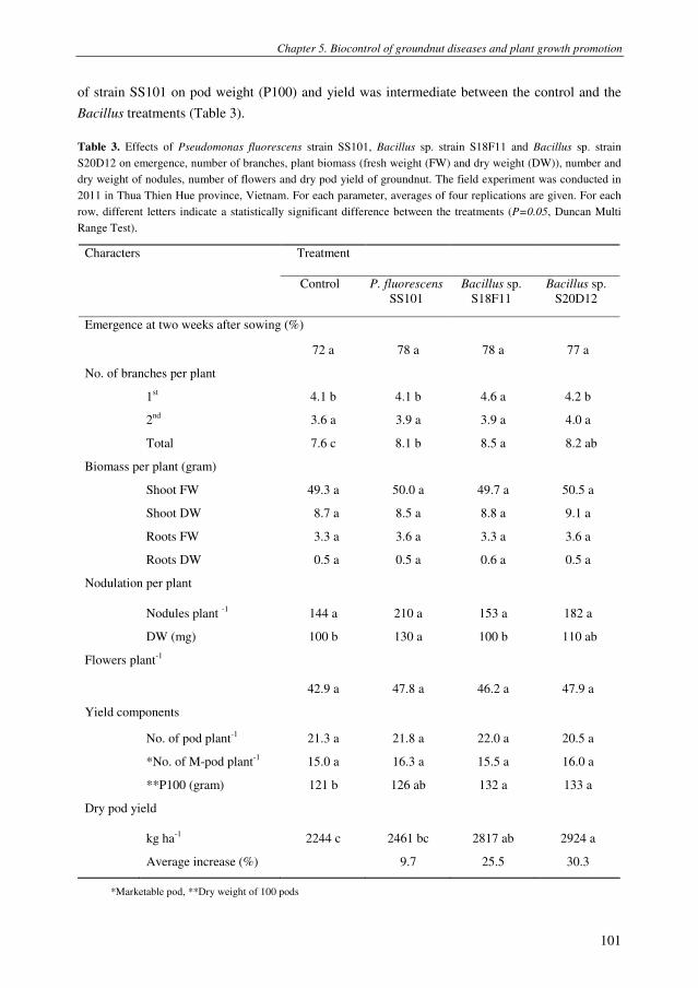
Chapter 5. Biocontrol of groundnut diseases and plant growth promotion
101
of strain SS101 on pod weight (P100) and yield was intermediate between the control and the
Bacillus treatments (Table 3).
Table 3. Effects of Pseudomonas fluorescens strain SS101, Bacillus sp. strain S18F11 and Bacillus sp. strain
S20D12 on emergence, number of branches, plant biomass (fresh weight (FW) and dry weight (DW)), number and
dry weight of nodules, number of flowers and dry pod yield of groundnut. The field experiment was conducted in
2011 in Thua Thien Hue province, Vietnam. For each parameter, averages of four replications are given. For each
row, different letters indicate a statistically significant difference between the treatments (P=0.05, Duncan Multi
Range Test).
Characters Treatment
Control P. fluorescens
SS101
Bacillus sp.
S18F11
Bacillus sp.
S20D12
Emergence at two weeks after sowing (%)
72 a 78 a 78 a 77 a
No. of branches per plant
1st 4.1 b 4.1 b 4.6 a 4.2 b
2nd
3.6 a 3.9 a 3.9 a 4.0 a
Total 7.6 c 8.1 b 8.5 a 8.2 ab
Biomass per plant (gram)
Shoot FW 49.3 a 50.0 a 49.7 a 50.5 a
Shoot DW 8.7 a 8.5 a 8.8 a 9.1 a
Roots FW 3.3 a 3.6 a 3.3 a 3.6 a
Roots DW 0.5 a 0.5 a 0.6 a 0.5 a
Nodulation per plant
Nodules plant -1
144 a 210 a 153 a 182 a
DW (mg) 100 b 130 a 100 b 110 ab
Flowers plant-1
42.9 a 47.8 a 46.2 a 47.9 a
Yield components
No. of pod plant-1
21.3 a 21.8 a 22.0 a 20.5 a
*No. of M-pod plant-1
15.0 a 16.3 a 15.5 a 16.0 a
**P100 (gram) 121 b 126 ab 132 a 133 a
Dry pod yield
kg ha-1
2244 c 2461 bc 2817 ab 2924 a
Average increase (%) 9.7 25.5 30.3
*Marketable pod, **Dry weight of 100 pods

Chapter 5. Biocontrol of groundnut diseases and plant growth promotion
102
Suppression of leaf spot by Pseudomonas and Bacillus
Bacillus and Pseudomonas species are well-known for their abilities to promote plant
growth and yield (Kloepper et al. 1980; van Loon 2007; Weller 2007; Lugtenberg and
Kamilova 2009). They can directly affect plant growth through, among others, facilitating
nutrient acquisition, the production of volatile compounds or plant growth hormones (van Loon
2007; Lugtenberg and Kamilova 2009). Indirect plant growth promotion involves suppression
of plant pathogens (van Loon 2007; Lugtenberg and Kamilova 2009). To evaluate the role of
pathogen suppression in the observed yield increases by strains SS101, S18F11 and S20D12,
the incidence and severity of several groundnut diseases were monitored in this field
experiment. These were the same root and leaf diseases as were monitored in the biocontrol
experiment described above. The results showed that none of the three bacterial strains
significantly reduced stem rot, damping-off, black collar rot and bacterial wilt (Table 4). None
of three bacterial strains affected nitrogen and carbon contents of groundnut nor macronutrient
concentrations in the soil before and after the experiment (Appendix 1B, Appendix 2).
However, all three strains significantly reduced leaf spot diseases, with the two Bacillus strains
having the most significant effects on disease incidence (Table 4). Both leaf spot pathogens can
affect plant growth and yield by reducing the available photosynthetic area via lesion formation
and leaflet abscission (Backman and Crawford 1984; McDonald et al. 1985; Savary and Zadoks
1992). Early and late leaf spots can cause severe defoliation and pod detachment at harvest
(Gremillion et al. 2011). The study by Backman and Crawford (1984) indicated that all levels of
defoliation resulted in yield losses and that there were no differences in yield loss per unit of
disease between C. arachidicola and C. personatum. Regression analyses by Bourgeois and
Boote (1992) further indicated that each 1% increase in necrotic leaf area, caused by C.
personatum, resulted in a 4% reduction in carbon exchange rate of infected leaflets. Their
observation that the affected area is four times larger than the visibly damaged area was
attributed to the fact that the infected area extends beyond the visible necrotic area. In addition,
other factors such as changes in the physiology of the leaf tissue surrounding the area invaded
by the pathogen, or the production of toxins, like cercosporin, may also contribute to the
reduced photosynthesis rate (Bourgeois et al. 1992; Daub and Ehrenshaft 2000).
Worldwide, yield losses caused by leaf spots range from 10% to more than 50%, but can
vary considerably from place to place and between seasons (Backman and Crawford 1984;
McDonald et al. 1985). In Vietnam, leaf spot diseases of groundnut may cause yield losses up
to 20-25% in some provinces in the North (Mehan and Hong 1994). To control leaf spots,
several strategies were recommended, including cultural measures, resistant cultivars,
fungicides or biological control (McDonald et al. 1985; Mehan and Hong 1994; Besler et al.
2006; Singh et al. 2011). Biological control of early and/or late leaf spot of groundnut has not
been studied extensively. Initial work done by Mitchell et al. (1987) showed that the
mycoparasitic fungus Dicyma pulvinata does not primarily act as a protectant against infections
by C. personatum, but can control the secondary spread of this pathogen. Foliar applications

Chapter 5. Biocontrol of groundnut diseases and plant growth promotion
103
with Bacillus cereus strain 304 (Kokalis-Burelle et al. 1992) or Pseudomonas fluorescens strain
Pf1 (Meena et al. 2002) showed promising results in the control of early and late leaf spot,
respectively. The results of our study further extend these findings and showed that application
of specific Pseudomonas and Bacillus strains to the seed bed of groundnut can significantly
reduce leaf spot diseases and substantially enhance pod yield.
Table 4. Effects of Pseudomonas fluorescens strain SS101, Bacillus sp. strain S18F11 and Bacillus sp. strain
S20D12 on the incidence and severity of different diseases of groundnut. The field experiment was conducted in
2011 in Thua Thien Hue province, Vietnam. For each of the parameters, average values of four replications are
given. For each row, different letters indicate a statistically significant difference between the treatments (P=0.05,
Duncan Multi Range Test).
Disease Treatment
Control P. fluorescens
SS101
Bacillus sp.
S18F11
Bacillus sp.
S20D12
Stem rot (Sclerotium rolfsii) (AUDPC) *
6.7 a 4.2 a 3.8 a 2.9 a
Damping off (Rhizoctonia solani) (%)
1.32 a 1.40 a 1.03 a 1.07 a
Black collar rot (Aspergillus niger) (AUDPC)
6.3 a 2.9 a 2.9 a 4.6 a
Bacterial wilt (Ralstonia solanacearum) (%)
1.84 a 1.63 a 0.83 a 1.39 a
Leaf spot (Cercospora arachidicola, Cercosporidium personatum) (%) **
DI-F 44.0 a 42.5 a 40.1 a 36.5 a
DS-F 17.3 b 15.8 ab 16.7 ab 13.0 a
DI-Peg 34.5 c 29.5 b 24.5 a 23.5 a
DS-Peg 14.2 b 11.4 a 9.1 a 8.8 a
Root colonization - 5.7 a 5.1 ab 4.8 b
*AUDPC- area under disease progress curve. **Fifty leaves of each plot were investigated for leaf spot diseases.
DI-F: disease incidence at flowering stage, DS-F: disease severity at flowering stage, DI-Peg: DI at peg stage,
DS-Peg: DS at peg stage.
The mechanisms underlying suppression of leaf spot diseases by strains SS101, S18F11
and S20D12 are not known yet. Induced systemic resistance (ISR) may be a potential
mechanism as was reported earlier by Kloepper et al. (2004) for biocontrol of other groundnut
diseases, i.e. black collar rot caused by A. niger. However, the elaborate study by Zhang et al.
(2001) with nineteen strains of ISR-bacilli and various chemical elicitors of pathogen resistance
indicated that groundnut is not systemically inducible in the same manner as other crops.
Chapter 5. Biocontrol of groundnut diseases and plant growth promotion
104
Whether induced systemic resistance is one of the mechanisms involved for the three bacterial
strains tested here remains to be investigated. In the field experiment, the population densities of
the applied bacterial strains were only monitored on the stem base and roots of groundnut
(Table 4), but not on the leaflets. Therefore, we cannot rule out direct antagonism between the
applied bacterial strains and the leaf spot pathogens.
Conclusions
Each of the five Pseudomonas and Bacillus strains tested in this study improved pod yield
of groundnut plants by 10 to 30%. Similar yield increases were observed in field experiments
that we conducted in 2010 in central Vietnam (Chapters 3 & 4). This consistency in yield
increases observed in two consecutive years in two independent field experiments at two
different locations holds great promise for further development of these bacterial strains as a
key component of an integrated strategy to manage multiple diseases of groundnut and/or to
improve yield.
Materials and Methods
Bacterial strains and culture conditions
P. fluorescens strain SS101, Pseudomonas sp. strains SH-C52 and R4D2, and Bacillus sp. strains S18F11
and S20D12 were cultured at 25°C on Pseudomonas Agar (PSA; Difco, France) and maintained in 40% glycerol at
-80°C in the Laboratory of Phytopathology, Wageningen University, the Netherlands. Phenotypic and genetic
characteristics of these five strains were described previously (Chapters 3 & 4). In the field experiments,
spontaneous rifampicin-resistant derivatives of these strains were used to monitor their population densities on
roots and stem base of groundnut plants by dilution plating on PSA-medium supplemented with rifampin (100 µg
ml-1
), chloroamphenicol (12.5 µg ml-1
), ampicillin (40 µg ml-1
) and delvocid (DSM, 100 µg ml-1
) (Chapters 3 & 4).
Biological control of S. rolfsii and other groundnut pathogens
The field experiment was conducted in 2011 in Thua Thien Hue province, Vietnam, where on average 15%
of the groundnut plants were naturally infected by S. rolfsii (Le et al. 2011). The experiment consisted of four
treatments, i.e. control, Pseudomonas sp. strain SH-C52, Pseudomonas sp. strain R4D2, and the chemical fungicide
tebuconazole (Folicur 250EC, Bayer Crop Science). The field experiment was laid out in a randomized complete
block design (RCBD) with four blocks as four replications and a plot size of 15 m2 (3 X 5 m) (Fig. 2). The distance
between rows was 30 cm and between plants 10 cm. Pseudomonas strains SH-C52 and R4D2 were grown on PSA
plates at 25°C for 48 h, harvested and washed with sterile water. Prior to sowing groundnut seeds, bacterial
suspensions were applied to the furrows at a final density of 106 cells per cm
2 soil. After 8 weeks of plant growth,
three plants per plot were collected to determine the population densities of the applied bacterial strains on the stem
base and roots of groundnut according to the methods described (Chapter 3). Folicur was applied three times during
the growth season, i.e. 20, 40 and 60 days after sowing, by spraying 250 g active ingredient (a.i.) ha-1
. The field soil
was fertilized with nitrogen (N) at 40 kg ha-1
, phosphorus (P2O5) at 60 kg ha-1
, potassium (K2O) at 60 kg ha-1
, and
calcium (Ca(OH)2) at 300 kg ha-1
. All phosphorus and calcium was applied at seed bed preparation. Seeds were
sown at a depth of 3-5 cm and covered with soil. When plants had three true leaves (seedling stage), 70% of the
nitrogen and 50% of the potassium were applied. The remainder of the N and K fertilizers was applied at
flowering. Emergence was monitored at 7, 10 and 13 days after sowing. Weeds were manually removed at four

Chapter 5. Biocontrol of groundnut diseases and plant growth promotion
105
developmental stages of the groundnut plants, i.e. seedling, flowering, peg and pod set stages, i.e. 4, 6, 8 and 10
weeks after sowing, respectively (Fig. 2).
A
B
C
D
E
F
Figure 2. Experimental design of the field experiment conducted in 2011 in Thua Thien Hue province, Vietnam.
A) Experimental field three days after sowing; B) Ten plants per plot were labeled at seedling stage (3 true leaves)
for measuring plant height, number of branches and branch length; C) Groundnut at seedling stage (5 to 6 true
leaves); D) Groundnut at peg stage; E) Sample collection at peg stage; F) Groundnut 10 days before harvesting.
Disease assessments
At each of the four developmental stages (seedling, flowering, peg and pod set), the incidence of stem rot
disease by S. rolfsii (Fig. 3A) was monitored in 1 m2 of each plot (~ 33 plants). To further enhance the incidence and

Chapter 5. Biocontrol of groundnut diseases and plant growth promotion
106
severity of stem rot disease, sclerotia of S. rolfsii strain H001 were inoculated into the field soil at a density of
approximately 100 sclerotia m-2
when the plants had reached the peg stage. Stem rot disease incidence was
assessed according to the methods described previously (Chapter 3). Black collar rot caused by A. niger was
assessed by scoring the number of plants with black collar rot symptoms (Fig. 3B) in 1 m2 for each plot. Damping-
off caused by R. solani (Fig. 3C) was monitored at the seedling stage by counting the number of infected plants in
10 m2
per plot. Bacterial wilt, caused by R. solanacearum, was monitored at pod set stage by counting the number
of wilted plants (Fig. 3D) in 10 m2 per plot.
A
B
C
D
E
F
Figure 3. Typical symptoms of major diseases of groundnut monitored in the 2011 field experiment in Thua Thien
Hue province, Vietnam. A) Stem rot (Sclerotium rolfsii); B) Black collar rot (Aspergillus niger); C) damping-off
(Rhizoctonia solani); D) Bacterial wilt (Ralstonia solanacearum); E) Early leaf spot (Cercospora arachidicola); F)
Late leaf spot (Cercosporidium personatum).
Leaf spots caused by Cercospora arachidicola and Cercosporidium personatum were assessed at flowering
and peg stage. Leaf spot incidence (LSI) is defined as the number of infected leaflets divided by the total number of
leaflets. Leaf spot severity (LSS) was rated using a scale of 0-5; 0 = no disease symptoms, 1 = <1% of the total
leaflet area is affected, 2 = 1- 10%, 3 = 10- 25%, 4 = 25- 50% and 5 = >50% of the total leaflet area is affected.
LSS was calculated based on the formula: LSS = [(1× number of leaflets rated as scale 1) + (2 × number of leaflets
rated as scale 2) + (3 × number of leaflets rated as scale 3) + (4 × number of leaflet rated as scale 4) +(5 × number
of leaflets rated as scale 5)] × 100 ÷ (5 × total number of leaflets). For each of the four plots per treatment, five
sites were randomly chosen and for each site ten leaflets were used for LSS assessment.
Plant growth promotion by Pseudomonas and Bacillus
Plant growth promotion by Pseudomonas fluorescens strain SS101 and Bacillus sp. strains S18F11 and
S20D12 was evaluated at the same time and at the same field site in Thua Thien Hue province as the biocontrol
experiment described above. Also the experimental set-up and conditions were the same as described above, except

Chapter 5. Biocontrol of groundnut diseases and plant growth promotion
107
that this experimental field was not inoculated with sclerotia of S. rolfsii. Next to the disease assessments described
above, plant height, branch length, nodulation, flower production and pod yield were determined. At the seedling
stage, ten plants per plot were randomly labelled to determine plant height, branch length, number of pods, number
of marketable pods and the weight of 100 pods (P100). During flowering time (from 42 to 64 days after sowing),
the number of flowers was monitored daily for five randomly selected plants per plot. Shoots and roots of three
randomly selected plants per plot were separated to determine fresh and dry weights. The number and dry weight
of root nodules was determined at the peg stage. To that end, three randomly selected plants per plot were
harvested; their root systems were washed to remove adhering soil and the number of nodules was counted.
Nodules were removed and dried in an oven for 3 days at 105°C to determine dry weight.
Statistical analysis
The Area Under the Disease Progress Curve (AUDPC) of stem rot caused by S. rolfsii and black collar rot
caused by A. niger was calculated using the method of Landa et al. (2002) and Kruijt et al. (2009). Disease
incidence and disease severity values were arcsin-transformed prior to statistical analysis. Statistical differences (P
< 0.05) between treatments were analyzed by ANOVA followed by the Duncan Multiple Range Test (SPSS
Statistics, USA). Normal distribution of the data and homogeneity of variances was tested prior to ANOVA.
References
Abd-Alla, E.F. and Ezzat (2003) Biocontrol of peanut southern blight (Sclerotium rolfsii) by Bacillus subtilis.
Journal of microbiology of the United Arab Republic 38, 207-216.
Abd-Allah, E.F. (2005) Effect of a Bacillus subtilis isolate on southern blight (Sclerotium rolfsii) and lipid
composition of peanut seeds. Phytoparasitica 33, 460-466.
Backman, P.A. and Crawford, M.A. (1984) Relationship between yield loss and severity of early and late leafspot
diseases of peanut. Phytopathology 74, 1101-1103.
Besler, B.A., Grichar, W.J., Starr, J.A., Senseman, S.A. and Lemon, R.G.J., A.J. (2006) Effects of peanut row
pattern, cultivar, and fungicides on control of southern stem rot, early leaf spot, and rust. Peanut Science 33,
1-6.
Bourgeois, G., Boote, K.J. and Burger, R.D. (1992) Leaflet and canopy photosynthesis of peanut affected by late
leaf spot. Agronomy Journal 84, 359-366.
Brenneman, T.B. and Murphy, A.P. (1991) Activity of tebuconazole on Cercosporidium personatum, a foliar
pathogen of peanut. Plant Disease 75, 699-703.
Brown, S.L. (2007) Peanut insects: ecology and control. In Encyclopedia of Pest Management ed. Pimentel, D.
pp.1 - 10: Taylor & Francis
Cattan, P. and Fleury, A. (1998) Flower production and growth in groundnut plants. European Journal of
Agronomy 8, 13-27.
D’aes, J., Maeyer, K.D., Pauwelyn, E. and Höfte, M. (2010) Biosurfactants in plant Pseudomonas interactions and
their importance to biocontrol. Environmental Microbiology Reports 2, 539-572.
Daub, M.E. and Ehrenshaft, M. (2000) The photoactivated Cercospora toxin cercosporin: contributions to plant
disease and fundamental biology. Annual Review of Phytopathology 38, 461-490.
Dey, R., Pal, K.K., Bhatt, D.M. and Chauhan, S.M. (2004) Growth promotion and yield enhancement of peanut
(Arachis hypogaea L.) by application of plant growth-promoting rhizobacteria. Microbiological Research
159, 371-394.
Fabra, A., Castro, S., Taurian, T., Angelini, J., Ibanez, F., Dardanelli, M., Tonelli, M., Bianucci, E. and Valetti, L.
(2010) Interaction among Arachis hypogaea L. (peanut) and beneficial soil microorganisms: how much is it
known? Critical Reviews in Microbiology 36, 179-194.

Chapter 5. Biocontrol of groundnut diseases and plant growth promotion
108
Florkowski, W.J. (1994) Groundnut production and trade. In The groundnut crop: a scientific basis for
improvement ed. Smartt, J. pp.1-22. London: Chapman & Hall.
Fravel, D.R. (2005) Commercialization and implementation of biocontrol. Annual Review of Phytopathology 43,
337-359.
Ganesan, P. and Gnanamanickam, S.S. (1987) Biological control of Sclerotium rolfsii Sacc. in peanut by
inoculation with Pseudomonas fluorescens. Soil Biology and Biochemistry 19, 35-38.
Gremillion, S.K., Culbreath, A.K., Gorbet, D.W., Mullinix, B.G., Pittman, R.N., Stevenson, K.L., Todd, J.W.,
Escobar, R.E. and Condori, M.M. (2011) Field evaluations of leaf spot resistance and yield in peanut
genotypes in the United States and Bolivia. Plant Disease 95, 263-268.
Höfte, M. and Altier, N. (2010) Fluorescent pseudomonads as biocontrol agents for sustainable agricultural
systems. Research in Microbiology 161, 464-471.
Kishore, G.K., Pande, S. and Podile, A.R. (2005a) Biological control of collar rot disease with broad-spectrum
antifungal bacteria associated with groundnut. Canadian Journal of Microbiology 51, 123-132.
Kishore, G.K., Pande, S., Rao, J.N. and Podile, A.R. (2005b) Pseudomonas aeruginosa inhibits the plant cell wall
degrading enzymes of Sclerotium rolfsii and reduces the severity of groundnut stem rot. European Journal
of Plant Pathology 113, 315-320.
Kloepper, J.W., Leong, J., Teintze, M. and N., S.M. (1980) Enhanced plant growth by siderophores produced by
plant groth-promoting rhizobactera. Nature 286, 885-886.
Kloepper, J.W., Ryu, C.M. and Zhang, S.A. (2004) Induced systemic resistance and promotion of plant growth by
Bacillus spp. Phytopathology 94, 1259-1266.
Kokalis-Burelle, N., Backman, P.A., Rodriguez-Kabana, R. and Ploper, L.D. (1992) Potential for biological control
of early leafspot of peanut using Bacillus cereus and chitin as foliar amendments. Biological Control 2, 321-
328.
Kruijt, M., Tran, H. and Raaijmakers, J.M. (2009) Functional, genetic and chemical characterization of
biosurfactants produced by plant growth-promoting Pseudomonas putida 267. Journal of Applied
Microbiology 107, 546-556.
Landa, B.B., Mavrodi, O.V., Raaijmakers, J.M., Gardener, B.B.M., Thomashow, L.S. and Weller, D.M. (2002)
Differential ability of genotypes of 2,4-diacetylphloroglucinol-producing Pseudomonas fluorescens strains
to colonize the roots of pea plants. Applied and Environmental Microbiology 68, 3226-3237.
Le, C.N. (2004) Study wilt diseases on groundnut and some methods to control them in Thua Thien Hue province.
Vietnam National Journal of Plant Protection 1, 9-15.
Le, C.N., Mendes, R., Kruijt, M. and Raaijmakers, J. (2011) Genetic and phenotypic diversity of Sclerotium rolfsii
Sacc. in groundnut fields in central Vietnam. Plant Disease (accepted with minor changes).
Lugtenberg, B. and Kamilova, F. (2009) Plant growth promoting rhizobacteria. Annual Review of Microbiology 63,
541-556.
McDonald, D., Subrahmanya, P., Gibbons, R.W. and Smith, D.H. (1985) Early and late leaf spots of groundnut.
Patancheru, A.P. 502 324 India: International Crops Research Institute for the Semi-Arid Tropics.
Mehan, V.K. and Hong, N.X. (1994 ) Disease constraints to groundnut production in Vietnam - research and
management strategies. In Newsletter 14. pp.8-11. Andhra Pradesh, India International Crops Research
Institute for the Semi-Arid Tropics.
Mendes, R., Kruijt, M., de Bruijn, I., Dekkers, E., van der Voort, M., Schneider, J.H.M., Piceno, Y.M., DeSantis,
T.Z., Andersen, G.L., Bakker, P. and Raaijmakers, J.M. (2011) Deciphering the rhizosphere microbiome for
disease-suppressive bacteria. Science 332, 1097-1100.
Middleton, K.J., Pande, S., Sharma, S.B. and Smith, D.H. (1994) Diseases. In The groundnut crop: a scientific
basis for improvement ed. Smartt, J. pp.336-394. London: Chapman & Hall.

Chapter 5. Biocontrol of groundnut diseases and plant growth promotion
109
Murugalakshmi, C.N., Anand, R. and Bhuvaneswari, K. (2009) Isolation, characterization and evaluvation of
Pseudomonas fluorescens against Sclerotium rolfsii as biocontrol agent. Asian Journal of Microbiology,
Biotechnology and Environmental Sciences 11, 653-656.
Nguyen, T.N., Tran, V.M., Nguyen, T.T. and Le, C.N. (2004) Research on groundnut diseases in Quang Binh
provice. National Agriculture and Rural Development 17, 337-342.
Ongena, M., Jourdan, E., Adam, A., Paquot, M., Brans, A., Joris, B., Arpigny, J.L. and Thonart, P. (2007) Surfactin
and fengycin lipopeptides of Bacillus subtilis as elicitors of induced systemic resistance in plants.
Environmental Microbiology 9, 1084-1090.
Pham, T.V. (1997) Status of integrated management of groundnut insect pests in Vietnam IAN 17, 47-48.
Porter, D.M., Smith, D.H. and Rodriguez - Kabana, R. (1984) Compendium of peanut diseases. St. Paul: American
Phytopathological Society.
Raaijmakers, J. (2006) Cyclic lipopeptide production by plant-associated Pseudomonas spp.: Diversity, activity,
biosynthesis, and regulation. Molecular Plant-Microbe Interactions 19, 699-710.
Raaijmakers, J.M., de Bruijn, I., Nybroe, O. and Ongena, M. (2010) Natural functions of lipopeptides from
Bacillus and Pseudomonas: more than surfactants and antibiotics. Fems Microbiology Reviews 34, 1037-
1062.
Ramantha Rao, V. and Murty, U.R. (1994) Botany - morphology and anatomy. In The Groundnut crop: A scientific
basis for improvement ed. Smartt, J. pp.43-89. London: Chapman & Hall.
Savage, G.P. and Keenan, J.I. (1994) The composition and nutritive value of groundnut kernels. In The Groundnut
crop: A scientific basis for improvement ed. Smartt, J. pp.173-213. London: Chapman & Hall.
Savary, S. and Zadoks, J.C. (1992) Analysis of crop loss in the multiple pathosystem groundnut-rust-late leaf spot.
I. Six experiments. Crop Protection 11, 99-109.
Savithiry, S. and Gnanamanickam, S.S. (1987) Bacterization of peanut with Pseudomonas fluorescens for
biological control of Rhizoctonia solani and for enhanced yield. Plant and Soil 102, 11-15.
Senthilraja, G., Anand, T., Durairaj, C., Kennedy, J.S., Suresh, S., Raguchander, T. and Samiyappan, R. (2010a) A
new microbial consortia containing entomopathogenic fungus, Beauveria bassiana and plant growth
promoting rhizobacteria, Pseudomonas fluorescens for simultaneous management of leafminers and collar
rot disease in groundnut. Biocontrol Science and Technology 20, 449-464.
Senthilraja, G., Anand, T., Durairaj, C., Raguchander, T. and Samiyappan, R. (2010b) Chitin-based bioformulation
of Beauveria bassiana and Pseudomonas fluorescens for improved control of leafminer and collar rot in
groundnut. Crop Protection 29, 1003-1010.
Shanmugam, V., Senthil, N., Raguchander, T., Ramanathan, A. and Samiyappan, R. (2002) Interaction of
Pseudomonas fluorescens with Rhizobium for their effect on the management of peanut root rot.
Phytoparasitica 30, 169-176.
Sharma, R.C. (2002) Fungal diseasse of groundnut. In Diseases of field crops eds. Gupta, V.K. and Paul, Y.S.
pp.262-278. New Delhi: Indus Publishing.
Shew, B.B. and Waliyar, F. (2007) Peanut diseases: ecology and control. In Encyclopedia of Pest Management ed.
Pimentel, D. pp.469–473: Taylor & Francis.
Singh, M.P., Erickson, J.E., Boote, K.J., Tillman, B.L., Jones, J.W. and van Bruggen, A.H.C. (2011) Late leaf spot
effects on growth, photosynthesis, and yield in peanut cultivars of differing resistance. Agronomy Journal
103, 85-91.
Taurian, T., Anzuay, M.S., Angelini, J.G., Tonelli, M.L., Luduena, L., Pena, D., Ibanez, F. and Fabra, A. (2010)
Phosphate-solubilizing peanut associated bacteria: screening for plant growth-promoting activities. Plant
and Soil 329, 421-431.
Tonelli, M.L., Furlan, A., Taurian, T., Castro, S. and Fabra, A. (2011) Peanut priming induced by biocontrol
agents. Physiological and Molecular Plant Pathology 75, 100-105.

Chapter 5. Biocontrol of groundnut diseases and plant growth promotion
110
Tonelli, M.L., Taurian, T., Ibanez, F., Angelini, J. and Fabra, A. (2010) Selection and in vitro characterization of
biocontrol agents with potential to protect peanut plants against fungal pathogens. Journal of Plant
Pathology 92, 73-82.
Tran, H., Ficke, A., Asiimwe, T., Höfte, M. and Raaijmakers, J.M. (2007) Role of the cyclic lipopeptide
massetolide A in biological control of Phytophthora infestans and in colonization of tomato plants by
Pseudomonas fluorescens. New Phytologist 175, 731-742.
van Loon, L. (2007) Plant responses to plant growth-promoting rhizobacteria. European Journal of Plant
Pathology 119, 243-254.
Weller, D.M. (2007) Pseudomonas biocontrol agents of soilborne pathogens: Looking back over 30 years.
Phytopathology 97, 250-256.
Wightman, J.A. and Ranga Rao, G.V. (1994) Groundnut pests. In The groundnut crop: a scientific basis for
improvement ed. Smartt, J. pp.395-479. London: Chapman & Hall.
Woodroof, J.G. (1983) Peanuts : production, processing, products. Westport: AVI.
Zhang, S., Reddy, M.S., Kokalis-Burelle, N., Wells, L.W., Nightengale, S.P. and Kloepper, J.W. (2001) Lack of
induced systemic resistance in peanut to late leaf spot disease by plant growth-promoting rhizobacteria and
chemical elicitors. Plant Disease 85, 879-884.

Chapter 5. Biocontrol of groundnut diseases and plant growth promotion
111
Appendix 1. Carbon (C), nitrogen (N), and C:N ratio in shoots of groundnut plants at peg stage grown in soil
untreated (Control) or treated with Pseudomonas, Bacillus, or the fungicide Folicur. A) Data from the field plot
inoculated with Sclerotium rolfsii; B) Data from the field plot without fungal inoculation. Both experiments were
conducted in 2011 in Thua Thien Hue province, Vietnam.
A
Treatment %C %N C:N ratio
Control 49.5 3.0 16.6
Pseudomonas sp. SH-C52 49.0 3.2 15.5
Pseudomonas sp. R4D2 48.3 3.3 14.8
Folicur 48.1 3.4 14.1
B
Treatment %C %N C:N ratio
Control 47.2 3.0 15.6
P. fluorescens SS101 47.6 3.5 13.4
Bacillus sp. S18F11 46.7 3.4 13.6
Bacillus sp. S20D12 47.9 3.3 14.6
Appendix 2. Macro-nutrients in the soil prior to and upon completion of the field experiment conducted in 2011 in
Thua Thien Hue province, Vietnam.
Sample pHKCl OM
(%)
Total N
(%)
Total K2O
(%)
Total P2O5
(%)
Available P2O5
(mg 100 g soil-1
)
Before EX 4.25 1.03 0.05 0.24 0.03 10.75
Control 4.30 1.40 0.04 0.22 0.03 12.65
P. fluorescens SS101 4.31 1.35 0.05 0.22 0.03 11.25
Bacillus sp. S18F11 4.22 1.29 0.05 0.23 0.03 13.68
Bacillus sp. S20D12 4.34 1.45 0.05 0.23 0.05 12.60

Chapter 5. Biocontrol of groundnut diseases and plant growth promotion
112
Chapter 6. Diversity of endophytic bacteria of groundnut nodules
Chapter 6
Diversity of endophytic bacteria of groundnut nodules and
their effects on plant growth and groundnut diseases
Le, C.N., Thai, T.H. and Raaijmakers, J.M.

Chapter 6. Diversity of endophytic bacteria of groundnut nodules
114

Chapter 6. Diversity of endophytic bacteria of groundnut nodules
115
Diversity of endophytic bacteria of groundnut nodules and their
effects on plant growth and groundnut diseases
Le, C.N.1,2
, Thai, T.H.3 and Raaijmakers, J.M.
1
1 Laboratory of Phytopathology, Wageningen University, the Netherlands
2 Department of Plant Protection, Hue University of Agriculture and Forestry, Vietnam
3 Department of Soil Sciences and Environment, Hue University of Agriculture and Forestry, Vietnam
Abstract
Legumes can overcome nitrogen limitation via symbiotic interactions with
rhizobia. In this study, bacteria were isolated from surface-sterilized nodules of
groundnut plants grown in farmer fields in central Vietnam. Approximately
100 bacterial isolates were grouped by BOX-PCR profiling and identified by
16S-rDNA sequencing. The results showed that the endophytic bacteria of
groundnut nodules were genetically diverse. Most of the isolates belonged to
the alpha-Proteobacteria, genus Sphingomonas. The other bacterial isolates
were classified as Rhizobium, Stenotrophomonas, Ralstonia and Burkholderia
species. One group of isolates belonged to the phylum Bacteroidetes, genus
Sphingobacterium. Most isolates did not induce nodule formation when re-
introduced on groundnut plants, except for some Sphingomonas isolates. Under
field conditions, Sphingomonas sp. strain HR32 and Rhizobium sp. strain HR9
suppressed leaf spot diseases of groundnut and increased the pod yield. The
potential of these endophytic bacteria as inoculants for groundnut cultivation
are discussed.
Keywords: nitrogen fixation, Arachis hypogaea, Rhizobium, Sphingomonas
Introduction
The low availability of nitrogen (N) in agricultural ecosystems often limits plant growth
and crop productivity. Legumes can overcome this nutrient limitation via symbiotic interactions
with rhizobia (Fabra et al. 2010). During this interaction, bacteria invade the plant root and
induce the formation of organelle-like structures called nodules. Inside the nodules, rhizobia
convert atmospheric di-nitrogen (N2) into ammonia, which is then used by the plant as a
nitrogen source (Broughton et al. 2000). N2 fixation is catalysed by the nitrogenase complex

Chapter 6. Diversity of endophytic bacteria of groundnut nodules
116
encoded by the nifHDK genes (Dixon and Kahn 2004). Rates of symbiotic N2 fixation in
legumes can vary with host, symbiont and environment, but rates as high as 600 kg N fixed ha-1
yr-1
have been reported in clover pastures (Unkovich and Pate 2000). Among the legume crops,
groundnut is cultivated in more than 80 countries and is the most important oil seed crop
worldwide (Rao and Murty 1994). To date, there are only few studies on N2 fixation in
groundnut. The available data suggest that N2 fixation is highly variable with rates ranging from
32 to 206 kg ha-1
(Giller et al. 1987; Bell and Wright 1994; Unkovich and Pate 2000).
Before entering the legume root tissue, rhizobia must move towards and colonize the root
surface (Fabra et al. 2010). In this process of recognition and adhesion to the root, symbiotic
signals are exchanged between the host plant and the bacterium to initiate nodule formation and
development. Flavonoids are released by plant roots and constitute important signals in
interactions between legumes and rhizobia (Broughton et al. 2000). Flavonoids belong to a
diverse family of aromatic compounds derived from secondary plant metabolism. Groundnut
produces some flavonoids such as chrysin, luteolin, apigenin, genistein that were reported to
induce expression of nod genes in rhizobia (Angelini et al. 2003). Depending on the host and
bacterium, these compounds activate the bacterial transcriptional regulator NodD, which in turn
induces the transcription of other nodulation genes (nod, nol and noe genes), whose products are
involved in the synthesis of the Nod factors (Spaink 2000). Nod factors are lipo-chito-
oligosaccharides that cause morphological changes in legume root hairs, leading to infection
thread formation, nodule development and symbiotic nitrogen fixation (Fabra et al. 2010).
Rhizobia enter roots via infection threads but can also enter the roots via wounds or via cavities
between undamaged epidermal cells. In groundnut, root nodules develop at the sites of lateral-
root emergence (Uheda et al. 2001) where the epidermis of the parent roots is broken (Boogerd
and van Rossum 1997).
Among the N2-fixing bacteria on groundnut, Bradyrhizobium species were the most
identified (van Rossum et al. 1995; Urtz and Elkan 1996; Zhang et al. 1999; Saleena et al.
2001). Morphological, physiological and molecular analyses indicated that groundnut
symbionts obtained from different geographical regions are highly diverse (reviewed in Fabra et
al. (2010)). Based on ITS-RFLP analysis and 16S-rDNA sequencing, Yang and Zhou (2008)
reported that isolates from nodules of groundnut in China were phylogenetically related to
Bradyrhizobium liaoningense, Bradyrhizobium japonicum and Bradyrhizobium yuanmingense.
Through 16S-rDNA sequencing and RFLP analysis, Taurian et al. (2006) and El-Akhal et al.
(2008) showed that also different Rhizobium species were associated with groundnut nodules. In
recent studies, various other bacterial genera, including Pseudomonas, Enterobacter and
Klebsiella species, were isolated from surface-sterilized nodules of groundnut (Ibanez et al.
2009). These bacteria were unable to induce nodule formation in groundnut plants, but
promoted plant growth when re-inoculated on groundnut (Ibanez et al. 2009). Also other
bacterial genera, including Inquilinus, Rhodopseudomonas, Phyllobacterium, Ochrobactrum,
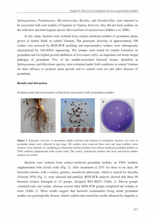
Chapter 6. Diversity of endophytic bacteria of groundnut nodules
117
Sphingomonas, Pseudomonas, Microbacterium, Bacillus, and Paenibacillus, were reported to
be associated with root nodules of legumes in Tunisia, however, they did not form nodules on
the wide host spectrum legume species Macroptilium atropurpureum (Zakhia et al. 2006).
In this study, bacteria were isolated from surface-sterilized nodules of groundnut plants
grown in farmer fields in central Vietnam. The genotypic diversity of approximately 100
isolates was assessed by BOX-PCR profiling and representative isolates were subsequently
characterized by 16S-rDNA sequencing. The isolates were tested for nodule formation on
groundnut and for hyphal growth inhibition of Sclerotium rolfsii, an important soil-borne fungal
pathogen of groundnut. Two of the nodule-associated bacterial strains, identified as
Sphingomonas and Rhizobium species, were evaluated under field conditions in central Vietnam
for their efficacy to promote plant growth and to control stem rot and other diseases of
groundnut.
Results and discussion
Isolation and characterization of bacteria associated with groundnut nodules
Figure 1. Schematic overview of groundnut nodule selection and isolation of endophytic bacteria. (A) roots of
groundnut plants were collected at peg stage, (B) nodules were removed from roots and large nodules (cross
sections) were selected, (C) morphology of bacterial colonies isolated from surface-sterilized groundnut nodules on
YMA medium supplemented with crystal violet. The watery, translucent colonies that were selected for further
analysis are circled.
Bacteria were isolated from surface-sterilized groundnut nodules on YMA medium
supplemented with crystal violet (Fig. 1). After incubation at 25°C for three to six days, 99
bacterial colonies with a watery, gummy, translucent phenotype, which is typical for rhizobia
(Vincent 1970; Fig. 1), were selected and purified. BOX-PCR analysis showed that these 99
bacterial isolates belonged to 23 groups, designed BG1-BG23 (Table 1). Eleven groups
contained only one isolate, whereas several other BOX-PCR groups comprised ten isolates or
more (Table 1). These results suggest that bacterial communities living inside groundnut
nodules are genotypically diverse, which confirm and extend the results obtained by Angelini et
A B C

Chapter 6. Diversity of endophytic bacteria of groundnut nodules
118
al. (2011), who analyzed, by a similar technique (i.e. Enterobacterial Repetitive Intergenic
Consensus (ERIC) PCR), the diversity of bacteria from nodules of groundnut plants grown in
Argentina.
Table 1. Genetic characterization of endophytic bacteria associated with nodules of groundnut plants grown in
farmer fields in central Vietnam. A total of 99 isolates was grouped by BOX-PCR profiling in 23 groups,
designated BG-1 through BG-23. One representative isolate of each group was tested for nodule formation on
groundnut roots under nethouse conditions. All 99 isolates were tested for their ability to inhibit hyphal growth of
Sclerotium rolfsii in vitro.
BOX-PCR
group*
16S-rDNA
sequenced
isolate**
Classification based on
16S-rDNA sequence
analysis
Nodule formation
(No. of nodules
plant-1
)
S. rolfsii
inhibition
in vitro
BG-1 (7) HR101 Sphingomonas sp. 0 -
BG-2 (14) HR32 Sphingomonas sp. 10 -
BG-3 (3) HR118 Sphingomonas sp. 3 -
BG-4 (23) HR71 Sphingomonas sp. 5 -
BG-5 (9) HR67 Sphingomonas sp. 4 -
BG-6 (4) HR64 Sphingomonas sp. 1 -
BG-7 (1) HR7 Stenotrophomonas sp. 0 -
BG-8 (1) HR9 Rhizobium sp. 0 -
BG-9 (1) HR10 Rhizobium sp. 1 -
BG-10 (10) HR42 Sphingobacterium sp. 6 -
BG-11 (2) HR54 Sphingomonas sp. 5 -
BG-12 (1) HR2 Sphingomonas sp. 0 -
BG-13 (1) HR21 Ralstonia sp. 0 -
BG-14 (1) HR27 Sphingomonas sp. 3 -
BG-15 (1) HR49 Sphingomonas sp. 0 -
BG-16 (2) HR51 Sphingomonas sp. 0 -
BG-17 (1) HR55 Sphingomonas sp. 0 -
BG-18 (6) HR75 Sphingomonas sp. 10 -
BG-19 (1) HR77 Burkholderia sp. 0 +
BG-20 (2) HR82 Sphingomonas sp. 0 -
BG-21 (1) HR94 Sphingomonas sp. 0 -
BG-22 (1) HR95 Sphingomonas sp. 0 -
BG-23 (6) HR5 Burkholderia sp. 0 +
Control 0 -
*The numbers of isolates belonging to a certain BOX-PCR group are indicated between brackets; **These
isolates were selected for 16S-rDNA sequencing (details see figure 2).
To further identify the bacterial isolates, 16S-rDNA sequencing was performed for one
isolate per BOX-PCR group. Ribosomal database comparisons revealed that almost all of the
isolates, i.e. 22 out of 23, belonged to the phylum Proteobacteria (Fig. 2). One isolate,
representative of BOX-PCR group BG-10, was closely related to Sphingobacterium species
within the phylum Bacteroidetes (Fig. 2). For the 22 isolates that were classified as

Chapter 6. Diversity of endophytic bacteria of groundnut nodules
119
Proteobacteria, seventeen belonged to the α-Proteobacteria, two to the β-Proteobacteria and
three to the γ-Proteobacteria (Fig. 2). Almost all of the isolates (i.e. 15 out of 17) within the α-
Proteobacteria clade were phylogenetically related to Sphingomonas species, whereas the two
isolates from BOX-PCR groups BG-8 and BG-9 were related to Rhizobium (Fig. 2). Zakhia et
al. (2006) described the isolation of Sphingomonas species from nodules of three different
legumes (Astragalus gombiformis, Lotus argenteus, and Calycotome villosa) in Tunisia. These
species were, however, not able to form nodules on Macroptilium atropurpureum, a legume
nodulated by a wide range of bacteria (Zakhia et al. 2006). Xie and Yokota (2006) reported that
Sphingomonas azotifigens, isolated from rice (Oryza sativa), was able to fix nitrogen based on
the acetylene reduction assay and nifH gene detection. Using the same detection assays, Videira
et al. (2009) also reported N-fixation by Sphingomonas isolates from rice in Brazil. They further
showed, by sequencing of 16S-rDNA and nifH fragments, that most of the nitrogen-fixing
isolates clustered apart from S. azotifigens (Videira et al. 2009). To our knowledge,
Sphingomonas species have also not been reported to form or associate with nodules on
groundnut. When re-introduced into autoclaved field soil, most Sphingomonas isolates obtained
in our study did not form nodules on groundnut roots (Table 1). However, for some of the
identified Sphingomonas isolates, in particular isolates H32 (BG-2) and HR75 (BG-18), nodules
were found on groundnut roots (Table 1). Whether these isolates colonized and penetrated the
root tissue more efficiently than the other isolates, and were actually responsible for nodule
formation is not known yet and needs to be investigated. Also the presence of nif and nod genes
as well as the ability of these isolates to fix nitrogen was not determined here and needs to be
addressed in future studies.
There are several studies that point to a possible role of Rhizobium species in nodule
formation on groundnut (Taurian et al. 2006; El-Akhal et al. 2009). In our study, however,
inoculation with isolates HR9 (BG-8) and HR10 (BG-9), which are closely related to Rhizobium
(Fig. 2), did not or only marginally (i.e. 1 nodule per plant) result in nodule formation on
groundnut plants (Table 1). Also isolates HR7, HR21, HR77 and HR5, close relatives of
Stenotrophomonas, Ralstonia or Burkholderia (Fig. 2), respectively, did not form nodules on
groundnut when re-introduced into autoclaved field soil. Ralstonia and Burkholderia species
have been reported to form nodules on legumes (Chen et al. 2003). For example, Ralstonia
taiwanensis LMG 19424 was reported to form nodules on Mimosa pudica (Chen et al. 2003).
N2-fixing Ralstonia was also isolated from Dalbergia species in Madagascar
(Rasolomampianina et al. 2005).

Chapter 6. Diversity of endophytic bacteria of groundnut nodules
120
HR71 BG-4 (23)
FJ158842 Sphingomonas sp.
HR64 BG-6 (4)
HR32 BG-2 (14)
HR75 BG-18 (6)
HR118 BG-3 (3)
HR82 BG-20 (2)
HR49 BG-15 (1)
HR95 BG-22 (1)
HR27 BG-14 (1)
HR2 BG-12 (1)
HR67 BG-5 (9)
HR94 BG-21 (1)
HR55 BG-17 (1)
EU931555 Sphingomonas paucimobilis
HR51 BG-16 (2)
NR 028634 Sphingomonas yabuuchiae
AB217471 Sphingomonas azotifigens
FJ455073 Sphingomonas sp.
HR101 BG-1 (7)
DQ840049 Sphingomonas sp.
EU826645 Rhizobium sp.
HR9 BG-8 (1)
HR10 BG-9 (1)
HR7 BG-7 (1)
FN645727 Stenotrophomonas maltophilia
HR54 BG-11 (2)
HR21 BG-13 (1)
DQ239898 Ralstonia mannitolilytica
AJ302311 Burkholderia sp.
AJ302312 Burkholderia sp.
AY391283 Burkholderia unamae
NR 041720 Burkholderia vietnamiensis
HR5 BG-23 (6)
HR77 BG-17 (1)
AB480713 Burkholderia sp.
HR42 BG-10 (10)
AJ496037 Sphingobacterium sp.
X56305 Gemmata obscuriglobus
99
40
99
99
89
80
99
99
88
99
99
99
81
9988
99
99
99
99
99
99
77
70
69
77
57
66
99
84
58
62
0.05
Pro
teo
ba
cte
ria
Ba
cte
roid
ete
s
Beta
Gamma
Alpha
Figure 2. Phylogenetic analysis of 16S-rDNA sequences of 23 bacterial isolates obtained from surface-sterilized
groundnut nodules. Each of these 23 isolates is a representative of 23 BOX-PCR groups (designated BG-1 to BG-
23) identified among a total of 99 isolates. The branch length in the tree indicates the percentage of sequence
dissimilarity and the numbers at the nodes indicate bootstrap values of 1,000 resamplings. In the tree, the name of
the isolates is followed by their BOX-PCR group; the number of isolates within each BOX-PCR group is shown
between brackets (see Table 1 for details).

Chapter 6. Diversity of endophytic bacteria of groundnut nodules
121
Burkholderia species are common inhabitants of the phytosphere and the internal tissues
of plants (Compant et al. 2008). They are well known as human, animal and plant pathogens,
but they can also exert beneficial effects on plants (Compant et al. 2008). Nitrogen fixation
appears to be common for multiple Burkholderia species. The first conclusive report on nodule
formation by Burkholderia was the study by Moulin et al. (2001). In their study, Burkholderia
sp. strain STM678 contained nifH and nodAB genes and formed 5 to 20 nodules on roots of a
M. atropurpureum plant. Other Burkholderia species reported to fix nitrogen are B.
vietnamiensis isolated from rice in Vietnam (Gillis et al. 1995), B. tropica from sugarcane (Reis
et al. 2004), B. unamae from maize (Caballero-Mellado et al. 2004) and B. silvatlantica (Perin
et al. 2006) from teosinte plants. The Burkholderia isolates (BG-19, BG-23) identified in our
study, however, did not form nodules on groundnut plants (Table 1). Instead, they were the only
among the nodule-associated isolates that inhibited hyphal growth of S. rolfsii (Table 1).
The Sphingobacterium sp. isolate obtained in our study (BG-10) did form nodules per
groundnut plant (Table 1). Also here, additional studies will be necessary to confirm these
results and to demonstrate N2-fixation. Previous studies indicated that a Sphingobacterium sp.
from the rhizosphere of caper plants (Capparis spinosa) was able to fix nitrogen (Andrade et al.
1997). Members of this genus were also reported to promote plant growth of corn (Marques et
al. 2010; Mehnaz et al. 2010) and to inhibit growth of some Fusarium species in vitro (Mehnaz
et al. 2010).
Plant growth promotion and biocontrol of groundnut diseases
For the field experiment conducted in 2011 in central Vietnam, bacterial isolates HR32
and HR9 were selected. Isolate HR32 is closely related to Sphingomonas sp., induced nodule
formation on groundnut, and represents one of the largest BOX-PCR groups among the
endophytic isolates of groundnut nodules (Table 1). Isolate HR9 was chosen because it is
closely related to Rhizobium, a well-known bacterial genus involved in N2-fixation and plant
growth promotion (Zakhia and de Lajudie 2001). The results of the field experiment showed
that both HR9 and HR32 did not significantly increase plant height or the length of the
dicotyledonary axes (data not shown). Moreover, no significant effects were observed on
seedling emergence, plant biomass, and the number of flowers (Table 2). Although there was a
tendency that both HR9 and HR32 increased the number of nodules per plant, these differences
were not statistically significant (Table 2). A similar trend was observed for the nitrogen content
of the groundnut plants treated with HR9 and HR32 (Appendix 1). Both isolates colonized the
roots of groundnut relatively well, reaching densities between 105-10
6 CFU g
-1 of root at the peg
stage (Table 3). However, both isolates could not be re-isolated from inside the nodules formed
on plants treated with these isolates (data not shown). Despite the fact that no significant effects
on plant biomass were found, both bacterial isolates did significantly enhance pod yield, with
increases of 15.8 and 18.5% relative to the untreated control (Table 2).

Chapter 6. Diversity of endophytic bacteria of groundnut nodules
122
Table 2. Effect of Rhizobium sp. isolate HR9 and Sphingomonas sp. isolate HR32 on emergence, number of
branches, plant biomass (fresh weight (FW) and dry weight (DW)), number and DW of root nodules, number of
flowers, yield components and dry pod yield of groundnut. The field experiment was conducted in 2011 in Thua
Thien Hue province, Vietnam. For each parameter, averages of four replications are given. For each row, different
letters indicate a statistically significant difference between the treatments (P=0.05, Duncan Multi Range Test).
Treatment
Control Rhizobium sp.
HR9
Sphingomonas sp.
HR32
Emergence at two weeks after sowing (%)
72 a 79 a 73 a
No. of branches per plant
1st 4.1 b 4.2 a 4.2 a
2nd
3.6 a 3. 8 a 3.9 a
Total 7.6 a 7.9 a 8.1 a
Biomass per plant at peg stage (gram)
Shoot FW 49.3 a 50.3 a 49.0 a
Shoot DW 8.7 a 9.0 a 8.7 a
Roots FW 3.3 a 3.6 a 3.3 a
Roots DW 0.5 a 0.5 a 0.5 a
Nodulation per plant at peg stage
No. of nodule plant -1
144 a 163 a 177 a
DW (mg) 100 a 120 a 90 a
No. of flowers plant-1
42.9 a 42.6 a 42.8 a
Yield components
No. of pod plant -1
21.0 a 21.3 a 23.0 a
* No. M-pod plant-1
15.0 a 14.2 a 15.0 a
** P100 pod (gram) 121 b 123 b 130 a
Dry pod yield
kg ha-1
2244 b 2598 a 2660 a
Increase (%) 15.8 18.5
*Marketable pods, **Weight of 100 pods.
There was no apparent correlation between these yield increases and the incidence of
several major soil-borne diseases: isolates HR9 and HR32 did not provide any control of stem
rot, damping-off, black collar rot and bacterial wilt (Table 3). However, both bacterial isolates
significantly reduced the incidence and the severity of leaf spots of groundnut, both at flowering
and at the peg stage (Table 3). Early and late leaf spots, caused by C. arachidicola and C.
personatum, respectively, seriously damage groundnut with yield reductions up to 50%
(McDonald et al. 1985). Current control methods mostly rely on the use of fungicides such as
penthiopyrad, azoxystrobin, pyraclostrobin and fluoxastrobin (Culbreath et al. 2009). To date,
there are only few studies on biological control of leaf spot diseases with beneficial

Chapter 6. Diversity of endophytic bacteria of groundnut nodules
123
microorganisms (Meena et al. 2002; Kishore et al. 2005; Meena et al. 2006; Galletti et al. 2008;
Chapter 5). The reduction of leaf spots of groundnut by HR9 and HR32 may have resulted in an
increase in photosynthetic leaf area and consequently in the observed yield increases relative to
the control. Whether isolates HR9 and HR32 colonize the leaf surfaces of groundnut and
interact directly with these fungal leaf pathogens remains to be determined. Another possible
mechanism involved in the biocontrol activity of both isolates may be induced systemic
resistance. Both mechanisms will be subject of future investigations.
Table 3. Colonization of roots of groundnut by Rhizobium sp. HR9 and Sphingomonas sp. HR32 and their effects
on the incidence and severity of multiple diseases of groundnut. The field experiment was conducted in 2011 in
Thua Thien Hue province, Vietnam. For each of the parameters, average values of four replications are given. For
each row, different letters indicate a statistically significant difference between the treatments (P=0.05, Duncan
Multi Range Test).
Characters Treatment
Control Rhizobium sp.
HR9
Sphingomonas
sp. HR32
Colonization (log cfu g-1
root) - 5.8 a 5.5 a
Stem rot (Sclerotium rolfsii) (AUDPC)*
6.7 a 4.6 a 5.8 a
Damping off (Rhizoctonia solani) (%)
1.33 a 0.83 a 1.28 a
Black collar rot (Aspergillus niger) (AUDPC)
6.3 a 5.4 a 4.6 a
Bacterial wilt (Ralstonia solanacearum) (%)
1.84 a 1.56 a 1.41 a
Leaf spot (Cercospora arachidicola, Cercosporidium personatum) (%)**
DI-F 44.0 b 36.5 a 34.5 a
DS-F 17.3 b 12.8 a 12.7 a
DI-Peg 34.5 a 29.5 a 29.5 a
DS-Peg 14.2 b 10.7 a 10.8 a
*AUDPC- area under disease progress curve. **Fifty leaves of each plot were investigated for leaf spot diseases.
DI-F: disease incidence at flowering stage, DS-F: disease severity at flowering stage, DI-Peg: DI at peg stage,
DS-Peg: DS at peg stage.
Materials and methods
Isolation of bacteria from groundnut nodules
Nodules were collected from roots of groundnut plants grown in ten farmer fields in Thua Thien Hue, a
province in central Vietnam. In each field, five groundnut plants (peg stage) were collected and ten nodules (on
average 2 per plant) were removed from the roots (Fig. 1). From these groundnut nodules, bacteria were isolated
based on the method used at the Laboratory of Microorganisms, Faculty of Agronomy, Hue University of
Agriculture and Forestry (HUAF), Vietnam. Briefly, the ten collected nodules were washed in 70% ethanol (v/v),
surface sterilized in 1% (v/v) sodium hypochlorite (NaOCl) for 3 min and washed three times with sterile distilled
Chapter 6. Diversity of endophytic bacteria of groundnut nodules
124
water. Sterilized nodules were crushed with a glass rod in a sterilized Eppendorf tube and 500 µl sterile distilled
water was added. Fifty µl of the suspension was plated in duplicate on Yeast Extract Mannitol Agar (YMA)
containing crystal violet (10 mg L-1
). Plates were incubated at 25°C for three to six days. After incubation, ten to
fifteen bacterial colonies with a wet, smooth surface were selected for each sample and purified on YMA plates. In
total, 99 purified isolates were purified and stored in 40% (v/v) glycerol stock at -20°C and -80°C for further
analysis.
Bacterial identification
The genotypic diversity of the bacterial isolates was investigated by BOX-PCR analysis according to
methods described by Tran et al. (2008). Amplicons from 200 to 5000 bp were scored visually for presence or
absence. Bacterial isolates with identical BOX-PCR profiles were considered to be genotypically identical.
Representative isolates of each BOX-PCR group were sent for 16S-rDNA sequencing at Macrogen Inc. (Seoul,
South Korea). The obtained forward and reverse sequences were assembled and edited in Vector NTI (Invitrogen,
version 8.0) and will be deposited in GenBank. For the phylogenetic analyses, the edited sequences were aligned to
sequences available in databases (http://www.ncbi.nlm.nih.gov/Genomes/). Sequences were trimmed to the same
size (~1,100 bp) and the phylogenetic tree was obtained with MEGA4 software (http://megasoftware.net).
Nodule formation on groundnut roots
Nodule formation by the bacterial isolates was tested in nethouse. The nethouse experiment was conducted
in 2010 at the Plant Protection Department, HUAF, Vietnam. Groundnut seeds were surface sterilized in 3% (v/v)
NaOCl for 15 min and rinsed three times with sterile distilled water. Sterilized seeds were soaked in sterile water
for 4 h at 25ºC and then incubated overnight in Petri dishes at 25ºC. Bacterial isolates were grown on YMA
medium plates at 25ºC, harvested after 3 days of incubation and adjusted to a density of 109
cell ml-1
. Natural clay
loam soil was collected from groundnut fields in Thua Thien Hue province, Vietnam. The soil was autoclaved
twice at 120ºC for 30 min with one night in between the two cycles. Autoclaved field soil was transferred to plastic
pots (250 g per pot) and two germinated groundnut seeds were sown per pot. The suspension of the bacterial isolate
was added to the soil to obtain a final density of 106 cells g
-1 soil. For each bacterial isolate (one treatment), two
pots were used. Two weeks after sowing, one plant was kept for every pot. Nodulation was assessed 45 days after
sowing. The groundnut plants and soil were taken out of the pots and soil was removed by watering. Then, the
nodules were visually observed and counted.
Hyphal growth inhibition of Sclerotium rolfsii
Inhibition of hyphal growth of S. rolfsii by the bacterial isolates was tested in dual culture assays according
to the method described by Kruijt et al. (2009). Briefly, bacterial isolates were spot-inoculated at the edge of a
1/5th
-strength potato dextrose agar plate (1/5th
PDA, pH 6.5). After incubation for 48 h at 25ºC, a 5-mm-diameter
agar plug of a 3-day-old culture of S. rolfsii strain H001 (Le et al. 2011) was placed in the centre of the 1/5th
PDA
plate and incubated at 25ºC. Inhibition of mycelial growth of S. rolfsii by the bacterial isolates was recorded 3-4
days after fungal inoculation.
Plant growth promotion and biological control
The experiment for plant growth promotion and biocontrol of groundnut diseases was conducted in 2011 in
a clay loam field in Thua Thien Hue province, Vietnam. The experiment consisted of three treatments, i.e. one
control treatment and two bacterial treatments, i.e. Rhizobium sp. strain HR9 and Sphingomonas sp. strain HR32.
Bacterial strains HR9 and HR32 were grown on YMA plates for 48 h at 25°C, harvested and washed with sterile
water. Prior to sowing groundnut seeds, bacterial suspensions were applied to the furrows at a final density of 106

Chapter 6. Diversity of endophytic bacteria of groundnut nodules
125
cells per cm2 soil. The field experiment was laid out in a randomized complete block design (RCBD) with four
blocks as four replicates with a plot size of 15 m2 (3 × 5 m).
The field soil was fertilized with nitrogen (N) at 40 kg/ha, phosphorus (P2O5) at 60 kg/ha, potassium (K2O)
at 60 kg/ha, and calcium (Ca(OH)2) at 300 kg/ha. Phosphorus and calcium were applied at the time of soil
preparation. Seeds were sown at a depth of 3-5 cm and covered with soil. When plants had three true leaves
(seedling stage), 70% of the nitrogen and 50% of the potassium was applied. The remainder of the N and K
fertilization was applied at flowering. Emergence was monitored at 7, 10 and 13 days after sowing. Weeds were
manually removed at four developmental stages of the groundnut plants, i.e. seedling, flowering, peg and pod set
stages (4, 6, 8 and 10 weeks after sowing).
To determine the effects of each of the bacterial strains on growth of groundnut, plant height, branch length,
nodulation, flower production, and yield were determined. At seedling stage, ten plants per plot were randomly
labelled to measure plant height and branch length. Flower production was monitored daily for five random plants
per plot during flowering time. The number and dry weight of nodules per plant root system was determined at the
peg stage. To that end, three plants per plot were harvested, their root systems were washed to remove adhering soil
and the number of nodules was counted. Nodules were removed and dried for at least 3 days at 105°C. Shoots and
roots of three plants per plot were separated for measuring fresh weight and dry weight. Three plants per plot were
collected to determine the population densities of the introduced bacterial strains HR9 and HR32. For both strains,
spontaneous rifampicin-resistant derivatives were generated and their densities on roots of groundnut were
determined according to the method described previously (Chapter 3). At harvest, the number of pods, marketable
pods and the weight of 100 pods were determined for the ten randomly labeled plants. In addition, some soil
properties, including pHKCl, OM (%), total nitrogen (%), potassium (%), total phosphorus (%), and available
phosphorus were analyzed prior to and upon completion of the field experiment.
Disease assessments
In the field experiment, the effects of the two bacterial strains HR9 and HR32 on the incidence and severity
of several groundnut diseases were also monitored. At each of the four developmental stages (seedling, flowering,
peg and pod set), the incidence of stem rot disease by Sclerotium rolfsii was monitored in 1 m2
of each plot (~33
plants).
Stem rot disease incidence was assessed according to the methods described previously (Chapter 5). Black
collar rot, caused by Aspergillus niger, was assessed by scoring the number of plants with black collar rot
symptoms in 1 m2 for each plot (Chapter 5). Damping-off, caused by Rhizoctonia solani, was monitored at the
seedling stage by counting the number of infected plants in 10 m2
per plot according to the methods described
previously (Chapter 5). Bacterial wilt, caused by Ralstonia solanacearum, was monitored at pod set stage by
counting the number of wilted plants in 10 m2 per plot (Chapter 5).
Leaf spots caused by Cercospora arachidicola and Cercosporidium personatum was assessed at flowering
and at the peg stage. Leaf spot incidence (LSI) is the number of infected leaflets divided by the total number of
monitored leaflets. Leaf spot severity (LSS) was rated using the following scale of 0-5, with 0 = no disease
symptoms, 1 = diseased area <1% of the leaflet area, 2 = diseased area of 1- 10%, 3 = diseased area of 10- 25%, 4
= diseased area of 25- 50% and 5 = diseased area >50%. LSS was calculated based on the formula: LSS = [(1×
number of leaflets rated as scale 1) + (2 × number of leaflets rated as scale 2) + (3 × number of leaflets rated as
scale 3) + (4 × number of leaflets rated as scale 4) +(5 × number of leaflets rated as scale 5)] × 100 ÷ (5 × total
number of leaflets). For each of the four plots per treatment, five sites were randomly chosen and for each site ten
leaflets were used for LSS assessment.

Chapter 6. Diversity of endophytic bacteria of groundnut nodules
126
References
Andrade, G., Esteban, E., Velasco, L., Lorite, M.J. and Bedmar, E.J. (1997) Isolation and identification of N2-
fixing microorganisms from the rhizosphere of Capparis spinosa L. Plant and Soil 197, 19-23.
Angelini, J., Castro, S. and Fabra, A. (2003) Alterations in root colonization and nodC gene induction in the
peanut-rhizobia interaction under acidic conditions. Plant Physiology and Biochemistry 41, 289-294.
Angelini, J., Ibáñez, F., Taurian, T., Tonelli, M.L., Valetti, L. and Fabra, A. (2011) A study on the prevalence of
bacteria that occupy nodules within single peanut plants. Current Microbiology 62, 1752-1759.
Bell, M. and Wright, G. (1994) The N2-fixing capacity of peanut cultivars with differing assimilate partitioning
characteristics. Australian Journal of Agricultural Research 45, 1455-1468.
Boogerd, F.C. and van Rossum, D. (1997) Nodulation of groundnut by Bradyrhizobium: a simple infection process
by crack entry. Fems Microbiology Reviews 21, 5-27.
Broughton, W.J., Jabbouri, S. and Perret, X. (2000) Keys to symbiotic harmony. Journal of Bacteriology 182,
5641-5652.
Caballero-Mellado, J., Martínez-Aguilar, L., Paredes-Valdez, G. and Santos, P.E.-d.l. (2004) Burkholderia unamae
sp. nov., an N2-fixing rhizospheric and endophytic species. International Journal of Systematic and
Evolutionary Microbiology 54, 1165-1172.
Chen, W.-M., Moulin, L., Bontemps, C., Vandamme, P., Bena, G. and Boivin-Masson, C. (2003) Legume
symbiotic nitrogen fixation by β-Proteobacteria is widespread in nature. Journal of Bacteriology 185, 7266-
7272.
Compant, S., Nowak, J., Coenye, T., Clement, C. and Barka, E.A. (2008) Diversity and occurrence of Burkholderia
spp. in the natural environment. Fems Microbiology Reviews 32, 607-626.
Culbreath, A.K., Brenneman, T.B., Kemerait Jr, R.C. and Hammes, G.G. (2009) Effect of the new pyrazole
carboxamide fungicide penthiopyrad on late leaf spot and stem rot of peanut. Pest Management Science 65,
66-73.
Dixon, R. and Kahn, D. (2004) Genetic regulation of biological nitrogen fixation. Nature Reviews Microbiology 2,
621-631.
El-Akhal, M.R., Rincón, A., Arenal, F., Lucas, M.M., El Mourabit, N., Barrijal, S. and Pueyo, J.J. (2008) Genetic
diversity and symbiotic efficiency of rhizobial isolates obtained from nodules of Arachis hypogaea in
northwestern Morocco. Soil Biology and Biochemistry 40, 2911-2914.
El-Akhal, M.R., Rincon, A., El Mourabit, N., Pueyo, J.J. and Barrijal, S. (2009) Phenotypic and genotypic
characterizations of rhizobia isolated from root nodules of peanut (Arachis hypogaea L.) grown in
Moroccan soils. Journal of Basic Microbiology 49, 415-425.
Fabra, A., Castro, S., Taurian, T., Angelini, J., Ibanez, F., Dardanelli, M., Tonelli, M., Bianucci, E. and Valetti, L.
(2010) Interaction among Arachis hypogaea L. (peanut) and beneficial soil microorganisms: how much is it
known? Critical Reviews in Microbiology 36, 179-194.
Galletti, S., Burzi, P., Cerato, C., Marinello, S. and Sala, E. (2008) Trichoderma as a potential biocontrol agent for
Cercospora leaf spot of sugar beet. BioControl 53, 917-930.
Giller, K.E., Nambiar, P.T.C., Srinivasa Rao, B., Dart, P.J. and Day, J.M. (1987) A comparison of nitrogen fixation
in genotypes of groundnut (Arachis hypogaea L.) using 15
N-isotope dilution. Biology and Fertility of Soils
5, 23-25.
Gillis, M., Van Van, T., Bardin, R., Goor, M., Hebbar, P., Willems, A., Segers, P., Kersters, K., Heulin, T. and
Fernandez, M.P. (1995) Polyphasic taxonomy in the genus Burkholderia leading to an emended description
of the genus and proposition of Burkholderia vietnamiensis sp. nov. for N2-fixing isolates from rice in
Vietnam. International Journal of Systematic Bacteriology 45, 274-289.

Chapter 6. Diversity of endophytic bacteria of groundnut nodules
127
Ibanez, F., Angelini, J., Taurian, T., Tonelli, M.L. and Fabra, A. (2009) Endophytic occupation of peanut root
nodules by opportunistic Gammaproteobacteria. Systematic and Applied Microbiology 32, 49-55.
Kishore, G.K., Pande, S. and Podile, A.R. (2005) Biological control of late leaf spot of peanut (Arachis hypogaea)
with chitinolytic bacteria. Phytopathology 95, 1157-1165.
Kruijt, M., Tran, H. and Raaijmakers, J.M. (2009) Functional, genetic and chemical characterization of
biosurfactants produced by plant growth-promoting Pseudomonas putida 267. Journal of Applied
Microbiology 107, 546-556.
Le, C.N., Mendes, R., Kruijt, M. and Raaijmakers, J. (2011) Genetic and phenotypic diversity of Sclerotium rolfsii
Sacc. in groundnut fields in central Vietnam. Plant Disease (accepted with minor changes).
Marques, A., Pires, C., Moreira, H., Rangel, A. and Castro, P.M.L. (2010) Assessment of the plant growth
promotion abilities of six bacterial isolates using Zea mays as indicator plant. Soil Biology & Biochemistry
42, 1229-1235.
McDonald, D., Subrahmanya, P., Gibbons, R.W. and Smith, D.H. (1985) Early and late leaf spots of groundnut.
Patancheru, A.P. 502 324 India: International Crops Research Institute for the Semi-Arid Tropics.
Meena, B., Marimuthu, T. and Velazhahan, R. (2006) Role of fluorescent pseudomonads in plant growth
promotion and biological control of late leaf spot of groundnut. Acta Phytopathologica et Entomologica
Hungarica 41, 203-212.
Meena, B., Radhajeyalakshmi, R., Marimuthu, T., Vidhyasekaran, P. and Velazhahan, R. (2002) Biological control
of groundnut late leaf spot and rust by seed and foliar applications of a powder formulation of Pseudomonas
fluorescens. Biocontrol Science and Technology 12, 195-204.
Mehnaz, S., Kowalik, T., Reynolds, B. and Lazarovits, G. (2010) Growth promoting effects of corn (Zea mays)
bacterial isolates under greenhouse and field conditions. Soil Biology and Biochemistry 42, 1848-1856.
Moulin, L., Munive, A., Dreyfus, B. and Boivin-Masson, C. (2001) Nodulation of legumes by members of the β-
subclass of Proteobacteria. Nature 411, 948-950.
Perin, L., Martínez-Aguilar, L., Paredes-Valdez, G., Baldani, J.I., Estrada-de los Santos, P., Reis, V.M. and
Caballero-Mellado, J. (2006) Burkholderia silvatlantica sp. nov., a diazotrophic bacterium associated with
sugar cane and maize. International Journal of Systematic and Evolutionary Microbiology 56, 1931-1937.
Rao, V.R. and Murty, U.R. (1994) Botany - morphology and anatomy. In The Groundnut crop: A scientific basis
for improvement ed. Smartt, J. pp.43-89. London: Chapman & Hall.
Rasolomampianina, R., Bailly, X., Fetiarison, R., Rabevohitra, R., Béna, G., Ramaroson, L., Raherimandimby, M.,
Moulin, L., De Lajudie, P., Dreyfus, B. and Avarre, J.C. (2005) Nitrogen-fixing nodules from rose wood
legume trees (Dalbergia spp.) endemic to Madagascar host seven different genera belonging to α- and β-
Proteobacteria. Molecular Ecology 14, 4135-4146.
Reis, V.M., Santos, P.E.-d.l., Tenorio-Salgado, S., Vogel, J., Stoffels, M., Guyon, S., Mavingui, P., Baldani,
V.L.D., Schmid, M., Baldani, J.I., Balandreau, J., Hartmann, A. and Caballero-Mellado, J. (2004)
Burkholderia tropica sp. nov., a novel nitrogen-fixing, plant-associated bacterium. International Journal of
Systematic and Evolutionary Microbiology 54, 2155-2162.
Saleena, L.M., Loganathan, P., Rangarajan, S. and Nair, S. (2001) Genetic diversity of Bradyrhizobium strains
isolated from Arachis hypogaea. Canadian Journal of Microbiology 47, 118-122.
Spaink, H.P. (2000) Root nodulation and infection factors produced by rhizobial bacteria. Annual Review of
Microbiology 54, 257-288.
Taurian, T., Ibañez, F., Fabra, A. and Aguilar, O.M. (2006) Genetic diversity of rhizobia nodulating Arachis
hypogaea L. in central Argentinean soils. Plant and Soil 282, 41-52.
Tran, H., Kruijt, M. and Raaijmakers, J.M. (2008) Diversity and activity of biosurfactant-producing Pseudomonas
in the rhizosphere of black pepper in Vietnam. Journal of Applied Microbiology 104, 839-851.

Chapter 6. Diversity of endophytic bacteria of groundnut nodules
128
Uheda, E., Daimon, H. and Yoshizako, F. (2001) Colonization and invasion of peanut (Arachis hypogaea L.) roots
by gusA-marked Bradyrhizobium sp. Canadian Journal of Botany 79, 733-738.
Unkovich, M.J. and Pate, J.S. (2000) An appraisal of recent field measurements of symbiotic N2 fixation by annual
legumes. Field Crops Research 65, 211-228.
Urtz, B.E. and Elkan, G.H. (1996) Genetic diversity among Bradyrhizobium isolates that effectively nodulate
peanut (Arachis hypogaea). Canadian Journal of Microbiology 42, 1121-1130.
van Rossum, D., Schuurmans, F.P., Gillis, M., Muyotcha, A., Vanverseveld, H.W., Stouthamer, A.H. and Boogerd,
F.C. (1995) Genetic and phenetic analyses of Bradyrhizobium strains nodulating peanut (Arachis hypogaea
L.) roots. Applied and Environmental Microbiology 61, 1599-1609.
Videira, S.S., De Araujo, J.L.S., Da Silva Rodrigues, L., Baldani, V.L.D. and Baldani, J.I. (2009) Occurrence and
diversity of nitrogen-fixing Sphingomonas bacteria associated with rice plants grown in Brazil. FEMS
Microbiology Letters 293, 11-19.
Vincent, J.M. (1970) A manual for the practical study of the root-nodule bacteria. London: Blackwell Scientific.
Xie, C.-H. and Yokota, A. (2006) Sphingomonas azotifigens sp. nov., a nitrogen-fixing bacterium isolated from the
roots of Oryza sativa. International Journal of Systematic and Evolutionary Microbiology 56, 889-893.
Yang, J.K. and Zhou, J.C. (2008) Diversity, phylogeny and host specificity of soybean and peanut bradyrhizobia.
Biology and Fertility of Soils 44, 843-851.
Zakhia, F. and de Lajudie, P. (2001) Taxonomy of rhizobia. Agronomie 21, 569-576.
Zakhia, F., Jeder, H., Willems, A., Gillis, M., Dreyfus, B. and de Lajudie, P. (2006) Diverse bacteria associated
with root nodules of spontaneous legumes in Tunisia and first report for nifH-like gene within the genera
Microbacterium and Starkeya. Microbial Ecology 51, 375-393.
Zhang, X., Nick, G., Kaijalainen, S., Terefework, Z., Paulin, L., Tighe, S.W., Graham, P.H. and Lindström, K.
(1999) Phylogeny and diversity of Bradyrhizobium strains isolated from the root nodules of peanut (Arachis
hypogaea) in Sichuan, China. Systematic and Applied Microbiology 22, 378-386.

Chapter 6. Diversity of endophytic bacteria of groundnut nodules
129
Appendix 1. Effect of Rhizobium sp. HR9 and Sphingomonas sp. HR32 on the total carbon carbon (%C) and total
nitrogen (%N) contents in shoots of groundnut plants at the peg stage. The groundnut plants were grown in the
field in 2011 in Thua Thien Hue, Vietnam. Averages of four replications are shown. For each of the parameters, no
statistically significant differences in %C and %N were observed between the treatments [(P%C=0.06, P%N=0.30)
except for C:N (PC:N=0.007) ANOVA statistical test]. An asterisk indicates a significant difference with the
untreated control.
Treatment %C %N C:N ratio
Control 47.2 3.0 15.6
Rhizobium sp. HR9 47.6 3.3 14.3 *
Sphingomonas sp. HR32 48.0 3.3 14.2 *

Chapter 6. Diversity of endophytic bacteria of groundnut nodules
130
Chapter 7. General discussion
Chapter 7
General discussion
Parts of this chapter are integrated in the review
“Stem rot of groundnut caused by the soil-borne pathogen Sclerotium rolfsii” (to be
submitted)

Chapter 7. General discussion
132
Chapter 7. General discussion
133
General discussion
Groundnut is one of the most important oil seed crops worldwide, but its production is
hampered by several pests and diseases. Among the soil-borne fungal diseases, stem rot caused
by Sclerotium rolfsii is a major yield-limiting factor in groundnut cultivation in many countries.
S. rolfsii has a broad host range of more than 500 plant species (Aycock 1966) and causes yield
losses ranging from 10-25% and sometimes even up to 80% (Mehan et al. 1994). In Vietnam,
yield losses due to stem rot disease have not been investigated yet, but it was reported that this
disease caused 5-11% plant death in farmer fields in north and central Vietnam (Le 2004;
Nguyen et al. 2004; Do 2006). Besides stem rot, several other diseases such as Rhizoctonia
damping-off (Rhizoctonia solani), black collar rot (Aspergillus niger), leaf spots (Cercospora
arachidicola, Cercosporidium personatum), and bacterial wilt (Ralstonia solanacearum) are
also widely distributed and damaging to groundnut plants (Mehan and Hong 1994; Le 2004;
Nguyen et al. 2004; Do 2006). In order to manage stem rot and other groundnut diseases,
fundamental knowledge of the distribution and genetic diversity of the pathogen populations is
essential.
Phenotypic and genetic diversity of S. rolfsii in central Vietnam
The survey that was conducted showed that the incidence of stem rot disease of groundnut
in central Vietnam ranged from approximately 5 to 25% (chapter 2). The results further
indicated a gradient in stem rot disease incidence across central Vietnam with an increase in
disease incidence from the northern to the southern field sites. This might be related to the
effect of temperature on disease development (Punja 1985), because along with the lower
latitude also the average daily temperature increases from the North to the South of Vietnam.
For two of the four provinces, the disease incidence of groundnut plants cultivated in clay loam
soil was significantly higher than for plants cultivated in sandy soil. This difference may be
related, in part, to the higher organic content of the clay loam soil, a characteristic that is known
to support germination of sclerotia and subsequent hyphal growth toward the host plant (Punja
1985).
Based on ITS-rDNA sequencing, S. rolfsii populations in groundnut fields in central
Vietnam appeared relatively uniform with three ITS-groups (1, 2 and 3). Most isolates were
closely related to reference isolates of S. rolfsii that were previously collected from groundnut,
tobacco and sweet pepper. Unfortunately, RAPD analysis as well as rep-PCR and ERIC-PCR
(McDonald et al. 2000) gave inconsistent/irreproducible results and were not considered useful
to assess the intraspecific diversity of the S. rolfsii isolates. Therefore, a range of other traits,
including pathogenicity, growth rate, sclerotial characteristics, mycelial compatibility and
tebuconazole sensitivity, were analysed. When comparing the sclerotial traits, the three isolates

Chapter 7. General discussion
134
in ITS-group 2 were clearly different from the isolates in ITS-groups 1 and 3: ITS-group 2
isolates produced substantially less sclerotia per plate and also the average maturation time and
sclerotial diameter were approximately two times higher than for isolates of ITS-groups 1 and 3.
These characteristics combined with the light brown colour of the sclerotia of these three ITS-
group 2 isolates were also reported for isolates classified morphologically or by LSU-
sequencing as S. rolfsii var. delphinii (Aycock 1966; Punja and Damiani 1996; Xu et al. 2010).
The genetic diversity of S. rolfsii isolates from central Vietnam was also investigated by
analysis of the mycelial compatibility. Within each of the three ITS-groups identified in our
study, a high variation in mycelial compatibility was observed. It should be emphasized,
however, that mycelial compatibility described in this thesis is based on macroscopic
observations in which hyphae of different isolates readily intermingle without forming a barrage
or inhibition zone. Therefore, the mycelial compatibility groups (MCG) found here cannot
automatically be considered as vegetative compatibility groups (VCG) or anastomosis groups
(AG). Microscopic analysis needs to be performed to confirm heterokaryon formation between
isolates.
The results further showed that the variation in sensitivity of S. rolfsii isolates from
groundnut fields in central Vietnam to the fungicide tebuconazole is substantially less than
reported earlier for the S. rolfsii population from Georgia, USA (Franke et al. 1998). This
provides a basis to evaluate the efficacy of this fungicide to control stem rot disease of
groundnut in central Vietnam. Combination with other control measures, including biological
control, is recommended to prevent resistance development as was observed previously for S.
rolfsii populations exposed repeatedly to this fungicide (Franke et al. 1998).
Biocontrol of stem rot by antagonistic bacteria
Pseudomonas
In a series of experiments conducted in the laboratory, growth chamber, nethouse and
field, we showed that specific Pseudomonas strains, producing either phenazines (PHZ) or
lipopeptides (LPs), suppressed hyphal growth of S. rolfsii and significantly reduced stem rot
disease of groundnut (chapter 3). By comparing the activities of wildtype Pseudomonas strains
with that of mutants disrupted in specific biosynthesis and regulatory genes, we demonstrated
that PHZ and specific LPs are key bioactive metabolites in biological control of stem rot disease
of groundnut. The results of the in vitro dual culture assays showed that strains SS101 and
SBW25, producing the viscosin-type LPs, and strain 267, producing putisolvins, did not or only
marginally inhibit hyphal growth of S. rolfsii. Lack of inhibitory activity of LPs was also
observed previously for viscosinamide and putisolvins in hyphal growth assays with oomycete
pathogens (Thrane et al. 1999; Gross et al. 2007; Kruijt et al. 2009). In contrast, thanamycin-
producing strain SH-C52 significantly inhibited hyphal growth of S. rolfsii, whereas its
Chapter 7. General discussion
135
thanamycin-deficient mutant was not effective. These results suggested differential activity of
structurally different LPs against the stem rot pathogen S. rolfsii, with thanamycin having
significant inhibitory effects on hyphal growth and plant infection. This presumed differential
activity of the four LPs is based on the assumption that each of the bacterial strains produces
similar amounts of the LPs on agar plates and in the groundnut rhizosphere. At this point,
however, there are no data available on the concentrations of each of the LPs produced on roots
of groundnut. Reporter-based studies (Keel et al. 1992) or advanced analytical analyses
(Ongena et al. 2007) should be conducted to confirm that the LPs are indeed produced in situ
and to determine the amounts produced.
Next to the well-characterized Pseudomonas strains, we also tested novel Pseudomonas
strains. We describe the isolation, characterization and biocontrol efficacy of Pseudomonas sp.
R4D2, a strain that was selected from a total of 3,360 indigenous bacterial isolates randomly
collected from the stem base and roots of groundnut plants grown in farmer fields in Vietnam
(chapter 4). Field experiments showed that treatment with strain R4D2 significantly reduced
the incidence of stem rot disease. The level of disease protection provided by this bacterial
strain was similar to that achieved by the fungicide tebuconazole. For Pseudomonas sp. strain
R4D2, two mutants were obtained that did not inhibit hyphal growth of S. rolfsii and were not
effective in controlling stem rot disease of groundnut under growth chamber conditions. Drop
collapse assays showed that strain R4D2 produces biosurfactant compound(s) that are not
produced by the two mutants. Although these results support the hypothesis that a biosurfactant
is responsible for biocontrol of S. rolfsii, additional genetic and biochemical analyses are
required to prove this and to strengthen the data.
Bacillus
In the past decade, several Bacillus species and strains have been studied for their efficacy
to control stem rot disease of groundnut (Abd-Alla and Ezzat 2003; Abd-Allah 2005; Abd-Allah
and El-Didamony 2007; Tonelli et al. 2011). For example, treatment of groundnut seeds with
Bacillus subtilis protected groundnut against S. rolfsii and significantly increased the number of
pods (Abd-Allah 2005). In this thesis, we describe the isolation of two promising Bacillus
strains, designated S20D12 and S18F11, from the stem base of groundnut plants grown in
farmer fields in central Vietnam (chapter 4). Phylogenetic analyses showed that strain S20D12
belongs to the same phylogenetic cluster as Bacillus amyloliquefaciens reference strains,
whereas strain S18F11 clustered with several Bacillus subtilis reference strains. Both Bacillus
species have been the subject of numerous studies on biological control of plant pathogens and
induced systemic resistance (Kloepper et al. 2004; Ongena and Jacques 2008). In the field
experiment that we conducted in Quang Nam province in 2010, both Bacillus strains provided
significant control of stem rot disease to a level similar to that of the fungicide Folicur.

Chapter 7. General discussion
136
Comparison of the biocontrol efficacy of Pseudomonas and Bacillus
The two field experiments described in this thesis (chapters 3, 4, 5) were conducted in
Vietnam in two consecutive years at two different locations: in 2010 in Quang Nam province
and in 2011 in Thua Thien Hue province. In both field experiments, several Pseudomonas and
Bacillus strains were tested for their efficacy to control stem rot and other groundnut diseases,
and to improve growth and yield. Despite the substantial difference in attainable pod yield
between the two locations, several of the applied bacterial strains increased pod yield of
groundnut consistently (Table 1). For example, Pseudomonas sp. strains SH-C52 and R4D2
significantly suppressed stem rot disease caused by S. rolfsii in the field experiments conducted
in 2010 (chapter 3) and in 2011 (chapter 5). Furthermore, Bacillus sp. strain S20D12
consistently improved pod yield in both years with increases in pod dry weight of 23-30%
(Table 1, chapters 4, 5). Despite the consistency in yield increases over two consecutive years,
the Bacillus strains did not consistently control stem rot disease. For example, in 2010 both
Bacillus strains significantly suppressed stem rot disease, but in 2011 they did not control this
disease nor several other soil-borne diseases such as Rhizoctonia damping-off, black collar rot
and bacterial wilt. However, both Bacillus strains significantly reduced leaf spot diseases
(chapter 5).
Table 1. Effect of the presence of various bacterial strains on pod yield of groundnut under field conditions in 2010
and 2011 in central Vietnam. The 2010 experiment was conducted on sandy soil in Quang Nam province, the 2011
experiment was conducted on clay loam soil condition in Thua Thien Hue province. Averages of three replicates
(2010) or four replicates (2011) are shown.
2010
Quang Nam province
2011
Thua Thien Hue province
Pod yield
(kg ha-1
)
% increase
relative to
control
Pod yield
(kg ha-1
)
% increase
relative to
control
Control 1220 - 2317 -
Folicur# 1300 8.3 2408 3.9
Pseudomonas sp. SH-C52 1190 -0.8 2565 * 10.7
Pseudomonas sp. R4D2 1320 10.0 2636 * 13.8
Control 1220 - 2244 -
P. fluorescens SS101 1370 * 14.2 2461 9.7
Bacillus sp. S18F11 1320 10.0 2817 * 25.5
Bacillus sp. S20D12 1480 * 23.3 2924 * 30.3
* significant (P<0.05) relative to the control, # fungicide treatment

Chapter 7. General discussion
137
Pseudomonas strains Bacillus strains
A
B
Figure 1. Correlations between stem rot disease incidence and pod yield in field experiments conducted in 2010 in
Quang Nam province (panel A) and in 2011 in Thua Thien Hue province (panel B). For both years, the effects of
Pseudomonas sp. strains R4D2 and SH-C52 (left) and of Bacillus sp. strains S18F11 and S20D12 (right) on stem
rot disease incidence and pod yield are given. AUDPC refers to the area under the disease progress curve for stem
rot disease of groundnut. For each treatment, the data of three or four replicates are given. For details see chapters
3, 4 & 5.
To determine the potential beneficial effects of biocontrol by the tested Pseudomonas and
Bacillus strains on pod yield, regression analyses were performed (Figures 1 and 2). The results
of these analyses showed that in the field experiment conducted in 2010, there was no
correlation between stem rot disease incidence and pod yield, neither for the two Pseudomonas
strains nor for the two Bacillus strains (Fig. 1A). However, for the field experiment conducted
in Hue province in 2011, there was a significant correlation between the reduction of stem rot
disease incidence by the two Pseudomonas strains and pod yield; for the two Bacillus strains,
the correlation was not significant (Fig. 1B). When considering other diseases of groundnut that
were monitored in the 2011 field experiment (chapter 5), a significant relationship was
P = 0.959 P = 0.666
P = 0.003 P = 0.365

Chapter 7. General discussion
138
observed between pod yield and the incidence and severity of leaf spot diseases in the Bacillus
treatments; however, no significant correlation was found for the two Pseudomonas strains (Fig.
2). Collectively, these results suggest that for the two Pseudomonas strains, increases in pod
yield correlated with suppression of stem rot disease, whereas for the Bacillus strains
suppression of leaf spot diseases appears to be a more important factor in the observed increases
in pod yield.
Pseudomonas strains A
Bacillus strains
B
Figure 2. Correlations between pod yield of groundnut and the incidence (panel A) or severity (panel B) of leaf
spot diseases caused by Cercospora and Cercosporidium species in the field experiment conducted in 2011 in Thua
Thien Hue province. The effects of Pseudomonas sp. strains R4D2 and SH-C52 (left) and of Bacillus sp. strains
S18F11 and S20D12 (right) on disease incidence/severity and pod yield are given. Disease incidence and severity
were assessed at the peg stage of the groundnut plants. For each treatment, the data of three or four replicates are
given. For details see chapter 5.
Endophytic bacteria of groundnut nodules as biocontrol agents
To address the question whether endophytic bacteria associated with nitrogen fixing root
nodules on groundnut have biocontrol properties, we set out to isolate and characterize these
P = 0.186
P = 0.076
P = 0.015
P = 0.016

Chapter 7. General discussion
139
bacteria. The results (chapter 6) showed that bacterial communities living inside groundnut
nodules are genotypically diverse, confirming and extending results obtained recently by
Angelini et al. (2011). Ribosomal database comparisons revealed that these isolates belong to
two bacterial phyla, i.e. Proteobacteria and Bacteroidetes. For the isolates that were classified as
Proteobacteria, the majority was phylogenetically related to Sphingomonas species and the
others to Rhizobium, Stenotrophomonas, Ralstonia, and Burkholderia spp. To our knowledge,
Sphingomonas species have not been reported to form or associate with nodules on groundnut.
When re-introduced into autoclaved field soil, most Sphingomonas isolates obtained in our
study did not form nodules on groundnut roots. However, in the presence of some of the
identified Sphingomonas isolates, in particular isolates H32 and HR75, nodules were found on
groundnut roots. Inoculation with isolates HR9 and HR10, which are closely related to
Rhizobium, did not or only marginally result in nodule formation on groundnut plants. Also
isolates HR7, HR21, HR77 and HR5, close relatives of Stenotrophomonas, Ralstonia or
Burkholderia, did not form nodules on groundnut. Additional studies will be necessary to
confirm these results and to determine if the Sphingomonas isolates H32 and HR75 are capable
of nodule formation and N2-fixation.
In the 2011 field experiment, both Rhizobium sp. strain HR9 and Sphingomonas sp. strain
HR32 did not significantly increase plant height, branch length, plant biomass or flower
production (chapter 6). Although there was a tendency that both HR9 and HR32 increased the
number of nodules per plant, these differences were not statistically significant. A similar trend
was observed for the nitrogen content of the groundnut plants treated with HR9 and HR32.
Despite the fact that no significant effects on plant biomass were found, both bacterial isolates
did significantly enhance pod yield with increases of 15.8 and 18.5%, respectively. There was
also no apparent correlation between these yield increases and the incidence of several major
soil-borne diseases: strains HR9 and HR32 did not provide any control of stem rot, damping-
off, black collar rot and bacterial wilt. However, both bacterial strains significantly reduced the
incidence and the severity of leaf spot diseases of groundnut. As described earlier for the two
Bacillus strains, the reduction of leaf spot diseases by HR9 and HR32 may have resulted in an
increase in photosynthetic leaf area and consequently in the observed pod yield increases.
Whether isolates HR9 and HR32 colonize the leaf surfaces of groundnut and interact directly
with these fungal leaf pathogens remains to be determined. Another possible mechanism
involved in the biocontrol activity of both bacterial strains may be induced systemic resistance.
Elucidating the mechanisms involved will be subject of future investigations.
Future perspectives
The observation that S. rolfsii populations from groundnut were relatively sensitive to
tebuconazole provides opportunities to use this fungicide to control stem rot disease in Vietnam.
Combination with other control measures, including biological control, is recommended to

Chapter 7. General discussion
140
prevent resistance development as was observed previously for S. rolfsii populations exposed
repeatedly to this fungicide. Several of the Pseudomonas strains described in this thesis are
particularly effective in controlling stem rot disease, whereas the Bacillus, Sphingomonas and
Rhizobium strains can be further exploited to control leaf spot diseases and to improve plant
growth and pod yield. Combining chemical pesticides and biocontrol agents for control of stem
rot disease has been evaluated before. For example, combining Trichoderma viride with
pentachloronitrobenzene, captan or aldrex T fungicides controlled stem rot disease on tomato
(Chima Wokocha 1990). Similarly, Trichoderma harzianum, Rhizobium sp., and carbendazim
were combined for management of stem rot of groundnut in India (Muthamilan and Jeyarajan
1996). In both studies, however, the level of disease control achieved by these combinations
was not significantly better than that provided by the biological control agents alone.
Experiments will be conducted to determine if combination of tebuconazole with Pseudomonas
or Bacillus strains leads to additive or synergistic effects on stem rot and other groundnut
diseases. Finally, registration and formulation of the antagonistic bacteria identified in this
thesis should be pursued in order to successfully use these microorganisms as an integral part of
future management practices to control stem rot and other groundnut diseases.
References
Abd-Alla, E.F. and Ezzat (2003) Biocontrol of peanut southern blight (Sclerotium rolfsii) by Bacillus subtilis.
Journal of microbiology of the United Arab Republic 38, 207-216.
Abd-Allah, E.F. (2005) Effect of a Bacillus subtilis isolate on southern blight (Sclerotium rolfsii) and lipid
composition of peanut seeds. Phytoparasitica 33, 460-466.
Abd-Allah, E.F. and El-Didamony, G. (2007) Effect of seed treatment of Arachis hypogaea with Bacillus subtilis
on nodulation in biocontrol of southern blight (Sclerotium rolfsii) disease. Phytoparasitica 35, 8-12.
Angelini, J., Ibáñez, F., Taurian, T., Tonelli, M.L., Valetti, L. and Fabra, A. (2011) A study on the prevalence of
bacteria that occupy nodules within single peanut plants. Current Microbiology 62, 1752-1759.
Aycock, R. (1966) Stem rot and other diseases caused by Sclerotium rolfsii or the status of Rolfs' fungus after 70
years. Raleigh: North Carolina State Univ.
Chima Wokocha, R. (1990) Integrated control of Sclerotium rolfsii infection of tomato in the Nigerian Savanna:
effect of Trichoderma viride and some fungicides. Crop Protection 9, 231-234.
Do, T.D. (2006) Survey on stem rot disease (Sclerotium rolfsii Sacc.) in Hanoi region in 2005-2006.
Franke, M.D., Brenneman, T.B., Stevenson, K.L. and Padgett, G.B. (1998) Sensitivity of isolates of Sclerotium
rolfsii from peanut in Georgia to selected fungicides. Plant Disease 82, 578-583.
Gross, H., Stockwell, V.O., Henkels, M.D., Nowak-Thompson, B., Loper, J.E. and Gerwick, W.H. (2007) The
genomisotopic approach: a systematic method to isolate products of orphan biosynthetic gene clusters.
Chem Biol 14, 53-63.
Keel, C., Schnider, U., Maurhofer, M., Voisard, C., Laville, J., Burger, U., Wirthner, P., Haas, D. and Defago, G.
(1992) Suppression of root diseases by Pseudomonas fluorescens CHA0 - importance of the bacterial
secondary metabolite 2,4-diacetylphloroglucinol. Molecular plant-microbe interactions 5, 4-13.
Kloepper, J.W., Ryu, C.M. and Zhang, S.A. (2004) Induced systemic resistance and promotion of plant growth by
Bacillus spp. Phytopathology 94, 1259-1266.

Chapter 7. General discussion
141
Kruijt, M., Tran, H. and Raaijmakers, J.M. (2009) Functional, genetic and chemical characterization of
biosurfactants produced by plant growth-promoting Pseudomonas putida 267. Journal of Applied
Microbiology 107, 546-556.
Le, C.N. (2004) Study wilt diseases on groundnut and some methods to control them in Thua Thien Hue province.
National Journal of Plant Protection (Vietnam) 1, 9-15.
McDonald, J.G., Wong, E. and White, G.P. (2000) Differentiation of Tilletia species by rep-PCR genomic
fingerprinting. Plant Disease 84, 1121-1125.
Mehan, V.K. and Hong, N.X. (1994) Disease constraints to groundnut production in Vietnam - research and
management strategies. In Newsletter 14. pp.8-11. Andhra Pradesh, India: International Crops Research
Institute for the Semi-Arid Tropics.
Mehan, V.K., Mayee, C.D. and McDonald, D. (1994) Management of Sclerotium rolfsii caused stem and pod rots
of groundnut - A critical review. International journal of pest management 40, 313-320.
Muthamilan, M. and Jeyarajan, R. (1996) Integrated management of Sclerotium root rot of groundnut involving
Trichoderma harzianum, Rhizobium and carbendazim. Indian journal of mycology and plant pathology 26,
204-209.
Nguyen, T.N., Tran, V.M., Nguyen, T.T. and Le, C.N. (2004) Research on groundnut diseases in Quang Binh
provice. National Agriculture and Rural Development (Vietnam) 17, 337-342.
Ongena, M., Jourdan, E., Adam, A., Paquot, M., Brans, A., Joris, B., Arpigny, J.L. and Thonart, P. (2007) Surfactin
and fengycin lipopeptides of Bacillus subtilis as elicitors of induced systemic resistance in plants.
Environmental Microbiology 9, 1084-1090.
Ongena, M. and Jacques, P. (2008) Bacillus lipopeptides: versatile weapons for plant disease biocontrol. Trends
Microbiol 16, 115-125.
Punja, Z.K. (1985) The biology, ecology, and control of Sclerotium rolfsii. Annual Review of Phytopathology 23,
97-127.
Punja, Z.K. and Damiani, A. (1996) Comparative growth, morphology, and physiology of three Sclerotium species.
Mycologia 88, 694-706.
Thrane, C., Olsson, S., Nielsen, T.H. and Sorensen, J. (1999) Vital fluorescent stains for detection of stress in
Pythium ultimum and Rhizoctonia solani challenged with viscosinamide from Pseudomonas fluorescens
DR54. Fems Microbiology Ecology 30, 11-23.
Tonelli, M.L., Furlan, A., Taurian, T., Castro, S. and Fabra, A. (2011) Peanut priming induced by biocontrol
agents. Physiological and Molecular Plant Pathology 75, 100-105.
Xu, Z.H., Harrington, T.C., Gleason, M.L. and Batzer, J.C. (2010) Phylogenetic placement of plant pathogenic
Sclerotium species among teleomorph genera. Mycologia 102, 337-346.

Chapter 7. General discussion
142

Summary
143
Summary
Groundnut (Arachis hypogaea L.) is an economically important legume crop in Vietnam
and many other countries worldwide. Stem rot disease, caused by the soil-borne fungus
Sclerotium rolfsii Sacc., is a major yield limiting factor in groundnut cultivation. Current control
methods mostly rely on the extensive use of fungicides and on cultural practices. Both methods
are not always effective and repeated fungicide use can lead to resistance development in the
pathogen population. To further improve disease control and to reduce the use of chemical
pesticides in groundnut cultivation, the feasibility of biological control of stem rot was
investigated. The project initially focused on evaluating the occurrence and severity of stem rot
disease in farmer fields in central Vietnam. It appeared that 5-25% of the groundnut plants in
farmer fields in central Vietnam were infected by S. rolfsii. From infected fields, S. rolfsii
isolates were collected and their genetic diversity was investigated as well as the variation in
sensitivity to tebuconazole, a fungicide commonly used to control stem rot disease. Based on
ITS-rDNA sequence analyses, three distinct groups were identified among a total of 103
randomly selected S. rolfsii field isolates, with the majority (n=90) in one ITS group. S. rolfsii
isolates originating from groundnut, tomato and taro were all pathogenic on groundnut and
relatively sensitive to tebuconazole. However, the isolates displayed substantial diversity in
various genetic and phenotypic traits, including mycelial compatibility, growth rate, and
sclerotial characteristics.
Subsequently, the efficacy of various beneficial bacteria to suppress stem rot disease was
investigated. First, the biocontrol activity of well-characterized antagonistic Pseudomonas
strains was evaluated in vitro and in growth chamber, nethouse and field experiments. Secondly,
indigenous groundnut-associated bacteria were isolated, their diversity was investigated and
their antifungal activity was analysed in nethouse and field experiments in central Vietnam.
Finally, endophytic bacteria living inside groundnut nodules were isolated, characterized and
tested for biocontrol and plant growth promotion under field conditions. For several of the most
promising bacterial strains, the mechanisms involved in biocontrol of stem rot disease were
investigated.
The experiments conducted with well-characterized Pseudomonas strains showed that
only phenazine-producing Pseudomonas chlororaphis strain Phz24 and Pseudomonas sp. strain
SH-C52, producing the chlorinated lipopeptide thanamycin, inhibited hyphal growth of S. rolfsii
and suppressed stem rot disease. Mutants of these strains that were deficient in phenazine or
thanamycin production did not show any activity against S. rolfsii, indicating that these
metabolites play an important role in suppression of stem rot disease. Other Pseudomonas
strains producing structurally different lipopeptides did not or only marginally inhibit growth of
S. rolfsii. These results suggested variation in sensitivity of the stem rot pathogen S. rolfsii for
structurally different lipopeptides produced by Pseudomonas.

Summary
144
Indigenous groundnut-associated bacteria with activity against S. rolfsii were classified by
16S-rDNA sequence analysis as γ-Proteobacteria (Pseudomonas), Bacteroidetes
(Chryseobacterium), and Firmicutes (Bacillus). Among these indigenous bacterial genera,
Pseudomonas sp. R4D2 consistently reduced stem rot disease under nethouse and field
conditions. The genes and bioactive compounds involved in the biocontrol activity of strain
R4D2 have not been identified yet, but phenotypic analyses suggest that biosurfactant
production plays a central role. Chryseobacterium sp. R4B3 did not reduce stem rot disease in
nethouse experiments, but was effective in suppression of stem rot disease under field
conditions. Finally, the two indigenous isolates that were identified as Bacillus spp. did not
reduce stem rot disease in nethouse experiments, but were effective in disease control under
field conditions. Furthermore, both Bacillus strains significantly increased pod yield with dry
weight increases of up to 30% relative to the control.
Endophytic bacteria of groundnut nodules appeared to be genetically diverse. The
majority was closely related to Sphingomonas while others were classified as Rhizobium,
Burkholderia, Stenotrophomonas, and Sphingobacterium species. In nethouse experiments,
occasionally, nodule formation on roots was observed on groundnut plants grown in soil
supplemented with some of these endophytic isolates, but further analyses are required to
establish that these isolates are indeed responsible for nodule formation and nitrogen fixation.
For two of the identified endophytic isolates, i.e. Rhizobium sp. HR9 and Sphingomonas sp.
HR32, field experiments showed that both strains were not effective against stem rot and other
soil-borne diseases of groundnut, but significantly reduced leaf spot diseases caused by
Cercospora and Cercosporidium spp. Application of these two endophytic bacterial strains also
led to increases in pod yield of groundnut under field conditions.
In conclusion, several of the bacterial strains tested in this study, in particular
Pseudomonas and Bacillus spp., significantly improved pod yield of groundnut by 10 to 30%.
The consistency in pathogen control and yield increase observed in two independent field
experiments at two different locations in two consecutive years, holds great promise for further
development of these bacterial strains as key components of an integrated strategy to manage
multiple diseases of groundnut and to improve yield.

Samenvatting
145
Samenvatting
Pinda (Arachis hypogaea L.) is een economisch belangrijk gewas in Vietnam en diverse
andere landen wereldwijd. Stengelrot, veroorzaakt door de bodemschimmel Sclerotium rolfsii
Sacc., is een belangrijke opbrengstbeperkende factor in de pindateelt. Momenteel wordt deze
ziekte voornamelijk bestreden door middel van fungiciden en teeltmaatregelen. Beide methoden
zijn echter niet altijd effectief en veelvuldig gebruik van fungiciden kan leiden tot resistentie-
ontwikkeling in de pathogeenpopulatie. Om de bestrijding van stengelrot te verbeteren en het
gebruik van fungiciden in de pindateelt te reduceren, hebben wij de haalbaarheid van
biologische bestrijding van deze ziekte bestudeerd. In dit onderzoek hebben we in eerste
instantie het vóórkomen en de ernst van stengelrot in pindavelden in Vietnam onderzocht.
Hieruit bleek dat 5 tot 25% van de pindaplanten in het veld geïnfecteerd was met S. rolfsii.
Daarnaast werden S. rolfsii isolaten verzameld waarvan de genetische diversiteit werd
bestudeerd alsmede hun gevoeligheid voor tebuconazole, een fungicide dat veelvuldig gebruikt
wordt om stengelrot te bestrijden. Op basis van ITS-rDNA sequenties van 103 willekeurig
geselecteerde S. rolfsii isolaten werden drie groepen geïdentificeerd; de meerderheid van de
isolaten (n=90) behoorde tot één ITS groep. S. rolfsii isolaten van pinda, tomaat en taro waren
allemaal pathogeen op pinda en relatief gevoelig voor tebuconazole. De S. rolfsii isolaten
vertoonden echter aanzienlijke diversiteit in verscheidene genetische en fenotypische
kenmerken, waaronder vegetatieve compatibiliteit, groeisnelheid en diverse eigenschappen van
sclerotiën, de overlevingsstructuren van deze bodemschimmel.
Vervolgens werd onderzocht of stengelrot onderdrukt kan worden door goedaardige
bacteriën die de pindaplant koloniseren. Allereerst werd de effectiviteit van bekende
Pseudomonas stammen geëvalueerd in in vitro experimenten en in klimaatcel-, kas- en
veldexperimenten. Tevens werden van pindaplanten bacteriën geïsoleerd en gekarakteriseerd;
een aantal hiervan werd vervolgens getest op hun schimmelremmende werking in kas- en
veldexperimenten in Vietnam. Tot slot werden ook endofytische bacteriën uit de
wortelknolletjes van pindaplanten geïsoleerd, gekarakteriseerd en getest op hun vermogen om
stengelrot te onderdrukken en plantengroei te bevorderen onder veldomstandigheden. Voor een
aantal van de meest veelbelovende isolaten werden de werkingsmechanismen die betrokken zijn
bij de onderdrukking van stengelrot nader onderzocht.
De experimenten met de bekende Pseudomonas stammen lieten zien dat alleen
Pseudomonas chlororaphis Phz24, producent van phenazines, en Pseudomonas sp. SH-52,
producent van het gechloreerde lipopeptide thanamycine, in staat waren de groei van S. rolfsii te
remmen en stengelrot te onderdrukken. Mutanten van deze stammen, die geen phenazine of
thanamycine kunnen produceren, verloren hun activiteit tegen S. rolfsii hetgeen een aanwijzing
is dat deze twee metabolieten een belangrijke rol spelen in biologische bestrijding van
stengelrot. Andere Pseudomonas stammen die structureel verschillende lipopeptiden produceren

Samenvatting
146
remden de groei van S. rolfsii niet of nauwelijks. Deze resultaten suggereren dat er variatie is in
gevoeligheid van S. rolfsii voor verschillende lipopeptiden geproduceerd door Pseudomonas.
De bacteriën die geïsoleerd waren van pindaplanten en activiteit vertoonden tegen
S. rolfsii werden op basis van 16S-rRNA analyses geclassificeerd als γ-Proteobacteriën
(Pseudomonas), Bacteroidetes (Chryseobacterium) en Firmicutes (Bacillus). Van deze
bacteriële genera was Pseudomonas sp. stam R4D2 effectief in onderdrukking van stengelrot
van pinda zowel in kas- als veldexperimenten. De genen en bioactieve stoffen betrokken bij
onderdrukking van stengelrot door R4D2 zijn nog niet opgehelderd, maar diverse analyses
suggereren dat de productie van zogenaamde biosurfactants een centrale rol speelt.
Chryseobacterium sp. R4B3 was niet in staat stengelrot te verminderen in kasexperimenten,
maar was wel effectief in de onderdrukking van stengelrot onder veldomstandigheden. De twee
isolaten die geïdentificeerd waren als Bacillus soorten bleken ook niet in staat om stengelrot te
onderdrukken in kasexperimenten, maar waren wel effectief onder veldomstandigheden.
Daarnaast zorgden deze Bacillus isolaten voor een significante toename in peulopbrengst tot
zelfs 30% ten opzichte van de niet-behandelde planten.
De bacteriën die geassocieerd zijn met stikstofbindende wortelknolletjes van pindaplanten
bleken genetisch divers te zijn. De meeste isolaten waren nauw verwant aan Sphingomonas
soorten, terwijl andere isolaten als Rhizobium, Burkholderia, Stenotrophomonas en
Sphingobacterium soorten geclassificeerd werden. In kasexperimenten werd slechts incidenteel
wortelknolvorming gevonden op pindaplanten die geteeld werden op grond waaraan deze
bacterie-isolaten waren toegevoegd. Vervolgexperimenten zijn nodig om vast te stellen of deze
bacterie-isolaten in staat zijn wortelknolletjes te vormen die stikstof kunnen binden. Twee van
deze bacterie-isolaten, te weten Rhizobium sp. HR9 en Sphingomonas sp. HR32, bleken niet in
staat om stengelrot en andere bodemgebonden pindaziekten te onderdrukken, maar gaven wel
een significante onderdrukking van bladvlekkenziekten van pinda veroorzaakt door Cercospora
en Cercosporidium soorten. Beide bacterie-isolaten zorgden ook voor een toename in
peulopbrengsten van pinda onder veldomstandigheden.
Samenvattend kan geconcludeerd worden dat een aantal van de in deze studie geteste
bacteriën, met name Pseudomonas en Bacillus stammen, de peulopbrengst van pindaplanten
aanzienlijk kan verhogen met toenames variërend van 10 tot 30%. De consistentie in ziekte-
onderdrukking en opbrengstverhoging in twee onafhankelijke veldexperimenten op twee
verschillende locaties in twee opeenvolgende jaren, is veelbelovend voor verdere implementatie
van deze bacterie-isolaten in een geïntegreerde strategie om meerdere pindaziekten te
onderdrukken en de opbrengst te verhogen.

Acknowledgements
147
Acknowledgements
The accomplishment of this thesis involved many people and organizations, who directly or
indirectly helped me during the last four years. Here, I would like to express my gratefulness to
all those involved.
“You cannot make your name without the help of your teachers”. My first and sincere
acknowledgements are to my supervisor, co-promotor and friend dr. Jos M. Raaijmakers. Jos,
your great and patient supervision during the last four years helped me to accomplish this thesis.
You actually brought me into the world of science. You not only helped me with scientific
supervision, but also in my daily life. You always stood behind me to help me in the laboratory
and in growth chamber experiments; to discuss and complete research proposals and all
chapters of this thesis; to collect samples in farmer fields in Vietnam under tropical conditions;
and to share great times during the conferences in Portland (USA) and Perth (Australia). Jos, I
will never forget the cakes you bought for me on the first day of my stay in Wageningen when
you introduced the city to me. Until now, performing my PhD program in your group has been
the best choice of my scientific life.
Next, I would like to give my special thanks to my promoter, prof. dr. Francine Govers.
Francine, thank you very much for your critical reading of the manuscripts, even at midnight.
Your bright ideas during discussions kept the project running on time. I also would like to give
my thanks to prof. dr. Pierre J.G.M. de Wit, who gave me a chance to conduct my PhD research
in the Laboratory of Phytopathology of the Wageningen University (WU).
I would like to give my thanks to dr. Macro Kruijt, who supervised me during the first
year of my PhD program. Marco, thank you for your help in every step of the laboratory work,
sequence editing, and writing of manuscripts. Your supervision not only helped me in science
but also in my daily life. As a paranymph, you helped me a lot with completing the thesis and
preparing for the defence eremony. I also would like to give my thanks to dr. Rodrigo Mendes,
who helped me a lot during the second and third year of my research, especially with
completing chapter two of my thesis. My external supervisor, dr. Peter A.H.M, Bakker (Utrecht
University) is also thanked. Peter, your discussions and suggestions at every meeting made my
study more focused on the objectives.
My thanks go out to other members of the Bacterial Ecology and Genomics group as well.
Especially to Chunxu, my paranymph, who helped me to prepare for the defence ceremony.
Judith, I would like to thank you very much for your great input to my research at group
meetings, and for your help with preparing the reception on the day of the defence. Irene, thank
for your help and guidance in the laboratory, especially during the first year of my PhD
program. The bike you fixed has been accompanying me for nearly four years. It was the first
time I had ever seen a girl fix a bike for a man. Ester, I would like to thank you very much for

Acknowledgements
148
your help in ordering and shipping materials to conduct the experiments in Vietnam. Other
members that I would like to thank are Andrews, Menno, Olesya, Kumar, Xu, Yiying, Dragana,
Emilie and Viviane, for a great time working together with you in a good group. My MSc
students Daniel Lopisso and Natasja Riksen also have my thanks. You are both very hard
working people who did a very good job.
I would like to give my thanks to Ali and Els for their help with the paper work; and to
Hans, Grardy and Henriek for ordering materials. Further thanks go to Jan, Rob, Peter, Patrick,
Ronnie, Bart, Wladimir, Matthieu, Harold, Klaas and other people in the Laboratory of
Phytopathology (WU) for technical help and scientific discussions. In addition, I would like to
thank Mr. Efren C. Altoveros (Calamba City, Philippines). Efren, the statistics book you gave
me and our discussions on data analysis helped me greatly with analyzing the data from field
experiments and incorporating these into the thesis.
During the field work in Vietnam, I received a lot of help from my students and
colleagues. I would like to give my thanks to my BSc and MSc students at Hue University of
Agriculture and Forestry (HUAF), Vietnam (Thuy, Giang, Linh, La, Ni, Nhan, Thu, Vu, and
Tu) for collection of samples and field trials. I also would like to give my thanks to Hoang
Trong Khang, Tran Thi Thu Ha, Le Van Hai, Tran Van Minh, Nguyen Minh Hieu, Ngyen Thi
Nguyet, Tran Thi Le, Tran Dang Hoa, Nguyen Vinh Truong, Le Dinh Huong, Tran Thi Xuan
An, Nguyen Quang Co and Ho Cong Hung (Faculty of Agronomy, HUAF) for their help and
support with the facilities in the field, and the nethouse and laboratory work in Vietnam. I
would also like to thank all the farmers who made their fields available for research. Many
thanks go to Ms. Nguyen Thi Ngoc Lien, the contact person at Vietnam International Education
Development (VIED) for her help to arrange the official papers and financial issues during the
last four years.
Acknowledgements go out to those organizations that were involved in and supported my
PhD project. Special thankfulness goes out to the Vietnamese government for financing of the
research project through project 322; to the Plant Sciences Group, Department of Plant Science,
Laboratory of Phytopathology, Wageningen University, the Netherlands, where the study was
conducted, for her provision of facilities as well as the enthusiasm and helpfulness of her staff;
and to the Department of Plant Protection, Faculty of Agronomy, HUAF for making available
the facilities to do research in laboratory and nethouse conditions in Vietnam.
Deep appreciation goes to all friends and relatives in Vietnam and overseas, especially to
those in the Netherlands for their direct or indirect help. Several sentences could not list all
these people. However, I would like to give my thanks to Mylène, Anna and Yannick for their
hospitality and the family atmosphere dinners. Many thanks also to my friends Doan Tin, Lan
Huong, Yen, Quang, Kim Dung, Quy Dung, Doan Thang, Ai, Thu Ha, Phuong Nam, Le Quyen,
Hong Trang, Ho Phuong, Van Anh, Long, Vu and other members of the Vietnamese Student

Acknowledgements
149
association in Wageningen (VSW), and Kim Anh in Utrecht, for your help and encouragement;
for all activities, and the moments we shared together during the last four years.
Most sincere gratefulness goes to my beloved mum and dad. This thesis is dedicated to my
dad’s memory. Dad, in my mind you are still here and always with me to share my success.
Kind appreciation is to my relatives’ contribution. In particular, I would like to give my thanks
to Thu, and Hang, my two my sisters in law, for taking care of my family during my stay in
Netherlands when I was doing my research.
Last but not least, I would like to thank my beloved wife Thai Thi Huyen, my daughter Le
Nhu Bang Chau and my coming child for their sacrifice; living without the help from their
husband and father. Huyen and Chau, you are my pride.
Time flies, yet you are forever in my mind. Once again, thank you very much for all.
Wageningen, 16 December 2011
Cuong N. Le

Curriculum vitae
150
Curriculum vitae
Cuong N. Le (Lê Như Cương – Vietnamese order) was born on November 15, 1974 in Ha
Tinh, Vietnam. He went to university in 1994 and obtained his bachelor degree of Plant
Protection in 1998 at the Hue University of Agriculture and Forestry (HUAF), Vietnam. He
worked at HUAF as an assistant lecturer from January 1999 to January 2001, after which he
continued to work as a lecturer on plant pathology at the Department of Plant Protection of
HUAF. From September 2001 to September 2003 he conducted his MSc study on wilt diseases
of groundnut and control methods at the same University.
In May 2005, he received a PhD scholarship from the Vietnamese government through
project 322 to study abroad. From January 2008 to December 2011, he conducted his PhD study
at the Laboratory of Phytopathology, Plant Sciences Group, Wageningen University, the
Netherlands. His study on diversity and biological control of Sclerotium rolfsii, causal agent of
stem rot of groundnut.
After finishing his PhD study in the Netherlands, he will continue his work at the
Department of Plant Protection, HUAF, Vietnam as a lecturer on plant pathology.
Correspondence address:
Mr. Cuong N. Le
Lecturer
Department of Plant Protection
Faculty of Agronomy
Hue University of Agriculture and Forestry
102 Phung Hung Street, Hue city, Vietnam
E-mail: [email protected]
151
Le Nhu Cuong16 December 2011
Phytopathology, Wageningen University
date
►
May 27, 2008
►
Jan-May 2008
►
To be submited
►
Mar 03-07, 2008
►
8,1 credits*
date
►
Jun 01, 2010
►
Sep 22, 2009
►
Oct 15-16, 2009
Nov 12, 2010
Jun 27, 2011
►
Aug 08, 2008
Sep 03, 2008
Sep 26, 2008
Jun 10, 2009
Sep 17, 2009
Sep 17, 2009
Sep 08, 2009
Sep 08, 2009
Oct 13, 2009
Oct 13, 2009
Nov 10, 2009
Nov 10, 2009
Sep 09, 2010
Oct 29, 2010
Aug 04, 2011
►
Dec 10-11, 2008
May 18-22, 2009
Apr 23-24, 2010
Sep 25-30, 2011
►
Jun 18-20, 2008
Dec 10, 2008
May 18-22, 2009
Apr 23-24, 2010
Aug 23-25, 2010
Sep 25-30, 2011
►
►
13,8 credits*
date
►
Jun 18-20, 2008
Oct 13-17, 2008
Aug 23-25, 2010
►
2008-2011
►
2009 - 2011
8,4 credits*
date
►
May 27-28, 2008
Sep 09,23 & Oct 21, 2008
Jun 29 -Jul 02, 2010
►
►
3,3 credits*
33.6
Excursions
IAB interview: I did not attend due to the field work in Vietnam
Education Statement of the Graduate School
Experimental Plant Sciences
First presentation of your project
Subtotal Start-up Phase
1) Start-up phase
Diversity and biological control of Sclerotium rolfsii, causal agent of stem and pod rot of groundnut in Vietnam
Diversity and biological control of Sclerotium rolfsii , causal agent of stem and pod rot of groundnut in Vietnam
Writing a review or book chapter
Writing or rewriting a project proposal
Issued to:
Review: "Stem rot of groundnut caused by the soil-borne pathogen Sclerotium rolfsii"
Date:
Group:
Molecular and Evolutionary Ecology (GEN-20306)
MSc courses
Laboratory use of isotopes
ALW meeting in Lunteren
Dr. Dagmar Hann: "Early events in Molecular plant-micro interaction"
Prof. dr. Olaf van Kooten: "Does plant physiology relate to consumer satisfaction?"
Dr. Daniela Nowara: "Envidence for plant-mediated gene silencing in the obligate biotrophic pathogen Blumeria gramini"
Seminars (series), workshops and symposia
CBS Phytopathology joint meeting
The first Centre for Soil Ecology day
Dr. Henkjan Schoonbeek: "Attack and defence mechanisms in the Botrytis cinerea - host interaction: roles of camalexin, ABC transporters and
oxalic acid"
Prof. dr. Harro Bouwmeester: "The interaction of plants with their invironments"
Prof. dr. Ton Bisseling: "From epigenetics to novel organelles and organns"
Dr. Rays H.Y. Jian: "Host-pathogen interaction drives genome plasticity in animal and plant pathogens"
Dr. Hiro Nonogaki: "Seeds, microRNA and Darwin"
Dr. Ricardo Oliva: "The Phytophthora infestans Avrblb2 effector: from one genome to hundreds of genomes"
2) Scientific Exposure
EPS theme symposia
EPS Symposium Ecology and Experimental Plant Sciences 2
NWO Lunteren days and other National Platforms
EPS PhD Student Day, Utrecht University (NL)
EPS PhD student days
Subtotal Personal Development
EPS courses or other PhD courses
4) Personal development
Membership of Board, Committee or PhD council
Organisation of PhD students day, course or conference
Techniques for Writing and Presenting a Scientific Paper
Molecular phylogenies: reconstruction and interpretation
Skill training courses
Project- and Time Management
Journal club
Subtotal In-Depth Studies
Literature studygroup Phytopathology
Individual research training
Field work in Vietnam
On the Evolution of Plant Pathogen interaction: from Principles to Practice
Summer school Rhizosphere Signalling
WGS Course: Information literacy, including Introduction Endnote
* A credit represents a normative study load of 28 hours of study.
TOTAL NUMBER OF CREDIT POINTS*Herewith the Graduate School declares that the PhD candidate has complied with the educational requirements set by the Educational Committee of
EPS which comprises of a minimum total of 30 ECTS credits
Prof. dr. Regine Kahmann: "Effectors of the plant-pathogen fungus Ustilago maydis "
3) In-Depth Studies
Oral: Plant pathology meeting in Hue, Vietnam
Poster:On the Evolution of Plant Pathogen interaction: from Principles to Practice
Oral: Workshop on Biotechnology in the center of Vietnam
Poster: International PGPR meeting, Portlnad, USA
Subtotal Scientific Exposure
Poster: Summer school Rhizosphere signaling, Wageningen, NL
International symposia and congresses
Workshop on Biotechnology in the center of Vietnam
International PGPR meeting, Portland, USA
Plant pathology conference in Hue, Vietnam
Oral: International Rhizosphere 3 meeting, Perth, Autralia
Presentations
International Rhizosphere 3 meeting in Perth, Australia
Prof. dr. Jack Leunissen: "The quest for orthologs"
Prof. dr. Rosie Bradshaw: "The genome of Dothistroma septosporum , a close relative of Cladosporium fulvum ; what have we learnt so far?"
Prof. dr. Pierre de Wit: "The molecular dialogue between pathogens and plants"
Prof. dr. Fred van Eeuwijk: "Statistical modeling of genotype to phenotype relations"
Prof. dr. Naoto Subia : "Chitin perception and signaling in rice and arabidopsis"

152
The research described in this thesis was performed at the Laboratory of
Phytopathology of Wageningen University, the Netherlands and Laboratory of Plant
Protection of Hue University of Agriculture and Forestry, Vietnam, under the Training
and Supervision Plan of the Graduate School 'Experimental Plant Sciences'.
This research was financially and supported by the Vietnamese Government through
project 322
Cover front page: Stem rot of groundnut at seedling stage
Cover back page: Groundnut field
Printed by Ponsen & Looijen b.v.