divergent roles for maize pan1 and pan2 receptor-like
TRANSCRIPT

Divergent Roles for Maize PAN1 and PAN2 Receptor-LikeProteins in Cytokinesis and Cell Morphogenesis1[W][OPEN]
Dena Sutimantanapi, Dianne Pater, and Laurie G. Smith*
Section of Cell and Developmental Biology, University of California San Diego, La Jolla, California 92093–0116
ORCID ID: 0000-0002-8294-1589 (L.G.S.).
Pangloss1 (PAN1) and PAN2 are leucine-rich repeat receptor-like proteins that function cooperatively to polarize the divisions ofsubsidiary mother cells (SMCs) during stomatal development in maize (Zea mays). PANs colocalize in SMCs, and both PAN1 andPAN2promote polarization of the actin cytoskeleton and nuclei in these cells. Here,we show that PAN1 andPAN2have additionalfunctions that are unequal or divergent. PAN1, but not PAN2, is localized to cell plates in all classes of dividing cells examined. pan1mutants exhibited no defects in cell plate formation or in the recruitment or removal of a variety of cell plate components; thus, theydid not demonstrate a function for PAN1 in cytokinesis. PAN2, in turn, plays a greater role than PAN1 in directing patterns ofpostmitotic cell expansion that determine the shapes of mature stomatal subsidiary cells and interstomatal cells. Localizationstudies indicate that PAN2 impacts subsidiary cell shape indirectly by stimulating localized cortical actin accumulation andpolarized growth in interstomatal cells. Localization of PAN1, Rho of Plants2, and PIN1a suggests that PAN2-dependent cellshape changes do not involve any of these proteins, indicating that PAN2 function is linked to actin polymerization by a differentmechanism in interstomatal cells comparedwith SMCs. Together, these results demonstrate that PAN1andPAN2are notdedicatedto SMC polarization but instead play broader roles in plant development. We speculate that PANs may function in all contexts toregulate polarized membrane trafficking either directly or indirectly via their influence on actin polymerization.
Leucine-rich repeat (LRR)-receptor-like kinases(RLKs) regulatemany aspects of plant development andphysiology. While few ligands have been definitivelyidentified, the general view of the function of theseproteins is that interaction of ligand(s) with the LRR-containing extracellular domains regulates the activityof the intracellular kinase domains, triggering down-stream cellular responses. While the kinase domains ofmany such receptor-like proteins appear to be catalyti-cally inactive (an estimated approximately 20% of allArabidopsis [Arabidopsis thaliana] RLKs, based on bio-informatics analyses; Castells and Casacuberta, 2007),many such “pseudokinases” nevertheless participate insignal transduction via interaction with active kinases(other LRR-RLKs or cytoplasmic kinases; Llompartet al., 2003; Boudeau et al., 2006; Rajakulendran andSicheri, 2010).
Wepreviously identified apair of LRR-RLKs inmaize(Zea mays), Pangloss1 (PAN1) and PAN2, which func-tion cooperatively to polarize the asymmetric divisionsof subsidiary mother cells (SMCs) during stomatal de-velopment (Cartwright et al., 2009; Zhang et al., 2012).
In response to hypothetical polarizing cues from theadjacent guard mother cell (GMC), premitotic SMCspolarize toward the GMC, involving migration of thenucleus to the site ofGMCcontact and the formationof apronounced enrichment of cortical F-actin at that site.Subsequently, SMCs divide asymmetrically to producesubsidiary cells flanking the GMC, which in turn dividesto produce a guard cell pair (Galatis and Apostolakos,2004). In pan1 and pan2 mutants, defects in premitoticSMC polarization (evident from a lack of nuclear po-larization and/or actin accumulation at the GMC contactsite) lead to abnormally oriented divisions producing ab-errantly shaped subsidiaries that often fail to differentiatecorrectly (Gallagher and Smith, 2000; Cartwright et al.,2009; Zhang et al., 2012). A function for PAN1 andPAN2 in responding to ligands produced by adjacentGMCs is consistent with the finding that both proteinsaccumulate in SMCs preferentially at the site of contactwith GMCs, prior to actin accumulation and nuclearpolarization to this site (Cartwright et al., 2009; Zhanget al., 2012). However, no ligands for PAN1 or PAN2have yet been identified. A synergistic increase in thefrequency of SMC polarity defects in pan1;pan2 doublemutants provides evidence of cooperative or partiallyredundant functions for these LRR-RLKs, but we foundno evidence that they physically interact as expected fora coreceptor pair (Zhang et al., 2012). Instead, localiza-tion studies revealed that these proteins act sequen-tially, with PAN2 functioning upstream of PAN1,because the polarized accumulation of PAN1 at GMCcontact sites of SMCs requires PAN2 but not vice versa(Cartwright et al., 2009; Zhang et al., 2012). The kinasedomains of both PAN1 and PAN2 are inactive in vitro,as predicted from the lack of certain amino acids needed
1 This work was supported by the National Science Foundation(grant no. IOS–1147265) and by the University of California Aca-demic Senate (research grant to L.G.S.).
* Address correspondence to [email protected] author responsible for distribution of materials integral to the
findings presented in this article in accordance with the policy de-scribed in the Instructions for Authors (www.plantphysiol.org) is:Laurie G. Smith ([email protected]).
[W] The online version of this article contains Web-only data.[OPEN] Articles can be viewed online without a subscription.www.plantphysiol.org/cgi/doi/10.1104/pp.113.232660
Plant Physiology�, April 2014, Vol. 164, pp. 1905–1917, www.plantphysiol.org � 2014 American Society of Plant Biologists. All Rights Reserved. 1905 www.plant.org on April 8, 2014 - Published by www.plantphysiol.orgDownloaded from
Copyright © 2014 American Society of Plant Biologists. All rights reserved.

for catalytic activity (Cartwright et al., 2009;Zhang et al.,2012), but both may function in signaling via a kinasedomain-mediated association with active kinases. Thedownstream events linking PAN function to premitoticSMC polarization are largely unknown, but PAN1functions cooperatively with, and physically interactswith, type I Rho of Plants (ROP) GTPases to promoteSMC polarization (Humphries et al., 2011). Consideringthat Rho family GTPases including ROPs in plants arewell known for their roles in regulating actin polymer-ization (Yalovsky et al., 2008), this finding suggests thatROPs link PAN1 to localized actin polymerization atGMCcontact sites.However, the functional significanceof localized actin accumulation at the GMC contact siteof SMCs is unclear.
In addition to their function in maize SMC polariza-tion, type I ROPs function in a variety of plant cellsundergoing polarized cell expansion to mediate the lo-calized accumulation of F-actin associated with local-ized expansion of the cell surface (Yalovsky et al., 2008).The roles of actin and ROPs in polarized cell growthhave been studied in the context of tip-growing pollentubes and root hairs as well as in epidermal pavementcells, where nonuniform patterns of cell expansion un-derlie the formation of lobes that interlock adjacent cellstogether. Arabidopsis ROP2 is enriched at sites of pave-ment cell lobe outgrowth, where it acts via the novelROP effector ROP-Interactive CRIB Domain-Containing4(RIC4) to stimulate localized cortical F-actin enrichment(Fu et al., 2005). Studies of the role of auxin and theauxin-binding protein ABP1 in this process support amodel in which ABP1-auxin interaction at the cell sur-face signals through ROP2 and RIC4 to promote actin-dependent localized accumulation of the auxin effluxcarrier PIN1, further increasing the local concentration ofauxinandestablishinga local feedback loop thatpromoteslocalized cell expansion (Xu et al., 2010; Yang and Lavagi,2012). In pollen tubes, actin filaments provide both long-and short-range guidance for vesicles trafficking to andfrom the growth site and may also directly influence ves-icle fusion and/or removal from the plasma membrane(Qin and Yang, 2011; Chebli et al., 2013). Thus, actin reg-ulation of membrane trafficking plays a central role inpolarized cell growth in all cell types where it has beenstudied, although the mechanisms by which actin influ-ences these processesmay not be the same in all cell types.
The cytoskeleton andmembrane trafficking also playcritical roles in plant cytokinesis. During somatic celldivisions, an actin- and microtubule-based structurecalled the phragmoplast forms between daughter nucleiafter mitosis and functions as a dynamic scaffold for theassembly of a new cell wall (cell plate) separating thedaughter cells (Jürgens, 2005). The cell plate is initiatedvia vesicle fusion at the phragmoplast equator andproceeds through a complex series of membrane-remodeling events to form a network of inter-connected tubules and eventually a continuous sheetperforated by plasmodesmata, involving ongoing fu-sion and fission of vesicles (Samuels et al., 1995; Seguí-Simarro et al., 2004). A wide variety of proteins regulating
vesicle targeting, fusion, and fission are localized to cellplate membranes, with variations in timing suggestingparticipation in distinct phases of cell plate formation(McMichael and Bednarek, 2013). The cell plate is also anactive site of cellwall andmembrane biosynthesis directedby enzymes that are recruited to the plate with character-istic timing (McMichael and Bednarek, 2013). Studieswithcytoskeleton-disrupting drugs have clearly demonstratedan essential role for phragmoplast microtubules in trans-porting vesicles to the cell plate, but the role of phragmo-plast F-actin is less clear (Jürgens, 2005). The mechanismsresponsible for the coordination and temporal regulationof the complex events underlying cell plate formationduring cytokinesis are largely unknown.
Here, we report new and unequal roles for PAN1 andPAN2 outside of SMCs. PAN1, but not PAN2, is local-ized to cell plates, although no essential function forPAN1 in cell plate formation was found via an analysisof panmutants. PAN2 plays a greater role than PAN1 inthe coordinated morphogenesis of interstomatal cellsand stomatal subsidiary cells that produce the charac-teristic shapes of maize stomata. Thus, PANs do notalways function cooperatively and have other roles be-sides the promotion of premitotic SMC polarization,with implications regarding the cellular processes inwhich these receptor-like proteins function.
RESULTS
PAN1 Is Recruited to Cell Plates in aPAN2-Independent Manner
PAN1 localization studies showed that in SMCs un-dergoing cytokinesis, PAN1 is enriched at cell plates aswell as at the site of contact with the adjacent GMC. Thiswas observed via live-cell imaging of native promoter-driven PAN1-yellow fluorescent protein (YFP; describedby Humphries et al., 2011) in combination with cyan flu-orescent protein (CFP)-tubulin to visualize phragmoplasts(Fig. 1A) and also via immunolocalization with a PAN1-specific antibody (Cartwright et al., 2009) with phalloidincounterstainingofphragmoplast F-actin (Fig. 1B).PAN1 ispresent at the earliest stage of cell plate formation in SMCs(Figure 1, arrowhead 1 in A and arrowhead in B), be-comingmore enriched at the cell plate later in areaswherethephragmoplast has alreadydisassembledandas the cellplate is attaching to the mother cell wall (Fig. 1A, arrow-heads 2 and 3). Shortly after completion of the new sub-sidiary cell wall, PAN1 returns to levels similar to thoseseen at the mother cell periphery (Fig. 1A, arrowhead 4).Notably, PAN1 enrichment in cell plates is not unique toSMCs, as it is also observed in symmetrically dividing leafepidermal cells; however, in these cells, it appears equallyenrichedatall stages of cell plate formation (Fig. 1, C–E).PAN1 is also enriched in cell plates of root cortical cells(Supplemental Fig. S1). Together, these observationssuggest a function for PAN1 in cell plate formation in allcell types. Thisfinding is consistentwith the observationthat PAN1 is expressed in a wide variety of tissues where
1906 Plant Physiol. Vol. 164, 2014
Sutimantanapi et al.
www.plant.org on April 8, 2014 - Published by www.plantphysiol.orgDownloaded from Copyright © 2014 American Society of Plant Biologists. All rights reserved.

cells are actively dividing, including embryos, ear andtassel primordia, and seedlingprimary roots (Cartwrightet al., 2009; Sekhon et al., 2011).
PAN1 and PAN2 colocalize in premitotic SMCs atthe site of GMC contact, and PAN2 is required for theaccumulation of PAN1 at this site (Cartwright et al.,2009; Zhang et al., 2012). However, immunolocalizationwith anti-PAN2 and imaging of native promoter-drivenPAN2-YFP (both described previously by Zhang et al.,2012) revealed no detectable localization of PAN2 at cellplates at any stage of SMC cytokinesis (Fig. 2, A–F).Consistent with this finding, PAN2 is not required forPAN1-YFP accumulation at cell plates (Fig. 2, G–J). Thus,PAN1 is recruited to cell plates by a PAN2-independentmechanism.
PAN1 Is Not Required for Cell Plate Formation
The observation that PAN1 is enriched at cell platesled us to ask whether there are defects in cell plate for-mation in SMCs or other cells in panmutants. AlthoughSMCwalls are oftenmisoriented in pan1 and pan2 singlemutants, and considerably more so in pan1;pan2 doublemutants (Cartwright et al., 2009; Zhang et al., 2012),analysis of epidermal cell walls and nuclei visualized byacriflavine or propidium iodide staining of fixed tissuesrevealed no incomplete ormissing cell walls in stomatalsubsidiaries or other epidermal cells in any of thesemutants (n . 800 cells per genotype). Since PAN1function in premitotic SMCs promotes localized actinpolymerization associated with polarization, we rea-soned that PAN1 could similarly function at the cellplate surface to promote the polymerization of actinfilaments in the phragmoplast. As illustrated inSupplemental Figure S2, F-actin was visualized in wild-type and pan mutant SMCs undergoing cytokinesis viaphalloidin staining, but no difference was observed inthe organization or density of F-actin in SMC phrag-moplasts whether oriented normally (SupplementalFig. S2B) or abnormally (Supplemental Fig. S2C; n = 26wild-type and 31 panmutant phragmoplasts examined).
To further investigate possible functions for PAN1 incell plate formation, we tested whether the recruitmentof a variety of other cell plate-localizedmolecules mightbe altered. The majority of research on cell plate for-mation has been carried out using Arabidopsis, andthere is a paucity of previously characterized tools forthe visualization of cell plate components in maize.Thus, we obtained antibodies raised against cell plate-localized proteins from other plants for which a maizehomolog is identifiable in the genome, and the portionused for antibody production is well conserved in themaize homolog. Results are presented for four suchantibodies that recognized maize proteins on immu-noblots consistent with the predicted size of maize ho-mologs (Supplemental Fig. S3) and labeled cell plateswith some specificity via immunolocalization (Fig. 3),suggesting that these antibodies indeed recognize ho-mologous proteins in maize. To facilitate analysis of the
Figure 1. PAN1 is enriched at cell plates. A, PAN1-YFP shown inmonochrome (top) and green (bottom). Asterisks mark GMCs. Arrow-heads 1 to 4 point to SMC cell plates at successive stages, as indicatedby the associated phragmoplast (magenta and marked with arrows atbottom). PAN1-YFP is enriched relative to mother cell walls in plates 2and 3. B, Immunolocalization of endogenous PAN1 shown in mono-chrome (top) and green (bottom), with actin labeling via phalloidinstaining shown in magenta at bottom. Asterisks mark GMCs. PAN1staining of an SMC cell plate is marked by the arrowhead (top), withthe associated phragmoplast marked by the arrow at bottom. Stainingof the phragmoplast itself does not exceed the background observedwhen pan1 protein null mutants are labeled in parallel. C to E, PAN1-YFP localization (monochrome at top and green at bottom) in threeseparate cells illustrating successive stages of cell plate formation, asindicated by the associated phragmoplasts (magenta) in transverselydividing epidermal cells. Arrowheads at top mark cell plates; arrows atbottom mark phragmoplasts. PAN1-YFP is enriched at cell plates rel-ative to the surrounding mother cell surface to an approximately equaldegree at all stages of cell plate development shown. Bar = 10 mm forall images.
Plant Physiol. Vol. 164, 2014 1907
Divergent Roles for Pangloss Receptor-Like Proteins
www.plant.org on April 8, 2014 - Published by www.plantphysiol.orgDownloaded from Copyright © 2014 American Society of Plant Biologists. All rights reserved.

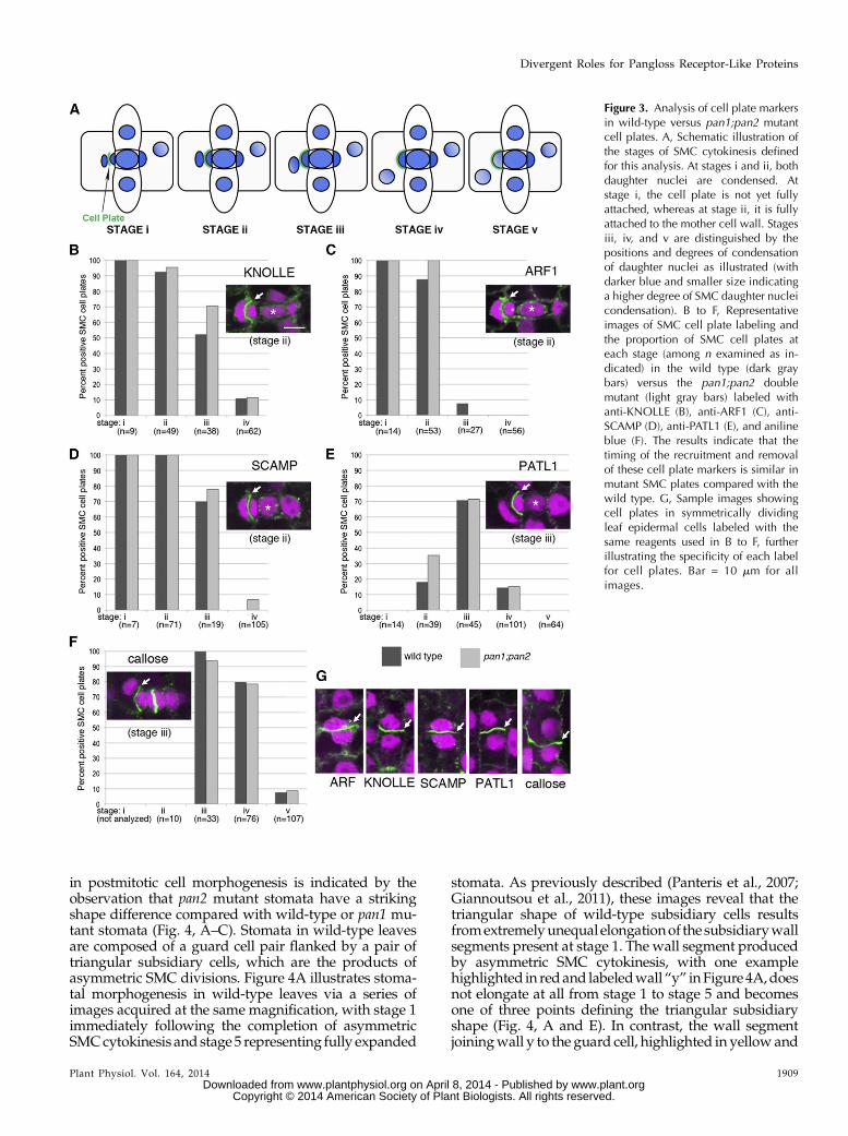
timing of the recruitment and removal of each cell platecomponent, we divided SMC cytokinesis into five stages(illustrated in Fig. 3A), with cell plate attachment occur-ring between stage i and stage ii and subsequent stagesdistinguished according to the position and degree ofdecondensation of the daughter nuclei.
KNOLLE is a cell plate-specific syntaxin that medi-ates the fusion of cell plate vesicles (Lukowitz et al.,1996; Lauber et al., 1997). ADP Ribosylation Factor1(ARF1) is a small GTPase that regulates the formationof vesicles at the Golgi and cell plate surface (Couchyet al., 2003), and Secretory Carrier Membrane Proteins(SCAMPs) are cell plate-localized proteins implicated inthe regulation of secretory and endocytic vesicle traf-ficking (Lam et al., 2008; Wang et al., 2010). Antibodiesraised againstArabidopsisKNOLLEandARF1GTPase,and rice (Oryza sativa) SCAMP, labeled similar propor-tions of wild-type and pan mutant SMC cell plates atstages i and ii, with anti-KNOLLE and anti-SCAMPalso labeling stage iii plates (Fig. 3, B–D). ArabidopsisPatellin1 (PATL1) is another putative regulator of vesi-cle formation, which localizes to cell plates at relativelylate stages (Peterman et al., 2004). Anti-PATL1 labeledwild-type and pan mutant SMC cell plates similarly,predominantly at stage iii (Fig. 3E). Finally, we exam-ined callose via aniline blue staining. Cell plates areinitially rich in callose, but callose is replaced with cel-lulose as cell plates mature (Samuels et al., 1995; Chenet al., 2009). Similar callose staining patterns were ob-served in wild-type and pan SMC cell plates at stages iiiand iv (Fig. 3F). Sample images illustrating the locali-zation of all five markers at each stage of SMC cell platedevelopment in each genotype are presented inSupplemental Figure S4. In summary, these experi-ments indicate that the timing of the recruitment andremoval of all cell plate components investigated issimilar in pan mutant SMC plates compared with thewild type. Together, our studies revealed no require-ment for PAN1 in any aspect of cell plate formation.
PAN2 Is Required for Coordinated Shape Changes inStomatal Subsidiaries and Interstomatal Cells
Like pan1, functions for pan2 outside of SMCs aresuggested by its expression in a variety of tissues whereno developing stomata are present, including elongatinginternodes, shoot apices, tassel primordia, and earlyembryos (Sekhon et al., 2011). Indeed, a function for pan2
Figure 2. PAN2 is undetectable at cell plates and not required for cellplate localization of PAN1. A to C, PAN2-YFP shown in monochrome(top) and green (bottom), along with CFP-tubulin in magenta, at suc-cessive stages of cell plate formation seen in three separate SMCs.Asterisks mark GMCs, which are flanked by PAN2-YFP patches inadjacent SMCs. Arrowheads point to the locations of cell plates (withno detectable PAN2-YFP), as indicated by the positions of the asso-ciated phragmoplasts seen at bottom. D to F, Anti-PAN2 stainingshown in monochrome (top) and green (bottom), along with propidiumiodide-stained nuclei shown in magenta, at successive stages of cy-tokinesis seen in three separate SMCs. Asterisks mark GMCs, whichare flanked by PAN2 patches in adjacent SMCs. Arrowheads point tothe locations of cell plates (with no detectable PAN2 staining), as in-dicated by the positions of associated nuclei. Bar = 10 mm for A to F.G and H, PAN1-YFP (monochrome in A and green in B) in two pan2-2mutant SMCs flanking GMCs (asterisks), with arrowheads pointing tocell plates. As indicated by the presence of a late-stage phragmoplast(CFP-tubulin signal; magenta in H), the SMC on the right is nearing thepoint of cell plate attachment while the SMC on the left has a recentlyattached cell plate. Although these SMCs lack PAN1-YFP patches ofnormal intensity at the site of GMC contact (e.g. as seen for the wildtype in Fig. 1), the PAN1-YFP signal level at cell plates is similar to thatof the wild type. Bar = 10 mm. I, Quantitative analysis of PAN1-YFPsignal intensity (arbitrary units as measured via ImageJ) in SMCs at
GMC contact sites (patch) and the cell plate (plate). J, As in I, but withresults shown as a ratio of signal measured at GMC contact sites versusthe cell plate. n = 62 cells analyzed for the wild type and 72 cellsanalyzed for pan2 mutants; error bars show SE. In wild-type SMCs,PAN1-YFP signal is approximately 3-fold stronger at GMC contact sitesthan at cell plates (patch-to-plate ratio of approximately 3). In pan2mutant SMCs, PAN1-YFP signal intensity is reduced at GMC contactsites but not at cell plates, reducing the patch-to-plate signal ratio toapproximately 1.
1908 Plant Physiol. Vol. 164, 2014
Sutimantanapi et al.
www.plant.org on April 8, 2014 - Published by www.plantphysiol.orgDownloaded from Copyright © 2014 American Society of Plant Biologists. All rights reserved.
in postmitotic cell morphogenesis is indicated by theobservation that pan2 mutant stomata have a strikingshape difference compared with wild-type or pan1 mu-tant stomata (Fig. 4, A–C). Stomata in wild-type leavesare composed of a guard cell pair flanked by a pair oftriangular subsidiary cells, which are the products ofasymmetric SMC divisions. Figure 4A illustrates stoma-tal morphogenesis in wild-type leaves via a series ofimages acquired at the same magnification, with stage 1immediately following the completion of asymmetricSMCcytokinesis and stage 5 representing fully expanded
stomata. As previously described (Panteris et al., 2007;Giannoutsou et al., 2011), these images reveal that thetriangular shape of wild-type subsidiary cells resultsfromextremelyunequal elongationof the subsidiarywallsegments present at stage 1. The wall segment producedby asymmetric SMC cytokinesis, with one examplehighlighted in redand labeledwall“y” inFigure 4A,doesnot elongate at all from stage 1 to stage 5 and becomesone of three points defining the triangular subsidiaryshape (Fig. 4, A and E). In contrast, the wall segmentjoiningwall y to the guard cell, highlighted in yellowand
Figure 3. Analysis of cell plate markersin wild-type versus pan1;pan2 mutantcell plates. A, Schematic illustration ofthe stages of SMC cytokinesis definedfor this analysis. At stages i and ii, bothdaughter nuclei are condensed. Atstage i, the cell plate is not yet fullyattached, whereas at stage ii, it is fullyattached to the mother cell wall. Stagesiii, iv, and v are distinguished by thepositions and degrees of condensationof daughter nuclei as illustrated (withdarker blue and smaller size indicatinga higher degree of SMC daughter nucleicondensation). B to F, Representativeimages of SMC cell plate labeling andthe proportion of SMC cell plates ateach stage (among n examined as in-dicated) in the wild type (dark graybars) versus the pan1;pan2 doublemutant (light gray bars) labeled withanti-KNOLLE (B), anti-ARF1 (C), anti-SCAMP (D), anti-PATL1 (E), and anilineblue (F). The results indicate that thetiming of the recruitment and removalof these cell plate markers is similar inmutant SMC plates compared with thewild type. G, Sample images showingcell plates in symmetrically dividingleaf epidermal cells labeled with thesame reagents used in B to F, furtherillustrating the specificity of each labelfor cell plates. Bar = 10 mm for allimages.
Plant Physiol. Vol. 164, 2014 1909
Divergent Roles for Pangloss Receptor-Like Proteins
www.plant.org on April 8, 2014 - Published by www.plantphysiol.orgDownloaded from Copyright © 2014 American Society of Plant Biologists. All rights reserved.

labeled wall “x” in Figure 4A, elongates extensively, asdoes the wall shared by the subsidiary and neighboringguard cell (Fig. 4, A and D). In concert with the acquisi-tion of triangular shape in subsidiary cells, points areformedon the endsof adjacent interstomatal cells (Fig. 4A,stage 5, arrows), indicating more rapid elongation of theinterstomatal wall segment shared with subsidiary cellscompared with that shared with guard cells.
When SMC divisions are oriented correctly, subsidi-ary cell shapes in pan1 and pan2 mutants are indistin-guishable from the wild type at birth (Fig. 4, comparestage 1 in B and C with that in A). At maturity, pan1mutant subsidiary cell shapes differ slightly from thewild type, with minor flattening of the point made bywall segment y associated with increased elongation ofthat segment (Fig. 1, B and E). In contrast, the shapes ofpan2 subsidiary cells at maturity are dramatically dif-ferent from thewild type, with long, flat sides producedby greatly increased elongation of wall y and reducedelongation ofwall x comparedwith thewild type (Fig. 4,C–E). The points on the ends of pan2 interstomatal cells
are correspondingly reduced (Fig. 4C). Only 20% to 30%of pan2 mutant SMCs divide aberrantly (Zhang et al.,2012), but all pan2 subsidiary cells exhibit this shapeabnormality (only the products of normally orientedSMC divisions are shown in Fig. 4). Thus, the pan2subsidiary cell and interstomatal cell shape defects de-scribed here arise after the completion of SMC division,are fully penetrant, and are separate from the occasionaldefects in subsidiary shape resulting from aberrantlyoriented divisions.
These observations raise the question of where PAN2functions to regulate themorphogenesis of stomatal andinterstomatal cells and how its localization in these cellsmay differ from that of PAN1. PAN1-YFP is barely de-tectable in expanding subsidiary and interstomatal cells,and virtually all detectable signal is at the subsidiarycell-guard cell interface (Fig. 5A). Consistent with alarger role in cellmorphogenesis, PAN2-YFP expressionlevels remain higher overall throughout the period ofepidermal cell expansion, with robust YFP signal ob-served at the surfaces of all cells (Fig. 5B). Notably, the
Figure 4. Analysis of stomatal morphogenesis in the wild type versus panmutants. A to C, Midplane confocal slices through theepidermal layer of leaves of the indicated genotypes expressing YFP-tubulin to permit the visualization of cell outlines. Stomatalmorphogenesis is divided into five stages. At stage 1, subsidiary cells have just formed; at stage 5, all cells are fully expanded.All images are shown at the same magnification to illustrate how cell shapes change as they expand. The wall produced byasymmetric cytokinesis in SMCs is defined as wall y, and one example in each image is highlighted in red. The subsidiary wallsegment linking wall y to the guard cells is defined as wall x, with one example in each image highlighted in yellow. Arrows inthe stage 5 wild-type image indicate points that form on the ends of interstomatal cells as subsidiary cells acquire a triangularshape. Bar = 14 mm for all images. D and E, Analysis of the lengths of wall x (D) and wall y (E) at each stage of stomatalmorphogenesis in plants of the indicated genotypes (n = 24–45 cells analyzed at each stage for each genotype; error bars showSD). The altered shapes of subsidiary cells in pan2 mutants result from increased elongation of wall y and decreased elongationof wall x.
1910 Plant Physiol. Vol. 164, 2014
Sutimantanapi et al.
www.plant.org on April 8, 2014 - Published by www.plantphysiol.orgDownloaded from Copyright © 2014 American Society of Plant Biologists. All rights reserved.
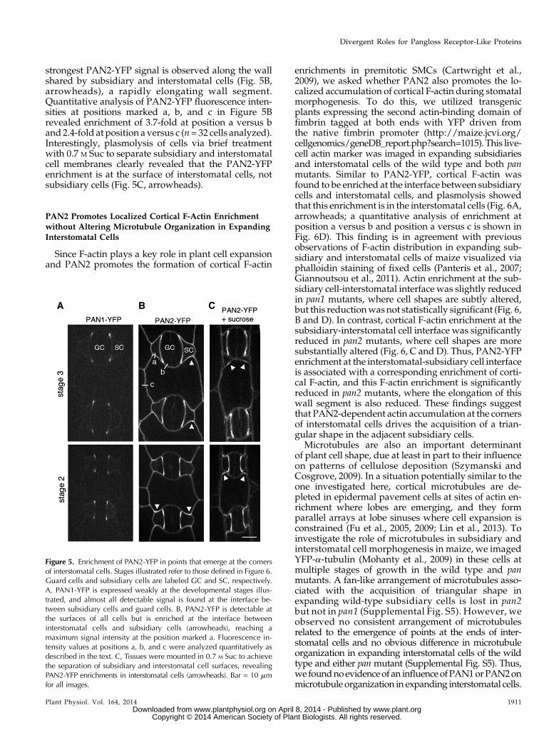
strongest PAN2-YFP signal is observed along the wallshared by subsidiary and interstomatal cells (Fig. 5B,arrowheads), a rapidly elongating wall segment.Quantitative analysis of PAN2-YFP fluorescence inten-sities at positions marked a, b, and c in Figure 5Brevealed enrichment of 3.7-fold at position a versus band 2.4-fold at position a versus c (n= 32 cells analyzed).Interestingly, plasmolysis of cells via brief treatmentwith 0.7 M Suc to separate subsidiary and interstomatalcell membranes clearly revealed that the PAN2-YFPenrichment is at the surface of interstomatal cells, notsubsidiary cells (Fig. 5C, arrowheads).
PAN2 Promotes Localized Cortical F-Actin Enrichmentwithout Altering Microtubule Organization in ExpandingInterstomatal Cells
Since F-actin plays a key role in plant cell expansionand PAN2 promotes the formation of cortical F-actin
enrichments in premitotic SMCs (Cartwright et al.,2009), we asked whether PAN2 also promotes the lo-calized accumulation of cortical F-actin during stomatalmorphogenesis. To do this, we utilized transgenicplants expressing the second actin-binding domain offimbrin tagged at both ends with YFP driven fromthe native fimbrin promoter (http://maize.jcvi.org/cellgenomics/geneDB_report.php?search=1015). This live-cell actin marker was imaged in expanding subsidiariesand interstomatal cells of the wild type and both panmutants. Similar to PAN2-YFP, cortical F-actin wasfound to be enriched at the interface between subsidiarycells and interstomatal cells, and plasmolysis showedthat this enrichment is in the interstomatal cells (Fig. 6A,arrowheads; a quantitative analysis of enrichment atposition a versus b and position a versus c is shown inFig. 6D). This finding is in agreement with previousobservations of F-actin distribution in expanding sub-sidiary and interstomatal cells of maize visualized viaphalloidin staining of fixed cells (Panteris et al., 2007;Giannoutsou et al., 2011). Actin enrichment at the sub-sidiary cell-interstomatal interface was slightly reducedin pan1 mutants, where cell shapes are subtly altered,but this reductionwas not statistically significant (Fig. 6,B and D). In contrast, cortical F-actin enrichment at thesubsidiary-interstomatal cell interface was significantlyreduced in pan2 mutants, where cell shapes are moresubstantially altered (Fig. 6, C and D). Thus, PAN2-YFPenrichment at the interstomatal-subsidiary cell interfaceis associated with a corresponding enrichment of corti-cal F-actin, and this F-actin enrichment is significantlyreduced in pan2 mutants, where the elongation of thiswall segment is also reduced. These findings suggestthat PAN2-dependent actin accumulation at the cornersof interstomatal cells drives the acquisition of a trian-gular shape in the adjacent subsidiary cells.
Microtubules are also an important determinantof plant cell shape, due at least in part to their influenceon patterns of cellulose deposition (Szymanski andCosgrove, 2009). In a situation potentially similar to theone investigated here, cortical microtubules are de-pleted in epidermal pavement cells at sites of actin en-richment where lobes are emerging, and they formparallel arrays at lobe sinuses where cell expansion isconstrained (Fu et al., 2005, 2009; Lin et al., 2013). Toinvestigate the role of microtubules in subsidiary andinterstomatal cell morphogenesis in maize, we imagedYFP-a-tubulin (Mohanty et al., 2009) in these cells atmultiple stages of growth in the wild type and panmutants. A fan-like arrangement of microtubules asso-ciated with the acquisition of triangular shape inexpanding wild-type subsidiary cells is lost in pan2but not in pan1 (Supplemental Fig. S5). However, weobserved no consistent arrangement of microtubulesrelated to the emergence of points at the ends of inter-stomatal cells and no obvious difference in microtubuleorganization in expanding interstomatal cells of the wildtype and either pan mutant (Supplemental Fig. S5). Thus,we foundnoevidenceof an influenceofPAN1orPAN2onmicrotubule organization in expanding interstomatal cells.
Figure 5. Enrichment of PAN2-YFP in points that emerge at the cornersof interstomatal cells. Stages illustrated refer to those defined in Figure 6.Guard cells and subsidiary cells are labeled GC and SC, respectively.A, PAN1-YFP is expressed weakly at the developmental stages illus-trated, and almost all detectable signal is found at the interface be-tween subsidiary cells and guard cells. B, PAN2-YFP is detectable atthe surfaces of all cells but is enriched at the interface betweeninterstomatal cells and subsidiary cells (arrowheads), reaching amaximum signal intensity at the position marked a. Fluorescence in-tensity values at positions a, b, and c were analyzed quantitatively asdescribed in the text. C, Tissues were mounted in 0.7 M Suc to achievethe separation of subsidiary and interstomatal cell surfaces, revealingPAN2-YFP enrichments in interstomatal cells (arrowheads). Bar = 10 mmfor all images.
Plant Physiol. Vol. 164, 2014 1911
Divergent Roles for Pangloss Receptor-Like Proteins
www.plant.org on April 8, 2014 - Published by www.plantphysiol.orgDownloaded from Copyright © 2014 American Society of Plant Biologists. All rights reserved.

Nonuniform Distributions of PIN1-YFP and YFP-ROP2 inExpanding Interstomatal and Subsidiary Cells ArePAN2 Independent
Since polarized localization of PIN1 auxin trans-porters is implicated in the mechanism of nonuniformcell expansion in epidermal pavement cells (Xu et al.,2010), we wondered whether PIN1 polarization inexpanding subsidiary or interstomatal cells of maize isassociatedwith nonuniform expansion in these cells. Toinvestigate this possibility, we analyzed the localizationof native promoter-driven PIN1-YFP (ZmPIN1a-YFP;Gallavotti et al., 2008). PIN1a-YFP is uniformly distrib-uted around the periphery of interstomatal cells at allstages of subsidiary morphogenesis in the wild type(Fig. 7A) and both pan mutants (Fig. 7, B and C). Al-though initially almost undetectable at the subsidiarycell face created by asymmetric SMC division (definedin Figure 4 as wall y), PIN1a-YFP rapidly becomeshighly enriched at this nonelongating face of wild-typesubsidiary cells and remains so throughout all sub-sequent stages of subsidiary expansion (Fig. 7A, arrow-heads). This association of PIN1awith the nonelongatingportion of the subsidiary cell surface is the opposite ofwhat has been observed in expanding Arabidopsispavement cells, where PIN1 and cortical F-actin are bothenriched at sites of lobe outgrowth (Xu et al., 2010). How-ever, the significance of PIN1a polarization in expandingmaize subsidiary cells is unknown. Remarkably, in spite
of the increased elongation of wall y in pan mutants,PIN1a-YFP remains highly polarized toward this por-tion of the cell surface in expanding pan1 and pan2mutant subsidiary cells, localizing as a patch (or a fo-cused chain of dots) at the center of this cell face as itelongates (Fig. 7, B and C, arrowheads). Thus, spatialcues mediating PIN1a targeting must still be present inpan mutant subsidiaries, albeit uncoupled from wallelongation control.
Finally, continuing to explore possible parallels withpavement cell lobe formation, where ROPs play a keyrole in regulating both cortical F-actin polymerizationand microtubule organization related to the emergenceof epidermal lobes (Fu et al., 2005, 2009; Lin et al., 2013),we investigated the localization of YFP-ROP2 (describedby Humphries et al., 2011) in expanding subsidiary andinterstomatal cells in maize and its dependence onPAN2. ROP2 is of further interest here because PAN1was found to physically interact with, and function co-operativelywith, type I ROPs to polarize SMCdivisions(Humphries et al., 2011). PAN2 acts upstream of PAN1in SMC polarization, so its function in these cells is alsorelated to type I ROPs, but a physical interaction be-tween PAN2 and ROP2 or other type I ROPs has notbeen demonstrated. YFP-ROP2 appears relatively uni-form in its distribution at the surface of expandingsubsidiary cells in both the wild type and pan2mutants(Fig. 8,AandB). YFP-ROP2 is conspicuouslydepleted atthe interface between interstomatal cells and guard cells
Figure 6. Cortical F-actin enrichment at the corners of interstomatal cells is reduced in pan2 mutants. A to C, YFP-ABD2-YFPlabeling illustrates the distribution of F-actin in expanding stomata and interstomatal cells at stages 2 and 3 in the wild type (A),pan1 (B), and pan2 (C). Arrowheads in A and B point to areas of cortical F-actin enrichment seen at the interface betweeninterstomatal cells and subsidiary cells in the absence of Suc. Mounting of wild-type tissue in 0.7 M Suc to achieve separationbetween subsidiary and interstomatal cells reveals that these cortical F-actin enrichments are in the interstomatal cells (ar-rowheads). D, Quantitative analysis of YFP-ABD2-YFP signal intensity ratios at position a versus b and position a versus c asdefined in A (n = 34–42 cells analyzed per genotype; error bars show SE). P values illustrate the significance of the differencesseen for each pan mutant relative to the wild type obtained via Student’s t test. Cortical F-actin enrichment at interstomatal cellcorners is significantly reduced (P , 0.01) in pan2 but not in pan1.
1912 Plant Physiol. Vol. 164, 2014
Sutimantanapi et al.
www.plant.org on April 8, 2014 - Published by www.plantphysiol.orgDownloaded from Copyright © 2014 American Society of Plant Biologists. All rights reserved.

(interface b in Fig. 8A) but is otherwise uniformly dis-tributed around the periphery of expanding interstomatalcells, with no enrichment at the interface with subsidiarycells (interface a in Fig. 8A; a quantitative analysis of signalintensity ratios confirming these conclusions is shown inFig. 8C). The nonuniform distribution of YFP-ROP2 at theinterstomatal cell surface does not appear related to therole of PAN2 in the morphogenesis of these cells, since nodifference was observed in YFP-ROP2 distribution inwild-type versus pan2mutant cells (Fig. 8, B andC). Thus,we find no evidence that PAN2 acts through ROP2 topromote localized cortical F-actin accumulation and non-uniform cell expansion.
DISCUSSION
Prior studies have demonstrated a cooperative func-tion for PAN1 and PAN2 in the polarization of SMC
divisions during stomatal development in maize and adependence of PAN1 on PAN2 for its polarized locali-zation in premitotic SMCs (Cartwright et al., 2009;Zhang et al., 2012). The work presented here demon-strates unequal or divergent roles for PAN1 and PAN2in other developmental processes.
PAN1 is enriched at developing cell plates, not onlyin SMCs but in all dividing cell types examined. Unlikethe situation in premitotic SMCs, PAN1 does not de-pend on PAN2 for its localization to cell plates, con-sistent with a lack of detectable PAN2 at cell plates. Infurther contrast to the situation in SMCs, PAN1 func-tion in cell plates could not be linked to F-actin poly-merization, since no difference in phragmoplast- or cellplate-associated F-actin was observed in pan mutants.Analysis of cytokinesis in pan mutants did not reveal aunique role for PAN1 in cell plate formation. The fre-quent misorientation of cell plates in pan1 mutantSMCs raises the possibility that PAN1 may function inattachment of the cell plate to the mother cell wallrather than in cell plate formation or maturation, asdemonstrated for the cell plate-localized, putative vesicle-trafficking regulator TPLATE (Van Damme et al., 2006,2011). However, cell platemisorientation is only observed
Figure 7. PAN1- and PAN2-independent polarization of ZmPIN1a-YFPin expanding subsidiary cells. All images are shown at the same mag-nification to illustrate cell enlargement at successive stages of stomatalmorphogenesis, numbered as illustrated in Figure 4. Arrowheads point tosubsidiary cell surface segment y produced by the asymmetric cytokinesisof subsidiary mother cells. ZmPIN1a-YFP becomes polarized toward thissegment of the cell surface shortly after its formation in the wild type (A).In pan1 (B) and especially in pan2 (C), where wall segment y elongatesmore than in the wild type, ZmPIN1a-YFP remains concentrated at thecenter of this segment, maintaining a polarized distribution similar to thatseen in the wild type. Bar = 10 mm for all images.
Figure 8. YFP-ROP2 localization in expanding stomata and inter-stomatal cells. A and B, The wild type (A) and the pan2 mutant (B) atstage 2 (bottom) and stage 3 (top). YFP-ROP2 distribution was notanalyzed in pan1 mutants because there was very little difference inthe shapes of subsidiary and interstomatal cells in pan1 mutantscompared with the wild type. The distribution of YFP-ROP2 on sub-sidiary versus interstomatal cell surfaces could not be determined viaplasmolysis experiments, because YFP-ROP2 signal was lost veryrapidly from the cell surface in the presence of Suc at concentrationssufficient to cause plasmolysis. Bar = 10 mm for all images. C, Ratios offluorescence intensities measured at positions a, b, and c as marked inA (n $ 25 cells analyzed per genotype). Error bars show SE. In bothgenotypes and stages, a modest enrichment of YFP-ROP2 was ob-served at positions a and c relative to position b. However, in contrastto YFP-ABD2-YFP and PAN2-YFP, no enrichment of YFP-ROP2 wasevident at position a versus position c. Moreover, no significant dif-ference was observed in any of the measured ratios when comparingthe wild type versus the pan2 mutant (P . 0.2 by Student’s t test for allthree comparisons).
Plant Physiol. Vol. 164, 2014 1913
Divergent Roles for Pangloss Receptor-Like Proteins
www.plant.org on April 8, 2014 - Published by www.plantphysiol.orgDownloaded from Copyright © 2014 American Society of Plant Biologists. All rights reserved.

in SMCs, even though PAN1 is localized to cell plates inall classes of dividing cells, arguing against this possi-bility and in favor of the idea that cell plate misorien-tation in both pan1 and pan2mutant SMCs results fromdefects in premitotic SMC polarity instead. There maybe a subtle defect in cell plate development in pan mu-tants that could be revealed by an analysis of additionalmolecular markers or of the timing of cell plate expan-sion or attachment. An alternative and likely possibility,in view of the large number of closely related LRR-RLKspresent in maize as in other plants, is that functionalredundancy between PAN1 and one ormore additionalLRR-RLKs conceals the functional significance of cellplate-localized PAN1. In any case, the observation of anLRR-RLK at the cell plate is interesting because it sug-gests ligand-mediated regulation of some aspect of cellplate or phragmoplast development. Thus, the interac-tion of LRR-RLKs such as PAN1with ligands producedinside the cell plate as early steps in plate biogenesis arecompleted may regulate the progression of cell plateformation (e.g. by triggering the recruitment of later-acting cell plate components).
Our study also showed that PAN2plays a greater rolethan PAN1 in the coordinated morphogenesis of sto-matal subsidiary and interstomatal cells after the com-pletion of stomatal divisions. PAN2 is enriched at thesurfaces of expanding interstomatal cells, where theycontact subsidiary cells, and stimulates a correspondingenrichment of cortical F-actin there. In pan2 mutants,reduced F-actin accumulation at this face of expandinginterstomatal cells is associated with a failure of pointsto emerge at the ends of these cells and with a con-comitant failure of neighboring subsidiary cells to ac-quire a triangular shape. Thus, as in SMCs, PAN2function in interstomatal cell morphogenesis is closelytied to localized actin polymerization. In premitoticSMCs, PAN2 is linked to localized actin polymerizationvia PAN1 and type I ROP GTPases (Cartwright et al.,2009; Humphries et al., 2011). However, in expandinginterstomatal cells, we find no enrichment of PAN1 orROP2 at the interface with subsidiary cells where PAN2and F-actin are enriched, and analysis of pan1 mutantsdemonstrates only a very minor role for PAN1 ininterstomatal and subsidiary cell morphogenesis.Thus, PAN2 appears to be linked to actin polymeri-zation via a different mechanism in interstomatalcells compared with SMCs, and this mechanism re-mains to be elucidated.
Fine actin filament networks at or near the plasmamembrane are associated with localized expansion ofthe cell surface in tip-growing cells (Vidali et al., 2001;Cárdenas et al., 2008; Dong et al., 2012) and expandingpavement cells (Frank and Smith, 2002; Fu et al., 2002).In tip-growing cells, the contribution of actin filamentsat the growth site has been difficult to separate from thatof cytoplasmic actin filament bundles that drive long-range transport of vesicles to and away from the tip, butevidence is accumulating that dynamic actin filamentnetworks at the growth site promote the accumulationand perhaps also the fusion with and/or removal of
vesicles from the plasma membrane (Qin and Yang,2011; Chebli et al., 2013). In pavement cells, corticalF-actin enriched at sites of lobe outgrowth has beenimplicated in the local suppression of PIN1 endocytosis,thereby promoting the localized enrichment of PIN1,leading to the local accumulation of auxin, which driveslocalized cell expansion (Nagawa et al., 2012). Thus,although we found no evidence of a similar role formaize PIN1a in interstomatal cell morphogenesis,findings for other cell types exhibiting polarized growthsuggest that PAN2-dependent cortical F-actin accumu-lation in interstomatal cells also promotes localized cellsurface expansion via the modulation of membraneand/or vesicle dynamics. This proposal is further sup-ported by the finding that F-actin promotes local ag-gregation of cortical endoplasmic reticulum at theemerging points on the ends of interstomatal cells,where the endoplasmic reticulummay facilitatemembrane-trafficking events supporting localized wall expansion(Giannoutsou et al., 2011).
Perhaps the most interesting facet of our results is theevidence they offer of a non-cell-autonomous influenceof interstomatal cells on the morphogenesis of neigh-boring subsidiary cells. Specifically, PAN2 and actinfunction at the interstomatal cell surface contactingsubsidiary wall x (as defined in Fig. 4) suppresses theelongation of subsidiary wall y. The enlargement ofsubsidiary cells in step with the neighboring guard cellscannot occur without the elongation of subsidiary wallx and/or y. Thus, redirection of growth towall y in pan2mutant subsidiary cells may be a response to the lack ofelongation of wall x, potentially mediated by the per-ception of physical forces such as wall or membranetension. Given the role of physical forces in orientingmicrotubules in plant cells (Landrein and Hamant,2013), this idea is consistent with our observation thatthe altered growth pattern in pan2 mutant subsidiarycells is associated with an alteration in microtubule or-ganization in these cells. It is interesting that excesselongation of wall y occurs in pan2 mutant subsidiarycells in spite of the correct localization of PIN1a to thecenter of this cell face, likely requiring continuous spa-tial regulation of membrane recycling to maintain PINpolarity, as in other cell types (Feraru and Friml, 2008).Thus, our findings suggest that some aspects of polar-ized membrane trafficking are preserved in expandingpan2 subsidiary cells (those required for PIN1a target-ing) while others are altered (those normally producingpreferential elongation ofwall x relative to y). Finally, inview of the putative function of PAN2 as a receptor andthe need for coordination of cell expansion betweeninterstomatal and subsidiary cells, it is interesting toconsider thepossibility that the subsidiary cell produces aligand to which PAN2 in the adjoining interstomatal cellresponds by promoting localized actin polymerizationand cell surface expansion. Thus, we speculate thatreceptor-mediated cell-cell interaction, as well as phys-ical forces, mediate the normal distribution of growth todifferent wall segments to achieve locally coordinatedcell shape changes during epidermal development.
1914 Plant Physiol. Vol. 164, 2014
Sutimantanapi et al.
www.plant.org on April 8, 2014 - Published by www.plantphysiol.orgDownloaded from Copyright © 2014 American Society of Plant Biologists. All rights reserved.

The findings reported here reveal that PAN1 andPAN2 have other functions in addition to their sharedfunction in the premitotic polarization of SMCs.Addingfurther to this conclusion, the closest relative of PAN2in Arabidopsis was recently identified as Guard CellHydrogen Peroxide Resistant1, which regulates guardcell aperture in mature leaves in response to abscisicacid and hydrogen peroxide (Hua et al., 2012). In play-ing multiple roles, PANs are similar to other receptorssuch as FERONIA (Cheung and Wu, 2011) and mem-bers of the ERECTA (van Zanten et al., 2009) and SERK(Chinchilla et al., 2009; Li, 2010) families in Arabidopsis.For example, ERECTA partners with closely relatedLRR-RLKs (ERECTA-LIKE proteins) to mediate inter-cellular signaling controlling the occurrence and orien-tation of asymmetric stomatal divisions (Pillitteri andTorii, 2012), promotes axis elongation and normalmorphogenesis of multiple aerial organs (van Zantenet al., 2009), and increases resistance to bacterial andfungal pathogens (Godiard et al., 2003; Llorente et al.,2005). Our finding of previously unrecognized roles forPAN1 and PAN2 demonstrates that these receptor-likeproteins are not dedicated to the perception of GMC-derived polarizing cues but also participate in cell platedevelopment (PAN1) or polarized cell growth (mainlyPAN2). We propose that PANs may function in allcontexts in the polarization of membrane trafficking,either directly or indirectly via their influence on actinpolymerization. The central importance of membranetrafficking in cell plate formation and polarized cellgrowth is well established, as discussed earlier. Thespatial regulation of membrane trafficking has not beenshown to play an essential role in the polarization ofplant cell division but generates the polarized distribu-tionof avariety of proteins in bothplant andanimal cells(Feraru and Friml, 2008; Apodaca et al., 2012). Thus, animportant role for polarized membrane trafficking inthe polarization of plant cell division is plausible.
MATERIALS AND METHODS
Plants and Growth Conditions
All maize (Zea mays) mutants employed in this study have been describedpreviously. Analysis of cytokinesis defects in pan mutants utilized pan1-Mu(Gallagher and Smith, 2000), pan1-ems (Cartwright et al., 2009), and pan1-Mu;pan2-O (Zhang et al., 2012) double mutants. Analysis of subsidiary and inter-stomatal morphogenesis defects utilized pan1-ems (Cartwright et al., 2009) andpan2-2 (Zhang et al., 2012) mutants. All mutants were backcrossed two or moretimes into the B73 wild-type background and compared with inbred B73 as thewild type. Transgenic plants expressing native promoter-driven PAN1-YFP,YFP-ROP2, and PAN2-YFP were described previously (Humphries et al.,2011; Zhang et al., 2012). Transgenics expressing native promoter-driven CFP-b-tubulin, YFP-a-tubulin, 2XYFP-ABD2 (the second actin-binding domain offimbrin tagged at both ends with YFP), and ZmPIN1a-YFP were generated asdescribed byMohanty et al. (2009) and at http://maize.jcvi.org/cellgenomics/index.php and were generously provided by Anne Sylvester (University ofWyoming). All transgenes were backcrossed two or more times to B73 and in-troduced into mutant backgrounds via crossing to mutants in the B73 back-ground. YFP- andCFP-tagged proteins were imaged in live cells after mountingtissues in distilled water. For imaging and phenotypic analysis, plants weregrown for 10 d to 4 weeks in a greenhouse maintained between 60°F and 90°Fwith natural lighting year round in La Jolla, California. Analysis of cell plate
localization and cytokinesis phenotypes utilized juvenile leaves (leaf 3 or 4);analysis of protein localization and cell morphology defects in stomatalsubsidiaries and interstomatal cells utilized adult leaf 8.
Staining of Actin, Callose, Cell Walls, and Nuclei
To analyze panmutants for cytokinesis defects, mature blade tissue from leaf3 or 4wasfixed and stainedwith acriflavine or 100mgmL21 propidium iodide asdescribed previously (Cleary and Smith, 1998; Hunter et al., 2012) andmountedin water (after propidium iodide staining) or saturated chloral hydrate (afteracriflavine staining) for imaging of epidermal walls and nuclei via confocalmicroscopy.
To stain callose in cell plates, the basal-most 2 cm of leaf 3 or 4 was cut into2-cm-long 3 2- to 3-mm-wide strips and fixed with formalin-acetic acid for 1 h.Following rehydration, tissues were stained with 0.1% (w/v) aniline blue inKPO4 buffer at pH 11 for 30 min, rinsed in phosphate-buffered saline, stainedwith 10 mg mL21 propidium iodide to label nuclei, rinsed again, and mounted inVectashield (Vector Laboratories) for imaging via confocal microscopy.
To labelphragmoplastactin, thebasal 2cmof leaf3or4wascut intostrips2cmlong 3 2 to 3 mm wide, fixed, and stained with Alexa Fluor 488-conjugatedphalloidin (Invitrogen) as described previously (Cartwright et al., 2009). Pro-pidium iodide at 10mgmL21 was used to stain nuclei. Samplesweremounted inVectashield (Vector Laboratories) for imaging via confocal microscopy.
Immunolocalization and Protein Gel Blotting
All antibodies used in this study were characterized previously. Anti-PAN1(Cartwright et al., 2009) and anti-PAN2 (Zhang et al., 2012) were generated inour laboratory. Anti-SCAMP (Lam et al., 2007) was raised against a rice (Oryzasativa) SCAMPproteinwhose closest relative inmaize is GRMZM2G041181 andwas provided by Liwen Jiang (Chinese University of Hong Kong). Anti-KNOLLE (Lukowitz et al., 1996) was raised against the Arabidopsis (Arabidopsisthaliana)KNOLLEprotein,whose closest relative inmaize isGRMZM2G100478, andwas provided by Gerd Jurgens (University of Tuebingen). Anti-PATL1 (Petermanet al., 2004)was raised againstArabidopsis PATL1,whose closest relative inmaize isGRMZM2G081652, andwas provided byKaye Peterman (Wellesley College). Anti-ARF1 was raised against Arabidopsis ARF1, whose closest relatives in maize areGRMZM2G105996, GRMZM2G357399, andGRMZM5G836182. This antibodywaspurchased fromAgrisera and is described at the productWeb page (http://www.agrisera.com/en/artiklar/plant_algal-cell-biology/compartment-markers/plant-Golgi-marker/arf1-adp-ribosylation-factor-1.html).
For tests of antibody specificity via protein gel blotting, proteins wereextracted from the basal 2 cm of leaves of 3- to 4-week-old B73maize plants afterremoval of leaveswith expanded sheaths.Membrane and soluble fractionswereprepared, separated via SDS-PAGE, and analyzed via immunoblotting as de-scribed previously (Cartwright et al., 2009). For immunolocalization, the basal2 cm of leaf 3 or 4 (containing cells at all stages of stomatal development) wasexcised from 10- to 20-d-old plants, cut into strips 2 cm long3 2 to 3 mmwide,fixed, and stained via awhole-mountproceduredescribedpreviously (Cartwrightet al., 2009). Primary antibodies were used for immunolocalization at thefollowing dilutions: affinity-purified anti-PAN1 and anti-PAN2 at 2 mg mL21,anti-ARF1 serum diluted to 1:2,000, affinity-purified anti-SCAMP at 1.5 mg mL21,anti-KNOLLE serum diluted to 1:1,000, and anti-PATL1 serum diluted to1:1,000. Binding of anti-KNOLLE, anti-ARF1, anti-SCAMP, and anti-PATL1antibodies was visualized by labeling with Alexa Fluor 488-conjugated sec-ondary antibodies (Invitrogen) diluted 1:200. Binding of anti-PAN1 and anti-PAN2 was visualized using Invitrogen Tyramide Signal Amplification kit 12following themanufacturer’s instructions.Nucleiwere subsequently counterstainedwith 10 mg mL21 propidium iodide (Sigma). Tissues were mounted in Vectashield(Vector Laboratories) and imaged via confocal microscopy as described below.
Confocal Microscopy and Image Analysis
Confocal imaging of fluorescence labeling was performed using a custom-assembled spinning-disk microscope system described previously (Walkeret al., 2007). YFP-tubulin images for subsidiary cell shape analysis were ac-quired with a Nikon 203 dry objective, and all other images were acquiredwith a Nikon 603 water-immersion objective. Alexa Fluor 488 was excitedwith an argon laser (488-nm line) and visualized with a Chroma HQ525/60emission filter. Propidium iodide was excited with an argon/krypton laser(568-nm line) and visualized with a Chroma HQ620/60 filter. YFP was excitedwith 514 nm and viewed with a Chroma HQ570/65 emission filter. CFP and
Plant Physiol. Vol. 164, 2014 1915
Divergent Roles for Pangloss Receptor-Like Proteins
www.plant.org on April 8, 2014 - Published by www.plantphysiol.orgDownloaded from Copyright © 2014 American Society of Plant Biologists. All rights reserved.

aniline blue were excited with a 440-nm laser and viewed with a ChromaHQ525/50 emission filter. Z projections of image stacks were produced usingImageJ version 1.36b or 1.47g (http://rsb.info.nih.gov/ij/). Further imageprocessing (adjustment of black levels, brightness, and contrast, production ofcolor merges, and figure preparation) was carried out using Adobe Photoshopversion 8.0 or 11.02, applying only linear adjustments to pixel values.
For quantitative analysis of PAN2-YFP, YFP-ABD2-YFP, and YFP-ROP2signal intensities, ImageJ version 1.47g was used to measure fluorescence in-tensities along the length of a line drawn at positions a, b, and c as indicated inFigures 5, 6, and 8 on Z projections of a standard number of focal planes for eachmarker. Minimum fluorescence intensity values (background signal) weresubtracted from maximum values, and ratios of these background-subtractedmaximumvalues (a to b and a to c)were calculated for individual cells at stages 2or 3 as defined in Figure 4 (one set of measurements per cell). For PAN2-YFPanalysis, 32 cells from 13 different wild-type individuals expressing PAN2-YFPwere analyzed. For YFP-ABD2-YFP, we analyzed 42 cells from two wild-typeindividuals, 34 cells from two pan2 mutant individuals, and 42 cells from twopan1mutant individuals. For YFP-ROP2, we analyzed 29 cells from three wild-type individuals and 25 cells from three pan2 individuals.
Quantitative analysis of PAN1-YFP intensities in SMCs (Fig. 2, I and J) wasconducted similarly except that single focal planes were used, and lines weredrawn through the cell plate and the GMC contact site in SMCs (n = 62 wildtype and 72 pan2) that had recently completed cytokinesis, as judged by thepresence of phragmoplast remnants and nuclear appearance, corresponding tothe stage when cell plate-associated PAN1 signal was highest.
Sequences for the genes and proteins investigated in this study can be found athttp://www.maizesequence.org (release 5b.60) as GRMZM2G034572_T01 andGRMZM2G034572_P01 (PAN2), GRMZM5G836190_T02 andGRMZM5G836190_P02(PAN1), GRMZM2G098643_T01 and GRMZM2G098643_P01 (PIN1a), andGRMZM5G846811_T01 and GRMZM5G846811_P01 (ROP2).
Supplemental Data
The following materials are available in the online version of this article.
Supplemental Figure S1. PAN1 localization at cell plates of root corticalcells.
Supplemental Figure S2. Analysis of phragmoplast F-actin in wild-typeand pan mutant SMCs.
Supplemental Figure S3. Protein gel-blot analysis of the specificities ofantibodies used in this study.
Supplemental Figure S4. Immunostaining with antibodies to cell platemarkers in wild-type and pan mutant SMCs undergoing cytokinesis.
Supplemental Figure S5. Microtubule organization visualized via imagingof YFP-a-tubulin in expanding subsidiary and interstomatal cells.
ACKNOWLEDGMENTS
We thank Xiaoguo Zhang for supplying the PAN2 immunolocalizationdata presented in Figure 2, Anne Sylvester for supplying transgenic linesexpressing ZmPIN1a-YFP, CFP-tubulin, YFP-tubulin, and YFP-ABD2-YFP,and Gerd Jurgens, Kaye Peterman, and Liwen Jiang for providing antibodiesused in this study.
Received November 15, 2013; accepted February 24, 2014; published February27, 2014.
LITERATURE CITED
Apodaca G, Gallo LI, Bryant DM (2012) Role of membrane traffic in thegeneration of epithelial cell asymmetry. Nat Cell Biol 14: 1235–1243
Boudeau J, Miranda-Saavedra D, Barton GJ, Alessi DR (2006) Emergingroles of pseudokinases. Trends Cell Biol 16: 443–452
Cárdenas L, Lovy-Wheeler A, Kunkel JG, Hepler PK (2008) Pollen tubegrowth oscillations and intracellular calcium levels are reversiblymodulated by actin polymerization. Plant Physiol 146: 1611–1621
Cartwright HN, Humphries JA, Smith LG (2009) PAN1: a receptor-likeprotein that promotes polarization of an asymmetric cell division inmaize. Science 323: 649–651
Castells E, Casacuberta JM (2007) Signalling through kinase-defectivedomains: the prevalence of atypical receptor-like kinases in plants.J Exp Bot 58: 3503–3511
Chebli Y, Kroeger J, Geitmann A (2013) Transport logistics in pollen tubes.Mol Plant 6: 1037–1052
Chen XY, Liu L, Lee E, Han X, Rim Y, Chu H, Kim SW, Sack F, Kim JY(2009) The Arabidopsis callose synthase gene GSL8 is required for cy-tokinesis and cell patterning. Plant Physiol 150: 105–113
Cheung AY, Wu HM (2011) THESEUS 1, FERONIA and relatives: a familyof cell wall-sensing receptor kinases? Curr Opin Plant Biol 14: 632–641
Chinchilla D, Shan L, He P, de Vries S, Kemmerling B (2009) One for all:the receptor-associated kinase BAK1. Trends Plant Sci 14: 535–541
Cleary AL, Smith LG (1998) The Tangled1 gene is required for spatialcontrol of cytoskeletal arrays associated with cell division during maizeleaf development. Plant Cell 10: 1875–1888
Couchy I, Bolte S, Crosnier MT, Brown S, Satiat-Jeunemaitre B (2003)Identification and localization of a beta-COP-like protein involved in themorphodynamics of the plant Golgi apparatus. J Exp Bot 54: 2053–2063
Dong H, Pei W, Haiyun R (2012) Actin fringe is correlated with tip growthvelocity of pollen tubes. Mol Plant 5: 1160–1162
Feraru E, Friml J (2008) PIN polar targeting. Plant Physiol 147: 1553–1559Frank MJ, Smith LG (2002) A small, novel protein highly conserved in
plants and animals promotes the polarized growth and division ofmaize leaf epidermal cells. Curr Biol 12: 849–853
Fu Y, Gu Y, Zheng Z, Wasteneys G, Yang Z (2005) Arabidopsis interdig-itating cell growth requires two antagonistic pathways with opposingaction on cell morphogenesis. Cell 120: 687–700
Fu Y, Li H, Yang Z (2002) The ROP2 GTPase controls the formation ofcortical fine F-actin and the early phase of directional cell expansionduring Arabidopsis organogenesis. Plant Cell 14: 777–794
Fu Y, Xu T, Zhu L, Wen M, Yang Z (2009) A ROP GTPase signalingpathway controls cortical microtubule ordering and cell expansion inArabidopsis. Curr Biol 19: 1827–1832
Galatis B, Apostolakos P (2004) The role of the cytoskeleton in the mor-phogenesis and function of stomatal complexes. New Phytol 161:613–639
Gallagher K, Smith LG (2000) Roles for polarity and nuclear determinantsin specifying daughter cell fates after an asymmetric cell division in themaize leaf. Curr Biol 10: 1229–1232
Gallavotti A, Yang Y, Schmidt RJ, Jackson D (2008) The Relationshipbetween auxin transport and maize branching. Plant Physiol 147:1913–1923
Giannoutsou EP, Apostolakos P, Galatis B (2011) Actin filament-organized local cortical endoplasmic reticulum aggregations in devel-oping stomatal complexes of grasses. Protoplasma 248: 373–390
Godiard L, Sauviac L, Torii KU, Grenon O, Mangin B, Grimsley NH,Marco Y (2003) ERECTA, an LRR receptor-like kinase protein controllingdevelopment pleiotropically affects resistance to bacterial wilt. Plant J 36:353–365
Hua D, Wang C, He J, Liao H, Duan Y, Zhu Z, Guo Y, Chen Z, Gong Z(2012) A plasma membrane receptor kinase, GHR1, mediates abscisicacid- and hydrogen peroxide-regulated stomatal movement in Arabi-dopsis. Plant Cell 24: 2546–2561
Humphries JA, Vejlupkova Z, Luo A, Meeley RB, Sylvester AW,Fowler JE, Smith LG (2011) ROP GTPases act with the receptor-likeprotein PAN1 to polarize asymmetric cell division in maize. Plant Cell23: 2273–2284
Hunter CT, Kirienko DH, Sylvester AW, Peter GF, McCarty DR, Koch KE(2012) Cellulose Synthase-Like D1 is integral to normal cell division, ex-pansion, and leaf development in maize. Plant Physiol 158: 708–724
Jürgens G (2005) Cytokinesis in higher plants. Annu Rev Plant Biol 56:281–299
Lam SK, Cai Y, Hillmer S, Robinson DG, Jiang L (2008) SCAMPs highlightthe developing cell plate during cytokinesis in tobacco BY-2 cells. PlantPhysiol 147: 1637–1645
Lam SK, Siu CL, Hillmer S, Jang S, An G, Robinson DG, Jiang L (2007)Rice SCAMP1 defines clathrin-coated, trans-golgi-located tubular-vesicular structures as an early endosome in tobacco BY-2 cells. PlantCell 19: 296–319
Landrein B, Hamant O (2013) How mechanical stress controls microtubulebehavior and morphogenesis in plants: history, experiments and re-visited theories. Plant J 75: 324–338
1916 Plant Physiol. Vol. 164, 2014
Sutimantanapi et al.
www.plant.org on April 8, 2014 - Published by www.plantphysiol.orgDownloaded from Copyright © 2014 American Society of Plant Biologists. All rights reserved.

Lauber MH, Waizenegger I, Steinmann T, Schwarz H, Mayer U, Hwang I,Lukowitz W, Jürgens G (1997) The Arabidopsis KNOLLE protein is acytokinesis-specific syntaxin. J Cell Biol 139: 1485–1493
Li J (2010) Multi-tasking of somatic embryogenesis receptor-like proteinkinases. Curr Opin Plant Biol 13: 509–514
Lin D, Cao L, Zhou Z, Zhu L, Ehrhardt D, Yang Z, Fu Y (2013) Rho GTPasesignaling activates microtubule severing to promote microtubule or-dering in Arabidopsis. Curr Biol 23: 290–297
Llompart B, Castells E, Río A, Roca R, Ferrando A, Stiefel V, Puigdomenech P,Casacuberta JM (2003) The direct activation of MIK, a germinal center kinase(GCK)-like kinase, by MARK, a maize atypical receptor kinase, suggests anew mechanism for signaling through kinase-dead receptors. J Biol Chem278: 48105–48111
Llorente F, Alonso-Blanco C, Sánchez-Rodriguez C, Jorda L, Molina A(2005) ERECTA receptor-like kinase and heterotrimeric G protein fromArabidopsis are required for resistance to the necrotrophic fungusPlectosphaerella cucumerina. Plant J 43: 165–180
Lukowitz W,Mayer U, Jürgens G (1996) Cytokinesis in the Arabidopsis embryoinvolves the syntaxin-related KNOLLE gene product. Cell 84: 61–71
McMichael CM, Bednarek SY (2013) Cytoskeletal and membrane dy-namics during higher plant cytokinesis. New Phytol 197: 1039–1057
Mohanty A, Luo A, DeBlasio S, Ling X, Yang Y, Tuthill DE, Williams KE,Hill D, Zadrozny T, Chan A, et al (2009) Advancing cell biology andfunctional genomics in maize using fluorescent protein-tagged lines.Plant Physiol 149: 601–605
Nagawa S, Xu T, Lin D, Dhonukshe P, Zhang X, Friml J, Scheres B, Fu Y,Yang Z (2012) ROP GTPase-dependent actin microfilaments promotePIN1 polarization by localized inhibition of clathrin-dependent endo-cytosis. PLoS Biol 10: e1001299
Panteris E, Galatis B, Quader H, Apostolakos P (2007) Cortical actin fila-ment organization in developing and functioning stomatal complexes ofZea mays and Triticum turgidum. Cell Motil Cytoskeleton 64: 531–548
Peterman TK, Ohol YM, McReynolds LJ, Luna EJ (2004) Patellin1, a novelSec14-like protein, localizes to the cell plate and binds phosphoinosi-tides. Plant Physiol 136: 3080–3094
Pillitteri LJ, Torii KU (2012) Mechanisms of stomatal development. AnnuRev Plant Biol 63: 591–614
Qin Y, Yang Z (2011) Rapid tip growth: insights from pollen tubes. SeminCell Dev Biol 22: 816–824
Rajakulendran T, Sicheri F (2010) Allosteric protein kinase regulation bypseudokinases: insights from STRAD. Sci Signal 3: pe8
Samuels AL, Giddings THJ Jr, Staehelin LA (1995) Cytokinesis in tobaccoBY-2 and root tip cells: a new model of cell plate formation in higherplants. J Cell Biol 130: 1345–1357
Seguí-Simarro JM, Austin JR II, White EA, Staehelin LA (2004) Electrontomographic analysis of somatic cell plate formation in meristematiccells of Arabidopsis preserved by high-pressure freezing. Plant Cell 16:836–856
Sekhon RS, Lin H, Childs KL, Hansey CN, Buell CR, de Leon N,Kaeppler SM (2011) Genome-wide atlas of transcription during maizedevelopment. Plant J 66: 553–563
Szymanski DB, Cosgrove DJ (2009) Dynamic coordination of cytoskeletaland cell wall systems during plant cell morphogenesis. Curr Biol 19:R800–R811
Van Damme D, Coutuer S, De Rycke R, Bouget FY, Inzé D, Geelen D(2006) Somatic cytokinesis and pollen maturation in Arabidopsis dependon TPLATE, which has domains similar to coat proteins. Plant Cell 18:3502–3518
Van Damme D, Gadeyne A, Vanstraelen M, Inzé D, Van Montagu MC,De Jaeger G, Russinova E, Geelen D (2011) Adaptin-like proteinTPLATE and clathrin recruitment during plant somatic cytokinesis oc-curs via two distinct pathways. Proc Natl Acad Sci USA 108: 615–620
van Zanten M, Snoek LB, Proveniers MC, Peeters AJ (2009) The manyfunctions of ERECTA. Trends Plant Sci 14: 214–218
Vidali L, McKenna ST, Hepler PK (2001) Actin polymerization is essentialfor pollen tube growth. Mol Biol Cell 12: 2534–2545
Walker KL, Müller S, Moss D, Ehrhardt DW, Smith LG (2007) Arabi-dopsis TANGLED identifies the division plane throughout mitosis andcytokinesis. Curr Biol 17: 1827–1836
Wang H, Tse YC, Law AH, Sun SS, Sun YB, Xu ZF, Hillmer S,Robinson DG, Jiang L (2010) Vacuolar sorting receptors (VSRs) andsecretory carrier membrane proteins (SCAMPs) are essential for pollen tubegrowth. Plant J 61: 826–838
Xu T, Wen M, Nagawa S, Fu Y, Chen JG, Wu MJ, Perrot-Rechenmann C,Friml J, Jones AM, Yang Z (2010) Cell surface- and rho GTPase-basedauxin signaling controls cellular interdigitation in Arabidopsis. Cell 143:99–110
Yalovsky S, Bloch D, Sorek N, Kost B (2008) Regulation of membranetrafficking, cytoskeleton dynamics, and cell polarity by ROP/RAC GTPases.Plant Physiol 147: 1527–1543
Yang Z, Lavagi I (2012) Spatial control of plasma membrane domains: ROPGTPase-based symmetry breaking. Curr Opin Plant Biol 15: 601–607
Zhang X, Facette M, Humphries JA, Shen Z, Park Y, Sutimantanapi D,Sylvester AW, Briggs SP, Smith LG (2012) Identification of PAN2 byquantitative proteomics as a leucine-rich repeat-receptor-like kinaseacting upstream of PAN1 to polarize cell division in maize. Plant Cell 24:4577–4589
Plant Physiol. Vol. 164, 2014 1917
Divergent Roles for Pangloss Receptor-Like Proteins
www.plant.org on April 8, 2014 - Published by www.plantphysiol.orgDownloaded from Copyright © 2014 American Society of Plant Biologists. All rights reserved.