distribution of disease write-up ves draft 2 july18 · mr alok tiwari (corresponding author)a...
TRANSCRIPT
University of Birmingham
A review of distribution of atherosclerosis in thelower limb arteries of patients with diabetes mellitusand peripheral vascular diseaseLowry, Danielle; Saeed, Mujahid; Narendran, Parth; Tiwari, Alok
DOI:10.1177/1538574418791622
License:None: All rights reserved
Document VersionPeer reviewed version
Citation for published version (Harvard):Lowry, D, Saeed, M, Narendran, P & Tiwari, A 2018, 'A review of distribution of atherosclerosis in the lower limbarteries of patients with diabetes mellitus and peripheral vascular disease', Vascular and Endovascular Surgery,vol. 52, no. 7, pp. 535-542. https://doi.org/10.1177/1538574418791622
Link to publication on Research at Birmingham portal
Publisher Rights Statement:Checked for eligibility 04/12/2018
This is a author-produced, peer-reviewed version of an article published in Vascular and Endovascular Surgery.https://doi.org/10.1177/1538574418791622
General rightsUnless a licence is specified above, all rights (including copyright and moral rights) in this document are retained by the authors and/or thecopyright holders. The express permission of the copyright holder must be obtained for any use of this material other than for purposespermitted by law.
•Users may freely distribute the URL that is used to identify this publication.•Users may download and/or print one copy of the publication from the University of Birmingham research portal for the purpose of privatestudy or non-commercial research.•User may use extracts from the document in line with the concept of ‘fair dealing’ under the Copyright, Designs and Patents Act 1988 (?)•Users may not further distribute the material nor use it for the purposes of commercial gain.
Where a licence is displayed above, please note the terms and conditions of the licence govern your use of this document.
When citing, please reference the published version.
Take down policyWhile the University of Birmingham exercises care and attention in making items available there are rare occasions when an item has beenuploaded in error or has been deemed to be commercially or otherwise sensitive.
If you believe that this is the case for this document, please contact [email protected] providing details and we will remove access tothe work immediately and investigate.
Download date: 19. Jul. 2020

The Distribution of Atherosclerosis in the Lower Limb Arteries of Patients
with Diabetes Mellitus and Peripheral Vascular Disease: A systematic
review
Miss Danielle Lowry a Degrees: MBChB, MRCS
Affiliation: University Hospitals Birmingham NHS Foundation Trust,
Dr Mujahid Saeedb Degrees: MBBS, MRCP(UK)(Endocrinology and Diabetes, FRCP(UK)
Affiliation: University Hospitals Birmingham NHS Foundation Trust
Dr Parth Narendranb,c Degrees: MBBS, BSc, MRCP. PhD
Affiliation: University Hospitals Birmingham NHS Foundation Trust,
Mr Alok Tiwari (Corresponding Author)a Degrees: MBBS, FRCSed, MD
Email: [email protected]
Affiliation: University Hospitals Birmingham NHS Foundation Trust,
a Vascular Department 6th Floor Nuffield House, University Hospitals Birmingham NHS
Foundation Trust, Queen Elizabeth Hospital Birmingham, Mindelsohn Way, Edgbaston,
Birmingham, B15 2WB, UK
b Diabetes Department, 5th Floor Nuffield House, University Hospitals Birmingham NHS
Foundation Trust, Queen Elizabeth Hospital Birmingham, Mindelsohn Way, Edgbaston,
Birmingham, B15 2WB, UK
c Institute of Metabolism and Systems Research, College of Medical and Dental Sciences,
University of Birmingham, Edgbaston, Birmingham, B15 2TT, UK
Abstract
Objective: There is a generally accepted hypothesis that patients with diabetes mellitus
(DM) have a higher burden of atherosclerotic disease below the knee compared to patients
without DM (NDM). The aim of this review was to summarize the evidence regarding this
hypothesis.
Methods: The literature was searched for papers that compared the anatomical distribution
of atherosclerotic disease in patients with DM and those without using radiological imaging.
Search terms used included “diabetes mellitus”, “peripheral vascular disease”, ‘distribution
of disease”, “angiography”, “computed tomography angiography” and “magnetic resonance
angiography”. Where possible, the number of patients with disease in each arterial segment
was extracted and included in a forest plot. A descriptive approach was taken when this was
not possible or a scoring system was used.
Results: Fourteen studies were included in the review and it was possible to summarise data
from nine of these in a forest plot. Fifteen different arterial segments were described,
however, the most commonly used segments that differentiated between proximal and
distal disease were aorto‐iliac (A‐I) (DM=466 patients, NDM=458), femoro‐popliteal (F‐P)
(DM=568, NDM=585), tibial (DM=306, NDM=417). The resulting forest plot showed that
those with DM were significantly less likely to have disease in the A‐I segment (OR 0.25
(0.15‐0.42)) and significantly more likely to have disease in the tibial segment (OR 1.94
(1.27‐2.96)). In the DM group, there was a trend towards relative sparing in the F‐P segment
but this does not reach significance (0.66 (0.33‐1.31)).
Conclusions: These results support the hypothesis that patients with DM are more likely to
have atherosclerotic disease in the tibial vessels than patients without DM. There is
however limited information on how individual vessels are affected. Further information on
this and a greater understanding of why the distal vessels are more affected are avenues for
future research.
Introduction
Infra‐popliteal disease is associated with critical limb ischaemia which is the final stage in
the disease course of peripheral arterial disease (PAD)1. The pattern of vascular disease
influences the options there are for revascularisation. Management of distal disease is more
challenging than proximal disease, although advances in this area are being made2‐5. Despite
these advances patients with distal disease have a higher risk of an amputation and shorter
amputation‐free survival6.
The prevalence of diabetes mellitus (DM) is increasing worldwide and is a major risk factor
for PAD7,8. Patients with DM have a predisposition towards a higher burden of
atherosclerotic disease below the knee compared to patients without DM (NDM). This is
considered to have an impact on both the treatment options available and prognosis
following revascularisation in patients with DM9,10.
This hypothesis of a higher burden of disease in the tibial arteries is widely accepted within
the medical community. The aim of this review was to summarise the quality of the
evidence supporting this hypothesis.
Methods
A literature search was performed using the search terms “diabetes mellitus”, “peripheral
vascular disease”, ‘distribution of disease”, “angiography”, “computed tomography
angiography” and “magnetic resonance angiography”. Synonyms and various combinations
were used in the search strategy which involved both MESH and keyword searches. Embase
and MEDLINE databases were searched including papers published from 1946 to present
day and in‐process citations. References from relevant studies were also scrutinised for
potential studies.
Papers were included if arterial imaging of the lower limb was undertaken using digital
subtraction angiography (DSA), Computed Tomography Angiography, or Magnetic
Resonance Angiography. They were excluded if the indication for imaging was not PAD, if
there was no separation of patients with and without DM or only patients with DM were
included. The final requirement was an anatomical description of the arteries affected by
atherosclerotic disease. This description could be given using a scoring system or
proportions of arterial segments affected.
Statistical analysis
For papers that included proportions of patients with PAD by arterial segment the number
of patients who had disease in each arterial segment was extracted by one author (DL).
These papers were included in a forest plot that was produced using Revman 5.3 (Review
Manager (RevMan) [Computer program]. Version 5.3. Copenhagen: The Nordic Cochrane
Centre, The Cochrane Collaboration, 2014.). Data were summarised as odds ratio (OR) with
95% confidence intervals.
Results
From the literature search, 151 potential papers were identified and following review of
titles, abstracts, full text and references 14 studies were included in the review (Figure 1).
The papers dated from 1964 to 2009 and were all cross‐sectional studies apart from one
cohort study11 and two case‐control studys12,13. The majority of papers did not state if their
analysis was by patient or by limb, in most, it appeared that a single treated limb was
included per patient11,13‐18. Four papers included all treated limbs19‐22, one paper included
both legs for all patients12, one paper analysed by lesion23 and one paper only used the data
from the left leg if there was bilateral imaging as they found the legs to be comparable24.
How risk factors for PAD were treated varied between papers. Four papers performed some
form of multivariate analysis to stratify for risk factors12,18,22,23, the majority of remaining
papers reported proportions of risk factors and comparability between groups however two
papers made no mention of risk factors14,19. No studies considered type I and type II DM
separately. One paper found a significant difference in the proportion of men and women in
their cohorts12 and one paper found significant differences in the proportions of smokers20.
Most papers had cohorts with a mean age in the mid‐sixties although two papers
deliberately selected young cohorts14,15 and two papers had older cohorts11,21. The majority
of cohorts consisted of approximately 60% men apart from Ozkan et al who had 85.9%
men18. The proportion of smokers in each group ranged from 13.5% to 83.2% (Table 1).
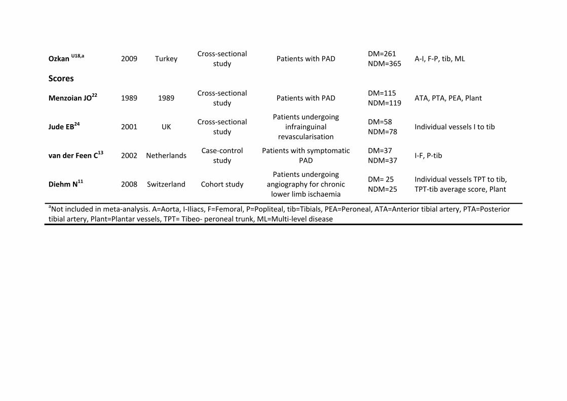
All the studies used angiography to visualise the arterial tree and in total 15 different
arterial segments were described (Table 2). The most commonly used segments that
differentiated between proximal and distal disease were aorto‐iliac (A‐I), femoro‐popliteal
(F‐P), tibial (Tib). Seven studies also included a category that represented disease at multiple
levels (ML). These segments were included in the forest plot along with smaller segments
that fitted in the same group. I.e. patients with disease in the popliteal artery could be
included in the F‐P group but those in a popliteal/tibial group could not be included. The
description of what constituted significant disease varied between papers. Of the papers
that described proportions of arterial segment involved five only included occlusions14,17,19‐
21, two defined a significant stenosis as involving more than 20% of the lumen15,16, two
defined it as more than 50%12,18 and in one paper the definition was not stated23.
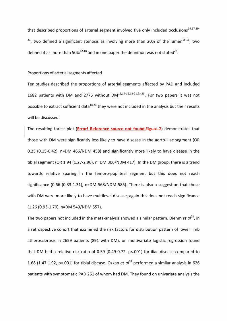
Proportions of arterial segments affected
Ten studies described the proportions of arterial segments affected by PAD and included
1682 patients with DM and 2775 without DM12,14‐16,18‐21,23,25. For two papers it was not
possible to extract sufficient data18,23 they were not included in the analysis but their results
will be discussed.
The resulting forest plot (Error! Reference source not found.Figure 2) demonstrates that
those with DM were significantly less likely to have disease in the aorto‐iliac segment (OR
0.25 (0.15‐0.42), n=DM 466/NDM 458) and significantly more likely to have disease in the
tibial segment (OR 1.94 (1.27‐2.96), n=DM 306/NDM 417). In the DM group, there is a trend
towards relative sparing in the femoro‐popliteal segment but this does not reach
significance (0.66 (0.33‐1.31), n=DM 568/NDM 585). There is also a suggestion that those
with DM were more likely to have multilevel disease, again this does not reach significance
(1.26 (0.93‐1.70), n=DM 549/NDM 557).
The two papers not included in the meta‐analysis showed a similar pattern. Diehm et al23, in
a retrospective cohort that examined the risk factors for distribution pattern of lower limb
atherosclerosis in 2659 patients (891 with DM), on multivariate logistic regression found
that DM had a relative risk ratio of 0.59 (0.49‐0.72, p<.001) for iliac disease compared to
1.68 (1.47‐1.92, p<.001) for tibial disease. Ozkan et al18 performed a similar analysis in 626
patients with symptomatic PAD 261 of whom had DM. They found on univariate analysis the
presence of DM was related to odds ratios of 0.56 (p=.001) for aorto‐iliac disease, 1.16
(p=.39) for femoro‐popliteal disease and 2.44 (p=.001) for tibial disease.
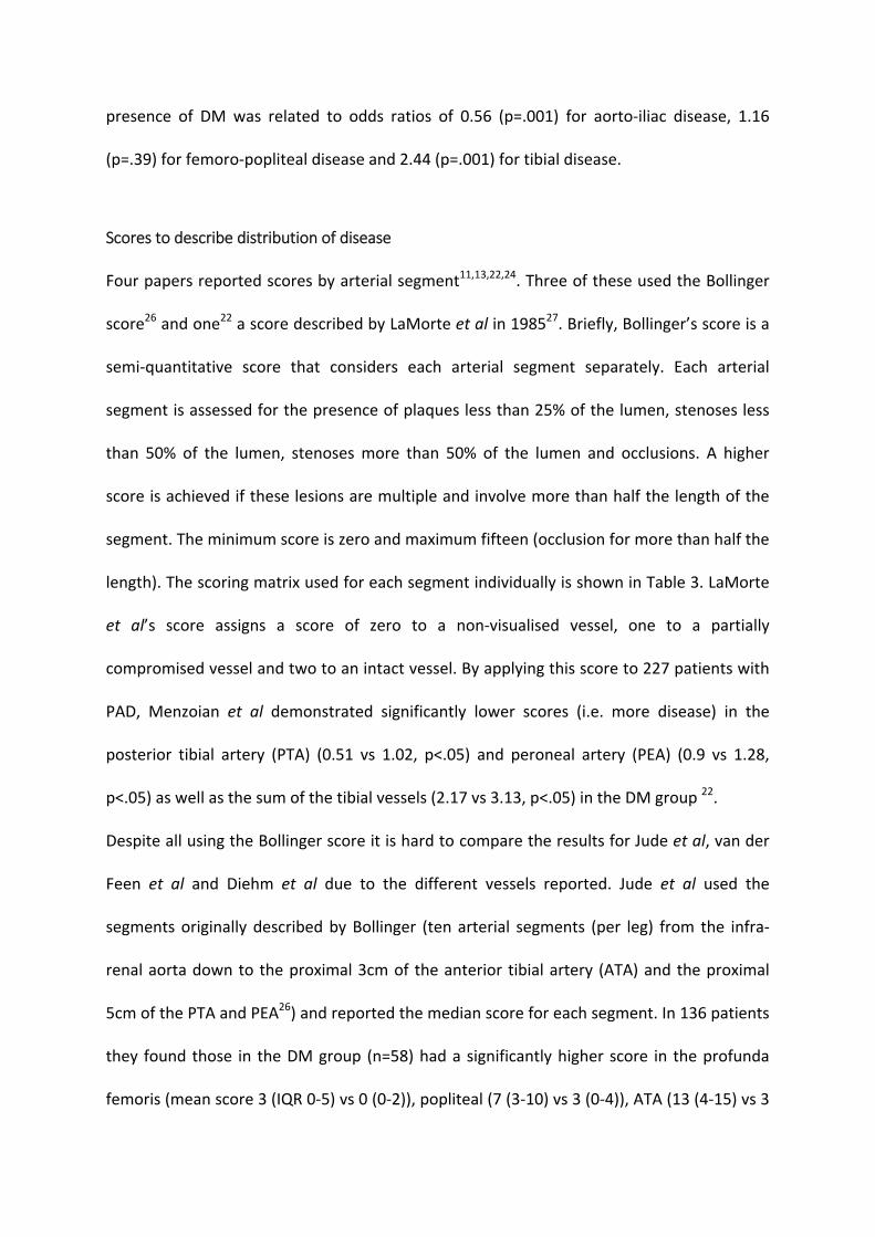
Scores to describe distribution of disease
Four papers reported scores by arterial segment11,13,22,24. Three of these used the Bollinger
score26 and one22 a score described by LaMorte et al in 198527. Briefly, Bollinger’s score is a
semi‐quantitative score that considers each arterial segment separately. Each arterial
segment is assessed for the presence of plaques less than 25% of the lumen, stenoses less
than 50% of the lumen, stenoses more than 50% of the lumen and occlusions. A higher
score is achieved if these lesions are multiple and involve more than half the length of the
segment. The minimum score is zero and maximum fifteen (occlusion for more than half the
length). The scoring matrix used for each segment individually is shown in Table 3. LaMorte
et al’s score assigns a score of zero to a non‐visualised vessel, one to a partially
compromised vessel and two to an intact vessel. By applying this score to 227 patients with
PAD, Menzoian et al demonstrated significantly lower scores (i.e. more disease) in the
posterior tibial artery (PTA) (0.51 vs 1.02, p<.05) and peroneal artery (PEA) (0.9 vs 1.28,
p<.05) as well as the sum of the tibial vessels (2.17 vs 3.13, p<.05) in the DM group 22.
Despite all using the Bollinger score it is hard to compare the results for Jude et al, van der
Feen et al and Diehm et al due to the different vessels reported. Jude et al used the
segments originally described by Bollinger (ten arterial segments (per leg) from the infra‐
renal aorta down to the proximal 3cm of the anterior tibial artery (ATA) and the proximal
5cm of the PTA and PEA26) and reported the median score for each segment. In 136 patients
they found those in the DM group (n=58) had a significantly higher score in the profunda
femoris (mean score 3 (IQR 0‐5) vs 0 (0‐2)), popliteal (7 (3‐10) vs 3 (0‐4)), ATA (13 (4‐15) vs 3
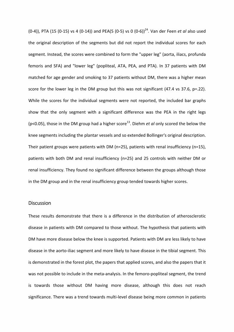
(0‐4)), PTA (15 (0‐15) vs 4 (0‐14)) and PEA(5 (0‐5) vs 0 (0‐6))24. Van der Feen et al also used
the original description of the segments but did not report the individual scores for each
segment. Instead, the scores were combined to form the "upper leg" (aorta, iliacs, profunda
femoris and SFA) and "lower leg" (popliteal, ATA, PEA, and PTA). In 37 patients with DM
matched for age gender and smoking to 37 patients without DM, there was a higher mean
score for the lower leg in the DM group but this was not significant (47.4 vs 37.6, p=.22).
While the scores for the individual segments were not reported, the included bar graphs
show that the only segment with a significant difference was the PEA in the right legs
(p<0.05), those in the DM group had a higher score13. Diehm et al only scored the below the
knee segments including the plantar vessels and so extended Bollinger's original description.
Their patient groups were patients with DM (n=25), patients with renal insufficiency (n=15),
patients with both DM and renal insufficiency (n=25) and 25 controls with neither DM or
renal insufficiency. They found no significant difference between the groups although those
in the DM group and in the renal insufficiency group tended towards higher scores.
Discussion
These results demonstrate that there is a difference in the distribution of atherosclerotic
disease in patients with DM compared to those without. The hypothesis that patients with
DM have more disease below the knee is supported. Patients with DM are less likely to have
disease in the aorto‐iliac segment and more likely to have disease in the tibial segment. This
is demonstrated in the forest plot, the papers that applied scores, and also the papers that it
was not possible to include in the meta‐analysis. In the femoro‐popliteal segment, the trend
is towards those without DM having more disease, although this does not reach
significance. There was a trend towards multi‐level disease being more common in patients
with DM. Four papers assessed the severity of disease in individual vessels rather than
segments11,22,24 although only Jude et al did for both above and below knee vessels 24. In
patients with and without DM, the least affected of the tibial vessels was consistently the
PEA.
The PEA as a target vessel for revascularisation has been considered to have limitations due
to success relying on indirect collateralisation to supply the forefoot28. The patency of the
PEA has also been demonstrated to be less critical in preventing amputation29. The
angiosome model holds that the areas supplied by the PEA are the anterior and lateral ankle
and plantar heel30. However increasingly the PEA has been shown to have multiple
collaterals and to commonly supply the pedal arteries and as such has comparable
outcomes for both surgical and endovascular revascularisation compared to other distal
target vessels31‐34.
DM is known to have an impact on both the presentation of PAD and outcomes following
revascularisation35,36. The distribution of atherosclerotic disease has also been shown to be
related to outcomes following revascularisation procedures in patients both with and
without DM37. The pathophysiology behind why patients with DM have increased PAD is
complex but thought to be related to a combination of down‐regulation of nitric oxide and
prostacyclin, upregulation of vasoconstrictors, apoptosis of endothelial cells, activated
coagulation, abnormal platelet activation and propensity towards plaque rupture38. There is
not any clear evidence why the distal vessels are predominantly affected and while these
results support the hypothesis that patients with DM have a more significant disease burden
below the knee they provide us with limited information on the degree to which individual
vessels or areas of vessels are affected.
A strength of the review is that all the papers used DSA as the imaging modality. DSA
remains the gold standard for imaging of the lower limbs and describing the anatomic
distribution of stenotic disease39. In the meta‐analysis, there was low heterogeneity
between the papers apart from those considering the femoro‐popliteal segment (I2=68%). A
major weakness of the review is the low quality of the papers included. They are all
observational studies, predominantly retrospective and so the body of evidence is low to
very low quality40. An attempt to assess the methodological quality of the papers using the
Newcastle‐Ottawa scale41 was made. However, all but three papers were cross‐sectional
studies making it not possible to apply the scale. There was consistency in the type of
patients selected with the majority of papers including patients with Fontaine II to IV
disease. However, one paper only included patients with intermittent claudication18, two
papers excluded those with intermittent claudication21,25 and three papers did not define
the patient group beyond symptomatic PAD15,19,20. As described in the results section there
was also variance in how significant disease was defined. Within each paper the
demographics for each group, when reported, were comparable (Table 1).
Additional weaknesses include that the papers are all relatively historical (earliest 1964,
latest 2009) and the variety in how the arterial segments were described and grouped
together. This grouping meant some data was not able to be included in the meta‐analysis
because the segment crossed the knee, weakening the data included. During data
collection, it was considered that improvements in the medical management of DM and
PAD may have had an impact on the distribution of disease. Evidence from high‐quality
randomised controlled trials on the importance of tight blood glucose control in relation to
the complications of DM was published in the late nineties42,43. When studies from prior to
the year 2000 were excluded from the meta‐analysis the trends remained the same

although the odds ratio for tibial disease was no longer significant (OR 1.99 (0.94‐4.24)). This
may suggest that optimising medical therapy has an impact on the degree of tibial disease.
This review was not designed to consider this question and so firm conclusions cannot be
drawn.
Conclusions
Patients with DM are more likely to have atherosclerotic disease in the tibial vessels
compared to patients without. The current published evidence supports this hypothesis.
There is very limited data on the degree to which individual vessels are affected. Further
information on this and a greater understanding of why the distal vessels are more affected
are avenues for future research. Studies that examine the impact of medical therapy on the
distribution of disease may also be valuable.
References
1. Becker F, Robert‐Ebadi H, Ricco JB, et al. Chapter I: Definitions, epidemiology, clinical presentation and prognosis. Eur J Vasc Endovasc Surg. 2011;42 Suppl 2:S4‐12.
2. Kok HK, Asadi H, Sheehan M, McGrath FP, Given MF, Lee MJ. Outcomes of infrapopliteal angioplasty for limb salvage based on the updated TASC II classification. Diagn Interv Radiol. 2017;23(5):360‐364.
3. Teraa M, Conte MS, Moll FL, Verhaar MC. Critical Limb Ischemia: Current Trends and Future Directions. J Am Heart Assoc. 2016;5(2).
4. Singh GD, Brinza EK, Hildebrand J, et al. Midterm Outcomes After Infrapopliteal Interventions in Patients With Critical Limb Ischemia Based on the TASC II Classification of Below‐the‐Knee Arteries. J Endovasc Ther. 2017;24(3):321‐330.
5. Jaff MR, White CJ, Hiatt WR, et al. An update on methods for revascularization and expansion of the TASC lesion classification to include below‐the‐knee arteries: A supplement to the inter‐society consensus for the management of peripheral arterial disease (TASC II): The TASC steering committee. Catheter Cardiovasc Interv. 2015;86(4):611‐625.
6. Mustapha JA, Diaz‐Sandoval LJ, Saab F. Infrapopliteal calcification patterns in critical limb ischemia: diagnostic, pathologic and therapeutic implications in the search for the endovascular holy grail. J Cardiovasc Surg (Torino). 2017;58(3):383‐401.
7. NCD Risk factor Collaboration (NCD‐RisC). Worldwide trends in diabetes since 1980: a pooled analysis of 751 population‐based studies with 4.4 million participants. Lancet. 2016;387(10027):1513‐1530.
8. World Health Organisation. Global Report on Diabetes. http://www.who.int/diabetes/global‐report/en/2016.
9. Forsythe RO, Jones KG, Hinchliffe RJ. Distal bypasses in patients with diabetes and infrapopliteal disease: technical considerations to achieve success. Int J Low Extrem Wounds. 2014;13(4):347‐362.
10. Wallaert JB, Nolan BW, Adams J, et al. The impact of diabetes on postoperative outcomes following lower‐extremity bypass surgery. J Vasc Surg. 2012;56(5):1317‐1323.
11. Diehm N, Rohrer S, Baumgartner I, Keo H, Do D, Kalka C. Distribution pattern of infrageniculate arterial obstructions in patients with diabetes mellitus and renal insufficiency ‐ implications for revascularization. Vasa. 2008;37(3):265‐273.
12. Haltmayer M, Mueller T, Horvath W, Luft C, Poelz W, Haidinger D. Impact of atherosclerotic risk factors on the anatomical distribution of peripheral arterial disease. Int Angiol. 2001;20(3):200‐207.
13. Van Der Feen C, Neijens FS, Kanters SDJM, Mali WPTM, Stolk RP, Banga JD. Angiographic distribution of lower extremity atherosclerosis in patients with and without diabetes. Diabetic Medicine. 2002;19(5):366‐370.
14. De Bakey ME, Crawford ES, Garrett HE, Cooley DA, Morris GC, Jr., Abbott JP. Occlusive Disease of the Lower Extremities in Patients 16 to 37 Years of Age. Ann Surg. 1964;159:873‐890.
15. Hansen ME, Valentine RJ, McIntire DD, Myers SI, Chervu A, Clagett GP. Age‐related differences in the distribution of peripheral atherosclerosis: when is atherosclerosis truly premature? Surgery. 1995;118(5):834‐839.
16. Kroger K, Buss C, Renzing‐Kohler K, Santosa F, Rudofsky G. Segmental manifestation of peripheral atherosclerosis and its association to risk factors. Vasa. 2000;29(3):199‐203.
17. Rueda CA, Nehler MR, Perry DJ, et al. Patterns of artery disease in 450 patients undergoing revascularization for critical limb ischemia: Implications for clinical trial design. Journal of Vascular Surgery. 2008;47(5):995‐1000.
18. Ozkan U, Oguzkurt L, Tercan F. Atherosclerotic risk factors and segmental distribution in symptomatic peripheral artery disease. J Vasc Interv Radiol. 2009;20(4):437‐441.
19. Haimovici H. Patterns of arteriosclerotic lesions of the lower extremity. Arch Surg. 1967;95(6):918‐933.
20. Ciavarella A, Silletti A, Mustacchio A, et al. Angiographic evaluation of the anatomic pattern of arterial obstructions in diabetic patients with critical limb ischaemia. Diabete Metab. 1993;19(6):586‐589.
21. Lazaris AM, Tsiamis AC, Fishwick G, Bolia A, Bell PR. Clinical outcome of primary infrainguinal subintimal angioplasty in diabetic patients with critical lower limb ischemia. J Endovasc Ther. 2004;11(4):447‐453.
22. Menzoian JO, LaMorte WW, Paniszyn CC, et al. Symptomatology and Anatomic Patterns of Peripheral Vascular Disease: Differing Impact of Smoking and Diabetes. Ann Vasc Surg. 1989;3(3):224‐228.
23. Diehm N, Shang A, Silvestro A, et al. Association of cardiovascular risk factors with pattern of lower limb atherosclerosis in 2659 patients undergoing angioplasty. Eur J Vasc Endovasc Surg. 2006;31(1):59‐63.
24. Jude EBO, S.O.; Chalmers, N.; Boulton, A.J.M. Peripheral Arterial Disease in Diabetic and Nondiabetic Patients. Diabetes Care. 2001;24:1433‐1437.
25. Rueda CA, Nehler MR, Perry DJ, et al. Patterns of artery disease in 450 patients undergoing revascularization for critical limb ischemia: implications for clinical trial design. J Vasc Surg. 2008;47(5):995‐999; discussion 999‐1000.
26. Bollinger A, Breddin K, Hess H, et al. Semiquantitative assessment of lower limb atherosclerosis from routine angiographic images. Atherosclerosis. 1981;38:339‐346.
27. LaMorte WW, Menzoian JO, Sidawy A, Heeren T. A new method for the prediction of peripheral vascular resistance from the preoperative angiogram. J Vasc Surg. 1985;2(5):703‐708.
28. Elliott BM, Robison JG, Brothers TE, Cross MA. Limitations of peroneal artery bypass grafting for limb salvage. J Vasc Surg. 1993;18(5):881‐888.
29. Faglia E, Clerici G, Clerissi J, et al. When is a technically successful peripheral angioplasty effective in preventing above‐the‐ankle amputation in diabetic patients with critical limb ischaemia? Diabet Med. 2007;24(8):823‐829.
30. Sumpio BE, Forsythe RO, Ziegler KR, van Baal JG, Lepantalo MJA, Hinchliffe RJ. Clinical implications of the angiosome model in peripheral vascular disease. Journal of Vascular Surgery. 2013;58(3):814‐826.
31. Karmody AM, Leather RP, Shah DM, Corson JD, Naraynsingh V. Peroneal artery bypass: a reappraisal of its value in limb salvage. J Vasc Surg. 1984;1(6):809‐816.
32. Ricco JB, Gargiulo M, Stella A, et al. Impact of angiosome‐ and nonangiosome‐targeted peroneal bypass on limb salvage and healing in patients with chronic limb‐threatening ischemia. J Vasc Surg. 2017;66(5):1479‐1487.
33. Dosluoglu HH, Cherr GS, Lall P, Harris LM, Dryjski ML. Peroneal artery‐only runoff following endovascular revascularizations is effective for limb salvage in patients with tissue loss. J Vasc Surg. 2008;48(1):137‐143.
34. Abularrage CJ, Conrad MF, Haurani MJ, et al. Long‐term outcomes of patients undergoing endovascular infrainguinal interventions with single‐vessel peroneal artery runoff. J Vasc Surg. 2011;53(4):1007‐1013.
35. DeRubertis BG, Pierce M, Ryer EJ, Trocciola S, Kent KC, Faries PL. Reduced primary patency rate in diabetic patients after percutaneous intervention results from more frequent presentation with limb‐threatening ischemia. J Vasc Surg. 2008;47(1):101‐108.
36. Abularrage CJ, Conrad MF, Hackney LA, et al. Long‐term outcomes of diabetic patients undergoing endovascular infrainguinal interventions. J Vasc Surg. 2010;52(2):314‐322.e311‐314.
37. Bradbury AW, Adam DJ, Bell J, et al. Bypass versus Angioplasty in Severe Ischaemia of the Leg (BASIL) trial: A description of the severity and extent of disease using the Bollinger angiogram scoring method and the TransAtlantic Inter‐Society Consensus II classification.[Erratum appears in J Vasc Surg. 2010 Dec;52(6):1751 Note: Bhattachary, V [corrected to Bhattacharya, V]]. J Vasc Surg. 2010;51(5 Suppl):32S‐42S.
38. Yang SL, Zhu LY, Han R, Sun LL, Li JX, Dou JT. Pathophysiology of peripheral arterial disease in diabetes mellitus. J Diabetes. 2017;9(2):133‐140.
39. Stoner MC, Calligaro KD, Chaer RA, et al. Reporting standards of the Society for Vascular Surgery for endovascular treatment of chronic lower extremity peripheral artery disease: Executive summary. J Vasc Surg. 2016;64(1):227‐228.
40. Balshem H, Helfand M, Schunemann HJ, et al. GRADE guidelines: 3. Rating the quality of evidence. J Clin Epidemiol. 2011;64(4):401‐406.
41. Wells G, Shea B, O'Connell D, et al. The Newcastle‐Ottawa Scale (NOS) for assessing the quality of nonrandomised studies in meta‐analyses. 2010; http://www.ohri.ca/programs/clinical_epidemiology/oxford.asp. Accessed January, 2018.
42. UK Prospective Diabetes Study (UKPDS) Group. Intensive blood‐glucose control with sulphonylureas or insulin compared with conventional treatment and risk of complications in patients with type 2 diabetes (UKPDS 33). The Lancet. 1998;352(9131):837‐853.
43. Group EoDIaCER. Epidemiology of Diabetes Interventions and Complications (EDIC): Design, implementation, and preliminary results of a long‐term follow‐up of the Diabetes Control and Complications Trial cohort. Diabetes Care. 1999;22(1):99‐111.
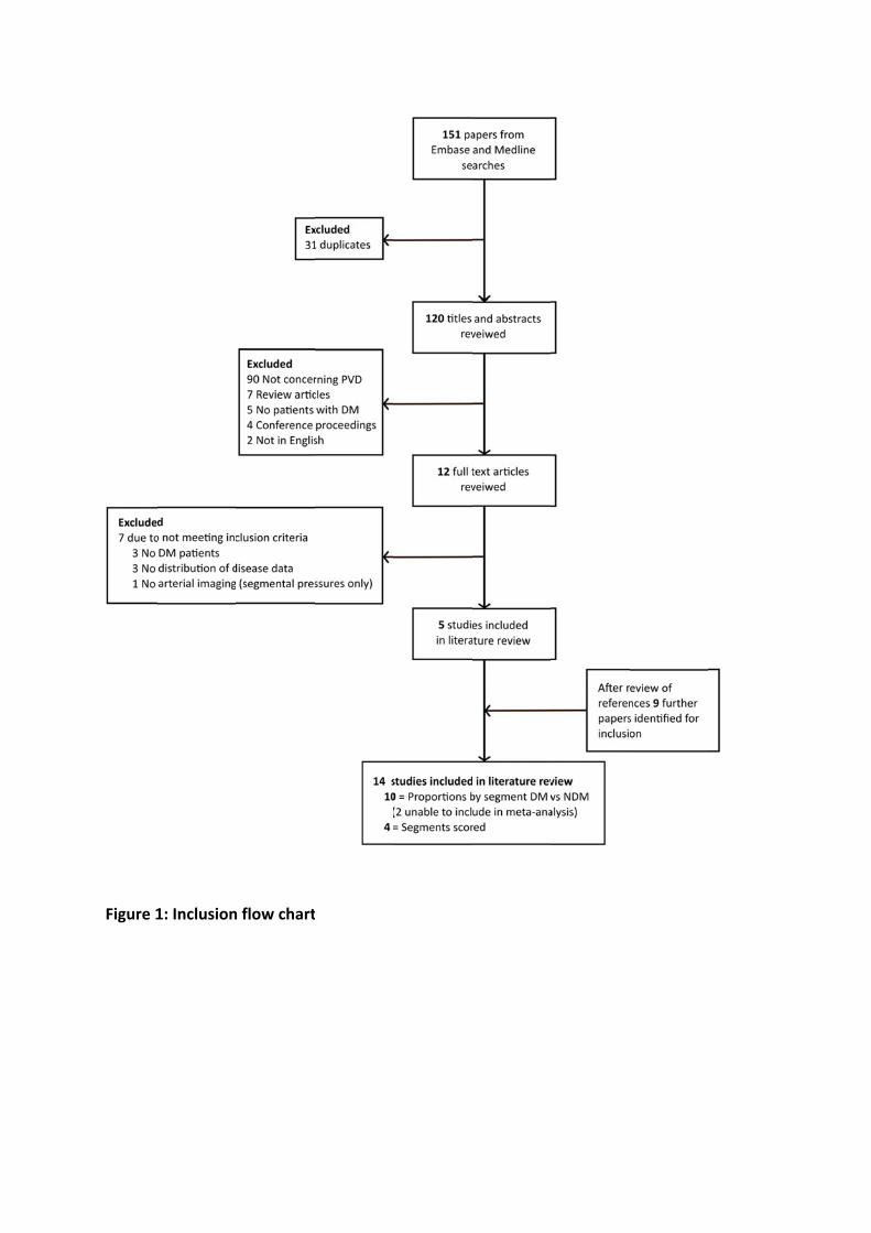
Figure 1
1: Inclusionn flow chartt
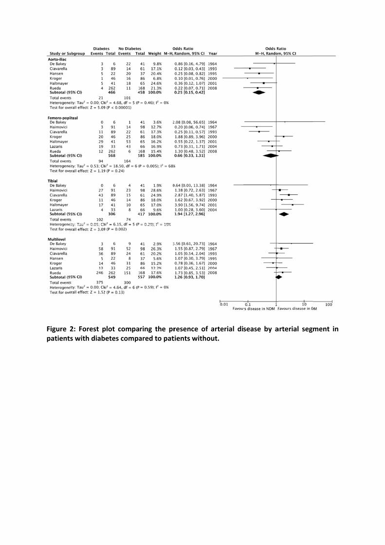
Figure patient
2: Forest pts with diab
plot compabetes compa
ring the prared to pat
resence of tients witho
arterial diout.
sease by aarterial segm
ment in
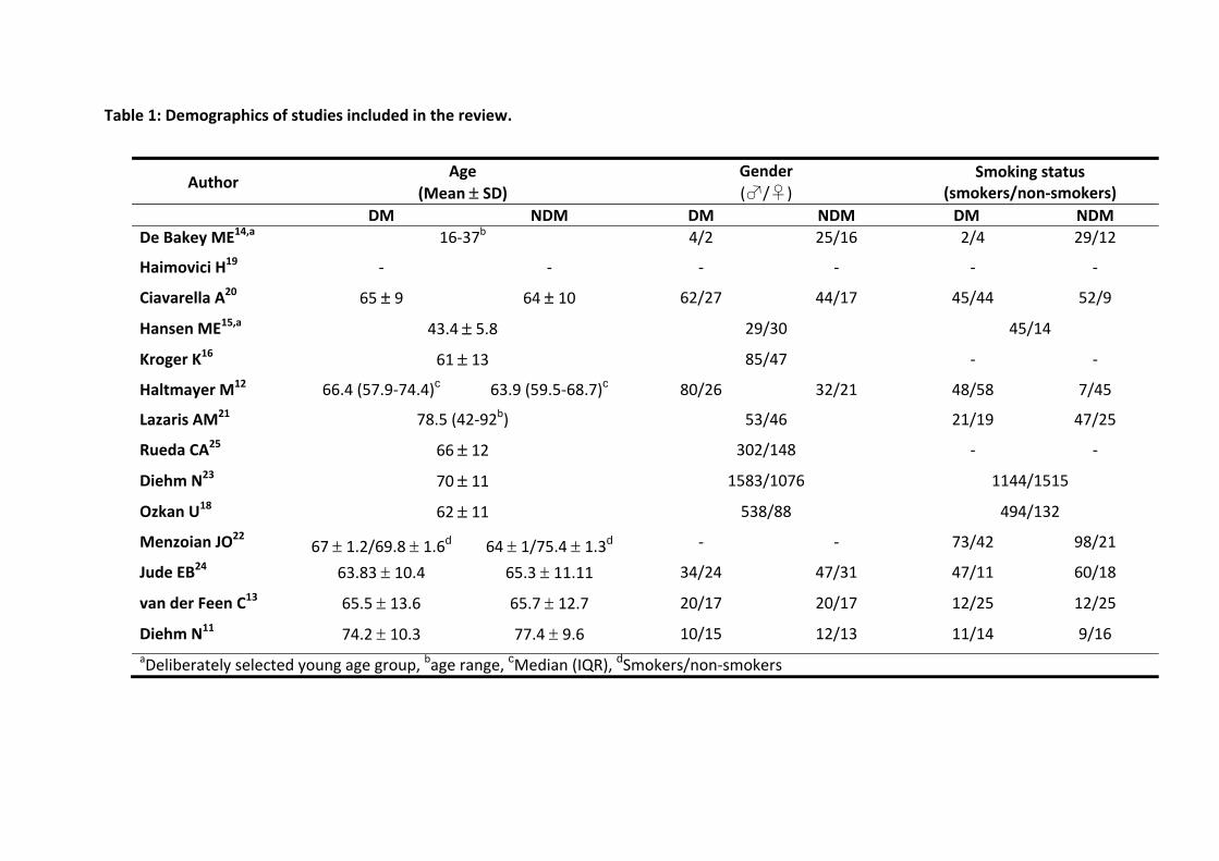
Table 1: Demographics of studies included in the review.
Author Age
(Mean SD) Gender
(♂/♀) Smoking status
(smokers/non‐smokers)
DM NDM DM NDM DM NDM
De Bakey ME14,a 16‐37b 4/2 25/16 2/4 29/12
Haimovici H19 ‐ ‐ ‐ ‐ ‐ ‐
Ciavarella A20 65 9 64 10 62/27 44/17 45/44 52/9
Hansen ME15,a 43.4 5.8 29/30 45/14
Kroger K16 61 13 85/47 ‐ ‐
Haltmayer M12 66.4 (57.9‐74.4)c 63.9 (59.5‐68.7)c 80/26 32/21 48/58 7/45
Lazaris AM21 78.5 (42‐92b) 53/46 21/19 47/25
Rueda CA25 66 12 302/148 ‐ ‐
Diehm N23 70 11 1583/1076 1144/1515
Ozkan U18 62 11 538/88 494/132
Menzoian JO22 67 1.2/69.8 1.6d 64 1/75.4 1.3d ‐ ‐ 73/42 98/21
Jude EB24 63.83 10.4 65.3 11.11 34/24 47/31 47/11 60/18
van der Feen C13 65.5 13.6 65.7 12.7 20/17 20/17 12/25 12/25
Diehm N11 74.2 10.3 77.4 9.6 10/15 12/13 11/14 9/16
aDeliberately selected young age group, bage range, cMedian (IQR), dSmokers/non‐smokers
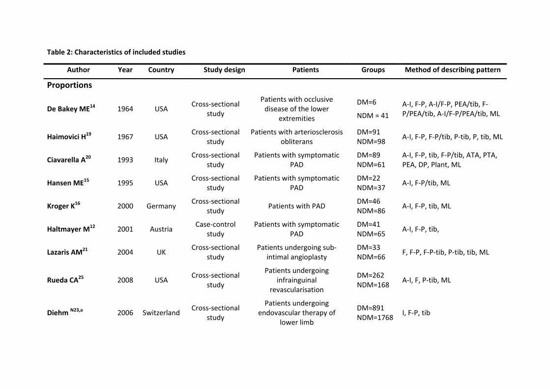
Table 2: Characteristics of included studies
Author Year Country Study design Patients Groups Method of describing pattern
Proportions
De Bakey ME14 1964 USA Cross‐sectional
study
Patients with occlusive disease of the lower
extremities
DM=6
NDM = 41
A‐I, F‐P, A‐I/F‐P, PEA/tib, F‐P/PEA/tib, A‐I/F‐P/PEA/tib, ML
Haimovici H19 1967 USA Cross‐sectional
study Patients with arteriosclerosis
obliterans DM=91 NDM=98
A‐I, F‐P, F‐P/tib, P‐tib, P, tib, ML
Ciavarella A20 1993 Italy Cross‐sectional
study Patients with symptomatic
PAD DM=89 NDM=61
A‐I, F‐P, tib, F‐P/tib, ATA, PTA, PEA, DP, Plant, ML
Hansen ME15 1995 USA Cross‐sectional
study Patients with symptomatic
PAD DM=22 NDM=37
A‐I, F‐P/tib, ML
Kroger K16 2000 Germany Cross‐sectional
study Patients with PAD
DM=46 NDM=86
A‐I, F‐P, tib, ML
Haltmayer M12 2001 Austria Case‐control
study Patients with symptomatic
PAD DM=41 NDM=65
A‐I, F‐P, tib,
Lazaris AM21 2004 UK Cross‐sectional
study Patients undergoing sub‐
intimal angioplasty DM=33 NDM=66
F, F‐P, F‐P‐tib, P‐tib, tib, ML
Rueda CA25 2008 USA Cross‐sectional
study
Patients undergoing infrainguinal
revascularisation
DM=262 NDM=168
A‐I, F, P‐tib, ML
Diehm N23,a 2006 Switzerland Cross‐sectional
study
Patients undergoing endovascular therapy of
lower limb
DM=891 NDM=1768
I, F‐P, tib
Ozkan U18,a 2009 Turkey Cross‐sectional
study Patients with PAD
DM=261 NDM=365
A‐I, F‐P, tib, ML
Scores
Menzoian JO22 1989 1989 Cross‐sectional
study Patients with PAD
DM=115 NDM=119
ATA, PTA, PEA, Plant
Jude EB24 2001 UK Cross‐sectional
study
Patients undergoing infrainguinal
revascularisation
DM=58 NDM=78
Individual vessels I to tib
van der Feen C13 2002 Netherlands Case‐control
study Patients with symptomatic
PAD DM=37 NDM=37
I‐F, P‐tib
Diehm N11 2008 Switzerland Cohort study Patients undergoing
angiography for chronic lower limb ischaemia
DM= 25 NDM=25
Individual vessels TPT to tib, TPT‐tib average score, Plant
aNot included in meta‐analysis. A=Aorta, I‐Iliacs, F=Femoral, P=Popliteal, tib=Tibials, PEA=Peroneal, ATA=Anterior tibial artery, PTA=Posterior tibial artery, Plant=Plantar vessels, TPT= Tibeo‐ peroneal trunk, ML=Multi‐level disease
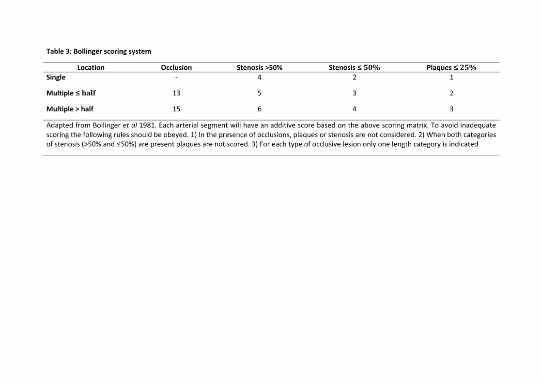
Table 3: Bollinger scoring system
Location Occlusion Stenosis >50% Stenosis ≤50% Plaques ≤25%
Single ‐ 4 2 1
Multiple ≤half 13 5 3 2
Multiple > half 15 6 4 3
Adapted from Bollinger et al 1981. Each arterial segment will have an additive score based on the above scoring matrix. To avoid inadequate scoring the following rules should be obeyed. 1) In the presence of occlusions, plaques or stenosis are not considered. 2) When both categories of stenosis (>50% and ≤50%) are present plaques are not scored. 3) For each type of occlusive lesion only one length category is indicated